Embed Size (px)
Citation preview
membranes
Article
The Early Fragmentation of a Bovine Dermis-Derived CollagenBarrier Membrane Contributes to TransmembraneousVascularization—A Possible Paradigm Shift for GuidedBone Regeneration
Eleni Kapogianni 1, Said Alkildani 2, Milena Radenkovic 3 , Xin Xiong 4, Rumen Krastev 4,5, Ignacio Stöwe 6,7 ,James Bielenstein 2, Ole Jung 7, Stevo Najman 8 , Mike Barbeck 2,9,*,† and Daniel Rothamel 10,11,†
�����������������
Citation: Kapogianni, E.; Alkildani,
S.; Radenkovic, M.; Xiong, X.; Krastev,
R.; Stöwe, I.; Bielenstein, J.; Jung, O.;
Najman, S.; Barbeck, M.; et al. The
Early Fragmentation of a Bovine
Dermis-Derived Collagen Barrier
Membrane Contributes to
Transmembraneous
Vascularization—A Possible
Paradigm Shift for Guided Bone
Regeneration. Membranes 2021, 11,
185. https://doi.org/10.3390/
membranes11030185
Academic Editor: Marina Pinheiro
Received: 25 February 2021
Accepted: 6 March 2021
Published: 9 March 2021
Publisher’s Note: MDPI stays neutral
with regard to jurisdictional claims in
published maps and institutional affil-
iations.
Copyright: © 2021 by the authors.
Licensee MDPI, Basel, Switzerland.
This article is an open access article
distributed under the terms and
conditions of the Creative Commons
Attribution (CC BY) license (https://
creativecommons.org/licenses/by/
4.0/).
1 Private Practice, 10623 Berlin, Germany; [email protected] BerlinAnalytix GmbH, 12109 Berlin, Germany; [email protected] (S.A.);
[email protected] (J.B.)3 Scientific Research Center for Biomedicine, Department for Cell and Tissue Engineering, Faculty of Medicine,
University of Nis, 18108 Nis, Serbia; [email protected] NMI Natural and Medical Sciences Institute at the University of Tübingen, 72770 Reutlingen, Germany;
[email protected] (X.X.); [email protected] (R.K.)5 Faculty of Applied Chemistry, Reutlingen University, 72762 Reutlingen, Germany6 Helios Klinikum Emil von Behring, Gefäßzentrum Berlin Südwest, 14165 Berlin, Germany;
[email protected] Clinic and Policlinic for Dermatology and Venereology, University Medical Center Rostock,
18057 Rostock, Germany; [email protected] Department of Biology and Human Genetics, Department for Cell and Tissue Engineering,
Faculty of Medicine, University of Nis, 18108 Nis, Serbia; [email protected] Department of Ceramic Materials, Chair of Advanced Ceramic Materials, Institute for Materials Science
and Technologies, Technical University of Berlin, 10587 Berlin, Germany10 Department of Oral and Maxillofacial Plastic Surgery, Evangelic Johanniter Hospital Bethesda
Mönchengladbach, 41061 Mönchengladbach, Germany; [email protected] Department of Oral and Maxillofacial Plastic Surgery, Heinrich-Heine Universität Düsseldorf,
40225 Düsseldorf, Germany* Correspondence: [email protected]; Tel.: +49-176-81022467† These authors contributed equally to this work.
Abstract: Collagen-based barrier membranes are an essential component in Guided Bone Regenera-tion (GBR) procedures. They act as cell-occlusive devices that should maintain a micromilieu wherebone tissue can grow, which in turn provides a stable bed for prosthetic implantation. However, thestanding time of collagen membranes has been a challenging area, as native membranes are oftenprematurely resorbed. Therefore, consolidation techniques, such as chemical cross-linking, havebeen used to enhance the structural integrity of the membranes, and by consequence, their standingtime. However, these techniques have cytotoxic tendencies and can cause exaggerated inflammationand in turn, premature resorption, and material failures. However, tissues from different extractionsites and animals are variably cross-linked. For the present in vivo study, a new collagen membranebased on bovine dermis was extracted and compared to a commercially available porcine-sourcedcollagen membrane extracted from the pericardium. The membranes were implanted in Wistar ratsfor up to 60 days. The analyses included well-established histopathological and histomorphometricalmethods, including histochemical and immunohistochemical staining procedures, to detect M1-and M2-macrophages as well as blood vessels. Initially, the results showed that both membranesremained intact up to day 30, while the bovine membrane was fragmented at day 60 with granulationtissue infiltrating the implantation beds. In contrast, the porcine membrane remained stable withoutsigns of material-dependent inflammatory processes. Therefore, the bovine membrane showed aspecial integration pattern as the fragments were found to be overlapping, providing secondaryporosity in combination with a transmembraneous vascularization. Altogether, the bovine membraneshowed comparable results to the porcine control group in terms of biocompatibility and standingtime. Moreover, blood vessels were found within the bovine membranes, which can potentially
Membranes 2021, 11, 185. https://doi.org/10.3390/membranes11030185 https://www.mdpi.com/journal/membranes
Membranes 2021, 11, 185 2 of 16
serve as an additional functionality of barrier membranes that conventional barrier membranes donot provide.
Keywords: tissue source; bovine collagen; porcine collagen; barrier membrane; Guided Bone Re-generation (GBR); tissue regeneration; transmembraneous vascularization; bovine dermis; porcinepericardium
1. Introduction
In dental implantology, the necessity for adequate quantity and quality of bone forimplant placement and its stabilization can require a bone augmentation procedure prior toimplantation. In this context, Guided Bone Regeneration (GBR) has become one of the mostestablished techniques for jawbone augmentation [1]. GBR is nearly always performedwith the use of a barrier membrane to seclude the bone defect from the infiltration ofsoft tissue and especially epithelial cells that could otherwise invade the defect area andinteract with the bone regeneration process [2]. By preventing the ingrowth of thesefaster growing cells, bone cells can repopulate the bone defect space, and a regenerativeenvironment can be established [3]. Nowadays, most often, resorbable membranes areused for most GBR procedures, as their application does not require a second surgery fortheir extraction as in the case of nonresorbable membranes [3]. In this context, collagen wasand is still used as a base material due to the belief that collagen—from every source andafter most of the processing techniques—is comparable in its physicochemical propertiesand always biocompatible due to its natural origin [4]. However, it has already beenshown that both the donor organism (e.g., allogeneic or xenogeneic sources) and mainlythe tissue source (e.g., dermis, pericardium or tendon tissue) have an influence on theintegration behavior of collagen-based biomaterials and factors, such as its standing time orits vascularization, which has a major impact on the material functionality in an intendedclinical indication [5,6]. Alongside decellularization and sterilization techniques, donororganisms and tissue sources are assumed to be the main factors that have an impact onthe physicochemical properties of the resulting biomaterial [7].
Therefore, it was revealed that the specific immunological tissue response to everybiomaterial depends on its individual physicochemical properties [8,9]. This can varyfrom a nearly bioinert response to a strong inflammatory reaction, associated with abscessformation or fibrotic capsule formation, as well as local and systemic consequences [10].In this context, the role of macrophages has recently been investigated, as these cells arekey elements in the tissue reaction cascade [11]. Macrophages are also thought to play animportant role in the framework of the tissue reaction to a biomaterial due to their secretionof cytokines [12]. Therefore, macrophages can roughly be divided into two subtypesbased on their overall expression profile: the M1 phenotype is pro-inflammatory, has beenshown to be especially present in the early healing phase and also seems to be involved inthe degradation process of biomaterials inducing a foreign body response, while the M2phenotype is an anti-inflammatory expressing reparative cytokine and is integrated in thetissue healing phase [13]. Biomaterial-associated multinucleated giant cells (BMGCs) areanother cell that may exhibit a pro-inflammatory (M1-BMGCs) or anti-inflammatory (M2-BMGCs) phenotype, which is equivalent to macrophages and dependent on the physicaland chemical properties of the biomaterial [14–16]. Moreover, it has been shown that thesecells are involved in the phagocytic degradation of different biomaterials expressing lyticenzymes [17]. In this context, it has been concluded that these “non-physiological cells”are only involved in material rejections or failures, but the research on this topic has shownthat their involvement in the tissue reaction to a specific biomaterial is strongly dependenton the type of biomaterial and its physicochemical properties [13]. Interestingly, it hasbeen assumed that their induction even in the case of collagen-based materials may becontraindicated, but there is still a lack of existing knowledge. Thus, it is highly important
Membranes 2021, 11, 185 3 of 16
to analyze the tissue reaction to a biomaterial for clarification of its specific inflammatorytissue reaction pattern with special consideration of its intended use.
In the context of GBR procedures, it has been understood that barrier membranesshould not only provide a barrier function, but they should optimally induce help tomodulate the microenvironment to increase bone remodeling [2]. It has been revealedthat angiogenesis has been emphasized to be an important factor strongly influencingthe outcome of bone healing [18]. It has been discussed that GBR membranes shouldallow for a so-called “transmembraneous vascularization”, allowing the formation of bloodvessels to increase the formation of new bone. However, it has also been shown that aningrowth of connective tissue is most often necessary to allow the simultaneous ingrowthof blood vessels [19]. This tissue ingrowth has been shown to correlate with the prematurebreakdown or fragmentation and an associated loss of barrier functionality of collagenmembranes [5]. Thus, it is of great importance to develop a next generation of resorbableGBR membranes with an adapted integration behavior that combines both the barrierfunctionality as well as a transmembraneous blood supply. In this context, it is also ofspecial interest to analyze the overall immune response to a membrane for evaluation ofthe local events that might additionally support the underlying bone regeneration process.
Furthermore, the alignment of the inflammatory cascade caused by the biomaterialcan influence aspects such as the response of anabolic tissue cells, e.g., osteoblasts [9].Moreover, it has been shown that different interactions between material-induced immunecells (macrophages and/or BMGCs) and tissue cells (osteoblasts or endothelial cells) existand may support the process of tissue regeneration at the molecular level [20]. During bonehealing, interactions between inflammatory response cells and bone remodeling cells havegained more importance [12]. It has been supposed that the next generation of biomaterialsshould integrate the immune system into regenerative strategies [8,21].
Altogether, it is believed that collagen membranes, even for GBR procedures, shouldprovide a long-standing time of several weeks up to 3–4 months for optimal clinicalresults in the course of jawbone healing. This assumption appears reasonable in thecase of multidimensional or bigger bone defects, but it has been reported in the case of“normal” or smaller defects (e.g., in the case of extraction sockets) that the application ofnative dermis-derived collagen membranes with a very short standing time contributesto comparable bone healing results [19]. In this context, it has been revealed that a fastmembrane degradation might also correlate with a higher or more pro-inflammatoryalignment of the material-associated tissue reaction [19]. As a consequence, it has beenrevealed that the faster tissue ingrowth also includes phagocyting cells, such as BMGCs,in concert with macrophages [22]. However, this fast membrane degradation is alsocombined with a higher transmembraneous vascularization that has been identified asan important co-factor for the material-related bone healing process [6]. Altogether, thequestion arises concerning which material factors are optimal for combining both the barrierfunctionality and the support for creation of a molecular microenvironment that triggersthe (underlying) process(es) of bone healing. Interestingly, even inflammatory cells, such asBMGCs and macrophages, have been shown to be potent sources of angiogenic molecules,such as the vascular endothelial growth factor (VEGF), so it might be of special interest tocreate membranes with a higher “inflammatory potential” to create the above-mentionedmicromilieu [23].
Particularly in the case of collagen, only a few studies have analyzed the differences invarious xenogeneic sources, and scarce knowledge exists about the consequences of the us-age of different animal sources as a basis for biomaterials, such as GBR membranes [24–26].Most of the membranes used in GBR procedures are based on porcine donor tissue. Gen-erally, fewer barrier membranes are used that are sourced from other animals or areof synthetic origin [3]. In this context, it has already been demonstrated that collagenmembranes derive from pigs, which have been treated using different purification andfabrication processes to induce varying tissue reactions [19]. Some collagen membranesare degraded by mononuclear cells during a very mild tissue reaction producing a long-
Membranes 2021, 11, 185 4 of 16
standing time, while other membranes induce stronger inflammatory reactions, involvingBMGCs that lead to a faster degradation combined with a lower standing time or prema-ture breakdown and loss of functionality. Altogether, these differing tissue reactions canbe attributed to differences in the preparation processes [7]. Although the influence ofdifferent processing methods on collagen-induced tissue reactions has been investigated,the influence of the collagen animal source is rarely analyzed. To fill this knowledge gap,the inflammatory tissue reactions to two collagen membranes from porcine and bovineorigins were analyzed in the present study. The membranes were implanted over a periodof up to 60 days by means of an established preclinical implantation model in Wistar rats.Furthermore, established histopathological and histomorphometrical analysis methods,as well as immunohistochemical detection methods of M1- and M2-macrophages, wereapplied [14,27].
2. Materials and Methods2.1. Bovine Collagen Membrane
The collagen membrane analyzed in the present study is based on a native collagenobtained from bovine skin. The collagen within the structure of the membrane is mostlycomposed of collagen Type I fibers. A hydrogel was prepared from the precursor tissueand then freeze-dried to create the membranes. The membranes were not chemicallycross-linked.
2.2. Porcine Collagen Membrane
The porcine collagen membrane analyzed in the present study is based on nativecollagen derived from pericardium using a decellularization process including a wet-chemical treatment, lyophilization and final sterilization by ethylene oxide gas. Themembrane was shown to fulfill the requirements of EN ISO 10993-1 and EN ISO 7405.
2.3. In Vivo Study
The Local Ethical Committee of the Faculty of Medicine (University of Niš, Serbia)authorized the in vivo experiments prior to the biomaterial implantations, based on deci-sion number 323-07-09101, on 2020-05/5, of the Veterinary Directorate of the Ministry ofAgriculture, Forestry and Water Management of the Republic of Serbia.
The preclinical in vivo experiments were conducted at the Faculty of Medicine in theUniversity of Niš (Serbia). Animal housing was conducted using standard conditions,i.e., water ad libitum, artificial light and regular rat pellets, as well as standard pre- andpostoperative care.
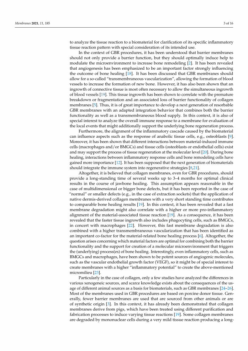
In total, 30 female, 10–12-week-old Wistar rats that were obtained from the MilitaryMedical Academy (Belgrade, Serbia) were randomly allocated into two study groups. Eachof the two study groups contained 15 experimental animals; 5 animals were sacrificed foreach group per time point (n = 5), i.e., 10, 30 and 60 days. The implantation was conductedfollowing the protocol described by Barbeck et al. [27–32]. In brief, the animals wereanesthetized via an intraperitoneal injection (10 mL ketamine [50 mg/mL] with 1.6 mLXylazine [2%]). After shaving and disinfection, an incision down to the subcutaneous tissuewithin the rostral subscapular region was made. Subsequently, a subcutaneous pocket wasbluntly built by a scissor, and the biomaterials were implanted into the pocket (Figure 1).Afterwards, the wounds were sutured.
After the respective study time points, the animals were euthanized with an overdoseof the above-mentioned anesthetics, and the implantation area together with the surround-ing tissue was explanted. Subsequently, the explanted tissue was fixed using a 4% formalinsolution for 24 h and then placed into PBS for the following histological workup process.
Membranes 2021, 11, 185 5 of 16Membranes 2021, 11, x FOR PEER REVIEW 5 of 17
Figure 1. (A) Schematic image of the implantation site. (B) Overview of a subcutaneous implanta-tion site of the porcine collagen membrane (PM) at day 30 postimplantation. CT = connective tis-sue; EP = epidermis (Azan-staining, “total scan”, 100 × magnification, scalebar = 1 mm).
After the respective study time points, the animals were euthanized with an overdose of the above-mentioned anesthetics, and the implantation area together with the sur-rounding tissue was explanted. Subsequently, the explanted tissue was fixed using a 4% formalin solution for 24 h and then placed into PBS for the following histological workup process.
2.3.1. Histology and Immunohistochemistry For histological workup, the tissue explants were initially cut into two segments of
identical dimensions and dehydrated using a series of increasing alcohol concentrations. After a xylol exposure, paraffin embedding was performed. Sections were prepared with a thickness of 3–5 μm by means of a rotation microtome (SLEE, Mainz, Germany). Three sections of every tissue explant were used for histochemical staining, i.e., hematoxylin and eosin (H&E), Movat Pentachrome and Azan.
Furthermore, four additional sections of every tissue explants were used for im-munohistochemical detection of macrophages. NF kappa ß-positive M1 macrophages and CD163-postive M2 macrophages were stained by means of antibodies against the pro- and anti-inflammatory molecules based on previously published methods [15]. Briefly, the slides were initially treated with citrate buffer and proteinase K in a water bath for 20 min that had a temperature of 96 °C and a pH 8. This was followed by equilibration using TBS-T buffer. Subsequently, the slides were prepared by H2O2 and avidin and biotin blocking solutions (Avidin/Biotin Blocking Kit, Vector Laboratories, Burlingame, CA, USA), incu-bated with the respective first antibody for 30 min, followed by incubation with the sec-ondary antibody (goat anti-rabbit IgG-B, sc-2040, 1:200, Santa Cruz Biotechnology, Dallas, TX, USA). Afterwards, the avidin–biotin–peroxidase complex (ThermoFisher Scientific,
Figure 1. (A) Schematic image of the implantation site. (B) Overview of a subcutaneous implantationsite of the porcine collagen membrane (PM) at day 30 postimplantation. CT = connective tissue;EP = epidermis (Azan-staining, “total scan”, 100× magnification, scalebar = 1 mm).
2.3.1. Histology and Immunohistochemistry
For histological workup, the tissue explants were initially cut into two segments ofidentical dimensions and dehydrated using a series of increasing alcohol concentrations.After a xylol exposure, paraffin embedding was performed. Sections were prepared witha thickness of 3–5 µm by means of a rotation microtome (SLEE, Mainz, Germany). Threesections of every tissue explant were used for histochemical staining, i.e., hematoxylin andeosin (H&E), Movat Pentachrome and Azan.
Furthermore, four additional sections of every tissue explants were used for im-munohistochemical detection of macrophages. NF kappa ß-positive M1 macrophages andCD163-postive M2 macrophages were stained by means of antibodies against the pro-and anti-inflammatory molecules based on previously published methods [15]. Briefly,the slides were initially treated with citrate buffer and proteinase K in a water bath for20 min that had a temperature of 96 ◦C and a pH 8. This was followed by equilibrationusing TBS-T buffer. Subsequently, the slides were prepared by H2O2 and avidin and bi-otin blocking solutions (Avidin/Biotin Blocking Kit, Vector Laboratories, Burlingame, CA,USA), incubated with the respective first antibody for 30 min, followed by incubation withthe secondary antibody (goat anti-rabbit IgG-B, sc-2040, 1:200, Santa Cruz Biotechnology,Dallas, TX, USA). Afterwards, the avidin–biotin–peroxidase complex (ThermoFisher Scien-tific, Henningsdorf, Germany) (30 min) was applied, and a counterstaining by hemalumwas conducted.
2.3.2. Histological Analysis
The histological analyses to study the tissue–biomaterial interactions within the im-plantation beds of the biomaterials and their surrounding tissue were conducted usingan Axio Imager M2 (Zeiss, Oberkochen, Germany) based on a protocol according to theDIN ISO 10993-6 as previously described [11,23,33–35]. These analyses focused on the
Membranes 2021, 11, 185 6 of 16
evaluation of the following parameters within the framework of the early and the latetissue response related to the implants: fibrosis; hemorrhage; necrosis; vascularization;and the presence of neutrophils, lymphocytes, plasma cells, macrophages and biomaterial-associated multinucleated giant cells (BMGCs). Finally, microphotographs were takenwith an Axiocam 506 color connected to a computer system running the ZEN Core (Zeiss,Oberkochen, Germany) connected to a microscope.
2.3.3. Histomorphometrical Analysis
The histomorphometrical analyses included the measurements of the occurrence ofanti-inflammatory and pro-inflammatory cells within the implant beds of the membranesas previously described [36]. Briefly, the slides stained by the aforementioned immunohis-tochemical methods were initially digitized. Then, the Image J software (National Institutesof Health, Bethesda, MD, USA) enabled the measurements of the stained cells within thetotal scans. At first, the defect area and the membrane area were manually marked, andtheir areas were determined. After that, the number of macrophages was also measuredvia a specially programmed plugin that allowed us to mark the area of the red stained cellsautomatically [37]. Finally, the cell numbers were related to the respective total area tocalculate the numbers of cells per mm2 (macrophages/mm2).
2.3.4. Statistical Analysis
Quantitative data are shown as mean ± standard deviation after an analysis of vari-ance (ANOVA), which enabled comparison of the data from the study groups via theGraphPad Prism 8.0 software (GraphPad Software Inc., La Jolla, CA, USA). Statisticaldifferences were designated as significant if p-values were less than 0.05 (* p ≤ 0.05), andhighly significant if p-values were less than 0.01 (** p ≤ 0.01), less than 0.001 (*** p ≤ 0.001)or less than 0.0001 (**** p ≤ 0.0001).
3. Results3.1. Histological (Qualitative) Analysis
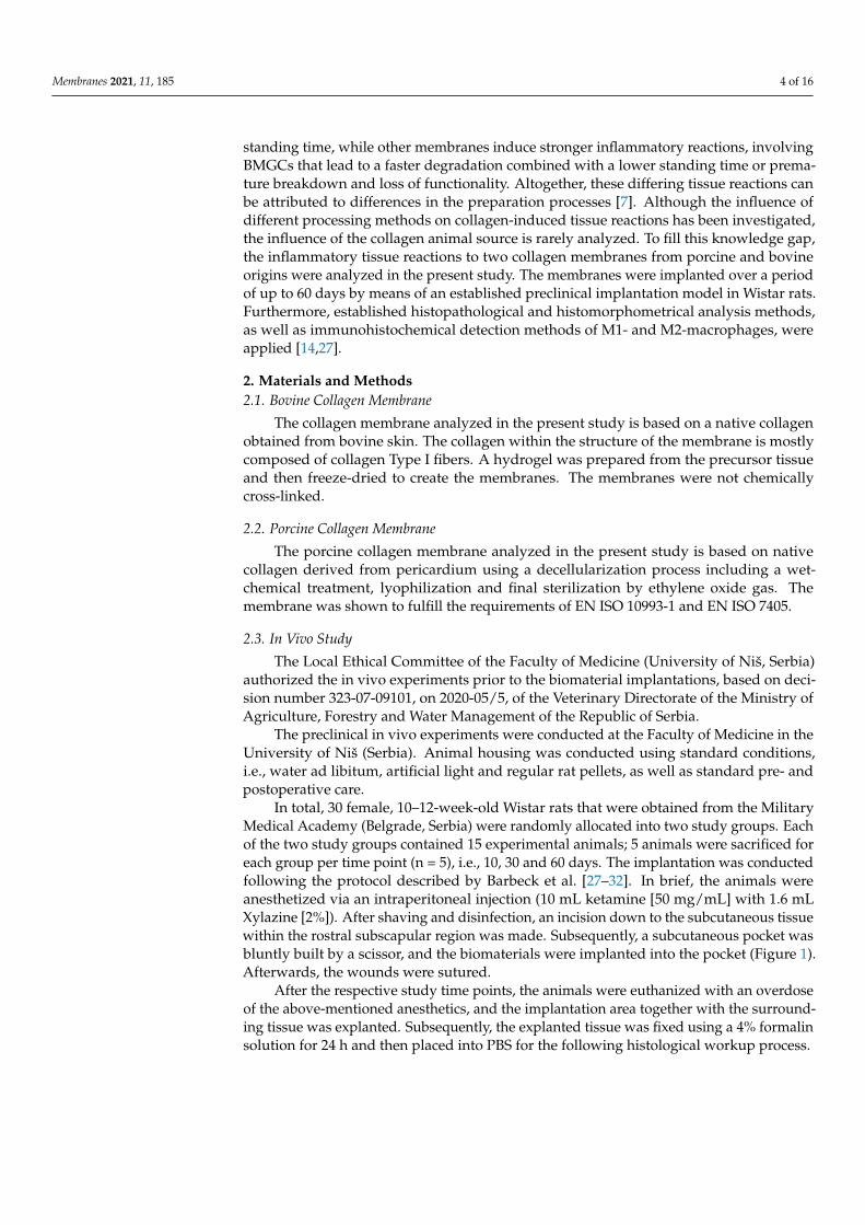
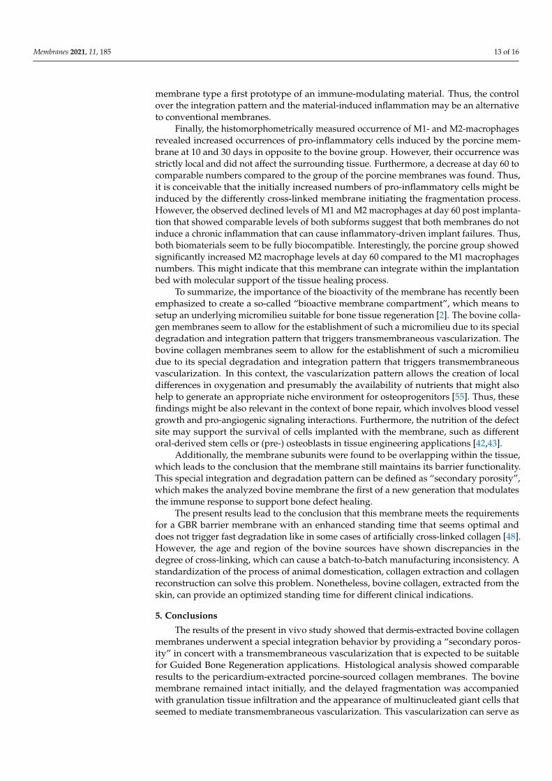
Histopathological analysis of the bovine collagen membrane at 10 days after im-plantation showed that the membrane was intact, showing no signs of a breakdown orfragmentation (Figure 1A). At the material surfaces, signs of a minor inflammatory tissue re-action were detected (Figure 2B). The reactive tissue was mostly composed of macrophages,granulocytes and fibroblasts at the surfaces and surrounding the membrane (Figure 2B).As for the porcine membrane, similar observations were noted at day 10 post implantation(Figure 2C,D).
At 30 days post implantation, the first signs of tissue infiltration were found in thecase of the bovine collagen membranes (Figure 1E). Therefore, an increased intensity ofthe reactive tissue could be detected (Figure 2E). The tissue was mainly composed of thesame cell types found at 10 days post implantation. Thus, mainly macrophages werefound at the material surfaces. Moreover, single biomaterial-induced multinucleatedgiant cells (BMGCs) were found at the surface of the bovine membrane at this time point(Figure 2F). Similar observations were made in the group of the porcine collagen membranethat was found to be completely intact at this time point (Figure 2G). Thus, a layer ofmacrophages was found attached to the membrane surfaces (Figure 2G). Nonetheless, noBMGCs appeared to be attaching to the porcine membrane (Figure 2H).
At day 60 post implantation, the bovine membrane appeared to be fragmented, asbig fragments of the membrane were found to be overlapping within the subcutaneousconnective tissue (Figure 2I). Furthermore, the reactive tissue infiltrated the interspacesof the membrane fragments (Figure 2I). Similar cell types to those observed at days 10and 30 were found within the surrounding tissue, i.e., mainly macrophages in concertwith single BMGCs (Figure 2J). Additionally, single vessels were observable within thetissue that infiltrated this membrane type (Figure 2J). The porcine membrane appearedintact with less intense reactive tissue response and infiltration (Figure 2K). Attached to the
Membranes 2021, 11, 185 7 of 16
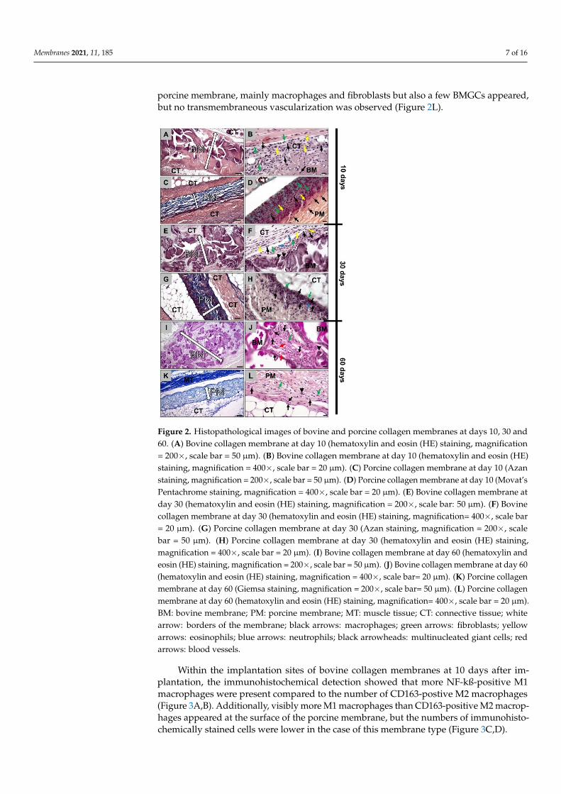
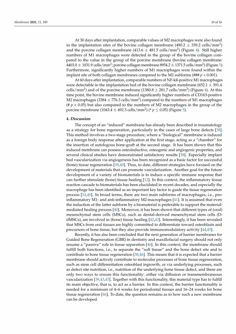
porcine membrane, mainly macrophages and fibroblasts but also a few BMGCs appeared,but no transmembraneous vascularization was observed (Figure 2L).
Membranes 2021, 11, x FOR PEER REVIEW 7 of 17
of macrophages was found attached to the membrane surfaces (Figure 2G). Nonetheless, no BMGCs appeared to be attaching to the porcine membrane (Figure 2H).
At day 60 post implantation, the bovine membrane appeared to be fragmented, as big fragments of the membrane were found to be overlapping within the subcutaneous connective tissue (Figure 2I). Furthermore, the reactive tissue infiltrated the interspaces of the membrane fragments (Figure 2I). Similar cell types to those observed at days 10 and 30 were found within the surrounding tissue, i.e., mainly macrophages in concert with single BMGCs (Figure 2J). Additionally, single vessels were observable within the tissue that infiltrated this membrane type (Figure 2J). The porcine membrane appeared intact with less intense reactive tissue response and infiltration (Figure 2K). Attached to the por-cine membrane, mainly macrophages and fibroblasts but also a few BMGCs appeared, but no transmembraneous vascularization was observed (Figure 2L).
Figure 2. Histopathological images of bovine and porcine collagen membranes at days 10, 30 and 60. (A) Bovine collagen membrane at day 10 (hematoxylin and eosin (HE) staining, magnification= 200 ×, scale bar = 50 μm). (B) Bovine collagen membrane at day 10 (hematoxylin and eosin (HE) staining, magnification = 400 ×, scale bar = 20 μm). (C) Porcine collagen membrane at day 10 (Azan staining, magnification = 200 ×, scale bar = 50 μm). (D) Porcine collagen membrane at day 10 (Movat’s Pentachrome staining, magnification = 400 ×, scale bar = 20 μm). (E) Bovine collagen membrane at day 30 (hematoxylin and eosin (HE) staining, magnification = 200 ×, scale bar: 50 μm). (F) Bovine collagen membrane at day 30 (hematoxylin and eosin (HE) staining, magnifica-tion= 400 ×, scale bar = 20 μm). (G) Porcine collagen membrane at day 30 (Azan staining, magnifi-cation = 200 ×, scale bar = 50 μm). (H) Porcine collagen membrane at day 30 (hematoxylin and eo-sin (HE) staining, magnification = 400 ×, scale bar = 20 μm). (I) Bovine collagen membrane at day 60 (hematoxylin and eosin (HE) staining, magnification = 200 ×, scale bar = 50 μm). (J) Bovine col-lagen membrane at day 60 (hematoxylin and eosin (HE) staining, magnification = 400 ×, scale bar= 20 μm). (K) Porcine collagen membrane at day 60 (Giemsa staining, magnification = 200 ×, scale bar= 50 μm). (L) Porcine collagen membrane at day 60 (hematoxylin and eosin (HE) staining, mag-nification= 400 ×, scale bar = 20 μm). BM: bovine membrane; PM: porcine membrane; MT: muscle tissue; CT: connective tissue; white arrow: borders of the membrane; black arrows: macrophages;
Figure 2. Histopathological images of bovine and porcine collagen membranes at days 10, 30 and60. (A) Bovine collagen membrane at day 10 (hematoxylin and eosin (HE) staining, magnification= 200×, scale bar = 50 µm). (B) Bovine collagen membrane at day 10 (hematoxylin and eosin (HE)staining, magnification = 400×, scale bar = 20 µm). (C) Porcine collagen membrane at day 10 (Azanstaining, magnification = 200×, scale bar = 50 µm). (D) Porcine collagen membrane at day 10 (Movat’sPentachrome staining, magnification = 400×, scale bar = 20 µm). (E) Bovine collagen membrane atday 30 (hematoxylin and eosin (HE) staining, magnification = 200×, scale bar: 50 µm). (F) Bovinecollagen membrane at day 30 (hematoxylin and eosin (HE) staining, magnification= 400×, scale bar= 20 µm). (G) Porcine collagen membrane at day 30 (Azan staining, magnification = 200×, scalebar = 50 µm). (H) Porcine collagen membrane at day 30 (hematoxylin and eosin (HE) staining,magnification = 400×, scale bar = 20 µm). (I) Bovine collagen membrane at day 60 (hematoxylin andeosin (HE) staining, magnification = 200×, scale bar = 50 µm). (J) Bovine collagen membrane at day 60(hematoxylin and eosin (HE) staining, magnification = 400×, scale bar= 20 µm). (K) Porcine collagenmembrane at day 60 (Giemsa staining, magnification = 200×, scale bar= 50 µm). (L) Porcine collagenmembrane at day 60 (hematoxylin and eosin (HE) staining, magnification= 400×, scale bar = 20 µm).BM: bovine membrane; PM: porcine membrane; MT: muscle tissue; CT: connective tissue; whitearrow: borders of the membrane; black arrows: macrophages; green arrows: fibroblasts; yellowarrows: eosinophils; blue arrows: neutrophils; black arrowheads: multinucleated giant cells; redarrows: blood vessels.
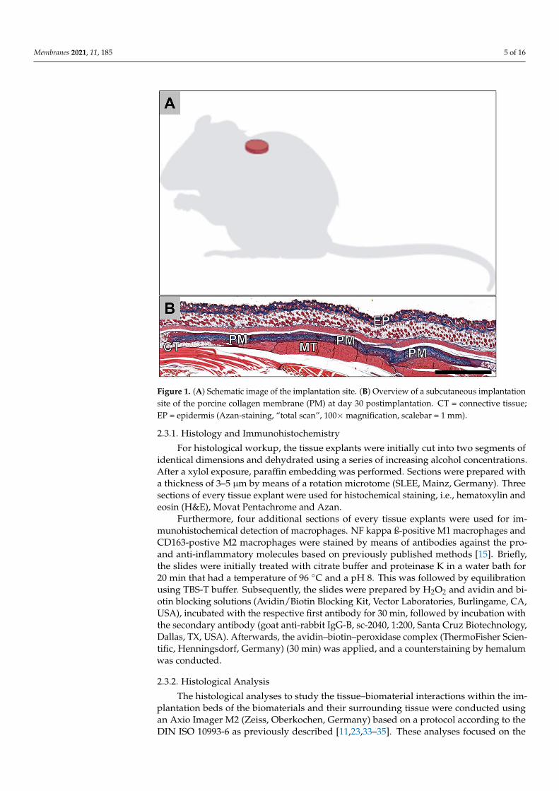
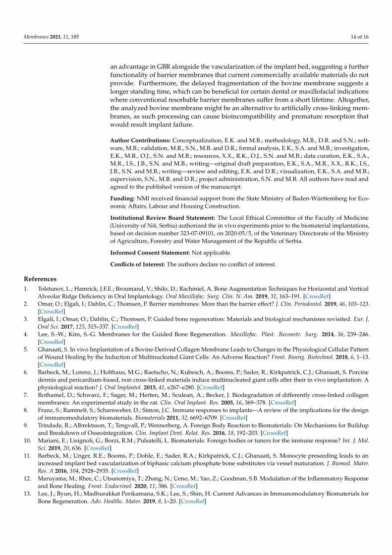
Within the implantation sites of bovine collagen membranes at 10 days after im-plantation, the immunohistochemical detection showed that more NF-kß-positive M1macrophages were present compared to the number of CD163-postive M2 macrophages(Figure 3A,B). Additionally, visibly more M1 macrophages than CD163-positive M2 macrop-hages appeared at the surface of the porcine membrane, but the numbers of immunohisto-chemically stained cells were lower in the case of this membrane type (Figure 3C,D).

Membranes 2021, 11, 185 8 of 16
Membranes 2021, 11, x FOR PEER REVIEW 8 of 17
green arrows: fibroblasts; yellow arrows: eosinophils; blue arrows: neutrophils; black arrowheads: multinucleated giant cells; red arrows: blood vessels.
Within the implantation sites of bovine collagen membranes at 10 days after implan-tation, the immunohistochemical detection showed that more NF-kß-positive M1 macro-phages were present compared to the number of CD163-postive M2 macrophages (Figure 3A,B). Additionally, visibly more M1 macrophages than CD163-positive M2 macrophages appeared at the surface of the porcine membrane, but the numbers of immunohistochem-ically stained cells were lower in the case of this membrane type (Figure 3C,D).
At day 30 post implantation, still more NF-kß-positive M1 macrophages than M2 macrophages appeared on the surface of both membranes (Figure 3E,F). In contrast to day 10, comparable amounts of CD136-positive M2 presence were found in both groups, while the numbers of the M1 macrophages were visibly higher in the group of the bovine mem-brane (Figure 3G,H).
At day 60 post implantation, both collagen membranes induced comparably lower numbers of M1 macrophages than at day 30 (Figure 3I–L). However, the numbers of M2 macrophages were found to be comparably low in the group of the bovine membrane, while the M2 macrophage number increased in the group of the porcine membrane (Fig-ure 3I–L).
Figure 3. Immunohistochemically stained slides show detection of CD163-positive M2 macro-phages (left column: A, C, E, G, I and K) and NF-kß-positive M1 macrophages (right column: B, D, F, H, J and L) into the implantation beds of both bovine and porcine collagen membranes at days 10, 30 and day 60 after implantation (all images: 400 × magnification; scale bars = 20 μm) (left: CD163 immunohistochemical staining; right: NF-kß immunohistochemical staining). BM: bovine membrane; PM: porcine membrane; CT: connective tissue; red arrows: CD163- and NF-kß-positive macrophages.
Figure 3. Immunohistochemically stained slides show detection of CD163-positive M2 macrophages(left column: A,C,E,G,I,K) and NF-kß-positive M1 macrophages (right column: B,D,F,H,J,L) intothe implantation beds of both bovine and porcine collagen membranes at days 10, 30 and day 60after implantation (all images: 400× magnification; scale bars = 20 µm) (left: CD163 immunohisto-chemical staining; right: NF-kß immunohistochemical staining). BM: bovine membrane; PM: porcinemembrane; CT: connective tissue; red arrows: CD163- and NF-kß-positive macrophages.
At day 30 post implantation, still more NF-kß-positive M1 macrophages than M2macrophages appeared on the surface of both membranes (Figure 3E,F). In contrast today 10, comparable amounts of CD136-positive M2 presence were found in both groups,while the numbers of the M1 macrophages were visibly higher in the group of the bovinemembrane (Figure 3G,H).
At day 60 post implantation, both collagen membranes induced comparably lowernumbers of M1 macrophages than at day 30 (Figure 3I–L). However, the numbers of M2macrophages were found to be comparably low in the group of the bovine membrane, whilethe M2 macrophage number increased in the group of the porcine membrane (Figure 3I–L).
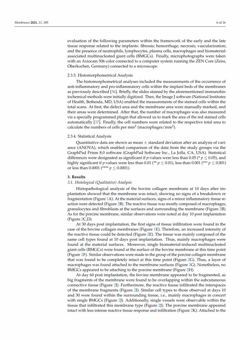
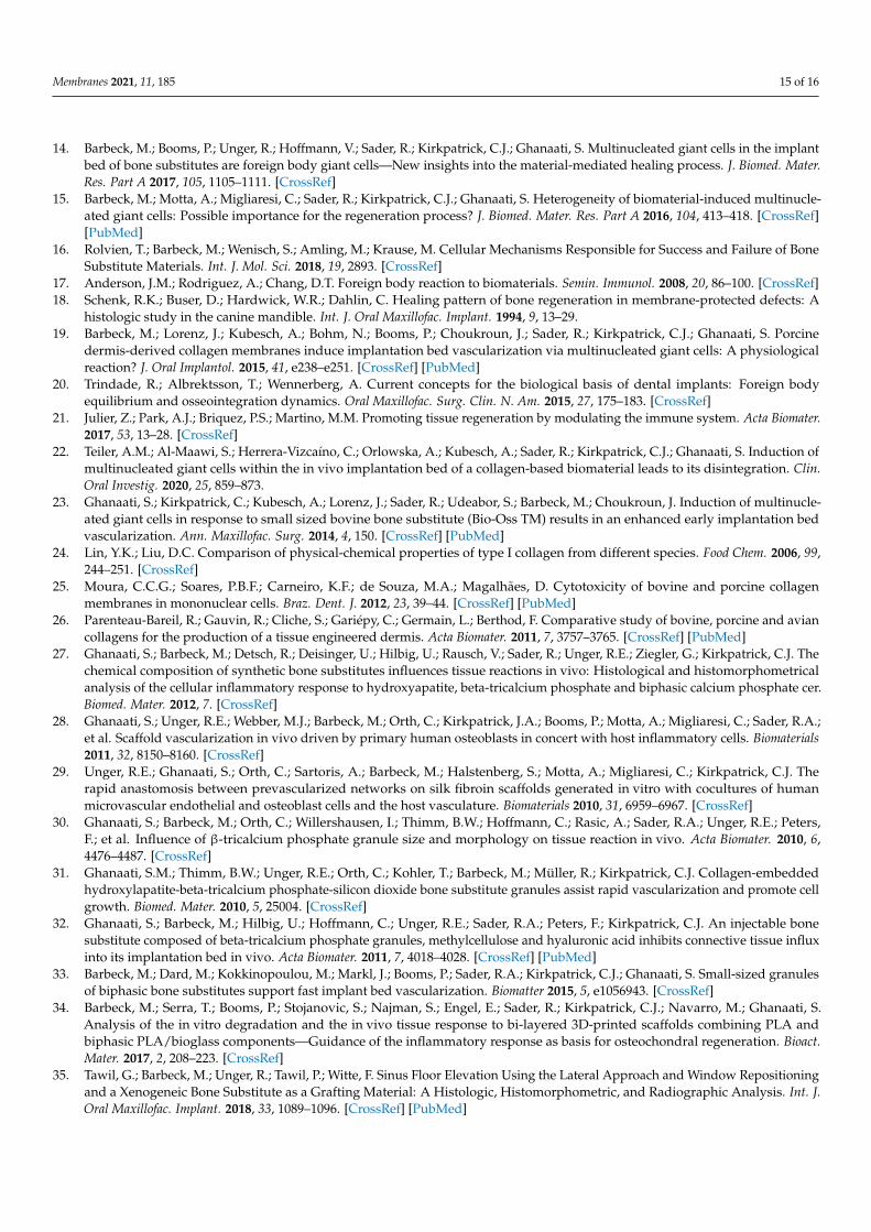
The immunohistochemical detection of blood vessels showed an elevated vasculariza-tion of the surrounding tissue around the bovine membrane comparative to the porcinemembrane at day 10 post implantation (Figure 4A,B). At day 30 post implantation, bothcollagen membranes exhibited a comparable vascularization within the connective tissuesurrounding the implants and at the tissue–membrane interface (Figure 4C,D). At 60 dayspostimplantation, vessels were found within the tissue that infiltrated the bovine membraneleading to a transmembraneous vascularization, while only single small blood vessels weredetected within the material bodies of the porcine collagen membrane (Figure 4E,F).
Membranes 2021, 11, 185 9 of 16
Membranes 2021, 11, x FOR PEER REVIEW 9 of 17
The immunohistochemical detection of blood vessels showed an elevated vasculari-zation of the surrounding tissue around the bovine membrane comparative to the porcine membrane at day 10 post implantation (Figure 4A,B). At day 30 post implantation, both collagen membranes exhibited a comparable vascularization within the connective tissue surrounding the implants and at the tissue–membrane interface (Figure 4C,D). At 60 days postimplantation, vessels were found within the tissue that infiltrated the bovine mem-brane leading to a transmembraneous vascularization, while only single small blood ves-sels were detected within the material bodies of the porcine collagen membrane (Figure 4E,F).
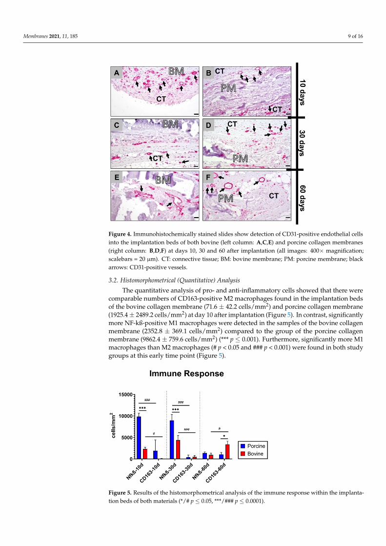
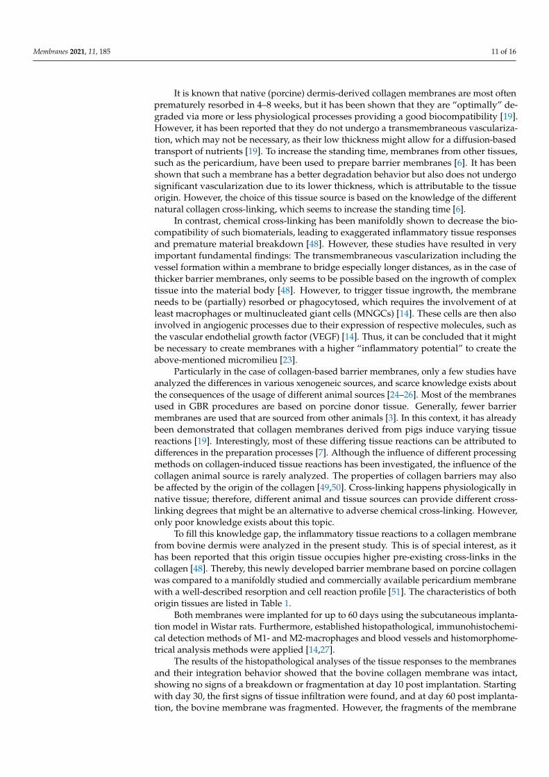
Figure 4. Immunohistochemically stained slides show detection of CD31-positive endothelial cells into the implantation beds of both bovine (left column: A, C and E) and porcine collagen mem-branes (right column: B, D and F) at days 10, 30 and 60 after implantation (all images: 400 × magni-fication; scalebars = 20 μm). CT: connective tissue; BM: bovine membrane; PM: porcine membrane; black arrows: CD31-positive vessels.
3.2. Histomorphometrical (Quantitative) Analysis The quantitative analysis of pro- and anti-inflammatory cells showed that there were
comparable numbers of CD163-positive M2 macrophages found in the implantation beds of the bovine collagen membrane (71.6 ± 42.2 cells/mm2) and porcine collagen membrane (1925.4 ± 2489.2 cells/mm2) at day 10 after implantation (Figure 5). In contrast, significantly more NF-kß-positive M1 macrophages were detected in the samples of the bovine colla-gen membrane (2352.8 ± 369.1 cells/mm2) compared to the group of the porcine collagen membrane (9862.4 ± 759.6 cells/mm2) (*** p ≤ 0.001). Furthermore, significantly more M1 macrophages than M2 macrophages (# p < 0.05 and ### p < 0.001) were found in both study groups at this early time point (Figure 5).
At 30 days after implantation, comparable values of M2 macrophages were also found in the implantation sites of the bovine collagen membrane (490.2 ± 339.2 cells/mm2) and the porcine collagen membrane (413.6 ± 481.5 cells/mm2) (Figure 4). Still higher num-bers of M1 macrophages were detected in the group of the bovine collagen compared to the value in the group of the porcine membrane (bovine collagen membrane: 4403.0 ± 1031.9 cells/mm2; porcine collagen membrane 8954.2 ± 1371.5 cells/mm2) (Figure 5). Fur-thermore, significantly higher numbers of M1 macrophages were found within the im-plant site of both collagen membranes compared to the M2 subforms (### p < 0.001).
Figure 4. Immunohistochemically stained slides show detection of CD31-positive endothelial cellsinto the implantation beds of both bovine (left column: A,C,E) and porcine collagen membranes(right column: B,D,F) at days 10, 30 and 60 after implantation (all images: 400× magnification;scalebars = 20 µm). CT: connective tissue; BM: bovine membrane; PM: porcine membrane; blackarrows: CD31-positive vessels.
3.2. Histomorphometrical (Quantitative) Analysis
The quantitative analysis of pro- and anti-inflammatory cells showed that there werecomparable numbers of CD163-positive M2 macrophages found in the implantation bedsof the bovine collagen membrane (71.6 ± 42.2 cells/mm2) and porcine collagen membrane(1925.4 ± 2489.2 cells/mm2) at day 10 after implantation (Figure 5). In contrast, significantlymore NF-kß-positive M1 macrophages were detected in the samples of the bovine collagenmembrane (2352.8 ± 369.1 cells/mm2) compared to the group of the porcine collagenmembrane (9862.4 ± 759.6 cells/mm2) (*** p ≤ 0.001). Furthermore, significantly more M1macrophages than M2 macrophages (# p < 0.05 and ### p < 0.001) were found in both studygroups at this early time point (Figure 5).
Membranes 2021, 11, x FOR PEER REVIEW 10 of 17
At 60 days after implantation, comparable numbers of NF-kß-positive M1 macro-phages were detectable in the implantation bed of the bovine collagen membrane (652.1 ± 391.4 cells/mm2) and of the porcine membrane (1380.8 ± 281.7 cells/mm2) (Figure 4). At this time point, the bovine membrane induced significantly higher numbers of CD163-positive M2 macrophages (3384 ± 776.3 cells/mm2) compared to the numbers of M1 mac-rophages (# p < 0.05) but also compared to the numbers of M2 macrophages in the group of the porcine membrane (1043.4 ± 492.3 cells/mm2) (* p ≤ 0.05) (Figure 5).
Figure 5. Results of the histomorphometrical analysis of the immune response within the implan-tation beds of both materials (*/# p ≤ 0.05, ***/### p ≤ 0.0001).
4. Discussion The concept of an “induced” membrane has already been described in traumatology
as a strategy for bone regeneration, particularly in the cases of large bone defects [38]. This method involves a two-stage procedure, where a “biological” membrane is induced as a foreign body response after application at the first stage, acting as a “chamber” for the insertion of autologous bone-graft at the second stage. It has been shown that this induced membrane can possess osteoinductive, osteogenic and angiogenic properties, and several clinical studies have demonstrated satisfactory results [38]. Especially implant bed vascu-larization via angiogenesis has been recognized as a basic factor for successful (bone) tis-sue regeneration [39,40]. Thus, to date, different strategies have focused on the develop-ment of materials that can promote vascularization. Another goal for the future develop-ment of a variety of biomaterials is to induce a specific immune response that can further stimulate (bone) tissue healing [12]. In this context, the inflammatory tissue reaction cas-cade to biomaterials has been elucidated in recent decades, and especially the macrophage has been identified as an important key factor to guide the tissue regeneration process [14,40]. In broad terms, there are two main subforms of macrophages, i.e., pro-inflamma-tory M1- and anti-inflammatory M2-macrophages [41]. It is assumed that even the induc-tion of the latter subform by a biomaterial is preferable to support the material-mediated healing process [40]. Moreover, it has been shown that different types of (oral) mesenchy-mal stem cells (MSCs), such as dental-derived mesenchymal stem cells (D-dMSCs), are involved in (bone) tissue healing [42,43]. Interestingly, it has been revealed that MSCs from oral tissues are highly committed to differentiate toward osteoblasts and precursors of bone tissue, but they also provide immunomodulatory activity [44,45].
Recently, it has also been concluded that the next generation of barrier membranes for Guided Bone Regeneration (GBR) in dentistry and maxillofacial surgery should not only resume a “passive” role in tissue separation [46]. In this context, the membrane should fulfill both functions, i.e., to separate the “soft tissue” and the bone defect site and
Figure 5. Results of the histomorphometrical analysis of the immune response within the implanta-tion beds of both materials (*/# p ≤ 0.05, ***/### p ≤ 0.0001).
Membranes 2021, 11, 185 10 of 16
At 30 days after implantation, comparable values of M2 macrophages were also foundin the implantation sites of the bovine collagen membrane (490.2 ± 339.2 cells/mm2)and the porcine collagen membrane (413.6 ± 481.5 cells/mm2) (Figure 4). Still highernumbers of M1 macrophages were detected in the group of the bovine collagen com-pared to the value in the group of the porcine membrane (bovine collagen membrane:4403.0 ± 1031.9 cells/mm2; porcine collagen membrane 8954.2± 1371.5 cells/mm2) (Figure 5).Furthermore, significantly higher numbers of M1 macrophages were found within theimplant site of both collagen membranes compared to the M2 subforms (### p < 0.001).
At 60 days after implantation, comparable numbers of NF-kß-positive M1 macrophageswere detectable in the implantation bed of the bovine collagen membrane (652.1 ± 391.4cells/mm2) and of the porcine membrane (1380.8 ± 281.7 cells/mm2) (Figure 4). At thistime point, the bovine membrane induced significantly higher numbers of CD163-positiveM2 macrophages (3384 ± 776.3 cells/mm2) compared to the numbers of M1 macrophages(# p < 0.05) but also compared to the numbers of M2 macrophages in the group of theporcine membrane (1043.4 ± 492.3 cells/mm2) (* p ≤ 0.05) (Figure 5).
4. Discussion
The concept of an “induced” membrane has already been described in traumatologyas a strategy for bone regeneration, particularly in the cases of large bone defects [38].This method involves a two-stage procedure, where a “biological” membrane is inducedas a foreign body response after application at the first stage, acting as a “chamber” forthe insertion of autologous bone-graft at the second stage. It has been shown that thisinduced membrane can possess osteoinductive, osteogenic and angiogenic properties, andseveral clinical studies have demonstrated satisfactory results [38]. Especially implantbed vascularization via angiogenesis has been recognized as a basic factor for successful(bone) tissue regeneration [39,40]. Thus, to date, different strategies have focused on thedevelopment of materials that can promote vascularization. Another goal for the futuredevelopment of a variety of biomaterials is to induce a specific immune response thatcan further stimulate (bone) tissue healing [12]. In this context, the inflammatory tissuereaction cascade to biomaterials has been elucidated in recent decades, and especially themacrophage has been identified as an important key factor to guide the tissue regenerationprocess [14,40]. In broad terms, there are two main subforms of macrophages, i.e., pro-inflammatory M1- and anti-inflammatory M2-macrophages [41]. It is assumed that eventhe induction of the latter subform by a biomaterial is preferable to support the material-mediated healing process [40]. Moreover, it has been shown that different types of (oral)mesenchymal stem cells (MSCs), such as dental-derived mesenchymal stem cells (D-dMSCs), are involved in (bone) tissue healing [42,43]. Interestingly, it has been revealedthat MSCs from oral tissues are highly committed to differentiate toward osteoblasts andprecursors of bone tissue, but they also provide immunomodulatory activity [44,45].
Recently, it has also been concluded that the next generation of barrier membranes forGuided Bone Regeneration (GBR) in dentistry and maxillofacial surgery should not onlyresume a “passive” role in tissue separation [46]. In this context, the membrane shouldfulfill both functions, i.e., to separate the “soft tissue” and the bone defect site and tocontribute to bone tissue regeneration [38,46]. This means that it is expected that a barriermembrane should actively contribute to molecular processes of bone tissue regeneration,such as stem cell differentiation osteoblast ingrowth, or via underlying processes, suchas defect site nutrition, i.e., nutrition of the underlying bone tissue defect, and there areonly two ways to ensure this functionality: either via diffusion or transmembraneousvascularization [39,43,47]. Together with this functionality, this material type has to fulfillits main objective, that is, to act as a barrier. In this context, the barrier functionality isneeded for a minimum of 4–6 weeks for periodontal tissues and 16–24 weeks for bonetissue regeneration [46]. To date, the question remains as to how such a new membranecan be developed.
Membranes 2021, 11, 185 11 of 16
It is known that native (porcine) dermis-derived collagen membranes are most oftenprematurely resorbed in 4–8 weeks, but it has been shown that they are “optimally” de-graded via more or less physiological processes providing a good biocompatibility [19].However, it has been reported that they do not undergo a transmembraneous vasculariza-tion, which may not be necessary, as their low thickness might allow for a diffusion-basedtransport of nutrients [19]. To increase the standing time, membranes from other tissues,such as the pericardium, have been used to prepare barrier membranes [6]. It has beenshown that such a membrane has a better degradation behavior but also does not undergosignificant vascularization due to its lower thickness, which is attributable to the tissueorigin. However, the choice of this tissue source is based on the knowledge of the differentnatural collagen cross-linking, which seems to increase the standing time [6].
In contrast, chemical cross-linking has been manifoldly shown to decrease the bio-compatibility of such biomaterials, leading to exaggerated inflammatory tissue responsesand premature material breakdown [48]. However, these studies have resulted in veryimportant fundamental findings: The transmembraneous vascularization including thevessel formation within a membrane to bridge especially longer distances, as in the case ofthicker barrier membranes, only seems to be possible based on the ingrowth of complextissue into the material body [48]. However, to trigger tissue ingrowth, the membraneneeds to be (partially) resorbed or phagocytosed, which requires the involvement of atleast macrophages or multinucleated giant cells (MNGCs) [14]. These cells are then alsoinvolved in angiogenic processes due to their expression of respective molecules, such asthe vascular endothelial growth factor (VEGF) [14]. Thus, it can be concluded that it mightbe necessary to create membranes with a higher “inflammatory potential” to create theabove-mentioned micromilieu [23].
Particularly in the case of collagen-based barrier membranes, only a few studies haveanalyzed the differences in various xenogeneic sources, and scarce knowledge exists aboutthe consequences of the usage of different animal sources [24–26]. Most of the membranesused in GBR procedures are based on porcine donor tissue. Generally, fewer barriermembranes are used that are sourced from other animals [3]. In this context, it has alreadybeen demonstrated that collagen membranes derived from pigs induce varying tissuereactions [19]. Interestingly, most of these differing tissue reactions can be attributed todifferences in the preparation processes [7]. Although the influence of different processingmethods on collagen-induced tissue reactions has been investigated, the influence of thecollagen animal source is rarely analyzed. The properties of collagen barriers may alsobe affected by the origin of the collagen [49,50]. Cross-linking happens physiologically innative tissue; therefore, different animal and tissue sources can provide different cross-linking degrees that might be an alternative to adverse chemical cross-linking. However,only poor knowledge exists about this topic.
To fill this knowledge gap, the inflammatory tissue reactions to a collagen membranefrom bovine dermis were analyzed in the present study. This is of special interest, as ithas been reported that this origin tissue occupies higher pre-existing cross-links in thecollagen [48]. Thereby, this newly developed barrier membrane based on porcine collagenwas compared to a manifoldly studied and commercially available pericardium membranewith a well-described resorption and cell reaction profile [51]. The characteristics of bothorigin tissues are listed in Table 1.
Both membranes were implanted for up to 60 days using the subcutaneous implanta-tion model in Wistar rats. Furthermore, established histopathological, immunohistochemi-cal detection methods of M1- and M2-macrophages and blood vessels and histomorphome-trical analysis methods were applied [14,27].
The results of the histopathological analyses of the tissue responses to the membranesand their integration behavior showed that the bovine collagen membrane was intact,showing no signs of a breakdown or fragmentation at day 10 post implantation. Startingwith day 30, the first signs of tissue infiltration were found, and at day 60 post implanta-tion, the bovine membrane was fragmented. However, the fragments of the membrane
Membranes 2021, 11, 185 12 of 16
were found to be overlapping within the subcutaneous connective tissue. Therefore, thismembrane type induced a tissue reaction, including both macrophages and multinucleatedgiant cells (MNGCs), that promoted this fragmentation process. Interestingly, the reactivetissue infiltrated the interspaces of the membrane fragments, and high numbers of vesselswere found within the tissue that infiltrated the membrane type. In contrast, the porcinemembrane remained intact until day 60 post implantation, inducing a less intense reactivetissue response and only a single cell infiltration of mononuclear cells. Furthermore, notransmembraneous vascularization was detected, but some single small vessels were foundwithin the membrane.
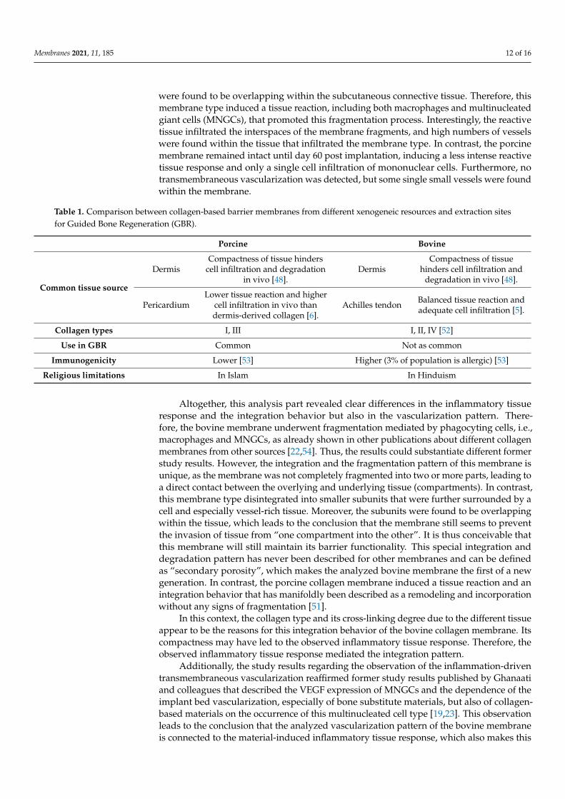
Table 1. Comparison between collagen-based barrier membranes from different xenogeneic resources and extraction sitesfor Guided Bone Regeneration (GBR).
Porcine Bovine
Common tissue source
DermisCompactness of tissue hinders
cell infiltration and degradationin vivo [48].
DermisCompactness of tissue
hinders cell infiltration anddegradation in vivo [48].
PericardiumLower tissue reaction and higher
cell infiltration in vivo thandermis-derived collagen [6].
Achilles tendon Balanced tissue reaction andadequate cell infiltration [5].
Collagen types I, III I, II, IV [52]
Use in GBR Common Not as common
Immunogenicity Lower [53] Higher (3% of population is allergic) [53]
Religious limitations In Islam In Hinduism
Altogether, this analysis part revealed clear differences in the inflammatory tissueresponse and the integration behavior but also in the vascularization pattern. There-fore, the bovine membrane underwent fragmentation mediated by phagocyting cells, i.e.,macrophages and MNGCs, as already shown in other publications about different collagenmembranes from other sources [22,54]. Thus, the results could substantiate different formerstudy results. However, the integration and the fragmentation pattern of this membrane isunique, as the membrane was not completely fragmented into two or more parts, leading toa direct contact between the overlying and underlying tissue (compartments). In contrast,this membrane type disintegrated into smaller subunits that were further surrounded by acell and especially vessel-rich tissue. Moreover, the subunits were found to be overlappingwithin the tissue, which leads to the conclusion that the membrane still seems to preventthe invasion of tissue from “one compartment into the other”. It is thus conceivable thatthis membrane will still maintain its barrier functionality. This special integration anddegradation pattern has never been described for other membranes and can be definedas “secondary porosity”, which makes the analyzed bovine membrane the first of a newgeneration. In contrast, the porcine collagen membrane induced a tissue reaction and anintegration behavior that has manifoldly been described as a remodeling and incorporationwithout any signs of fragmentation [51].
In this context, the collagen type and its cross-linking degree due to the different tissueappear to be the reasons for this integration behavior of the bovine collagen membrane. Itscompactness may have led to the observed inflammatory tissue response. Therefore, theobserved inflammatory tissue response mediated the integration pattern.
Additionally, the study results regarding the observation of the inflammation-driventransmembraneous vascularization reaffirmed former study results published by Ghanaatiand colleagues that described the VEGF expression of MNGCs and the dependence of theimplant bed vascularization, especially of bone substitute materials, but also of collagen-based materials on the occurrence of this multinucleated cell type [19,23]. This observationleads to the conclusion that the analyzed vascularization pattern of the bovine membraneis connected to the material-induced inflammatory tissue response, which also makes this
Membranes 2021, 11, 185 13 of 16
membrane type a first prototype of an immune-modulating material. Thus, the controlover the integration pattern and the material-induced inflammation may be an alternativeto conventional membranes.
Finally, the histomorphometrically measured occurrence of M1- and M2-macrophagesrevealed increased occurrences of pro-inflammatory cells induced by the porcine mem-brane at 10 and 30 days in opposite to the bovine group. However, their occurrence wasstrictly local and did not affect the surrounding tissue. Furthermore, a decrease at day 60 tocomparable numbers compared to the group of the porcine membranes was found. Thus,it is conceivable that the initially increased numbers of pro-inflammatory cells might beinduced by the differently cross-linked membrane initiating the fragmentation process.However, the observed declined levels of M1 and M2 macrophages at day 60 post implanta-tion that showed comparable levels of both subforms suggest that both membranes do notinduce a chronic inflammation that can cause inflammatory-driven implant failures. Thus,both biomaterials seem to be fully biocompatible. Interestingly, the porcine group showedsignificantly increased M2 macrophage levels at day 60 compared to the M1 macrophagesnumbers. This might indicate that this membrane can integrate within the implantationbed with molecular support of the tissue healing process.
To summarize, the importance of the bioactivity of the membrane has recently beenemphasized to create a so-called “bioactive membrane compartment”, which means tosetup an underlying micromilieu suitable for bone tissue regeneration [2]. The bovine colla-gen membranes seem to allow for the establishment of such a micromilieu due to its specialdegradation and integration pattern that triggers transmembraneous vascularization. Thebovine collagen membranes seem to allow for the establishment of such a micromilieudue to its special degradation and integration pattern that triggers transmembraneousvascularization. In this context, the vascularization pattern allows the creation of localdifferences in oxygenation and presumably the availability of nutrients that might alsohelp to generate an appropriate niche environment for osteoprogenitors [55]. Thus, thesefindings might be also relevant in the context of bone repair, which involves blood vesselgrowth and pro-angiogenic signaling interactions. Furthermore, the nutrition of the defectsite may support the survival of cells implanted with the membrane, such as differentoral-derived stem cells or (pre-) osteoblasts in tissue engineering applications [42,43].
Additionally, the membrane subunits were found to be overlapping within the tissue,which leads to the conclusion that the membrane still maintains its barrier functionality.This special integration and degradation pattern can be defined as “secondary porosity”,which makes the analyzed bovine membrane the first of a new generation that modulatesthe immune response to support bone defect healing.
The present results lead to the conclusion that this membrane meets the requirementsfor a GBR barrier membrane with an enhanced standing time that seems optimal anddoes not trigger fast degradation like in some cases of artificially cross-linked collagen [48].However, the age and region of the bovine sources have shown discrepancies in thedegree of cross-linking, which can cause a batch-to-batch manufacturing inconsistency. Astandardization of the process of animal domestication, collagen extraction and collagenreconstruction can solve this problem. Nonetheless, bovine collagen, extracted from theskin, can provide an optimized standing time for different clinical indications.
5. Conclusions
The results of the present in vivo study showed that dermis-extracted bovine collagenmembranes underwent a special integration behavior by providing a “secondary poros-ity” in concert with a transmembraneous vascularization that is expected to be suitablefor Guided Bone Regeneration applications. Histological analysis showed comparableresults to the pericardium-extracted porcine-sourced collagen membranes. The bovinemembrane remained intact initially, and the delayed fragmentation was accompaniedwith granulation tissue infiltration and the appearance of multinucleated giant cells thatseemed to mediate transmembraneous vascularization. This vascularization can serve as
Membranes 2021, 11, 185 14 of 16
an advantage in GBR alongside the vascularization of the implant bed, suggesting a furtherfunctionality of barrier membranes that current commercially available materials do notprovide. Furthermore, the delayed fragmentation of the bovine membrane suggests alonger standing time, which can be beneficial for certain dental or maxillofacial indicationswhere conventional resorbable barrier membranes suffer from a short lifetime. Altogether,the analyzed bovine membrane might be an alternative to artificially cross-linking mem-branes, as such processing can cause bioincompatibility and premature resorption thatwould result implant failure.
Author Contributions: Conceptualization, E.K. and M.B.; methodology, M.B., D.R. and S.N.; soft-ware, M.B.; validation, M.R., S.N., M.B. and D.R.; formal analysis, E.K., S.A. and M.B.; investigation,E.K., M.R., O.J., S.N. and M.B.; resources, X.X., R.K., O.J., S.N. and M.B.; data curation, E.K., S.A.,M.R., I.S., J.B., S.N. and M.B.; writing—original draft preparation, E.K., S.A., M.R., X.X., R.K., I.S.,J.B., S.N. and M.B.; writing—review and editing, E.K. and D.R.; visualization, E.K., S.A. and M.B.;supervision, S.N., M.B. and D.R.; project administration, S.N. and M.B. All authors have read andagreed to the published version of the manuscript.
Funding: NMI received financial support from the State Ministry of Baden-Württemberg for Eco-nomic Affairs, Labour and Housing Construction.
Institutional Review Board Statement: The Local Ethical Committee of the Faculty of Medicine(University of Niš, Serbia) authorized the in vivo experiments prior to the biomaterial implantations,based on decision number 323-07-09101, on 2020-05/5, of the Veterinary Directorate of the Ministryof Agriculture, Forestry and Water Management of the Republic of Serbia.
Informed Consent Statement: Not applicable.
Conflicts of Interest: The authors declare no conflict of interest.
References1. Tolstunov, L.; Hamrick, J.F.E.; Broumand, V.; Shilo, D.; Rachmiel, A. Bone Augmentation Techniques for Horizontal and Vertical
Alveolar Ridge Deficiency in Oral Implantology. Oral Maxillofac. Surg. Clin. N. Am. 2019, 31, 163–191. [CrossRef]2. Omar, O.; Elgali, I.; Dahlin, C.; Thomsen, P. Barrier membranes: More than the barrier effect? J. Clin. Periodontol. 2019, 46, 103–123.
[CrossRef]3. Elgali, I.; Omar, O.; Dahlin, C.; Thomsen, P. Guided bone regeneration: Materials and biological mechanisms revisited. Eur. J.
Oral Sci. 2017, 125, 315–337. [CrossRef]4. Lee, S.-W.; Kim, S.-G. Membranes for the Guided Bone Regeneration. Maxillofac. Plast. Reconstr. Surg. 2014, 36, 239–246.
[CrossRef]5. Ghanaati, S. In vivo Implantation of a Bovine-Derived Collagen Membrane Leads to Changes in the Physiological Cellular Pattern
of Wound Healing by the Induction of Multinucleated Giant Cells: An Adverse Reaction? Front. Bioeng. Biotechnol. 2018, 6, 1–13.[CrossRef]
6. Barbeck, M.; Lorenz, J.; Holthaus, M.G.; Raetscho, N.; Kubesch, A.; Booms, P.; Sader, R.; Kirkpatrick, C.J.; Ghanaati, S. Porcinedermis and pericardium-based, non cross-linked materials induce multinucleated giant cells after their in vivo implantation: Aphysiological reaction? J. Oral Implantol. 2015, 41, e267–e280. [CrossRef]
7. Rothamel, D.; Schwarz, F.; Sager, M.; Herten, M.; Sculean, A.; Becker, J. Biodegradation of differently cross-linked collagenmembranes: An experimental study in the rat. Clin. Oral Implant. Res. 2005, 16, 369–378. [CrossRef]
8. Franz, S.; Rammelt, S.; Scharnweber, D.; Simon, J.C. Immune responses to implants—A review of the implications for the designof immunomodulatory biomaterials. Biomaterials 2011, 32, 6692–6709. [CrossRef]
9. Trindade, R.; Albrektsson, T.; Tengvall, P.; Wennerberg, A. Foreign Body Reaction to Biomaterials: On Mechanisms for Buildupand Breakdown of Osseointegration. Clin. Implant Dent. Relat. Res. 2016, 18, 192–203. [CrossRef]
10. Mariani, E.; Lisignoli, G.; Borzì, R.M.; Pulsatelli, L. Biomaterials: Foreign bodies or tuners for the immune response? Int. J. Mol.Sci. 2019, 20, 636. [CrossRef]
11. Barbeck, M.; Unger, R.E.; Booms, P.; Dohle, E.; Sader, R.A.; Kirkpatrick, C.J.; Ghanaati, S. Monocyte preseeding leads to anincreased implant bed vascularization of biphasic calcium phosphate bone substitutes via vessel maturation. J. Biomed. Mater.Res. A 2016, 104, 2928–2935. [CrossRef]
12. Maruyama, M.; Rhee, C.; Utsunomiya, T.; Zhang, N.; Ueno, M.; Yao, Z.; Goodman, S.B. Modulation of the Inflammatory Responseand Bone Healing. Front. Endocrinol. 2020, 11, 386. [CrossRef]
13. Lee, J.; Byun, H.; Madhurakkat Perikamana, S.K.; Lee, S.; Shin, H. Current Advances in Immunomodulatory Biomaterials forBone Regeneration. Adv. Healthc. Mater. 2019, 8, 1–20. [CrossRef]
Membranes 2021, 11, 185 15 of 16
14. Barbeck, M.; Booms, P.; Unger, R.; Hoffmann, V.; Sader, R.; Kirkpatrick, C.J.; Ghanaati, S. Multinucleated giant cells in the implantbed of bone substitutes are foreign body giant cells—New insights into the material-mediated healing process. J. Biomed. Mater.Res. Part A 2017, 105, 1105–1111. [CrossRef]
15. Barbeck, M.; Motta, A.; Migliaresi, C.; Sader, R.; Kirkpatrick, C.J.; Ghanaati, S. Heterogeneity of biomaterial-induced multinucle-ated giant cells: Possible importance for the regeneration process? J. Biomed. Mater. Res. Part A 2016, 104, 413–418. [CrossRef][PubMed]
16. Rolvien, T.; Barbeck, M.; Wenisch, S.; Amling, M.; Krause, M. Cellular Mechanisms Responsible for Success and Failure of BoneSubstitute Materials. Int. J. Mol. Sci. 2018, 19, 2893. [CrossRef]
17. Anderson, J.M.; Rodriguez, A.; Chang, D.T. Foreign body reaction to biomaterials. Semin. Immunol. 2008, 20, 86–100. [CrossRef]18. Schenk, R.K.; Buser, D.; Hardwick, W.R.; Dahlin, C. Healing pattern of bone regeneration in membrane-protected defects: A
histologic study in the canine mandible. Int. J. Oral Maxillofac. Implant. 1994, 9, 13–29.19. Barbeck, M.; Lorenz, J.; Kubesch, A.; Bohm, N.; Booms, P.; Choukroun, J.; Sader, R.; Kirkpatrick, C.J.; Ghanaati, S. Porcine
dermis-derived collagen membranes induce implantation bed vascularization via multinucleated giant cells: A physiologicalreaction? J. Oral Implantol. 2015, 41, e238–e251. [CrossRef] [PubMed]
20. Trindade, R.; Albrektsson, T.; Wennerberg, A. Current concepts for the biological basis of dental implants: Foreign bodyequilibrium and osseointegration dynamics. Oral Maxillofac. Surg. Clin. N. Am. 2015, 27, 175–183. [CrossRef]
21. Julier, Z.; Park, A.J.; Briquez, P.S.; Martino, M.M. Promoting tissue regeneration by modulating the immune system. Acta Biomater.2017, 53, 13–28. [CrossRef]
22. Teiler, A.M.; Al-Maawi, S.; Herrera-Vizcaíno, C.; Orlowska, A.; Kubesch, A.; Sader, R.; Kirkpatrick, C.J.; Ghanaati, S. Induction ofmultinucleated giant cells within the in vivo implantation bed of a collagen-based biomaterial leads to its disintegration. Clin.Oral Investig. 2020, 25, 859–873.
23. Ghanaati, S.; Kirkpatrick, C.; Kubesch, A.; Lorenz, J.; Sader, R.; Udeabor, S.; Barbeck, M.; Choukroun, J. Induction of multinucle-ated giant cells in response to small sized bovine bone substitute (Bio-Oss TM) results in an enhanced early implantation bedvascularization. Ann. Maxillofac. Surg. 2014, 4, 150. [CrossRef] [PubMed]
24. Lin, Y.K.; Liu, D.C. Comparison of physical-chemical properties of type I collagen from different species. Food Chem. 2006, 99,244–251. [CrossRef]
25. Moura, C.C.G.; Soares, P.B.F.; Carneiro, K.F.; de Souza, M.A.; Magalhães, D. Cytotoxicity of bovine and porcine collagenmembranes in mononuclear cells. Braz. Dent. J. 2012, 23, 39–44. [CrossRef] [PubMed]
26. Parenteau-Bareil, R.; Gauvin, R.; Cliche, S.; Gariépy, C.; Germain, L.; Berthod, F. Comparative study of bovine, porcine and aviancollagens for the production of a tissue engineered dermis. Acta Biomater. 2011, 7, 3757–3765. [CrossRef] [PubMed]
27. Ghanaati, S.; Barbeck, M.; Detsch, R.; Deisinger, U.; Hilbig, U.; Rausch, V.; Sader, R.; Unger, R.E.; Ziegler, G.; Kirkpatrick, C.J. Thechemical composition of synthetic bone substitutes influences tissue reactions in vivo: Histological and histomorphometricalanalysis of the cellular inflammatory response to hydroxyapatite, beta-tricalcium phosphate and biphasic calcium phosphate cer.Biomed. Mater. 2012, 7. [CrossRef]
28. Ghanaati, S.; Unger, R.E.; Webber, M.J.; Barbeck, M.; Orth, C.; Kirkpatrick, J.A.; Booms, P.; Motta, A.; Migliaresi, C.; Sader, R.A.;et al. Scaffold vascularization in vivo driven by primary human osteoblasts in concert with host inflammatory cells. Biomaterials2011, 32, 8150–8160. [CrossRef]
29. Unger, R.E.; Ghanaati, S.; Orth, C.; Sartoris, A.; Barbeck, M.; Halstenberg, S.; Motta, A.; Migliaresi, C.; Kirkpatrick, C.J. Therapid anastomosis between prevascularized networks on silk fibroin scaffolds generated in vitro with cocultures of humanmicrovascular endothelial and osteoblast cells and the host vasculature. Biomaterials 2010, 31, 6959–6967. [CrossRef]
30. Ghanaati, S.; Barbeck, M.; Orth, C.; Willershausen, I.; Thimm, B.W.; Hoffmann, C.; Rasic, A.; Sader, R.A.; Unger, R.E.; Peters,F.; et al. Influence of β-tricalcium phosphate granule size and morphology on tissue reaction in vivo. Acta Biomater. 2010, 6,4476–4487. [CrossRef]
31. Ghanaati, S.M.; Thimm, B.W.; Unger, R.E.; Orth, C.; Kohler, T.; Barbeck, M.; Müller, R.; Kirkpatrick, C.J. Collagen-embeddedhydroxylapatite-beta-tricalcium phosphate-silicon dioxide bone substitute granules assist rapid vascularization and promote cellgrowth. Biomed. Mater. 2010, 5, 25004. [CrossRef]
32. Ghanaati, S.; Barbeck, M.; Hilbig, U.; Hoffmann, C.; Unger, R.E.; Sader, R.A.; Peters, F.; Kirkpatrick, C.J. An injectable bonesubstitute composed of beta-tricalcium phosphate granules, methylcellulose and hyaluronic acid inhibits connective tissue influxinto its implantation bed in vivo. Acta Biomater. 2011, 7, 4018–4028. [CrossRef] [PubMed]
33. Barbeck, M.; Dard, M.; Kokkinopoulou, M.; Markl, J.; Booms, P.; Sader, R.A.; Kirkpatrick, C.J.; Ghanaati, S. Small-sized granulesof biphasic bone substitutes support fast implant bed vascularization. Biomatter 2015, 5, e1056943. [CrossRef]
34. Barbeck, M.; Serra, T.; Booms, P.; Stojanovic, S.; Najman, S.; Engel, E.; Sader, R.; Kirkpatrick, C.J.; Navarro, M.; Ghanaati, S.Analysis of the in vitro degradation and the in vivo tissue response to bi-layered 3D-printed scaffolds combining PLA andbiphasic PLA/bioglass components—Guidance of the inflammatory response as basis for osteochondral regeneration. Bioact.Mater. 2017, 2, 208–223. [CrossRef]
35. Tawil, G.; Barbeck, M.; Unger, R.; Tawil, P.; Witte, F. Sinus Floor Elevation Using the Lateral Approach and Window Repositioningand a Xenogeneic Bone Substitute as a Grafting Material: A Histologic, Histomorphometric, and Radiographic Analysis. Int. J.Oral Maxillofac. Implant. 2018, 33, 1089–1096. [CrossRef] [PubMed]
Membranes 2021, 11, 185 16 of 16
36. Barbeck, M.; Najman, S.; Stojanovic, S.; Mitic, Ž.; Živkovic, J.M.; Choukroun, J.; Kovacevic, P.; Sader, R.; Kirkpatrick, C.J.; Ghanaati,S. Addition of blood to a phycogenic bone substitute leads to increased in vivo vascularization. Biomed. Mater. 2015, 10, 055007.[CrossRef]
37. Lindner, C.; Pröhl, A.; Abels, M.; Löffler, T.; Batinic, M.; Jung, O.; Barbeck, M. Specialized Histological and HistomorphometricalAnalytical Methods for Biocompatibility Testing of Biomaterials for Maxillofacial Surgery in (Pre-) Clinical Studies. In Vivo 2020,34, 3137–3152. [CrossRef] [PubMed]
38. Masquelet, A.C.; Begue, T. The Concept of Induced Membrane for Reconstruction of Long Bone Defects. Orthop. Clin. N. Am.2010, 41, 27–37. [CrossRef] [PubMed]
39. Schwarz, F.; Rothamel, D.; Herten, M.; Sager, M.; Becker, J. Angiogenesis pattern of native and cross-linked collagen membranes:An immunohistochemical study in the rat. Clin. Oral Implant. Res. 2006, 17, 403–409. [CrossRef]
40. Peric Kacarevic, Ž.; Rider, P.; Alkildani, S.; Retnasingh, S.; Pejakic, M.; Schnettler, R.; Gosau, M.; Smeets, R.; Jung, O.; Barbeck, M.An introduction to bone tissue engineering. Int. J. Artif. Organs 2020, 43, 69–86. [CrossRef] [PubMed]
41. Miron, R.J.; Bosshardt, D.D. Multinucleated Giant Cells: Good Guys or Bad Guys? Tissue Eng. Part B Rev. 2018, 24, 53–65.[CrossRef]
42. Spagnuolo, G.; Codispoti, B.; Marrelli, M.; Rengo, C.; Rengo, S.; Tatullo, M. Commitment of Oral-Derived Stem Cells in Dentaland Maxillofacial Applications. Dent. J. 2018, 6, 72. [CrossRef] [PubMed]
43. Ballini, A.; Boccaccio, A.; Saini, R.; Van Pham, P.; Tatullo, M. Dental-Derived Stem Cells and Their Secretome and Interactionswith Bioscaffolds/Biomaterials in Regenerative Medicine: From the In Vitro Research to Translational Applications. Stem Cells Int.2017, 2017, 1–3. [CrossRef]
44. Pierdomenico, L.; Bonsi, L.; Calvitti, M.; Rondelli, D.; Arpinati, M.; Chirumbolo, G.; Becchetti, E.; Marchionni, C.; Alviano, F.;Fossati, V.; et al. Multipotent mesenchymal stem cells with immunosuppressive activity can be easily isolated from dental pulp.Transplantation 2005, 80, 836–842. [CrossRef]
45. Yamaza, T.; Kentaro, A.; Chen, C.; Liu, Y.; Shi, Y.; Gronthos, S.; Wang, S.; Shi, S. Immunomodulatory properties of stem cells fromhuman exfoliated deciduous teeth. Stem Cell Res. Ther. 2010, 1. [CrossRef]
46. Rozalia, D.; George, I.M.; Giorgio, M.C.; Peter, V.G. The role of barrier membranes for guided bone regeneration and restorationof large bone defects: Current experimental and clinical evidence. BMC Med. 2012, 10, 1. [CrossRef]
47. Suhaimi, H.; Bhusan Das, D. Glucose diffusion in tissue engineering membranes and scaffolds. Rev. Chem. Eng. 2016, 32, 629–650.[CrossRef]
48. Delgado, L.M.; Bayon, Y.; Pandit, A.; Zeuglogi, D. To cross-link or not to cross-link? Cross-linking associated foreign body response ofcollagen-based devices. 2015, 084946, 1–50.
49. Zheng, M.H.; Chen, J.; Kirilak, Y.; Willers, C.; Xu, J.; Wood, D. Porcine small intestine submucosa (SIS) is not an acellularcollagenous matrix and contains porcine DNA: Possible implications in human implantation. J. Biomed. Mater. Res. B Appl.Biomater. 2005, 73, 61–67. [CrossRef] [PubMed]
50. Behring, J.; Junker, R.; Walboomers, X.F.; Chessnut, B.; Jansen, J.A. Toward guided tissue and bone regeneration: Morphology,attachment, proliferation, and migration of cells cultured on collagen barrier membranes. A systematic review. Odontology 2008,96, 1–11. [CrossRef]
51. Biocompatibility and Biodegradation of a Native Porcine Pericardium Membrane: Results of In Vitro and In Vivo Examinations—PubMed. Available online: https://pubmed.ncbi.nlm.nih.gov/22299091/ (accessed on 22 February 2021).
52. Davison-Kotler, E.; Marshall, W.S.; García-Gareta, E. Sources of collagen for biomaterials in skin wound healing. Bioengineering2019, 6, 56. [CrossRef]
53. Lynn, A.K.; Yannas, I.V.; Bonfield, W. Antigenicity and Immunogenicity of Collagen. J. Biomed. Res. 2004, 343–354. [CrossRef][PubMed]
54. Lorenz, J.; Kubesch, A.; Korzinskas, T.; Barbeck, M.; Landes, C.; Sader, R.A.; Kirkpatrick, C.J.; Ghanaati, S. TRAP-PositiveMultinucleated Giant Cells Are Foreign Body Giant Cells Rather Than Osteoclasts: Results from a Split-Mouth Study in Humans.J Oral Implant. 2015, 41, e257-66. [CrossRef] [PubMed]
55. Kusumbe, A.P.; Adams, R.H. Osteoclast progenitors promote bone vascularization and osteogenesis. Nat. Med. 2014, 20,1238–1240. [CrossRef] [PubMed]