Embed Size (px)
Citation preview
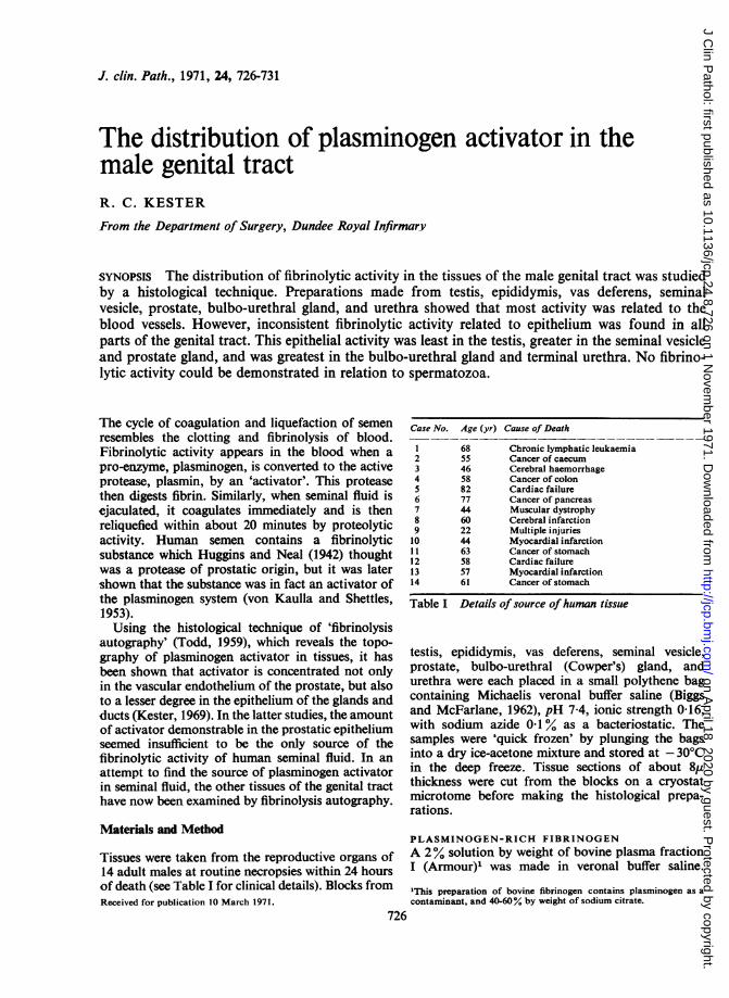
J. clin. Path., 1971, 24, 726-731
The distribution of plasminogen activator in themale genital tractR. C. KESTER
From the Department of Surgery, Dundee Royal Infirmary
SYNOPSIS The distribution of fibrinolytic activity in the tissues of the male genital tract was studiedby a histological technique. Preparations made from testis, epididymis, vas deferens, seminalvesicle, prostate, bulbo-urethral gland, and urethra showed that most activity was related to theblood vessels. However, inconsistent fibrinolytic activity related to epithelium was found in allparts of the genital tract. This epithelial activity was least in the testis, greater in the seminal vesicleand prostate gland, and was greatest in the bulbo-urethral gland and terminal urethra. No fibrino-lytic activity could be demonstrated in relation to spermatozoa.
The cycle of coagulation and liquefaction of semenresembles the clotting and fibrinolysis of blood.Fibrinolytic activity appears in the blood when apro-enzyme, plasminogen, is converted to the activeprotease, plasmin, by an 'activator'. This proteasethen digests fibrin. Similarly, when seminal fluid isejaculated, it coagulates immediately and is thenreliquefied within about 20 minutes by proteolyticactivity. Human semen contains a fibrinolyticsubstance which Huggins and Neal (1942) thoughtwas a protease of prostatic origin, but it was latershown that the substance was in fact an activator ofthe plasminogen system (von Kaulla and Shettles,1953).Using the histological technique of 'fibrinolysis
autography' (Todd, 1959), which reveals the topo-graphy of plasminogen activator in tissues, it hasbeen shown that activator is concentrated not onlyin the vascular endothelium of the prostate, but alsoto a lesser degree in the epithelium of the glands andducts (Kester, 1969). In the latter studies, the amountof activator demonstrable in the prostatic epitheliumseemed insufficient to be the only source of thefibrinolytic activity of human seminal fluid. In anattempt to find the source of plasminogen activatorin seminal fluid, the other tissues of the genital tracthave now been examined by fibrinolysis autography.
Materials and Method
Tissues were taken from the reproductive organs of14 adult males at routine necropsies within 24 hoursof death (see Table I for clinical details). Blocks fromReceived for publication 10 March 1971.
Case No. Age (yr) Cause of Death
1 68 Chronic lymphatic leukaemia2 55 Cancer of caecum3 46 Cerebral haemorrhage4 58 Cancer of colon5 82 Cardiac failure6 77 Cancer of pancreas7 44 Muscular dystrophy8 60 Cerebral infarction9 22 Multiple injuries10 44 Myocardial infarction11 63 Cancer of stomach12 58 Cardiac failure13 57 Myocardial infarction14 61 Cancer of stomach
Table I Details of source of human tissue
testis, epididymis, vas deferens, seminal vesicle,prostate, bulbo-urethral (Cowper's) gland, andurethra were each placed in a small polythene bagcontaining Michaelis veronal buffer saline (Biggsand McFarlane, 1962), pH 7-4, ionic strength 0-16,with sodium azide 0-1 % as a bacteriostatic. Thesamples were 'quick frozen' by plunging the bagsinto a dry ice-acetone mixture and stored at - 30°Cin the deep freeze. Tissue sections of about 8,uthickness were cut from the blocks on a cryostatmicrotome before making the histological prepa-rations.
PLASMINOGEN-RICH FIBRINOGENA 2% solution by weight of bovine plasma fractionI (Armour)' was made in veronal buffer saline.
'This preparation of bovine fibrinogen contains plasminogen as acontaminant, and 40-60% by weight of sodium citrate.
726
on April 18, 2020 by guest. P
rotected by copyright.http://jcp.bm
j.com/
J Clin P
athol: first published as 10.1136/jcp.24.8.726 on 1 Novem
ber 1971. Dow
nloaded from
The distribution ofplasminogen activator in the male genital tract
THROMBIN SOLUTIONThrombin, topical (bovine origin) (Parke Davis):stock solution 1,000 units/ml in 50% glycerol, andthis was diluted to 20 units/ml in Michaelis buffersaline to obtain a working solution.
CELLOPHANE SHEETCellophane PT 300-400 gauge (British CellophaneLtd).
NEUTRAL BUFFERED FORMALIN SOLUTION(LILLIE, 1965)Harris's haematoxylin (Harris, 1900).The histochemical technique was that of fibrino-
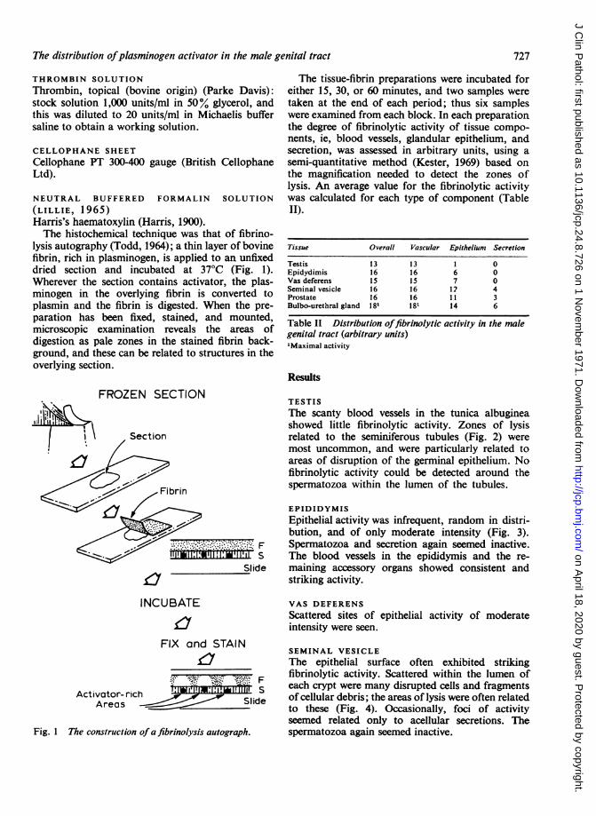
lysis autography (Todd, 1964); a thin layer of bovinefibrin, rich in plasminogen, is applied to an unfixeddried section and incubated at 37°C (Fig. 1).Wherever the section contains activator, the plas-minogen in the overlying fibrin is converted toplasmin and the fibrin is digested. When the pre-paration has been fixed, stained, and mounted,microscopic examination reveals the areas ofdigestion as pale zones in the stained fibrin back-ground, and these can be related to structures in theoverlying section.
FROZEN SECTION
Section
Fibrin
Slide
INCUBATE
£7FIX and STAIN
3 F
Activator- rich _g**g * SAreas d lide
Fig. 1 The construction ofa fibrinolysis autograph.
The tissue-fibrin preparations were incubated foreither 15, 30, or 60 minutes, and two samples weretaken at the end of each period; thus six sampleswere examined from each block. In each preparationthe degree of fibrinolytic activity of tissue compo-nents, ie, blood vessels, glandular epithelium, andsecretion, was assessed in arbitrary units, using asemi-quantitative method (Kester, 1969) based onthe magnification needed to detect the zones oflysis. An average value for the fibrinolytic activitywas calculated for each type of component (TableII).
Tissue Overall Vascular Epitheliuni Secretion
Testis 13 13 1 0Epidydimis 16 16 6 0Vas deferens 15 15 7 0Seminal vesicle 16 16 12 4Prostate 16 16 1 1 3Bulbo-urethral gland 181 181 14 6
Table II Distribution offibrinolytic activity in the malegenital tract (arbitrary units)'Maximal activity
Results
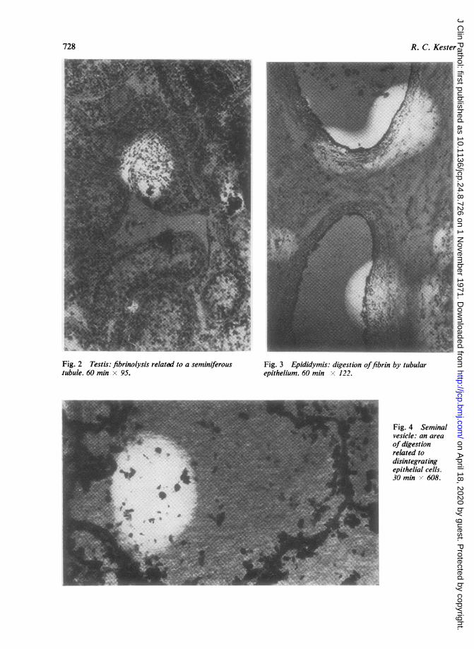
TESTISThe scanty blood vessels in the tunica albugineashowed little fibrinolytic activity. Zones of lysisrelated to the seminiferous tubules (Fig. 2) weremost uncommon, and were particularly related toareas of disruption of the germinal epithelium. Nofibrinolytic activity could be detected around thespermatozoa within the lumen of the tubules.
EPIDIDYMISEpithelial activity was infrequent, random in distri-bution, and of only moderate intensity (Fig. 3).Spermatozoa and secretion again seemed inactive.The blood vessels in the epididymis and the re-maining accessory organs showed consistent andstriking activity.
VAS DEFERENSScattered sites of epithelial activity of moderateintensity were seen.
SEMINAL VESICLEThe epithelial surface often exhibited strikingfibrinolytic activity. Scattered within the lumen ofeach crypt were many disrupted cells and fragmentsof cellular debris; the areas of lysis were often relatedto these (Fig. 4). Occasionally, foci of activityseemed related only to acellular secretions. Thespermatozoa again seemed inactive.
727
on April 18, 2020 by guest. P
rotected by copyright.http://jcp.bm
j.com/
J Clin P
athol: first published as 10.1136/jcp.24.8.726 on 1 Novem
ber 1971. Dow
nloaded from
R. C. Kester*w ~~~~~~~~~~~~~~~~~~~~~~. .wS
Fig. 2 Testis: fibrinolysis related to a seminiferoustubule. 60 min x 95.
Fig. 3 Epididymis: digestion offibrin by tubularepithelium. 60 min x 122.
Fig. 4 Seminalvesicle: an areaofdigestionrelated todisintegratingepithelial cells.30 min > 608.
728
on April 18, 2020 by guest. P
rotected by copyright.http://jcp.bm
j.com/
J Clin P
athol: first published as 10.1136/jcp.24.8.726 on 1 Novem
ber 1971. Dow
nloaded from
The distribution ofplasnunogen activator in the male genital tract
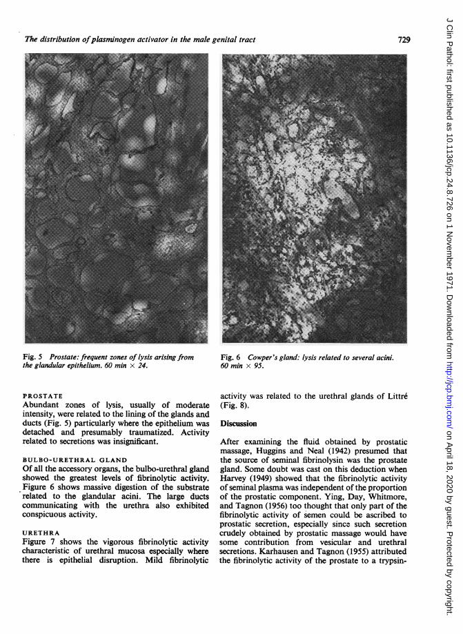
Fig. 5 Prostate: frequent zones of lysis arising fromthe glandular epithelium. 60 min x 24.
PROSTATEAbundant zones of lysis, usually of moderateintensity, were related to the lining of the glands andducts (Fig. 5) particularly where the epithelium wasdetached and presumably traumatized. Activityrelated to secretions was insignificant.
BULBO-URETHRAL GLANDOf all the accessory organs, the bulbo-urethral glandshowed the greatest levels of fibrinolytic activity.Figure 6 shows massive digestion of the substraterelated to the glandular acini. The large ductscommunicating with the urethra also exhibitedconspicuous activity.
URETHRAFigure 7 shows the vigorous fibrinolytic activitycharacteristic of urethral mucosa especially wherethere is epithelial disruption. Mild fibrinolytic
Fig. 6 Cowper's gland: lysis related to several acini.60 min x 95.
activity was related to the urethral glands of Littre(Fig. 8).
Discussion
After examining the fluid obtained by prostaticmassage, Huggins and Neal (1942) presumed thatthe source of seminal fibrinolysin was the prostategland. Some doubt was cast on this deduction whenHarvey (1949) showed that the fibrinolytic activityof seminal plasma was independent of the proportionof the prostatic component. Ying, Day, Whitmore,and Tagnon (1956) too thought that only part of thefibrinolytic activity of semen could be ascribed toprostatic secretion, especially since such secretioncrudely obtained by prostatic massage would havesome contribution from vesicular and urethralsecretions. Karhausen and Tagnon (1955) attributedthe fibrinolytic activity of the prostate to a trypsin-
729
on April 18, 2020 by guest. P
rotected by copyright.http://jcp.bm
j.com/
J Clin P
athol: first published as 10.1136/jcp.24.8.726 on 1 Novem
ber 1971. Dow
nloaded from

R. C. Kester
Fig. 7 Prostatic urethra: prominent activity from Fig. 8 Urethra: mildfibrinolytic activity arising frommucosa, especially where disrupted. 30 min x 95. the glands ofLittre. 30 min x 95.
like protease rather than to the presence of plasmin-ogen activator. Although the prostate is rich inplasminogeh activator (Albrechtsen, 1957;Rasmussen, Albrechtsen, and Astrup, 1958;Rasmussen and Albrechtsen, 1960), neverthelessthe histological technique shows that the activatoris largely confined to the vessels, making it unlikelythat the prostate contributes much fibrinolyticactivity to the ejaculate.The normal secretory processes in male accessory
glands are accompanied by definite changes inepithelial structure varying from desquamation tocell rupture (Mann, 1964). Analysis of the glandularfluid confinrns the presence not only of wholeepithelial cells but also of glandular debris. In thehistological preparations, fibrinolytic activity re-latedto such debriswasconspicuous. This observation
is consistent with that of Pandolfi and Astrup (1967)that damage or disruption of corneal epitheliumenhances the fibrinolytic activity of these cells.
It has been shown in the present studies that theglandular epithelium of the male assessory genitalorgans contains plasminogen activator in varyingamounts. Although the fibrinolytic activity of bloodvessels is consistently high throughout the genitaltract (except in the testis), the fibrinolytic activityof epithelium increases along the genital tract andis greatest in both the epithelium and secretion ofCowper's gland. The full significance of the fibrin-olysis in seminal plasma is not yet clear. Sincefibrinolytic activity may be necessary to main-tain the patency of the urinary tract by digestingfibrinous deposits (Astrup and Sterndorff, 1952;Ladehoff, 1960; Charlton, 1966), fibrinolysis may
730
on April 18, 2020 by guest. P
rotected by copyright.http://jcp.bm
j.com/
J Clin P
athol: first published as 10.1136/jcp.24.8.726 on 1 Novem
ber 1971. Dow
nloaded from
The distribution ofplasminogen activator in the male genital tract
similarly be necessary to maintain semen in a fluidstate within the genital passages (Mann, 1964).However, the high concentration of activator in thelast secretion to be added to the ejaculate-thatfrom Cowper's glands-would suggest that thefibrinolytic activity had its main effects afterejaculation by liquefying the seminal coagulum andthus facilitating the migration of spermatozoa intothe uterine cavity. It is also possible that plasminplays a part in acrosome rupture, the process of'sperm capacitation', and in penetration of the zonapellucida.Huggins and Neal (1942) have shown that the
fibrinolytic activity of semen was in the seminalplasma rather than in the spermatozoa; this hasbeen confirmed using centrifugation methodschecked by microscopy (Kester, 1970). In thepresent experiment no activator was demonstratedin spermatozoa, although Tympanidis and Astrup(1968) found that human spermatozoa in vaginalsmears exhibited significant fibrinolysis. It is possiblethat in their material the spermatozoa were coated byseminal plasma containing activator, or that theyhad been activated by contact with vaginal orcervical secretions.
I am deeply indebted to Dr Alastair S. Todd for hiscontinued interest and support, and to Mrs AnneNunn for technical advice. I wish to thank ProfessorD. M. Douglas for laboratory facilities, Miss MaryBenstead, medical artist, for her assistance, Mr TomKing for his invaluable help in preparing the photo-micrographs, and Miss Joyce Devlin for typing thescript. I am grateful to the Board of Management,Dundee General Teaching Hospitals, for a researchgrant.
References
Albrechtsen, 0. K. (1957). The fi,rinolytic activity of human tissues.Brit. J. Haemat., 3, 284-291.
Astrup, T., and Sterndorff, I. (1952). An activator of plasminogen innormal urine. Proc. Soc. exp. Biol. (N. Y.), 81, 675-678.
Biggs, R., and Macfarlane, R. G. (1962). Hunan Blood Coagilationand its Disorders, 3rd ed., p. 370. Blackwell, Oxford.
Charlton, C. A. C. (1966). Pathogenesis of urolithiasis: relation ofurinary fibrinolytic activity to nondialyzable urinary solids.Surg. Forum, 17, 503-505.
Harris, H. F. (1900). On the rapid conversion of haematoxylin intohaematein in staining, reactions. J. appl. Micro., 3, 777-780.
Harvey, C. (1949). Fibrinolysin in human semen. A method of assay,and some preliminary observations. Proc. Soc. Study. Fert.(Edinb.), 1, 11-17.
Huggins, C., and Neal, W. (1942). Coagulation and liquefaction ofsemen. Proteolytic enzymes and citrate in prostatic fluid. J.exp. Med., 76, 527-541.
Karhausen, L., and Tagnon, H. (1955). Le syndrome de fibrinolyseprostatique: nature de l'activit6 prot6olytique de la prostate.Acta clin. beig., 10, 471.476.
von. Kaulla, K. N., and Shettles, L. B. (1953). Relationship betweenhuman seminal fluid and the fibrinolytic system. Proc. Sor.Exp. Biol. (N. Y.), 83, 692-694.
Kester, R. C. (1969). Plasminogen activator in the human prostate.J. clin. Path., 22, 442.446.
Kester, R. C. (1970). The Distribution of Plasminogen Activator in theMale Genital Tract. ChM Thesis, University of Cape Town.
Ladehoff, A. (1960). The content of plasminogen activator in thehuman urinary tract. Scand. J. clin. Lab. Invest., 12, 136-139.
Lillie, R. D. (1965). Histopathologie Technic and Practical Histo-chemistry, 3rd ed., p. 38. McGraw Hill, New York.
Mann, T. (1964). The Biochemistry ofSemen and of the Male Repro-ductive Tract, 2nd ed. Methuen, London.
Pandolfi, M., and Astrup, T. (1967). A histochemical study of thefibrinolytic activity: cornea, conjunctiva, and lacrimal gland.Arch. Ophthal., 77, 258-264.
Rasmussen, J., and Albrechtsen, 0. K. (1960). Characterisation of thefibrinolytic components in the human prostate. Scand. J. clin.Lab. Invest., 12, 261-268.
Rasmussen, J., Albrechtsen, 0. K., and Astrup, T. (1958). Thefibrinolytic activity in the human prostate and in seminalfluid. Proceedings 6th Congress of the European Society ofHaematology, vol. 2, p. 494. Karger, Basle.
Todd, A. S. (1959). The histological localisation of fibrinclysinactivator. J. Path. Bact., 78, 281-283.
Todd, A. S. (1964). Localization of fibrinolytic activity in tissues. Erit.med. Bull., 20, 210-212.
Tympanidis, K., and Astrup, T. (1968). Fibrinolytic activity of rat,rabbit and human sperm cells. Proc. Soc. exp. Biol. (N. Y.), 129,179-182.
Ying, S. H., Day, E., Whitmore, W. F., and Tagnon, H. J. (1956).Fibrinolytic activity in human prostatic fluiid and semen. Fertil.and Steril., 7, 80-87.
731
on April 18, 2020 by guest. P
rotected by copyright.http://jcp.bm
j.com/
J Clin P
athol: first published as 10.1136/jcp.24.8.726 on 1 Novem
ber 1971. Dow
nloaded from