Embed Size (px)
Citation preview
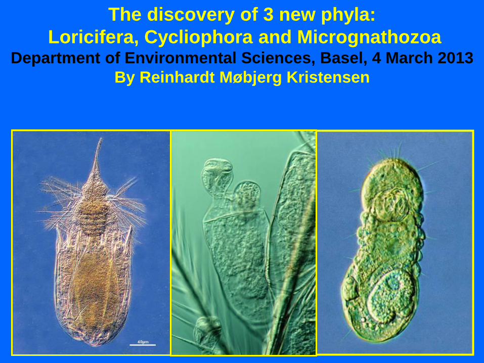
The discovery of 3 new phyla:
Loricifera, Cycliophora and Micrognathozoa Department of Environmental Sciences, Basel, 4 March 2013
By Reinhardt Møbjerg Kristensen
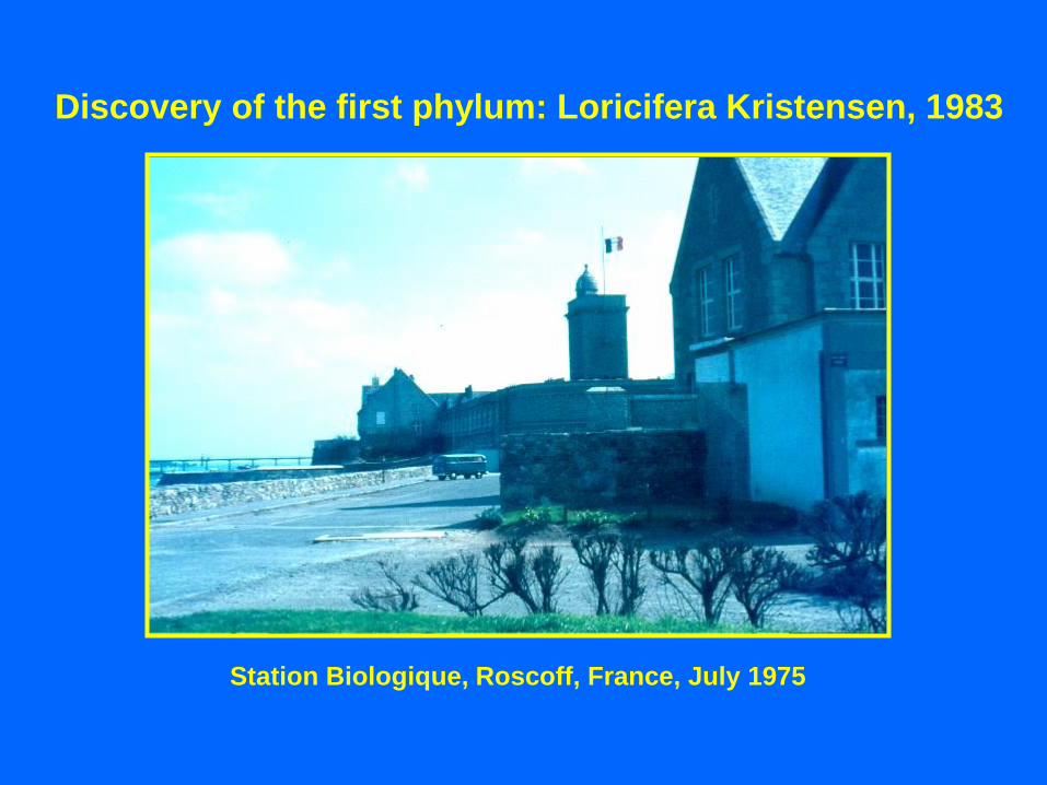
Station Biologique, Roscoff, France, July 1975
Discovery of the first phylum: Loricifera Kristensen, 1983
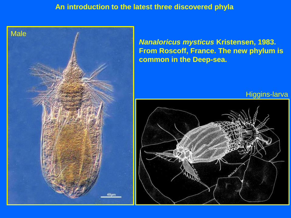
Nanaloricus mysticus Kristensen, 1983.
From Roscoff, France. The new phylum is
common in the Deep-sea.
An introduction to the latest three discovered phyla
Male
Higgins-larva
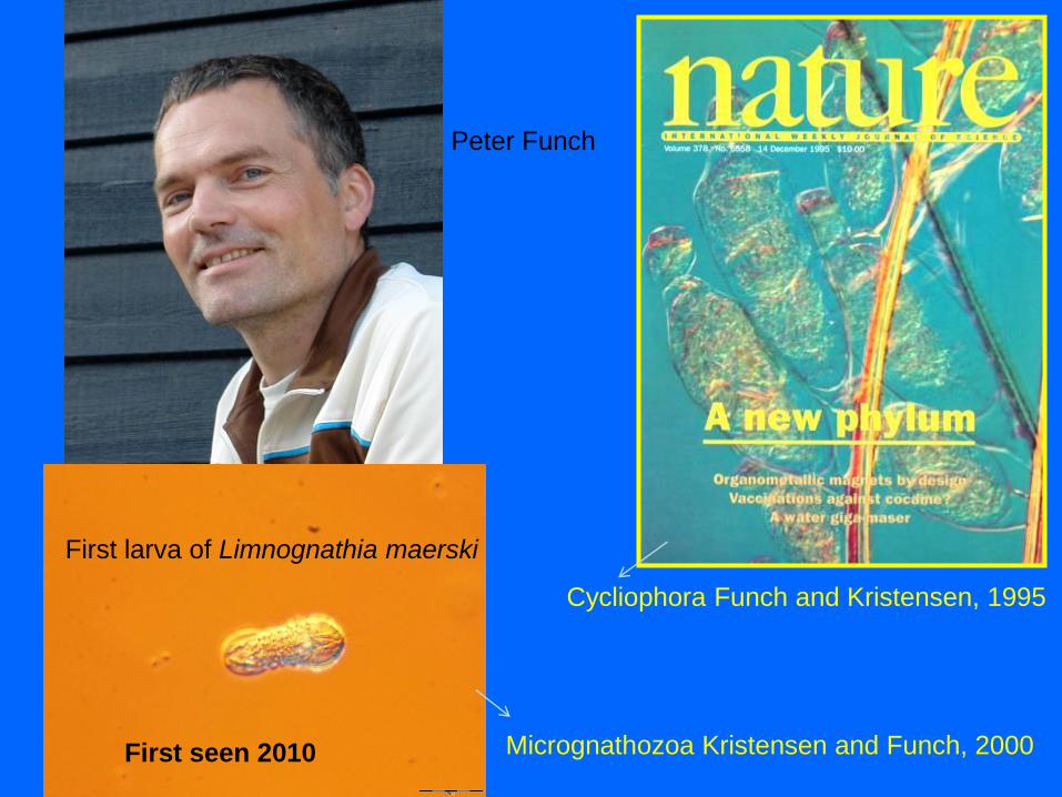
Peter Funch
Cycliophora Funch and Kristensen, 1995
Micrognathozoa Kristensen and Funch, 2000
First larva of Limnognathia maerski
First seen 2010

An introduction to the latest three discovered phyla
Three new phyla: The cladogram of Sørensen et al. 2000
Totally outdated

Dunn et al., 2008: EST
140 genes and ”new” 34 metazoans
ATOL-NSF programme 2008
“Assembling the Tree of Life”
in Copenhagen
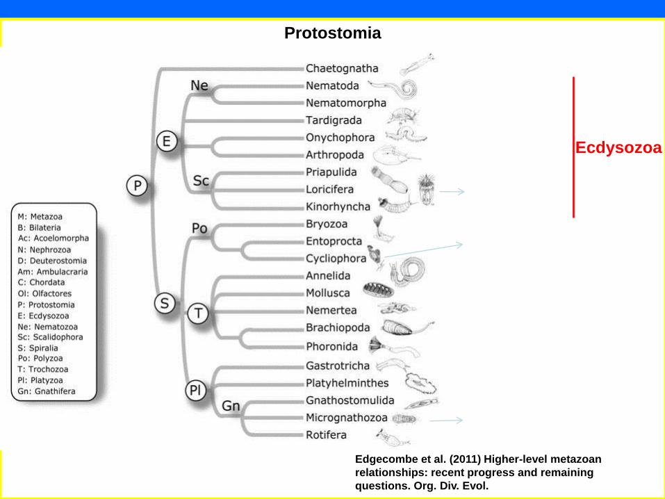
Edgecombe et al. (2011) Higher-level metazoan
relationships: recent progress and remaining
questions. Org. Div. Evol.
Protostomia
Ecdysozoa

3
6
4 5
7
6. Tardigrada
4. Loricifera
5. Polychaeta
7. Copepoda &
Tantalocarida
8. Aplacophora
1. Nematoda
6
4
5
1
3. Kinorhyncha
Interstitial fauna
from carbonate sand (2002)
M.V. Sørensen
& R.P. Higgins
J.G. Hansen &
A. Jørgensen
I. Heiner; R. Neves
K. Worsaae
2. Gastrotricha G. Gad
C. Clausen
R. Huys & P. Funch
A. Jørgensen
2
8

50 µm
Hamlet larva, Helsingør 1975 Higgins larve, Florida 1983

Samplings on Faroe Bank, BIOFAR Project 1989
1. Anchor dredge 2. Box core 3. Higgins’ Meiobenthic sledge
Mentor: Robert P. Higgins, Smithsonian Institution

Pliciloricus enigmaticus
The first known Loricifera from the Eastward-Expedition, USA (1974)
Computer graphic

Pliciloricus gracilis Higgins and Kristensen, 1986 Adult Larva
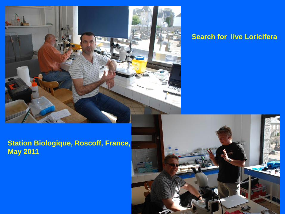
Station Biologique, Roscoff, France,
May 2011
Search for live Loricifera
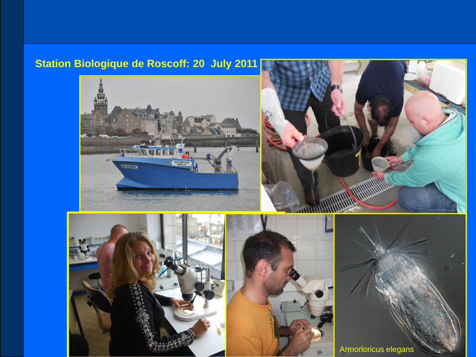
Station Biologique de Roscoff: 20 July 2011
Armorloricus elegans

Higgins-larva of Nanaloricus from Roscoff, 2005
Trenzen ar Skoden, 50 m
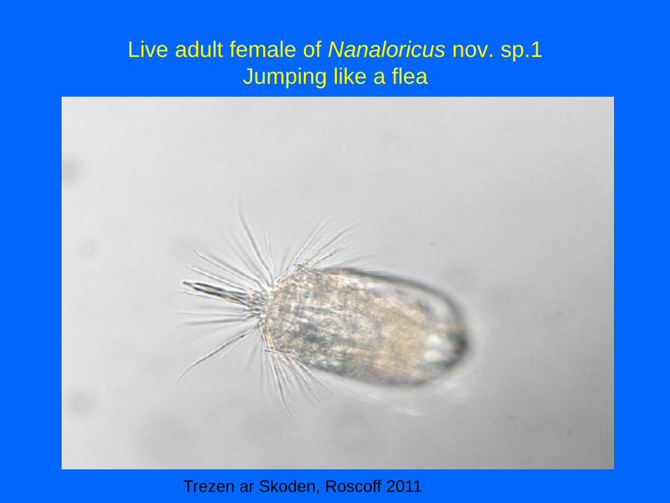
Live adult female of Nanaloricus nov. sp.1
Jumping like a flea
Trezen ar Skoden, Roscoff 2011

The life cycle of
the family
Nanaloricidae
Nanaloricus mysticus, Roscoff
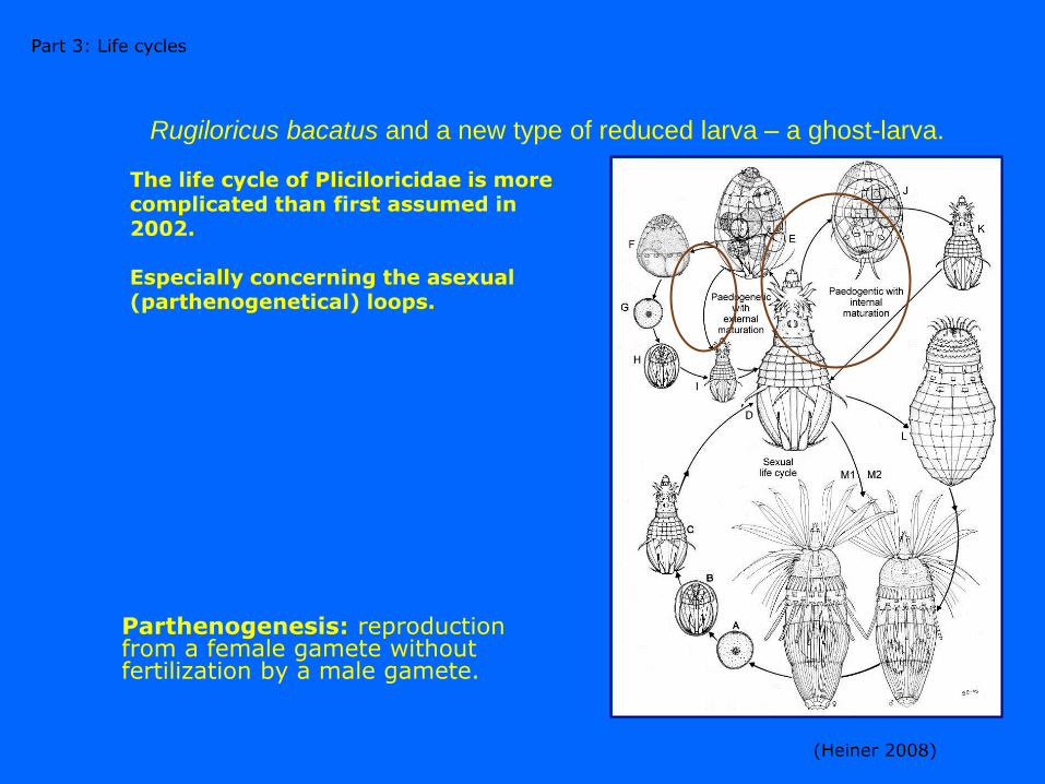
The life cycle of Pliciloricidae is more complicated than first assumed in 2002. Especially concerning the asexual (parthenogenetical) loops.
Rugiloricus bacatus and a new type of reduced larva – a ghost-larva.
(Heiner 2008)
Part 3: Life cycles
Parthenogenesis: reproduction from a female gamete without fertilization by a male gamete.

Photos of Yoshihisa Shirayama: The first deep sea loriciferans
Pliciloricus hadalis Kristensen and Shirayama, 1988: 8260 m depth
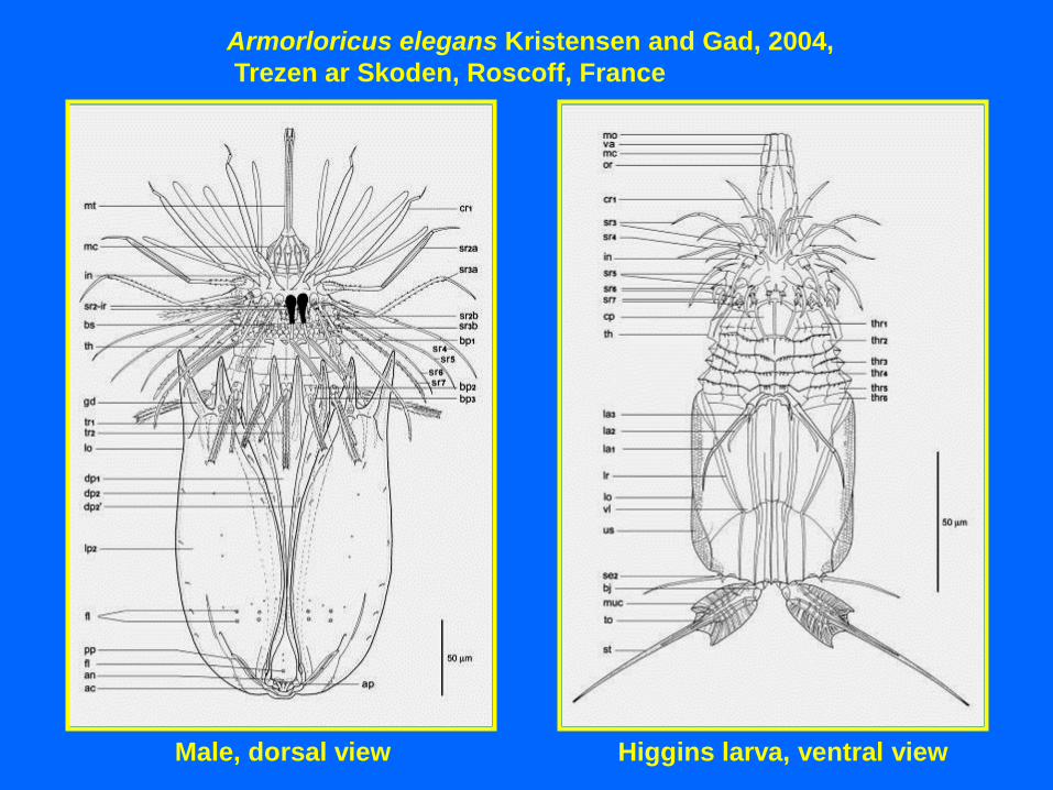
Armorloricus elegans Kristensen and Gad, 2004,
Trezen ar Skoden, Roscoff, France
Male, dorsal view Higgins larva, ventral view
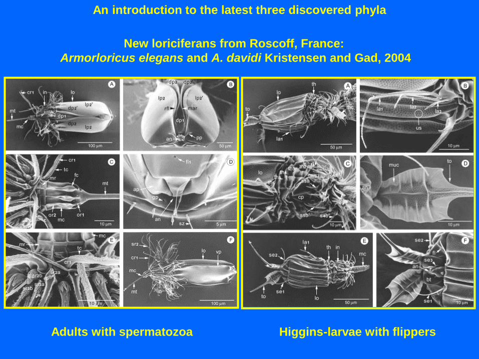
An introduction to the latest three discovered phyla
New loriciferans from Roscoff, France:
Armorloricus elegans and A. davidi Kristensen and Gad, 2004
Adults with spermatozoa Higgins-larvae with flippers

TEM of N. mysticus
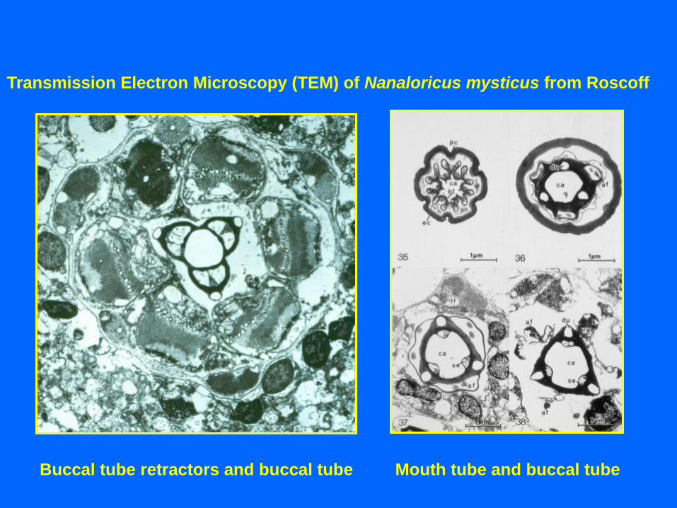
Transmission Electron Microscopy (TEM) of Nanaloricus mysticus from Roscoff
Buccal tube retractors and buccal tube Mouth tube and buccal tube
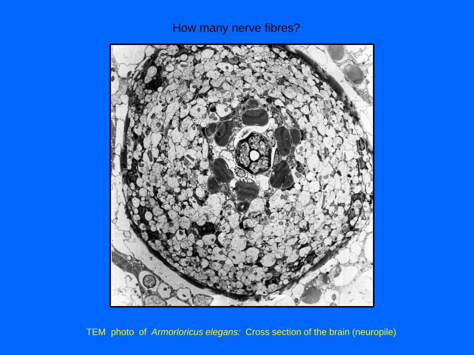
TEM photo of Armorloricus elegans: Cross section of the brain (neuropile)
How many nerve fibres?
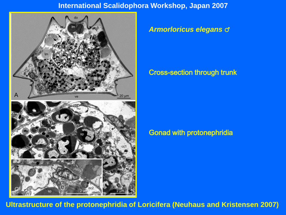
International Scalidophora Workshop, Japan 2007
Ultrastructure of the protonephridia of Loricifera (Neuhaus and Kristensen 2007)
Armorloricus elegans ♂
Cross-section through trunk
Gonad with protonephridia
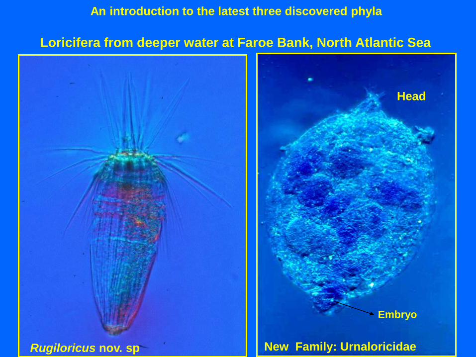
Rugiloricus nov. sp
Loricifera from deeper water at Faroe Bank, North Atlantic Sea
An introduction to the latest three discovered phyla
Head
New Family: Urnaloricidae
Embryo

New familly from the Faroe Bank
International Scalidophora Workshop, Japan 2007
Urnaloricidae: Ghost- or Megalarva

The life cycle of the New Order
?
?
Pedogenesis:
The cyst-like ghost larva retains
the cuticles of the postlarva and
the adult. Out of the cyst-like
ghost larva comes a normal
Higgins-larva.
The cyst-like larva’s armature
A
B C
D
E
International Scalidophora Workshop, Japan 2007

Since its discovery
assigned to 3
families:
Nanaloricidae
Pliciloricidae
Urnaloricidae
• About 30 species described
(Heiner, 2008)
Nanaloricus, Armorloricus, Phoeniciloricus,
Spinoloricus, Culexiregiloricus,
Australoricus
Pliciloricus, Rugiloricus, Titaniloricus
Urnaloricus
An introduction to the latest three discovered phyla
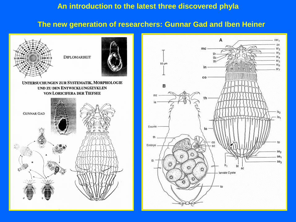
The new generation of researchers: Gunnar Gad and Iben Heiner

Molecular data does not support Scalidophora
Kinorhyncha Nematoda Priapulida Loricifera Nematomorpha
Scalidophora Nematoida
Plesiomorphies?
Adult morphology
18S rRNA
• Introvert + mouth cone
• Scalids
• Papillar sense organs (flosculi)
• Protonephridia
• Unpaired dorsal and ventral nerve
strings
• Loss of circular muscles
• Loss of sperm flagellum
• Both sexes with cloaca
• Vermiform habitus
Sørensen et al., 2008

Loricifera Nematomorpha
• Hexaradial symmetry patterns in
pharynx, opposed to pentaradial
symmetry patterns in Kinorhyncha
and Priapulida
Loriciferan – Nematomorph sister groups?
Martin Vinther Sørensen’ theory
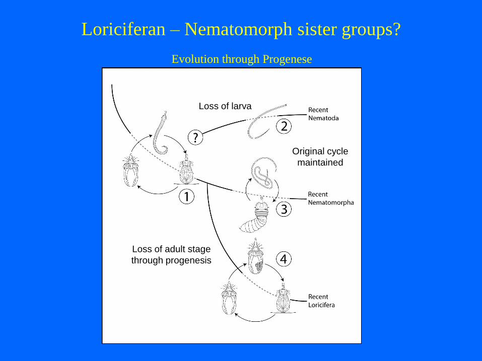
Evolution through Progenese
Loss of larva
Original cycle
maintained
Loss of adult stage
through progenesis
Loriciferan – Nematomorph sister groups?

Ricardo Neves: Roscoff 2011
Myoanatomy: Phallacidin DAPI-staining
Nanaloricus nov sp. from Trezen ar Skoden
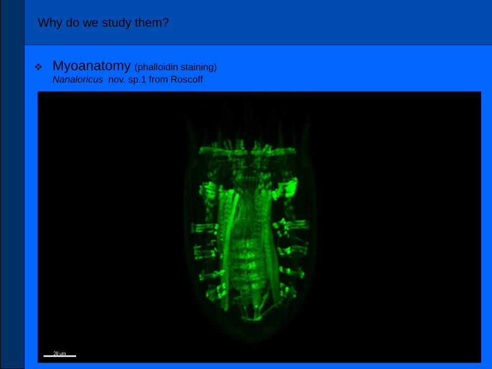
Why do we study them?
Myoanatomy (phalloidin staining)
Nanaloricus nov. sp.1 from Roscoff
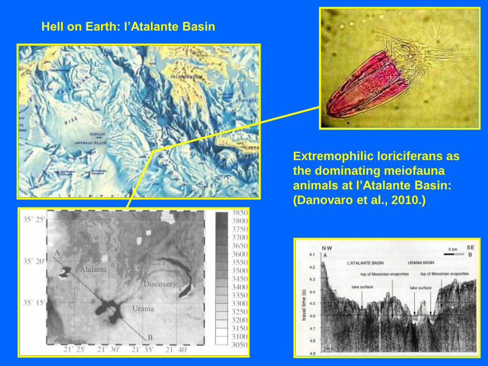
Extremophilic loriciferans as
the dominating meiofauna
animals at l’Atalante Basin:
(Danovaro et al., 2010.)
Hell on Earth: l’Atalante Basin
Extremophilic loriciferans as
the dominating meiofauna
animals at l’Atalante Basin:
( Danovaro et al, 2010)
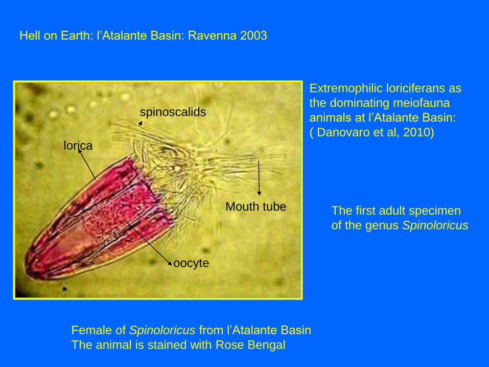
Hell on Earth: l’Atalante Basin: Ravenna 2003
Female of Spinoloricus from l’Atalante Basin
The animal is stained with Rose Bengal
Mouth tube
lorica
oocyte
spinoscalids
The first adult specimen
of the genus Spinoloricus

Holotypic female of Spinoloricus nov. sp. from l’Atalante Bassinet
Dorsal Ventral Dorsal

The DHABs
• The deep hypersaline anoxic basins Bannock,
Urania, Discovery, l’Atalante represent unique
deep-sea environments:
- Remains of hypersaline waters of the Miocene period (23x106 yrs ago)
- Separated for several thousand years from the surrounding SW environment
- Characterized by “extreme values” of the following parameters:
. High salinity (above 30%)
. Absence of light
. Elevated pressure (> 300 atm)
. Variable pH values (from acidic to alkaline, depending on the basin)
. Gas emission (Urania, l’Atalante basin)

Modus – the ROW
who took all the samples
The reseach vessel: R/V Urania
Expeditions to L’Atalante in 1998, 2005 and 2008

Evaporit containing Synechoccus Photomicrograph of Haloarcula
in a NaCl crystal (Nature, 2001)
Gypsum crystal
Expedition to the Urania Basin in 1998

1. Light Microscopy
In the first expedition, specimens belonging to three animal
Phyla were observed inner part of the L’Atalante basin:
Nematoda Copepoda Loricifera
All specimens were initially stained with Rose Bengal.
All copepods were empty exuviae, nematodes were only weakly
stained whereas all of the loriciferans were intensely colored

4. Ultra-structural analyses by TEM
Ultra-structural analyses revealed
the lack of mitochondria, which are
replaced by hydrogenosome-like
organelles H (a-c).
TEM revealed:
the presence of hydrogenosome
fields (c) similar to those reported
in anaerobic ciliates;
the presence of rod-shaped
structures (d-f), like prokaryotes P,
in proximity to the
hydrogenosome-like organelles.
H
a
P
e
P
H H d
P
P
f
H
H
H
H
H
H H
H
H
c
b
H Marginal
plate

Altings Oprindelse - Origins 2012
Sirius Passet in 2011 (Photo: Martin Stein)
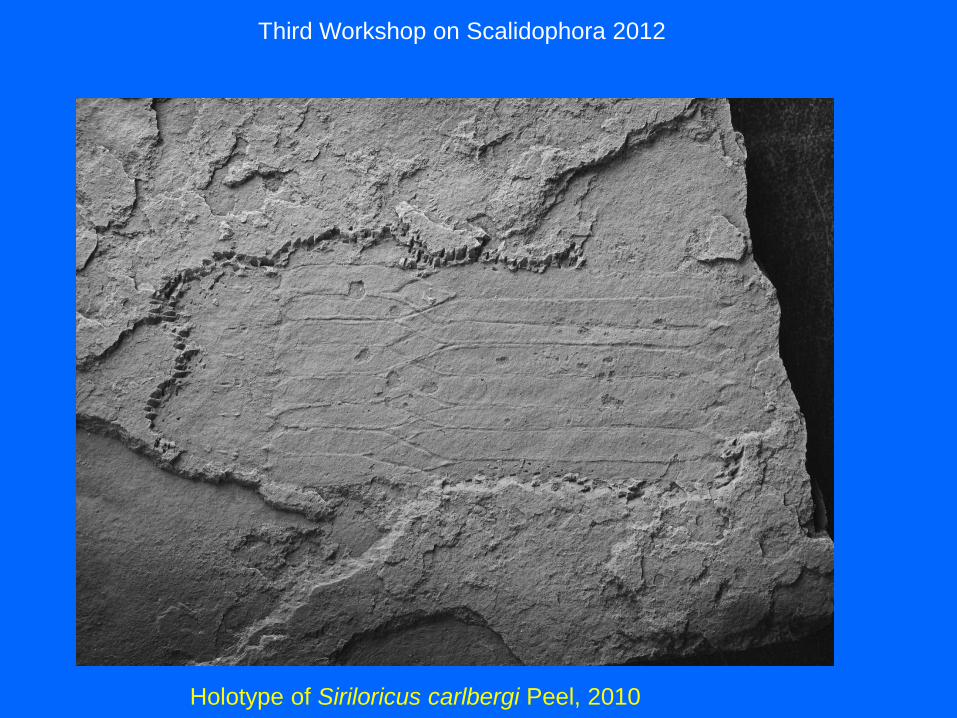
Third Workshop on Scalidophora 2012
Holotype of Siriloricus carlbergi Peel, 2010
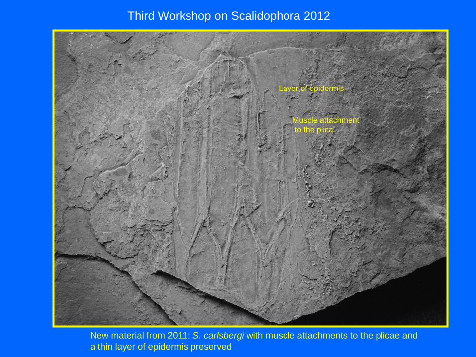
Third Workshop on Scalidophora 2012
New material from 2011: S. carlsbergi with muscle attachments to the plicae and
a thin layer of epidermis preserved
Muscle attachment
to the plica
Layer of epidermis
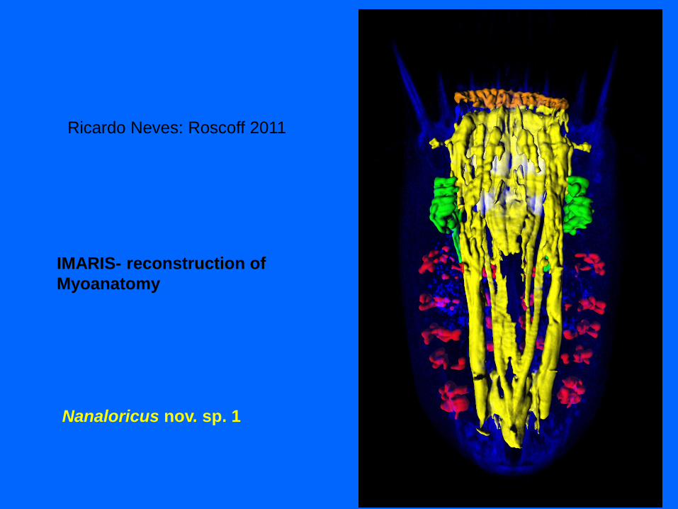
Ricardo Neves: Roscoff 2011
IMARIS- reconstruction of
Myoanatomy
Nanaloricus nov. sp. 1

Third Workshop on Scalidophora 2012
Martin Stein’s 3-D reconstruction of holotype
mouth tube
introvert
anterior
lorIca
posterior
lorIca
anal field
1 caudal spike
pro plate

Discovery of the second phylum: Cycliophora
Funch and Kristensen, 1995
Tjärnö 1997: Symbion pandora on the mouth limbs of Norway Lobster

Kaldbak Marine Biological Laboratory, 1998,
Faroe Islands
Symbion pandora (Cycliophora)
from mouth limbs of Norway
lobster (Nephrops norvegicus) in
Kaldbak Fjord, 1990. Cover
photo of Nature, 1995.
An introduction to the latest three discovered phyla

the overall myoanatomy of cycliophorans resembles that
found in bdelloid rotifers
(Brakenhoff, 1937) (Hochberg and
Litvaitis, 2000) (Santo, 2001)
- however, the insertion in the body integument is clearly different
Symbion pandora was believe to be a
marine Rotifera
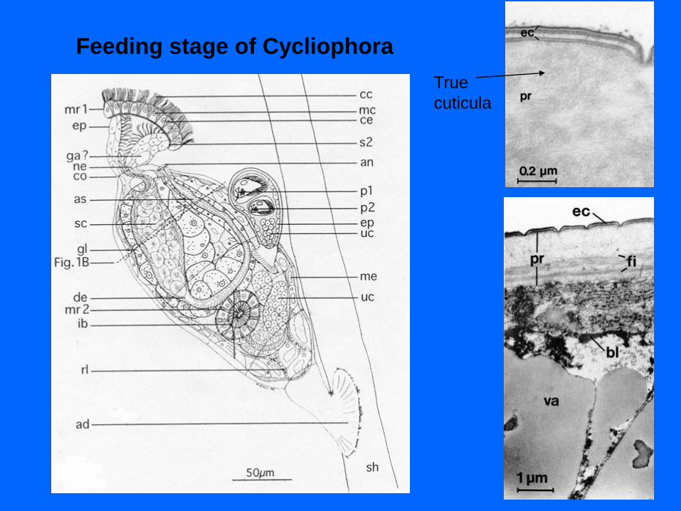
Feeding stage of Cycliophora
True
cuticula

An introduction to the latest three discovered phyla
Symbion pandora on Norway Lobster,
Kaldbak Fjord, Faroe Islands, 1990
Symbion americanus on
American Lobster,
Maine, USA, 1998
An introduction to the latest three discovered phyla

An introduction to the latest three discovered phyla
Pandora larva Dwarf female
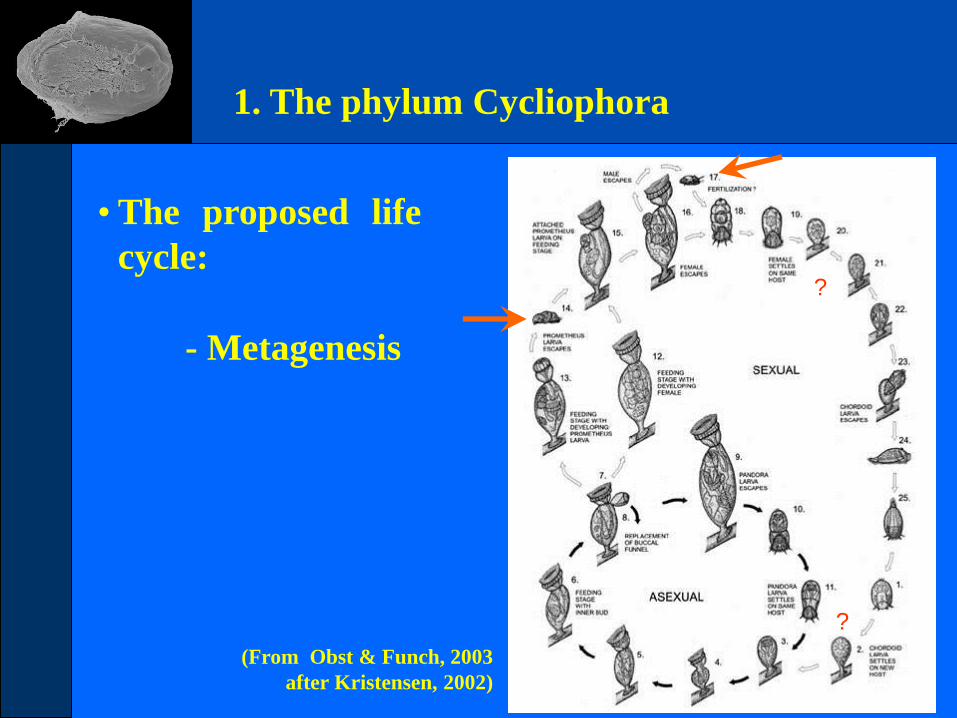
• The proposed life
cycle:
(From Obst & Funch, 2003
after Kristensen, 2002)
- Metagenesis
1. The phylum Cycliophora
?
?
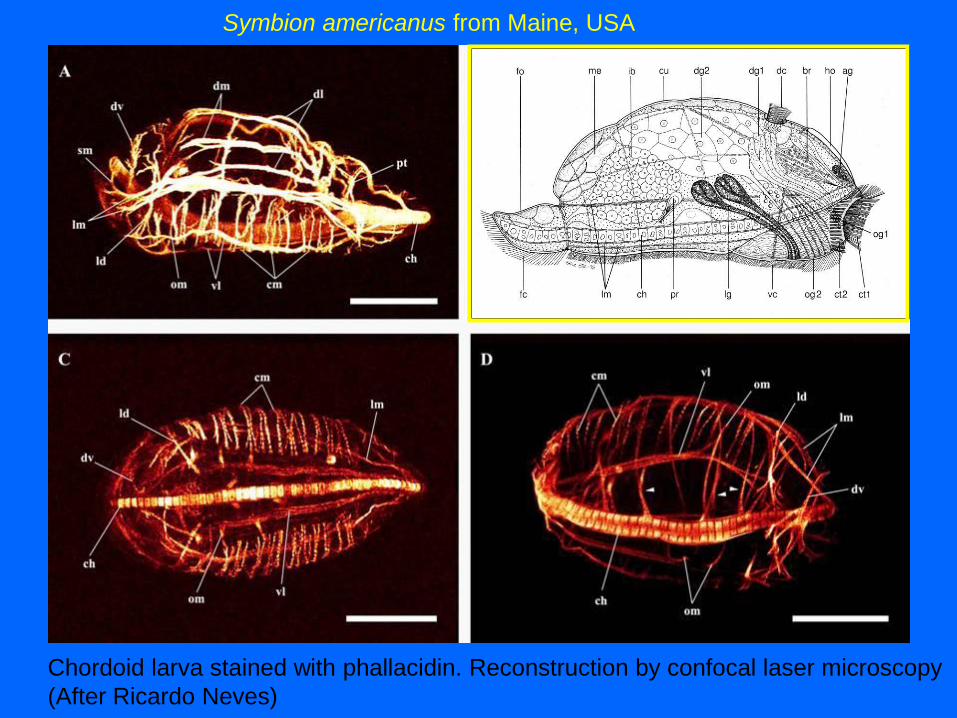
Chordoid larva stained with phallacidin. Reconstruction by confocal laser microscopy
(After Ricardo Neves)
Symbion americanus from Maine, USA

2. The second described species
Symbion americanus lives attached to the mouthparts of
Homarus americanus.
(Adapted from Obst et al., 2006)
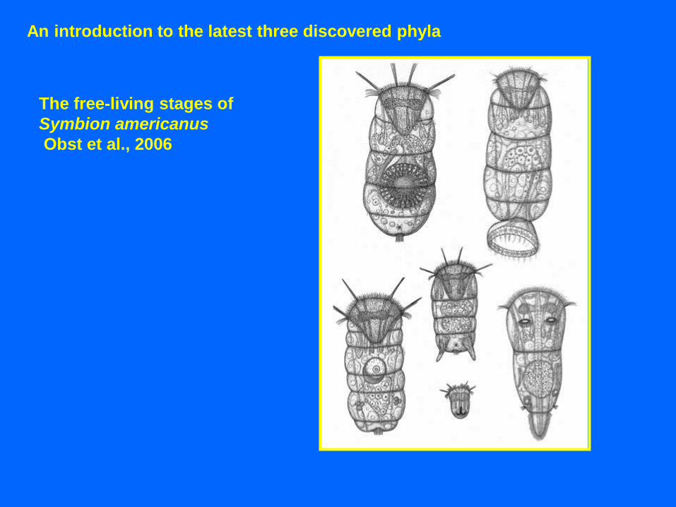
The free-living stages of
Symbion americanus
Obst et al., 2006
An introduction to the latest three discovered phyla

3. Results
frontal ciliated field (fc)
lateral sensoria (la)
penis (pe)
ventral ciliated field (vc)
The gross morphology
S. americanus
5 mm

3. Results
The ultrastructure
cerebral glands (ce)
medial glands (mg)
brain (ga)
penial sheath (ps)
Prometheus larva (pl)
testis (te)
5 mm
S. pandora

Are there
more cycliophorans ?
Nephropsis atlantica
900 - 1400 m
Nephropidae
(52 described species)
48 from continental slopes and deeper
12 from the continental shelf

Discovery of the third phylum: Micrognathozoa
Kristensen and Funch, 2000
Danish Arctic Station, Qeqertarsuaq, June 2004

Limnognathia maerski, Kristensen and Funch, 2000
(Micrognathozoa)

An introduction to the latest three discovered phyla
Arctic Biological Field Course
to Mudderbugten and Kvandalen
August 1994.
More than 30 new homothermic
springs were discovered.
Discovery of a Shangri-La in
Greenland – The Valley of
Angelica (Kvandalen).

An introduction to the latest three discovered phyla
Homothermic spring in Østerlien, summer and winter

The first sledge-expedition
to Mudderbugten, April 1978.
Field Course in Arctic Biology,
base camp at Isunngua-
Spring, August 1994.
Discovery of Micrognathozoa,
Limnognathia maerski
Kristensen & Funch, 2000.
An introduction to the latest three discovered phyla

An introduction to the latest three discovered phyla
Royal Lecture

An introduction to the latest three discovered phyla
Ventral view of the first known
Micrognathozoan: Limnognathia maerski
described by Kristensen & Funch (2000).
The species has recently been found in
Sub-Antarctic (Crozet Islands) and
Wales.
The animal is only about 0.1 mm

An introduction to the latest three discovered phyla
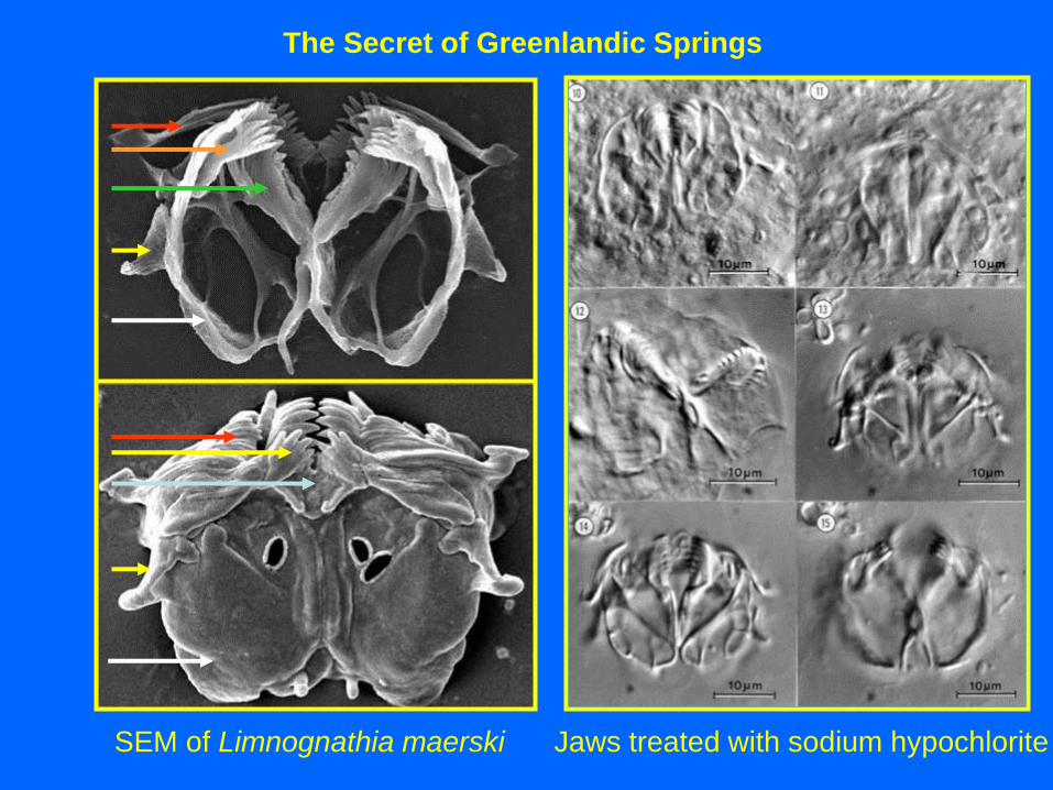
Scanning electron microscopy
of Limnognathia maerski
Lateral view
Ventral view
Winter egg

The Secret of Greenlandic Springs
Transmission Electron Microscopy (TEM) of Limnognathia maerski
The Secret of Greenlandic Springs
SEM of Limnognathia maerski Jaws treated with sodium hypochlorite

An introduction to the latest three discovered phyla
3-D reconstruction of the jaws
of Limnognathia maerski
Martin V. Sørensen
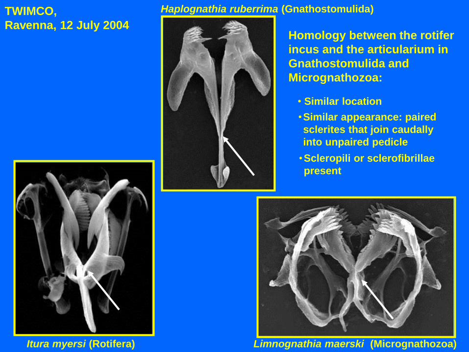
Itura myersi (Rotifera) Limnognathia maerski (Micrognathozoa)
Haplognathia ruberrima (Gnathostomulida)
Homology between the rotifer
incus and the articularium in
Gnathostomulida and
Micrognathozoa:
• Similar location
•Scleropili or sclerofibrillae
present
•Similar appearance: paired
sclerites that join caudally
into unpaired pedicle
TWIMCO,
Ravenna, 12 July 2004

An introduction to the latest
three discovered phyla
Phylogeny of
Kristensen and
Funch, 2000

H. ruberrima G. paradoxa L. maerski Seison sp. K. serrulata
An introduction to the latest three discovered phyla
-- Jaws composed on lucent rods
Incus/main jaw/articularium present
-- Mallei present
-- Basal plate and suspen-
sorium present
-- Basal plate
and fibularium
-- Cellular epidermis with intracellular
lamina
-- Syncytial epidermis
Loss of body ciliation
Wheel organ
-- Reversal to monociliated
cells
Sørensen´s phylogeny

The Secret of Greenlandic Springs
Giribet et al. 2004
new molecular
data

Arctic Workshop 2010: ”The tree of Life/ATol-team”

Arctic Workshop 2010: Photo of Limnognathia maerski

Arctic Workshop 2010: Limnognathia maerski
Katrine Worsaae: Myoanatomy (phalloidin, depth-coded)
Bergen-connection

Arctic Workshop 2010: Tree of Life/ATol
K. Worsaae 2011: cilia, nephridia, gonoducts & nerves
anti-acetylated α-tubulin (depth-coded)
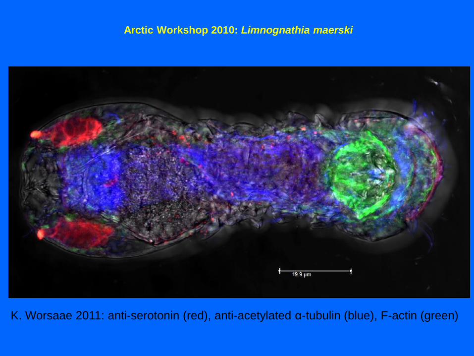
Arctic Workshop 2010: Limnognathia maerski
K. Worsaae 2011: anti-serotonin (red), anti-acetylated α-tubulin (blue), F-actin (green)
Acknowledgements to the steering committee of BIOFAR, the
ATOL-NSF programme “The Tree of Life”, the crews of the
different research vessels, Carlsberg Foundation and Danish
Research Agency.
Special thanks to:
Ricardo Neves, Katrine Worsaae, Roberto Bertolani, Gonzalo
Giribet, Bjørki Geyti, Robert P. Higgins, Stine Elle, Eyðfinn
Magnussen, Aslak Jørgensen, Peter Funch, Martin V. Sørensen,
Jesper G. Hansen, Gunnar Gad, Majken Them Jensen and Iben
Heiner.