Embed Size (px)
Citation preview
Nebojsa Paunkovic1,
Jane Paunkovic2
1. Professor of MegatrendUniversity, Belgrade, Serbia.
2. Poliklinika “Paunkovic”,Zajecar, Serbia
✬✬✬
Keywords: Thyrotropin receptorantibody – Graves’ disease –Diagnostic criteria – Clinicalimportance of criteria
Correspondence address: Prof. Dr Nebojsa Paunkovic, Poliklinika “Paunkovic”, 19000 Zajecar, Serbia,E-mail: [email protected]
Received:
21 December 2006
Accepted revised:
24 March 2007
The diagnostic criteria of Graves’ disease and especiallythe thyrotropin receptor antibody; our own experience
www.nuclmed.gr Hellenic Journal of Nuclear Medicine ñ May - August 2007
AbstractIt is generaly accepted that the thyrotropin receptor antibody (TRAb) has a stimulating activity and isthe major pathogenic factor in Graves’ disease (GD). In spite of that, TRAb is not routinely examinedin clinical practice. The aim of this article is to briefly review the subject and suggest protocols for thediagnosis, treatment and follow-up of patients with GD based on our own studies and referring es-pecially to TRAb. Clinical symptoms and signs and thyroid hormones may have poor sensitivity orspecificity, especially in cases of endocrine ophthalmopathy and subclinical hyperthyroidism. Inthese cases the TRAb test is 98% sensitive and specific with a diagnostic accuracy of almost 99%. Bythis test it is possible to differentiate between autoimmune and other forms of thyrotoxicosis such asautonomous hyperthyroidism, destructive thyroiditis, iodine induced hyperthyroidism etc. Antithyroiddrugs decrease serum TRAb levels and also induce immune remission. If after treatment TRAb re-mains increased as in about 30% of our cases, patients will relapse. In pregnant women with GD thefollow-up of serum TRAb levels is also important as predictive of immune thyroid disease in the new-born. Data presented in this article confirm that the determination of serum TRAb levels in some rarehyperthyroid disorders, such as associated autoimmune and autonomous forms and in epidemio-logical studies, is also justified.
Hell J Nucl Med 2007; 10(2): 89-94
Introduction – Historical background
Autoimmune hyperthyroidism (Graves’ or Basedow’s disease) is the result of an au-toimmune stimulatory activity to the thyroid gland and probably to some extrathyroidtissues like retrobulbar tissue [1, 2]. Adams and Purves (1956) were the first to ob-
serve that in patients with Graves’ disease (GD) there was a stimulatory activity different fromthyroid stimulating hormone (TSH) [3]. This substance was then called: “long acting thyroidstimulator” (LATS). It was shown later that LATS was an antibody of the immunoblobulintype, reacting with the membrane of thyrocytes [4].
Others have since described the concept of hormone-receptor activity [5] and recently themolecular basis of thyroid stimulating hormone receptor (TSH-R or TR) and its antibody [6,7]. Also, the cloning of the recombinant human TSH-R has been described. The expressionof human TSH-R has been described on transfected Chinese hamster ovary cells (CHO) [8]and on the leukemia cell line, K562 [9].
TSH-R is now being examined either by a bioassay or by a radio-receptor assay. The ba-sis of the bioassay test is the measurement of a biological response induced after a stimula-tory effect of TSH-R. This response may be the generation of cAMP, the release of T3, theformation of colloid droplets, etc. As a model of study, slices of thyroid tissue or thyroid cellsculture were used [10-16]. Our method of using thyroid cells suspension is considered sim-ple and reliable [17].
Adams and Kennedy (1967) were the first to detect by a radio-receptor assay (RRA) agamma globulin which was protecting LATS from neutralization [18]. Smith and Hall (1974)showed that “stimulatory immunoglobulins” from sera of patients with GD, inhibit the bind-ing of TSH to thyroid cell membranes [19]; this was the base for the TSH antibody radioas-say. Modifications of the first assay followed [20-22]. The modification of Morgenthaler(1999) has been noticeable: he used species specific, human, TSH receptor, and very prac-tical technologic procedure - solid phase separation [22].
TSHR or TR antibody (TRAb) is considered an important pathogenic factor for autoim-
Special Review Article
CM
YK
CM
YK
C MYK C MYK
C MYK C MYK
89
89
Hellenic Journal of Nuclear Medicine ñ May - August 2007 www.nuclmed.gr90
mune hyperthyroidism and has been used: a) for the diagnosisand the differential diagnosis of GD, [23, 24]. b) to monitortreatment or predict remission or relapse of GD, [25, 26]. c)for early detection of neonatal hyper- or hypothyroidism andfor its differential diagnosis [27] and d) to predict the evolutionof Graves’ ophthalmopathy [1, 2, 28].
Although it is generally accepted that TRAb has a stimu-latory activity and is the major pathogenic factor in GD, it isnot routinely examined in clinical practice. The aim of this ar-ticle based on our studies, is to suggest diagnostic protocols es-pecially referring to the importance of TRAb.
The diagnosis of autoimmune hyperthyroidismThe diagnosis of GD is based on: a) the clinical signs andsymptoms of the hypermetabolic state, b) the laboratory find-ings i.e. elevated free thyroid hormones and suppressed ultra-sensitive TSH, c) functional tests such as radionuclide uptaketests, d) the presence of ophthalmopathy, e) specific antibod-ies like the TRAb and f) ultrasonography findings.
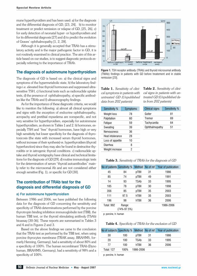
As for the importance of these diagnostic criteria, we wouldlike to mention the following: a) almost all clinical symptomsand signs with the exception of endocrine ophthalmopathy,acropachy and pretibial myxedema are nonspecific, and notvery sensitive for hyperthyroidism, especially for autoimmunehyperthyroidism, as shown in Tables 1 and 2. b) hormones, es-pecially TSH and “free” thyroid hormones, have high or veryhigh sensitivity but lower specificity for the diagnosis of thyro-toxicosis (the state with increased serum thyroid hormones,without increase of their synthesis) vs. hyperthyroidism (thyroidhyperfunction) since they may also be found in destructive thy-roiditis or in iatrogenic thyroid conditions. c) radionuclide up-take and thyroid scintigraphy have clinical and technical limita-tions for the diagnosis of GD [29]. d) routine immunologic testsfor the determination of serum “thyroid autoantibodies” main-ly refer to the microsomal Ab and are not considered eitherenough sensitive (Fig. 1), or specific for GD [30].
The contribution of TRAb test for thediagnosis and differential diagnosis of GD
a) For autoimmune hyperthyroidism
Between 1986 and 2006, we have published the followingdata for the diagnosis of GD concerning the sensitivity andspecificity of TRAb determinations performed by the porcinethyrotropin binding inhibition immunoglobulin test (TBII), thehuman TBII test, or the thyroid stimulating antibody (TSAb)bioassay [30-36]. These reports are summarized in Tables 3and 4 and in Figures 2 and 3.
Based on the above findings we came to the conclusionthat the TRAb test as performed by the TBII test, when usingporcine thyrocytes membrane (TRAK assay, BRAHMS, for-merly Henning, Germany), had a sensitivity of about 80% anda specificity of 100%. The human recombinant TRAb (Dynohuman, BRAHMS, Germany), had a sensitivity of 98% and aspecificity of 100%.
Special Review Article
C MYKC
MY
KC
MY
KC MYK
C MYK C MYK
90
Weight loss 78
Palpitation 60
Fatigue 59
Sweating 39
Nervousness 36
Heat intolerance 28
Loss of appetite 10
Diarrhea 8
Weight increase 2
Sensitivity % Symptoms
Goiter 81
Tremor 69
Tachycardia 64
Ophthalmopathy 51
Clinical signs Sensitivity %
45 84 pTBII 31 1986
85 74 pTBII 49 1991
14 93 TSAb 33 1995
185 78 pTBII 30 1998
356 85 pTBII 35 2003
111 97.5 hTBII 35 2003
196 98 hTBII 36 2006
Total: 992 Range: 1986-2006(74%-97.5%)
p: porcine, h: human
Method No of refNo of patients Sensitivity % Year of publication
Table 1. Sensitivity of clini-cal symptoms in patients withuntreated GD (Unpublisheddata from 202 patients)
Table 2. Sensitivity of clini-cal signs in patients with un-treated GD (Unpublished da-ta from 202 patients)
Figure 1. TSH receptor antibody (TRAb) and thyroid microsomal antibody(TMAb) findings in patients with GD before treatement and in stableremission [23].
Table 3. Sensitivity of TRAb for the diagnosis of GD
30 100 pTBII 31 1986
20 100 TSAb 33 1995
77 100 hTBII 36 2006
Total: 127 100% 1986-2006
p: porcine, h: human
Method No of refNo of subjects Specificity % Year of publication
Table 4. Specificity of TRAb for the exclusion of GD

www.nuclmed.gr Hellenic Journal of Nuclear Medicine ñ May - August 2007
The significantly higher binding inhibition of labeled TSHon the recombinant human TSH receptor (rhTR) compared tothe porcine extracted and solubilized TSH receptor, is con-sidered the main factor for the improved diagnostic sensitivityof rhTR [35, 37-39] (Fig. 3).
b) Differential diagnosis of GD
It is the opinion of the authors that all TRAb positive hyper-thyroid patients have autoimmune hyperthyroidism (GD)while all TRAb negative have some other forms of hyperthy-roidism such as autonomous or iatrogenous. Thus clinicalpractice if diagnosis is be based on the clinical signs and symp-toms, hormone levels, etc as mentioned above, may be inac-curate. Serum determination of TRAb is not a routine proce-dure, and in cases of suspected GD or in associated GD andPlummer’s disease if TRAb is negative, may lead to uncertaindiagnoses. In such cases we suggest that another more sensi-tive TRAb test be used, or the same TRAb test be repeated. In cases of suspected GD with negative TRAb, we have foundthat half the patients after being re-examined by using a moresensitive or the same TRAb test, became TRAb positive. Inthe other half, the clinical status was re-evaluated and theywere finally diagnosed as not having GD (Fig. 4) [36]. Others
have reported similar experience [38, 39]. Some authors be-lieve that there exists a condition of disseminated thyroid au-tonomy which shows some similarities with the true negativeTRAb, GD [38-40].
Routine TRAb determinations may be important in someepidemiological studies. In an epidemiologic report we havebeen able to differentiate autoimmune from the other forms ofhyperthyroidism [41].
c) For associated GD and Plummer’s disease (PD)It has been statistically shown that some patients with GD mayalso have PD (multinodular toxic goiter and toxic adenoma). Ithas been reported that this case is found more often than if itwas just a «statistical» phenomenon [42,43]. Also, some pa-tients with PD may develop GD after being treated with 131I[44,45]. We have reported some 20 patients with this entity[46]. The findings of one of them are shown in Figure 5.
The contribution of the TRAb test to monitortreatment of GD
Antithyroid drugsThiourea derivatives thionamides: mercaptoimidazole andpropylthiouracil are the most often used antithyroid drugs.
Special Review Article
CM
YK
CM
YK
C MYK C MYK
C MYK C MYK
91
91
Figure 2. Percent generated serum cAMP levels in patients with GD (be-fore treatment, under methimazole-Th, in remission and in the controlgroup [32].
0
100
200
300
400
500
600
700
800
Graves'd.
% c
AM
P
Under treatment Remission Euthyroidism
Figure 3. A. Individual values of TSH binding inhibition (TBI) in untreatedpatients with GD, tested by two assays. B. Average values of TBI in thesame analysis [35].
Figure 4. The result of re-evaluation of diagnosis and TRAb determinationin a group of “TRAb negative” GD patients (n=51) [36].
Figure 5. Scintigraphic images of a patient MD, 46 years, female, with as-sociated Graves’ and Plummer’s disease [46]. A. Data from 1984. Diffusethyroid scintiscan with 131I with ‘’warm’’ nodule in the lower part of the leftlobe; hypermetabolic state. Total T4: 210 nmol/l. TBII: strongly positive(310 U/l). Treated with methimazole (MMI). Remission and cessation ofMMI after one year. B. Control check-up data on1990. Autonomous hy-perfunctional nodule in the scintiscan. T4 normal: 130 nmol/l, T3 elevated:3.5 nmol/l and TBII negative: 5 U/L. Treated with 814 MBq of 131I. C.Control check-up on 1992. A ‘’warm’’ nodule in the scintiscan in the leftlobe with partially suppresed paranodular tissue of the right lobe. T4 nor-mal: 120 nmol/l.
Hellenic Journal of Nuclear Medicine ñ May - August 2007 www.nuclmed.gr92
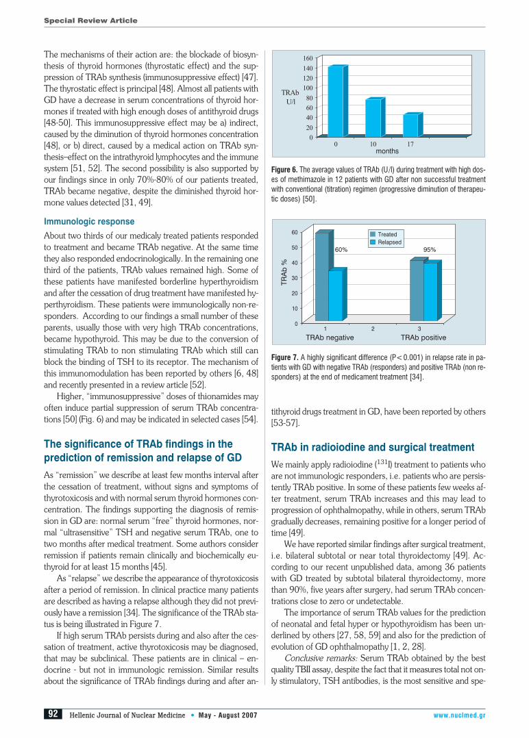
The mechanisms of their action are: the blockade of biosyn-thesis of thyroid hormones (thyrostatic effect) and the sup-pression of TRAb synthesis (immunosuppressive effect) [47].The thyrostatic effect is principal [48]. Almost all patients withGD have a decrease in serum concentrations of thyroid hor-mones if treated with high enough doses of antithyroid drugs[48-50]. This immunosuppressive effect may be a) indirect,caused by the diminution of thyroid hormones concentration[48], or b) direct, caused by a medical action on TRAb syn-thesis–effect on the intrathyroid lymphocytes and the immunesystem [51, 52]. The second possibility is also supported byour findings since in only 70%-80% of our patients treated,TRAb became negative, despite the diminished thyroid hor-mone values detected [31, 49].
Immunologic response
About two thirds of our medicaly treated patients respondedto treatment and became TRAb negative. At the same timethey also responded endocrinologically. In the remaining onethird of the patients, TRAb values remained high. Some ofthese patients have manifested borderline hyperthyroidismand after the cessation of drug treatment have manifested hy-perthyroidism. These patients were immunologically non-re-sponders. According to our findings a small number of theseparents, usually those with very high TRAb concentrations,became hypothyroid. This may be due to the conversion ofstimulating TRAb to non stimulating TRAb which still canblock the binding of TSH to its receptor. The mechanism ofthis immunomodulation has been reported by others [6, 48]and recently presented in a review article [52].
Higher, “immunosuppressive” doses of thionamides mayoften induce partial suppression of serum TRAb concentra-tions [50] (Fig. 6) and may be indicated in selected cases [54].
The significance of TRAb findings in theprediction of remission and relapse of GD
As “remission” we describe at least few months interval afterthe cessation of treatment, without signs and symptoms ofthyrotoxicosis and with normal serum thyroid hormones con-centration. The findings supporting the diagnosis of remis-sion in GD are: normal serum “free” thyroid hormones, nor-mal “ultrasensitive” TSH and negative serum TRAb, one totwo months after medical treatment. Some authors considerremission if patients remain clinically and biochemically eu-thyroid for at least 15 months [45].
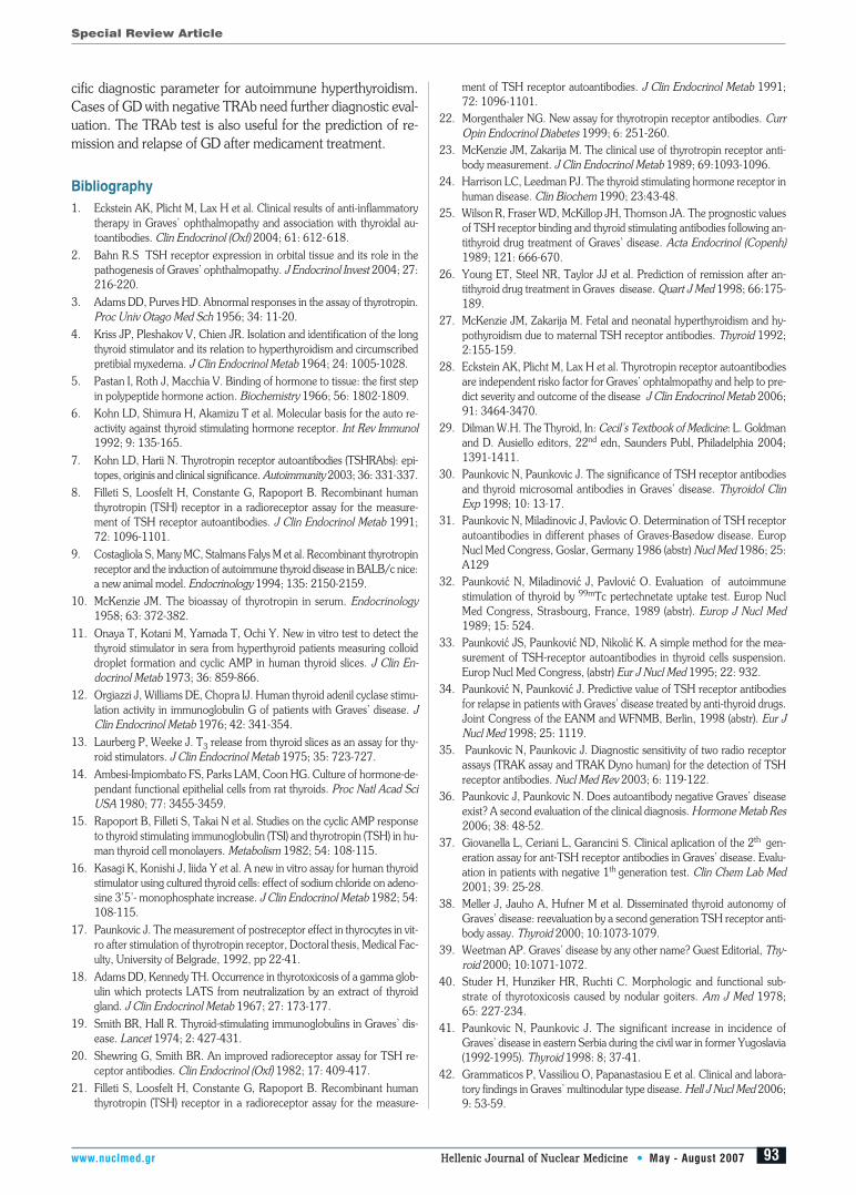
As “relapse” we describe the appearance of thyrotoxicosisafter a period of remission. In clinical practice many patientsare described as having a relapse although they did not previ-ously have a remission [34]. The significance of the TRAb sta-tus is being illustrated in Figure 7.
If high serum TRAb persists during and also after the ces-sation of treatment, active thyrotoxicosis may be diagnosed,that may be subclinical. These patients are in clinical – en-docrine - but not in immunologic remission. Similar resultsabout the significance of TRAb findings during and after an-
tithyroid drugs treatment in GD, have been reported by others[53-57].
TRAb in radioiodine and surgical treatmentWe mainly apply radioiodine (131I) treatment to patients whoare not immunologic responders, i.e. patients who are persis-tently TRAb positive. In some of these patients few weeks af-ter treatment, serum TRAb increases and this may lead toprogression of ophthalmopathy, while in others, serum TRAbgradually decreases, remaining positive for a longer period oftime [49].
We have reported similar findings after surgical treatment,i.e. bilateral subtotal or near total thyroidectomy [49]. Ac-cording to our recent unpublished data, among 36 patientswith GD treated by subtotal bilateral thyroidectomy, morethan 90%, five years after surgery, had serum TRAb concen-trations close to zero or undetectable.
The importance of serum TRAb values for the predictionof neonatal and fetal hyper or hypothyroidism has been un-derlined by others [27, 58, 59] and also for the prediction ofevolution of GD ophthalmopathy [1, 2, 28].
Conclusive remarks: Serum TRAb obtained by the bestquality TBII assay, despite the fact that it measures total not on-ly stimulatory, TSH antibodies, is the most sensitive and spe-
Special Review Article
C MYKC
MY
KC
MY
KC MYK
C MYK C MYK
92
Figure 6. The average values of TRAb (U/l) during treatment with high dos-es of methimazole in 12 patients with GD after non successful treatmentwith conventional (titration) regimen (progressive diminution of therapeu-tic doses) [50].
months
Figure 7. A highly significant difference (P<0.001) in relapse rate in pa-tients with GD with negative TRAb (responders) and positive TRAb (non re-sponders) at the end of medicament treatment [34].
0
10
20
30
40
50
60
1 2 3
TRAb negative TRAb positive
95%60%
TRA
b %
TreatedRelapsed
www.nuclmed.gr Hellenic Journal of Nuclear Medicine ñ May - August 2007
cific diagnostic parameter for autoimmune hyperthyroidism.Cases of GD with negative TRAb need further diagnostic eval-uation. The TRAb test is also useful for the prediction of re-mission and relapse of GD after medicament treatment.
Bibliography1. Eckstein AK, Plicht M, Lax H et al. Clinical results of anti-inflammatory
therapy in Graves’ ophthalmopathy and association with thyroidal au-toantibodies. Clin Endocrinol (Oxf) 2004; 61: 612-618.
2. Bahn R.S TSH receptor expression in orbital tissue and its role in thepathogenesis of Graves’ ophthalmopathy. J Endocrinol Invest 2004; 27:216-220.
3. Adams DD, Purves HD. Abnormal responses in the assay of thyrotropin.Proc Univ Otago Med Sch 1956; 34: 11-20.
4. Kriss JP, Pleshakov V, Chien JR. Isolation and identification of the longthyroid stimulator and its relation to hyperthyroidism and circumscribedpretibial myxedema. J Clin Endocrinol Metab 1964; 24: 1005-1028.
5. Pastan I, Roth J, Macchia V. Binding of hormone to tissue: the first stepin polypeptide hormone action. Biochemistry 1966; 56: 1802-1809.
6. Kohn LD, Shimura H, Akamizu T et al. Molecular basis for the auto re-activity against thyroid stimulating hormone receptor. Int Rev Immunol1992; 9: 135-165.
7. Kohn LD, Harii N. Thyrotropin receptor autoantibodies (TSHRAbs): epi-topes, originis and clinical significance. Autoimmunity 2003; 36: 331-337.
8. Filleti S, Loosfelt H, Constante G, Rapoport B. Recombinant humanthyrotropin (TSH) receptor in a radioreceptor assay for the measure-ment of TSH receptor autoantibodies. J Clin Endocrinol Metab 1991;72: 1096-1101.
9. Costagliola S, Many MC, Stalmans Falys M et al. Recombinant thyrotropinreceptor and the induction of autoimmune thyroid disease in BALB/c nice:a new animal model. Endocrinology 1994; 135: 2150-2159.
10. McKenzie JM. The bioassay of thyrotropin in serum. Endocrinology1958; 63: 372-382.
11. Onaya T, Kotani M, Yamada T, Ochi Y. New in vitro test to detect thethyroid stimulator in sera from hyperthyroid patients measuring colloiddroplet formation and cyclic AMP in human thyroid slices. J Clin En-docrinol Metab 1973; 36: 859-866.
12. Orgiazzi J, Williams DE, Chopra IJ. Human thyroid adenil cyclase stimu-lation activity in immunoglobulin G of patients with Graves’ disease. JClin Endocrinol Metab 1976; 42: 341-354.
13. Laurberg P, Weeke J. T3 release from thyroid slices as an assay for thy-roid stimulators. J Clin Endocrinol Metab 1975; 35: 723-727.
14. Ambesi-Impiombato FS, Parks LAM, Coon HG. Culture of hormone-de-pendant functional epithelial cells from rat thyroids. Proc Natl Acad SciUSA 1980; 77: 3455-3459.
15. Rapoport B, Filleti S, Takai N et al. Studies on the cyclic AMP responseto thyroid stimulating immunoglobulin (TSI) and thyrotropin (TSH) in hu-man thyroid cell monolayers. Metabolism 1982; 54: 108-115.
16. Kasagi K, Konishi J, Iiida Y et al. A new in vitro assay for human thyroidstimulator using cultured thyroid cells: effect of sodium chloride on adeno-sine 3’5’- monophosphate increase. J Clin Endocrinol Metab 1982; 54:108-115.
17. Paunkovic J. The measurement of postreceptor effect in thyrocytes in vit-ro after stimulation of thyrotropin receptor, Doctoral thesis, Medical Fac-ulty, University of Belgrade, 1992, pp 22-41.
18. Adams DD, Kennedy TH. Occurrence in thyrotoxicosis of a gamma glob-ulin which protects LATS from neutralization by an extract of thyroidgland. J Clin Endocrinol Metab 1967; 27: 173-177.
19. Smith BR, Hall R. Thyroid-stimulating immunoglobulins in Graves’ dis-ease. Lancet 1974; 2: 427-431.
20. Shewring G, Smith BR. An improved radioreceptor assay for TSH re-ceptor antibodies. Clin Endocrinol (Oxf) 1982; 17: 409-417.
21. Filleti S, Loosfelt H, Constante G, Rapoport B. Recombinant humanthyrotropin (TSH) receptor in a radioreceptor assay for the measure-
ment of TSH receptor autoantibodies. J Clin Endocrinol Metab 1991;72: 1096-1101.
22. Morgenthaler NG. New assay for thyrotropin receptor antibodies. CurrOpin Endocrinol Diabetes 1999; 6: 251-260.
23. McKenzie JM, Zakarija M. The clinical use of thyrotropin receptor anti-body measurement. J Clin Endocrinol Metab 1989; 69:1093-1096.
24. Harrison LC, Leedman PJ. The thyroid stimulating hormone receptor inhuman disease. Clin Biochem 1990; 23:43-48.
25. Wilson R, Fraser WD, McKillop JH, Thomson JA. The prognostic valuesof TSH receptor binding and thyroid stimulating antibodies following an-tithyroid drug treatment of Graves’ disease. Acta Endocrinol (Copenh)1989; 121: 666-670.
26. Young ET, Steel NR, Taylor JJ et al. Prediction of remission after an-tithyroid drug treatment in Graves disease. Quart J Med 1998; 66:175-189.
27. McKenzie JM, Zakarija M. Fetal and neonatal hyperthyroidism and hy-pothyroidism due to maternal TSH receptor antibodies. Thyroid 1992;2:155-159.
28. Eckstein AK, Plicht M, Lax H et al. Thyrotropin receptor autoantibodiesare independent risko factor for Graves’ ophtalmopathy and help to pre-dict severity and outcome of the disease J Clin Endocrinol Metab 2006;91: 3464-3470.
29. Dilman W.H. The Thyroid, In: Cecil’s Textbook of Medicine: L. Goldmanand D. Ausiello editors, 22nd edn, Saunders Publ, Philadelphia 2004;1391-1411.
30. Paunkovic N, Paunkovic J. The significance of TSH receptor antibodiesand thyroid microsomal antibodies in Graves’ disease. Thyroidol ClinExp 1998; 10: 13-17.
31. Paunkovic N, Miladinovic J, Pavlovic O. Determination of TSH receptorautoantibodies in different phases of Graves-Basedow disease. EuropNucl Med Congress, Goslar, Germany 1986 (abstr) Nucl Med 1986; 25:A129
32. Paunkovi ′c N, Miladinovi ′c J, Pavlovi ′c O. Evaluation of autoimmunestimulation of thyroid by 99mTc pertechnetate uptake test. Europ NuclMed Congress, Strasbourg, France, 1989 (abstr). Europ J Nucl Med1989; 15: 524.
33. Paunkovi ′c JS, Paunkovi ′c ND, Nikoli ′c K. A simple method for the mea-surement of TSH-receptor autoantibodies in thyroid cells suspension.Europ Nucl Med Congress, (abstr) Eur J Nucl Med 1995; 22: 932.
34. Paunkovi ′c N, Paunkovi ′c J. Predictive value of TSH receptor antibodiesfor relapse in patients with Graves’ disease treated by anti-thyroid drugs.Joint Congress of the EANM and WFNMB, Berlin, 1998 (abstr). Eur JNucl Med 1998; 25: 1119.
35. Paunkovic N, Paunkovic J. Diagnostic sensitivity of two radio receptorassays (TRAK assay and TRAK Dyno human) for the detection of TSHreceptor antibodies. Nucl Med Rev 2003; 6: 119-122.
36. Paunkovic J, Paunkovic N. Does autoantibody negative Graves’ diseaseexist? A second evaluation of the clinical diagnosis. Hormone Metab Res2006; 38: 48-52.
37. Giovanella L, Ceriani L, Garancini S. Clinical aplication of the 2th gen-eration assay for ant-TSH receptor antibodies in Graves’ disease. Evalu-ation in patients with negative 1th generation test. Clin Chem Lab Med2001; 39: 25-28.
38. Meller J, Jauho A, Hufner M et al. Disseminated thyroid autonomy ofGraves’ disease: reevaluation by a second generation TSH receptor anti-body assay. Thyroid 2000; 10:1073-1079.
39. Weetman AP. Graves’ disease by any other name? Guest Editorial, Thy-roid 2000; 10:1071-1072.
40. Studer H, Hunziker HR, Ruchti C. Morphologic and functional sub-strate of thyrotoxicosis caused by nodular goiters. Am J Med 1978;65: 227-234.
41. Paunkovic N, Paunkovic J. The significant increase in incidence ofGraves’ disease in eastern Serbia during the civil war in former Yugoslavia(1992-1995). Thyroid 1998: 8; 37-41.
42. Grammaticos P, Vassiliou O, Papanastasiou E et al. Clinical and labora-tory findings in Graves’ multinodular type disease. Hell J Nucl Med 2006;9: 53-59.
Special Review Article
CM
YK
CM
YK
C MYK C MYK
C MYK C MYK
93
93

Hellenic Journal of Nuclear Medicine ñ May - August 2007 www.nuclmed.gr94
43. Bulow Pedersen I, Knudsen N, Perrild H et al. TSH-receptor antibodymeasurement for differentiation of hyperthyroidism into Graves’ diseaseand multinodular toxic goiter: a comparison of two competitive bindingassays. Clin Endocrinol 2001; 55: 381-390.
44. Chiovato L, Santini F, Vitti P et al. Appearance of thyroid stimulating an-tibody and Graves’ disease after radioiodine therapy for toxic nodulargoiter. Clin Endocrinol (Oxf) 1994; 40: 803-806.
45. Nygaard B, Faber J, Vejle A et al. Transition of nodular toxic goiter to au-toimmune hyperthyroidism triggered by 131I therapy. Thyroid 1999; 5:477-481.
46. Paunkovic N, Paunkovic J. Associated Graves’ disease and Plummer’sdisease. Hell J Nucl Med 2003; 6: 44-47.
47. Cooper DS. Antithyroid drugs. New Engl J Med 2005; 352: 905-917.
48. Volpe R. Immunoregulation in autoimmune thyroid disease. N Engl JMed 1987; 316: 44-46.
49. Paunkovi ′c N, Paunkovi ′c J, Pavlovi ′c O. Values of TSH receptor autoan-tibodies (TRAb) in patients with treated Graves’ disease. Radiol Jugosl1991; 25: 319-323.
50. Paunkovic N, Paunkovic J. The outcome of medical treatment with highdoses of methimazole on serum level of TSH receptor antibodies in pa-tients with Graves’ disease previously treated with titration doses. Ab-stract book of 12th International Thyroid Congress, Kyoto. (abst). En-docrine Journal 2000; 47, Suppl:190: 336
51. Bernet V, Burman K. Autoimmune thyroid disease, In: Rich RR, Clinical Im-munology – Principles and Practice, Mosby, St. Louis 1996; 1482-1502.
52. Volpe R. The immunomodulatory effects of anti-thyroid drugs are medi-ated via actions on thyroid cells, affecting thyrocyte-immunocyte signal-ing: a review. Curr Pharm Des 2001; 7: 491-500.
53. Michelangeli V, Poon C, Taft J et al. The prognostic value of thyrotropinreceptor antibody measurement in the early stages of treatment ofGraves’ disease with antithyroid drugs. Thyroid 1998; 8: 119-124.
54. Werner RS, Romaldini JH, Farah CS et al. Serum thyroid stimulating an-tibody, thyroglobulin levels, and thyroid suppressibility measurement aspredictors of the outcome of combined methimazole and triiodothyroninetherapy in Graves’ disease. Thyroid 1991; 1: 293-299.
55. Li J, Gao H, Xu L. The prognostic value of thyroid stimulating antibodiesafter antithyroid drug treatment of Graves’ disease. Chung Hua I HsuehTsa Chih 1994; 74: 218-220.
56. Winsa B, Dahlberg A, Jansson R et al. Factors influencing the outcome ofthyrostatic drug therapy in Graves’ disease. Acta Endocrinol (Copenh)1990; 122: 722-728.
57. Maugendre D, Massart C. Clinical value of a new TSH binding inchibito-ry activiry assay using human TSH receptors in the follow-up of antithy-roid drug treated Graves’ disease. Comparison with thyroid stimulatingantibody bioassay. Clin Endocrinol 2001; 54: 89-96.
58. Polak M, Leger J, Luton D et al. Fetal cord blood sampling in the diag-nosis and the treatment of fetal hyperthyroidism in the offsprings of a eu-thyroid mother, producing thyroid stimulating immunoglobulins. Ann En-docrinol (Paris) 1997; 58: 338-342.
59. Laurberg P, Nygaard B, Glinoer D et al. Guidelines for THS-receptor an-tibody measurements in pregnancy: results of an evidence-based sympo-sium organized by the European Thyroid Association. Eur J Endocrinol1998; 139: 584-586.
[
Special Review Article
C MYKC
MY
KC
MY
KC MYK
C MYK C MYK
94