Embed Size (px)
Citation preview

The Diagnosis and Surgical Removal of a Dentigerous Cyst Associated with an Unerupted
Mandibular Left First Premolar in a Shih Tzu
Brett Beckman, DVM, FAVD, DAVDC, DAAPM
Affiliated Veterinary Specialists, Orlando, Florida
Florida Veterinary Dentistry and Oral Surgery, Punta Gorda, Florida
Animal Emergency Center of Sandy Springs, Atlanta, GA
www.veterinarydentistry.net
Introduction
Dentigerous cysts arise from odontogenic epithelium associated with an unerupted adult
tooth. (1,2,3,4,5,6,7,8) Gingival changes associated with these cysts can be minimal to non-
existent. Without adequate assessment of edentulous areas conditions that result in patient
discomfort and tissue destruction may go undetected. This case report describes the diagnosis
and surgical treatment of a dentigerous cyst associated with a seemingly missing mandibular left
first premolar (305) with minimal visible gross gingival pathology.
History:
A three year old, male neutered, Shih Tzu weighing 8 kg presented for an annual examination
in March 2002. No prior oral care had been provided by the owner. Dental cleaning had never
been performed.
Diagnostics:
Physical examination of the patient was within normal limits. Oral examination revealed a
gingivitis index of II, a calculus index of II, and a plaque index of II.(9) There was a marked
distolingual rotation of the maxillary right (106) and left (206) second premolar teeth and the
maxillary right (107) and left (207) third premolar teeth. Lingual displacement of the
mandibular left (302) and right (402) second incisor teeth was noted. The patient was missing
the mandibular left first (305) and second (306) premolar teeth and the mandibular right second
premolar tooth (406). Close inspection revealed a firm one millimeter nodule present dorsally in
amelanotic gingiva six millimeters distal to the left mandibular labial frenulum (Figure 1). No
additional abnormalities were present. The patient was kept for complete dental radiographs and
dental cleaning pending an appropriate pre-anesthetic evaluation.

The differential diagnosis for the nodule in amelanotic gingiva associated with the missing
tooth included gingival overgrowth obscuring a retained root fragment from previous tooth
fracture, abscess(2,5), neoplasia,(2,4,5,7,8,10) fibrous lesion of the mandible (2) and
cyst.(1,2,3,4,5,6,7,8,11)
Figure 1
A small amelanotic region is present just distal to the frenulum.
A small nodule can be seen in the gingiva.
Pre-anesthetic blood testing included a complete blood count, ALT, BUN and glucose which
were all within normal limits. Pre-anesthetic ECGa analysis showed a normal sinus rhythm,
normal complexes and a heart rate of 106. Mucous membrane color, capillary refill time, pulse
character and chest auscultation were all within normal limits.
Butorphanolb
(3.2 mg IM) was given 30 minutes preoperatively and a 20 gauge intravenous
catheterc placed in the right cephalic vein. General anesthesia was induced with ketamine
d (40
mg IV) and valiume (2 mg IV). The patient was intubated with a 5.5 mm cuffed endotracheal
tubef and the cuff was gently inflated. The animal was maintained with isoflurane
g (2.0-2.5%)
and oxygen (1.0 L/min)(12) using a semi-closed anesthetic delivery systemh, placed on a water
circulated heating padi and leads for the ECG monitor
b positioned. A balanced electrolyte
solutionj was administered at 10ml/kg/hr.(12) Temperature, respiration, pulse and capillary refill
time were regularly recorded by a technician.

A complete oral examination was performed and abnormalities noted on the dental chart. In
addition to the changes discovered during the initial oral examination slight bleeding was present
upon probing the buccal aspect of teeth 206 and 207 at the furcation. All pocket depths were
within normal limits. The oral cavity was thoroughly rinsed with 0.12 % chlorhexidinek
solution. Complete supragingival and subgingival scalingl was performed. The teeth were
polished using a disposable prophy anglem
on a slow speed handpiecen and polishing paste
o. The
oral cavity was rinsed thoroughly with saline followed again by 0.12 % chlorhexidinek solution.
Dental radiographsp were taken of all teeth. Radiographic findings included a rounded, one
centimeter diameter lucency in the left mandible. It extended from the mesial aspect of the
mandibular left third premolar tooth (307) to the central aspect of the root of the mandibular left
canine tooth (304). (Figure 2) The apex of the mandibular left first premolar tooth (305) was
displaced in a coronal and distal direction and the root was tipped distally. The distal cusp of
tooth 305 rested just above the level of the alveolar crest and bone was absent at this interface.
Figure 2
The cyst has displaced the unerrupted premolar and can be
seen surrounding a portion of the mandibular left canine tooth (304).

Diagnosis:
Based on the radiographic appearance of this lucency and its association with unerrupted
tooth 305 a tentative diagnosis of a dentigerous cyst was made1. The tentative diagnosis was
relayed to the owner.
Treatment Plan:
The treatment plan included pre-operative and post-operative pain management, aspiration of
the cyst for cytologic evaluation, and complete removal of the lining to prevent recurrence.
Placement of a synthetic bone implantq prior to closure in order to fill the defect and maintain the
alveolar ridge was also planned.
Treatment:
The patient was placed in right lateral recumbency. A mental nerve block was performed by
injecting bupivicainer (1.5 mg), using a tuberculin syringe and a 25 gauge 1 inch needle
s, at the
entrance to the left middle mental foramen and holding digital pressure over the foramen for
sixty seconds. The surgical site was thoroughly lavaged with 0.12 % chlorhexidineh solution.
A #15 Bard Parker bladet mounted on a No. 7 scalpel handle
u was used to make two divergent
vertical releasing incisions starting in the gingiva at the lingual aspect of the mandible and
extending six millimeters beyond the mucogingival line. One incision was made at the mesial
angle of tooth 307 and the other three millimeters distal to tooth 304. The incisions were
connected by incising the gingiva on the mandibular crest. A No. 2 Molt periosteal elevatorv
was used to elevate the flap apically to expose the area. The distal portion of the crown of tooth
305 could be visualized with a portion of the cyst lining adhered to the neck of the tooth.(Figure
3) The cyst opening could be seen as a lytic area in the bone mesial and buccal to tooth 304.
Tooth 305 was easily extracted with a dental luxatorw using minimal effort. The fluid in the cyst
was clear and was aspirated into a 3 cc syringe with a 22 gauge needlex and kept for cytology. A
No. 4 round bury in a high speed delivery system
n was used to remove a small portion of alveolar
bone to aid in cyst visualization and curettage. The cyst lining could be visualized adhered to the
exposed bone and a portion of the root of tooth 304 and extended around the root, lingually and

bucally. (Figure 4) The cyst lining was grasped with an ophthalmic thumb forcepz and (a
portion) was peeled from the bone. Remnants of the lining were gently scraped from the walls
using a No. 2 Molt periosteal elevatorv being careful not to damage the exposed root of tooth
304. (Figure 5) Following removal of the cyst lining the defect was thoroughly lavaged with
salineaa
, 24% EDTAbb
was applied to the exposed cementum for two minutes and rinsed from the
site with salineaa
. A synthetic bone implant q
was placed in the defect to the level of alveolar
crestal bone. (Figure 6) The gingiva was sutured in a simple interrupted pattern using Mayo
Hagar needle holderscc
and 4-0 monocryl with a 3/8” NFS-2 cutting needledd
. (Figure 7) A post-
op radiograph showed complete fill of the cystic site with bioglassq. (Figure 8) Ketoprofen
ee (16
mg IM) was administered for pain control. The cyst lining was sent for histopathology.
Cytology of the contents of the cyst was unremarkable, containing only occasional red blood
cells.
Figure 3
A portion of the crown of the mandibular left first premolar (305) could be
visualized with a portion of the cyst lining adhered to the neck of the tooth.
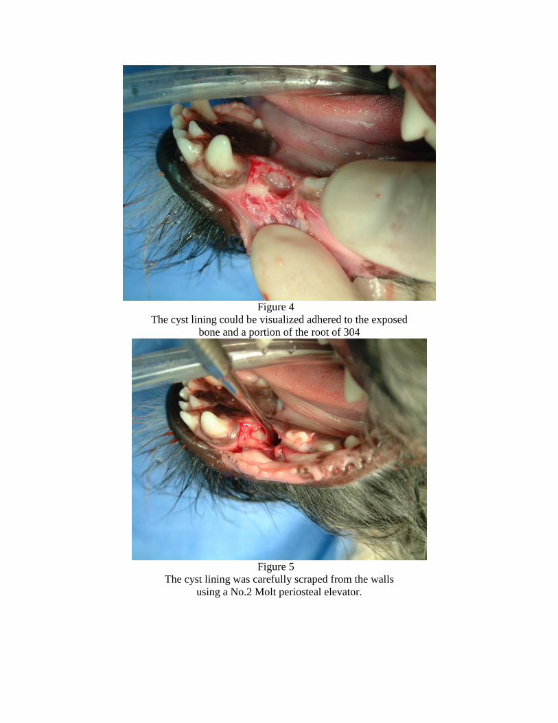
Figure 4
The cyst lining could be visualized adhered to the exposed
bone and a portion of the root of 304
Figure 5
The cyst lining was carefully scraped from the walls
using a No.2 Molt periosteal elevator.

Figure 6
A synthetic bone material was placed to fill the defect and help
maintain the alveolar ridge.
Figure 7
The mucoperiosteal flap was sutured with 4-0 monocryl
in a simple interrupted pattern

Figure 8
Complete fill of the cyst site with synthetic
bone material immediately post-op.
Post-op Care
The patient was placed on a blanket in recovery and carefully monitored. At the first sign of
swallowing the endotracheal tubeg was deflated and removed. The patient was monitored until
sternal recumbency was achieved. Head bobbing was encountered and the patient was held by a
technician until able to stand without danger of self-trauma. Prior to discharge the IV catheterd
was removed and a light pressure bandage placed over the catheter site to aid in hemostasis.
A recheck was scheduled for two weeks post-op to monitor healing of the surgical site.
Instructions were to feed soft food only for a period of two weeks eliminating access to anything
in the environment that was hard that could be chewed. Ketoprofenee
was dispensed in a
flavored oral suspension (10mg/ml) with instructions to give 8 mg every twenty four hours for
three days starting twenty four hours post-op. Two days following discharge the owner was
contacted by phone. The patient showed no adverse effects from the procedure.
A diagnosis of odontogenic cyst was confirmed by histopathology three days later. The
results were relayed to the owner by phone.

Follow-up/Results
A recheck examination two weeks post-op showed complete healing of the surgical site. The
sutures were intact. The owner was instructed to resume feeding the regular kibble diet and to
start brushing the teeth daily paying particular attention to buccal aspect of teeth 206 and 207 at
the furcation. CET toothpasteff was dispensed. An appointment for dental cleaning and
radiography of the surgical site was made for October to evaluate for possible return of the cyst
due to inadequate removal of the lining and to evaluate tooth 304 for external resorption or
endodontic complications.
The patient presented for possible foreign body ingestion in June. During sedation for survey
abdominal radiography, a dental radiographq was taken of the surgical site. Radiography
revealed an increase in bone density in the area of the former cyst. (Figure 9) The periodontal
ligament space on the distal aspect of tooth 304 was present although widened slightly in the
coronal one-third.
The patient returned for oral examination, radiography, and cleaning in December.
Radiography revealed normal bone and periodontal ligament space surrounding tooth 304.
(Figures 10 and 11) The gross appearance was normal. (Figure 12)
Figure 9
Radiography revealed an increase in bone density in the area of the former cyst
at seven months post-op.

Figure 10
Radographic appearance at 12 months post-op. Healing is complete
with no indication of damage to the canine tooth.
Figure 11

An alternate view at 12 months shows no indication
of damage to tooth (304)
Figure 12
Gross appearance at 12 months post-op
Discussion
This case demonstrates the importance of radiographic evaluation of even minor gingival
lesions associated with edentulous areas in the oral cavity. Several conditions can be considered
in the initial differential list for minor gingival lesions as the one described in this case in
association with a missing secondary tooth. A fractured tooth with gingival overgrowth
obscuring root fragment(s) should be considered. Teeth may be ankylosed or impacted both
preventing eruption but unassociated with cyst formation (13). Other possibilities include
abscesses (2,5) and fibrous lesions of the mandible(2). A simple cyst may exist without
association with an impacted tooth.(5,11) Finally, dentigerous cysts are odontogenic cysts
associated with a unerrupted tooth and can present as described in this case.(1,2,3,4,5,6,7,8)
Dentigerous cysts are generally benign however the veterinary literature describes cases in which
they have given rise to odontomas(9), ameloblastomas (7), carcinomas (8), and ameloblastic
fibro-odontomas (4).
Cysts of odontogenic origin arise from cells of the developing tooth. Further classification is
based upon histopathology and at times radiographic appearance. A dentigerous cyst is a type of
odontogenic cyst that arises from the epithelial components of the developing tooth follicle or its

remnants and occurs infrequently. (1,2,3,4,5,6,7,8) Two types of dentigerous cysts have been
described in the veterinary literature.(1) An eruption cyst results from the expanding follicular
space around the crown of a tooth as it erupts and generally requires no treatment. A follicular
cyst arises from the developing tooth follicle surrounding the neck of the tooth. The
radiolucency surrounding the crown of the unerrupted tooth in this case is typical of a follicular
cyst. Based upon histopathology and radiographic appearance this cyst could be classified
accordingly. Follicular cysts are locally aggressive lesions that should be approached early in
their course to attempt to remove the cyst lining in its entirety eliminating further destruction of
tissue. (3,5)
In this case the adjacent canine tooth was partially surrounded by the cyst. Although no
visible signs of damage were present grossly or radiographically initially or on follow-up, this
tooth should be monitored for signs of root resorption, ankylosis and endodontic disease.
Conclusion
Missing teeth with or without gingival lesions can be associated with a variety of benign and
potentially pathologic conditions. Detection of pathology can be gained with the help of
radiography. Therapeutic decisions should be made based upon radiographic findings. Early
surgical intervention is important in preventing tissue destruction where cysts are present in
conjunction with unerrupted teeth. Radiographic follow-up is important in assessing the success
of cyst removal and for continued monitoring of adjacent teeth for after-effects of cyst
impingement.

Products
a) EKG analyzer, Vetronics, Lafayette, IN
b) Torbugesic, Fort Dodge Animal Health, Fort Dodge, IA
c) Surflo intravenous catheter, Terumo Medical Corp, Elkton, MD
d) Ketaset, Fort Dodge Animal Health, Fort Dodge, IA
e) Valium, Abbott Laboratories, N Chicago, IL
f) 5-0 Endotracheal Tube, Rusch, Deluth, GA
g) IsoFlo, Abbott Laboratories, N Chicago, IL
h) VMS Anesthesia Machine, Matrix Medical, Inc., Orchard Park, NY
i) T Pump, Gaymar Industries, Orchard Park, NY
j) Lactated Ringer’s solution, Abbott Labs, N Chicago, IL
k) Chlorhexidine, First Priority, Elgin, IL
l) Neosonic, Amdent, Cherry Hill, NJ
m) Disposable prophy angle, Carlile Labs, Rockwell Centre, NY
n) High Speed Delivery System, Beaverstate Dental, Tualatin, OR
o) Prophy 1 Paste, Carlile Labs, Rockville Centre, NY
p) DentX Image Vet X70, AFP Imaging, Elmsford, NY
q) Consil, Nutramax Laboratories, Baltimore, MD
r) Bupivicaine, Abbott Laboratories, N Chicago, IL
s) Tuberculin syringe and needle, Nipro Mecical Corp, Miami, FL
t) No. 15 surgical blade, Carlile Labs, Rockville Centre, NY
u) Scalpel handle, Spectrum, Stow, OH
v) No. 2 Molt periosteal elevator, Hu-Friendy Co, Chicago, IL
w) L-5S Luxator, Cislak Manufacturing Inc., Glenview, IL
x) 3 cc syringe and 22 gauge needle, Nipro Mecical Corp, Miami, FL
y) No. 4 round bur, Carlile Labs, Rockville Centre, NY
z) Ophthalmic Thumb forceps, Spectrum, Stow, OH
aa) 0.9% saline solution, Abbott Labs, N Chicago, IL
bb) Prefgel, Biora, Inc., Chicago, IL
cc) Mayo-Hagar needle holders, Spectrum, Stow, OH

dd) 4-0 Monocryl, Ethicon, Inc. Somerville, NJ
ee) Ketofen, Fort Dodge Animal Health, Fort Dodge, IA
ff) CET Toothpaste, Virbac, Ft. Worth, TX
References
1. Wiggs RB, Lobprise HB: Veterinary Dentistry Principles and Practice. Philadelphia,
Lippincott 131 (1997)
2. Anderson JG, Harvey CE. Odontogenic Cysts. J Vet Dent 10(4):5-9 (1993)
3.. Tholen M, Hoyt RF: Oral Pathology. In Bojrab MJ, Tholen M: Small Animal Oral Medicine
and Surgery. Philadelphia, Lea & Febiger 38 (1990)
4. Kramek BA, O’Brien, TD, Smith, FO. Diagnosis and Removal of a Dentigerous Cyst
Complicated by Ameloblastic Fibro-odontoma in a Dog. J Vet Dent 13(1):9-11 (1996)
5. Lobprise HB, Wiggs RB. Dentigerous Cyst in A Dog. J Vet Dent 9(1):13-15 (1992)
6. Rossman LE, Garber DA, Harvey CE. Disorders of the Teeth. In:Harvey CE ed. Veterinary
Dentistry. Philadelphia: WB Saunders Co 88 (1985)
7. Rashmir-Raven A Debowes RM, Case WC, Gatewood DM, Clem MF. Dentigerous Cysts.
Compen. Cont. Educ. Pract. Vet 12(8):1120-1126 (1990)
8. Poulet FM, Valentine BA, Summers BA. A Survey of Epithelial Odontogenic Tumors and
Cysts in Dogs and Cats. Vet Path 29:369-380 (1992)
9. Wiggs RB, Lobprise HB. Veterinary Dentistry Principles and Practice. Philadelphia,
Lippincott, 352-355 (1997)
10. Verstraete, F: Odontogenic Tumours, in Proceedings from the 26th
World Congress of the
World Small Animal Veterinary Association 216 (2001)
11. Burk RL. Radiology. In: Bojrab MJ, Tholen M, eds. Small Animal Oral Medicine and
Surgery. Philadelphia: Lea & Febiger, 64 (1990)
12. McKelvey D, Hollingshead KW, Small Animal Anesthesia, Mosby Pub. Co. St. Louis, MO
16-22 (1998)
13. Wiggs RB, Lobprise HB: Veterinary Dentistry Principles and Practice. Philadelphia,
Lippincott, 179 (1997)


![Tooth Bar Manual[1]2. Using the inside width dimension from your bucket, measure the Tooth Bar from left to right, then mark that distance on the Tooth Bar, (This mark is for reference](https://img.pdfslide.us/doc/110x75/604cde6ac6cf006409340b85/tooth-bar-manual1-2-using-the-inside-width-dimension-from-your-bucket-measure.jpg)