Embed Size (px)
Citation preview


L I E> R.AFLYOF THE
U N IVLRSITYor ILLINOIS
570.SILL
cop. 2

The person charging this material is re-sponsible for its return to the Hbrary fromwhich It was withdrawn on or before theLatest Date stamped below.
Theft, mutilation, and underlining of booksare reasons for disciplinary action and mayresult in dismissal from the University.
UNIVERSITY OF lUINOIS LIBRARY AT URBANA-CHAMPAIGN
BM^LDUiGUSfcONb*
APRAPR1
1 8 \m8
^ (
JAN 3 1^95
yAN30 1993
30 1993
JAN 3 1993
L161— O-1096


THEDEVELOPMENTALANATOMYOF ISOETES
DOMINICK J. PAOLILLO, JR.
LINOIS BIOLOGICAL MONOGRAP
1
THE UNIVERSITY OF ILLINOIS PRESS. URBANA
ILLINOIS BIOLOGICAL MONOGRAPHS
Volumes 1 through 24 contained four issues each and were available through
subscription. Beginning with number 25 (issued in 1957), each publication is
numbered consecutively. No subscriptions are available, but standing orders
are accepted for forthcoming numbers. Prices of previous issues still in print
are listed below, and these may be purchased from the University of Illinois
Press, Urbana, Illinois. Requests for exchange arrangements should be ad-
dressed to the Exchange Department, University Library, Urbana, Illinois.
Baker, Frank Collins (1922): The Molluscan Fauna of the Big Vermilion River, Illinois, with
Special Reference to Its Modification as the Result of Pollution by Sewage and Manufacturing
Wastes. 15 pis. Vol. 7, No. 2. $1.25.
Balduf, W. V. (1959): Obligatory and Facultative Insects in Rose Hips. 12 pis. No. 26.
$3.50.
Cregan, Sister Mary Bertha (1941): Generic Relationships of the Dolichopodidae (Diptera)
Based on a Study of the Mouth Parts. 30 pis. Vol. 18, No. 1. $1.00.
Fisher, Harvey I., and Goodman, Donald C. (1955): The Myology of the Whooping Crane,
Grus americana. 40 figs. Vol. 24, No. 2. $2.50.
Gambill, William G., Jr. (1953): The Leguminosae of Illinois. Vol. 22, No. 4. $3.00.
Goodnight, Clarence James (1940): The Branchiobdellidae (Oligochaeta) of North Amer-
ican Crayfishes. 3 pis. Vol. 17, No. 3. $1.00.
Gutberlet, John Earl (1915): On the Osteology of Some of the Loricati. 5 pis. Vol. 2,
No. 2. $.50.
Heiss, Elizabeth M. (1938): A Classification of the Larvae and Puparia of the Syrphidae
of Illinois, Exclusive of Aquatic Forms. 17 pis. Vol. 16, No. 4. $1.50.
Higley, Ruth (1918): Morphology and Biology of Some Turbellaria from the Mississippi
Basin. 3 pis. Vol. 4, No. 3. $1.25.
Hoffmeister, Donald F. (1951): A Taxonomic and Evolutionary Study of the Pinon Mouse,
Peromyscus fruei. 5 pis. 24 figs. Vol. 21, No. 4. Cloth only, $3.50.
and Goodpaster, Woodrow W. (1954): The Mammals of the Huachuca Mountains,
Southeastern Arizona. 27 figs. Vol. 24, No. 1. $3.00.
Hopkins, Sewell Hepburn (1934): The Papillose Allocreadiidae—A Study of Their Mor-
phology, Life Histories, and Relationships. 4 pis. 6 figs. Vol. 13, No. 2. $1.00.
Humes, Arthur Grover (1942): The Morphology, Taxonomy, and Bionomics of the Nemer-
tean Genus Carcinonemertes. 4 pis. 1 map. Vol. 18, No. 4. $1.50.
Kendeigh, S. Charles (1941): Territorial and Mating Behavior of the House Wren. 32 figs.
Vol. 18, No. 3. $1.50.
(1952): Parental Care and Its Evolution in Birds. 35 figs. Vol. 22, Nos. 1-3. $4.00.
Kramer, Sol (1950): The Morphology and Phylogeny of Auchenorhynchous Hemoptero
(Insecta). 6 charts. 16 pis. Vol. 20, No. 4. $2.00.
Kudo, Richard Roksabro (1944): Morphology and Development of Nosema nofabilis Kudo;
Parasitic in Sphaerospora polymorpha Davis, a Parasite of Opsanus tau and O. fce/o. 12
pis. 7 figs. Vol. 20, No. 1. $1.25.
Liem, Karel F. (1963): The Comparative Osteology and Phylogeny of the Anabantoidei
(Teleostei, Pisces). 104 figs. No. 30. $3.50.
Meglitsch, Paul A. (1940): Cytological Observations on Endamoeba blaftae. 8 pis. Vol. 17,
No. 4. $1.00.
Morgan, Jeanne (1959): The Morphology and Anatomy of American Species of the GenusPsaronius. 82 figs. No. 27. $3.00.
Ray, James Davis, Jr. (1956): The Genus Lysimachia in the New World. 20 pis. 11 maps.
Vol. 24, Nos. 3-4. $2.50,

THE DEVELOPMENTAL ANATOMY OF ISOETES

Digitized by tine Internet Archive
in 2011 with funding from
University of Illinois Urbana-Champaign
http://www.archive.org/details/developmentalana31paol

THE DEVELOPMENTALANATOMY
OF ISOETES
DOMINIGK J. PAOLILLO, JR.
ILLINOIS BIOLOGICAL MONOGRAPHS
31
THE UNIVERSITY OF ILLINOIS PRESS • URBANA • 1963

Board of Editors:
Francis J. Kruidenier, Theodore Delevoryas,
R. D. DeMoss, James G. Sternburg, and Aubrey B. Taylor
THIS MONOGRAPH IS A CONTRIBUTION FROM THE DEPARTMENT OF
BOTANY, UNIVERSITY OF ILLINOIS. DISTRIBUTED APRIL 29, 1963
© 1963 by the Board of Trustees of the University of
Illinois. Manufactured in the United States of America.
Library of Congress Catalog Card No. 63-10316.

>p
CONTENTS
INTRODUCTION 1
MATERIALS AND METHODS 2
THE GENERAL ORGANIZATION OF THE SPOROPHYTE 3
,TERMINOLOGY 7
THE SHOOT 8
REVIEW OF THE LITERATURE 8
OBSERVATIONS AND DISCUSSION 15
SUMMARY AND CONCLUSIONS 51
THE ROOT-PRODUCING MERISTEM 52
REVIEW OF THE LITERATLTRE 52
OBSERVATIONS AND DISCUSSION 57
SUMMARY AND CONCLUSIONS 75
THE APICAL MERISTEM OF THE ROOT 76
REVIEW OF THE LITERATURE 76
OBSERVATIONS AND DISCUSSION 78
SUMMARY AND CONCLUSIONS 82
GENERAL SUMMARY AND CONCLUSIONS 82
LITERATURE CITED 84
PLATES 89
INDEX 129

INTRODUCTION
In Isoetes, the relationships of some of the plant parts are unique,
and the structure of the sporophyte is difficult to interpret and explain.
One of the best ways to secure an understanding of this structure is to
compare plants of different ages to determine the relationships of the
component parts throughout ontogeny. Sporophytes from sporeling to
adult stages have been examined in this study. Observations are re-
corded on the growth of the shoot tip, lateral meristem, and root-pro-
ducing meristem, and on procambial differentiation, root initiation, and
the growth of the apical meristem of the root. To enhance the continuity
of the report, the review of the literature has been divided into three
parts. The pertinent original observations follow each section of the
literature review in sequence.
This study was completed at the University of California at Davis
during the tenure of a National Science Foundation Predoctoral Fellow-
ship. I wish to express my sincere thanks to Dr. E. M. Gifford, Jr., for
guidance throughout the course of this investigation. Thanks are also
due to Dr. K. Esau and Dr. L. K. Mann for critical review of the original
manuscript and suggested improvements on the original draft.
Mr. W. Russell assisted me in locating populations of Isoetes howellii
and I. nuttallii. Dr. S. C. Tucker supplied me with several specimens of
I. braunii. Dr. E. M. Gifford, Jr., placed the departmental collection of
slides of I. howellii at my disposal. I am indebted to Dr. H. B. Currier
for the use of his Ortholux microscope for fluorescence microscopy and
to Mr. H. B. Tepper for assistance with staining procedures.

THE DEVELOPMENTAL ANATOMY OF IsOetCS
MATERIALS AND METHODS
Isoetes howellii Engelm., 7. nuttallii A. Br., and I. hraunii Dur. were
examined in this investigation. 1. howellii and 1. nuttallii were obtained
from vernal pools in Lake Co., Calif. I. nuttallii was also collected from
a moist sod over granite rock in El Dorado Co., Calif. Several specimens
of 7. hraunii, collected under one foot of water at Deming Lake, Minn.,
were given to me by Dr. S. C. Tucker. 7. howellii and 7. nuttallii were
collected near the beginning of the growing season (Feb. and March).
Young and old plants were obtained in the field. All of the plants col-
lected showed signs of new vegetative growth. Some plants were fixed
directly after collection, whereas others were fixed after they were trans-
planted and grown for one month in the greenhouse in sand cultures
inundated with half-strength Hoagland's solution. Sporelings of 7.
howellii were obtained from spores that were sown in distilled water,
tap water, and half-strength Hoagland's solution in Syracuse watchglasses.
Specimens were fixed in Craf III (Sass, 1958, p. 18), in Regaud's
formaldehyde-dichromate mixture (Conn, Darrow, and Emmel, 1960,
p. 14) with eight days of postchroming, and in Formalin-Aceto-Alcohol
(Conn et al., 1960, p. 7) prepared with 50 per cent ethanol. Fixation
times and washing were according to the recommendations given in
the references consulted for formulae. Materials were dehydrated with
a normal butyl-ethanol series, infiltrated with Fisher tissuemat, and sec-
tioned serially at 7-12 /*. The principal combinations of stains used for
morphological studies were hematoxylin-safranine-fast green and chlora-
zol black-acid fuchsin-malachite green-martius yellow. (Details of these
schedules are contained in the dissertation on which this report is based.
This dissertation is filed in the University of California Library, Davis.
)
Other staining schedules used were as follows: Regaud's hematoxylin
( Conn et al, 1960, p. 213 ) ; Heidenhain's hematoxyfin ( Conn et al, 1960,
p. 182, using acidified iron-alum as the mordant, and using Johansen's,
1940, p. 50, mixture of the dye); tannic acid-iron chloride-safranine
(Foster, 1934, with the addition of fast green in clove oil); tannic acid-
iron chloride-lacmoid (Cheadle, Gifi^ord, and Esau, 1953); aniline blue
fluorescence (Currier and Strugger, 1956); mercuric bromphenol blue
(Mazia, Brewer, and Alfert, 1953); periodic acid-Schiff's reagent (Click,
1949, p. 44); aqueous pyronine Y (Tepper and Gifford, 1962); and the
Fuelgen reaction (Johansen, 1940, pp. 95-97).
Most of the specimens examined were 7. howellii. All of the develop-
mental investigations were limited to this species. Examination of the
MATERIALS AND METHODS
other two species served to support and modify some of the concepts
developed. Both I. howellii and I. brounii are typically two-lobed. /.
nuttallii is typically three-lobed. For two-lobed specimens, longitudinal
sections were made parallel and perpendicular to the basal groove, i.e.,
between and across the two lobes, respectively.
THE GENERAL ORGANIZATION OF THE SPOROPHYTE
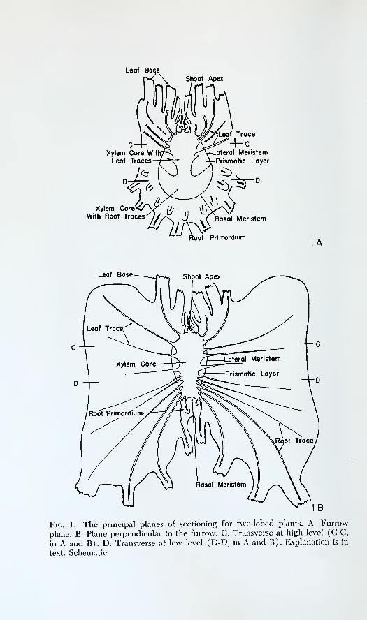
The principal planes of sectioning for two-lobed specimens are illus-
trated in figure 1. This figure indicates the locations of the meristematic
tissues in relation to the whole plant. Figure lA represents the sporo-
phyte as it is seen in a median longitudinal section in the plane passing
through the basal furrow. In the text, this plane of sectioning is referred
to as the plane of the furrow or the furrow plane. The shoot apex is at
the bottom of a conical depression. Vertical expansion of the cortex
raises the bases of the leaves above the level of the shoot apex. The por-
tion of the xylem core that bears the leaf traces is obconical, whereas
the portion that bears the root traces is convex on the lower perimeter
and forms a "horn" at each side. The outline of the xylem core of the
whole stele has been compared to that of a garden edging-tool, an anchor,
and a vegetable chopper (Foster and Gifford, 1959, p. 172). The cam-
bium is composed of two parts: the lateral meristem and the basal meri-
stem. The lateral meristem produces the so-called prismatic layer toward
the inside. This layer obtains its name from the prismatic form of its
cells and is also called the secondary vascular tissue. The basal meristem
augments the root-bearing portion of the stele and produces the sur-
rounding ground tissue, in which the root primordia are organized. Amedian section in the furrow plane does not show any root primordium
attached to the basal meristem.
Figure IB shows the appearance of a median longitudinal section at
right angles to the furrow plane. Both lobes of the plant are represented
in the figure. Again the shoot apex appears at the base of the conical
depression. The shape of the portion of the xylem core that bears leaf
traces is the same as in figure lA, but the shape of the portion that
bears root traces is different. This difference indicates that the root-
bearing portion of the stele is flattened in the plane of the basal furrow.
The root traces are arranged in an orthostichy and obscure the relation-
ship between the basal and lateral meristems.
Figure IC is a transverse section taken at a level through the broadest
Leaf Base
Xylem Core With;
Leaf Traces
Xylem CoreWith Root Traces
Shoot Apex
ateral Meristem
Prismatic Layer
Basal Meristem
Root Primordium
Leaf Base Shoot Apex
Fig. 1. The principal planes of sectioning for two-lobed plants. A. Furrow
plane. B. Plane perpendicular to the furrow. C. Transverse at high level (C-C,
in A and B). D. Transverse at low level (D-D, in A and B). Explanation is in
text. Schematic.


THE DEVELOPMENTAL ANATOMY OF ISOeteS
part of the leaf-bearing portion of the stele (level C-C, in A and B).
The figure is arranged with the basal furrow running from left to right
for comparison with figure lA. At all levels of sectioning, the stem is
divided into lobes. At the level of sectioning in figure IC, the lateral
meristem appears circular. Figure ID shows the appearance of a trans-
verse section taken at a low level in the plant (level D-D, in A and B)and is arranged with the furrow running from top to bottom for com-
parison with figure IB. The arrangement of root traces near the basal
meristem in ID is similar to that in IB, and the root traces again ob-
scure the relationship between the basal and lateral meristems. The basal
meristem is arranged in the form of a ribbon on the convex underside of
the root-bearing portion of the stele and therefore appears at two loca-
tions in figure ID (cf. level D-D in A).
Orthostichies of root traces are inserted on the root-bearing portion
of the stele in several to many places. Figure IB shows a vertical or-
thostichy because a longitudinal section at right angles to the furrow
plane is represented. Figure ID is taken at a level where two nearly
horizontal orthostichies are represented. The remainder of the orthos-
tichies of root traces are inserted obliquely on the root-bearing portion
of the stele. It may be argued that the various insertions of the or-
thostichies contradict the meaning of the word because an orthostichy
should be vertical in the plant. However, with respect to the basal meri-
stem, the arrangements of root traces at different locations are similar.
It is convenient to designate the arrangement of root traces one above
the other in median longitudinal section as an orthostichy and to apply
the same designation to similar arrangements elsewhere on the root-
bearing portion of the stele.
For additional orientation, three diagrams representing sporophytes
in the first three plastochrons are given as figure 2. All my figures of
sporophytes in the first three plastochrons are drawn and photographedfrom sections in the plane containing the first leaf, foot, and first root.
This plane corresponds to the longitudinal plane of sectioning at right
angles to the furrow plane in plants where the basal furrow can be
recognized. While the plant has a distichous phyllotaxy, the insertions
of all the leaves are contained in this plane. The second root also appears
in this plane. The first leaf and root form the beginnings of one lobe of
the plant; the second leaf and root form the beginnings of the other.
The basal furrow forms between these two lobes (fig. 2C). The shoot
apex is directly above the basal furrow. Additional figures illustrating
the transition from sporophytes with one leaf to sporophytes with manyleaves are contained in Baldwin's (1933) account of the early develop-
ment of the sporophyte.

TERMINOLOGY
First Ligule
Second Leaf
^y>—Procambium
\ \ \« Sheath of the" First Leaf
SecondRoot
Location of
the Furrow
Fig. 2. Three stages in the development of a young sporophyte. A. One leaf.
B. Two leaves. C. Three leaves. The plane of sectioning in A, B, C is the
same plane that contains all the leaf traces while the plant is in a ¥2 phyllotaxy.
The basal furrow forms between the first and second roots in a plane at right
angles of the plane of sectioning of the figures. Schematic.
TERMINOLOGY
Parke (1959) had discussed the problem of terminology in studies of
the apical meristem of the shoot. He defined the shoot apex as that por-
tion of the shoot tip above the youngest leaf primordium. The shoot tip
consists of the shoot apex and varying portions of the surrounding tissues,
including leaf primordia and young leaves. Parke's definition of the
shoot apex is convenient for conical apexes when the primordia arise
on the flank of the apex. However, a median longitudinal section does
not necessarily include the youngest primordium, and in such a circum-
stance it is difficult to mark the limits of the apex.
Some longitudinal sections of Isoetes show a flattened surface to the
left and right of the apical mound (e.g., pi. 2). I designate this flattened
region part of the region of leaf formation. It is best to exclude all this
THE DEVELOPMENTAL ANATOMY OF IsoeteS
region from the shoot apex because young primordia may be located in
the region of leaf formation outside of the median plane. Under these
conditions, the shoot apex of Isoetes may be less than that portion of
the shoot tip above the youngest leaf primordium seen in a median
section.
Because Isoetes is protostelic, the term plerome designates the pro-
cambium above the stele. The term stele, as it has been applied to
Isoetes by Lang (1915b), indicates the vascular tissues and associated
parenchyma formed from the procambium. Lang (1915b) believed that
the lateral meristem and its derivatives are extrastelar in origin. If the
lateral meristem arises in the procambium (West and Takeda, 1915),
the concept of the stele may be extended to include the prismatic layer.
In plants with few leaves, my figures are labeled according to the suc-
cession of leaves on the plant, with L^, Lg, Lg . . . ; L^ designates the
oldest leaf. For plants with several to many leaves, the leaves are num-bered from the youngest to the oldest, as P^, Pg, P3 . . . .
THE SHOOT
REVIEW OF THE LITERATURE
Fomi of the shoot apex. Hofmeister's (1862) report of an apical cell
in the shoot apex of Isoetes was challenged by Bruchmann (1874) and
Hegelmaier (1874). Hegelmaier related the formation of cells to the
activity of the surface layer of the entire apex, whereas Bruchmannidentified a small group of superficial initials within the apex. Farmer
(1890) maintained that there was never any substantial evidence for
an apical cell in his preparations of I. lactistris, the same species
studied by Hofmeister and Bruchmann. Scott and Hill (1900) intro-
duced evidence for the existence of an apical cell in at least somespecimens of I. hijstrix, and Lang (1915b) did not exclude the possi-
bility that an apical cell exists in some plants of I. lacustris. Other work-
ers (Stokey, 1909; West and Takeda, 1915; Weber, 1922; Liebig, 1931;
Bhambie, 1957; Sharma, 1961) have rejected the idea of an apical cell
in the shoot apexes of various species of Isoetes. Bruchmann (1874),
Hegelmaier (1874), and Rauh and Falk (1959b) have allowed the possi-
bility that the configurations found in the apex may vary among plants
of different ages, but Bruchmann ( 1874 ) found no apical cell in young
or old plants of /. lacustris.
Hofmeister (1862), Bruchmann (1874), and West and Takeda (1915)
THE SHOOT
illustrated the shoot apex of Isoetes as a conical or dome-shaped mass,
but Farmer (1890) asserted that the apex is flat. Scott and Hill (1900)
came to the same conclusion and reported that they could determine the
location of the median section of the shoot only by counting serial sec-
tions between opposing leaf primordia and choosing the median section
of this sequence. One may question the validity of such a procedure in
a plant with a spiral phyllotaxy because opposing primordia are of dif-
ferent ages and at different distances from the center of the apex. More
important, however, is the possibility that Scott and Hill may have
erroneously identified the shoot apex as a leaf primordium. They re-
marked that Bruchmann's (1874) illustrations of conical apexes in I.
lacustris bear a "suspicious resemblance" to leaf primordia. But Bruch-
mann's figures agree with those given by Hofmeister (1862) and Lang
(1915b) for I. lacustris. One may conclude either that Scott and Hill
did not recognize the apexes of their plants, or that the apex of /. hystrix
is so flat that these authors remained unconvinced by Bruchmann's illus-
trations of conical apexes in I. lacustris. West and Takeda (1915) ex-
amined slides of /. hystrix which had been prepared and studied by
Farmer. They reported that the shoot apex of 7. hystrix is conical. It maybe suggested, therefore, that Scott and Hill (1900) had an erroneous
concept of the topography of the shoot tip. One may also ask why Scott
and Hill (1900) maintained that their evidence explained Hofmeister's
findings on the apical cell, when Hofmeister reported that the apical
cell is at the summit of a conical apex and Scott and Hill maintained
that the apex is flat. The situation is complicated by Lang's (1915b)
report that the apex of I. lacustris usually forms a slight conical projec-
tion but is sometimes flat. However, Lang has also commented that
Bruchmann's (1874) figures are accurate. It may be concluded that the
shape of the apex can vary among plants of the same species. Inter-
specific variations also exist. For example, the figures given for I. coro-
mandeliana by Bhambie (1957) illustrate apexes which are more elon-
gate than those illustrated for other species of Isoetes.
La Motte ( 1937 ) reported that the shoot apex and probably all of the
permanent tissues of the sporophyte originate from a single quadrant of
the embryo. Mitotic activity is delayed in this quadrant until late in
embryogeny. Thus, the apex appears to arise laterally on the embryo.
The poles of the embryo are occupied by the tip of the first leaf and
the tip of the first root. Baldwin (1933) wrote that the "characteristic
leaf growing region of the adult sporophyte" is well defined at the time
of the origin of the seventh leaf. Baldwin paid little attention to cellular
details in the shoot tip of the young plant. He used outline drawings to
indicate the several stages recognized in the development of the young

10 THE DEVELOPMENTAL ANATOMY OF IsoetCS
sporopliyte. These are not detailed enough to determine the character
of the shoot apex during the first few plastochrons. In the figures of
Bruchmann (1874) and Campbell (1891), the shoot apex may be dis-
tinguished from the youngest leaf primordium during the second and
third plastochrons. Although this distinction cannot be made on the out-
line drawings given for comparable stages of development by Baldwin
(1933) and La Motte (1937), it can be made in the photomicrograph
published with La Motte's (1937) account.
Bruchmann (1874), Bhambie (1957), and Sharma (1961) recognized
that the form of the apex changes during ontogeny. Bruchmann ( 1874
)
reported that the topography of the relatively flat apex of the sporeling
is subjected to plastochronic changes and that leaf primordia are formed
in the tissues surrounding the conical apex of the adult plant.
Function of the shoot apex in the shoot tip. Hofmeister (1862) attrib-
uted the origin of the cells of the shoot of Isoetes to the activity of an
apical cell. In his scheme of growth, the shoot apex is responsible for
the production of cells which later enter into organogenesis and histo-
genesis in other parts of the growing tip of the plant. Likewise, Bruch-
mann (1874) and Hegelmaier (1874) forwarded the concept that all
the cells of the shoot have their ultimate origin in the shoot apex. Hegel-
maier felt that the orientation of cell walls and cell files leaves no doubt
that the superficial layer of the apex contributes cells inwardly, and that
these multiply to give a plerome directly below the apex and cortical
cells lateral to the apex. Bruchmann gave the same general scheme of
growth from the apex, although his concept of apical initials restricts
the location of the initiating group of cells more than the concept of a
meristematic surface layer formulated by Hegelmaier.
The account rendered by Bruchmann offers the opportunity to raise
an important point. Bruchmann (1874) wrote that the summital cells
of the shoot "sind die Meristem-Initialgruppe" of all of the surrounding
cells and "haben als solche auch die Aufgabe, den in Folge der Blatt-
bildung verbrauchten Scheiteltheil bei Erweiterung desselben wieder
zu ersetzen. Die Blattbildung ist hier zwar sehr langsam und daher auch
die Thiltigheit der Stammscheitel-Initialen triige, immerhin aber lasst
sie sich verfolgen." If attention is drawn to the word trage, it becomes
clear that Bruchmann did not necessarily regard the apical initials as
mitotically highly active. His concept of initials must have been related
to the ultimate source of cells rather than to relative rates of mitotic
activity. Furthermore, in the scheme of growth proposed by Hofmeister
(1862), emphasis is placed on the division of the derivatives of the
apical cell. Whereas the apical initial or initials are the source of all
the cells in the shoot in the schemes of Bruchmann, Hegelmaier, and

THE SHOOT 11
Hofmeister, the initials are not regarded as sites of organogenesis and
do not enter directly into the production of the plant body. The apical
initials are regarded as histologically undifferentiated cells. The multi-
plication and differentiation of their derivatives furnishes the materials
for the growth of the shoot.
Both Bruchmann (1874) and Hegelmaier (1874) recognized anti-
clinal and perichnal divisions in the superficial cells of the apex. The
scheme of cell division given by Bhambie (1957) for the shoot tip
of I. coromandeJiana closely resembles that given by Popham (1951)
for the shoot tip of his typical angiosperm. Bhambie (1957) did not
apply either dermatogen or tunica to the superficial layer of the shoot
tip, because periclinal divisions occur in the superficial layer during leaf
formation. Bhambie did not state clearly the exact location of the sites
of leaf formation in relation to the apical cone. From his diagram, it
appears that leaf primordia arise in the tissues surrounding the base of
the cone. If this is the case, no periclinal divisions occur within the super-
ficial layer of the apex of the species he investigated. Sharma ( 1961
)
has reported that the shoot apex of 7. sampathkwnarani has an outer
"epidermal" layer in which the cells divide mostly anticlinally, and an
inner mass of cells that divide in all planes.
Differentiation of tissues in the primary plant body. Most authors
have recognized a plerome above the centrally located vascular tissues
of the stem (Hofmeister, 1862; Bruchmann, 1874; Hegelmaier, 1874;
Scott and Hill, 1900; West and Takeda, 1915; Lang, 1915b). Because
Isoetes is protostelic, the plerome is composed entirely of procambium.
Although it has been admitted (West and Takeda, 1915) that this ple-
rome is rather poorly defined, Lang's (1915b) photographs offer sub-
stantial evidence for its existence. Stokey (1909) has reported that there
is no indication of a procambial strand above the mature stele in the
stems of the several species she investigated. Farmer (1890) and Stokey
(1909) argued that the vascular tissues of the stem represent a sym-
podium of leaf traces and that there is no cauline portion to the stele.
Farmer (1890) pointed out that the distinction between cauline and
foliar portions in the stele of Isoetes is not easily made and that the
distinction may exist more in the mind of the investigator than in the
plant. He emphasized, however, that there is clearly no cauline bundle
in the young plant. But this point was freely admitted by most workers.
West and Takeda (1915), for example, stated that there is general
agreement about the absence of a cauline portion in the stele of the
young plant. With Bruchmann (1874), they maintained that a cauline
portion does exist in later stages of development. No statement has been
made as to when in ontogeny the transition is accomplished.
12 THE DEVELOPMENTAL ANATOMY OF IsOCteS
Scott and Hill (1900) and Stokey (1909) reported that Hofmeister
(1862) regarded the stele of the mature sporophyte as a composite of
leaf traces. Weber (1922) commented that these reports were in conflict
with his knowledge of Hofmeister's work, but the Ray Society Transla-
tion that served as a reference for Scott and Hill and for Stokey was
not available to Weber. West and Takeda (1915) used the reference
that was quoted by Scott and Hill and Stokey (i.e., Hofmeister, 1862)
and correctly reported that Hofmeister recognized cauline tracheids in
the mature plant.
Lang (1915b) recognized a peripheral and central portion in the
xylem cylinder of the stems of several species of Isoetes. Leaf traces are
attached tliroughout the peripheral portion of the xylem but do not affect
the arrangement of tracheids in the central portion, which is regarded
as wholly cauline. Lang ( 1915b ) suggested that the cauline procambiumgives rise to both xylem and phloem. Hegelmaier (1874), however,
stated that the xylem core is "das Umwandlungsproduct des ganzen
Pleroms." Bruchmann (1874) reported that there is no distinct boundary
at the periphery of the core of xylary procambium. Lang (1915b) em-
phasized the absence of a definite boundary between the procambiumand the surrounding tissues. He stated that radial seriation of cells maybe traced from the peripheral portion of the xylem core, through the
primary phloem, into the cortex.
Whereas Lang (1915b) and West and Takeda (1915) concluded that
primary phloem occurs in the stem, Scott and Hill (1900) reported that
cauline primary phloem is absent, except under certain circumstances
(see later). They believed that the first tangential divisions around the
procambial xylem core are cambial divisions. Hegelmaier's ( 1874 ) ideas
on the question are close to those of Scott and Hill, and Stokey (1909)
offered a somewhat similar concept. For these authors, the primary
vascular tissues found within the stem are confined to the xylem core at
the center of the plant and the vascular tissues of the leaf and root
traces.
Differentiation of procambium to the leaves. The accounts that are
available in the literature suggest that differentiation of a leaf trace takes
place in the primary cortex and that the trace is initially inclined out-
ward. Hegelmaier (1874) offered that:
An den Stellen, welche in ihrer Lage den Anfiingen von Bliittern entsprechen,
andert sich jene nach einwarts geneigte Richtung der tangentialen Scheide-
wande in eine auswarts ansteigende, und es enstehen hier zarte, steil voninnen und unten nach aussen und oben gerichtete Zellenbiindel, die ersten
Aniagen der Blattstrange, deren Anfange somit in nachster Niihe des Scheitels
sich differenziren und deren spater hinzuwachsende Theile, entsprechend der
Richtung, welche die centripetal sich vermehrenden Radialreihen der Rinden-

THE SHOOT 13
zellen annehmen, einen mehr und mehr dem wagrechten sich nahrenden
Verlauf bekommen, in welchem endlich ein Theil der Zellen bekanntlich
schrauben- und ringformige Verdickungen erfahrt.
Hegelmaier's account is rendered without the use of illustrations. Ac-
cording to Fanner (1890), "The leaf trace originates in the division of
a row of cells, in an upward and outward direction, which more or less
irregularly connect the base of the leaf rudiment with the central part
of the stem, at the apex of the woody portion of the bundle. Thence the
divisions proceed upward into the leaf and downward into the stem."
Farmer's account is also rendered without the use of illustrations. West
and Takeda (1915) offered the following description of the initiation
of a leaf trace:
That part of the procambial strand of the foliar bundle which traverses the
primary cortex is differentiated at a very early stage in the development of
the leaf. It originates by the division of certain cells in the primary cortex,
which retain their meristematic character for a considerable period. A strand
of small cells, easily distinguishable by their relatively large nuclei, is pro-
duced in an upward and outward direction. The upper extremity of the strand
extends to the base of the young leaf. Connection of the piimary xylem and
primary phloem of the stem-stele is established by the downward prolonga-
tion of the procambial strand, the tissues of which are differentiated from
the 'parenchymatous mantle.'
The figure cited by West and Takeda in support of this description shows
a trace in an advanced stage of development.
Rauh and Falk (1959b) reported that the differentiation of the pro-
cambial traces of Stylites (a member of the Isoetaceae that was re-
cently described by Amstutz, 1957) is basipetal, and that the traces dif-
ferentiate in the primary cortex and reach the axial procambium before
any cytohistological differentiation can be detected in the latter at the
place of attachment of the trace. To evaluate these ideas and the re-
ports on procambial differentiation in the leaf traces of Isoetes, one must
determine if the differentiation of the axial procambium is strictly com-
parable to the differentiation of the procambium of the leaf traces. Onemust also detennine if the designation primary cortex is appropriate for
the tissue which gives rise to the leaf traces. These problems are dis-
cussed in a later section of this report.
The cambium and secondary growth. The cambium is composed of
two parts: the lateral meristem and the basal meristem. All authors have
treated the lateral meristem as part of the cambium. The basal meristem,
which has been treated in several ways, is discussed later in this report.
In the present section of the literature review, the term cambium will
be used to designate the lateral meristem only. This usage allows facility
in treating the literature, because most of the discussions of the cambium
relate only to the lateral meristem.
14 THE DEVELOPMENTAL ANATOMY OF ISOeteS
There is some difiference of opinion on the place of origin of the
cambium in the mature plant. Scott and Hill (1900) reported that
the cambium originates in the cells immediately outside the axial xylary
procambium. Stokey ( 1909 ) stated that the cambium begins its activity
in the parenchyma that surrounds the central core of xylem. West and
Takeda (1915) indicated that the cambium originates in the outer por-
tion of the parenchymatous mantle, which builds the parenchyma sheath,
the axial primary phloem, and the cambium as concentric layers around
the core of xylem. In their view, the cambium arises from a part
of the procambium, for the parenchymatous mantle is considered a
part of the plerome. West and Takeda (1915) and Lang (1915b) be-
lieved that the cambium begins its activity at the level of the youngest
mature phloem. Although Lang (1915b) agreed that in large plants the
cambium originates in the layer of cells adjacent and external to the
cauline primary phloem, he chose to designate the primary phloem as
the outer limit of the stele. In his opinion, therefore, the cambium is
extrastelar in origin because it originates in the primary cortex. A logi-
cal consequence of this opinion is the exclusion of the prismatic layer
from the stele.
For Stylites, Rauh and Falk ( 1959b ) reported that the cambium takes
its origin from the innermost layer of the primary cortex, which borders
on the primary phloem. They designated the cambium as a secondary
meristem and maintained a distinction between the cell layer that gives
rise to the cambium and the so-called primary meristem, which gives rise
to both the primary phloem and a parenchyma sheath that surrounds
the xylem core. A discontinuity of cell files is shown at this location in
their diagram of the origin of the cambium, but such a discontinuity is
lacking in photographs and drawings distributed throughout the rest
of the text.
For the sporeling, Hofmeister (1862) reported that a mantle of cam-bium is formed during the first growing season and that the cambiumarises from the layer of parenchyma adjacent and external to the axial
tracheids. Stokey (1909) reported that cambial activity begins early in
the ontogeny of the sporophyte. Her figure 3, cited in support of the
early origin of the cambium, shows a plant that is probably in its first
season of growth.
All authors have agreed that the outer derivatives of the cambium dif-
ferentiate as parenchyma of the secondary cortex, but Lang ( 1915b ) held
that the activity of the cambium toward the outer side has been greatly
overrated. He attributed a large portion of the cortical tissues to the
extension of the primary cortex. Most authors have agreed that the
corky layers of the surface of the plant are formed by the suberization
THE SHOOT 15
and death of the outermost cells of the cortex, but West and Takeda
(1915) have reported a cork cambium in the parts of the plant where
the basal furrow forms.
The inner derivatives of the lateral meristem have been the subject
of much discussion. Weber (1922), Rauh and Falk (1959b), and La-
moureux (1961) have given detailed reviews of the hterature on the
morphological nature of the prismatic layer. This layer has been de-
scribed as undifferentiated parenchyma (von Mohl, 1845), as secondary
xylem (Smith, 1900; Stokey, 1909), and as a mixture of sieve elements,
tracheary elements, and parenchyma (Russow, 1872; Scott and Hill,
1900; West and Takeda, 1915). Arguments advanced in favor of the
presence of sieve elements in the prismatic layer depend on the presence
of callose in the putative sieve elements (Esau, Cheadle, and Gilford,
1953; Lamoureux, 1961), the continuity of the innermost derivatives of
the prismatic layer with the functional sieve elements of leaf traces
(Russow, 1872; Scott and Hill, 1900), and cytological details of the
putative sieve elements (Lamoureux, 1961). Various arguments have
been advanced against the presence of sieve elements in the prismatic
layer. These are based on the obscurity of the physiological function of
the prismatic layer (Smith, 1900), the similarity of the sieve areas to
pits on the walls of parenchyma cells and the impurity of the callose
deposits (Weber, 1922), or the assertion that the putative sieve elements
are actually immature tracheids (Stokey, 1909).
The question of whether the prismatic layer should be called xylem
or phloem is unimportant. It has been reported that both sieve elements
and tracheary elements differentiate in the prismatic layer (Russow,
1872). Recently, secondary vascular tissue has been used to designate
the prismatic layer (Foster and Gilford, 1959; Lamoureux, 1961). Tliis
designation may be especially apt because the composition of the pris-
matic layer varies, tracheary elements being absent in some species
(West and Takeda, 1915).
OBSERVATIONS AND DISCUSSION
Form of the shoot apex. The shoot apex is organized in the young
sporophyte. A careful study of the figures available in the literature
(Hofmeister, 1862; Bruchmann, 1874; Keinitz-GerlofiF, 1881; Campbell,
1891; La Motte, 1937) and of a number of original preparations suggests
that the shoot apex cannot be distinguished from the surrounding cells
on morphological (figs. 3, 4) or cytological grounds until the advent of
the second plastochron, A median sagittal section of a sporophyte with
only one leaf may show a group of superficial cells between the ligule
16 THE DEVELOPMENTAL ANATOMY OF IsOCteS
of the leaf and the portion of the sheathing leaf base that appears in
the section (fig. 4). No distinction can be made between the cells that
will give rise to the shoot apex and those that will furnish the second leaf
primordium.
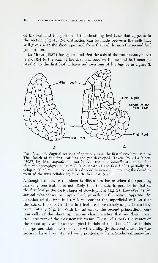
La Motte ( 1937 ) has speculated that the axis of the rudimentary shoot
is parallel to the axis of the first leaf because the second leaf emerges
parallel to the first leaf. I have redrawn one of his figures as figure 3.
Ligule
Sheath of theFirst Leaf
First Root
Figs. 3 and 4. Sagittal sections of sporophytes in the first plastochron. Fig. 3.
The sheath of the first leaf has not yet developed. Taken from La Motte(1937, fig. 13). Magnification not known. Fig. 4. /. howellii at a stage older
than the sporophyte in figure 3. The sheath of the first leaf is partially de-
veloped. The ligule mother cell has divided transversely, initiating the develop-
ment of the multicellular ligule of the first leaf. X 390.
Although the axis of the shoot is difficult to locate when the sporeling
has only one leaf, it is not likely that this axis is parallel to that of
the first leaf in the early stages of development ( fig. 3 ) . However, as the
second plastochron is approached, growth in the region opposite the
insertion of the first leaf tends to reorient the superficial cells so that
the axis of the shoot and the first leaf are more closely aligned than they
were initially (fig. 4). With the advent of the second primordium, cer-
tain cells of the shoot tip assume characteristics that set them apart
from the rest of the meristematic tissue. These cells mark the center of
the shoot apex and are the apical initials of the shoot. These initials
enlarge and stain less deeply or with a slightly different hue after the
sections have been stained with progressive hematoxylin-safranine-fast
THE SHOOT 17
green (pi. 1, A, B, C, at arrows), but they are no more vacuolated than
the adjacent cells.
During the initiation of the second primordium, tilting of the apical
surfaces continues so that an acute angle is formed between the surface
of the first leaf and the combined surfaces of the shoot apex and the
second leaf (pi. 1, A, B, C). As the second primordium grows, it be-
comes closely appressed to the first leaf (pi. 1, D, E, at L2). The angle
between the surfaces of the first leaf and the shoot apex becomes very
acute, and there is no pronounced topographic distinction between the
shoot apex and the second primordium, even after the ligule mother cell
of the second leaf is differentiated (pi. 1, E, at Lio). The broadening of
the apex by the multiplication and expansion of cells is accompanied
by a shift in the surface of the apex toward a plane more nearly perpen-
dicular to the adaxial surfaces of the first two leaves. As the development
of the third leaf proceeds, the topographic boundaries of the apex be-
come more distinct (figs. 5, 6). The apex assumes the form of a small
dome. The dome is very low, and the apical initials are deeper than the
apex. The apex may be so narrow that the tops of the apical initials
account for all of the apex.
The arrangement of leaves in a two-lobed specimen is distichous for
the first 10-15 plastochrons, and the shoot tip is not radially symmetrical
through these early stages of development. In a longitudinal section at
right angles to the basal furrow (figs. 5, 6, 8, 9) the leaves are to the
left and right of the apex. The apex may measure from 15-50 /x in width
by 3-15 /x in height between periods of leaf initiation. The form of the
shoot apex of the young plant is subject to plastochronic variations. Theapical initials are usually raised in a small dome (fig. 9). Leaf primordia
arise in the tissues next to the base of this dome, at least as early as the
third plastochron and for all subsequent periods of initiation. In the
young plant, the initiation of a leaf primordium may so alter the to-
pography of the shoot tip that the apex appears to have no height above
the axil of the primordium (fig. 8). At some stages of the plastochron,
the boundaries of the apex are indistinct, so that it is difficult to obtain
exactly comparable measurements from one apex to another.
As the phyllotaxy changes from distichous to spiral, the apex en-
larges, and the topography of the apex becomes more stabilized. The
apex forms a cone in the shoot tip. Plastochronic changes in the apex
become less distinct or nonexistent. Rauh and Falk (1959b) have re-
ported that in Stylites plastochronic changes in the form of the apex maybe detected in young plants but not in old plants. Bruchmann (1874)
has reported plastochronic changes in the shoot apex of young plants
of /. lacustris.
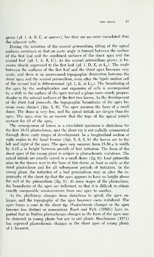
Figs. 5, 6, 7, 8 and 9. Legend on facing page.

THE SHOOT 19
Shortly after the spiral phyllotaxy becomes well established, the apical
cone is about twice as broad as high, and the apical initials occupy
the distal region of the cone (pi. 2, A). At this stage, the apex may
measure 50-75 fi in width by 20-30 /x in height. As the plant increases in
size, the apex may also enlarge. At the same time the apical initials be-
come less distinct, and the apex becomes covered with a uniform layer
of cells that are similar to the cells covering the tissues flanking the apex
(pi. 2, B, C). The uniform character of the superficial cells of the ma-
ture apex makes it impossible to single out a particular group of cells
as apical initials on any grounds other than position.
While the apical initials are still distinguishable on the basis of their
staining reaction, they comprise a group of cells of similar size. How-
ever, several of the specimens examined offer clear indication that a
single cell dominated the apical group at the time of fixation(pi. 3, A, B,
C, arrows). Whether or not such a cell is regarded as an apical cell
depends on one's concept of an apical cell and on one's emphasis on
Figs. 5, 6, 7, 8, and 9. Median longitudinal sections of young sporophytes of
I. howellii. X 500. Figs. 5 and 6. Plants in the third plastochron. Figure 6
represents a more advanced stage of the plastochron than figure 5. In both
figures, the surface of the shoot apex is more or less at right angles to the
adaxial surfaces of the second and third leaves. Fig. 7. Plant with 8-9 leaves.
Sectioned parallel to the developing basal furrow. The sheathing base of
each leaf appears to the left and right of the shoot apex. Fig. 8. Plant with
8-9 leaves. Sectioned at right angles to the plane of the fun-ow (sectioned
in the sagittal plane). The plant was still in a V2 phyllotaxy. The shoot apex
has no height above the axil of P^ because of the stage of the plastochron.
Fig. 9. Plant cut at right angles to the basal furrow. Fixed at the time the
phyllotaxy was changing from Vz to spiral. P^ represents the edge of the
youngest primordium and not the median portion. The apex has some height
above the axil of the youngest primordium. In all figures, the stippled cells
are members of their respective groups of apical initials. Other cells in these
groups appear in sections adjacent to those illustrated. P1-P4, youngest to
oldest leaves, counting from the shoot apex.

20 THE DEVELOPMENTAL ANATOMY OF IsoeteS
short-term versus long-term cell configurations. In the first dozen or
more plastochrons, such a cell would not be easily maintained at the
summit of the apex. Plastochronic variations in topography would alter
the spatial relations of the cells of the apex so that no one cell would
remain continuously elevated above the others. As plastochronic changes
become less important, an increased stability of the configurations in the
apex may be expected to result. However, the configurations that show
a single large cell at the summit of the apex are not stable throughout
ontogeny, for no such configuration is found in old plants. The apparent
apical cell has been found only in plants that were near the change from
distichous to spiral leaf arrangement. The low frequency of occurrence
of the apparent apical cell (about one plant in 15-20 of the appropriate
size) indicates that such a cell may dominate the apex for only a short
period in ontogeny or that it may never occur at all in some plants.
The evidence available does not support Hofmeister's (1862) report
of extreme regularity of division from two or three cutting faces of an
apical cell that is supposed to exist throughout the life of every sporo-
phyte. The evidence oflFered by Scott and Hill (1900) for the existence of
an apical cell in at least some plants of I. hijstrix could be considered
at this time. It is possible, however, that these authors failed to iden-
tify apexes properly in their materials, and their conclusions cannot
be accepted without reservations. On the other hand, the assertion byBhambie (1957) that the configurations shown in his cell net drawings
exclude the possibility of an apical cell is clearly unfounded. In most of
his figures the summit of the apex is dominated by a single cell. Thepattern of the cell net below this cell would not reflect the presence or
absence of an apical cell so much as it would the number of cutting
faces possessed by an apical cell and any symplastic growth adjustments
among the cells in and behind the apex. As a matter of fact, certain of
Bhambie's drawings and my plate 3, A, B, C, may be interpreted as evi-
dence for the existence of an apical cell which divides mostly anticlinally
(and alternately, with respect to the single plane of sectioning) andrarely periclinally. In my opinion, an undue emphasis on configurations
like those shown in plate 3, rather than poor microtechnique as suggested
by Bhambie (1957), led to the formulation of the concept that every
plant of every species of Isoetes has an apical cell (Hofmeister, 1862).
Bhambie (1957), in arguing against the existence of an apical cell,
has emphasized the lack of an extensively regular cell net which an
apical cell might be expected to generate (cf. Hegelmaier, 1874; Rauh
and Falk, 1959b). But it may also be argued that the summital cell in
some small plants of Isoetes, like the apical cell of Equisetum, is in posi-
tion to displace other cells from the center of the apex by its own

THE SHOOT 21
growth and division. In diis sense, the summital cell is at least the
analog of the apical cell of Eqtiisetum. Regardless of the interpretation,
one observes that the form of the apex just before and after the transition
from distichous to spiral phyllotaxy lends itself to the domination of the
summital region by a single cell. As the plant matures, the cells of the
apex become more equivalent, even at the summital region, for this
region of the apex flattens as the plant ages (pi. 2, C )
.
Popham ( 1960 ) described Isoetes as an example of a plant "in which
the apex of the very young sporophyte exhibits a single apical cell . . .
whereas an unstratified layer of cells dividing in many planes may be
observed in apices of older and adult sporophytes." In an earlier paper
(Popham, 1951), a similar opinion is implied but not stated. In /.
howeUii, the presence of a large summital cell in some small plants oflFers
no guarantee that a similar condition occurs in the ontogeny of all
plants. During the second plastochron, a definite group of equivalent
apical initials is present (pi. 1, B, C, arrows; cf. Bruchmann, 1874). The
occurrence of a single summital cell depends on the elevation of the
apex into a dome or cone, and I assume that the occuiTence of a large
summital cell is the result of growth adjustments within the group of
apical initials in their topographic setting in the apex.
The changes that occur in the shape of the apex of /. howellii during
the ontogeny of the sporophyte parallel those reported for /. coro-
mandeliana by Bhambie (1957). In the latter species, the form of the
apex changes from a flattened dome (plants with 6-15 leaves) to an
elongate cone (plants with about 20 leaves), with a tendency toward
flattening as the plant approaches old age. In 7. howellii, the apex of a
plant with a V2 phyllotaxy is only slightly raised above the surround-
ing tissue (figs. 5-7, 9). With the establishment of the spiral phyllotaxy,
the apex is further elevated, and becomes a sharp cone which is about
twice as broad as it is high(pi. 2, A, B ) . In the largest plants available,
the apexes were clearly elevated above the surrounding tissue, with their
widths about three to five times as great as their heights.
Several circumstances detract from any generalization which might
be made. First of all, there is no objective way of determining the age
of the plants I collected in the field. The plants may attain a certain
maximum size, but there is no guarantee that the largest plant is the
oldest. The external dimensions of a specimen are determined by the
balance between growth and deterioration of its tissues. New increments
of tissue are added to the plant by its meristems, and older tissues are
lost from the outside by decay. After sufficient time has elapsed, two
plants of different ages can attain the same size. But this size need not
be stable, because the plant may decrease in size if the equifibrium be-

22 THE DEVELOPMENTAL ANATOMY OF IsOCteS
tween growth and decay is disturbed. One might anticipate that the
internal structure of a plant would be a better indication of its age.
However, the increments of secondary vascular tissue are not layered in
the early stages of growth in I. howellii. Likewise, the number of series
of roots (see later section) produced by a plant in a season of growth
could vary from plant to plant or from year to year. Two plants of
similar external size and form may have apexes of rather different form,
even though the plants are approximately equal in the number of series
of roots they have possessed (pi. 4, A, B). To make comparisons valid,
it is also necessary to eliminate variations caused by differences in the
stage of the plastochron. It has already been mentioned that plastochronic
changes in the apexes of adult plants are not observed. Comparisons of
the apexes of different adult plants on the basis of the external andinternal form of the plants are valid, but the diflficulty with these com-parisons is that there is no objective way to relate them to chrono-
logical age.
Bhambie ( 1957 ) has reported that the "form of the apex is apparently
correlated with the mode of growth of the axis. In very young plants, as
also in mature ones, radial growth is more pronounced than vertical
growth, while in the prime youth of its life the plant elongates most rap-
idly and possesses, though for a short time, a conical apex." No data onthe rates of elongation support this statement.
In Isoetes, the internodes do not elongate after they are formed (Hof-
meister, 1862). The elongation of a stem during a growing season de-
pends directly on the number of leaves that are added along the axis
of the stem during a single season. This number may be estimated bythe number of leaves in a rosette, because the rosette is renewed each
year so that the number of leaves on the plant is not cumulative. In
/. howellii the height between successive traces on the stele is the samein plants with conical apexes as it is in plants that are much older andthat have broader apexes than their younger counterparts. The number of
leaves in the rosette of large plants is greater than the number of leaves
in the rosettes of small plants. Thus, the increment of length added to
the axis each season is larger in large plants than it is in small plants. It
follows that the conical form of the shoot apex is not correlated with the
highest rate of elongation. I cannot suggest what factors are respon-
sible for the variations in fonn of the apex through ontogeny. Apparently,
however, the change is not related in any simple fashion to the rate of
elongation of the plant.
Cytological details of the shoot apex and the adjacent tissues. Theuse of Regaud's fixative has facilitated the study of vacuoles, mito-
chondria, and plastids in the cells of the shoot tip. The discussion of

THE SHOOT 23
these cell components is based on the examination of specimens pre-
pared after Regaud's fixation and begins with a description of the shoot
tips of mature plants(pi. 4, C, D )
.
Bhambie (1957) has reported that the cells of the superficial layer
of the apex of 7. coromandeliana are less vacuolate than the subjacent
cells. In /. howellii the vacuoles of the superficial layer are smaller than
those of the subjacent cells. The transition from small to large vacuoles
may occur very abruptly along the axis of the plant so that large vacuoles
are found in the second layer of cells (pi. 4, C). More often, the transi-
tion occurs gradually, so that several cell layers are traversed before
cells with large vacuoles are encountered (pi. 4, D). The mitochondria
of cells of the superficial layer are granular and short, rodlike forms;
they are seldom elongated. In the cells with larger vacuoles, the mito-
chondria are long and threadlike, attaining a length of several microns
(pi. 5, A, at M, and opposite pi. 5), and granular mitochondria are in-
conspicuous or absent.
Because a separate publication has been devoted to the plastids of
7. howellii (Paolillo, 1962), they are mentioned only briefly here. Dur-
ing the interphase period in a meristematic cell, typically only one plastid
is present (Stewart, 1948). Tliis plastid may be called a proplastid be-
cause it is immature. With Regaud's fixation, the proplastid in its least
differentiated state is revealed to be a flattened object, often circular.
It stains very deeply at the rim and lightly in the interior after treat-
ment with hematoxylin and other protein stains. This staining reaction
is obtained for proplastids in the superficial cells of the shoot apex, in
young leaf primordia, in the intercalary meristematic tissues of growing
leaves, and in the apical meristem of the root. For convenience in de-
scription, I call the proplastid in this condition the imdifferentiated
plastid. The plastids of the cells of the superficial layer of the apex are
undifferentiated plastids (pi. 5, A, at U PI). Below the superficial layer,
the plastids are less condensed in form than the undifferentiated plas-
tids (pi. 5, A, B, at PI). Most of the plastids of the internal tissue of the
apex have a deeply staining reticulum and a lightly staining ground
substance (see opposite pi. 5).
The cytological characteristics of the superficial layer of the apex
and of the region of leaf formation surrounding the base of the apex
are essentiafly the same. The cytological characteristics of young leaf
primordia also resemble those of the superficial layer of the apex. With
regard to this similarity of cytological characteristics in cells of the shoot
apex and sites of leaf formation, Isoetes resembles such gymnosperms as
Ephedra (Dayes-Dujeu, 1957) and Cnjptomeria (Tribot, 1961) and

24 THE DEVELOPMENTAL ANATOMY OF IsoetCS
contrasts with some angiosperms (Buvat, 1952) in which the leaf pri-
mordia diflFer cytologically from cells of the apex.
All of the comments on the vacuoles, mitochondria, and plastids of
the superficial layer of the apex of the mature plant apply to the apexes
of young plants, with a few minor differences. In small plants, the tran-
sition to cells with large vacuoles occurs in the second layer. In young
plants with conical apexes, the transition extends over whatever num-
ber of cell layers are present in the apex. Two plants among twenty
young specimens examined after Regaud's fixation showed exceptionally
large cells at the summits of sharply conical apexes, and the vacuoles
of these cells were intermediate in size between those typical for the
superficial layer and those of the underlying tissue.
When acid fixation and a staining schedule containing safranine are
used, the most marked difference between the apical initials and the
adjacent cells in small plants is the lack of safranine in the apical initials.
This difference in staining with safranine may persist until after the
spiral phyllotaxy is established (pi. 2, A). In small plants, fixation with
Regaud's fluid and staining with Regaud's hematoxylin revealed no
special cytological properties for the apical initials, except that the twoplants mentioned in the previous paragraph showed some difference in
vacuolation between the summital cells and the other superficial cells
of the shoot tip. In small plants, the apical initials do not stain dis-
tinctively with acid fuchsin or with mercuric bromphenol blue. In large
plants, the superficial layer of the apex stains uniformly in all of the
staining schedules I have used, and the apical initials have no distinctive
staining properties. The most consistent feature in plants of all ages is
the deeper staining of the superficial layer compared to the underlying
tissues.
In the staining schedules used, safranine is regressed with acidified
alcohol and acid fuchsin is regressed with basified alcohol. From the
observation that these two dyes produce different results in delimiting
the apical initials of young plants, it may be suggested that the apical
initials of these plants differ from the adjacent cells in some, but not
all, of their stainable constituents. To determine if the differential stain-
ing properties of the apical initials might result from a lower content of
stainable RNA, ten small plants, of the size that might contain distinct
apical initials, and one large plant were stained with pyronine Y. This
stain may be used as an indicator of RNA in plant cells (Tepper andGifford, 1962) and is more or less specific for this compound under cer-
tain conditions. The materials were fixed in Graf III and FAA, and the
stain had to be applied for several hours or overnight in order to obtain
an intensity of staining equivalent to that obtained in six minutes in

THE SHOOT 25
higher plants in the same 2 per cent aqueous solution. It was also neces-
sary to reduce the washing of the stained materials (in n-butyl alcohol)
to the minimum required for dehydration in order to prevent complete
loss of stain. No tests were made to determine what constituents other
than RNA contributed to the staining, although it is obvious that py-
ronine Y stains more than RNA in plant tissues (Tepper and Gifford,
1962). In 7. howellii, starch grains, secondary walls of tracheary ele-
ments, and callose deposits stain with pyronine Y. Large starch grains
are absent from the apex (see later), and secondary walls and masses
of callose are absent from the shoot tip, so there may be a close re-
lationship between pyronine staining and stainable RNA content within
the shoot tip. Even if this relationship exists, the morphological sig-
nificance of the phenomenon is an open question. With these reserva-
tions in mind, the results of staining with pyronine Y will be discussed.
In the small plants, there was some indication that the cells which
corresponded to the apical initials seen after safranine staining were
more lightly stained than the surrounding cells. In the large plant, no
difiFerences in staining could be detected within the superficial layer of
the apex. In both the large and small plants, the superficial layer of the
apex and of the region of leaf formation was more deeply stained than
the underlying tissues. The adaxial cells of the youngest leaf primordium
of a given shoot tip stained more deeply than any of the cells of the
apex, but differences in staining were much less pronounced between
the apex and surrounding cells where no leaf primordium was present.
The most striking differences in staining are not among cells within
the apex, but between the cells of the apex and those of the young leaf
primordia. These differences are demonstrated to varying degrees with
pyronine Y, safranine, Regaud's hematoxylin, and other hematoxylins.
Safranine, used regressively after progressive hematoxylin, brings out
the differences most dramatically. The staining differences which do
exist separate the shoot tip into the same two regions that one would
designate on other grounds: the region of the shoot apex and the region
of leaf formation. In the shoot apex, the superficial layer stains more
deeply than the underlying layers. In the region of leaf formation, the
superficial cells closely resemble their counterparts in the shoot apex,
except where they are directly concerned with leaf formation. In the
latter case they stain more deeply than any of the cells of the shoot apex.
The distribution of starch can be studied by the use of IKI solution
or by the application of periodic acid followed by Schiff's reagent (PAS
reaction). Both of these staining techniques are appropriate for the de-
tection of large starch grains, but the PAS reaction may be used for the
coloration of very minute particles. One may assume that small particles
26 THE DEVELOPMENTAL ANATOMY OF IsOetCS
that are limited to plastids and have the same color as the large starch
grains are also starch granules. If this assumption is correct, the PAS
reaction enables one to detect smaller starch granules than may be
easily detected with the IKI reaction. The PAS reaction, therefore, pro-
vides a useful tool for the study of the distribution of starch in the
shoot tip.
Starch may or may not be detected by the PAS reaction in the sum-
mital region of the apex (pi. 5, C, D, E, F). Likewise, starch may be
present or absent in young leaf primordia (pi. 5, C, E, F). The starch
grains found at the summit of the apex are always minute (pi. 5, D),
but large grains of starch may occur at the base and tip of a young leaf
primordium (pi. 5, F). Bhambie (1957) has reported that starch is ab-
sent from the shoot apex and young leaf primordia of I. coronmndeliana.
In /. howellii, all of the four possible combinations of the presence and
absence of starch in the shoot apex and the leaf primordia occur. Too
few specimens have been treated with the PAS reaction to allow any
definite statement on the conditions that determine the distribution of
starch in the shoot tip.
Function of the shoot apex in the shoot tip. The function of the shoot
apex must be considered in relation to the function of the entire shoot tip.
Plants with the spiral phyllotaxy well established (pi. 2, A, B, C, and
older) will be considered first. It may be freely admitted that mitoses
are infrequently encountered within the limits of the shoot apex. Bruch-
mann (1874) has suggested that the frequency of mitoses in the apical
initials may be low, and Stewart (1948) reported that the cells of the
apex are seldom found in division. I have located mitoses in median or
near median longitudinal sections of nine out of eighty apexes from
plants with a spiral phyllotaxy. The search for mitotic figures was not
designed to obtain data on the relative frequencies of mitoses in the
apex, but rather to determine whether or not the pattern of the cell net
is a reliable indicator of the location and orientation of mitoses within
the apex. The arrangement of cell walls was used by Bruchmann ( 1874
)
and Hegelmaier (1874) in the formulation of their concepts of the
growth of the shoot tip. The concept that apical growth may be in-
terpreted by the cell net pattern is supported by my observations on
the location and orientation of mitotic figures within the apex. The fol-
lowing discussion, therefore, is based primarily on the pattern of the
cell net, and information on the location and orientation of mitotic
figures is introduced where it is pertinent.
One of the striking features of the cell net of Isoctes is the presence
of radial files of cells beneath the surface of the shoot tip. As Lang(1915b) and others have pointed out, these radial files are a manifesta-
THE SHOOT 27
tion of the primary thickening of the plant body. Near the shoot apex,
the files do not extend indefinitely into the cortex. Instead they bend
upward and terminate at the bases of the leaves (pi. 6, A, B). The
uppermost files are the shortest. New leaves are added to the rosette
from the inside and are displaced outward. It may be assumed, there-
fore, that the short files found below the distal part of the shoot tip are
extended to the length of the longer files found below them. At the same
time, it is obvious that new files are added on top of the older files. If
this were not the case, the files near the shoot apex would become in-
definite in length, and it is a matter of observation that this does not
happen. One must, therefore, determine the origin of new radial files.
The uppermost radial file in a section may be found to lie parallel to
the surface of the region of leaf formation where the latter has not been
raised in connection with the fonuation of a leaf (pis. 2, B, C; 6, A, B,
at F). The cell file and the surface layer separate where the latter rises
over the contour of the apex. The wedge of cells at this location in the
section is in position to forai a new radial file, which would be inserted
above the older cell file as both are extended outward (pis. 2, B, C;
6, A, at W ) . This wedge could originate either from a transverse division,
with reference to the shoot axis, of a cell near the inner end of the older
file, or from a cell which has been contributed downward from within
the shoot apex. Cell net patterns and division figures which indicate
both of these origins have been observed. Growth of the wedge into
a new file results in the transfer of cells at the base of the apex to the
region of leaf initiation. That such a transfer of cells does occur is sup-
ported by the observation that a young leaf primordium may be found
to abut directly on the base of the apex (pi. 2, B). The region of leaf
formation may then be regenerated from the apex at that particular site.
The spiral phyllotaxy of the adult plant allows for the uniform transfer
of cells from the apex to the region of leaf formation along all radii
of the plant.
Because some of the cells within the apex are in a position to form
new radiating files, it is important to know the source of the internal
cells of the apex. The observation of mitoses and the cell net pattern
indicate that the superficial layer along the flanks of the apex contributes
cells inwardly (figs. 10, 11; and pis. 4, B; 5, F; 6, A, at PF). These in-
ward derivatives are then in a position to produce the radial files of
cells mentioned above. Periclinal divisions also occur in and near the
summital cells of the apex, so that derivatives of the superficial layer
are contributed inwardly along the axis of the plant (pis. 4, C; 6, A;
at PS). These derivatives are not necessarily in a position to contribute
to the radial files, and need never do so in large apexes. In small apexes,

28 THE DEVELOPMENTAL ANATOMY OF IsoeteS
Figs. 10, 11, 12, and 13. Apexes of mature plants of 7. howellii as seen in
longitudinal sections. Figs. 10 and 11. Apexes showing periclinal divisions in
the superficial layer of the apex. X 400. Figs. 12 and 13. Apexes of larger
plants than those of figs. 10 and 11, showing an increase in number or in
width of cell files toward the summit as an expression of lateral expansion in
the summit region. X 350.
such derivatives might produce new cells both vertically and horizon-
tally in the shoot tip. Mitoses and cell net patterns which indicate a
contribution from the summit to the flanks of the apex have also been
observed. Anticlinal divisions occur at the summit of the apex (pi. 6, B,
at AS). The cell net pattern of an increasing number of files toward
the summit in some apexes also leads one to believe that these apexes
were growing in the summital region before the time of fixation (figs. 12,
13).
The apexes of young plants consist of relatively few cells. The mitotic
activity within the apexes of small plants cannot be accounted for by
growth of the apex itself, because the size of the apex does not increase
markedly in the first dozen plastochrons. The contribution from the
apex to other regions of the shoot tip is more direct in small plants than
in large ones. This contribution is indicated by the cell net pattern and
by mitotic figures.
The above analysis agrees in essentials with the accounts of Bruch-
mann (1874) and Hegelmaier (1874), but in particulars supports
Bruchmann's more restricted concept of a group of apical initials rather
than Hegelmaier's concept of an apical cell surface. The group of apical
initials present within the apex is distinguishable with certain staining
THE SHOOT 29
techniques until after the spiral phyllotaxy is well established. Subse-
quently, the summital cells are not visibly different from the adjacent
cells of the superficial layer. Popham ( 1951 ) has registered some objec-
tion to the use of the temi apical initials when the distal axial cells are
not distinguished on the basis of size, shape, and plane of cell division
from the adjacent cells. He argued "The term apical initials,' however,
would seem to imply (1) a group of cells showing a distinctive 'fixed
or regular scheme of segmentation' and (2) a group of cells ultimately
responsible for the initiation of all cells of the shoot apex." For the
definition of initials, the second of these two criteria may be accepted
without hesitation, but the first need not be accepted. Popham (1951)
apparently regarded both criteria as necessary for the definition of
initials. Yet the second criterion does not require that the summital cells
be distinguishable from the adjacent cells on the basis of size, shape, and
plane of cell division. The term apical initials may be used in a func-
tional sense, i.e., to indicate a group of cells responsible for the initiation
of the other cells of the shoot apex. It is precisely because of their posi-
tion that these cells can furnish derivatives to regenerate the rest of the
shoot apex. Position and function are the intrinsic properties of initials.
The distinctive size and staining properties of the apical initials of young
plants are only incidental to the concept that these cells are the initials
of the shoot.
If the concept of initials is related to the ultimate origin of cells, rather
than to the relative rates of cell division of different regions of the shoot
tip, the concept of the anneau initial ( Buvat, 1952; Gifford, 1954 ) should
be rejected for Isoetes. The region of leaf formation in the shoot tip of
Isoetes may be considered a more active region of growth than the
shoot apex, but the shoot apex furnishes cells to replenish the region of
leaf formation as the latter is used up in organogenesis. The region
of leaf formation is not an independent region of the shoot tip, although
the exact nature and frequency of contributions to this region from the
apex cannot be estimated. Cell divisions can be found in the shoot apex,
and because cells are contributed from the apex to the portions of
the shoot tip involved in histo- and organogenesis, the shoot apex con-
tains the initials of the shoot. The initials are a group of cells in the
distal region of the apex. No claim is made that the apex itself is a
a region of organogenesis. My interpretation of the concept of apical
initials in Isoetes is in agreement with Bruchmann's (1874). The desig-
nation initials does not have to be applied only to cells which are mitoti-
cally highly active ( Paohllo and Gifford, 1961 )
.
Differentiation of tissues in the primary plant body. The purpose of
the following discussion is to explain the structure of the primary plant

30 THE DEVELOPMENTAL ANATOMY OF IsOCteS
body of Isoetes in relation to the function of the shoot tip. To facihtate
this discussion, a brief account of the tissues present in the primary plant
body is given. Only large, well-grown plants are considered at this point.
The differentiation of tissues in the primary body of young plants is de-
scribed together with the differentiation of procambium to the leaf pri-
mordia.
As Lang (1915b) found for other species, the primary xylem of the
protostele of I. howelUi can be divided into a peripheral and a central
part (pi. 7, A, and D at PX and CX). The boundary between the two
portions of the xylem cannot be drawn with certainty, and the propor-
tions of the two vary with the specimen. The number of tracheids in the
peripheral xylem may be low (pi. 7, B) or high (pi. 7, C), The distinc-
tion between central and peripheral xylem in I. howellii and 7. nuttallii
is usually less marked than that in the species studied by Lang (1915b),
and much less marked than in Shjlites ( Rauh and Falk, 1959b ) . Most of
the cells of the central xylem are procumbent, whereas the cells of the
peripheral xylem may be erect and may be arranged in radial files (pi.
7, A, B, C). Directly outside of the peripheral xylem, one to several
cell layers differentiate as a parenchyma sheath (pi. 7, A, at Par S), andin contact with the parenchyma sheath is a layer of primary phloem(pi. 7, A, B, at PSE). When the primary sieve elements first mature,
they are bounded by residual procambium and primary cortex, but di-
rectly below this level they are separated from the primary cortex bythe cambium and cambial derivatives. During secondary growth, the
primary phloem is usually obhterated.
An understanding of the differentiation of the tissues of the pri-
mary plant body may be obtained by relating these tissues to the growth
of the shoot tip. The portion of the shoot tip under consideration has beendivided into the shoot apex and the region of leaf formation. The apical
initials of the shoot are located witliin the superficial layer of the
shoot apex and contribute cells laterally to the superficial layer of the
region of leaf formation and basipetally to the interior of the plant.
The internal cells of the shoot apex enter into vertical and lateral growthof the shoot. The cells away from the axis of the plant grow to form the
radial cell files which underlie the region of leaf formation and the
bases of leaves. On and near the axis of the plant, the cells contributed
by the shoot apex eventually add to the centrally located procambiumof the stem. The procambium is seated upon the primary vascular tissues
and has the form of a tapering mound or a blunt solid cone because there
is considerable broadening of the procambium before its elements finally
mature.
According to Lang (1915b) the primary phloem is the outer limit of

THE SHOOT 31
the stele. This arbitrary boundary need not be accepted because the
cells inside and outside of the primary phloem may occur in the same
radial files as the phloem elements (pi. 7, A, B). Radial seriation in the
peripheral region of the xylem indicates that the presence of radial files
of cells cannot be used to delimit the procambium if the peripheral
xylem is considered a part of the primary plant body. Lang (1915b)
himself has pointed out that cell files may be continuous from the pe-
ripheral portion of the xylem into the cortex. Bruchmann (1874) also
reported that there is no sharp boundary to the procambium.
West and Takeda (1915) designated the outer portion of the pro-
cambium as the parenchymatous mantle. This mantle surrounds the
xylary procambium and gives rise to the parenchyma sheath, the pri-
mary phloem, and the cambium. Rauh and Falk (1959b) recognized a
similar layer, said to give rise to the parenchyma sheath and the primary
phloem of Stylites, and called it the primary meristem. Although these
authors maintained that this meristematic sheath arises in the cortex,
such a distinction cannot be consistently made in their figures. In Isoetes,
the meristematic layer comparable to that which gives rise to the pri-
mary phloem of Stylites is inseparable from the rest of the procam-
bium. It is but a small step to change the terms parenchymatous mantle
(West and Takeda, 1915) and the primary meristem (Rauh and Falk,
1959b) to procambium. The use of the term procambium is justified
because (1) in ultimate origin there is no clear-cut discontinuity be-
tween the xylary core and the surrounding cell layers; (2) the leaf
traces are attached to this tissue from the time they are formed; (3) the
cells near the outer limit of this tissue differentiate as phloem.
Where leaf traces do not intervene between mature and immature
tissues in a longitudinal section( pi. 7, A, at PSE ) , one may project up-
ward the approximate limits of the procambium. The trajectory fol-
lowed in this projection leads upwards and inwards from the location
of the primary phloem. As best as can be determined by the size, shape,
orientation, and staining properties of cells, the approximate boundary
of the procambium can be followed to a level two or three cell layers
below the surface of the region of leaf formation, and then it tapers
abruptly toward the axis of the plant. In designating the primary meri-
stematic tissues, one might call the tissue adjacent to the procambium
the ground meristem. The procambium and ground meristem are not
readily separable from one another because a common primary meri-
stematic activity of the tissue below the shoot apex and region of leaf
formation is responsible for additions to both the procambium and the
ground meristem.
In a longitudinal section, the region of leaf formation is located

32 THE DEVELOPMENTAL ANATOMY OF IsoetCS
above the peripheral region of the procambial cyHnder (pis. 6, A, B;
7, A, D). Therefore the designation of the tissue underlying the region
of leaf formation as primary cortex ( Hegelmaier, 1874; West and Takeda,
1915; Rauh and Falk, 1959b) is subject to criticism. The designation of
this tissue as ground meristem is also inaccurate because at the same
level in the plant the tissue near the axis of the shoot cannot be readily
identified as procambium. The tissue intervening between the procam-
bium and the surface of the shoot tip is histologically undifferentiated.
Bruchmann ( 1874 ) reported that the upper limits of the procambium
fade into an undifferentiated tissue which he called the "Urmeristem."
This histologically undifferentiated region has special cytological fea-
tures. In median longitudinal sections of specimens fixed in Regaud's
fixative and stained with Regaud's hematoxylin, cells containing con-
spicuous, long mitochondria are found beneath and within the shoot
apex (pi. 5, A, at M; opposite pi. 5). Unfortunately, the chondriome is not
photogenic (pi. 4, C, D). The region of cells containing the conspicuous
chondriome varies in depth and grades off below into a region wherethe chondriome is inconspicuous. This latter region coincides with the
centrally located portion of the procambial cylinder. The lateral limits
of the region containing the conspicuous chondriome underlie the region
of leaf formation. As is the case for the procambium, there are no sharp
boundaries for the region containing the conspicuous chondriome.
Centrally this undifferentiated tissue yields to the acropetal dif-
ferentiation of procambium, whereas laterally it differentiates into the
ground meristem, except where leaf traces are formed. At these loca-
tions, procambium is formed in the lateral position also (see later).
The advantage of using the above concepts is not so much in recording
new or different observations as in allowing a coherent terminological
treatment which clarifies the processes of differentiation. If the tissue
underlying the region of leaf formation is not regarded as cortex, the
cortex does not give rise to leaf traces and varying portions of what mayrightfully be called the procambial cylinder. The problem is not one
of mere semantics, because the designation of tissue within the shoot tip
as primary cortex is not justified on the basis of the degree of morpholog-
ical differentiation of this tissue. Moreover, the leaf traces arise in the
tissue underlying the region of leaf formation, and are only subsequently
carried away from the shoot apex. The tissue in which the leaf traces
arise is the periphery of the histologically undifferentiated tissue below
the surface of the shoot tip. Avoidance of the use of primaiy cortex to
designate this histologically undifferentiated tissue can assist in a better
understanding of the process of the differentiation of procambial leaf
traces.

THE SHOOT 33
Differentiation of procambium to the leaves. A number of circum-
stances make it difficult to observe the differentiation of procambium to
the youngest leaves of mature plants of Isoetes. First, the youngest leaves
are crowded at the base of the apex, and the differentiation of procam-
bium from the level of one leaf to that of the next occurs over a very
small vertical height. This height may be no more than two cell layers.
Second, a layer of parenchyma differentiates completely around the xylem
of the leaf trace. At the insertion of the trace on the stele, the paren-
chyma cells of the xylem sheath are oriented with the axis of the leaf
trace rather than with the vertical axis of the plant. In a section passing
adjacent to a leaf trace, continuity of the axial vascular elements above
and below the trace appears to be lacking. The leaf traces are numerous
and closely placed on the stele. Consequently, only fortuitous longi-
tudinal sections show the continuity of the primary phloem with the
region of procambial differentiation to the youngest leaves (pi. 8, A).
A further difficulty arises out of the artifacts of sectioning resulting
from the orientation of certain cells in the vicinity of the shoot apex.
At the level of the mature stele, the cells of the prismatic layer are
oriented so that their long axes are nearly vertical in the plant. A trans-
verse section, therefore, gives an accurate idea of the radial dimensions
of these cells ( see opposite pi. 8, at 1 ). In the region of procambial dif-
ferentiation, however, the cells are inclined inward so that a trans-
verse section gives an exaggerated radial dimension for these cells (op-
posite pi. 8, at 2). The situation is further aggravated by the increase
in the tangential dimensions of the cells on the periphery of the xylem
core as the stele matures. At the periphery of the mature stele the tan-
gential and vertical dimensions of cells are approximately equal, whereas
in the region of procambial differentiation the tangential dimensions of
cells are about one-half the vertical dimensions (opposite pi. 8, at 1
and 2). With their small tangential dimensions and their exaggerated
radial dimensions, the cells in the region of procambial differentiation
to the youngest leaves do not resemble the cells at the periphery of the
matiu-e stele in transverse sections, and their continuity with the latter
may be easily overlooked.
Most of the accounts of procambial leaf traces in Isoetes give the im-
pression that the initial course of the trace is upward and outward in
the primary cortex. However, Liebig ( 1931 ) has mentioned that the
traces are initially vertical in their orientation and assume an upwardand outward course during later growth. The observations which are
reported below support Liebig's opinion, and it may be suggested that
other investigators have reported on advanced stages in the develop-
ment of a trace rather than on the initial stages of its formation. The

34 THE DEVELOPMENTAL ANATOMY OF IsoetCS
reports of suspended leaf traces are, then, even more remarkable but
are referable to a fourth difficulty which is encountered in the study
of procambial differentiation.
The cells of the leaf traces undergo the changes typical of procambial
differentiation. They become elongate and densely cytoplasmic before
they begin to mature as xylem and phloem elements (pi. 7, D, at LT).
The cells outside of the xylem core, which mature as the primary phloem
of the stem and are in continuity with the first formed phloem of the leaf
trace, do not elongate and do not become densely cytoplasmic before
they mature as phloem (pi. 7, A, D, at PSE). If their phloic nature is
accepted (see later) it may be argued that they are procambial at some
stage in their development. However, it is not difficult to acknowledge
that the contrast between the procambium of the leaf trace and the
procambium of the axial phloem of the stem can give the impression of
a suspended leaf trace, even when the two are actually observed in
contact with one another, especially if the plane containing the leaf
trace does not exactly coincide with the plane of sectioning (pi. 7, D,
left side). The report of basipetal differentiation of leaf trace procam-
bium in Stylites (Rauh and Falk, 1959b) may be explained in this way,
for the figures offered in support of basipetal differentiation show only
traces in advanced stages of development.
As Hegelmaier (1874) has reported, the tangential walls of the cells
beneath the region of leaf formation slant inward (pis. 6, A, B; 7, A, D;
8, A). As seen in longitudinal sections, the cells of this region form a
bridge for the differentiation of procambium from the mound of pro-
cambium below to the base of the forming leaf. The exact time, in re-
lation to the ontogeny of the leaf, that these cells differentiate as pro-
cambium may be somewhat variable or even a matter for debate.
Throughout the following discussion, the term procambium will beapplied to those cells which may be distinguished by their position anddegree of morphological differentiation in terms of form, staining prop-
erties, or both as the precursors of vascular tissue.
Procambium is differentiated to the base of an emerging leaf whenthe leaf is still very close to the apex. The procambium differentiates
across the two or three cell layers intervening between the axial pro-
cambium and the base of the growing leaf primordium. The procambial
strand is not clearly defined in longitudinal sections, even at the time
the ligule mother cell is visible (pi. 8, B), but in transverse sections
indications of traces may be detected to leaf primordia that do not
show any sign of a ligule mother cell. The leaf trace follows a path
determined by the tangential walls of the cells that compose it so that
the trace is initially straight and stands nearly vertical or slants inward

THE SHOOT 35
depending on the specimen (pi. 8, B, at LT). A new trace lies on a
tangent from the approximate edge of the pre-existing axial procambium.
Differentiation of the axial procambium is acropetal. Whether or not
differentiation of the trace is also acropetal is difficult to establish, be-
cause this differentiation takes place over two or three cell layers. Noconclusive evidence has been obtained on this point.
Leaf traces are subjected to the radial displacement which occurs
along all radii of the plant. Thirteen plastochrons elapse before a trace is
formed directly inside an older trace. As radial displacement proceeds,
the tissue that adjoins the inner side of a trace differentiates as ground
meristem and then as cortical parenchyma. Meanwhile, the axial pro-
cambium expands laterally throughout, and the cells at its periphery be-
come more erect. During this expansion, the base of a trace assumes an
upward and outward course, but the upper part maintains nearly its
original direction (pi. 8, C, D, E). As the radial files of cells of the
ground tissue continue to grow, the upper part of the trace is movedfurther and further from the axis. The lower part of the trace assumes
the direction of the radial files and keeps pace with the radial expan-
sion of the shoot by intercalary growth as the base of the leaf is carried
away from the axis of the plant. The trace becomes sharply bent below
the base of its leaf (pi. 8, F).
When the trace is first established, the continuity of its tissues with
those of the axial procambium is not strongly expressed, but changes
in the orientation of cells are consummated before the tissues of the
trace and stele mature. As the base of a trace assumes a more and morenearly horizontal position in the plant, its cells become more nearly
aligned with other procumbent cells in the procambial cylinder (pi. 6,
A, at CXLT). These latter cells form the connection between the xylary
procambium of the trace and that of the procambial core, and their
arrangement allows for the attachment of the trace throughout the pe-
ripheral region of the primary xylem (pi. 7, B, C, at CXLT). At the
same time, meristematic activity continues on the abaxial side of the
trace. The radially oriented files of cells which result from this ac-
tivity contact the procumbent elements of the base of the leaf trace.
These files are a manifestation of the general radial growth of the plant,
but because of their location their component cells differentiate in a
particular fashion. Some of these cells become the axial primary phloem,
differentiating as sieve elements in contact with the first-matured sieve
elements of the leaf trace (pi. 7, B, at PSE).
These phenomena may also be followed in cross sections, but a par-
ticular limitation of cross sections must be kept in mind. The radial
files of cells near the shoot tip bend upward. A transverse section.

36 THE DEVELOPMENTAL ANATOMY OF IsoetCS
therefore, is an artificial plane of sectioning with respect to the growth
of the plant. Instead of extended cell files, short portions of many files
appear in transverse sections. Along a single radius in one section, por-
tions of successively older files are encountered away from the axis of
the plant. The arrangement observed is not as orderly as that found in
longitudinal sections.
The photographs taken to illustrate the characteristic appearance of
leaf traces of various ages in transverse sections (pis. 9-12) are from a
single large plant. This particular series of sections, cut at 10 /a, is slightly
oblique, but the majority of transverse series are somewhat oblique in
their orientation. Because of this obliquity some leaf traces appear moredistinct than others. The first level of sectioning illustrated (pi. 9, A)is 10 fi below the summit of the apex. All the visible transections of
leaf traces and leaf primordia are numbered. The lowest number is given
to the youngest primordium in the section. In this particular plant, P4
is approximately 10 /x high and shows no indication of a ligule mothercell. All of the older primordia and leaves have multicellular ligules (pi.
9, A). The smallest of these is the two-celled hgule of P^. In the next
section (not illustrated), three younger leaf sites can be identified,
and the youngest of these is called P^ in this account. Whether this leaf
site should be called P^ or I^ (first incipient primordium) is arbitrarily
decided, because there is no sharp distinction between P^ and I^. Aslight uplifting of the surface occurs coincident with anticlinal divisions
in the superficial layer of the region of leaf formation and an increase
in the stainabihty of the resulting cells. Thus, the initial indications of
the formation of a leaf site are not readily separable from the changes
in the contour of the shoot tip which accompany leaf initiation. It is
reasonable, as well as convenient, to designate the youngest identifiable
leaf site in transverse sections as P^.
Ten fji lower, or 30 /x below the summit of the apex (pi. 9, B), the
obliquity of sectioning is manifest. The level of sectioning in the lower
part of the photo is higher in the plant than that in the upper part of
the photo. The cell net below P^(pi. 9, B, at 1 ) is not very distinctive,
but below Po (pi. 9, B, enclosed in hexagon) a distinctive group of
cells may be identified. These cells are a part of the trace to Po ( abbrevi-
ated TPo). The traces along the arc from TP4 to TPg (clockwise) are
somewhat obscure because of the obliquity of sectioning. In this arc,
the bases of cells of the superficial layer of the region of the leaf forma-
tion are probably represented, while the complementary arc ( 4-5 counter
clockwise) contains cells of the histologically undifferentiated tissue
underlying the region of leaf formation as well as leaf traces and ground
meristem. In a section 10 /x deeper (pi. 10, A) TP5 may be distinguished.

THE SHOOT 37
but TP2 cannot be seen. The cells underlying TPo are indicated (pi. 10,
A, at 2). These cells are on the perimeter of the procambial cylinder,
and it can be seen that this perimeter passes inside TP-, comes adjacent
to TP^, underlies P^, adjoins TP^, underlies TP3, passes inside of TPg,
and adjoins TP-. Its relationship to the traces in the arc 4-5 ( clockwise
)
is somewhat obscured by the obliquity of sectioning. Ten fx. deeper
(pi. 10, B), TP^ has disappeared in the perimeter of the procambial
core, and TPr, is on the perimeter of the procambial core. The shape of
the cells of TP,- in this section (pi. 10, B, at 5) resembles that of the
cells of TP. at a level 20 fx higher (pi. 9, B, at 2). In plate 10, B, TP^
still adjoins the perimeter of the procambial core. Twenty /n lower (pi.
11, A), TP5 has disappeared in the perimeter of the procambial core.
TP^, and TP^ are attached to this perimeter (pi. 11, A, at 6 and 7), and
the cells at these locations resemble those at the attachment of TPg at
the level 20 /x higher (pi. 10, B, at 5). Abaxial to TP^, a radial file of
cells (pi. 11, A, at arrow) of the ground tissue may be seen. The cells
of this file differ in tangential dimensions from the cells of TPg.
Thirty /x lower (pi. 11, B), the cells attaching TPg to the procambial
cylinder (pi. 11, B, at 9) may be compared with their counterparts for
TPg and TP^ (pi. 11, A, at 6 and 7). Radial files of cells appear at the
attachment of TPj^ (pi. 11, B, at 12), and the base of TP^o adjoins TP^^,
whereas the base of TP„ adjoins TPj^. Ten fx lower (pi. 11, C), radial
files of cells appear under the attachment of TP-,2 (ph 11» C, at 12),
whereas at a level an additional 20 /x deeper, the cells in this position
(pi. 11, D, at 12) are confluent with similar radial files at the base of
TPj, (pi. 11, D, at 17). Twenty fi deeper (pi. 12, A), TPoq is attached
to the procambial cylinder, and radial files of cells are seen at this loca-
tion (pi. 12, A, at 20) and under the attachment of TPj- (pi. 12, A, at
17). Sections 20 /x deeper (pi. 12, B), an additional 10 fx deeper (pi. 12,
C), and again 20 /x deeper (pi. 12, D) show the attachment of TPgg
and some younger traces. The field of view has been changed between
plate 12, C, and 12, D, to show TPo^ and TP^g which do not appear in
plate 12, A, B, or C. Adjacent to the attachments of TP^o to TPog (pi.
12, A, B, C, D), radial files of cells fill the bays between leaf traces.
The inner members of these files mature as part of the peripheral xylem
and often as tracheids(pi. 12, D, at 21 ) . The cells outside of the xylem
core mature as a sheath of parenchyma, and those outside of the latter
become sieve elements. The over-all picture obtained is the same as that
seen in longitudinal sections (pi. 7, A, B, C, D), except that the length
of the radial files of cells cannot be fully appreciated in transverse
sections because the files are tilted with respect to the horizontal plane
(pl.7, A, B, C, D).

38 THE DEVELOPMENTAL ANATOMY OF IsoetCS
Directly above the level of maturation of the conducting elements of
the xylem and phloem, radial files of cells are distributed all around the
procambial cylinder. At higher levels, the radial files are most pro-
nounced at the bases of leaf traces (pi. 11, B, C, D, at 12). The highest
mature phloem in the stele is also associated with the attachment of a
leaf trace (pi. 12, A, B, at Ph, near 19). It may be suggested that the
original sequence of initiation of leaf traces is followed acropetally by
tangential divisions on the periphery of the axial procambium. These
divisions occur while the bases of the leaf traces are assuming a hori-
zontal position, so that the base of a trace is ultimately embedded in
radial files of cells (pi. 8, F). Stokey (1909) interpreted parts of these
files as the inner derivatives of the cambium and described the situation
by reporting that there is an overgrowth of the leaf traces by the sec-
ondary tissues. In the present account these files are regarded as entirely
primary to the level where the first axial sieve elements are matured
(see later).
In plants that have a distichous leaf arrangement, the procambiumto the youngest leaf primordium diff^erentiates directly above the pro-
cambium to the second older primordium. The traces of the primordia
are bent sharply and the cells of a given trace seem to be continuous
with the cells of traces to primordia on the opposite side of the stem
(pi. 13, A). Median longitudinal sections in the plane of the basal groove
give the impression that the stele and procambial cylinder of the youngplant is a miniature of that of the older plants (pi. 13, B). However, all
of the mature and immature vascular elements, except those in the pris-
matic layer, are oriented with respect to the leaves and roots. In other
words, practically all vascular elements participate in the formation of
leaf and root traces (pi. 13, A). The process of differentiation of the pro-
cambium of the leaf trace could not be followed in the materials avail-
able. In all of the plants with distichous arrangement of the leaves,
primordia which showed ligule mother cells also showed a clearly de-
fined trace in longitudinal section. No suspended traces were found in
any of these plants.
In longitudinal sections in the plane of the basal furrow, sieve ele-
ments are located to the left and right of the tracheids of the leaf traces
(pi. 13, B, at PSE). It is noteworthy that sieve elements form in lat-
eral positions in association with trace tissues. Away from the axis of
the plant, the phloem of the trace is on the abaxial or underside of the
trace. Near the axis, the phloem is found in two lateral groups. Only
the tracheary elements reach the axis, and thus the integrity of the
xylem core is preserved. The phloem of a trace in a plant with spiral
phyllotaxy is abaxial throughout its extent and connects directly with
THE SHOOT 39
the axial sieve elements beneath the attachment of the trace to the
stele.
When the phyllotaxy changes to spiral, the leaf primordia begin to
form over regions that do not contain traces to older primordia. Eventu-
ally, median sections of leaf primordia may be seen in longitudinal
sections taken in the plane of the basal furrow. From this time onward,
the adult pattern of procambial differentiation is established, but the
stele of the plant is still too small to show the differentiation of central
and peripheral xylem. Because of the spiral insertion of leaves the
newly formed portions of the stele assume a circular or polygonal cross-
sectional outline. Tangential divisions at the periphery of the stele are
more pronounced than in the younger plants. The stem has primary
sieve elements which are not directly a part of the leaf traces, although
all of the tracheids which differentiate are members of leaf traces. The
obconical growth of the stele increases the cross-sectional area of the
xylem core so that leaf traces no longer account for all of the tracheids
that are formed in the stem. For the specimen in which the transition
to this condition was found, the xylem core measured 270 //, across at the
level of the highest mature tracheids and 540 fx in height from the level
of the tracheids of the original leaves of the plant to the level of the
highest mature tracheids. In contrast, the width of the combined traces
of the first few leaves is about 35 fx, and that of the xylem core of a
large plant may be 1,400 /x or more. The height of the tallest leaf-bearing
stele observed exceeded 7,000 fx (7 mm.).Esau (1954) has interpreted the foliar portion of the vascular tissue
of the shoot to be that portion which differentiates with a definite posi-
tional relationship to the leaves. Cauline vascular tissues do not showa definite positional relationship to leaves. The terms cauline and foliar
have been applied to the stele of Isoetes in this sense throughout the
literature. In I. howellii, all of the procambial tissues and all of the
mature primary vascular tissues of the shoot show a positional relation-
ship to leaves as long as the plant is in a distichous phyllotaxy. After the
spiral phyllotaxy is established, the leaf traces become separated enough
so that some primary cauline phloem is formed. The formation of cauline
tracheids is not accomplished until later in ontogeny, when the primary
xylem core has increased in diameter much beyond that of the sporeling.
It has been suggested (Lance-Nougarede and Loiseau, 1960) that
the pith of higher plants is a cauline tissue region. Rauh and Falk
(1959b) considered the central region of the primary xylem core of
Stylites as the homolog of the pith in higher plants. Lang (1915b) main-
tained both that the central xylem of Isoetes is wholly cauline, and that
part of the peripheral xylem among the attachments of the leaf traces

40 THE DEVELOPMENTAL ANATOMY OF IsOCteS
can be considered cauline. Wetmore ( 1943 ) has reported that in certain
protostehc lycopods a central column of cauline procambium may be
identified below the shoot apex and that this column may even ascend
above the level of the youngest leaf trace. In all plants of I. howellii that
have a spiral phyllotaxy, there is a core of procambium that initially
shows little relationship to the leaves. As differentiation and maturation
of the leaf traces proceed, portions of the procambial core assume a
positional relationship to the leaves. In large plants, this positional re-
lationship does not extend to the center of the xylem core. In smaller
plants, the leaf traces may account for all the tracheids of the shoot,
even when indications of an axial core of procambium are present
(pi. 2, A, at Pr).
Scott and Hill (1900) reported centripetal maturation of the xylem
core of /. hystrix. West and Takeda ( 1915 ) reported that xylem matura-
tion begins at the center of the stem in /. japonica, but Ogura (1938)
recognized a parenchymatous pith in the same species. Lang (1915b)
located the protoxylem of the shoot at the attachments of the leaf traces,
i.e., at the inner limit of the peripheral xylem. In Sttjlites (Rauh and
Falk, 1959b), the maturation of the first tracheids at the inner limit of
the peripheral portion of the xylem is easily seen because of the markeddistinction between central and peripheral xylem. In I. howellii, the
highest tracheids of the xylem core are associated with the bases of leaf
traces (pis. 7, A and D, at T; 12, C, at T at 22). These locations indi-
cate the inner extent of the peripheral xylem. The location of the first
mature xylem elements is a reflection of the degree of development of
the central portion of the xylem. In other words, centrifugal maturation
of the xylem would be expected when no central portion of the xylem
core is recognizable. It may be suggested, therefore, that the apparent
direction of maturation varies with the age and vigor of the specimen
investigated. This type of variation might account for the reports of
centrifugal (West and Takeda, 1915) and centripetal (Scott and Hill,
1900) maturation of xylem in Isoetes. Any designation of the direction
of maturation that can be made is subject to the reservation that matura-
tion throughout the xylem core of Isoetes takes place over very few
cell layers (pi. 7, A, D). In Stylites, where the shoot is generally more
elongate than in Isoetes, the maturation of primary xylem is completed
over many cell layers (Rauh and Falk, 1959b).
The cambium and secondary growth. The cambium is composed of
two parts: the lateral meristem and the basal meristem (fig. lA). In
this section of the report, the term cambimn designates the lateral
meristem only.
There is some difference of opinion on the place of origin of the

THE SHOOT 41
cambium in mature plants. In one view, the first tangential divisions
around the core of xylary procambium are recognized as cambial ( Scott
and Hill, 1900); in another, the cambium differentiates outside the pri-
mary phloem at a level where the latter is mature (Lang, 1915b; West
and Takeda, 1915). The second view has been adopted in this study.
The early divisions around the core of xylary procambium are regarded
as part of the growth of the procambium. If one adopts this interpre-
tation and recognizes that there is no clear-cut boundary between the
procambium and the adjacent ground meristem, then it is possible
to designate the layer of cells outside of the primary phloem as part of
the procambium. According to this view, the cambium would arise from
part of the procambium (cf. West and Takeda, 1915). Primary growth
passes over to secondary growth in a continuous process of radial ex-
pansion. Cambial activity represents a sustained meristematic activity
in a particular part of the procambium. Esau (1953, p. 381) has re-
marked that "The procambium and the cambium may be looked upon
as two developmental stages of the same meristem." This general state-
ment can be applied to Isoefes. However, the cambium of Isoefes arises
outside of the primary phloem, so that it is anomalous in its place of
origin, as well as in its subsequent activity. Nonetheless, the primary
plant body of Isoefes is directly comparable to that of other protostelic
species (cf. Lang, 1915b). A core of xylem is surrounded by a sheath
of parenchyma that is in turn surrounded by the primary phloem.
In the higher plants, tangential divisions that produce radial files of
cells may occur high in the procambium with respect to the level of
mature tissue (Esau, 1953, p. 381). Radial files may exist in bundles
where no secondary growth occurs, as in Zea (Esau, 1943). Thus, radial
seriation alone is not a reliable criterion for determining the presence of
a cambium. In Isoetes, the first tangential divisions around the core
of xylary procambium occur at a level higher than the level of mature
vascular elements, and Scott and Hill (1900) regarded these divisions
as cambial. It is more fruitful and convenient to recognize these divisions
as part of the primary thickening of the plant (Lang, 1915b; Rauh and
Falk, 1959b ) rather than to designate them as a manifestation of cambial
activity.
The cambium may be regarded as arising at the level at which the
primary phloem matures. This is a natural definition, because the sieve
elements which appear in the primary axial phloem differentiate centrip-
etally (cf. Rauh and Falk, 1959b). Then, by definition, the sieve ele-
ments which are added abaxial to the first formed sieve elements (cen-
trifugally) are secondary, but there is no other morphological boundary
between primary and secondary phloem.

42 THE DEVELOPMENTAL ANATOMY OF IsOetCS
To understand the origin of the cambium during the ontogeny of the
sporeling one must have a clear understanding of the arrangement of
the vascular tissues while the plant has only a few leaves (see fig. 2).
The xylem and the phloem of the first leaf is continuous with the xylem
and the phloem of the first root. The vascular bundles of both of these
organs are collateral, and the phloem is on the abaxial side of the
bundles. Tracheids differentiate across the axis of the plant to the sec-
ond leaf and the second root. The phloem of the second leaf and root is
also abaxial. The phloem of the first leaf and root stands in relation to
that of the second leaf and root as one side of the letter H stands in
relation to the other, with the connection between the two sides being
formed by sieve elements which are located out of the median sagittal
plane of sectioning, for median longitudinal sections in the plane of the
basal groove show that there are phloem elements to the left and right
of the axial tracheids in the young plant (pi. 13, B, at PSE). Phloemdifferentiates in these locations when the trace of the second leaf ma-tures. Additional sieve elements differentiate next to the abaxial face
of the first-formed sieve elements in this position (pi. 13, B, at SSE).
By the time the plant has several leaves, radial seriation of cells is noted
in sections in the furrow plane (pi. 13, B). Such seriation is not pro-
nounced in the plane containing the leaf traces because the traces are
so closely placed that they adjoin one another with little intervening
tissue (pi. 13, A).
One has two alternatives in the treatment of the first sieve elements
formed in the lateral positions in sections in the plane of the basal fur-
row: they may be designated as primary or as secondary phloem. If
the cambium arises in the layer of parenchyma surrounding the first
tracheids (Hofmeister, 1862; Stokey, 1909) the lateral sieve elements are
secondary. As a consequence, the cambium would be active in the plant
before two leaf traces have matured and it would contribute cells to
the leaf traces. On the other hand, if the lateral elements which are
first formed are considered primary, then a parallel between the youngest
and the oldest plants is immediately evident. The innermost and most
acropetal axial sieve elements may be regarded as primary (pi. 13, B,
at PSE). The choice between these two alternatives is of no import to
the phenomena discussed, but designating the first lateral sieve elements
as primary allows a consistent treatment of the description of the pri-
mary plant body throughout ontogeny. From this perspective the
cambium is interpreted as arising in the procambium outside of the
primary phloem. This interpretation is also consistent for sporophytes
of all ages. Stokey (1909) reported that the cambium arises in the layer
of parenchyma surrounding the xylary core, and this description, too,
THE SHOOT 43
could be consistent for plants of all ages. The matter once again re-
solves itself into a choice of a point of view. The perspective that has
been adopted in this report allows a direct comparison between the pri-
mary plant body of Isoetes and other protostelic forms (cf. Lang, 1915b)
and has been chosen for that reason.
When the plant is young, the inward derivatives of the cambium dif-
ferentiate as sieve elements only(pi. 13, B, C, at SSE ) . In larger plants
of I. howellii, the inner derivatives of the cambium may differentiate as
layers of sieve elements alternating with layers of parenchyma (pi. 13,
D), and some or many of the cells in the layers of parenchyma may dif-
ferentiate as tracheary elements (pi. 13, E, at ST). In I. nuttallii, the
prismatic layer consists almost entirely of secondary sieve elements,
with few parenchyma cells.
The tracheids of the secondary xylem of I. howellii occur both in
small groups and as extensive layers of cells, depending on the speci-
men examined. Secondary xylem has not been found in very small plants,
and is absent in even some very large plants. The tracheids which occur
in the layers of parenchyma may replace the parenchyma entirely in
some locations. The presence of secondary tracheids is dependent on
the presence of alternating layers of secondary sieve elements and
parenchyma, but in some plants where layering occurs, no secondary
tracheids are found. The pattern of formation of tracheids does not ap-
pear to be fixed from layer to layer, and one cannot use the position
of tracheids in one layer to predict the position of tracheids in another
layer.
Rauh and Falk (1959b) have reported that there are at present no
diagnostic studies on the origin of secondary tracheids in Isoetes, and
no comparable tissue occurs in Stylites. They have suggested that the
explanation of the occurrence of layers of secondary tracheids in Isoetes
might be the same as for alternation of xylem and phloem in Beta, i.e.,
multiple cambia. Investigations on I. howellii indicate that this is not
the case. Maturing tracheids may be found nearly in contact with the
inside of the cambial region (pi, 13, F). More often, the cambium sur-
rounds a layer of parenchyma located outside of a layer of sieve ele-
ments, or it surrounds a layer of sieve elements located outside a layer
of parenchyma that contains tracheids. Because of the unpredictability
of the pattern of maturation of the secondary xylem elements, it is im-
possible to relate these observations directly in a sequence of matura-
tion. However, because there are no indications that phloem maturation
occurs outside of the cambium, no similarity to the multiple cambia of
Beta is apparent.
Based on present evidence, the following sequence is proposed. The

44 THE DEVELOPMENTAL ANATOMY OF IsOeteS
cambium produces a layer of parenchyma. Some of these cells begin
to mature as tracheitis while the cambium produces a layer of sieve ele-
ments. As the latter elements mature, maturation of the tracheids con-
tinues. Whether or not the cells differentiating as tracheids diflFerentiate
first as parenchyma cells has not been determined. It appears that at
least some of the cells which differentiate as tracheids are distinct from
parenchyma cells at their formation. As far as the order of matura-
tion within a parenchyma band is concerned, immature tracheids have
been found ( 1 ) inside, ( 2 ) outside, ( 3 ) both inside and outside of
mature tracheids within a single file of cambial derivatives. All of these
variations can be found in one plant.
Scott and Hill (1900) reported multiple cambia in 7. hystrix. Theyv^TOte that sometimes a normal cambium differentiates around the xylem
core, and that this cambium contributes secondary xylem inwardly. This
first-formed cambium is said to be supplanted by another cambium,which is anomalous in its activity and which arises outside of the first-
formed axial phloem. They interpreted the presence of the normal cam-bium inside of the first-formed axial sieve elements to mean that these
sieve elements are primary, whereas if the cambium is anomalous from
the start, these first sieve elements are secondary. If the perspective used
in this study is applied, the situation can be explained more simply. Scott
and Hill interpreted the first tangential divisions around the procambial
xylem core as cambial divisions, but I have interpreted them as part of
the growth of the procambium (cf. Lang, 1915b; West and Takeda,
1915). According to my interpretation, the "secondary xylem" formedby the "normal cambium" comprises late-maturing primary tracheids
differentiating in the part of the procambium which normally matures
as cells of the xylem parenchyma sheath (pi. 13, G, at LMT). Lang(1915b) has already pointed out this possibility in comparing his ob-
servations on I. lactistris with those of Scott and Hill on 7. hystrix. If
it is held that the cambium always arises outside of the primary (first-
formed) axial sieve elements, all of the cells from the cambium inward
originate from procambium, regardless of the presence or absence of
late-maturing primary tracheids.
Similar late-maturing primary tracheids occur in Stylites. Rauh and
Falk (1959b) have described these tracheids as secondary xylem aris-
ing from some of the derivatives of the primary meristem. The other
derivatives of this meristem differentiate as primary sieve elements and
parenchyma cells. The so-called secondary xylem arises centripetal to
the primary phloem, and is appressed to the peripheral primary xylem
in the location where the parenchyma sheath might otherwise differen-
tiate. The use of the term secondary for these tracheids is a misnomer,

THE SHOOT 45
because they correspond to the late-maturing primary tracheids that
may be formed in 7. howellii and in other species of Isoetes (Lang,
1915b). Scott and Hill (1900) and Rauh and Falk (1959b) have arrived
at the same name, secondary xylem, for these late-maturing primary
tracheids, although their interpretations of the origin of these cells are
different.
Scott and Hill (1900) also reported that in two specimens they found
evidence that a second anomalous cambium had arisen inside of the first.
Both of the cambia in each plant produced sieve elements, parenchyma,
and tracheids on their inner faces. Therefore, these multiple cambia in
7. hijstrix cannot be compared directly with the multiple cambia of
Beta, because in Beta each cambium produces xylem toward the inside
and phloem toward the outside.
As Lang ( 1915b ) has noted, secondary xylem may be in contact with
the abaxial face of the primary phloem. A layer of tracheids embedded
in parenchyma may be found external to the first-formed sieve elements
(pi. 14, A). Lang (1915b) suggested that such an occurrence provides
a convenient way of showing the transition from primary to secondary
growth, because at this location the first secondary elements are tra-
cheids, whereas the primary phloem consists of sieve elements. Whenthe first secondary vascular elements are sieve elements, there is no
visible boundary between mature primary and secondary elements (pi.
14, A, at PSE+ SSE).
Mature secondary tracheids are easily identified. They are empty
and their secondary wall thickenings are in the form of rings and helices.
They resemble the tracheids of the primary xylem and, except for size
and form, also the tracheids of the leaf and root traces. The recognition
of secondary sieve elements in the cambial derivatives seems to have
caused considerable difficulty for some workers. The secondary sieve ele-
ments resemble the primary axial sieve elements in all details and, ex-
cept for shape and size, also the sieve elements of the leaf and root
traces. The cytological characteristics which are discussed below pertain
to all of the sieve elements of the plant. The sieve elements of the pris-
matic layer, which are cambial derivatives, are singled out for dis-
cussion because it is the phloic nature of these elements that has been
denied.
Russow (1872) recognized sieve elements in the prismatic layer. Esau
et ah (1953) and Lamoureux (1961) have demonstrated the presence
of callose in this tissue by using modern techniques (Cheadle et al.,
1953). Stokey (1909) reported that all of the elements of the prismatic
layer which do not mature as parenchyma or as tracheids are actually
immature tracheary elements. She based her conclusions mainly on wall
46 THE DEVELOPMENTAL ANATOMY OF ISOCteS
characteristics, and she failed to detect callose in any cells of the pris-
matic layer with the use of anihne blue. Lamoureux ( 1961 ) investigated
both I. howellii and 7. nuttallii and demonstrated callose deposits in the
sieve elements of the prismatic layer. The latter of these two species
was investigated by Stokey ( 1909 ) . Both of these species have been used
in the present report. My observations support those of Lamoureux
(1961), which indicate the presence of sieve elements in the prismatic
layer.
The most cogent argument which may be advanced in support of the
phloic nature of some of the elements of the prismatic layer is based
upon several lines of evidence taken in unison. First, callose is deposited
on thin areas in the walls. These deposits can be detected with the ani-
line blue fluorescence techniques (pi. 14, B) described by Currier and
Strugger (1956), and with resorcin blue (pi. 14, C) by the method of
Cheadle et al. (1953). The callose deposits may become definitive and
fill the lumen of a cell (pi. 14, D). Second, the cytoplasm of the sieve
elements appears clearer, and the nuclei are smaller and more chromatic
than those of the parenchyma cells (pi. 14, E). Third, there is con-
tinuity between the prismatic layer, the primary axial phloem, and the
phloem of leaf traces (pi. 7, B, at PSE). All of these observations have
been reported in the literature, and the evidence offered in this report
is only confirmation of earlier accounts ( cf . Lamoureux, 1961 )
.
Weber ( 1922 ) waged the most comprehensive attack on the argu-
ments given above. He reported that the thin areas on the walls of the
sieve elements are not different from the pits of parenchyma cells. Healso applied a battery of chemical tests to the definitive callose deposits
and concluded that callose, cellulose, pectic substances, and proteins
are present in these deposits. He argued that the presence of protein dis-
quahfied the cells containing the deposits as phloem. He further pointed
out that callose is not unique to sieve elements. The last observation is
supported by recent studies (Eschrich, 1956; Currier, 1957). In I.
howellii, I have found callose in the ligule (pi. 14, F) and in parenchymacells elsewhere in the plant (pi. 14, G, H).The conclusion offered by Weber (1922) is that the putative sieve
elements are only a specialized type of parenchyma cell. This alternative
designation of the sieve elements may be only a matter of teraiinology.
But Weber's arguments are subject to criticism on other grounds. Thecallose deposits in the parenchyma of the leaf and root and in the
parenchyma of the primary xylem core of the stem are found in cells
affected by disruptive growth. Large air cavities form in the leaf and
in the root. The cells of the ligule sepai"ate as the Hgule grows. Thexylem core is stretched laterally by the continued expansion of the

THE SHOOT 47
primary meristematic tissues above the mature stele so that large inter-
cellular spaces form in the xylem core. It is possible that injury caused
during disruptive growth could induce the formation of callose in the
affected cells. The sieve elements accumulate large quantities of callose,
even though they are not subjected to disruptive growth of the type
found in the ligule and in the other places where I have found callose
in 7. howellii. The callose deposits found in sieve elements begin as
small accumulations on the thin areas in the walls and appear when no
such deposits are present in the adjacent parenchyma cells of the pris-
matic layer. Together with the accumulation of callose, the sieve ele-
ments show a clear cytoplasm, chromatic nuclei, and thicker walls than
parenchyma cells (cf. Rauh and Falk, 1959b). This combination of
characters distinguishes the sieve elements of Isoetes from parenchyma
cells and identifies them with the sieve elements of other plants. In his
arguments, Weber (1922) has pointed out that individually the charac-
teristics of the sieve elements do not offer conclusive evidence for their
phloic nature. However, it is the combination of characteristics which
makes the recognition of sieve elements possible.
Weber regarded his observations on the impurity of the callose masses
as evidence against the presence of sieve elements. The concept of sieve
elements, however, does not require that the callose accumulated must
be pure. Weber's (1922) observations are instructive for the under-
standing of the composition of the definitive callose masses, and several
new observations may be added to them. The following discussion ap-
plies only to large masses of callose. The fluorescence reaction and re-
sorcin blue staining are the only reactions which yield positive results
with both large and small deposits of callose in I. howellii.
When stained in the PAS reaction, large masses of callose are colored
slightly more than by Schiff's reagent applied after no hydrolysis treat-
ment. Callose is supposed to be a ^-1, 3 glucan (Kessler, 1958), and
periodic acid should form aldehydes only on the end groups of the
callose chains. A weak staining reaction with Schiff's reagent would
then be expected. If cellulose and/or pectic substances are present as
impurities in the callose masses, they may add to the staining reaction.
My attempts to identify pectic substances with ruthenium red and with
the Prussian blue precipitate (Rawlins, 1933, pp. 43-44) have yielded
no conclusive results.
Mercuric bromphenol blue (Mazia et al., 1953) has been used for
a comparison with the impressive list of reactions for protein used by
Weber (1922). This compound stains the definitive callose masses of
Isoetes a deep blue or purple. To determine whether or not the callose
deposits of other plants also stain with mercuric bromphenol blue.
48 THE DEVELOPMENTAL ANATOMY OF IsoetCS
trumpet hyphae of Nereocystis were stained. The callose plugs in these
hyphae stained deeply with both mercuric bromphenol blue and re-
sorcin blue. Parker and Philpott (1961) have reported, without com-
ment, that mercuric bromphenol blue stains the callose plugs in trumpet
hyphae of Macrocystis. E. M. Engleman (personal communication) has
stained the callose deposits of the sieve elements of Impatiens sultani
with mercuric bromphenol blue. If this dye colors only protein in the
callose deposits of plant cells, protein may be mixed with callose in a
variety of plants. On the other hand, mercuric bromphenol blue maystain callose deposits because it actually stains callose as well as pro-
teins in plant cells. The extent to which callose gives a positive reaction
with mercuric bromphenol blue and with the tests for protein used byWeber (1922) remains an open question. At present, Weber's tests stand
as substantial evidence for the presence of protein in the callose deposits
of Isoetes.
The definitive callose of Isoetes stains with an aqueous solution of
pyronine Y. The walls of the sieve elements, starch grains, and secondary
walls of the tracheids give a similar staining reaction. It is not sug-
gested that these staining reactions indicate the presence of RNA, be-
cause Tapper and Gifford (1962) found that removal of RNA failed to
inhibit the affinity for pyronine Y in secondary walls and in the hila of
starch grains of Chenopodium album. Further tests are required to ex-
plain the affinity of various substances for pyronine Y. Other dyes whichstain the definitive callose of Isoetes are chlorazol black, crystal violet,
hematoxylin, and safranine (cf. Foster and Gilford, 1959, p. 175). Thestaining reactions carried out to date do not contradict Weber's idea
that callose, pectic substances, cellulose, and proteins are present in
the definitive callose masses. However, the strong reaction of definitive
callose with such reagents as mercuric bromphenol blue and pyronine Ypoint out the difficulty of interpreting microchemical tests. Careful and
extensive investigations with enzyme and chemical hydrolyses are re-
quired before a comprehensive appraisal of the composition of the de-
finitive callose of Isoetes can be made.
Attention will now be given to the matters of the external derivatives
of the cambium and the proportion of secondary and primary cortex in
a plant at any time. If one adopts the view that the first tangential di-
visions around the core of xylary procambium are cambial (Scott andHill, 1900), then radial files of cells very close to the apex would con-
tain secondary elements and the multiplication of these elements would
produce a large quantity of secondary cortex, even above the level of
maturation of the xylem and phloem. When the cambium is said to
arise outside of the mature primary axial phloem (Lang, 1915b), a wide

THE SHOOT 49
primary cortex surrounds the cambium where it originates. It is in this
context that the question of the relative amounts of secondary and pri-
mary cortex is real.
Lang ( 1915b ) has postulated that most of the cortex found on a plant
at a given time is formed by the extension of the primary cortex. His
hypothesis is based on the discovery of plants in which little or no
secondary vascular tissues had been formed by the cambium. He be-
lieved that the sparcity of secondary vascular tissues indicated an in-
activity of the cambium. Because the cortex was no smaller in these
plants than in plants where a large prismatic layer was present, he
concluded that the cambium does not produce much of the cortex. An
alternative explanation would be that the cambium was unifacial in the
specimens showing Httle or no secondary vascular tissues, so that de-
rivative tissues were formed on only one side of the cambium.
West and Takeda (1915) have suggested that all of the primary cor-
tex is soon replaced at the level of secondary growth. It has also been
suggested that secondary cortex replaces the cortical tissue which is
sloughed off at the outer extremities of the plant at seasonal intervals
(von Mohl, 1845). It cannot be denied that there is a rapid loss of cor-
tical cells by sloughing in many species of Isoetes (von Mohl, 1845;
Eames, 1936, p. 55). However, at the cellular level there is no clear
distinction between the primary and secondary cortex of Isoetes, and,
as Lang (1915b) has reported, the leaf and root traces are stretched
throughout their lengths and not only at the level of the cambium. Were
the latter the case it might indicate that growth were restricted to the
region of the cambium.
Figure 41 given by Eames (1936, p. 55) is drawn as if the traces to
leaves and roots were broken in the region of the cambium during the
season in which their respective leaves and roots become nonfunctional.
To this extent, the figure is incorrectly drawn. Traces to old leaves and
roots may be detected throughout the cortex after the leaves and roots
have been lost from the surface of the plant (pi. 15, A), and these traces
may remain functional because their sieve elements do not become
crushed or blocked during at least the first season after a leaf or root
separates from the plant (pi. 15, B; cf. Hofmeister, 1862). Eventually,
a trace is stretched so thin that all remnants of it are lost at the level
of the cambium. The cambium grows over the stump of the trace (cf.
Hofmeister, 1862) and covers it with secondary vascular tissues. From
the time the trace is broken at the level of the cambium, the newly pro-
duced secondary cortex includes no remnant of the trace. Thus, if the
cortex can be shown to have no remnant of leaf traces at a given level,
it is entirely secondary at that level. Near the bottom of a leaf-bearing

50 THE DEVELOPMENTAL ANATOMY OF IsOCteS
portion of the stele of one old plant no remnant of leaf traces was found
in the cortex. The height of the leaf-bearing portion of the stele of this
plant exceeded 7,000 ix. At a level between 2,000 and 2,500 fx below the
uppermost mature tracheids, the leaf traces were stretched so thin that
they were hardly perceptible. At the lowest level of the leaf-bearing
portion of the stele, the cortex of this large plant was all secondary,
beyond a doubt.
Among the many plants examined, few were tall enough to give any
indication that the oldest leaf traces had been stretched beyond recog-
nition. However, another line of evidence can be used to judge the pro-
duction of a secondary cortex. Farmer (1890) has pointed out that the
e£Fect of radial or near radial divisions in the cambium can be traced
in the derivatives of the cambium. But cell files may be followed awayfrom the cambium for only a fraction of the total width of the cortex,
because the formation of intercellular spaces with the rounding-off of
cells near the central region of the cortex destroys the continuity of cell
files. In one plant of 7. howellii (pi. 15, C, D) I observed a transition
from one cell to two cells separated by a radial wall, on the inside of
the cambium (pi. 15, D, from Par to SSE). The number of cambial cells
matched the number of sieve elements (pi. 15, D, at LM), and three
boundaries could be followed outward from the radial walls of these
cambial cells (pi. 15, C, D, at a, b, and c). The middle boundary could
be followed for many cell layers into the cortex(pi. 15, C, at b ) but only
two cell layers into the secondary vascular tissue. The conclusion is that
the quantity of secondary cortex produced during a given time is greater
than the quantity of secondary vascular tissue produced during the same
time.
The peripheral cells of the cortex become emptied of reserve ma-
terials. In some specimens, a broad, lightly staining region of cells occurs
on the periphery of the plant (pi. 15, A, at NEC). At the very edge of
the plant, a corky layer is formed by the compaction of the surface
cells. No cork cambium can be identified, but indications are that the
cells undergo divisions before they die, and the increase in cell number
assists in making the peripheral tissues compact (pi. 15, E). The efi^ect
is much like that obtained with the so-called storied cork of some mono-
cotyledons (Esau, 1960, pp. 151-152). West and Takeda (1915) reported
a cork cambium in the region of the cortex where the basal furrow
forms. The cells of this region become quite long in response to the
tensions which cause the ultimate tearing of cells in the formation of
the furrow. Division figures may be located among these cells (pi. 15,
F, arrows). However, no generative layer can be recognized, and the
designation of a cork cambium is not justified.

THE SHOOT 51
SUMMARY AND CONCLUSIONS
The shoot apex of I. howelUi originates with the advent of the second
plastochron. During the second plastochron the apex remains topo-
graphically indistinct from the second leaf. In the third plastochron the
form of the apex begins to resemble that of the apexes of older plants,
but is subject to plastochronic changes until the spiral phyllotaxy is well
established. As the plant matures, plastochronic variations in the form
of the apex subside and disappear. The apex continues to enlarge and
shows a tendency toward broadening as the plant ages. While the plant
is still small, the apical initials may be larger and lighter staining (after
treatment with some dyes) than the adjacent cells. In older plants, the
superficial layer of the apex stains uniformly, and the apical initials maybe designated only on the basis of their position in the apex. The super-
ficial layer of the apex stains more deeply than the underlying tissues
in plants of all ages. Cytologically, the superficial layer of the apex re-
sembles the superficial layer of the region of leaf formation and young
leaf primordia. Leaf primordia show greater basophilia than the shoot
apex.
The apex contains a group of apical initials from the time the apex
can be distinguished. At about the time the phyllotaxy of the plant
changes from distichous to spiral, the form of the apex lends itself to
domination by a single cell of the original apical group of initials. There
is no evidence that this configuration persists, and the idea that an apical
cell, in the sense of Hofmeister (1862), exists throughout the life of the
plant cannot be supported. The superficial layer of the apex contributes
cells laterally to the superficial layer of the region of leaf formation and
inwardly to the rest of the shoot tip. The internal derivatives grow ver-
tically and laterally to form a region of histologically undifferentiated
tissue, which yields to the differentiation of the procambial core from
below and to that of leaf trace procambium and ground meristem lat-
erally. Leaf traces differentiate to primordia as young as Po, and ini-
tially are vertical or slant inward to the primordia. The horizontal course
of the lower portion of the trace is a consequence of radial expansion of
the shoot.
The primary plant body of the shoot consists of a core of primary
xylem, a xylem parenchyma sheath, primary phloem, residual procam-
bium, and primary cortex, as well as leaf traces and leaf tissues. The
cambium arises in the layer of procambium remaining outside of the
primary phloem and gives rise to secondary vascular tissue from its inner
face. The secondary vascular tissue (the prismatic layer) consists of
sieve elements, parenchyma cells, and tracheids in variable proportions,
depending on the species, age, and size of the plant as well as on un-
ILLINOIS LIBRARY.
52 THE DEVELOPMENTAL ANATOMY OF Isoetes
known conditions. Externally the cambium produces a secondary cortex.
The immediate outer derivatives of the cambium are meristematic, and
meristematic activity continues for some distance from the cambium. Thesecondary cortex ultimately replaces all of the primary cortex, but the
proportion of primary to secondary cortex in a plant at a given time is
not easily determined.
THE ROOT-PRODUCING MERISTEM
REVIEW OF THE LITERATURE
The location of roots and the sequence of root initiation with respect
to the root-producing meristem. Von Mohl (1845) pointed out that in
a two-lobed specimen the youngest roots arise in two lines that are
parallel to the root-producing meristem. One line of the two is dis-
placed toward each lobe of the plant because the root-producing meri-
stem is extended in the plane of the basal furrow. New roots continue
to arise from the furrow as the older roots are displaced from the medianplane of the plant. Thus, the order of root initiation of successive lines
of roots appears to be basipetal with respect to the shoot apex, or acrop-
etal with respect to the root-producing meristem. Von Mohl felt that
this sequence of root initiation set Isoetes apart from other vascular
cryptogams, where the sequence of root initiation is acropetal with
respect to the shoot apex. Hofmeister (1862) investigated the sequenceof root initiation within each line, or series, of roots and found that for
each series, the roots nearest the axis of the plant are the oldest, andthose that are nearest the ends of the furrow (nearest the shoot apex)
emerge last. This sequence of initiation was said to unite Isoetes with
the other vascular cryptogams.
Both Hofmeister (1862) and von Mohl (1845) reported on /. lacustris,
but each had emphasized a different aspect of the initiation of the
roots. There are two sequences of root initiation that must be considered:
this point is essential to an understanding of the literature. The se-
quence emphasized by von Mohl ( 1845 ) is the sequence of initiation of
successive lines, or series, of roots. The sequence emphasized by Hof-
meister ( 1862 ) is the sequence of initiation of roots within a series.
Scott and Hill (1900) reinvestigated the sequence of root initiation,
and they determined that Hofmeister's (1862) report was correct. Theywrote that "the relative age of roots could be determined by their state
of development, the oldest root-traces having their vascular tissues more

THE ROOT-PRODUCING MERISTEM 53
or less obliterated, while the youngest were still wholly meristematic,
and all the intermediate stages were represented." No proof was given
that the state of development of a root is an indicator of its age. This
was assumed to be true.
Stokey ( 1909 ) endorsed the idea that the sequence of initiation within
a series of roots is acropetal with respect to the shoot apex. Lang (1915a)
expressed some hesitancy, but also accepted this point of view. West
and Takeda ( 1915 ) made a radical departure from Hofmeister's concept
when they claimed that all the roots of a given series are initiated simul-
taneously. To explain the emergence of roots of a series in a sequence
that is acropetal with respect to the shoot apex, they argued that "It so
happens that the nearer the long axis of the corm a root is differen-
tiated, the narrower the zone of cortical parenchyma it has to traverse
before reaching the surface; it follows, therefore, that in any given series,
all the roots of which are differentiated simultaneously, that the root
which has to travel the shortest distance before reaching the surface . . .
will be the first of that series to emerge from the cortex." West and
Takeda (1915) carried out their principal investigations on 7. japonica
and applied the results to /. lacustris, which had been studied by von
Mohl (1845), Hofmeister (1862), and Stokey (1909). The latter two
authors had come to the conclusion that the sequence of initiation within
a series is acropetal with respect to the shoot apex.
Schoute (1938) asserted that West and Takeda (1915) offered no
evidence in support of their statement that all the roots of a series are
initiated simultaneously. He also asserted that their explanation that
the acropetal sequence of emergence results from the variable thickness
of the cortex is contradicted by the drawings of longitudinal sections in
the furrow plane, as given by von Mohl (1845) and Lang (1915a).
Schoute considered that the concept of acropetal origin of the roots
should be reinstated. However, the view of West and Takeda (1915)
that all roots of a series are of the same age has become established in
recent textbooks on plant morphology (Eames, 1936, pp. 52-53, 56; Fos-
ter and Gilford, 1959, p. 174).
Eames (1936, p. 56) has reported that each series of roots is "uni-
formly placed" with relation to the older series, so that members of suc-
cessive series line up to form rows. Hence, each root, or each root trace,
occurs in two lines. The first of these is the series of initiation parallel
to the basal furrow. The second is perpendicular to the first and has
been called an orthostichy by Lang (1915a). Little emphasis has been
placed on the orthostichies of roots, although they are amply illustrated
in the Hterature (von Mohl, 1845; Hofmeister, 1862; Scott and Hill,
1900; Stokey, 1909; Lang, 1915a; West and Takeda, 1915; Liebig, 1931).

54 THE DEVELOPMENTAL ANATOMY OF IsoetCS
Schoute (1938) asserted that each new series of roots "forms its mem-bers without any relation to the roots of the previous row; there is no
alternation or juxtaposition to be observed; such a spatial relation more-
over would hardly be expected in view of the different ages of adjacent
roots (of the different series)." Schoute (1938) cited figures given by
Scott and Hill ( 1900 ) in support of his assertion, but these figures ( Scott
and Hill, 1900, text figs. 11, 12) do not support his opinion, because series
and orthostichies are represented in both figures.
The morphological nature of the root-producing meristem. One hun-
dred and twenty years ago, von Mohl (1845) proposed three alternatives
for the designation of the root-producing meristem of Isoetes: (1) the
basal meristem is a short main root, (2) is some type of primary meri-
stem on a par with the apical meristem of the shoot, or (3) is a part
of the cambium. Von Mohl placed the technical solution of the problem
within the scope of ontogenetic and developmental studies of the sporo-
phyte. Although the appropriate information was not at his disposal,
he speculated that there is probably no sustained growth of the pri-
mary root of the sporeling of Isoetes. He concluded that if this is the
case, the root-producing meristem is not the homolog of a primary root.
On the other hand, his investigations of the sequence of initiation of
successive series of roots and certain similarities between the growth of
the leaf- and the root-bearing portions of the plant supported the idea
that the basal meristem, or root-producing meristem, is a primary meri-
stem on a par with the shoot apex. He concluded that "der untere Theil
der aufsteigenden Achse sich in den Verhaltnissen seines Wachsthumesganz nach Art eines urspriinglichen Caudex descendens verhalt, ohne
dass man ihn desshalb wirklich als solchen betrachten darf."
For the most part, subsequent discussions of this controversy have
been amplifications of one or another of the alternatives proposed byvon Mohl (1845). Braun (1847) championed the idea that the root-
producing meristem is a short primary root. Hofmeister (1862) regarded
the root-producing meristem as a portion of the cambium. Scott and
Hill (1900) strongly advanced the same opinion as Hofmeister. Lang
(1915a) restated von Mohl's position, discussing both sides of the ques-
tion. He pointed out that during ontogeny initials of the lateral meri-
stem may be converted into initials of the root-producing meristem. This
conversion of initials and the continuity of the cambium with the basal
meristem were taken as indications of a close relationship between the
lateral and basal meristems. They were not considered proof that the
root-producing meristem is part of the cambium. Lang recognized that,
for each series, the sequence of appeai-ance of roots along the basal
furrow is acropetal with respect to the shoot apex. This sequence makes
THE ROOT-PRODUCING MERISTEM 55
a comparison with the acropetal sequence of adventitious roots in other
vascular cryptogams attractive. However, Lang (1915a) proposed that
the sequence of initiation of roots in a single orthostichy is acropetal
with respect to the basal meristem and allows a direct comparison with
the formation of lateral roots on the main roots of higher plants. Lang
further suggested that the many similarities of the morphology of the
leaf-bearing and root-bearing portions of the plant make a direct com-
parison of their respective "apical meristems" possible. He concluded
that although the root-producing meristem may not arise as a primary
meristem, or as a main root, its growth produces a structure comparable
to the shoot (cf. von Mohl, 1845).
West and Takeda (1915) also regarded the basal meristem as a pri-
mary meristem on a rank with the apical meristem of the shoot. They
wrote: "This meristematic tissue may possibly be regarded as a part of
the cambium; but it is present in the embryonic plant (cf. Hofmeister,
1862, pi. 48, fig. 3), persists throughout the life of the plant, and is
quite distinct both in origin and appearance from the true cambium. It
therefore seemed advisable to distinguish between the two tissues; the
term 'primary meristem' is therefore employed with reference to the
former." Their reference to Hofmeister's figure was made in spite of
the fact that Hofmeister (1862, p. 357) considered the lateral and the
basal meristems to be parts of one cambium. Moreover, the figure cited
represents a plant in the third plastochron, and the designation of this
plant as "embryonic" is misleading. Their assertions as to the origin and
appearance of the basal meristem are left unsupported. Weber (1922)
argued that the basal meristem must be considered a part of the cam-
bium because it can never be found in the young plant before the lateral
meristem or separated from it. Baldwin (1933) reported that the basal
meristem arises in the tissue between the first two roots so that at least
these two roots are formed without benefit of the activity of the basal
meristem.
Liebig ( 1931 ) claimed that all of her median longitudinal sections of
Isoetes supported the thesis of Braun (1847) that "Die Wurzeln sind
Seitenwurzeln einer bloss nicht verlangerten Hauptwurzel." She argued
that if all the roots of Isoetes were adventitious their vascular attach-
ments would be closely associated with individual leaves, as is the
case for the roots of the ferns. Concentrating on longitudinal sections
at right angles to the furrow plane, Liebig emphasized the parallel be-
tween the acropetal sequence, with respect to the basal meristem, of
members of successive series in the same orthostichy and the acropetal
sequence of root initiation on the main roots of higher forms (cf. Lang,
1915a). Schoute (1938) vigorously attacked the position maintained by
56 THE DEVELOPMENTAL ANATOMY OF Isoetes
Liebig (1931). He stated that the sequence of root initiation estab-
hshed by Scott and Hill (1900) is acropetal with respect to the shoot
apex, and he considered that no direct comparisons can be made be-
tween root initiation in Isoetes and root initiation on the main roots of
higher forms. Schoute failed to note that the sequence he referred to
applies to the order of initiation of roots within a series, and not to the
sequence of formation of successive series. The latter sequence is acrop-
etal with respect to the root-producing meristem, as has been indi-
cated by Scott and Hill (1900) and all other workers who have in-
vestigated the matter.
Liebig ( 1931 ) , like other authors, recognized that the basal meristem
is distributed as a narrow ribbon over the base of the centrally located
vascular tissues and is not directly comparable to the tap root of higher
forms. She assumed that Isoetes has undergone many modifications of
its original form in migrating to semiaquatic and aquatic habitats. Amongthe modifications is the degeneration of the taproot, so that in present
forms the root-producing meristem now occupies the place and performs
the function of the original taproot. She admitted that the absence of
intermediate forms prevents the elucidation of the derivation of the
present condition. She wrote, however, "Da diese Zone die Funktion der
Hauptwurzel versieht, gebe ich ihr auch diesen Namen." If analogy is
intended, rather than homology, Liebig's interpretation of the basal meri-
stem of Isoetes is much like that of West and Takeda (1915). These
authors stated that the tissue intervening between the basal meristem
and the surface of the furrow is in many ways analogous to a root cap.
This statement justifies the assumption that the basal meristem itself is
in some way analogous to the apical meristem of a root.
Liebig (1931) gave little attention to the origin of the first root, but
her account and those of Hofmeister (1862), Bruchmann (1874), Kienitz-
Gerloff (1881), and La Motte (1937) indicate that the first root is the
"main root" of the plant and has no sustained growth. The second root
arises at the base of the second leaf (Hofmeister, 1862; Bruchmann,
1874) in a manner which may be termed adventitious in the sense that
the tenn is applied to the roots of the ferns. The basal meristem arises
from the tissue located between the first tsvo roots (Baldwin, 1933) and
not as the first root meristem of the plant. Technically, it is certain that
the basal meristem is not a primary root. The alternatives available are
to designate the root-producing meristem as some type of primary meri-
stem on a par with the shoot apex or to designate it as a part of the
cambium. The essence of this question is whether or not the basal meri-
stem produces primary or secondary tissues. This subject shall receive
further treatment in a later section.

THE ROOT-PRODUCING MERISTEM 57
OBSERVATIONS AND DISCUSSION
The location of roots and the sequence of root initiation with respect
to the root-producing meristem. The discussions of the location of roots
within the plant body of Isoetes have been given in the literature with-
out definitions of the spatial an-angements that have been described. To
allow precise discussion, the series and orthostichies of roots may be
characterized in terms of their morphological properties. Only two-lobed
specimens will be considered. Plate 16, A, is a photograph of a mature
sporophyte, in which orthostichies of root traces are demonstrated. This
photograph of a whole plant is taken in a plane parallel to that passing
through the basal furrow. The root traces appear at the surface as nearly
vertical rows because the members of an orthostichy are attached one
above the other on the sides of the root-bearing portion of the stele. Theuppermost traces appear the smallest because they are the oldest and
have been stretched the most during the growth of the cortex in which
the traces are embedded. If the surface of a plant is viewed from the
underside, with the basal furrow running from left to right, the orthos-
tichies of root attachments will appear in vertical lines (pi. 16, B). Anorthostichy is then seen to cross the basal furrow so that it is represented
on both lobes of a plant (pi. 16, B, at to 0).
Plate 16, B, shows the presence of series of roots (S and S'). A series
is most easily recognized when the live roots of a plant are limited to
one row along each side of the basal furrow ( fig. 14 ) . If each of the two
rows constitutes a series, the properties of a series can be described.
The three characteristics of a series recorded in figure 14 are ( 1 ) a series
is more or less parallel to the basal groove, (2) inside each root of a
series a large air cavity forms toward the side away from the furrow
and the vascular bundle is placed toward the side facing the furrow, and
(3) the members of a series do not occupy the same orthostichies as the
members of the series most similar in age on the opposite side of the
furrow. The first of these properties has been emphasized in the litera-
ture. The second can be deduced from the observation that all of the
roots appearing on a single lobe of a two-lobed specimen show the same
eccentric organization with respect to the furrow plane of the plant
(Hofmeister, 1862; Ogura, 1938). The last of the three characteristics of
a series has not been reported previously and deserves further attention
because of its bearing on the arrangement of roots in orthostichies. If
each root is contained in a series and in an orthostichy, each of the roots
represented on the plant in figure 14 must be in a different orthostichy
because the members of the same orthostichy should be afigned at the
base of the plant (pi. 16, B). It is concluded that members of the same

58 THE DEVELOPMENTAL ANATOMY OF IsoeteS
Prthostichies
BasalFurrow, orGroove
Fig. 14. Sketch of the lower surface of a mature sporophyte in which the
Hving roots were limited to one row along each side of the basal furrow. Thesetwo rows each form a series of roots. The members of the two series are onalternate orthostichies. Schematic. The abbreviation "orth." is used for "orthos-
tichy" in figures 15 to 19.
series occupy alternate orthostichies. This alternation can be seen in
plate 16, B, by comparing series S and S'.
A precise designation of the location of roots or root traces is obtained
when the locations are represented graphically. For this purpose, it is
best to view transverse sections of two-lobed specimens in a standard-
ized position so that the basal furrow runs from left to right. A drawing
of the section may then be related to a set of coordinates by the follow-
ing procedure. A straight line is drawn through the middle of the furrow
and is regarded as the x-axis. Series of roots lie more or less parallel to
the X-axis. The perpendicular bisector of the x-axis is drawn and is namedthe y-axis. Orthostichies appear in cross-sections along lines which are
more or less parallel to the y-axis. In some plants the y-axis contains an
orthostichy. Such an orthostichy is the zero orthostichy (0-orthostichy),
which is the most basal in the plant. Orthostichies located to the right
of the y-axis receive values that are consecutive and increasingly posi-

THE ROOT-PRODUCING MERISTEM 59
tive. Orthostichies located to the left of the y-axis receive values that are
consecutive and increasingly negative. The series above the x-axis are
given values that are consecutive and increasingly positive, and the
series below the x-axis receive values that are consecutive and in-
creasingly negative.
The properties of a series have been obtained by the inspection of
plants with one row of live roots on each lobe, but it may be assumed
that these properties hold when more than one series of live roots is
present on each lobe. It must be further assumed that, developmentally,
some of the members of a series are close enough to each other and
distinct enough from the next younger series on the same lobe so that
differences in maturation of successive series can be detected at some
place on a lobe of a plant and a reference series for the assignment of
roots can be obtained. The reference series of one lobe has a counterpart,
similar in stage of development or maturation, on the opposite lobe.
The location of a root or a root trace in a section can be designated
by naming the orthostichy and the series to which it belongs. The nu-
merical values of the orthostichies and series of roots are determined
after data are collected from serial transverse sections, so that the young-
est series of root primordia may be recognized. At least one of the two
series of root primordia closest to the x-axis receives the value of 1
(+ or — , depending on location). If the youngest series of root pri-
mordia on each of the two lobes are matched in their degree of differ-
entiation, one is designated series 1 and the other is designated series
— 1. If the youngest series has no counterpart, it receives the designa-
tion 1 or — 1. The younger series yield information on the sequence of
root initiation, but it is necessary to study several of the older series
on each lobe in order to make a correct determination of the members
of each series. The number of series studied may be arbitrarily limited,
but the number of orthostichies that can be studied is limited by the
insertion of the orthostichies with reference to the plane of sectioning.
Whereas orthostichies near the y-axis are sectioned nearly transversely,
those far from the y-axis are sectioned very obliquely so that they are
ill-defined in the sections.
Figure 15 shows the relative positions of the innermost live roots at
the surface of a plant which had more than one series of live roots on
each lobe. Four series of roots are represented in figure 15 because mem-bers of the same series occupy alternate orthostichies. The roots are
crowded because they all emerge from the plant in the basal furrow.
In figure 16, which is taken at a higher level in the same plant, absolute
displacement from the x-axis justifies the separation of the traces from
the ten roots in figure 15 into four series. Figure 16 further demonstrates
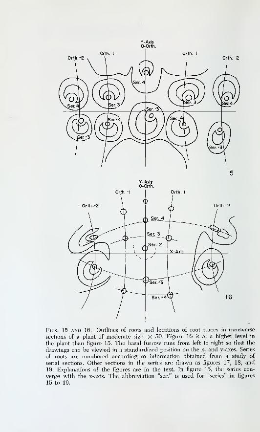
Orth.-2Orth. I
Orth. 2
Figs. 15 and 16. Outlines of roots and locations of root traces in transverse
sections of a plant of moderate size. X 50. Figure 16 is at a higher level in
the plant than figure 15. The basal furrow runs from left to right so that the
drawings can be viewed in a standardized position on the x- and y-axes. Series
of roots are numbered according to information obtained from a study of
serial sections. Other sections in the series are drawn as figures 17, 18, and
19. Explanations of the figures are in the text. In figure 15, the series con-
verge with the X-axis. The abbreviation "ser." is used for "series" in figures
15 to 19.
THE ROOT-PRODUCING MERISTEM 61
the validity of assigning members of the same series to alternate orthos-
tichies. If the assignment were made otherwise, the regularity of the
series would be destroyed. Figure 17, taken from a section near the
basal meristem, shows that the traces become crowded as their attach-
ments to the stele are approached. In figure 18 all of the traces and
primordia located in series 4 to —4 and in orthostichies 2 to —2 are
shown. The irregularities in the location of the sections of traces and
primordia are explained later.
In figure 18, series —1 contains primordia in the 0-orthostichy and in
orthostichies — 2 and 2. Of these three primordia the one on the 0-orthos-
tichy is the most mature. The other two are about equal to one another
in maturity. In series 1, which is comparable in degree of maturation
to series — 1, primordia are present in orthostichies — 1 and 1. These
two primordia are similar in degree of maturation, are less mature than
the centrally located primordium in series — 1 (the one on the 0-orthos-
tichy), and are more mature than the laterally placed primordia in series
— 1 (the ones on orthostichies —2 and 2). A similar situation prevails
in the series 2 and — 2. For series 2, the primordia that are in orthos-
tichies —2 and 2 are less mature than the primordium that is in the
0-orthostichy. They are also less mature than both the roots of series
— 2, which are in the orthostichies — 1 and 1. Figure 19 represents these
relationships. Each zigzag line connects primordia in a pair of series of
comparable maturation. Starting in each line with the primordium on
the 0-orthostichy, one can follow a single bidirectional sequence of a
decreasing degree of maturation because the 0-orthostichy contains the
most mature primordium in a pair of comparable series and because
the members of the pair of comparable series occupy alternate orthos-
tichies.
West and Takeda ( 1915 ) stated that all of the roots of a series are
initiated simultaneously. One may infer that the primordia of the same
series should be found in the same state of maturation because West
and Takeda explained the appearance of roots in an acropetal sequence
at the surface of the plant only on the basis of variations in the thick-
ness of the cortex. This inference is contradicted by my observations
and by those of Scott and Hill (1900). The latter authors interpreted
the maturation differences of root primordia of a series as indications
of age differences among the primordia of a series. But differences in
the maturation stage of the primordia of a series would not preclude the
possibility of simultaneous initiation of a series if the direction of the
maturation sequence were acropetal. If a simultaneous initiation of a
series is to be rejected, one must show that the extent of the youngest
series is less than that of the next older series, at least in some cases.
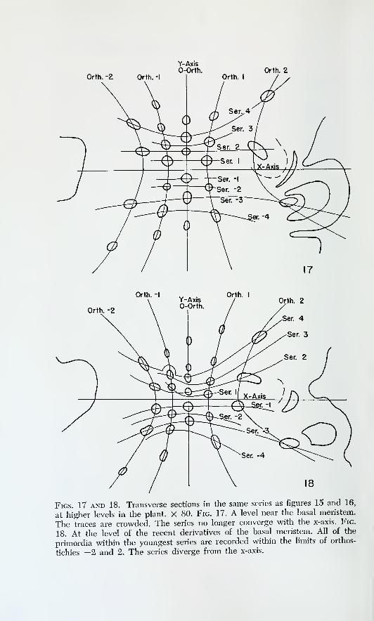
Orth. -2
Y-Axis0-Orth. Orth. 2
Orth. -IY-Axis
Orth. I
Orth. -2
Figs. 17 and 18. Transverse sections in the same series as figures 15 and 16,
at higher levels in the plant. X 80. Fig. 17. A level near the basal meristem.
The traces are crowded. The series no longer converge with the x-axis. Fig.
18. At the level of the recent derivatives of the basal meristem. All of the
primordia within the youngest series are recorded within the limits of orthos-
tichies —2 and 2. The series diverge from the x-axis.

THE ROOT-PRODUCING MERISTEM 63
Y-Axis0-Orth.
Orth. -Orth. I
Orth. -2Orth. 2
# = Ser. I and -I
0=Ser. 2 and -2
19Fig. 19. A portion of figure 18. The two zigzag lines (broken one connecting
empty spots; solid one connecting solid spots) trace bidirectional sequences
of decreasing maturation. Each sequence starts on the 0-orthostichy and in-
cludes members of the pair of series most comparable in maturation.
Scott and Hill (1900) represented roots in series with different lengths
in their text figure 11, and their explanation that the youngest series
drawn is shortest because it is incompletely initiated is feasible. But
their figure offers no conclusive evidence because it represents only an
isolated section and does not necessarily show the youngest series of
primordia in the plant.
Figure 20 is a graphic condensation of the data obtained from a series
of transverse sections of a plant of the same size as that in figures 13
to 16. Orthostichies — 2 to 2 are represented over the range of series
6 to — 6. The coordinates used in this figure are rectangular abstractions
of the actual spatial relationships of the roots in the original sections.
All techniques of observation are the same as in the previous example.
There is a greater number of series on the -j-y side of the graph than

6
5
4
3
2
CO-I
-2
-3
-4
-5
-6
6's
5's
w 4 s
Q> 3 s
2's
I's
T-2-1012Orthostichies
20
-2-10 12Orthostichies
22
0>
6 • •5 • • •4 • •3 •2 • •
•
-I
-2 •-3 • •-4 • • •-5 • •-6 • • •
6s
5's
(A 4'sa>
3'sCO
2's
-2-1 12 3
Orthostichies
21
-2-1 12 3
Orthostichies
23Figs. 20, 21, 22, and 23. Graphic condensations of data on the location of
root traces and root primordia in two plants. Fig. 20. Data from a plant sim-
ilar in size to that in figures 15 to 19. Series 1 and series —1 are incomplete.
Fig. 21. Data from a larger plant than that in figure 20. No 0-ortliostichy is
present. Series 3, series —2, series 1, and series —1 are incomplete. Fig. 22.
Rearrangement of data of figure 20, combining series of comparable matura-
tion in a single row (e.g., series 6 and series —6 combine to give the rowmarked 6's). This arrangement emphasizes the lateral (acropetal) extent of
the combined pairs of series. Fig. 23. Rearrangement of data of figure 21.
Similar to figure 22 in arrangement.
THE ROOT-PRODUCING MERISTEM 65
on the —y side. All of the available spaces on the graph are taken except
in the 1 and —1 series. The 3-2-3-2 alternation at successive y values
indicates that initiation of primordia has been regular for all of the roots
represented. The most important aspect of the graph is that series 1
is limited in its extent. Figure 21 is a condensation of the data from a
larger plant. The range of orthostichies represented is from —2 to 3,
and there is no 0-orthostichy. Again there are more series of roots on
the -fy side of the graph than on the —y side. Series 1 is limited to one
primordium. In this plant, however, series —2 and 3 are also limited
in extent, though not so much as series 1. Figures 22 and 23 illustrate
the data of figures 20 and 21, respectively, in another form, so that the
acropetal (lateral on the graph) extent of the combined pairs of series
of comparable maturation is emphasized.
The one primordium of series 1 in figure 20 is on the O-orthostichy.
This is the same orthostichy as that occupied by the most mature pri-
mordium in the combined pair of series 1 and — 1 of figure 19. Similarly,
the primordium in series 1 and orthostichy 1 of figure 21 is very near
the y-axis, although in this particular plant no orthostichy was actually
on the y-axis. Figure 19 may be thought of as a later stage in the initi-
ation of series than figures 20 and 21. Because the most basal orthostichy
is the first occupied in a series and also contains the most mature pri-
mordium in the more complete series (fig. 17), one may conclude that
the sequence of maturation coincides with the sequence of initiation
and that differences in maturation are reflections of differences in age.
It is now possible to recite a few of the observations and conclusions
obtained in this section. Each root is contained in an orthostichy and
in a series. The series are roughly parallel to the basal furrow, and the
orthostichies cross the basal furrow. The delimitation of series has been
described and applied, and the conclusion is reached that members of
a series occupy alternate orthostichies. Initiation of the members of a
series follows an acropetal bidirectional sequence that begins at the base
of the plant and proceeds at both ends toward the shoot apex. The mem-bers of two series of comparable age from opposite lobes may be inte-
grated into a single acropetal and bidirectional sequence of initiation
because the members of these series occupy alternate orthostichies. Notwo roots in the same orthostichy are of the same age. Successively
younger members of an orthostichy are displaced to opposite sides of
the basal meristem. The place where an orthostichy crosses the basal
meristem may be designated a site of root initiation.
The observations recorded above may be summarized in two ways.
With emphasis on the series, the initiation of a series may be said to
begin in the orthostichies at the base of the plant and to continue in a

66 THE DEVELOPMENTAL ANATOMY OF IsOCteS
bidirectional and acropetal sequence by contributions from alternate
sites of root initiation. However, the members of comparable series can
be integrated into a single sequence of initiation. Emphasizing the
orthostichies, one may state that each site of root initiation participates
in each acropetal wave or period of root initiation. The most basal site
is the first to produce a root in a new wave of initiation. A bidirectional
acropetal sequence follows, and alternate sites of root initiation con-
tribute roots to the same side of the basal furrow. The formation of roots
in comparable pairs of series is only incidental to the regular operation
of the sites of root initiation.
The formation of primordia at one side and the other of the basal
meristem allows the distribution of roots toward both lobes of the plant.
Whether the site of root initiation is one site that contributes primordia
to alternate sides or two sites that alternate in their activity cannot
be decided on morphological grounds. The region between opposing
root traces is uniform in its appearance until the advent of a new root
primordium. I cannot suggest what it is that provides for the regular
distribution of activity during successive waves of root initiation. How-ever, regularity is not complete in all plants. In a few specimens twoadjacent orthostichies were found operating in phase in their contribu-
tions to the two lobes of the plant. In a few other plants, there wereindications that root primordia had formed at the center of the basal
meristem rather than only at its sides. In these plants, remnants of root
traces were found embedded on the center line of longitudinal sections
taken in a plane at right angles to the basal furrow (pi. 16, C, at RRT).Ordinarily, root traces are displaced laterally. The traces along the
center lines of the sections were stretched vertically. No permanent im-
pairment of the activity of the sites of root initiation could be detected.
My observations on young plants are incomplete. Available data indi-
cate that where three orthostichies can be studied, two may operate in
phase while the third is out of phase. Each site of initiation alternates
between lobes in its contribution of new primordia, and the bidirectional
sequence of initiation of a series is acropetal with respect to the shoot
apex. The pattern suggested is the same as that given for the first six
roots of I. laciistris by Hofmeister (1862) where the third and fourth
roots are displaced toward opposite sides of the basal furrow (fig. 24).
The sequence is supposed to be repeated for additional roots (Hof-
meister, 1862), and the pattern may persist in the young sporophyte.
The transition to the mature pattern requires further study. Baldwin
( 1933 ) has reported a sequence of initiation for the first roots of I. engel-
manni which indicates that adjacent sites of root initiation are all in
phase, because the third and fourth roots are displaced to the same side

THE ROOT-PRODUCING MERISTEM 67
Primary Root Primary Root
Figs. 24 and 25. Schematic representations of the relative positions of the
first six roots of sporophytes of Isoefes. The locations are tentatively placed in
series and in orthostichies. Fig. 24. Location of the first six roots of I. laciisfris
according to Hofmeister (1862). The sites of root initiation of two orthos-
tichies may be in phase in the production of primordia on alteiTiate sides of
the basal meristem. Fig. 25. Location of the first six roots of /. engelmanni
according to Baldwin (1933). The sites of root initiation for all three orthos-
tichies may be in phase in the production of root primordia on alternate sides
of the basal meristem.
of the furrow as the first root (fig. 25). The variability of the patterns
of initiation in young plants has not been assayed and the exact relation-
ship of the pattern of initiation in young plants to the pattern in mature
plants is not known.
Attention may now be given to the curvature seen in series and orthos-
tichies in cross-sections taken at various levels of the plant (figs. 15-18).
One might expect that the lines connecting the series would converge
toward the x-axis in transverse sections, because of the bidirectional and
acropetal sequence of root initiation. The primordia are displaced from
the basal meristem after they are formed, and the oldest member of a
series should be displaced the farthest from the median plane. The ex-
pected spatial arrangement is found at low levels in the plant, but not
at high levels. To explain this variation, one needs to acknowledge that
the basal meristem lies in a plane that is convex when viewed from
below. The attachments of roots in the 0-orthostichy are encountered
first in serial transverse sections taken in an upward sequence in a plant,

THE DEVELOPMENTAL ANATOMY OF IsOCteS
High Level
-Y
X
X
D
\2
6'Intermediate
Level
Y
X
X
Low Level
B
26
Fig. 26. Schematic representation of the relative positions of successi\'e mem-bers of the same series. Trace 1 is in a more basal orthostichy (is closer to the
y-axis) than trace 2. A. Superimposition of successive members in longitudinal
view at right angles to the furrow plane. B, C, D. Appearance of the series
segment 1-2 in transverse sections at various levels represented in A. The in-
clination of segment 1-2 with respect to the x-axis varies with the level of
sectioning. For comparison, see figures 15 to 17.

THE ROOT-PRODUCING MERISTEM 69
and the attachments of the traces of other orthostichies are seen in
higher sections. Further, the root traces are curved within the plane that
contains all of the root traces of a given orthostichy (pi. 16, C). Suc-
cessive members of the same series are attached at different levels in
the plant. A schematic superimposition of the appearance of successive
members of a series, as seen in longitudinal sections perpendicular to
the furrow plane, is given as figure 26, A. Root 1 is closer to the median
plane than root 2. Transverse sections viewed in relation to x- and y-axes
would show the series segment 1-2 converging with the x-axis at low
levels (fig. 26, B), parallel to the x-axis at intermediate levels (fig. 26,
C), and diverging from the x-axis at a level near the attachment of the
trace of root 1 (fig. 26, D). The three inclinations of series segments
with respect to the x-axis are encountered in the above order in serial
sections of the plant taken in an upward sequence (figs. 16-18). The
series in figure 16 converge toward the x-axis, but in figures 17 and 18
there is an increasing tendency toward divergence from the x-axis.
In transverse sections observed in the standardized position, all orthos-
tichies except the 0-orthostichy diverge from the y-axis. The intercept of
two flat planes is a straight line, and the appearance of the 0-orthostichy
as a straight line in transverse sections (figs. 16-18) indicates that the
root traces of the 0-orthostichy are contained in a fiat plane. The more
acropetal orthostichies appear as curved lines in transverse sections;
their root traces are, therefore, contained in curved planes. In other
words, the growth vector parallel to the x-axis in a given transverse
section is not constant at different values of y. The data obtained for
the curvature of orthostichies in cross-sections cannot be easily used
in a quantitative description of the growth of the plant. A cross-section
represents an artificial plane of reference because the basal meristem
is curved. Transverse sections do not reveal different orthostichies at
comparable levels. Further, because the planes containing all but the
0-orthostichy are curved, the curvature of the line connecting mem-bers of the orthostichies in cross-sections varies according to the ob-
liquity of sectioning for a given orthostichy. Distortion of this kind
increases toward the ends of the basal meristem. As a final difficulty,,
the curved plane containing the root traces of an orthostichy is not
the surface of a cone but approaches a portion of the surface of the-
solid of rotation of a hyperbola or parabola. The rotational axis of
the solid that is bounded by the curved plane is oblique to the axis
of the plant, and the properties of the curved surface are not easily dis-
covered in transverse sections.
The morphological nature of the root-producing meristem. As von
Mohl (1845) has pointed out, if the first root of the sporeling has a

70 THE DEVELOPMENTAL ANATOMY OF IsOCteS
short and definite life span, the root-producing meristem cannot be
correctly designated the homolog of a main root. Bruchmann (1874)
has reported that the first root of the sporeling is short-lived. My observa-
tions support Bruchmann's, and the obvious conclusion is that the basal
meristem is not the main root of the plant. The alternative designations
available are: (1) the basal meristem is some type of primary meristem
other than a main root; (2) the basal meristem is a part of the cam-
bium. Throughout the following discussion, the terms hasal meristem
and root-producing meristem will be used as synonyms. The term lateral
m^eristem will be used to designate the portion of the cambium which
is located above the basal meristem.
Baldwin ( 1933 ) has reported that the basal meristem is organized after
two roots are present on the sporophyte. For ten or more plastochrons
following this event, the plant remains in a V2 phyllotaxy. The medianplane of sectioning perpendicular to the furrow contains all of the leaf
traces and the 0-orthostichy of root traces and exposes the site of root
initiation for the 0-orthostichy (pi. 13, A, at BM). The relationship be-
tween the basal meristem and the rest of the plant is obscured by the
root traces. However, in the plane of sectioning that coincides with the
furrow plane, a different picture is obtained. The basal meristem (pi. 13,
B, at BM) is found to be continuous with the lateral meristem (pi. 13, B,
at LM ) . This continuity is established during the origin of the basal andlateral meristems, and is very striking while the root-bearing portion
of the stele is small (pi. 17, A, at BM and LM). The basal and lateral
meristems originate in the periphery of the procambial strand that forms
across the axis of the plant during the formation of the traces to the
second leaf and root. Viewed in the furrow plane of sectioning, this
strand is circular in outline. Tracheids form at the center of the strand
and sieve elements form laterally. It has already been mentioned that
the lateral meristem originates outside of the first sieve elements. Di-
visions in the lower part of the strand produce the basal meristem and
complete the origin of the cambium. No direction or sequence in the
formation of the cambium has been observed in the materials avail-
able. The lateral and basal meristems of a young plant together form
a U in sections in the furrow plane (pi. 17, A), and repeated tangential
divisions produce derivatives inside and outside of the basal and lateral
meristems. Radial seriation of files of cells is evident when the plant
has several leaves.
It is reasonable to suggest that the similarity of origin and the con-
tinuity of the basal and lateral meristems indicate that these meristems
may be regarded as parts of the cambium. Thus, both the basal and the
lateral meristems are active in secondary growth. However, from the
earliest time of their activity the lateral and basal meristems produce

THE ROOT-PRODUCING MERISTEM 71
inner derivatives which mature as different types of cells (pi. 13, B).
The lateral meristem at first produces sieve elements on its inner face
(pi. 13, B, at SSE), whereas the basal meristem produces tracheary ele-
ments (pi. 13, B, at T). As the plant grows, the extent of the basal meri-
stem increases. The production of inner derivatives from the basal
meristem adds to the mass of the root-bearing portion of the stele. All
of these inner derivatives are regarded as secondary tissues.
As the plant ages, the lateral meristem may produce parenchyma and
tracheids on its inner face in addition to sieve elements (pi. 13, E). Thebasal meristem may produce parenchyma as well as tracheids on its
inner face, and a layering of these elements parallel to the basal meri-
stem is often noted (pi. 17, B; cf. Hofmeister, 1862, pi. 52, fig. 6). Thelateral layers of parenchyma are confluent with tracheary elements de-
rived from the basal meristem. When secondary xylem is formed inside
the lateral meristem, the tracheids differentiate among the cells of the
parenchyma layers. In view of the acropetal sequence of maturation of
root series, one may postulate that the differentiation of tracheids in the
derivatives of the lateral meristem is an expression of the acropetal
maturation of tracheary elements along the series of root traces at their
attachments to the stele. The continuation of this acropetal maturation
would produce tracheary elements in the lateral layers of parenchymabeyond the uppermost root primordia.
As the perimeter of the root-bearing portion of the stele increases andthe length of the basal meristem becomes greater, a conversion of initials
may occur at the ends of the basal meristem. The evidence for this con-
version comes from the interpretation of cell files that were derived
from cambial activity. Within radial files of cells that are inside the
limits of the basal meristem, the oldest cells of a file may be sieve ele-
ments, whereas sieve elements are absent near the initials in the meri-
stem (pi. 17, C, within brackets at FC). The old sieve elements (pi. 17,
C, at OSSE) were produced as derivatives of the lateral meristem. Thepresence of root primordia external to the initials of the cell files (pi. 17,
C, at HP) indicates that the initials were in the basal meristem at the
time of fixation and suggests that a conversion of initials has occurred.
This conversion allows the root-producing meristem to progress toward
the shoot apex. The acropetal progression of the root-producing meri-
stem makes it possible to obtain transverse sections in which the attach-
ments of root traces appear inside the limits of the lateral meristem and
outside secondary sieve elements (pi. 18, A, B, at BBT). Again, it maybe noted that these tracheary elements at the bases of root traces are
confluent with a layer of parenchyma inside the lateral meristem (pi. 18,
B, at BBT and Par). The appearance of a transverse section such as that
in plate 18, B, is very similar to the appearance of a cross-section of

72 THE DEVELOPMENTAL ANATOMY OF IsOCteS
a stem of Stylites at a level where the so-called "Wurzelstele" is present
(Rauh and Falk, 1959b). Judging from the description given by Rauh
and Falk (1959b), I believe that the acropetal formation of the root
stele of Stylites involves the same processes as the acropetal progression
of the root-producing meristem of Isoetes. The course of differentia-
tion of the inner derivatives of the cambium is altered from one that
can produce sieve elements to one that produces parenchyma cells and
tracheary elements but no sieve elements. At the same time, the outer
derivatives of the meristem attain the ability to form root primordia. Thecauses of these changes are not known.
The idea that the basal meristem is a primary meristem (Lang, 1915a;
West and Takeda, 1915) may be better understood if attention is turned
to the appearance of the root-bearing portion of the stele in a longitu-
dinal section taken at right angles to the basal furrow. Such a section
may contain an orthostichy of roots (pi. 16, C), or may pass between
orthostichies (pi. 18, C). In the first case, the arrangement of root traces
is like the arrangement of leaf traces while the plant has a V2 phyllotaxy.
In the second case, the relation of the basal meristem to mature tissues
in the lower portion of the plant is similar to the relation of the shoot
apex to mature tissues in the upper portion of the plant (cf. pis. 7, A,
and 18, C). The comparison between the basal meristem and a pri-
mary meristem is attractive if attention is confined to longitudinal sec-
tions perpendicular to the furrow plane. But information from transverse
sections should be examined before the comparison is accepted as valid.
In plate 18, B, near the top, there is no need for hesitation in designating
the layers of clear cells as layers of secondary sieve elements (SSE).
Toward the right, however, root traces are present, indicating that at
that location the initials of the lateral meristem have been converted to
initials of the basal meristem. Were it not for these root traces, the outer-
most layer of phloem would be continuous at the right in the photo.
At a higher level in the plant, the phloem layer is continuous around
the right side of the stele and at that level would be designated sec-
ondary throughout its extent. At the level of sectioning in plate 18, B,
the phloem adjacent to the root traces must be designated primary if
the root-producing meristem is a primary meristem (cf. West and
Takeda, 1915). Because the basal meristem is greatly extended in the
form of a ribbon, the transition between primary and secondary phloem
takes place along a line parallel to the edge of the basal meristem. Start-
ing with one horn of the root stele, this line descends in an arc, convex
from below, and rises to the other horn of the stele, crosses to the other
side of the meristem, and descends in another arc to rise again to the
first horn of the stele, and crosses the end of the basal meristem back
THE ROOT-PRODUCING MERISTEM 73
to the starting point. In the median plane of sectioning at right angles
to the basal furrow (pi. 18, C), the transition from secondary to pri-
mary phloem oflFers no conceptual difficulties. At the ends of the ribbon-
shaped meristem, however, the conversion of initials leads to difficulties
of description. If the concept that the basal meristem produces primary
tissues is maintained, the conversion of initials requires that initials pro-
ducing secondary vascular tissues revert to the production of primary
tissues at the ends of the basal meristem. Likewise, the production of
secondary cortex must revert to the production of primary cortex.
In choosing the appropriate designation for the root-producing meri-
stem, it is reasonable to assert that one should strive for a concept which
allows a unified description of the growth of the entire plant. Lang(1915a) argued that the conversion of initials at the boundary between
the lateral and basal meristems does not "prove " that these meristems
are both part of the cambium. Certainly this relationship cannot be
proved in a rigorous sense. When the basal meristem is regarded as a
part of the cambium, however, terminological difficulties are avoided,
and a clear description of the growth results. If one adopts this concept,
the processes of growth in the basal portion of the plant can be sum-
marized as follows.
Early in the life of the plant, the lateral and basal meristems begin
their activity together as parts of the cambium. The differentiation of
their respective derivatives is different from the first stages of cambial
activity. The basal, or root-producing, part of the cambium does not
produce sieve elements on its inner face, but the lateral portion of the
cambium does. The first several roots form in close association with the
first several leaves (Hofmeister, 1862; Bruchmann, 1874), but soon root
primordia begin to form at the bases of older roots (Hofmeister, 1862).
Sites of root initiation are established in the basal meristem and orthos-
tichies of roots are produced. The inner derivatives of the basal meristem
add to the root-bearing portion of the stele, and these derivatives differ-
entiate as tracheids and parenchyma cells which are attached to the
bases of the root traces. Root primordia are initiated in the outer deriva-
tives of the basal meristem (Scott and Hill, 1900) in the secondary cortex.
Because the newest roots are located at the edge of the basal meristem,
the phloem matures toward the edge of the meristem in conjunction
with the formation of sieve elements in the procambial root traces.
The arrangement of leaves changes into a spiral as the plant ages.
Hence, the leaf-bearing portion of the stele becomes radially symmet-
rical. The arrangement of roots at a site of root initiation remains in a
y-2. "rhizotaxy," and orthostichies are formed. Hence, the root-bearing
portion of the stele does not become radially symmetrical. The ends of
74 THE DEVELOPMENTAL ANATOMY OF IsoetCS
the ribbon-shaped basal meristem advance acropetally, so that at these
locations the inner derivatives of the cambium cease to differentiate as
sieve elements, while the outer derivatives of the cambium attain the
ability to differentiate as root primordia.
If the basal meristem is regarded as a part of the cambium, the roots
of Isoetes may be thought of as adventitious. Liebig ( 1931 ) argued that
if the roots of Isoetes are actually adventitious, they should arise in
association with leaves, as do the adventitious roots of other vascular
cryptogams. A root primordium in the mature plant is organized at
the edge of the basal meristem, at the base of an older root trace, but
there is no reason why the appellation adventitious should be any less
applicable than if the root had been organized at the base of a leaf
trace. The continued formation of root primordia along a particular part
of the cambium allows a convenient mechanism for the direct and simple
attachment of the collateral traces of the roots. The xylem of the root
traces attaches directly to the xylem of the stele, and the phloem, which
in all root traces is abaxial with reference to the median plane of the
furrow, connects directly to the secondary sieve elements produced
toward the inside of the lateral meristem.
For the root-bearing portion of the stele, the designation of a periph-
eral and central portion is not possible, even in large plants, and it
appears that all of the tracheids of the root-bearing portion of the stele
are differentiated with positional relationship to root traces. Although
the shortness of the tracheids in this part of the stele makes their orien-
tation somewhat obscure, the layering found in the inner derivatives of
the basal meristem (pi. 17, B) is an indication of the relationship of
the tracheids to the bases of the series of root traces.
The difference between the kinds of derivatives found inside the basal
and lateral meristems does not destroy the continuity of the cambiumbetween the lateral and basal locations. Examples from other species
may be cited to indicate that a cambium may produce both xylem and
phloem from its inner surface, and in varying patterns (Esau, 1960, p.
253). In these species, the derivatives of a single initial in the cambiummay differentiate as phloem for a limited time and then as xylem, so
that a parallel for the conversion of initials at the ends of the basal meri-
stem is available. It must be recognized, however, that the pattern of
root initiation and the acropetal progression of the root-producing meri-
stem of Isoetes are quite orderly. For this reason, it is profitable to dis-
tinguish between the basal meristem and the lateral meristem, even
though they are parts of the same cambium. At any given time, the root-
producing meristem may be treated as a bounded portion of the cam-
bium, recognizable on the basis of the presence of root primordia at its

THE ROOT-PRODUCING MERISTEM 75
external face. The mere presence of tracheary elements inside the cam-
bium does not identify that portion of the cambium as the basal meristem
because the lateral meristem also produces derivatives which differen-
tiate as tracheids, and these are in layers confluent with tracheids in the
root-bearing portion of the stele. The presence of elements adjacent to
the internal face of the cambium does identify that portion of the cam-
bium as the lateral meristem, because the inner derivatives of the basal
meristem do not differentiate as sieve elements.
Rauh and Falk ( 1959b ) compared the structure of Isoetes and Stylites.
They concluded that the root-bearing portions of the steles of the two
genera are directly comparable. I agree with their conclusion. How-
ever, their suggestion that the basal and lateral meristems are not di-
rectly and mutually related at their origin in either genus cannot be
accepted, because it makes an unnecessary and artificial distinction be-
tween the growth of the upper and lower portions of the plant. Their
idea is based on the study of the acropetal progression of the "Wurzel-
stele" along the axis of the plant. The sporophyte of Stylites decays from
the base upward as the plant ages, and it is not difficult to acknowledge
that the relationship of the "Wurzelstele" to the lower extremity of the
leaf-bearing portion of the stele is obscure in a mature plant. Rauh and
Falk (1959a) discussed the sporeling of Stylites, but did not describe
the earliest activity of the root-producing meristem.
The apparent separation of the stele of Isoetes into leaf-bearing and
root-bearing portions has led to the designation of the lower portion of
the plant as a rhizomorph ( West and Takeda, 1915 ) and as a rhizophore
( Lang, 1915a; Foster and Gifford, 1959, p. 174 ) . I can offer no objection
to either of these terms if they are used in a descriptive sense and if the
implication that the basal meristem is a primary meristem is avoided.
The whole sporophyte, exclusive of leaves and external roots, has been
called a stem, a stock (Lang, 1915a), and a corm (Foster and Gifford,
1959, p. 172). All of these are suitable descriptive terms.
SUMMARY AND CONCLUSIONS
The roots of Isoetes are arranged in series and in orthostichies. Anorthostichy is produced by the formation of root primordia on a site of
root initiation and by the alternate displacement of primordia to one
side and the other of the basal meristem. Each wave of root initiation
is bidirectional and is acropetal with respect to the shoot apex, because
it begins in the most basal orthostichy. During each wave of root initi-
ation, alternate sites of root initiation contribute primordia to the same
side of the basal meristem. Two series of comparable age are thus pro-

76 THE DEVELOPMENTAL ANATOMY OF IsOetCS
duced, and these series are displaced to opposite lobes of the plant. The
two comparable series do not contain sets of roots of exactly equivalent
ages.
The basal, or root-producing, meristem and the lateral meristem are
best regarded as parts of a cambium. It is possible to distinguish be-
tween the basal and lateral meristems on the basis of the differentiation
of their respective derivatives. These meristems originate together, as
parts of the same cambium, and during ontogeny portions of the lateral
meristem may be added to the basal meristem. An acropetal progression
of the ends of the basal meristem results from this conversion of initials.
A parallel development occurs in Stylites (Rauh and Falk, 1959b), but
in Stylites the acropetal progression is much more pronounced than in
Isoetes. The root-bearing portions of the steles of Isoetes and Stylites are
probably homologous ( cf . Rauh and Falk, 1959b )
.
THE APICAL MERISTEM OF THE ROOT
REVIEW OF THE LITERATURE
The process of root initiation. The apical meristem of the root is or-
ganized during root initiation. Little has been reported on root initiation
in mature plants, but Bruchmann (1874) has given a detailed account
of the initiation of the first several roots of the sporeling. He considered
the first root to be exogenous and all the later roots endogenous. His
decision was based on the locations of the first divisions associated with
root formation. Bruchmann recognized histogens in the root apex andasserted that the differentiation of the initials of these histogens proceeds
in basipetal order. The first initial recognized is that of the calyptro-
dermatogen. For the first root this initial is set off by a periclinal division
in the superficial layer of the plant, but for subsequent roots the di-
vision is in hypodermal tissue. The course of further differentiation of
initials of the second and later roots depends on the distance between
the initial of the calyptro-dermatogen and the existing vascular tissues
of the plant. If sufficient cell layers are present at the origin of the first
initial, the other initials differentiate directly from available cells. If
only one or two layers are present between the initial of the calyptro-
dermatogen and the vascular tissues, divisions occur to produce the
number of cell layers required for the differentiation of the initials of
the remaining histogens.

THE APICAL MERISTEM OF THE ROOT 77
Farmer (1890) agreed that the first root is exogenous. Campbell
(1891) suggested that the first root is a primary organ and also re-
corded that the second root is recognized as a mass of dividing cells
before its characteristic organization is obtained. Scott and Hill ( 1900
)
reported that root primordia are differentiated in the external deriva-
tives of the basal meristem but gave no further details. Hofmeister
(1862) stated that the original direction of the axis of the root pri-
mordium in large plants is oblique to the vertical axis of the plant, and
Liebig (1931) reported that the primordia are seldom found in a ver-
tical position. Hofmeister (1862) also reported that the root traces are
curved, so that they form an arc which is concave with respect to the
median plane of the furrow. West and Takeda (1915) have asserted
that the roots penetrate the cortex actively, but Lang (1915a) has
stated that the roots are carried to the surface by the growth of the
cortex and begin active apical growth after the surface is reached. Onthis basis, Lang (1915a) suggested a similarity between the roots of
Isoefes and the leaves, which originate exogenously.
The organization of the apical meristem of the root. Hofmeister (1862)
believed that the root of Isoetes grows with an apical cell. Naegeli and
Leitgeb (1868) supported Hofmeister's opinion, but they could adduce
no positive evidence for the existence of the apical cell. Instead, they
came to their conclusion by eliminating, to their own satisfaction, the
other possibilities they could suggest. However, they also considered
that the plerome of the root grows with its own initial. Farmer (1890)
severely criticized Naegeli and Leitgeb (1868), charging that their ob-
servations were not consistent with their hypothesis. Farmer suggested
that the existence of a plerome initial precludes the existence of an
apical cell because the latter should act as the initial of all tissues.
Naegeli and Leitgeb (1868) had suggested, however, that the apical
cell may be rather inactive.
Bruchmann ( 1874 ) reported the existence of several histogens in the
root apex of Isoetes: a calyptro-dermatogen, an outer periblem, an inner
periblem, and a plerome. The calyptro-dermatogen and the outer and
inner periblem layers were supposed to be supplied by the activity of
their respective layers of initials, and the plerome was said to grow with
a single initial. Farmer (1890) asserted that the boundary between the
outer cortex and the inner cortex is sharp and that this boundary can
be traced through the initials of the root. He criticized Bruchmann
( 1874 ) for not recognizing the continuity between the outer cortex, epi-
dermis, and root cap. Farmer recognized a single initial for the pro-
cambium (plerome), a layer of initials for the inner cortex, and a layer
of initials furnishing cells for all other regions of the root. Campbell

78 THE DEVELOPMENTAL ANATOMY OF IsOCteS
( 1891 ) reported that the plerome has several initials and that an ad-
ditional layer of initials furnishes cells to all other tissues of the root.
Mager (1907) adopted Farmer's (1890) concept of the organization of
the root apex, whereas Liebig (1931) followed Bruchmann (1874).
The concept of the organization of the root of Isoetes outlined by
Kienitz-Gerlolf (1881) stands apart from all others. He maintained that
the root has an undifferentiated meristem. In other words, the initials
cannot be designated in terms of the mature tissues of the root. It is
only away from the region of the initials that the distinctions amongtissues become obvious. He oflFered many figures in an effort to refute
the concept of Bruchmann ( 1874 ) , and some of these were said to showthat the distinction between the plerome and the inner periblem can-
not be maintained. The concept of Kienitz-Gerloff also came under the
scrutiny of Farmer (1890). Farmer charged that the evidence proposed
by Kienitz-Gerloff cannot be accepted because the figures presented
show the procambial strand on the axis of the root. Farmer maintained
that the longitudinal plane of sectioning at right angles to that in
Kienitz-Gerloff's figures (i.e., the plane that would show the procambiumeccentrically placed) is the only one that shows the proper relationship
of the plerome to the other tissues.
The dichotomy of the root. Bruchmann (1874) asserted that in the
development of the dichotomy of the root the plerome initial takes the
initiative by dividing longitudinally with reference to the axis of the
root. Tlie first dichotomy occurs in the plane which shows the procam-
bium on the axis of the root. After the plerome initial divides, the initials
of the other histogens organize themselves with respect to the twonewly formed plerome initials. Sister apexes remain in a common root
cap for some time. Naegeli and Leitgeb ( 1868 ) have reported that fifth-
order dichotomies may be found within a single root cap. Because
branching is cruciate and sister apexes diverge from each other at a
slight angle from the axis of the root, longitudinal sections which showthe apexes in proper relationship to mature tissues are difficult to ob-
tain. Naegeli and Leitgeb offered a figure of a section of a root in which
dichotomies had occurred and stated that the figure does not disclose
the apexes of the root. However, Bruchmann (1874) chose to cite this
figure (Naegeli and Leitgeb, 1868, pi. 19, fig. 12) as a correct repre-
sentation of the organization of the apical meristem of the root.
OBSERVATIONS AND DISCUSSION
The process of root initiation. I have not made sufficient observa-
tions on young sporophytes to allow any statement on the origin of the

THE APICAL MERISTEM OF THE ROOT 79
first root. I have observed the origin of the second root by the enlarge-
ment and division of hypodermal cells at the base of the second leaf
(pi. 1, D, at R., ), but my observations are too incomplete to reveal the
relationship of these first-recognized cells to the final organization of
the meristem of the root. Greater attention has been given to the initia-
tion of roots in plants with several to many leaves. A plant of this size
has a well-established basal meristem, and root primordia are differen-
tiated in the external derivatives of the basal meristem. The time at
which the apical meristem of the root primordium is organized in re-
lation to the length of the primordium varies with the thickness of the
cortex. In young plants, where the cortex is thin, the apical meristenr
is organized before the primordium becomes extended. In older plants,
where the cortex outside of the basal meristem is composed of manylayers of cells, the primordium may increase in length before any ac-
tivity of an organized apical meristem is apparent (pi. 18, D). This
variation in the time of organization of the meristem is consistent with
the observation that the first few roots, which are formed close to the
surface of the sporeling, have organized meristems directly after they
are first recognizable and before any extension occurs (Bruchmann,
1874).
A primordium first appears as a group of cells with deeply staining
cytoplasm and chromatic nuclei. This group of cells is located at the
edge of the basal meristem with the axis of the group slightly inclined
away from the axis of the plant. The plastids of a newly differentiated
primordium have the condensed form of the undifferentiated plastids
found in the superficial layer of the shoot apex (pi. 5, A, at UPl). Else-
where in the basal meristem, the plastids resemble those of the internal
cells of the apex (pi. 5, A, B, at Pi). The starch content of a cell in a
young primordium is much smaller than that of a cell in the surrounding
ground tissue (pi. 18, E).
A procambial strand is differentiated in the growing primordium. This
event may occur before the complete organization of the apical meri-
stem (pi. 18, D). The procambial trace is usually curved, so that it is
concave with respect to the median furrow plane. The tip of the trace
is very close to the median furrow plane of the plant and is displaced
practically straight downward along the central plane, but the attach-
ment of the trace is displaced laterally. The curvature of the trace is
maintained as the trace is extended. The roots do not penetrate the
cortex actively until the tip is close to the surface. In young plants,
the penetration may occur soon after the primordium is organized. Thetip of a primordium is carried outward along the median plane, so that
the root breaks out of the cortex within the basal furrow. After a root

80 THE DEVELOPMENTAL ANATOMY OF IsOCteS
emerges, lateral displacement is very pronounced. Displacement at the
surface of the plant exceeds that nearer the attachment of the trace, so
that the original curvature of the trace is reversed as the trace ages.
The organization of the apical meristem of the root. I have observed
the apical meristems of the following types of roots and root primordia:
(1) the first root, unextended, (2) the first root, extended, (3) root pri-
mordia in young plants, (4) root primordia in old plants, (5) roots of
mature plants after emergence from the cortex but before complete
extension, (6) roots of mature plants after complete extension. The
information obtained from these various sources agrees very well, so
it appears that the organization of root apexes in 1. howellii does not
vary greatly during ontogeny. The apical meristem can be described in
terms of the differentiated tissue regions of the root. These regions are
the root cap, the epidermis, the outer cortex, the inner cortex, and the
procambium (pi. 19, A). The procambium is near the side of the root
that faces the furrow. The adaxial side of the procambium differentiates
as xylem. The inner cortex is thickest on the abaxial side of the root
but breaks down to form a large cavity in this location (cf. fig. 14).
The eccentricity of the procambium and inner cortex is manifest to the
region of the initials.
In agreement with Farmer (1890), I have found that the most definite
boundary among tissues occurs between the inner cortex and the outer
cortex. This boundary can be followed through the region of initials
(pi. 19, B, C, D, between OC and IC ) . The outer cortex, epidermis, and
root cap have a common origin. In contrast to Naegeli and Leitgeb
(1868), Bruchmann (1874), Farmer (1890), Mager (1907), and Liebig
( 1931 ) , I find that the procambium does not have its own permanent
initials. The distinction between the initials of the procambium and
those of the inner cortex is lacking in most of the apexes examined. Tothis extent, my observations agree with those of Kienitz-Gerloff (1881),
but the concept of an undifferentiated meristem cannot be accepted
because the boundary between the inner cortex and the outer cortex
can be traced through the initials. Although Campbell (1891) reported
that the procambium has several initials of its own, his figures reveal that
he did not distinguish between the procambium and the inner cortex in
his preparations. Therefore, the cells he identified as the "initials of
the plerome" are actually the initials common to the inner cortex andprocambium.
In some apexes, the procambium does not appear continuous with
the initials of the inner cortex. This may indicate that the organization
of the apex varies among roots or that the procambium may grow for
certain periods with its own initials, which are displaced by new con-

THE APICAL MERISTEM OF THE ROOT 81
tributions from the cells generating the inner cortex. These contributions
may be related to the dichotomy of the root apex.
There is no indication of a quiescent center (Clowes, 1959) in the
roots of Isoetes. Division figures are observed in the initials. Divisions
are observed near the apex even in extended roots. Bruchmann (1874)
has reported an early curtailment of apical growth in the first root of
7. lactistris, but my observations on I. howellii do not support his find-
ings.
The dichotomy of the root. Several stages of the dichotomy of the
apex of the root have been observed. Plate 19, E and F, show two of
these stages. The following account is tentative because the early stages
of dichotomy were observed in relatively few roots. The first dichotomy
occurs near the time the root emerges from the cortex. At this time, a
distinction between the procambium and the initials of the inner cortex
may be possible. The distal cells of the procambium and of the inner
cortex cease to act as initials. The procambium becomes broad and
blunt at the tip (pi. 19, E, at Pr), and the distal cells of the inner cortex
subdivide (pi. 19, E, below tip of procambium). Two or more cells
laterally placed among the initials of the inner cortex maintain their
size (pi. 19, E, at arrows).
These cells enlarge and divide so that their derivatives near the axis
are contributed to the procambium. These derivatives become the new
temporary initials of the procambium. At this stage in the dichotomy,
the procambium appears to be continuous with the initials of the inner
cortex. The configuration of the apexes in plate 19, F, can account for
the configuration of the apex in plate 19, C and D. Therefore, it is rea-
sonable to assume that some of the longitudinal sections which show an
apparent continuity of the procambium with the initials of the inner
cortex show the apex after the initiation of a dichotomy. Other dichoto-
mies follow soon after the first. The production of new temporary initials
for the procambium by the initials of the outer cortex allows multiple
dichotomies to occur with the production of relatively few new layers
of cells. The production of new apexes within one root cap does not
contribute appreciably to the elongation of the root.
Bruchmann (1874) believed that the procambium takes the initiative
in a dichotomy. My observations do not contradict this idea, but it is
also possible that the initials of the inner cortex and the initials of the
procambium act together in the dichotomy. Bruchmann's figure of a
dichotomy (1874, fig. 20) represents a stage earlier than that given here
in plate 19, E. In his figure, the distal cells of the inner cortex have not
yet divided into smaller cells. Bruchmann believed that the procam-
bium is distinct throughout the life of the root. He did not describe

82 THE DEVELOPMENTAL ANATOMY OF IsoeteS
any reorganization of the apex during the dichotomy. Farmer (1890)
reported that his observations did not conflict with the ideas forwarded
by Bruchmann (1874), but gave no details on his own observations.
SUMMARY AND CONCLUSIONS
Root primordia are differentiated in the external derivatives of the
basal meristem. Elongation of a primordium may or may not precede
the organization of its apical meristem, depending on the thickness of
the cortex outside of the basal meristem. Root primordia do not actively
penetrate the cortex until they are very close to the surface of the plant.
The apical meristem of the root is composed of a layer of initials which
gives rise to the cells of the outer cortex, epidennis, and root cap, and
of a group of initials common to the inner cortex and the procambium.
The distal cells of the procambium may function as temporary initials,
but are replaced during a dichotomy and possibly at other times.
GENERAL SUMMARY AND CONCLUSIONS
Synoptic treatments of the principal observations and conclusions are
given at the end of each section of this report, and there is no need to
repeat these summaries here. Instead, a few closing remarks will be
made. In relation to the previous literature on Isoetes, this report is
synthetic to the extent that it incorporates the concepts of many workers
into one description. The literature on Isoetes spans a century and a
half, and it is not surprising that many pertinent observations have been
made. I have tried to develop the ideas available in the literature andto determine which observations are common to the many descriptions
of Isoetes.
Principal among the concepts which have been treated with different
perspectives are the origin of the cambium and the nature of the basal
meristem. I recognize that the choice of perspectives in the treatment
of some aspects of these concepts is subjective, and that descriptions
can be logically constructed within more than one perspective. Theperspectives chosen in this study facilitate description of the wholeplant. All of the meristematic tissues of the corm have been considered
together, and the consequences of a concept in one part of the plant are
applied to the description of other parts of the plant. The perspectives
employed in this report also yield a description which facilitates com-

GENERAL SUMMARY AND CONCLUSIONS 83
parison of Isoetes with other plants. Here, the concepts of primary vas-
cular differentiation and of the primary plant body are most strongly
affected.
The use of a particular concept may provide a terminology which
avoids many semantic difficulties, but the benefits do not end there.
Description is the first stage in obtaining an understanding of growth
and form. The benefits of description are enhanced if the perspectives
adopted are styled to reveal the most instructive analogies and homol-
ogies. From this standpoint, it is best to regard the tissue underlying
the shoot apex as a histologically undifferentiated tissue which furnishes
cells to the procambium and the ground meristem. It is best to regard
the early tangential divisions around the procambial xylem core as a
part of the growth of the procambium. Also, there is ample justification
for regarding the lateral meristem and the basal meristem as parts of
one cambium. Each of these ideas has appeared in the literature. Theyoccur together in this report as the combination of concepts which are
supported by my observations.
84
LITERATURE CITED
Amstutz, E.
1957. Stylites, a new genus of Isoetaceae. Ann. Mo. bot. Gdn. 44:121-123.
Baldwin, W. K. W.1933. The organization of the young sporophyte of Isoetes Engelmanni
A. Br. Trans, roy. Soc. Can., ser. 3, sec. 5, 27:1-30.
Bhambie, S.
1957. Studies in pteridophytes. I. The shoot apex of Isoetes coromandeliana
L. J. Indian bot. Soc. 36:491-502.
Braun, a.
1847. Weitere Bemerkungen iiber Isoetes. Flora, Jena 30:33-36.
Bruchmann, H.
1874. Ueber Anlage und Wachsthum der Wurzeln von Lycopodium undIsoetes. Jena. Z. Natui-w. 8:522-578.
Buvat, R.
1952. Structure, evolution et fonctionnement du meristeme apical de quel-
ques dicotyledones. Ann. Sci. nat., botan., ser. 11, 13:199-300.
Campbell, D. H.1891. Contributions to the life-history of Isoetes. Ann. Bot., Lond. 5:231-
258.
Cheadle, V. I., E. M. GiFFORD, Jr., and K. Esau1953. A staining combination for phloem and contiguous tissues. Stain
Tech. 28:49-53.
Clowes, F. A. L.
1959. Apical meristems of roots. Biol. Rev. 34:501-529.
CoNN", H. J., M. A. Darrow, and V. M. Emmel1960. Staining procedures used by the biological stain commission. 2nd ed.
Baltimore, Md.: Williams and Wilkins Co. 289 pp.
LITERATURE CITED 85
Currier, H. B.
1957. Callose substance in plant cells. Am. J. Bot. 44:478-488.
Currier, H. B., and S. Strugger1956. Aniline blue and fluorescence microscopy of callose in bulb scales
of Allium cepa L. Protoplasma 45:552-559.
Da\'es-Dujeu, M.1957. Etude du point vegetatif et des meristemes intercalaires de YEphedra
monostachya. Rev. gen. Bot. 64:41-75.
Eames, a. J.
1936. Morphology of vascular plants. Lower groups. New York: McGraw-Hill, 433 pp.
Esau, K.
1943. Ontogeny of the vascular bundle in Zea mays. Hilgardia 15:327-368.
1953. Plant anatomy. New York: John Wiley and Sons. 735 pp.1954. Primary vascular differentiation in plants. Biol. Rev. 29:46-86.
1960. Anatomy of seed plants. New York: John Wiley and Sons. 376 pp.Esau, K., V. I. Cheadle, and E. M. Gifford, Jr.
1953. Comparative structure and possible trends of specialization of the
phloem. Am. J. Bot. 40:9-19.
ESCHRICH, W.1956. Kallose. Protoplasma 47:487-530.
Farmer, J. B.
1890. On Isoetes laciistris L. Ann. Bot., Lond. 5:37-62.
Foster, A. S.
1934. The use of tannic acid and iron chloride for staining cell walls in
meristematic tissue. Stain Tech. 9:91-92.
Foster, A. S., and E. M. Gifford, Jr.
1959. Comparative morphology of vascular plants. San Francisco, Calif.:
Freeman and Co. 555 pp.Gifford, E. M., Jr.
1954. The shoot apex in angiosperms. Bot. Rev. 20:477-529.Click, D.
1949. Techniques of histo- and cytochemistry. New York: Interscience Pub-lishers. 531 pp.
Hegelmaier, F.
1874. Zur Kenntniss einiger Lycopodinen. Bot. Ztg. 32:481-488, 497-505,513-524.
HOFMEISTER, W.1862. On the germination, development, and fructification of the higher
Cryptogamia, and on the fructification of the Coniferae. Ray Soc.
Publ. 22. 506 pp.JOHANSEN, D. A.
1940. Plant microtechnique. New York: McGraw-Hill. 523 pp.Kessler, G.
1958. Zur Charakterisieiiing der Siebrohrenkallose. Ber. schweiz. bot. Ges.
68:5-43.
Kienitz-Gerloff, F.
1881. t)ber Wachsthum und Zelltheilung und die Entwickelung des Em-bryos von Isoetes lacitstris. Bot. Ztg. 39:761-770, 785-795.
86 THE DEVELOPMENTAL ANATOMY OF IsoetCS
La Motte, C.
1937. Morphology and orientation of the embryo of Isoetes. Ann. Bot. (n.s.)
1:695-716.
Lamoureux, C. H.
1961. Comparative studies on phloem of vascular cryptogams. Unpublished
Ph.D. dissertation, Univ. of California.
Lance-Nougarede, a., and J. Loiseau
1960. Sur la structure et le fonctionnement du meristem vegetatif de quel-
ques Angiospermes aquatiques ou semi-aquatiques depourvues de
moelle. C. R. Soc. Biol., Paris 250:4438-40.
Lang, W. H.1915a. III. Studies in the morphology of Isoetes. I. The general morphology
of the stock of Isoetes lacustris. Mem. Proc. Manchr. Lit. Phil. Soc.
59:1-28.
1915b. III. Studies on the morphology of Isoetes. II. The analysis of the
stele of the shoot of Isoetes lacustris in the light of mature structure
and apical development. Mem. Proc. Manchr. Lit. Phil. Soc. 59:29-57.
LlEBIG, J.
1931. Erganzungen zur Entwicklungsgeschichte von Isoetes lacustris L.
Flora, Jena 125:321-358.
Mager, H.
1907. Beitrager zur Anatomie der physiologischen Scheiden der Pterido-
phyten. Bibl. bot., Stuttgart 14 (66): 1-58.
Mazia, D., p. a. Brewer, and M. Alfert1953. The cytological staining and measurement of protein with mercuric
bromphenol blue. Biol. Bull., Woods Hole 104:57-67.
MOHL, H. VON1845. Ueber den Bau des Stammes von Isoetes lacustris. In Vermischte
Schriften botanischen Inhalts. Tubingen: L. F. Fues. Pp. 122-128.
Naegeli, C, and H. Leitgeb1868. Entstehung und Wachstum den Wurzeln. Beitr. wiss. Bot. 4.
Ogura, Y.
1938. Anatomie der Vegetationsorgane der Pteridophyten. In Linsbauer,
Handbuch der Pflanzenanatomie. Berlin: Gebriider Bomtrager. 476
pp.Paolillo, D. J., Jr.
1962. The plastids of Isoetes howellii. Am. J. Bot. 49:590-598.
Paolillo, D. J., Jr., and E. M. Gifford, Jr.
1961. Plastochronic changes and the concept of apical initials in Ephedraaltissima. Am. J. Bot. 48:8-16.
Parke, R. V.
1959. Growth periodicity and the shoot tip of Abies concolor. Am. J. Bot.
46:110-118.
Parker, J., and D. E. Philpott1961. The ultra-structure of sieve plates of Macroctjstis pyrifera. Bull. Tor-
rey bot. CI. 88:85-90.
POPHAM, R. A.
1951. Principal types of vegetative shoot apex organization in vascular
plants. Ohio J. Sci. 51:249-270.

LITERATURE CITED 87
1960. Variability among vegetative shoot apices. Bull. Torrey bot. CI. 87:
139-150.
Rauh, W., and H. Falk1959a. Stylites E. Amstutz, eine neue Isoetacee aus den Hochandern Perus.
1. Teil: Morphologic, Anatomic, und Entwicklungsgeschichte der
Vegetationsorgane. S.B. hcidclberg. Akad. Wiss. 1959:1-83.
1959b. Stylites E. Amstutz, eine neue Isoetacee aus den Hochandern Perus.
2. Teil: Zur Anatomic des Stammes mit besonderer Beriichsichtigung
der Vcrdikungsprozesse. S.B. hcidclberg. Akad. Wiss. 1959:87-160.
Rawlins, T. E.
1933. Phytopathological and botanical research methods. New York: Wiley
and Sons. 156 pp.Russow, E.
1872. Vergleichcnde Untersuchungen betreffend die Histiologie (Histio-
graphie und Histiogcnic) der vegetativen und sporenbildenden Or-
gane und die Entwickelung der Sporen der Leitbiindcl-Kryptogamcn,
mit Beriicksichtigung der Histiologie der Phanerogamen, ausgehend
von der Betrachtung der Marsiliaceen. Acad. Imp. Sci. St. Peters-
bourg, Mem., ser. 7, 19:i-ix, 1-207.
Sass, J. E.
1958. Botanical microtechnique. 3rd ed. Ames: Iowa State College Press.
228 pp.SCHOUTE, J. C.
1938. Morphology. In Fr. Verdoorn, Manual of pteridology. The Hague:Martinus Nijhoff. Pp. 1-64.
Scott, D. H., and T. G. Hill1900. The structure of Isoetes Hystrix. Ann. Bot., Lond. 14:413-454.
Sharma, U.
1961. Further contributions to our knowledge of Isoetes sampathumaraniRao, L. N. Part III. Shoot apex. J. Indian bot. Soc. 40:201-205.
Smith, R. W.1900. The structure and development of the sporophylls and sporangia of
Isoetes. Bot. Gaz. 29:225-258, 323-346.
Stewart, W. N.
1948. A study of the plastids in the cells of the mature sporophyte of
Isoetes. Bot. Gaz. 110:281-300.
Stokey, a. G.
1909. The anatomy of Isoetes. Bot. Gaz. 47:311-335.
Tepper, H. B., and E. M. Gifford, Jr.
1962. Detection of ribonucleic acid with pyronin. Stain Tech. 37:52-53.
Tribot, R.
1961. Le point vegetatif du Cryptomeria japonica Don. Phyllotaxie et vas-
cularisation des axes epicotyles. Rev. Cytol. Biol. Vegetales 23:1-48.
Weber, U.
1922. Zur Anatomic und Systematik der Gattung Isoetes L. Hedwigia 63:
219-262.
West, C, and H. Takeda1915. On Isoetes japonica A. Br. Trans. Linn. Soc. Lond. (Bot.), ser. 2,
8:331-376.
Wetmore, R. H.
1943. Leaf-stem relationships in the vascular plants. Torreya 43:16-28.
PLATES
All plates, except plate 16, B, are of Isoetes howellii. Plate 16, B, is of /.
braunnii.
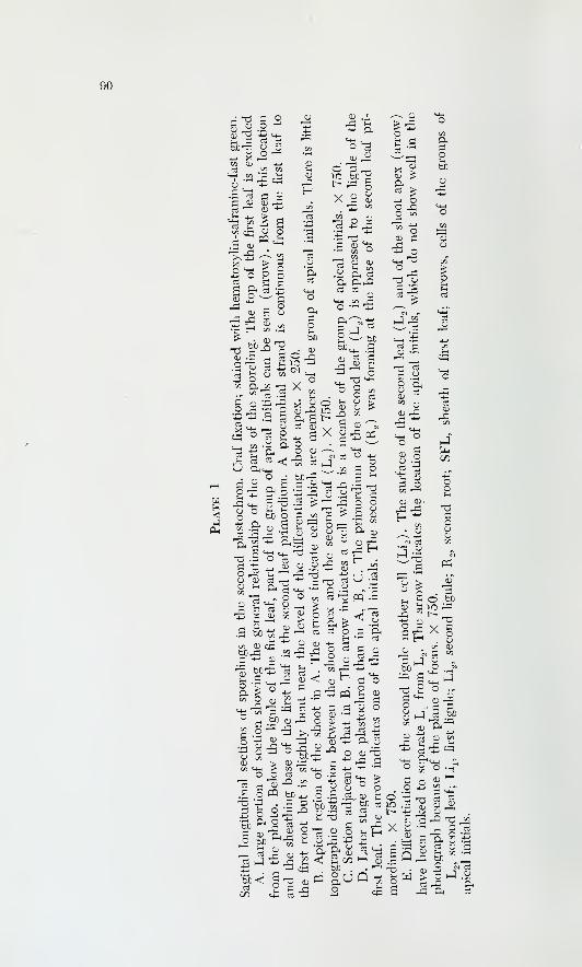
90
S^ .2"^
•^ p 03 Cu
+j X 1—
I
3J -i
O 3 rrt
c ^^ a<a p
2 =5
X a>Qj
G
03 <L) <D
a aM-H M r-i
O •'-' 411
^ o "
•2 1 -a a
S M-< „ ^ Ctf QJ
p ; bC
a a
M-l
U
x^
'"' o
rn" O
t! Cii
.S2 fc5
.- — '-^ arG MH O TSO O '-I '-' "SO bD O Gw .iir D " i-i
^ ^ ^ -n ^
2 ^
t: ri3 -^
^ ^ o
o a3
!^ G*^ G TJ
-13
E—
I
^ r "I "S
.50
G ^.'G bO-K
en OJ CCbJD^G -^ <D
'^ a; oen ^ p
55 rS -G.-H ^ ^
03 T3 PQ .G
^ .a
- « G
G aC (T)
^ ±1
m
C
G
'5bGo
3? 3 "^ 0) obO O ^ O
S .^ J2 _G
o ^ "^ ;g"^ „^ .^*^4-1 P* e/:i en m-iO O rt O^ 'O) -° .22
O « bC 4-+3.53?^ o
^ G
•G .^
W3 '^'S
bC en 'OOi ;^ oi
^ o(U Inbc a03
^^
o'n .a
-M oO =n
CD G•£ o
a:a
04GO
J
9 ft
CD <r>'
Go
tf
S o bc
X
Jen 0);i en
OO 04
^ a:^ JGo
ti G -5 CC
Go
^" aen . ,,
a ^ cc bjo . . ^ T-j
bC<J03
4-- cc
a -Gm g.uQ
X i!i
a"^gQ
G '^ ^ r—j en
CD -^ ^ i; '—
I
0^ rtCD tH en^ bC
O ?,
> O I—
I
rG ftft

92
Plate 2
Median longitudinal sections of shoot tips. X 580. All three plants had a well-
established spiral phyllotaxy. The size of the plant increases from A to C.
A. Lightly staining group of apical initials still visible in the apex. Foster's
(1934) stain, plus fast green. Sectioned perpendicular to the furrow plane.
B. Uniformly stained superficial layer. Stained with hematoxylin-safranine-
fast green. Sectioned in the furrow plane.
C. No distinct group of apical initials. Stained with hematoxylin-safranine-
fast green. The staining reaction of the superficial layer of the shoot apex is
much the same as that of the superficial layer of the region of leaf formation
to the left and the right of the apex. Sectioned in the furrow plane.
F, files of cells parallel to the superficial layer; Pr, top of procambial cylinder
of the stem; W, wedges of cells in position to form new cell files.
93
# C/w
94
Plate 3
Longitudinal sections of apexes dominated by single cells. X 590. The size
of the plant increases from A to C. All three plants were near the change from
distichous to spiral phyllotaxy. The arrow in each case indicates the cell most
highly elevated in the apex. In A and B, this cell is the largest in the apex.
In no case was the summital cell unique in staining properties. A and B werestained with hematoxylin-safranine-fast green. C was stained with the chlor-
azol-acid fuchsin-malachite green combination. A and B are sectioned in the
furrow plane; C perpendicular to the furrow plane.

95
A
%:»-''*'^# #.*:^-,*'9*:
^ . 1 ^^
A#
«#fjr»
4 J.4«4?^\ B
96
Plate 4
Median longitudinal sections of shoot tips of mature sporophytes.
A and B. Apexes of two plants with nearly identical external dimensions.
Each of the plants contained approximately forty series of roots. Both sectioned
in the furrow plane. Both X 290.
C and D. Transition in size of vacuoles from the superficial to subjacent
layers in the apex. In C the transition to large vacuoles is abrupt. In D it is
gradual. Mitochondria and plastids are not conspicuous in these photos. Both
Regaud's fixation and Regaud's hematoxylin. Both X 700.
PF, periclinal wall in the superficial layer on the flank of the apex; PS, peri-
clinal wall in the summital region.

97
PF
/ I# *
• *>
- -/
• • K-^
• 4

Plate 5
Cytological details in the shoot tip.
A, B, and drawing below. Portions of adjacent longitudinal sections from
a large apex. The plastids of the superficial layer (A, at UPl) are undiflFeren-
tiated and are more condensed in form than those in the cells of the under-
lying tissues (A and B, at PI). The cell in A containing the identified
mitochondria (M) is drawn below this legend. The mitochondrion identified
in the drawing is one of those identified in A. The mitochondria are drawn
as they appear in the peripheral cytoplasm, so that vacuoles are not adequately
represented. The length of the mitochondria is manifest. The plastids drawn
have a darkly staining reticulum and a lightly staining ground substance. Part
of one of these plastids is visible in A (Pi). Both A and B, Regaud's fixation
and Regaud's hematoxylin. Both X 1,700. Drawing X 1,500.
C, D, E, F. Distribution of starch in the shoot tip is revealed by the PASreaction. Longitudinal sections. In C and D, starch is absent from the leaf
primordia and present in the summital cells of the shoot apex. D is at a higher
magnification and shows only the summital cells of C. The starch grains in
the summital cells are minute. C, X 500; D, X 880. In E, starch is absent
from the leaf primordia and nearly absent from the summital cells of the
apex. A few starch grains appear in an adjacent section (not shown) in the
summital cells. X 330. In F, starch is present in the leaf primordia but absent
from the summital cells of the apex. The starch grains in the leaf primordia
are larger than those shown in the apex in D. X 330.
M, mitochondrion; PF, periclinal wall in the superficial layer on the flank
of the apex; Pi, plastid; UPl, undifferentiated plastid.
Mitochondrion
Nucleus
Plastid
cf. A
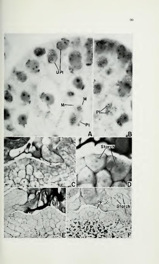
99
\PI %
k , ^ •
v.. J

100
Plate 6
Longitudinal sections of shoot tips of mature plants.
A and B. The arrangement of parts is similar in these two figures, but the
shoot apex and the subjacent cells appear broader in B than in A. Radial
files of cells underlie the region of leaf formation. The tangential walls of
cells slant inward at these locations (near F). In A, the cells at CXLT were
differentiating with a positional relationship to a leaf trace and were to pro-
vide the attachment of the xylem of the trace throughout the peripheral por-
tion of the primary xylem of the stele. Both X 300.
AS, anticlinal metaphase in the summit region; CXLT, cells to form the
connection of the xylem of the leaf trace to that of the cauline stele; F, files
of cells parallel to the superficial layer; PF, periclinal wall in the superficial
layer on the flank of the apex; PS, periclinal wall in the superficial layer in
the summital region; W, wedge of cells in position to form new cell file.

101
^ # if
f ' PS ^*»»j^'
tfllll^ll-
i
5s ^.-•
^%«Leaf Trace
/
A

102
Plate 7
Longitudinal sections showing mature and procambial portions of the stele.
A. Side and upper portions of the stele, plus adjacent tissues. From the
primary sieve elements (PSE) the approximate limits of the procambial core
can be followed upward and inward. The youngest leaf primordia stand abovethe periphery of the stele. Beneath the primordia, the tangential walls of the
cells slant inward. The xylem is composed of a central and a peripheral por-
tion and is surrounded by a sheath of parenchyma cells (Par S). The tracheids
at T are at the base of a leaf trace which is visible in adjacent sections. Thesetracheids are at the approximate inner limit of the peripheral portion of the
xylem core. X 160.
B. Peripheral portion of mature stele. The cells at CXLT have differentiated
with a positional relationship to a leaf trace (LT) and form the attachment
of the xylem of the trace to the cauline tracheids of the stele. The axial pri-
mary sieve elements (PSE) are continuous with the sieve elements of the
leaf trace. Scattered tracheids are present in the peripheral xylem (PX).
X 160.
C. Similar to B, but with many tracheids at the edge of the peripheral
xylem (PX). X 160.
D. Shoot tip and subjacent tissues, showing an arrangement similar to that
in A. The tracheids at T are associated with a leaf trace which is visible in
adjacent sections. X 190.
CX, central portion of xylem core; CXLT, cells forming the connection
of the xylem of the leaf trace to the cauline tracheids; LT, leaf trace; Par S,
parenchyma sheath; PSE, primary sieve elements; PX, peripheral portion of
the xylem core; T, tracheids, in each case associated with a leaf trace visible
in adjacent sections.

103
*«><'
CXLT" ' '•
• ^^LT\
1-tPSE
PSE
^ParS/ / . CXLT
Central Xylem Peripheral Xy1em a
B
LT
^m
PSE
A.
^-i

104
Plate 8
Stages in the development of leaf traces as seen in longitudinal sections. Theaxis of the shoot is vertical in all figures.
A and drawing below. Continuity of peripheral region of the stele with the
region of formation of procambium to the leaves. The section is median for a
young leaf primordium ( LP ) . The shoot apex is to the right, at the margin
of the photo. The tangential walls of the cells beneath the leaf primordium
are inclined inward. The cells marked 1 and 2 are for reference to the draw-
ing of A that is below this legend. The rectangles above the numbers in the
drawing represent the appearance of cells 1 and 2 in transverse sections. For
cell 1, the radial dimension is not greatly exaggerated in transverse section,
but this is not true for cell 2. The tangential dimensions of the rectangles
above the numbers in the drawing are representative of cells found in the
appropriate locations in transverse sections. A, X 300, drawing, X 150.
B. Primordium with ligule mother cell. The leaf trace (LT) slants inward.
The shoot apex is off to the right. Pr indicates cells of the procambial core.
X 370.
C and D. Serial sections of a primordium (ca. P-) with a multicellular
ligule. The leaf trace (LT) was in the initial stages of bending. X 340.
E. Later stage in the bending of a leaf trace. The procambial leaf trace
(LT) stains more deeply than the radial rows of procambial cells at the margin
of the procambial core (Pr). X 460.
F. Late stage in the bending of a leaf trace. The attachment of the leaf
trace is not perfectly shown, but it can be seen that radial files of narrow cells
occur along the base of the trace on its abaxial side. X 325.
LP, leaf primordium; LT, leaf trace; Pr, cells of the procambial core; PSE,
primary sieve elements.
Cf. A

105
it*>s -'./
. LT-
PSE. **•
i.
#T*:
LT. LT
';:s^-;'Fi
106
Plates 9 through 12 are serial transverse sections of the upper portion of a
large plant, showing leaf traces and their attachments to the vascular core
of the stem. Explanations are in the text. The numbers mark the locations
of leaves or primordia and their respective traces.
Plate 9
Distances in ^ from the summit of the apex are: A, 10; B, 30. X 340.
SA, shoot apex.

107
\^W
,
4<
!3
•a* y . f
Ligules
5 :
8
-. SA •
Ligulesl— '#/
'I- <#i;
914
• . •
sm^. •/**'12.
• 10
5—
.
•^^i1 «
%•
*4%J»rii \
-..-J' r&. '»
•:>*«yv: .

108
Plate 10
Distances in ^ from the summit of the apex are: A, 40; B, 50. X 340.
The vertical Hnes on B mark the approximate lateral limits of plate 11,
A, B, C, D.

109
•7. %4r^^^iS^TT'
^.i^;^-^-my%

110
Plate 11
Distances in ^ from the summit of the apex are: A, 70; B, 100; C, 110; D, 130.
X 340.

Ill

112
P. c
S^ a
"^
M-l TSoa;3C/3 ^<D
^
^ ^
C fCH
T3 4-1
c lu d."^ 1—1
cS-MCCD
Q^
of s6"
1—
1
^H
p;
1303 u03
03 CDCD1—
1
cq 4_,-̂M
m"w ^ .r.
3I on 1—1
^ I—
1
T—
1
•-' <(D m
T3 '6-
a a j-i
ft s,G CO QJ
rt aOS ^ 03
^ C/3 cu
rS:_( sc3 c Mh
^ (U-t-J
•-a
,H asc3 -1-1
M^ 3 .^ «C/2 cu (X)^ CS]
m ^
2 PQ ^4-< fi 03
•T-H ;_,
_G <r0) •^
^ of ^f^2 1-H C
03 ^^M-l
^< OS2 o;
. rJ:5
"13 C (J ft
Oh (U r.
CUcu
<

r ^r^BF^ •
f
inCVJ
0) \
\OJCVJ
^
^* JP i«fc^**£. *
x«?1lynP-S'W^ .f
p*--' sr
'^M' ?
CMCM
i?=*-tr<>'O)
s
7oCM
/X^;^COCM
CM
<r/tK;T-/V»\t

114
Plate 13
Relationship of tissues in young and old sporophytes.
A. Sporophyte with several leaves. Longitudinal section in a plane perpen-
dicular to the furrow plane. All of the vascular tissues have a positional re-
lationship to the leaves and the roots. The relationship of the basal meristem
(BM) to the rest of the plant is obscured by the presence of root traces (RT).
X 370.
B. Sporophyte with several leaves. Longitudinal section in the furrow plane.
The basal meristem (BM) is continuous with the lateral meristem (LM).The lateral meristem has produced some secondary sieve elements (SSE).
Some primary sieve elements are obHterated (Obi). The basal meristem has
produced tracheids (T) on its inner face. X 230.
C. D, E. Longitudinal sections showing the derivatives of the lateral meri-
stem in plants of increasing age. The axis is vertical in all of the photos. In C,
only secondary sieve elements (SSE) have differentiated inside of the lateral
meristem (LM). X 260. In D, secondary sieve elements and parenchyma
(Par) have differentiated in layers inside the lateral meristem. X 260. In E,
secondary tracheids (ST) have differentiated among parenchyma cells in alter-
nating layers with secondary sieve elements. X 250.
F. Transverse section showing immature secondary tracheids (ST) nearly
in contact with the lateral meristem (LM). X 300.
G. Late maturing tracheids (LMT) in the parenchyma sheath outside of
the peripheral xylem (PX). Longitudinal section; axis vertical. X 300.
BM, basal meristem; LM, lateral meristem; LMT, late-maturing tracheids;
LT, leaf trace; Obi, obliterated primary sieve elements; P^, P^, Pg, youngest,
second youngest, and third youngest leaf primordia, respectively; PSE, pri-
mary sieve elements; PX, peripheral xylem; RT, root trace; SSE, secondary
sieve elements; ST, secondary tracheids inside lateral meristem; T, secondary
tracheids inside basal meristem.
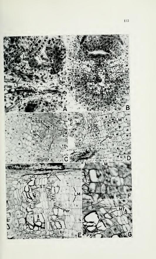
115
.- - , ... :v -
v<^
'v.'."'-N
««
,,i ] Par- .rji^fc^-, v>-'^'^"\
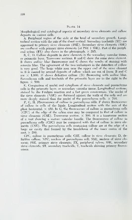
116
Plate 14
Morphological and cytological aspects of secondary sieve elements and callose
deposits in various cells.
A. Peripheral region of the stele at the level of secondary growth. Longi-
tudinal section with the axis of the shoot vertical. Secondary tracheids (ST) are
appressed to primary sieve elements (PSE). Secondary sieve elements (SSE)
are confluent with primary sieve elements ( at PSE + SSE ) . Part of the periph-
eral xylem (PX) also shows in the photograph. X 285.
B, C, D. Callose deposits in sieve elements in the secondary vascular tissue,
or prismatic layer. Transverse sections. B and C show the same sieve element.
B shows aniline blue fluorescence and C shows the results of staining with
resorcin blue. The agreement of the two techniques in the detection of callose
is very good. The large white area near the upper end of the sieve element
in B is caused by several deposits of callose which are out of focus. B and Care X 1,000. D shows definitive callose (D) fluorescing with aniline blue.
Parenchyma cells and tracheids of the prismatic layer are to the right in the
figure. X 500.
E. Comparison of nuclei and cytoplasm of sieve elements and parenchyma
cells in the prismatic layer, or secondary vascular tissue. Longitudinal sections;
stained by the Feulgen reaction and a fast green counterstain. The nuclei of
the sieve elements (NSE) are flattened against the walls of the cells and are
more deeply stained than the nuclei of the parenchyma cells. X 700.
F, G, H. Fluorescence of callose in parenchyma cells. F shows fluorescence
of callose in cells of the ligule. Longitudinal section with the axis of the
plant horizontal. X 450. In G, the fluorescence of callose in parenchyma cells
(CPC) at the edge of the xylem core may be compared to that of callose in
sieve elements (CSE). Transverse section. X 500. H is a transverse section
of a root showing a mature vascular bundle. The fluorescence of callose in
parenchyma cells (CPC) may be compared with that of callose in sieve ele-
ments ( CSE ) . The parenchyma cells containing callose are at the edge of a
large air cavity that formed by the breakdown of the inner cortex of the
root. X 290.
CPC, callose in parenchyma cells; CSE, callose in sieve elements; D, de-
finitive callose; NPC, nucleus of parenchyma cefl; NSE, nucleus of sieve ele-
ment; PSE, primary sieve elements; PX, peripheral xylem; SSE, secondary
sieve elements; ST, secondary tracheids; T, tracheids showing primary fluores-
cence.

117

118
Plate 15
Various aspects of cortical tissues in mature plants.
A. Upper portion of a longitudinal section of a large plant. Leaf traces
(LT) to leaves already lost from the plant may be seen. The peripheral por-
tion of the cortex is composed of nearly empty cells (NEC). X 40.
B. Tissues from the upper (unmarked) trace in A. Functional sieve ele-
ments (SE) are present. Crushed tracheary elements appear at CT. Similar
tissues were in the other trace (marked LT) in A. X 380.
C. D. The effect of radial division in a cambial initial, as seen in cell files
in a transverse section. D is at a higher magnification than C. The boundaries
of the cell files at a, b, and c match the radial walls of the initials in the
lateral meristem. Explanation is in the text. C, X 270; D, X 500.
E. Periphery of plant, away from basal furrow. Longitudinal section. Thecells are compactly arranged. The straight walls (Walls) indicate that the
cells of the cortex subdivide before they enter into formation of the corky
layer which covers the sporophyte. X 250.
F. Periphery of plant where the furrow was fonning. Transverse section.
The furrow was forming from the right. The cells indicated by the arrows
show division figures. X 220.
CT, crushed tracheary elements; LM, lateral meristem; LT, leaf trace;
NEC, nearly empty cells; Par, parenchyma; SE, sieve elements; SSE, sec-
ondary sieve elements; ST, secondary tracheids; arrows, cells showing division
figures; a, b, c, radial boundaries matching the radial walls of initials in the
lateral meristem.

119
NEC
/<y \'
fs-
\ .0
—
--^-^hJU
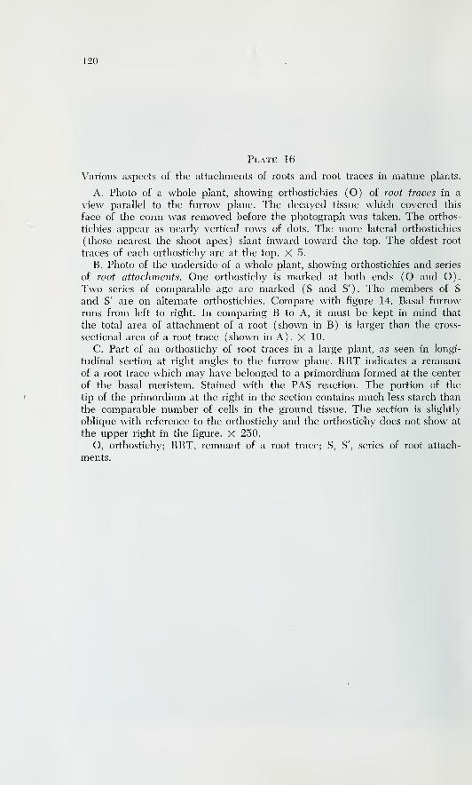
120
Plate 16
Various aspects of the attachments of roots and root traces in mature plants.
A. Photo of a whole plant, showing orthostichies (O) of root traces in a
view parallel to the furrow plane. The decayed tissue which covered this
face of the corm was removed before the photograph was taken. The orthos-
tichies appear as nearly vertical rows of dots. The more lateral orthostichies
(those nearest the shoot apex) slant inward toward the top. The oldest root
traces of each orthostichy are at the top. X 5.
B. Photo of the underside of a whole plant, showing orthostichies and series
of root attachments. One orthostichy is marked at both ends (O and O).
Two series of comparable age are marked ( S and S' ) . The members of S
and S' are on alternate orthostichies. Compare with figure 14. Basal furrow
runs from left to right. In comparing B to A, it must be kept in mind that
the total area of attachment of a root (shown in B) is larger than the cross-
sectional area of a root trace (shown in A). X 10.
C. Part of an orthostichy of root traces in a large plant, as seen in longi-
tudinal section at right angles to the furrow plane. RRT indicates a remnant
of a root trace which may have belonged to a primordium formed at the center
of the basal meristem. Stained with the PAS reaction. The portion of the
tip of the primordium at the right in the section contains much less starch than
the comparable number of cells in the ground tissue. The section is slightly
oblique with reference to the orthostichy and the orthostichy does not show at
the upper right in the figure. X 250.
O, orthostichy; RRT, remnant of a root trace; S, S', series of root attach-
ments.
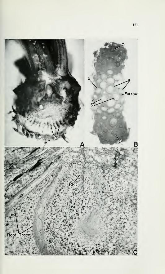
121

122
Plate 17
The relationship of the cambium to derivative tissues.
A. Longitudinal section in the furrow plane of a young plant. The basal
meristem (BM) and lateral meristem (LM) are confluent at the right but not
at the left because of obliquity of sectioning. The lateral meristem has pro-
duced secondary sieve elements (SSE) in contact with the primary sieve
elements (PSE ) . Primary sieve elements are obliterated in part ( Obi ) . At the
upper part of the stele, primary sieve elements (PSE) are still intact. At the
left, a root trace (RT) is attached to the stele. X 350.
B. Basal meristem (BM) and derivatives, as seen in longitudinal section
in the furrow plane in a large plant. The tracheids (T) and parenchyma (Par)
which compose the inner derivatives of the basal meristem are weakly layered
in a direction parallel to the basal meristem. X 210.
C. Basal and lateral portions of the stele of a plant of moderate size, as
seen in longitudinal section in the furrow plane. The section is off median so
that the bases of some root traces may be seen. The uppermost root primor-
dium (RP) marks the upper limit of the basal meristem. Some files of cells
below this limit (within brackets at FC) contain old secondary sieve ele-
ments (OSSE), and this indicates that the course of differentiation of the
derivatives of the initials of these files has changed from one that produces
sieve elements to one producing no sieve elements. The initials of these files
were in the lateral meristem at one time (while the inner derivatives of the
initials matured as sieve elements), although they were in the basal meristem
at the time of fixation. Secondary tracheids (ST) inside the basal meristem are
confluent with parenchyma (Par) inside the lateral meristem. This plant hadnot yet produced secondary tracheids inside of the lateral meristem at the
time of fixation. X 170.
BM, basal meristem; FC, files of cells containing sieve elements; LM, lateral
meristem; Obi, obliterated primary phloem; OSSE, old secondary sieve ele-
ments; Par, parenchyma; PSE, primary sieve elements; RP, root primordium;
RT, root trace; SSE, secondary sieve element; ST, secondary tracheid; T,
tracheids.

123
. .'.•-."•v..: •• :\/ /
,- r '^ »

124
Plate 18
Various aspects of the basal and lateral meristems and of young root pri-
mordia.
A. Transverse section of a large plant in which the basal meristem has ad-
vanced toward the shoot apex. At the center of the photo is a portion of the
stele bearing leaf traces (LT). Secondary sieve elements (SSE), which were
produced by the lateral meristem (LM), occur between this location and
the bases of root traces (BRT). The lateral meristem was once operative
throughout the entire perimeter of the stele at this level in the plant but
has been converted, in part, to the basal meristem (left and right extremities
of the stele). The lateral meristem has produced several layers of secondary
tracheids (ST). X 60.
B. Similar to A, but at a higher magnification. From another large plant.
Tracheids at the bases of root traces (BRT) are confluent with parenchyma
(Par) produced by the lateral meristem (LM). X 150.
C. Relationship of the basal meristem (BM) to mature vascular tissues.
Longitudinal section perpendicular to the plane of the furrow and passing
between orthostichies. Secondary sieve elements (SSE) differentiate toward
the basal meristem. X 300.
D. Root primordium showing no distinct indications of organized apical
activity. A procambial strand (Pr) has differentiated. Longitudinal section
perpendicular to the plane of the furrow in a plant of moderate size. X 290.
E. Low starch content in an organizing primordium. Section perpendicular
to the furrow plane and from a large plant. The starch content of the root
primordium (RP) is lower than that of the ground tissue. Compare with plate
16, C. X 250.
BM, basal meristem; BRT, bases of root traces; LM, lateral meristem;
LT, leaf trace; Pr, procambium; RP, root primordium; RT, root trace; SSE,
secondary sieve element; ST, secondary tracheids.

125
\
STST
RT
BRT-%. N
\RT
VSSE LT
.BRT
\
LM
/\ A
*-s^. \ , • jj«M» Tysj, •& *' r \ kst lit I iiimiAfci.i'Mni
^
^^,
'

126
Plate 19
Transverse and longitudinal sections of root tips.
A. Transverse section of a root tip of a partially extended root of a mature
plant. The procambium (Pr) has an eccentric position in the section. This
eccentricity depends on the development of the inner cortex (IC). X 80.
B. Longitudinal section of the tip of a partly extended primary root of a
sporeling. The root cap (RC), epidermis (E), and outer cortex (OC) share
a common origin. The cell net indicates continuity between the procambium(Pr) and the initials of the inner cortex (IIC). The inner cortex (IC) does
not develop markedly in the primary root. X 430.
C. D. Adjacent longitudinal sections showing continuity of the procam-
bium (Pr) and the initials of the inner cortex. A sharp boundary exists be-
tween the outer cortex (OC) and the inner cortex (IC). The apex represented
is one among several within the dichotomized tip of a partially extended root
of a mature plant. Cells 1 and 2 appear in both sections. X 700.
E, F. Stages in the dichotomy of an apex. F is a later stage than E. Longi-
tudinal sections of roots emerging from the cortex of a small plant. The inked
lines follow the tip of the procambium (Pr). In F, the lines are open belowbecause the procambium is continuous with the initials of the inner cortex
( at arrows in F ) . This continuity was caused by a contribution to the pro-
cambium from cells among the initials of the inner cortex (arrows in E).
E, X 500. F, X 420.
E, epidermis; IC, inner cortex; IIC, initials of inner cortex; OC, outer cortex;
Pr, procambium; RC, root cap; 1 and 2, cells appearing in both C and D;arrows, cells among the initials of the inner cortex.

127
, E


INDEX
129
Anneaii initial, 29Apical cell: in root, 77; in shoot, 8,
19-21
Apical initials: differentiation of, in
young shoot, 16, 19, 24, 28-29
Basal meristem, 3, 13, 54-56, 69-75
Callose, 15, 45-48
Gambia: multiple, 43, 45Cambium, 3, 13-15, 40-45; origin of,
14, 40-43
Cauline vascular tissue, 11-12, 39Cork, 14, 50; storied, 50Cork cambium, 15, 50
Cortex: primary, 32, 48-49; second-
ary, 14, 48-50
Dichotomy of root, 78
Foliar vascular tissue, 11-12, 39
Furrow plane of sectioning, 3
Ground meristem, 31-32
Histogens in root, 76-77
Initials: concept of, 10, 29; conver-
sion of, in cambium, 73
Lateral meristem,Cambium
3, 13. See also
Mitoses: in root apex, 81; in shoot
apex, 26, 28
Orthostichies of roots, 3, 6, 53-54; de-
fined, 57
Parenchyma: secondary, 43, 71
Parenchyma sheath, 30, 37Parenchymatous mantle, 14, 31
Phloem: primary, 12, 30, 35; second-
ary, 43, 45, 71
Phyliotaxy, 17
Pith, 39-40
Planes of sectioning, 3
Plastids: in root primordium, 79; in
shoot apex, 23Plastochronic changes, 17, 20
Plerome, 8, 11
Primary meristem of Stylites, 14, 31
Primary tissues: differentiation of, 11-
12, 29-32
Prismatic layer, 3, 15
Procambium, 11, 30-31, 79; of leaves,
12-13, 33-40; of root primordium,
79

130 THE DEVELOPMENTAL ANATOMY OF IsoetCS
Region of leaf formation, 7
Rhizomorph, 75Rhizophore, 75Ribonucleic acid: stain for, in shoot
apex, 24-25
Root: origin of tissvies in, 80; pri-
mary, 54-56
Root apex: dichotomy of, 78; organ-
ization of, 77-78, 80-81
Root initiation: location of, 52-54, 57-
69; process of, 76-77, 78-80; se-
quence of, 61-65; site of, 65Root maturation: sequence of, 61
Root-producing meristem, 54, 69. Seealso Basal meristem
Roots: adventitious, 55, 56, 74; en-
dogenous, 76; exogenous, 76; kinds
observed, 80
Secondary meristem: of Shjlites, 14
Secondary vascular tissue. See Pris-
matic layer
Series of roots, 52-54; defined, 57
Shoot apex: defined, 7; cytology of,
22-26; form of, 8-10, 15-22; func-
tion of, 10-11, 26-29; mitoses in,
26, 28; origin of, 9-10, 15-17; size
of, 19
Shoot tip, 7
Standardized position: for transverse
sections, 58Starch: in root primordium, 79; in
shoot tip, 25-26
Stele, 8
Wurtzelstele: of Stijlites, 72, 75
X-axis: defined, 58Xylem: central, 12, 30, 39-40; cen-
trifugal matmation of, 40; centrip-
etal maturation of, 40; peripheral,
12, 30, 37, 39-40; secondary, 43,
71
Y-axis: defined, 58
Roback, Selwyn S. (1954): The Evolution and Taxonomy of the Sarcophaginae (Diptera,
Sarcophagidae). 9 charts. 34 pis. Vol. 23, Nos. 3-4. $4.00.
Rominger, James M. (1962): Taxonomy of Setaria (Gramineae) in North America. 15 maps.
6 pis. No. 29. $3.00.
Schoof, Herbert Frederick (1942): The Genus Conotrachelus Dejeon (Coleoptera, Curculion-
idae) in the North Central United States. 9 pis. Vol. 19, No. 3. $1.50.
Selander, Richard B. (I960): Bionomics, Systemafics, and Phylogeny of Lytta, a Genus of
Blister Beetles {Coleopiera, Meloidae). 350 figs. No. 28. $4.50.
Smith, Frank (1923): The Colciferous Glands of Lumbricidae and Dipiocordio. 12 pis. Vol. 9,
No. 1. $1.25.
Solheim, Wilhelm Gerhard (1930): Morphological Studies of the Genus Cercospora. 4 pis.
Vol. 12, No. 1. $1.00.
Spooner, Charles Stockman (1938): The Phylogeny of the Hemipfera, Based on a Study
of the Head Capsule. 24 pis. 2 figs. Vol. 16, No. 3. $1.00.
Sprague, Victor (1941): Studies on Gregarina blaftarum with Particular Reference to the
Chromosome Cycle. 6 pis. 2 figs. Vol. 18, No. 2. $.75.
Stannard, Lewis J., Jr. (1957): The Phylogeny and Classification of the North American
Genera of the Suborder Tubulifera (Thysanopfera ) . 14 pis. No. 25. $2.50.
Stevens, Frank Lincoln (1924): Parasitic Fungi from British Guiana and Trinidad. 19 pis.
1 map. Vol. 8, No. 3. $1.25.
and Ryan, Sister Mary Hilaire (1939): The Microthyrioceae. Vol. 17, No. 2. $1.50.
Tehon, Leo Roy (1935): A Monographic Rearrangement of Lophodermium. 5 pis. Vol. 13,
No. 4. $2.00.
Van Cleave, Harley J. (1953): Acanthocephalo of North American Mammals. 13 pis. 10
figs. Vol. 23, Nos. 1-2. $4.00.
Yuncker, Truman George (1921): Revision of the North American and West Indian Species
of Cuscuta. 13 pis. Vol. 6, Nos. 2-3. $2.00.


UNivERsrrv of illinois-urbana
3 0112 065097245