Embed Size (px)
Citation preview

THE DEVELOPMENT OF SELECTIVE INHIBITORS FOR ER -MANNOSIDASE I
BY COMBINATORIAL MODIFICATION OF KIFUNENSINE AND
DEOXYMANNOJIRIMYCIN
by
JESSICA CARDOT
(Under the Direction of Geert-Jan Boons)
ABSTRACT
Selective inhibitors of endoplasmic reticulum -mannosidase I (ER Man I) have
the potential to be used for treatment of a number of genetic diseases. Although potent inhibitors
for this enzyme have been described, in general these compounds display poor selectivity and
often inhibit Golgi -mannosidase I. To address this difficulty, we have synthesized and
combinatorial modified kifunensine, which is a potent GH47 -mannosidase I inhibitor, and 1-
deoxymannojirimycin, a weak -mannosidase I inhibitor. Kifunensine was modified by a
hydrazone moiety, which could easily be extended by reaction with a range of aldehydes. The
library of hydrazone-linked analogues was screened for inhibition of human ER Man I and mouse
Golgi Man I. It was found that a pyridine functionalized kifunensine analog selectively inhibited
ER Man I. Based on this finding, a second generation of analogues was prepared in which a
functionalized pyridine scaffold was coupled to kifunensine through the hydrazone moiety. 1-
Deoxymannojirimycin was modified by a hydrazine moiety, which could easily be extended by
reaction with a range of aldehydes. The library of hydrazone-linked DMJ analogues was screened
for inhibition of human ER Man I and mouse Golgi Man I.
INDEX WORDS: Mannosidase Inhibitors, Kifunensine, Deoxymannojirimycin
THE DEVELOPMENT OF SELECTIVE INHIBITORS FOR ER -MANNOSIDASE I
BY COMBINATORIAL MODIFICATION OF KIFUNENSINE AND
DEOXYMANNOJIRIMYCIN
by
JESSICA CARDOT
BS Chemistry, Nazareth College of Rochester, 2006
A Dissertation Submitted to the Graduate Faculty of The University of Georgia in Partial
Fulfillment of the Requirements for the Degree
DOCTOR OF CHEMISTRY
ATHENS, GEORGIA
2011

© 2011
Jessica Cardot
All Rights Reserved

THE DEVELOPMENT OF SELECTIVE INHIBITORS FOR ER -MANNOSIDASE I
BY COMBINATORIAL MODIFICATION OF KIFUNENSINE AND
DEOXYMANNOJIRIMYCIN
by
JESSICA CARDOT
Major Professor: Geert-Jan Boons
Committee: Kelley Moremen
Robert phillips
Electronic Version Approved:
Maureen Grasso
Dean of the Graduate School
The University of Georgia
December 2011

iv
DEDICATION
I would like to dedicate this dissertation to my parents, Andy and Jan Cardot, and
my brother Andy Cardot for all of their love and support.

v
TABLE OF CONTENTS
Page
ABBREVIATIONS ........................................................................................................ ix
LIST OF FIGURES ......................................................................................................... x
LIST OF FIGURES ........................................................................................................ xi
CHAPTER
1 INTRODUCTION AND LITERATURE REVIEW ........................................ 1
Introduction and Literature Review .......................................................... 1
N-Linked Glycoproteins ........................................................................... 1
Biosynthetic Pathway of N-Linked Glycans .............................................. 1
Biosynthesis of N-Glycans in the ER ........................................................ 4
N-Glycans Direct and Indirect Roles in Glycoprotein Foilding ................. 8
Calnexin/Calreticulin Cycle ...................................................................... 9
ER-Associated Degradation (ERAD) ...................................................... 12
Diseases Associated with ERAD ............................................................ 13
Inverting and Retaining Enzymes ........................................................... 14
Glycosyl Hydrolase Family 47 (GH 47) .................................................. 16
Mannosidase Inhibitors ........................................................................... 18
References .............................................................................................. 23

vi
2 THE DEVELOPMENT OF SELECTIVE INHIBITORS FOR ER -
MANNOSIDASE I BY COMBINATORIAL MODIFICATION OF
KIFUNENSINE ........................................................................................... 28
Abstract .................................................................................................. 29
Introduction and Literature Review ........................................................ 29
Results and Discussion ........................................................................... 32
Synthesis of Azidolactone ...................................................................... 33
Synthesis of Bisacetonide Kifunensine ................................................... 34
Synthesis of Novel Hydrazone Modified KIF ......................................... 35
Synthesis of Novel Kifunensine Analogues ........................................... 36
Cellular Study of Pyridine KIF ............................................................... 43
Conclusion ............................................................................................. 45
Experimental Section .............................................................................. 45
References .............................................................................................. 60
3 THE DEVELOPMENT OF SELECTIVE INHIBITORS FOR ER -
MANNOSIDASE I BY COMBINATORIAL MODIFICATION OF
DEOXYMANNOJIRIMYCIN ..................................................................... 62
Abstract .................................................................................................. 63
Introduction and Literature Review ........................................................ 63
Results and Discussion ........................................................................... 65
Synthesis of Hydrazine DMJ .................................................................. 65
Synthesis of Hydrazone DMJ Analogues ................................................ 66
Conclusion ............................................................................................. 68
vii
Experimental Section .............................................................................. 69
4 THE EXTRACYCTOPLASMIC DOMAIN OF THE MYCOBACTERIUM
TUBERCULOSIS SER/THR KINASE PKnB BINDS SPECIFIC
MUROPEPTIDES AND IS REQUIRED FOR PKnB LOCALIZATION ..... 75
Abstract .................................................................................................. 77
Author Summary .................................................................................... 78
Introduction and Literature Review ........................................................ 79
Results.................................................................................................... 81
Binding of PGN fragments to the extracytoplasmic domain of PknB ...... 81
Muropeptides stimulate resuscitation of dormant M. tuberculosis cells ... 86
PknB is present in the cell envelope of M. tuberculosis ........................... 89
PknB localizes to the septum and poles, and the extracytoplasmic domain
is required for proper PknB localization .................................................. 91
Discussion .............................................................................................. 93
Materials and Methods ........................................................................... 98
Strains, media, and recombinant plasmid construction and protein
production .............................................................................................. 98
Chemical Synthesis of PGN fragments ................................................. 100
Surface plasmon resonance binding assays and kinetic analysis ............ 101
Conditioned medium preparation .......................................................... 103
Dormancy and resuscitation assay ........................................................ 103
Growth stimulation assay...................................................................... 104
Microscopy .......................................................................................... 105

viii
Western Blotting .................................................................................. 105
Acknowledgments ................................................................................ 106 "[Click here and type Subheading]" 104
References ............................................................................................ 107
Supporting Figures and Tables.............................................................. 113
5 CONCLUSIONS ........................................................................................ 120

ix
ABBREVIATIONS
Ac…………………………….. …………………………………………………….Acetyl
Ac2O…………………………………………………………………….Acetic Anhydride
C……………………………………………………………………………………Celsius
DCM …………………………………………………………………….Dichloromethane
DMAP………………………………………………………. N,N-dimethylaminopyridine
DMF……………………………………………………………..N,N-dimethylformamide
DMJ ……………………………………………………………...1-Deoxymannojirimycin
DMP……………………………………………………………..Dess Martin Periodinane
EDC………………… 1-ethyl-3-(3-dimethylaminopropyl) carbodiimide hydrochloride)
ERAD…………………………………. Endoplasmic Reticulum Associated Degradation
ER Man I……………………………………… Endoplasmic Reticulum -mannosidase I
Et2O ………………………………………………………………………….Diethyl ether
EtOH……………………………………………………………………………….Ethanol
HCl………………………………………………………………………Hydrochloric acid
HOBt……………………………………………………………..N-Hydroxybenzotriazole
KIF…………………………………………………………………………….Kifunensine
Me…………………………………………………………………………………...Methyl
MeOH…………………………………………………………………………….Methanol
NMR………………………………………………………….Nuclear magnetic resonance
NaN3 ……………………………………………………………………….. Sodium azide
NaNO2 ……………………………………………………………………. Sodium Nitrite
NaOMe ………………………………………………………………...Sodium methoxide
OST……………………………………………………………..Oligosaccharyltransferase
Ph……………………………………………………………………………………Phenyl
Piv………………………………………………………………………………….Pivoyal
Piv-Cl……………………………………………………………Trimethyl acetyl chloride
TBAF……………………………………………………..Tetrabutylammonium Fluoride
TFA …………………………………………………………………...Trifluoroacetic acid
THF………………………………………………………………………..Tetrahydrofuran

x
LIST OF TABLES
Page
Table 1: Diseases associated with ERAD ....................................................................... 13
Table 2: Ki Inhibition data for kifunensine analogues 15-2............................................. 38
Table 3: Ki Inhibition data for pyridine kifunensine analogues 28-40 ............................. 42
Table 4: Ki data for DMJ analogues 46-50 ..................................................................... 68
Table 5: Affinity of synthetic muropeptides for the extracytoplasmic domain of M.
tuberculosis PknB .............................................................................................. 84
Table 6: Resuscitation of dormant M. tuberculosis cultures ............................................ 87
Table S1: Kinetic binding parameters for the interaction of synthetic muropeptides with
the PASTA domains of M. tuberculosis PknB .................................................. 118

xi
LIST OF FIGURES
Page
Figure 1: Core Glc3Man9GlcNAc2 structure ..................................................................... 2
Figure 2: Biosynthetic scheme for N-glycans in the ER .................................................... 5
Figure 3: Catalytic site and amide nitrogen activation ...................................................... 7
Figure 4: The Calnexin/Calreticulin cycle ...................................................................... 10
Figure 5: General inverting and retaining enzyme mechanisms ...................................... 15
Figure 6: Model for the structure and catalytic residues for ER Man I used in mutagenesis
studies described in this paper ............................................................................ 17
Figure 7: Known -mannosidase inhibitors ................................................................. 18
Figure 8: Stereo views of the active site of dGMII with bound DMJ (B) and Swainsonine
(C) molecules ..................................................................................................... 20
Figure 9: Co-crystal structure of Kifunensine in the binding pocket of ER Man I ........... 22
Figure 10: Structures of compounds in initial library screening, 15-27 ........................... 37
Figure 11: Functionalized Pyridine dihydrazone kifunensine analogues, 28-39 .............. 41
Figure 12: Effect of inhibitor treatment on the endoglycosidase sensitivity of a reporter
glycoprotein expressed in mammalian cells ........................................................ 44
Figure 13: Structures of DMJ analogues or initial library screening, 46-50 .................... 67
Figure 14: Structures of synthetic muropeptides used in the binding and phenotypic
assays................................................................................................................. 82

xii
Figure 15: Sensorgram of a compound with relatively high binding affinity for ED-
PknB……………………………………………………………………………...83
Figure 16: Growth stimulation assay of low inoculum cultures of M. tuberculosis ......... 88
Figure 17: Subcellular localization of M. tuberculosis PknB .......................................... 90
Figure 18: Localization of PknB to sites of peptidoglycan turnover in the mycobacterial
cell ..................................................................................................................... 92
Figure 19: Model of PknB localization and activation by interaction of its
extracytoplasmic domain with peptidoglycan fragments ..................................... 97
Figure S1: T-Coffee alignment of the four PASTA domains of M. tuberculosis PknB and
the single PASTA domain of PBP2 .................................................................. 113
Figure S2: SDS-PAGE gel showing expression and purification of ED-PknB .............. 114
Figure S3: Sensorgrams of compounds tested in the Biacore binding experiments ....... 115
Figure S4: Immunoblot of subcellular fractions of M. smegmatis showing localization of
the RFP-PknB kinase domain fusion ................................................................ 116
Figure S5: Live cell imaging of M. smegmati ............................................................... 117

1
CHAPTER 1
INTRODUCTION AND LITERATURE REVIEW
Introduction and Literature Review
N-linked Glycoproteins
The most common covalent modification in eukaryotic cells is N-linked
glycosylation.1 The majority of proteins synthesized in the ER are in fact N-linked
glycoproteins which are responsible for a diverse set of cellular functions.
Glycoproteins serve as ligands for many recognition processes such as modulation of
immune response, enhanced solubility, facilitation of protein orientation, protein rigidity,
regulation of protein turnover, protein stabilization against proteolysis, as well as to
mediate interactions with pathogens. Not only is N-linked glycosylation the most
prevalent covalent modification but no other modification is so widely utilized for a
variety of purposes.1,2
Biosynthetic Pathway of N-glycans
Although the N-glycans expressed on the cell service contain complex
oligosaccharide motifs, they all stem from a basic core structure, Glc3Man9GlcNAc2
(Figure 1). 3

2
c
e
j
d
h
a
(1-4)
(1-4)
(1-6)
(1-6)
(1-3)
(1-3)
i k
f
g
b
l
m
n
(1-2) (1-2)
(1-2)
(1-2)
(1-3)
(1-3)
(1-2)
A
B C
Figure 1: Core oligosaccharide structure containing three glucose residues (purple), nine mannose residues
(green) and two N-acetyl-glucosamine residues (teal), Glc3Man9GlcNAc2. The monosaccharide subunits
highlighted in bold circles (a-g) are glycosylated to the glycan on the cytosolic surface, where as the rest
are added lumenally. Residues d,f,g,l are responsible for interactions with the calnexin/calreticulin cycle.
Where as mannose residue i is cleaved by ER Man I, which is believed to be a key enzyme ERAD.

3
The biosynthetic pathway of N-glycans is shared between the ER and Golgi
apparatus. The core oligosaccharide and the polypeptide are synthesized in the ER and
cytosol at which point an N-glycosylation reaction occurs between the Glc3Man9GlcNAc2
and the side chain of an Asn residue of the polypeptide facilitated by an oligosaccharide
transferase (OST) enzyme. The newly formed glycoprotein will undergo numerous
trimming steps to allow for the proper folding and oligomerization to the native form.
Once the native form is achieved the glycoprotein is translocated to the Golgi
compartment at which point it is subjected to numerous trimming and elongation steps to
form the complex glycans found on mature glycoproteins. The modifications to the
glycan in the ER result in limited diversification because the alterations are shared by all
glycoproteins. However, these modifications in the ER appear to correspond to
glycoprotein folding, quality control, sorting, degradation, and secretion. The various
configurations of the N-linked glycan in the ER are not merely just intermediates in a
biosynthetic pathway but each have a specific function that determines the fate of the
individual glycoproteins. These biosynthetic intermediates play a vital role in three main
phases of the glycoproteins existence. Phase 1 occurs solely in the ER where truncated
versions of the core oligosaccharide are necessary for proper protein folding and quality
control. The second phase occurs in the ER, Golgi and trans-Golgi network and
determines the intracellular transport and targeting. The final phase is solely in the Golgi
and takes place after extensive modifications of the Glycan portion giving rise to a
tremendous amount of diversification. The change from structural uniformity in the ER
to structural complexity in the Golgi corresponds to the marked change in glycoprotein
4
function. Phase 1 or glycoprotein biosynthesis in the ER will be discussed in detail as it
pertains to the current dissertation work.
Biosynthesis of N-glycans in the ER
In the ER during the early stages of the secretory pathway, the N-glycans have a
common role in promoting protein folding, quality control and certain sorting events.
The core glycan is produced in the ER membrane by monosaccharyl-transferases and is
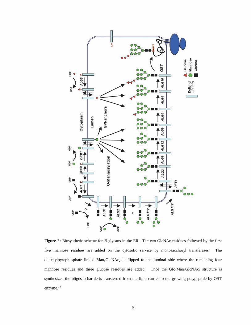
added to the protein as Glc3Man9GlcNAc2 in the lumen of the ER (Figure 2). 12
5
Figure 2: Biosynthetic scheme for N-glycans in the ER. The two GlcNAc residues followed by the first
five mannose residues are added on the cytosolic service by monosacchoryl transferases. The
dolichylpyrophosphate linked Man5GlcNAc2 is flipped to the luminal side where the remaining four
mannose residues and three glucose residues are added. Once the Glc3Man9GlcNAc2 structure is
synthesized the oligosaccharide is transferred from the lipid carrier to the growing polypeptide by OST
enzyme.13

6
The monosaccharides are first attached to a lipid carrier by monosaccharyl-
transferases in the cytosol.1,11
Once the heptasaccharide is synthesized, the sugar unit is
rotated to the lumenal side catalyzed by an ATP-independent bi-directional flippase.12
The glycosylase enzymes in both the lumenal and cytosol compartments of the ER are
highly specific for individual sugar moieties; thus allowing for the linear synthesis of the
branched oligosaccharide.16
The final step in the core oligosaccharide synthesis is the
addition of the terminal -1,2-glucose residue which is essential for sufficient recognition
by the OST enzyme that transfers the Glc3Man9GlcNAc2 from the lipid carrier to the Asn
side chain of the glycosylation sequon AsN-X-Ser/Thr forming an N-glycosidic bond.17,18
For many years it was widely accepted that this sequon was necessary because of the
relatively chemically inert nature of the Asn side chain nitrogen.19
The polypeptide
would form a loop which allows for the side chain hydroxyls of Ser/Thr to interact with
the amide side chain of Asn thereby increasing the nucleophilic nature of nitrogen. The
need for this loop explained why the X amino acid could not be a proline residue do to
the rigid cyclic side chain.21,22
Once the Glc3Man9GlcNAc2 is added to the polypeptide,
protein folding can commence. However, recent studies preformed by Locher and co-
workers47
brought new insights to the OST mode of action. The catalytic subunit, STT3,
of the OST enzyme has homologues in bacteria and archea. Locher and coworkers
obtained an x-ray crystal structure of a bacterial OST in complex with an acceptor protein
(Figure 3).47

7
Figure 3. Catalytic site and amide nitrogen activation. a, Transmembrane and periplasmic domains of
PglB are coloured blue and green, respectively. Selected side chains are in ball and stick representation,
with carbon atoms coloured cyan and green for transmembrane and periplasmic domain residues,
respectively, and yellow for receptor peptide. Grey dashed lines indicate hydrogen bonds or intereactions
with divalent cation M2+. b, Chemical structure of the catalytic site, indicating interactions as in a. Blue and
yellow dashed lines indicate transmembrane domain and peptide backbones, respectively. c, Presumed
mechanism of amide nitrogen activation. Yellow dashed lines indicate peptide backbone. The amido group
of free acceptor Asn features electron delocalization, indicated by resonance. When bound to PglB, the
amido group may form hydrogen bonds with the catalytically essential D56 and E319, requiring rotation
around the C-N bond (red arrow). 47
As previously discussed in this section, previous explanations for the amide
activation included direct involvement of the Ser/Thr residues; however, this new X-ray
structure illustrates that the Thr residue is firmly bound to the side chains of the STT3
protein and not involved with the amide motif (figure 3a).47
Locher and coworkers
propose that the Asn residue forms hydrogen bonds with STT3 and through the rotation
required for these interactions the double bond character of the amide residue is
abolished, thus increasing the nucleophilicity of the nitrogen (figure 3b-c).47
These
findings offer new insights to the catalytic mechanism of the OST enzyme.

8
N-Glycans Direct and Indirect Roles in Glycoprotein Folding
The large, polar carbohydrate moiety directly affects the properties of the
polypeptide chain and can profoundly change the conformation.19
The polar saccharide
unit is likely to affect local protein structure by orienting the polypeptide segment
towards the surface of the protein domains. The sugar unit also limits the
conformational space available for protein folding resulting in a more rigid structure.
Furthermore, interactions between the acetamido group of the first GlcNAc residues and
the polypeptide encourage -turns. These interactions are believed to help stabilize and
promote local structure in a peptide, as well as potentially effecting global folding. In
comparisons studies of native glycoproteins verse the noN-glycosylated counter parts it
was demonstrated that the presence of the glycan helps to increase stability, solubility,
and resistance to proteases.20-22
However, it is important to note that although the glycan
aids in the initial protein folding the glycans are not usually important for maintaining the
overall folded protein structure. It has been shown that when N-glycosylation is inhibited
the most common effect is aggregation of misfolded proteins that never reach a
functional state. The importance of the glycan on protein folding is highly dependent on
the protein and the physiological context. Some proteins are completely dependent on
glycosylation, others are semi-dependent where as others display no dependence at all.
In some cases, each individual glycosylation site has been shown to be insignificant
individually, but if all glycosylation sites are knocked out then the protein function is
compromised. These findings again indicate that glycans play not only a role in local
protein folding but also can have global effects.
9
Glycoproteins also bind to lectins in the ER which indirectly affect protein
folding. It is thought that the glycans act as sorting signals that can be modified by the
cell to reveal the folding status of a protein; thus giving glycans an indirect role in protein
folding as well. The use of glycans as a sorting and quality control mechanism was
postulated due to four main observations. The first being that N-linked glycans retain a
glucose residue on the A branch of the glycan in misfolded glycoproteins, which
undergoes de- and re-glucosidation.43
The second, ER glucosyltransferase is selective for
misfolded glycoproteins.14
The third observation was that newly synthesized
glycoproteins bind selectively to resident ER protein Calnexin.45
The final observation
was that the binding of the glycoprotein to Calnexin is blocked by ER glucosidase
inhibitors.46
These findings have lead to the widely accepted model of the
Calnexin/Calreticulin Cycle.
Calnexin/Calreticulin Cycle
Binding to the Calnexin/Calreticulin cycle is one of the most important indirect effects
that the N-glycans have on protein folding (Figure 4).46

10
Figure 4: The Calnexin/Calreticulin cycle. Once the oligosaccharide is transferred to the polypeptide, the
outermost glucose residues (n and m from figure 1) are rapidly removed. The mono-glucosidated structure
is recognized by calnexin or calreticulin and the N-glycan binds to one of the lectins. Once bond, the
glycoprotein is exposed to ERp57, an ER cofactor, that promotes protein folding through disulfide bond
formation. Once the glycoprotein has folded, the final glucose residue is cleaved and the glycoprotein is
released from the lectin. If the Glycoprotein is not folded then the N-glycan is re-gucosylated and sent
through the calnexin/calreticulin cycle again. 46
The N-glycan acts as a signaling system for binding to the lectins in the ER acting
as a pre-requisite for entry into the cycle. Once the Glc3Man9GlcNAc2 is covalently
attached to the protein the outer most glucose residue is rapidly removed by ER-
Glucosidase I, with subsequent removal of the second glucose residue by ER-Glucosidase
II. The monoglucosylated core glycan then acts as a binding ligand to Calnexin or
Calreticulin lectin. Calnexin, being a membrane bound lectin, and Calreticulin, being a
soluble lectin, both are homologous ER lections that transiently bind to nearly all newly
synthesized glycoproteins. These two lectins serve as molecular gate keepers preventing

11
aggregation as well as export of incompletely folded proteins from the ER. Both lectins
are highly asymmetric and contain a long, curved hydrophilic peptide arm. This arm is
thought to interact with co-chaperones or form a region to protect the bound substrate
against premature degradation; however, it is not believed to form the glycoprotein
binding site.23-27
The Calnexin/Calreticulin cycle not only protects glycoprotein from
premature degradation, but also exposes the substrates to ERp57, an ER cofactor that is a
thiol-disulfide oxidoreductase. This cofactor aids in proper disulfide formation during
the ongoing folding process in the ER. ERp57 temporarily creates disulfide bonds
between the cysteine residues of the Calnexin/Calreticulin bound glycoprotein and itself
resulting in key intermediates in oxidation and isomerization events that lead to the
formation of correctly paired disulfide bonds between cysteine residues within the
glycoprotein. Although the interaction with the Calnexin/Calreticulin cycle tends to
slow down the rate of the folding process there tends to be an increase in folding
efficiency. The release of the glycoprotein form the cycle is obtained once ER-
Glucosidase II removes the final glucose residue. If the glycoprotein has reached its
native conformation then it is exported to the Golgi apparatus, however, if the
glycoprotein is still misfolded then it is recognized by ER-Glucosyltransferase (ER-GT)
enzyme. The ER-GT serves as a folding sensor for the glycoprotein because only
incompletely folded glycoproteins are re-glucosylated.; at which point the glycoprotein
can rebind to the Calnexin/Calreticulin cycle and continue the folding process. The
glycoprotein can be de-glucosylated and re-glucosylated until it is properly folded,
oligomerized, or degraded. If the glycoprotein fails to fold upon numerous turns through

12
this cycle then they are sequestered in the ER eventually degraded. This degradation
process is known as ER-associated degradation (ERAD).
ER-Associated Degradation (ERAD)
The process known as ERAD is a biological process full of checks and balances.
Misfolded and unassembled glycoproteins should be targeted for disposal, but folding
intermediates should not. How does the ER distinguish between aberrant glycoproteins
and glycoproteins that just have not completed the folding process? A widely accepted
mechanism is that ERAD functions on a time scale. Newly synthesized proteins are
given a lag period, usually 30-90 minutes, to allow them ample time to fold. Although
the timer mechanism is widely accepted it does not explain how glycoproteins whose
final destination is the ER are not degraded after the lag period. To explain this aspect, it
is proposed that the timer mechanism works in conjunction with a folding sensor. It has
been shown that the ERAD process is delayed by the trimming of the mannose residues
on the core glycan structure. The most important mannosidase in the ER is ER--
mannosidase I (ER Man I) which is responsible for selectively removing the terminal
mannose residue (i) of the B branch of the core glycan.35
It has been shown that if the ER
Man I is genetically mutated or inhibited by a mannosidase inhibitor, then glycoprotein
degradation is significantly slower.37
On the other hand, if ER Man I is over expressed
then the degradation process is increased.38
ER Man I is a slow acting enzyme and is
likely to act as the timer protecting newly synthesized glycoproteins from early
degradation. Once the mannose is trimmed from the B branch by ER Man I, it is thought
that ER degradatioN-enhancing -mannosidase-like protein (EDEM) competes with the
Calnexin/Calreticulin cycle for substrates.44
Therefore, impairing ER Man I could give

13
the glycoprotein more time to reach the native conformation before being recognized be
ERAD and sent to the cytosol for disposal.
Diseases Associated with ERAD
Diseases associated with ERAD are usually caused by the ERAD machinery
being overwhelmed by misfolded glycoproteins causing a buildup of aberrant proteins in
the cellular matrix. The molecular basis for several human diseases such as cystic
fibrosis, familial hypercholesterolemia, a heritable form of pulmonary emphysema and
many others can be associated with the ERAD process (Table 1).
Table 1: A selection of diseases associated with ERAD
ERAD Substrate Associated Disease
a1-protease inhibitor Emphysema liver disease
AquaporiN-2 Nephrogenic diabetes insipidus
-hexosaminidase Tay-Sachs disease
CD4 AIDS
Collagen Osteogenesis Imperfecta
Connexin Charcot-Marie-tooth disease
DF508 CFTR Cystic fibrosis
Fibrinogen Familial hypofibrinogenemia
HMG-CoA reductase Heart disease
Some heritable forms of pulmonary emphysema are caused by impaired secretion
of misfolded genetic variants of human 1-antitrypsin (AAT) from the liver hepatocytes.
Human AAT helps protect lung elastin fibers from elastolytic destruction. This
monomeric glycoprotein is part of the serine proteinase inhibitor superfamily and
defective intracellular transport of misfolded glycoproteins diminishes the levels of this

14
inhibitor in the system. Lower levels of AAT result in the elastolytic destruction of lung
elastin. It has been observed that multiple rounds of binding to the Calnexin cycle are
required for proper folding of the newly synthesized AAT glycoproteins into functional
conformations. Sifers et. al. investigated the role of intracellular ER mannosidases in the
quality control machinery of the cell by looking at the intracellular degradation of
misfolded AAT. It was determined that modification of the glycoprotein by ER Man I
mediates the proteosomal degradation of terminally misfolded AAT. Their findings
provided insight that ER Man I activity plays a significant role in glycoprotein folding
and quality control.
Inverting and Retaining Enzymes
There are two major mechanisms in which almost all glycosidic bonds can be
hydrolyzed by an enzyme: inverting and retaining. Both mechanisms require two
residues, a proton donor and a nucleophile/base (Figure 5). The proton donor is
positioned above the sugar residue and within hydrogen bonding distance of the anomeric

15
oxygen for both mechanisms.
Figure 5: General inverting and retaining enzyme mechanisms for most glycosidase enzymes.
The difference in the mechanisms occurs on the base/nucleophile side. In an inverting
mechanism, the enzyme residue acts as a base and will deprotonate a water molecule
which subsequently attacks the anomeric center displacing the ligand. The distance
between the enzyme residue and the sugar molecule must be large enough to
accommodate a water molecule. On the other hand, the enzyme is much closer to the
sugar molecule in the retaining mechanism due to the enzyme acting directly as the
nucleophile. Once the ligand is displaced a water molecule can attack the anomeric

16
center, releasing the enzyme and resulting in a sugar residue with the original anomeric
configuration.
The catalytic mechanism and classification of these enzymes is dependent on
sequence similarities, and preferred substrates. However, inverting and retaining
enzymes can be further classified into CAZy glycosyl hydrolase (GH) families based on
product stereochemistry, bond specificity, and inhibitor structure. Therefore, -
mannosidases within the same CAZy GH family will have remarkably similar catalytic
binding domains and substrate specificity.
Glycosyl Hydrolase Family 47 (GH 47)
Although nearly all glycosidase enzymes have the two common inverting or
retaining mechanisms previously discussed, GH47 mannosidases have a distinct catalytic
mechanism setting them apart from other glycosidases. The GH47 family contains three
subfamilies: ER Man I, Golgi Man IA-IC (-1,2-mannosidases), and EDEM1-3 (ER
stress induced proteins). ER Man I is believed to play a vital role in N-glycan
biosynthesis and degradation.
To help determine the catalytic mechanism of GH47 enzymes Dr Moremen and
coworkers evaluated the conformational changes of the glycan during hydrolysis and the
various residues involved in the process.49
Figure 6 demonstrates the unusual 1C4
conformation that the inhibitor substrate (dMNJ) adopts in the catalytic site.49,50

17
Figure 6: Model for the structure and catalytic residues for ER Man I used in mutagenesis studies
described in this paper. The structure of the co-complex between human ER Man I and dMNJ (PDB
1FO2(20)) was used to select potential residues involved in catalysis. The end (A) and side (B) views of the
human ER Man I ribbon diagram display the ()7 barrel structure with the inhibitor (dMNJ;stick
representation) bound in the core of the barrel directly coordinated to the proteiN-bound Ca2+ ion (blue
space fill) as highlighted by the blue circle. The residues examined in this study are shown in the stereo
diagram (C), where the stick representation of dMNJ is shown in yellow, the Ca2+ ion is shown in blue
space fill, and the relevant residues described throughout are shown as white stick diagrams. The small red
space fill structures connected through cyan dotted lines to the Ca2+
ion are water molecules that coordinate
Ca2+ ion. The small green space fill structures connected through cyan dotted lines to the Ca2+ ion
represent the carbonyl oxygen and O-- of Thr688, the only protein residue directly coordinated to the Ca2+
ion. Proposed acid, base and nucleophile trajectories as described throughout are illustrated with magenta
dotted lines. Hydrogen bonds between Glu330 and Arg334 as well as Glu599 and His524 are shown as green
dotted lines.49

18
Moremen and co-workers describe a unique inverting hydrolytic mechanism in
which a novel 3H4 sugar conformation is proposed for the exploded transition state.
49
Although the GH47 enzymes have been implicated in nascent glycoprotein
disposal, their roles and actual involvement is under investigation. Therefore, the
development of a selective inhibitor that could distinguish between the various GH47
enzymes would not only be a target for drug development but would also aid in the
understanding of the degradation pathway.
Mannosidase Inhibitors
Polyhydroxylated iminosugars are widely known as strong glycosidase inhibitors
and have demonstrated promising results as therapeutic agents. Although iminosugars
are strong glycosidase inhibitors there have been limited successful mannosidase
inhibitors developed (Figure 7). The lack of safe and efficacious glycosidase inhibitors is
largely dictated by the promiscuity and lack of selectivity towards glycosidase families
inherent of these iminosugars. There are many potent inhibitors in the literature and
modification of these known substrates with motifs to increase selectivity is a promising
avenue for drug development.
HO
HO
N
HO
HO
NH
O
ONH
HO
HO
HO
HO
N
HO
HO
H
HO
H
Swainsonine Kifunensine 1-Deoxymannojirimycin
Figure 7: Known -mannosidase inhibitors
19
Numerous -mannosidase inhibitors have been discovered from natural sources
as well as manually synthesized. 1-Deoxymannojirimycin was isolated from legume
Lonchocarpus sericeus. Originally thought to selectively inhibit Golgi mannosidase
enzymes, however recent studies have determined that DMJ does in fact inhibit both the
ER and Golgi mannosidases; however, the potency of DMJ is limited. The poor
inhibition can be attributed to the flexibility of the substrate leading to an entropically
unfavorable interaction due to the large amount of energy required to obtain the 1C4
conformation needed in the binding pocket.
Swainsonine, an indolizidine alkaloid, was the first reported glycoprotein
processing mannosidase inhibitor and was found to be a potent Golgi -mannosidase II
(GMII) inhibitor. GMII is a GH 38 family enzyme and is a key enzyme in the Golgi
processing pathway responsible for the trimming of two mannose residues. Kuntz and co-
workers published co-crystal structures of DMJ and swainsonine bound in the catalytic
site of GMII (figure 8) .50
20
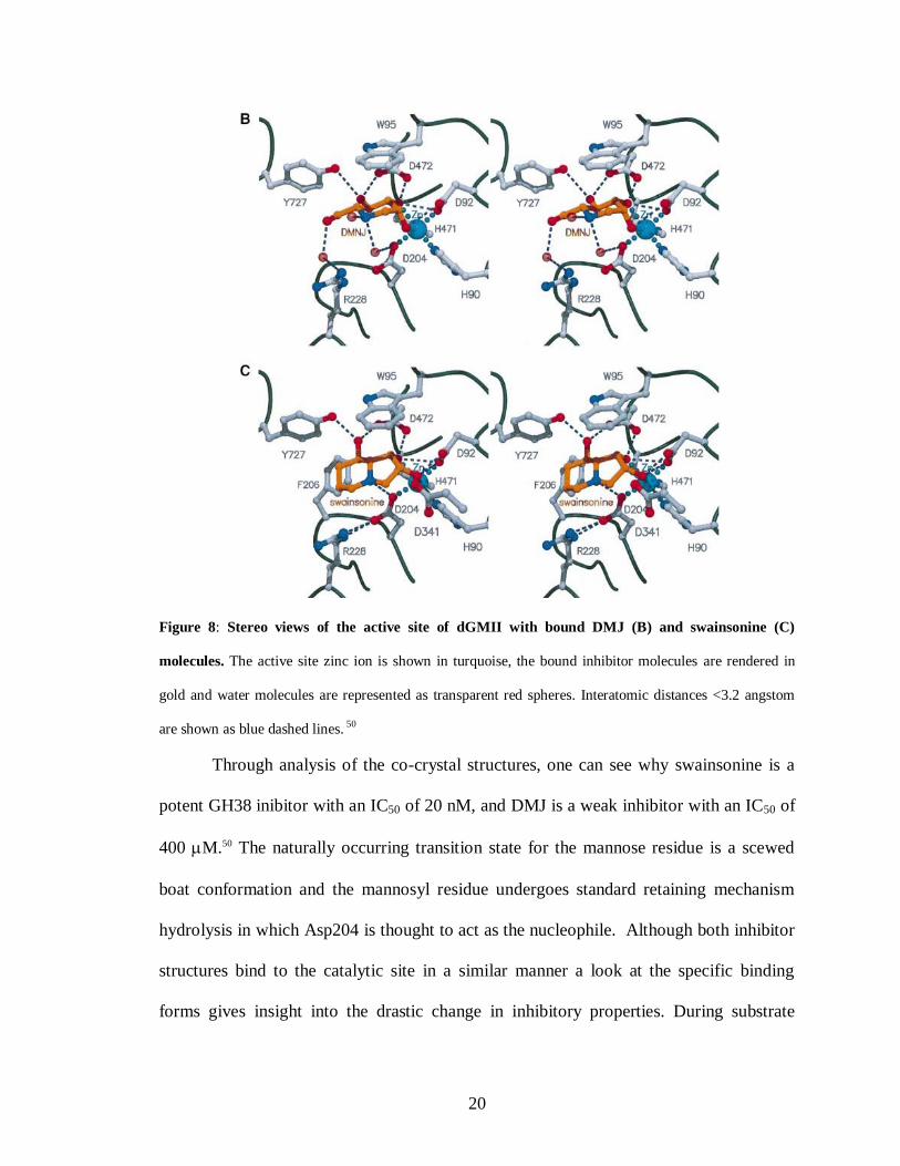
Figure 8: Stereo views of the active site of dGMII with bound DMJ (B) and swainsonine (C)
molecules. The active site zinc ion is shown in turquoise, the bound inhibitor molecules are rendered in
gold and water molecules are represented as transparent red spheres. Interatomic distances <3.2 angstom
are shown as blue dashed lines. 50
Through analysis of the co-crystal structures, one can see why swainsonine is a
potent GH38 inibitor with an IC50 of 20 nM, and DMJ is a weak inhibitor with an IC50 of
400 M.50 The naturally occurring transition state for the mannose residue is a scewed
boat conformation and the mannosyl residue undergoes standard retaining mechanism
hydrolysis in which Asp204 is thought to act as the nucleophile. Although both inhibitor
structures bind to the catalytic site in a similar manner a look at the specific binding
forms gives insight into the drastic change in inhibitory properties. During substrate
21
binding to the enzyme, binding of the C2 and C3 hydroxyls of swainsonine to the Zn ion
flattens the 5-membered ring of swainsonine. Flattening of this ring causes the molecule
to orient in such a way that the bridge head nitrogen, which mimics C1 in the natural
substrate, is brought significantly closer to the nucleophilic Asp204. On the other hand, a
look at DMJ in the binding site illustrates that the endocyclic nitrogen is oriented farther
away from the nucleophilic amino acid residue Asp204 caused by the O2 and O3
hydroxyls coordinating with the Zn ion. These differences in orientation could lead to
the significant difference in potency between swainsonine and DMJ.
Although swainsonine was shown to reduce tumor growth and metastasis with
limited side effects in preliminary clinical trials for treatment of late-stage cancer,
swainsonine was later found to inhibit lysosomal mannosidases when tested in vivo; thus
resulting in the animals developing lysosomal storage disease symptoms.
Kifunensine was discovered in actinomycete Kitasatosporia kifunense 9482, and
is an alkaloid corresponding to the cyclic oxamide derivative of 1-amino mannojirimycin.
Kifunensine has been reported to inhibit both the ER and Golgi mannosidase enzymes in
nanomolar concentrations; thus making it one of the most potent and effective
glycoprotein processing inhibitors to date for GH47 enzymes. The bicyclic nature of KIF
naturally locks the substrate in the 1C4 conformation making the substrate an entropically
favorable inhibitor that mimics the transition state of the naturally occurring mannosyl
residue. However, this compound is not selective and will inhibit both the ER and Golgi
enzymes.
Through modification of Kifunensine there is a potential to synthesize new potent
and selective inhibitors. The two most common methods for iminosugars modification
22
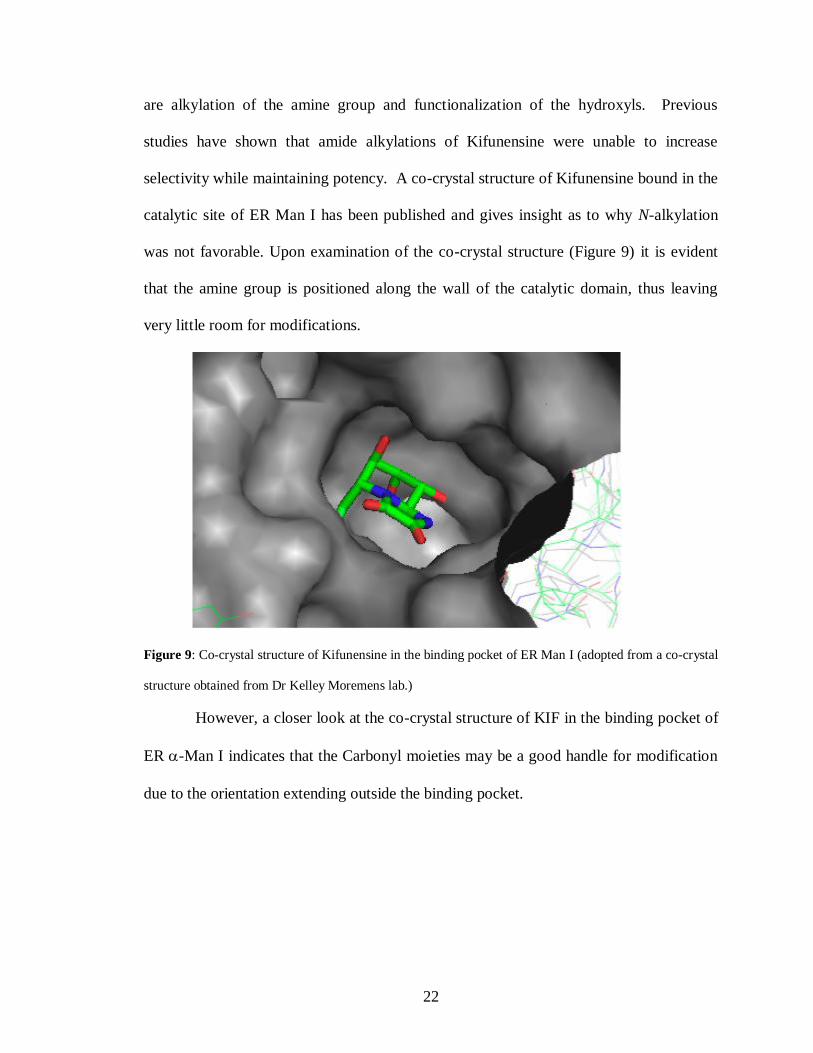
are alkylation of the amine group and functionalization of the hydroxyls. Previous
studies have shown that amide alkylations of Kifunensine were unable to increase
selectivity while maintaining potency. A co-crystal structure of Kifunensine bound in the
catalytic site of ER Man I has been published and gives insight as to why N-alkylation
was not favorable. Upon examination of the co-crystal structure (Figure 9) it is evident
that the amine group is positioned along the wall of the catalytic domain, thus leaving
very little room for modifications.
Figure 9: Co-crystal structure of Kifunensine in the binding pocket of ER Man I (adopted from a co-crystal
structure obtained from Dr Kelley Moremens lab.)
However, a closer look at the co-crystal structure of KIF in the binding pocket of
ER -Man I indicates that the Carbonyl moieties may be a good handle for modification
due to the orientation extending outside the binding pocket.

23
REFERENCES
1. Kornfeld R, Kornfeld S. Assembly of asparagine-linked oligosaccharides, Annu. Rev.
Biochem, 1985,54, 631-64.
2. Trombetta ES. Biological roles of oligosaccharides: all of the theories are correct
Glycobiology, 1993, 3, 97-130.
3. Apweiler R, Hermjakob H, Sharon N. On the frequency of protein glycosylation, as
deduced from analysis of the SWISS-PROT database, Biochim. Biophys. Acta, 1999,
1473, 4-8.
4. Wormald MR, Petrescu AJ, Pao YL, Glithero A, Elliot T, Dwek RA. Conformational
Studies of Oligosaccharides and Glycopeptides: Complementarity of NMR, X-ray
Crystallography, and Molecular Modelling, Chem. Rev, 2002, 102, 371-86.
5. Petrescu AJ, Milac A-L, Petrescu SM, Dwek RA, Wormald MR. Statistical analysis
of the protein environment of N-glycosylation sites: implications for occupancy,
structure, and folding, Glycobiology, 2004,14,103-14.
6. Rudd PM, Wormald MR, Stanfield RL, Huang M, Mattsson N, et. Al. Roles for
glycosylation of cell surface receptors involved in cellular immune recognition, J.
Mol. Biol., 1999, 293, 351-66.
7. Lowe JB, Marth JD. A Genetic Approach to Mammalian Glycan Function, Annu.
Rev. Biochem., 2003, 72, 643-91.
8. Weis WI, Drickamer K. Structural Basis of LectiN-Carbohydrate Recognition, Annu.
Rev. Biochem. 1996, 65, 441-73.
9. Lis H, Sharon N. Lectins: Carbohydrate-Specific Proteins That Mediate Cellular
Recognition, Chem. Rev. 1998, 98, 637-74.
10. Burda P, Aebi M. The dolichol pathway of N-linked glycosylation, Biochim Biophys.
Acta., 1999,1426, 239-57.
11. Hirschberg CB, Snider MD. Topography of glycosylation in the rough endoplasmic
reticulum and Golgi apparatus, Annu. Rev. Biochem, 1987, 56, 63-87.
12. Helenius J, Ng DTW, Marolda CL, Walter P, Valvano MA, Aebi M. Translocation of
lipid-linked oligosaccharides across the ER membrane requires Rft1 protein, Nature,
2002,415, 447-50.
13. Balasubramanian K, Schroit AJ. Aminophospholipid asymmetry: A matter of life and
death, Annu. Rev. Physiol., 2003, 65,701-34.

24
14. Helenius J, Aebi M. Transmembrane movement of dolichol linked carbohydrates
during N-glycoprotein biosynthesis in the endoplasmic reticulum, Semin. Cell Dev.
Biol,. 2002,13,71-78.
15. Burda P, Jakob CA, Beinhauer J, Hegemann JH, Aebi M. Ordered assembly of the
asymmetrically branched lipid-linked oligosaccharide in the endoplasmic reticulum is
ensured by the substrate specificity of the individual glycosyltransferases,
Glycobiology, 1999, 9, 617-25.
16. Burda P, Aebi M. The ALG10 locus of Saccharomyces cerevisiae encodes the alpha-
1,2 glucosyltransferase of the endoplasmic reticulum: the terminal glucose of the
lipid-linked oligosaccharide is required for efficient N-linked glycosylation,
Glycobiology, 1998, 8, 455-62.
17. Spiro RG. Glucose residues as key determinants in the biosynthesis and quality
control of glycoprote, J. Biol. Chem., 2000, 275, 35657-60.
18. Nilsson IM, von Heijne G. Determination of the distance between the
oligosaccharyltransferase active site and the endoplasmic reticulum membrane, J.
Biol. Chem., 1993, 268, 5798-801.
19. Helenius A. How N-linked oligosaccharides affect glycoprotein folding in the
endoplasmic reticulum, Mol. Biol. Cell, 1994, 5, 253-65.
20. Imberty A, Perez S. Stereochemistry of the N-Glycosylation Sites in Glycoproteins,
Protein Eng., 1995, 8, 699-709.
21. Wormald MR, Dwek RA. Struct. Fold. Des., 1999, 7, R155-60.
22. Kundra R, Kornfeld S. Asparagine-linked oligosaccharides protect Lamp-1 and
Lamp-2 from intracellular proteolysis, J. Biol. Chem., 1999, 274, 31039-46.
23. Ellgaard L, Molinari M, Helenius A. Setting the standards: quality control in the
secretory path- way, Science, 1999, 286, 1882-88.
24. Parodi A. Protein glucosylation and its role in protein folding, Annu. Rev. Biochem.,
2000, 69, 9-93.
25. Schrag JD, Bergeron JJ, Li Y, Borisova S, Hahn M, et al. The Structure of calnexin,
an ER chaperone involved in quality control of protein folding, Mol. Cell, 2001, 8,
633-44.
26. Chevet E, Cameron PH, Pelletier MF, Thomas DY, Bergeron JJ. The endoplasmic
reticulum: integration of protein folding, quality control, signaling and degradation,
Curr. Opin. Struct. Biol,. 2001, 11, 120-24.

25
27. Ellgaard L, Helenius A. Quality control in the endoplasmic reticulum, Nat. Rev. Mol.
Cell Biol., 2003, 4, 181-91.
28. Tsai B, Ye Y, Rapoport TA. Retro-translocation of proteins from the endoplasmic
reticulutn into the cytosol, Nat. Rev. Mol. Cell Biol, 2002, 3, 246-55.
29. Kostova Z, Wolf DH. For whom the bell tolls: protein quality control of the
endoplasmic reticulum and the ubiquitin–proteasome connection, EMBO J., 2003, 22,
2309-17.
30. McCracken AA. Evolving questions and paradigm shifts in endoplasmic-reticulum-
associated degradation (ERAD), BioEssays, 2003, 25, 868-77.
31. Yoshida Y. A novel role for N-glycans in the ERAD system, J. Biochem. 2003, 134,
183-90.
32. Lippincott-Schwartz J, Bonifacino JS, Yuan LC, Klausner RD. Degradation from the
endoplasmic reticulum: disposing of newly synthesized proteins, Cell, 1988, 54, 209-
20.
33. Molinari M, Calanca V, Galli C, Lucca P, Paganetti P. Role of EDEM in the release
of misfolded glycoproteins from the calnexin cycle, Science, 2003, 299, 1397-400.
34. Mancini R, Aebi M, Helenius A. Multiple endoplasmic reticulum-associated
pathways degrade mutant yeast carboxypeptidase Y in mammalian cells, J. Biol.
Chem., 2003, 278, 46895-905.
35. Jakob CA, Burda P, Roth J, Aebi M. Degradation of misfolded endoplasmic
reticulum glycoproteins in Saccharomyces cerevisiae is determined by a specific
oligosaccharide structure, J. Cell Biol., 1998, 142, 1223-33.
36. Herscovics A. Structure and function of Class I alpha 1,2-mannosidases involved in
glycoprotein synthesis and endoplasmic reticulum quality control, Biochimie, 2001,
83, 757-62.
37. Liu Y, Choudhury P, Cabral CM, Sifers RN. Oligosaccharide Modification in the
Early Secretory Pathway Directs the Selection of a Misfolded Glycoprotein for
Degradation by the Proteasome, J. Biol. Chem., 1999, 274, 5861-67.
38. Wu Y, Swulius MT, Moremen KW, Sifers RN. Elucidation of the molecular logic by
which misfolded α1-antitrypsin is preferentially selected for degradation, Proc. Natl.
Acad. Sci. USA, 2003, 100, 8229-34.

26
39. Jakob CA, Bodmer D, Spirig U, Battig P, Marcil A, et al. Htm1p, a mannosidase-like
protein, is involved in glycoprotein degradation in yeast, EMBO Rep. 2001, 2, 423-
30.
40. Nakatsukasa K, Nishikawa S, Hosokawa N, Nagata K, Endo T. Mnl1p, an α-
Mannosidase-like Protein in Yeast Saccharomyces cerevisiae, Is Required for
Endoplasmic Reticulum-associated Degradation of Glycoproteins, J. Biol. Chem.,
2001, 276, 8635-38.
41. Hosokawa N, Wada I, Hasegawa K, Yorihuzi T, Tremblay LO, et al. Mnl1p, an alpha
-mannosidase-like protein in yeast Saccharomyces cerevisiae, is required for
endoplasmic reticulum-associated degradation of glycoproteins, EMBO Rep. 2001, 2,
415-22.
42. van Anken E, Romijn EP, Maggioni C, Mexghrani A, Sitia R, et al. Sequential waves
of functionally related proteins are expressed when B cells prepare for antibody
secretion, Immunity, 2003, 18, 243-53.
43. Suh K, Bergmann JE, Gabel CA. Selective retention of monoglucosylated high
mannose oligosaccharides by a class of mutant vesicular stomatitis virus G proteins. J. Cell Biol., 1989, 108, 811-19.
44. Sousa MC, Ferreri-Garcia MA, Parodi AJ. Recognition of the oligosaccharide and
protein moieties of glycoproteins by the UDP-Glc: glycoprotein glucosyltransferase.
Biochemistry, 1992, 31, 97-105
45. Ou WJ, Cameron PH, Thomas DY, Bergeron JJ. Association of
folding intermediates of glycoproteins with calnexin during protein maturation.
Nature, 1993, 364, 771-76.
46. Hammond C, Helenius A. Folding of VSV G protein: sequential interaction with BiP
and calnexin, Science, 1994, 266, 456-58.
47. Lizal C, Gerber S, Numao S, Aebi M, Locher K. X-ray Structure of a Bacterial
Oligosaccharyltransferase, Nature, 2011, 474, 350-55
48. Karaveg K, Siriwardena A, Tempel W, Liu ZJ, Glushka J, Wang BC, Moremen K.
Mechanism of Class 1 (Glycosylhydrolase Family 47) a-Mannosidases Involved in N-
glycan Processing and Endoplasmic Reticulum Quality Control, J. Bio. Chem., 2005,
280, 16197-16207.

27
49. van den Elsen J.M.H., Kuntz D.A., Rose D.R. Structure of Golgi a-mannosidase II: a
target for inhibition of growth and metastasis of cancer cells, The EMBO Journal,
2001, 20, 3008-3017.
28
CHAPTER 2
THE DEVELOPMENT OF SELECTIVE INHIBITORS FOR ER α-MANNOSIDASE I
BY COMBINATORIAL MODIFICATION OF KIFUNENSINE
Jessica Cardot, Yong Xiang, Kelley Moremen, Geert-Jan Boons
To be submitted to J. Am. Chem. Soc.

29
Abstract
Selective inhibitorsfendoplasmic reticulum -mannosidase I (ER Man I) have
the potential to be used for treatment of a number of genetic diseases. Although potent
inhibitors for this enzyme have been described, in general these compounds display poor
selectivity and often inhibit Golgi -mannosidase I. To address this difficulty, we have
synthesized and combinatorial modified kifunensine, which is a potent GH47 -
mannosidase I inhibitor. Thus, kifunensine was modified by a hydrazone moiety, which
could easily be extended by reaction with a range of aldehydes. The library of hydrazone-
linked analogues was screened for inhibition of human ER Man I and mouse Golgi Man
I. It was found that a pyridine functionalized kifunensine analog selectively inhibited ER
Man I. Based on this finding, a second generation of analogues was prepared in which a
functionalized pyridine scaffold was coupled to kifunensine through a hydrazone moiety.
Introduction and Literature Review
N-glycans are widely expressed on the cell surface of eukaryotic cells and are
responsible for many biological processes. Several diseases are specifically associated
with disruption or breakdown of N-glycan biosynthesis, such as lysosomal storage
disease and neurological disorders. 1, 2
The N-glycan biosynthetic pathway consists of a
cascade of trimming and elongation steps performed by various glycosyl hydrolase and
transferase enzymes. During this cascade, if a nascent protein is detected the endoplasmic
reticulum contains a quality control mechanism known as endoplasmic reticulum
associated degradation (ERAD) to ensure that only native glycoproteins are translocated
to the Golgi apparatus for further elongation and expression. The nascent glycoproteins

30
are sequestered in the ER and translocated to the cytosol for disposal. 1, 2
ER--
mannosidase I (ER Man I) is a predominant enzyme in ERAD and is responsible for the
trimming of a specific mannose residue from the core Man9GlcNAc2 structure during the
maturation of the glycoproteins. Several studies have shown that ER Man I inhibition
results in a decrease in nascent glycoprotein disposal, whereas overexpression of ER Man
I lead to an increase in glycoprotein disposal. These results lead to the proposed model
that trimming of the Man9GlcNAc2 core to the Man8GlcNAc2 structure of nascent or
incompletely folded glycoproteins was the rate-determining step in initiating glycoprotein
disposal, thus making ER Man I a promising target for drug therapies for diseases
associated with misfolded glycoproteins. 3, 4
Although there are many glycosidases involved in N-glycan biosynthesis they can
be categorized by two main catalytic subtypes; inverting and retaining mechanisms.
Catalytic mechanism and classification is dependent mainly on sequence similarit ies, and
preferred substrates. However, inverting and retaining enzymes can be further classified
into CAZy glycosyl hydrolase (GH) families based on product stereochemistry, bond
specificity, and inhibitor structure. Therefore, -mannosidases within the same CAZy
GH family will have remarkably similar catalytic binding domains and substrate
specificity.
ER Man I is a GH47 enzyme that is believed to play a vital role in N-glycan
biosynthesis and degradation. The GH47 family contains three subfamilies: ER Man I
and Golgi Man IA-IC (-1,2-mannosidases), and EDEM1-3 (ER stress induced proteins).
GH47 enzymes are one of three GH families that are calcium dependent. Although the
GH47 enzymes have been implicated in nascent glycoprotein disposal, their roles and

31
actual involvement is under investigation. Therefore, the development of a selective
inhibitor that could distinguish between the various GH47 enzymes would not only be a
target for drug development but would also aid in the understanding of the degradation
pathway.
Polyhydroxylated iminosugars are widely known as strong glycosidase inhibitors
and have demonstrated promising results as therapeutic agents. Although iminosugars
are potent glycosidase inhibitors there have been limited successful therapeutics
developed. The lack of safe and efficacious glycosidase inhibitors is largely due to
promiscuity and lack of selectivity towards glycosidase families inherent of these
substrates. Kifunensine, a naturally occurring alkaloid, is a potent GH47 -mannosidase
inhibitor, however, kifunensine cannot differentiate between different GH47 enzymes
such as ER Man I and Golgi Man I.7 Golgi Man I is a critical enzyme in N-glycan
maturation and elongation, therefore inhibition of this enzyme is problematic for proper
cellular function. 5 Due to the fact that there are many potent inhibitors in the literature,
modification of these known compounds with motifs to increase selectivity is a promising
avenue for drug development.
The two most common methods for iminosugar modifications are alkylation of
the amine group and functionalization of the hydroxyls. Previous studies have shown
that amine alkylations of kifunensine were unable to increase selectivity while
maintaining potency.10
A co-crystal structure of kifunensine bound in the catalytic site of
ER Man I has since been published and gives insight to why N-alkylation was not
favorable. Upon examination of the co-crystal structure it is evident that the amine group
is positioned along the wall of the catalytic domain, thus leaving very little room for
32
modifications; however, the carbonyl groups are oriented in a plan that appears to be
directed outside the catalytic site. We envisaged that modification of the kifunensine
carbonyls with various functionalities could potentially increase selectivity while
maintaining high potency. Here we report the first selective modification of the C2
carbonyl of kifunensine resulting in a selective potent inhibitor. Our strategy employed a
chemoselective coupling between a kifunensine derivative functionalized with a
hydrazone moiety at the C2 position. The hydrazone analogue was utilized as a lead
substrate in the combinatorial synthesis of a small library of kifunensine analogues using
a small selection of commercially available aldehydes. The resulting dihydrazone-based
library was tested against GH47 inverting enzymes Human ER Man I, and Mouse Golgi
Man I for selective inhibition. A pyridine functionalized KIF analogue demonstrated
high selectivity and potency. The pyridine functionalized analogue underwent further
biological testing to determine if the potency and selectivity would transfer from a
mocrotiter plate assay to a cell study.
Results and Discussion
The initial challenge was to devise a synthetic strategy that could distinguish
between the C2 and C3 carbonyls and allow selective installation of a chemical handle
for diversification. This motif had to be highly selective and efficient to be a viable
linker. A hydrazone group was determined to be an appealing option for library synthesis
due to the highly selective and robust reaction with aldehydes, even in the presence of
complex substrates.

33
Synthesis of Azidolactone
Commercially available L-gulonic--lactone is an inexpensive starting material
that allows for the synthesis of compound 6 in five synthetic manipulations. The first
step was isopropylidene protection of the two diols with copper sulfate, acetone with a
catalytic amount of sulfuric acid to achieve the desired bisacetonide 2 in a quantitative
reaction.8 Selective removal of the 5,6-O-isopropylidene of 2 by treatment with acetic
acid/water (7/1), at room temperature, afforded compound 3 in 93 % yield. Silyl
protection of the primary alcohol using tert-butyldimethylsilyl chloride (TBS-Cl) and
imidazole in DMF furnished silyl ether 4 in 89 % yield.9 The secondary alcohol
underwent intermediate triflation by treatment with triflic anhydride and pyridine in
DCM. The crude triflate was charged with sodium azide in DMF to afford the desired
azidolactone 6 in 72 % yield over two steps.9, 10
(Scheme 1)
O
O
O
O
OH
HO
O
O
HO
OH
OH
HO
O
O
O
O
O
O
H2SO4, CuSO4 Acetone
quant.
CH3COOH, H2O (7:1)
O
O
O
O
OH
TBSO
TBS-Cl, imidazole, DMF89 %
O
O
O
O
N3
TBSO
O
O
O
O
OTf
TBSO
Tf2O, Pyridine, DCMNaN3, DMF
1 2 3
456
93 %
72 % over 2 steps
Scheme 1: Synthetic scheme for azido lactone 6.

34
Synthesis of Bisacetonide KIF
Reduction of lactone 6 using sodium borohydride afforded diol 7 in 85 % yield,11
which underwent selective protection of the primary alcohol as a pivaloyl ester using Piv-
Cl and pyridine to give 8 in an 80 % yield. The silyl ether of 8 was selectively removed
by treatment with TBAF in THF to afford a second diol, which was transformed into
acetonide 9 using 2-methoxypropene and catalytic PTSA in 92 % yield.12
Treatment of
9 with lithium aluminum hydride effectively reduced both the azide and ester moieties to
achieve the essential amine 10 in a 90 % yield. Under standard peptide coupling
conditions, using HOBt and EDC, oxamic acid was coupled to amine 10 affording the
precursor to bisacetonide KIF, diamide 11.13
The yield of the coupling reaction ranged
from 70-90 % yield depending on the quality of EDC and MP-carbonate used. DMP
oxidation of the primary alcohol to an aldehyde allowed for the subsequent cyclization of
the diamide in the presence of ammonia in methanol to afford bisacetonide KIF 12 in a
yield of 33-55 % over two steps.10
This reaction relies on the quality of DMP as well as
ensuring removal of excess DMP and the DMP by-product formed during the oxidation
prior the introduction of ammonia, which is difficult do to since the intermediate
aldehyde decomposes on silica gel (Scheme 2).

35
OO
OO
N3TBSO
OHOH
OO
N3TBSO
OHOPiv
OO
N3TBSO
O
O
N3
OPivO
O
O
O
NH2
OHO
O
O
O
NH
OHO
O
NH2
O
O
O
O
N
O
O
NH
O
O
NaBH4, EtOH
85 %
PivCl, Pyr, DCM
1. TBAF, THF, -20 oC, 93 %
2. 2-methoxy propene,
PTSA, CH2Cl2, 92 %
1. DMP
2. NH3/MeOH
46 % over 2 steps
LiAlH4, Et2O
HOBt, EDC, Oxamic acid, DMF, MP-Carbonate resin
6 7 8
91011
12
80 %
90 %70-90 %
Scheme 2: Synthesis of bisacetonide Kifunensine
Synthesis of Novel Hydrazone Modified KIF
The synthesis of a novel hydrazone KIF began with the selective thionation of the
C2 carbonyl of KIF utilizing Lawesson’s reagent. During the reaction, the secondary
amide of the C2 position coordinates with the Lawesson’s reagent fascilitating the
transformation preferentially occurring at the C2 position and not the C3 carbonyl. Once
12 was synthesized, the bicyclic diamide was subjected to Lawesson’s thionation reagent,
which converted the secondary amide into thioamide 13 in yields ranging from 65-90 %.
The success of this reaction relies on two features, i: that any remaining DMP/by-
products from the previous reaction are removed, and ii: that the Lawessons reagent is of
high quality.14
Deprotection of 13 with 75 % TFA/water to deblock the acetonide
protecting groups resulted in the formation thioKIF in quantitative yields. Treatment of

36
14 with hydrazine hydrate resulted in the hydrazone analogue 15 in 83 % yield.15
Hydrazone KIF is a suitable candidate for combinatorial modification (Scheme 3).
O
O
N
O
O
NH
O
O
O
O
N
O
O
NH
O
S
HO
HO
N
HO
HO
NH
O
SLawessons reagent TFA/H2O
quant.65-90 %
HO
HO
N
HO
HO
NH
O
NHydrazine Hydrate
EtOH, 83 %NH2
Scheme 3: Synthesis of novel Hydrazone KIF
Synthesis of Novel Kifunensine Analogues
With the successful synthesis of hydrazone functionalized KIF and the purchase
of fourteen different aldehydes including aromatic, heterocyclic, and aliphatic aldehydes,
a small library of dihydrazone functionalized KIF analogues was synthesized. Hydrazone
15 was dissolved in 1 % AcOH in DMSO followed by the addition of the selected
aldehyde (Figure 10).

37
HO
HO
N
HO
HO
NH
O
N
NH2
HO
HO
N
HO
HO
NH
O
NN
R1 % AcOH, DMSO
H R
O
R=
16
OH
OH
20
S
24
Br
17
N
21
25
F
18
N
NH
22
26
OMe
OH
19
O
23
27
Figure 10: Structures of compounds in initial library screening, 15-27
The reaction was monitored by HRMS-MALDI-TOF. Once complete, the
compounds were purified by HPLC and screened for inhibition and selectivity towards
ER Man I, and Golgi Man I. (Table 2).

38
Table 2: Ki Inhibition data for kifunensine analogues 15-27.
Inhibitor ER Man I (M) Golgi Man I (M) Golgi/ER
Kifunensine 0.06 0.08 1.0
Hydrazone (15) 0.83 0.68 1.0
16 1.94 3.10 1.5
17 2.31 1.30 0.5
18 0.93 2.32 2.5
19 1.70 0.86 0.5
20 0.27 0.98 3.5
21 0.07 0.66 9.5
22 0.69 2.10 3.0
23 1.60 2.00 1.5
24 1.05 2.40 2.5
25 1.62 1.90 1.0
26 1.00 1.92 2.0
27 1.50 2.00 1.5
The initial modification to kifunensine with a hydrazone moiety resulted in
approximately a 10-fold decrease in activity along with no selectivity. It is known that
many proteins contain phenylalanine, tryptophan, and tyrosine residues each of which
contain an aromatic side chain. The aromatic substituents are generally buried within the
more hydrophobic core and help stabilize the structure of the protein through pi-pi
stacking interactions. With this in mind, a range of aromatic substituents was coupled to
Kif through the hydrazone linker in order to promote possible pi-pi stacking interactions

39
within the catalytic site. A baseline reaction with benzaldehyde, 16, resulted in a
decrease in activity. Moderately deactivated aromatic substituents, compounds 17 and 18,
had comparable inhibition to 16, but were still less effective than both kifunensine and
the hydrazone. A moderately activated aromatic substituent, containing an electron
donating hydroxy functionality, 19, resulted in a minor increase in activity. Although 19
had comparable activity to the hydrazone, no selectivity was achieved. The more strongly
activated diol substituted aromatic compound 20 resulted in a 4-fold increase in activity
compared to the hydrazone along with 3-fold selectivity towards the ER-Man I enzyme.
These results indicate that a more electron rich aromatic ring has more favorable
interactions with the ER-Man I catalytic site in respect to the more electron deficient
aromatics.
Heterocyclic aromatic rings were then investigated. The pyridine functionalized
analogue 21 demonstrated a significant increase in activity compared the hydrazone
analogue with a Ki of 0.07 M for ER Man I, which is the same as kifunensine.
Interestingly, analogue 21 had a Ki of 0.66 mM for the Golgi Man I enzyme, resulting in
a 9.5-fold preference to the ER enzyme over the Golgi enzyme. To date, this is the first
selective inhibitor for ER-Man I that maintained potency. The introduction of the ring
nitrogen in analogue 21 led to a 28-fold increase in activity for the ER enzyme and only a
4.5-fold increase in activity for the Golgi enzyme, as seen in the comparison of analogues
16 and 21. These two analogues have comparable configurations but differ in electronics.
In contrast to benzene, the electron density of pyridine is not evenly distributed resulting
in a dipole, as well as the nitrogen is slightly basic, one or both of these properties may
influence the increased binding affinity.

40
A series of analogues containing heterocyclic aromatic substituents was
synthesized including imidazole (22), furan (23), and thiophene (24) functionalized
kifunensine. Of these heterocycles, the imidazole functionalized derivative demonstrated
the best results with an ER Man I Ki comparable to the parent hydrazone, as well as 3-
fold selectivity towards the ER. These results indicate that the nitrogen atom plays a key
role in the interactions between enzyme and substrate
Long aliphatic chain substituents, analogues 25, 26, and 27, were synthesized and
yielded no selectivity, and low potency.
A small library of functionalized pyridine analogues was synthesized from
commercially available aldehydes to investigate substituent effects on potency and
selectivity (Figure 11).

41
HO
HO
N
HO
HO
NH
O
N
NH2
HO
HO
N
HO
HO
NH
O
NN
R1 % AcOH, DMSO
H R
O
NH2N
28
29
N
OMe
30
N Cl
31
N
BocHN
32
N
NHPiv
33
N
NHPiv
34
NH
O
35
N
36
N
O
37
N
HN
38
NCOOH
39
N
N
40
Figure 11: Functionalized Pyridine dihydrazone kifunensine analogues, 28-40
R =

42
Table 3: Ki Inhibition data for pyridine kifunensine analogues 28-40
Inhibitor ER Man I (M) Golgi Man I (M) Golgi/ER
Kifunensine 0.06 0.08 1.3
Pyridine-Kif, 21 0.07 0.66 9.5
28 1.29 2.67 2.0
29 0.37 0.91 2.5
30 1.48 3.55 2.5
31 0.18 0.80 4.5
32 1.08 5.00 4.5
33 2.25 11.23 5.0
34 0.86 3.06 3.5
35 1.26 4.73 4.0
36 0.62 3.23 5.5
37 0.81 2.79 3.5
38 1.41 5.20 4.0
39 1.29 8.55 6.5
40 0.39 1.70 4.5
From the functionalized pyridine series, there were eight analogues that
demonstrated greater than 4-fold selectivity towards the ER; however the inhibition was
mediocre. It appears that the selectivity is caused more by the Golgi enzyme having
unfavorable interactions with the bulkier substituents rather than positive interactions
with the ER enzyme, evident by the high Ki values. However, the 4-chloropyridine and
pyrimidine functionalized Kifunensine analogues, 16 and 25 respectively, demonstrated

43
4.5 fold selectivity and high nanomolar inhibition. This set of analogues indicates that the
Golgi enzyme does not have the ability to accept larger substrates into the binding pocket
in comparison to the ER enzyme.
Cellular Study of Pyridine Kifunensine
Analogue 21 was subjected to an endoglycosidase F (Endo F) sensitivity assay to
determine influences on N-glycan processing in the Golgi apparatus. Endo F resistant
glycans are mature glycans that have been extended and elaborated in the core structure,
where as Endo F sensitive glycans are glycans that are in earlier stages of the
biosynthesis. In other words, if the inhibitor inhibits Golgi Man I then the N-glycans will
contain the core high mannose structure needed for recognition by Endo F thereby being
Endo F sensitive. As seen in figure 12, compound 21 takes approximately 10-25 mM
concentration to transition from Endo F resistant to Endo F sensitive glycans indicating
that 21 is approximate 5-20 fold worse at inhibiting Golgi Man I than Kif. Kifunensine
was used as a standard and was found to take approximately 5 mM concentration to
convert from Endo F resistant to Endo F sensitive glycans.

44
Figure 12: Effect of inhibitor treatment on the endoglycosidase sensitivity of a reporter glycoprotein
expressed in mammalian cells. HEK293 cells were transfected with an expression construct encoding a
soluble secreted form the rat sialyltransferase, ST6GAL1, a protein containing 2 complex type type N-
glycan structures. Following transfection the cells were treated for 4 days with the indicated inhibitor
concentration and the conditioned medium containing the recombinant ST6GAL1 was harvested, incubated
with recombinant endoglycosidase F1 (endoF1) at room temperature overnight, resovled by SDS-PAGE,
and immunoblotted using an antibody specific for rat ST6AGL1. ST6AGL1 secreted into the medium from
untreated cell cultures was resistant to endoF1 cleavage since the glycans wereall complex type. Inhibition
of mannose processing in vivo resulted in conversion of the glycans from endoF1-resistant structures to
structures that were sensitive to cleavage by endoF1. Glycan cleavage resulted in a faster mobility of
ST6AGL1 by SDS-PAGE. The data indicate that treatment of cells with >5 mM Kif resulted in conversion
to endoF1 sensitive glycan structures, while concentrations >25 mM of the Kif-derivative were required to
produce endoF1 sensitive glycans on ST6GAL1.
These results correspond to the initial Ki studies in which 21 demonstrated 10-
fold worse inhibition of Golgi Man I in respect to Kif, however, 21 and Kif had nearly
identical ER Man I inhibition.

45
Conclusions
In summary, the design and synthesis of a novel C2 hydrazone functionalized
Kifunensine analogue was achieved. Through this synthetic route a library of
dihydrazone functionalized Kifunensine analogues was prepared and tested for inhibitory
activity against Human ER Man I and Mouse Golgi Man I. A pyridine functionalized
derivative, 21, was found to be a potent and selective inhibitor of ER Man I. Analogue
21 had a Ki of 0.07 M for ER Man I and 0.66 M for Golgi Man I indicating a 10-fold
preference for the ER enzyme. The selectivity was further demonstrated through an Endo
F sensitivity assay in which compound 21 was around 5-10 fold worse than Kif at
inhibiting downstream N-glycan processing.
Acknowledgments- I would like to thank the Moremen laboratory for performing all of
the Ki assays and cellular studies for our inhibitor molecules.
Experimental Section
General Methods and Materials
All chemicals were commercially available and used without further purification.
Reactions were carried out using anhydrous solvents under an Argon atmosphere unless
otherwise stated. Anhydrous DCM was distilled from calcium hydride under a nitrogen
atmosphere. All column chromatography was carried out on Silica gel 60 (EM Science
70-230 mesh), and reactions were monitored using TLC (EM science, kiesel gel 60 F254
on aluminum) stained with cerium sulfate/ ammonium molybdate solution.

46
2,3:5,6-Di-isopropylidene-L-gulunolactone (2) L-gulonic--lactone (1) (25 g, 0.14 mol)
was dissolved in acetone (820 mL) and charged with anhydrous CuSO4 (89.6 g, 0.56
mol) and catalytic H2SO4 ( 2.55 mL). The reaction mixture was stirred under argon for 48
hrs until all starting material and intermediate mono protected intermediate were
consumed. Once complete, the reaction was filtered through a buchner funnel and the
filtrate was quenched with NaHCO3. The solids were again filtered and the filtrate was
concentrated on a rotovap to an oily solid residue. Recrystallization from EtOAc yields
pure product as an off white solid in quantitative yields (36 g). 1H NMR (300 MHz,
CDCl3) δ 4.83 (d, J = 5.6 Hz, H-3, 1H), 4.74 (dd, J = 5.6, 3.5 Hz, H-2, 1H), 4.56 – 4.35
(m, 2H), 4.22 (dd, J = 8.9, 6.3 Hz, H-5, 1H), 3.82 (dd, J = 8.8, 6.0 Hz, 1H), 1.56 (s, 2H),
1.47 (s, 6H), 1.40 (s, 3H), 1.38 (s, 3H). Calculated MW was 258.27 MALDI-TOF found
[M+H] 259.27.
2,3-isopropylidene-L-gulunolactone (3) 2,3:5,6-Diisopropylidene-L-gulunolactone 2
(36 g, 0.14 mol) was dissolved in AcOH/Water (7/1, 240 mL) and was stirred at r.t. for
24-48 hrs until no starting material remained. Once complete, the solvents were removed
under reduced pressure and the oily solids were recrystallized from EtOAc in 93 % yield
as light yellow solids (28.3 g). Calculated MW was 218.20, MALDI-TOF found [M+H]
219.20.
6-O-tert-butyldimethylsilyl-2,3-O-isopropylidene-L-gulunolactone (4) 2,3-
isopropylidene-L-gulunolactone ( 28.3 g 0.13 mol) was dissolved in anhydrous DMF and
cooled to -40 oC. To the cooled solution, tert-butyldimethylsilyl chloride (21.5 g 0.14

47
mol) and imidazole (13.26 g .20 mol) were added and the reaction was stirred at -40 oC
for 2 hrs and checked by TLC. Once no starting material remained the reaction was
warmed to r.t. and DMF was removed under reduced pressure. The crude material was
dissolved in DCM (300 mL) and Brine (300 mL). The organic layer was set aside and the
Brine was extracted by DCM (2 x 200 mL). The combined organic layers were dried
over MgSO4, filtered and evaporated to dryness. The colorless oil was purified by flash
silica gel chromatography with hexanes/EtOAc (3/1) mobile phase resulting in clear oil in
89 % yield (38.4 g). 1H NMR (300 MHz, CDCl3) δ 5.06 – 4.65 (m, 1H), 4.50 (dd, J = 6.5,
3.4 Hz, 1H), 3.96 (dd, J = 5.5, 3.0 Hz, 1H), 3.73 (dd, J = 4.5, 1.9 Hz, 1H), 2.75 (d, J = 3.6
Hz, 1H), 1.34 (d, J = 23.8 Hz, 6H), 0.81 (s, 9H), -0.00 (s, 6H). Calculated MW was
332.46, MALDI-TOF found [M+Na] 355.45.
(3S,4S,5R)-5-[(1’R)-1’-Azido-2’-tert-butyldimethylsilanyloxyethyl]-3,4-
isopropylidenedioxytetrahydrofuraN-2-one (6) 6-O-tert-butyldimethylsilyl-2,3-O-
isopropylidene-L-gulunolactone 4 (30.5 g 0.09 mol) was dissolved in dry DCM (250 mL)
and cooled to – 30 oC. Anhydrous pyridine (22.2 mL 0.28 mol) was charged to the vessel
and Tf2O (31 mL 0.18 mol) was added slowly over 5 minutes. The reaction was stirred at
-30 oC for 1 hr. The reaction progress was monitored by TLC (Hexanes/EtOAc 1/1) and
when no starting material remained the reaction was warmed to r.t. diluted with DCM
and washed with 1 N HCl ( 200 mL) followed by water (200 mL), and a final wash with
saturated aqueous Na2CO3 (200 mL). The DCM layer was dried with MgSO4, filtered
and concentrated under reduce pressure. The crude triflate (5) was dissolved in dry DMF
(200 mL) and sodium azide (17.6 g, 0.27 mol) was added. The reaction was stirred

48
overnight under argon. The reaction mixture was diluted with EtOAc (200 mL) and
Brine (200 mL). The EtOAc layer was set aside and the brine layer was extracted with
EtOAc (2 x 200 mL). The combined EtOAc layers were dried with MgSO4, filtered and
concentrated. The crude material was purified by flash silica gel chromatography with
Hexanes/EtOAc (3/1) to give an off white solid (23.2 g). 5-Azido-6-O-tert-
butyldimethylsilyl-5-deoxy-2,3-O-isopropylidene-D-mannonolactone was obtained in 72
% over two steps. 1H NMR (300 MHz, CDCl3) δ 4.96 – 4.56 (m, 2H), 4.29 (dd, J = 10.0,
3.1 Hz, 1H), 3.95 (d, J = 10.8 Hz, 1H), 3.71 (dd, J = 12.0, 6.1 Hz, 1H), 1.34 (d, J = 23.8
Hz, 6H), 0.91 (s, 9H), -0.11 (s, 6H). Calculated MW was 357.48, MALDI-TOF found
[M+H] 358.45.
(2R, 3R, 4R, 5R)-5-Azido-6-tert-butyldimethylsilyl-5-deoxy-2,3-isopropylidene-
dioxy-1,4-hexanediol (7) (3S,4S,5R)-5-[(1’R)-1’-Azido-2’-tert-
butyldimethylsilanyloxyethyl]-3,4-isopropylidenedioxytetrahydrofuraN-2-one (23.2 g,
0.06 mol) was dissolved in EtOH (600 mL) and charged with sodium borohydride (4.9 g
37.8 mol) at r.t.. The reaction was stirred for 4 hrs under argon, TLC showed no more
starting material. The reaction was quenched with the addition of ammonium chloride
until no more effervescence. The solution was filtered and concentrated to afford an oily
white solid. The crude was purified by flash silica gel chromatography with
Hexanes/EtOAc (3/1) eluent to afford (2R, 3R, 4R, 5R)-5-Azido-6-O-tert-
butyldimethylsilyl-5-deoxy-2,3-O-isopropylidene-dioxy-1,4-hexanediol as a clear oil
(19.9 g) in 85 % yield. 1H NMR (300 MHz, CDCl3) δ 4.41 (dd, J =1.4, 7.3 Hz, 1H), 4.29
(m, 1H), 4.09 (dd, J = 3.1, 10.6 Hz, 1H), 3.8-3.9 (m, 2H), 3.79 (dd, J = 4.4, 12.4 Hz, 1H),

49
3.60 (dd, J = 1.4, 9.1 Hz, 1H), 3.46 (m, 1H), 1.34 (d, J = 23.8 Hz, 6H), 0.91 (s, 9H), -
0.11 (s, 6H). Calculated MW was 361.51, MALDI-TOF found [M+H] 362.50.
(2R, 3R, 4R, 5R)-2-Azido-1-tert-butyldimethylsilyl-4,5-isopropylidene-dioxy-6-
pivaloyl-3-hexanol (8) (2R, 3R, 4R, 5R)-5-Azido-6-tert-butyldimethylsilyl-5-deoxy-2,3-
isopropylidene-dioxy-1,4-hexanediol (19.9 g, 0.06 mol) was dissolved in dry DCM (200
mL) and charged with anhydrous pyridine (11.1 mL 0.14 mol) and trimethylacetyl
chloride (8.1 mL 0.07 mol). The reaction was stirred at r.t. overnight. Once complete,
the reaction was diluted with DCM and washed with water (2 x 200 mL). The organic
layer was dried with MgSO4, filtered and concentrated in vacuo. The crude material was
purified by flash silica gel chromatography with Hexanes/EtOAc (20/1) mobile phase
giving (2R, 3R, 4R, 5R)-2-Azido-1-O-tert-butyldimethylsilyl-4,5-O-isopropylidene-
dioxy-6-pivaloyl-3-hexanol (19.6 g) in 80 % yield. 1H NMR (300 MHz, CDCl3) δ 4.53 –
4.19 (m, 2H), 3.98 (dd, J = 10.6, 3.1 Hz, 2H), 3.77 (dd, J = 10.6, 6.3 Hz, 1H), 3.51 (t, J =
9.1 Hz, 1H), 3.29 (ddd, J = 9.2, 6.3, 3.1 Hz, 1H), 2.33 (d, J = 9.1 Hz, 1H), 1.40 (s, 3H),
1.29 (s, 3H), 1.11 (s, 9H), 0.82 (s, 9H), 0.00 (d, J = 1.8 Hz, 6H). Calculated MW was
445.63, MALDI-TOF found [M+H] 446.60.
(2R, 3R, 4R, 5R)-2-Azido-1,3:4,5-O-di-isopropylidene-dioxy-6-pivaloyl-3-hexanol (9)
(2R, 3R, 4R, 5R)-2-Azido-1-O-tert-butyldimethylsilyl-4,5-O-isopropylidene-dioxy-6-
pivaloyl-3-hexanol (19.6 g, 0.04 mol) was dissolved in THF (500 mL) and cooled to -20
oC. Tetra-N-butylammonium fluoride (5.3 mL, 1 M solution in THF, 0.05 mol) was
added to the cooled solution and stirred for 1 hr and monitored by TLC. Once no starting
50
material remained, the solution was warmed to r.t. and poured into brine, extracted with
EtOAc, dried with MgSO4,filtered and evaporated. The yellow oil ((2R, 3R, 4R, 5R)-2-
Azido-1,3:4,5-O-diisopropylidene-dioxy-6-pivaloyl-3-hexanol, 13.4 g 0.04 mol) was
dissolved in dry DCM (200 mL) at r.t.. PTSA (0.14 g, 0.81 mmol) and 2-
methoxypropene (7.7 mL, 0.08 mol) were added to the solution and the reaction was
stirred for 30 min. The reaction was quenched with water and extracted with DCM (3 x
200 mL). The combined organic layers were dried with MgSO4, filtered and
concentrated. The crude material was purified by flash silica gel chromatography using
Hexanes/EtOAc (6/1) as an eluent. (2R, 3R, 4R, 5R)-2-Azido-1,3:4,5-O-
diisopropylidene-dioxy-6-pivaloyl-3-hexanol (13.8 g) was obtained in 93 % yield. 1H
NMR (300 MHz, CDCl3) δ 4.53 – 4.36 (m, 1H), 4.36 – 4.28 (m, 1H), 3.86 (dd, J = 10.1,
3.0 Hz, 1H), 3.72 – 3.60 (m, 1H), 3.49 (ddd, J = 9.3, 6.3, 3.0 Hz, 1H), 3.25 (s, 1H), 2.37
(d, J = 9.2 Hz, 1H), 1.50 (s, 3H), 1.40 (s, 3H), 1.37 (s, 9H), 1.21 (s, 3H). Calculated MW
was 371.43, MALDI-TOF found [M+H] 372.40.
(2R, 3R, 4R, 5R)-5-Amino-2,3:4,6-diisopropylidene-dioxy-hexanol (10) (2R, 3R, 4R,
5R)-2-Azido-1,3:4,5-di-isopropylidene-dioxy-6-pivaloyl-3-hexanol (13.8 g, 0.04 mol)
was dissolved in diethyl ether (600 mL) and charged with lithium aluminum hydride (4.6
g 0.12 mol) under an argon atmosphere. The reaction was stirred for 2 hrs and checked
by TLC. Once no starting material remained the reaction was quenched with the slow
successive addition of water (5.0 mL), 10 % NaOH (5.0 mL) and water (15.0 mL). The
ether was decanted off and the water was extracted with EtOAc (2 x 50 mL) The organic
layers were combined, dried with MgSO4, filtered and evaporated to a white fluff (10.0
51
g). The crude amine was pure enough to be used for the next step. Calculated MW was
261.31, MALDI-TOF found [M+H] 262.29.
(2R, 3R, 4R, 5R)- 2,3:4,6-diisopropylidene-5-oxamoylamino alcohol (11) (2R, 3R, 4R,
5R)-5-Amino-2,3:4,6-diisopropylidene-dioxy-hexanol (10 g, 0.04 mol) was dissolved in
DMF (300 mL). Oxamic acid (5.3 g 0.06 mol), EDC (11.5 g 0.06 mol) and HOBt (8.1 g
mol) were added to the solution and stirred for 3 hrs. After 3 hrs MP-Carbonate resin
(Loading capacity 3.01 mmol/g, 59.8 g 0.18 mol) was added to the solution and the
reaction was stirred overnight. Once complete, the reaction mixture was filtered through
a cotton plug and washed with DCM (30 mL) to remove the resin. The solvents were
evaporated and the crude material was purified with flash silica gel chromatography
using EtOAc/Hexanes/TEA (10/1/0.1) as a solvent system. (2R, 3R, 4R, 5R)- 2,3:4,6-
diisopropylidene-5-oxamoylamino alcohol was obtained as an off white solid in 90 %
yield (11.9 g). Calculated MW was 332.35, MALDI-TOF found [M+Na] 355.30.
(3aS,3bS,6bR,10aR,10bS)-2,2,9,9-Tetramethyl-1,3,8,10-tetraoxacyclohexa-
[e]cyclopenta[g]-5,6-dione-4-azaiN-dolizidine (diisopropylidene Kifunensine) (12)
(2R, 3R, 4R, 5R)- 2,3:4,6-diisopropylidene-5-oxamoylamino alcohol (1.34 g, 4.0 mmol)
was dissolved in dry DCM (60 mL) and fresh Dess Martin Periodinane (2.1 g, 4.8 mmol)
was added to the solution. The reaction mixture was stirred for 4 hrs at r.t. under argon.
The reaction was diluted in diethyl ether (250 mL) and filtered through a celite pad. The
organics were evaporated to dryness. The crude aldehyde intermediate was dissolved in
7.0 N NH3 in MeOH (5.7 mL, 0.04 mol) and stirred overnight. The reaction was

52
concentrated and purified by flash silica gel chromatography using Hexanes/EtOAc (1/3)
mobile phase. (3aS,3bS,6bR,10aR,10bS)-2,2,9,9-Tetramethyl-1,3,8,10-
tetraoxacyclohexa-[e]cyclopenta[g]-5,6-dione-4-azaiN-dolizidine (.69 g) was synthesized
in 55 % yield over two steps. Calculated MW was 312.32, MALDI-TOF found [M+Na]
335.30.
Thio Analogue of Diisopropylidene Kifunensine 13 (3aS,3bS,6bR,10aR,10bS)-2,2,9,9-
Tetramethyl-1,3,8,10-tetraoxacyclohexa-[e]cyclopenta[g]-5,6-dione-4-azaiN-dolizidine
200 mg, 0.64 mmol) was dissolved in anhydrous Toluene (10 mL) under an argon
atmosphere and heated to reflux (~80 oC). Lawessons reagent (129. mg 0.32 mmol) was
added to the refluxing solution. The resulting reaction mixture was refluxed for 2 hrs.
The reactions was monitored by MALDI-TOF, and once no more starting material was
present the toluene was removed under reduced pressure and crude product was purified
by flash silica gel chromatography using Hexanes/EtOAc (1/2) as an eluent gave 189 mg
in 90 % yield. 1H NMR (500 MHz, CDCl3) δ 4.82 (d, J = 8.6 Hz, 1H), 4.66 (dd, J = 11.0,
4.9 Hz, 1H), 4.32 (t, J = 8.0 Hz, 1H), 4.13 (dd, J = 11.3, 8.0 Hz, 1H), 4.02 (t, J = 8.3 Hz,
1H), 3.73 (t, J = 10.7 Hz, 1H), 3.56 (td, J = 10.6, 4.7 Hz, 1H), 1.50 (s, 6H), 1.42 (s, 3H),
1.31 (s, 3H).Calculated MW was 328.38 . MALDI-TOF found [M+H] 329.36.
Thio analogue of Kifunensine 14 Thio analogue of diisopropylidene Kifunensine 13
(189 mg, 0.58 mmol) was dissolved in 3/1 TFA/Water (4 mL) and stirred for 1 hr.
Reaction progress was monitored by MALDI-TOF and once global deprotection was
complete the reaction mixture was diluted in water and lyophilized. Crude product (143

53
mg, quantitative yield) was pure enough to be used in the next reaction. 1H NMR (500
MHz, CD3OD) δ 4.92 (d, J = 8.9 Hz, 1H), 4.33 – 4.22 (m, 1H), 4.01 – 3.80 (m, 3H), 3.67
(dd, J = 11.8, 5.2 Hz, 1H), 3.46 (d, J = 9.0 Hz, 1H), 3.21 (s, 2H). Calculated MW was
248.26, MALDI-TOF found [M+H] 249.25.
Hydrazone analogue of Kifunensine 15 Thio analogue of Kifunensine (143 mg, 0.58
mmol) was dissolved in EtOH (40 mL) under argon and Hydrazine hydrate was added
(0.28 mL, 0.58 mmol) to the reaction at 45 oC. The reaction was stirred for 2 hrs and
checked by MALDI-TOF. Once complete, the solvents were evaporated and the crude
material was purified by a HPLC equipped with a semi-preparative agilent zorbax C18
column with a flow rate of 1 mL/min, mobile phase A was 0.1 % TFA in water, and
mobile phase B was 0.1 % TFA in acetonitrile. Isolated product was 118 g 83 % yield.
1H NMR (500 MHz, d2o) 5.18 (d, J = 9.3 Hz, 1H), 4.28 (dd, J = 9.5, 4.4 Hz, 1H), 4.06 (d,
J = 3.1 Hz, 1H), 3.99 (t, J = 3.1 Hz, 1H), 3.87 (dd, J = 12.0, 9.7 Hz, 1H), 3.71 (dd, J =
12.1, 4.6 Hz, 1H), 3.62 (dd, J = 9.3, 3.0 Hz, 1H). Calculated MW was 246. 22, MALDI-
TOF found [M+H] 247.19.
General Procedure for Hydrazone Coupling Reaction
Compound 15 (5 mg, 0.02 mmol ) was dissolved in 1 % AcOH in DMSO (1 mL) in a
small glass vial. The aldehyde was added (0.04 mmol) and the vials were stirred for 48
hrs. The reaction progress was monitored by MALDI-TOF and once no starting material
was observed in the MALDI-TOF spectrum the reaction was stopped. The solvents were
frozen using liquid nitrogen and then the samples were lyophilized. Once the DMSO was

54
removed the samples were dissolved in Water/Acetonitrile (1/1) and purified by HPLC
equipped with a semi-preparative agilent zorbax C18 column with a flow rate of 1
mL/min, mobile phase A was 0.1 % TFA in water, and mobile phase B was 0.1 % TFA in
acetonitrile. The samples were lyophilized and submitted for inhibition studies.
General Procedure for Inhibition Study
Each kifunensine analogue was tested for inhibitory activity at three concentrations (0.05,
0.5, and 5 mM) using recombinant human ER mannosidase I or Golgi Man IA. ER Man
I assays were performed using 20 g of enzyme and Man9-GlcNAc2-PA (20 M) as
substrate in a final volume of 20 L containing 20 mM MES/NaOH, pH 7.0, 150 mM
NaCl, and 5 mM CaCl2. The enzyme reactions were allowed to proceed for 15 min at 37
oC and were stopped by addition of 20 L of 1.25 M Tris-HCl pH 7.6. The human ER
Man I product, Man8-GlcNac2-PA, was resolved from Man9GlcNac2-PA by HPLC on a
Hypersil APS-2 NH2 column. Man9GlcNac2-PA was prepared by reductive
pyridylamination of Man9GlcNac2 isolated from crude soybean agglutinin. Golgi Man IA
assays were performed using 10 g of enzyme in a reaction containing 20 mM potassium
phosphate, pH 6.0, 150 mM NaCl, 5mM Man--1,2-Man--O-CH3 in a total volume of
25 L. Enzyme reactions were allowed to proceed for 30 min at 37 oC and terminated by
the additionof 25 L of 1.25 Tris-HCl, pH 7.6. The amount of mannose released was
quantified using a glucose oxidase and peroxidase reagent. The recombinant enzymes
were expressed and purified. For determination of the compound inhibition, enzyme
activity was plotter as a percentage of residual activity (compared to no added
compound) versus compound concentration and the IC50 values were estimated from the

55
plots. All inhibitory assays and cellular studies were performed by the Dr Kelley
Moremen lab at the CCRC of the University of Georgia.
Benzaldehyde Derivative (16) 1H NMR (500 MHz, d2o) 8.44 (s, 1H), 7.78 (d, J = 7.3
Hz, 2H), 7.47 (dt, J = 26.7, 7.2 Hz, 3H), 5.31 (d, J = 9.3 Hz, 1H), 4.36 (dd, J = 9.5, 4.4
Hz, 1H), 4.12 (d, J = 3.4 Hz, 1H), 4.04 (t, J = 3.0 Hz, 1H), 3.93 (dd, J = 12.0, 9.7 Hz,
1H), 3.74 (m, 10.6, 3.8 Hz, 2H). Calculated MW = 334.33, MALDI-TOF found [M+H]=
335.30.
4-Bromobenzaldehyde Derivative (17) 1H NMR (500 MHz, d2o) 8.32 (s, 1H), 7.61 (dd,
J = 21.6, 8.6 Hz, 4H), 5.29 (d, J = 9.3 Hz, 1H), 4.35 (dd, J = 9.5, 4.6 Hz, 1H), 4.11 (d, J =
3.3 Hz, 1H), 4.04 (t, J = 3.2 Hz, 1H), 3.92 (dd, J = 12.0, 9.6 Hz, 1H), 3.77 (dd, J = 12.1,
4.6 Hz, 1H), 3.70 (dd, J = 9.2, 2.9 Hz, 1H). Calculated MW = 413.22, MALDI-TOF
found [M+H]= 414.22.
4-Fluorobenzaldehyde Derivative (18) 1H NMR (500 MHz,) 8.43 (s, 1H), 7.81 (dd, J =
8.5, 5.6 Hz, 2H), 7.17 (t, J = 8.8 Hz, 2H), 5.33 (d, J = 9.2 Hz, 1H), 4.36 (dd, J = 9.5, 4.4
Hz, 1H), 4.12 (d, J = 3.2 Hz, 1H), 4.04 (s, 1H), 3.93 (dd, J = 11.9, 9.8 Hz, 1H).Calculated
MW = 352.32, MALDI-TOF found [M+H]= 353.30.
3-Hydroxy-4-Methoxybenzaldehyde Derivative (19) 1H NMR (500 MHz, d2o) 8.44 (s,
1H), 7.40 (d, J = 8.7 Hz, 1H), 6.57 (dd, J = 8.7, 2.2 Hz, 1H), 6.48 (d, J = 2.1 Hz, 1H),
5.22 (d, J = 9.3 Hz, 1H), 4.35 (dd, J = 9.6, 4.4 Hz, 2H), 4.10 (d, J = 3.2 Hz, 1H), 4.03 (s,

56
1H), 3.98 (m, 1H) 3.78 (m, 1H), 3.78 (s, 3H), 3.73 (m, 1H), 3.67 (dd, J = 9.2, 2.9 Hz,
1H). Calculated MW = 380.35, MALDI-TOF found [M+H]= 381.32.
3,4-Dihydroxybenzaldehyde Derivative (20) 1H NMR (500 MHz, d2o) 9.52 (s, 1H),
8.19 (s, 1H), 7.27 (d, J = 2.0 Hz, 1H), 7.13 (dd, J = 8.3, 1.9 Hz, 1H), 6.85 (d, J = 8.2 Hz,
1H), 5.31 (d, J = 9.3 Hz, 1H), 4.35 (dd, J = 9.5, 4.5 Hz, 1H), 4.11 (d, J = 3.6 Hz, 1H),
4.03 (t, J = 3.2 Hz, 1H), 3.92 (dd, J = 12.1, 9.6 Hz, 1H), 3.85- 3.69 (m, 1H). Calculated
MW = 366.33, MALDI-TOF found [M+H]= 367.30.
4-Pyridinecarboxaldehyde Derivative (21) 1H NMR (500 MHz, d2o) 8.71 (d, J = 6.6
Hz, 2H), 8.46 (s, 1H), 8.29 (d, J = 6.6 Hz, 2H), 5.25 (d, J = 9.4 Hz, 1H), 4.31 (dd, J = 9.4,
4.4 Hz, 1H), 4.09 (d, J = 3.3 Hz, 1H), 4.02 (t, J = 2.8 Hz, 1H), 3.91 (dd, J = 11.8, 9.8 Hz,
1H), 3.75 (dd, J = 12.1, 4.5 Hz, 1H), 3.58 (dd, J = 9.3, 2.9 Hz, 1H). Calculated MW
=335.32 , MALDI-TOF found [M+H]= 336.28.
4- Imidazolecarboxaldehyde Derivative (22) 1H NMR (500 MHz, d2o) 8.75 (d, J = 0.7
Hz, 1H), 8.35 (s, 1H), 7.79 (d, J = 1.1 Hz, 1H), 5.23 (d, J = 9.3 Hz, 1H), 4.32 (dd, J = 9.6,
4.5 Hz, 1H), 4.09 (dd, J = 3.6, 0.6 Hz, 1H), 4.01 (t, J = 3.3 Hz, 1H), 3.90 (dd, J = 12.0,
9.6 Hz, 1H), 3.75 (dd, J = 12.1, 4.6 Hz, 1H), 3.61 (dd, J = 9.3, 3.0 Hz, 1H). Calculated
MW = 324.29, MALDI-TOF found [M+H]= 325.27.
Furaldehyde Derivative (23) 1H NMR (500 MHz, d2o) 8.27 (s, 1H), 7.68 (s, 1H), 7.03
(d, J = 3.3 Hz, 1H), 6.63 -6.53 (m, 1H), 5.30 (d, J = 9.2 Hz, 2H), 4.35 (dd, J = 9.3, 4.3

57
Hz, 2H), 4.11 (d, J = 3.2 Hz, 1H), 4.05 -3.31 (m, 2H). Calculated MW = 324.29,
MALDI-TOF found [M+H]= 330.27.
3-Methyl-2-thiophene carboxaldehyde Derivative (24) 1H NMR (500 MHz, D2O) 8.64
(s, 1H), 7.52 (d, J = 5.1 Hz, 1H), 6.95 (d, J = 5.1 Hz, 1H), 5.29 (d, J = 9.2 Hz, 1H), 4.36
(dd, J = 9.7, 4.4 Hz, 1H), 4.11 (d, J = 3.3 Hz, 1H), 4.08 -3.77 (m, 1H), 3.77 - 3.65 (m,
2H), 2.31 (s, 3H). Calculated MW =354.38 , MALDI-TOF found [M+H]= 355.35
1-Hexanal Derivative (25) 1H NMR (500 MHz, D2O) 8.23- 7.69 (m, 1H), 5.20 (d, J =
9.3 Hz, 1H), 4.35 (m, 1H), 4.12-3.05 (m, 2H), 3.84 (m, 1H), 3.62 (m, 1H), 2.32 (dd, J =
13.4, 7.3 Hz, 2H), 1.76- 1.25 (m, 2H), 1.22 (dd, J = 13.2, 9.4 Hz, 4H), 0.77 (dd, J = 13.2,
7.0 Hz, 3H). Calculated MW =328.36 , MALDI-TOF found [M+H]=329.34
trans-1,2-Hexenal Derivative (26) 1H NMR (500 MHz, D2O) 8.03 (d, J = 9.5 Hz, 1H),
6.57 (m, 1H), 6.29 (m, 1H), 5.27 (d, J = 9.4 Hz, 1H), 4.33 (dd, J = 9.4, 4.3 Hz, 1H), 4.0-
4.2 (m, 2H), 3.9 (m, 1H), 3.78 (m, 1H), 3.67 (d, J = 9.5 Hz, 1H), 2.18 (dd, J = 14.3, 7.1
Hz, 2H), 1.40 (td, J = 14.6, 7.3 Hz, 2H), 0.81 (t, J = 7.4 Hz, 3H). Calculated MW =
326.35, MALDI-TOF found [M+H]= 327.31.
trans-1,2-Nonenal Derivative (27) 1H NMR (500 MHz, d2o) 8.01 (d, J = 9.5 Hz, 1H),
6.54(m, 1H), 6.27 (dd, J = 15.5, 9.6 Hz, 1H), 5.23 (d, J = 9.1 Hz, 1H), 4.35 (m, 1H), 4.03
(m, 2H), 3.93 (dd, J = 40.0, 27.9 Hz, 1H), 3.75 (dd, J = 12.0, 4.6 Hz, 1H), 3.64 (dd, J =
9.2, 3.0 Hz, 1H), 2.20 (dd, J = 14.0, 7.0 Hz, 2H), 1.56-1.15 (m, 2H), 1.12 (d, J = 23.3 Hz,

58
6H), 0.75 (d, J = 6.8 Hz, 3H). Calculated MW = 368.43, MALDI-TOF found [M+H]=
369.40.
4-Amino-Pyridine-3-Carboxaldehyde Derivative (28) Calculated MW = 350.33,
MALDI-TOF found [M+H]= 351.35
(2-Formyl-pyridine-3-yl) Derivative (29) Calculated MW =350.33, MALDI-TOF
found [M+H]= 351.35
2-Methoxy Pyridine Derivative (30) 1H NMR (500 MHz, d2o) 8.63 (s, 1H), 8.28 (dd, J
= 7.6, 1.7 Hz, 1H), 8.17 (dd, J = 5.1, 1.9 Hz, 1H), 7.07 (dd, J = 7.5, 5.2 Hz, 1H), 5.34 (d,
J = 9.3 Hz, 1H), 4.37 (dd, J = 9.6, 4.5 Hz, 1H), 4.13 (d, J = 3.2 Hz, 1H), 4.05 (t, J = 3.2
Hz, 1H), 3.99- 3.88 (m, 3H), 3.76 (ddd, J = 12.2, 10.7, 3.8 Hz, 2H). Calculated MW =
365.34, MALDI-TOF found [M+H]= 366.39.
4-Chloro Pyridine Derivative (31) Calculated MW = 369.76, MALDI-TOF found
[M+H]= 370.80
(2-Formyl-pyridiN-3-yl)-carbamic acid tert-butyl ester Derivative (32) Calculated
MW = 450.45, MALDI-TOF found [M+H]= 451.50
N-(3-Formyl-pyridiN-2-yl)-2,2-dimethyl-propionamide Derivative (33) Calculated
MW = 434.45, MALDI-TOF found [M+H]= 435.50

59
N-(4-Formyl-pyridiN-3-yl)-2,2-dimethyl-propionamide Derivative (34) Calculated
MW = 434.45, MALDI-TOF found [M+H]= 435.50
2-Hydroxy-4-Pyridine Carboxaldehyde Derivative (35) Calculated MW = 351.31,
MALDI-TOF found [M+H]= 352.32.
6-Quinoline Derivative (36) Calculated MW = 385.37, MALDI-TOF found [M+H]=
386.39
Furo(3,2-bypyridine) Carboxaldehyde Derivative (37) Calculated MW = 375.34 ,
MALDI-TOF found [M+H]= 376.39
1H-Pyrrole[3,2-b]-pyridine-6-carboxaldehyde Derivative (38) Calculated MW =
374.35, MALDI-TOF found [M+H]= 375.40
3-Formylindol-1-yl-acetic Acid Derivative (39) Calculated MW = 431.40, MALDI-
TOF found [M+H]= 432.45

60
REFERENCE
1. Elbein, A. D., Inhibitors of the Biosynthesis and Processing of N-Linked
Oligosaccharide Chains. Annual Review of Biochemistry 1987, 56, 497-534.
2. Vallee, F.; Karaveg, K.; Herscovics, A.; Moremen, K. W.; Howell, P. L., Structural
basis for catalysis and inhibition of N-glycan processing class I alpha 1,2-
mannosidases. Journal of Biological Chemistry 2000, 275 (52), 41287-41298.
3. Molinari, M.; Calanca, V.; Galli, C.; Lucca, P.; Paganetti, P., Role of EDEM in the
release of misfolded glycoproteins from the calnexin cycle. Science 2003, 299 (5611),
1397-1400.
4. Oda, Y.; Hosokawa, N.; Wada, I.; Nagata, K., EDEM as an acceptor of terminally
misfolded glycoproteins released from calnexin. Science 2003, 299 (5611), 1394-
1397.
5. Moremen, K. W.; Molinari, M., N-linked glycan recognition and processing: the
molecular basis of endoplasmic reticulum quality control. Current Opinion in
Structural Biology 2006, 16 (5), 592-599.
6. Fuhrmann, U.; Bause, E.; Legler, G.; Ploegh, H., Novel Mannosidase Inhibitor
Blocking Conversion of High Mannose to Complex Oligosaccharides. Nature 1984,
307 (5953), 755-758.
7. Kayakiri, H.; Takase, S.; Shibata, T.; Okamoto, M.; Terano, H.; Hashimoto, M.;
Tada, T.; Koda, S., Structure of Kifunensine, a New Immunomodulator Isolated from
an Actinomycete. Journal of Organic Chemistry 1989, 54 (17), 4015-4016.
8. Sano, H.; Sugai, S., Synthesis of an Optically-Active Carbocyclic Derivative of (+)-
Hydantocidin. TetrahedroN-Asymmetry 1995, 6 (5), 1143-1150.
9. Fleet, G. W. J.; Ramsden, N. G.; Witty, D. R., Practical Synthesis of
Deoxymannojirimycin and Mannonolactam from L-Gulonolactone - Synthesis of L-
Deoxymannojirimycin and L-Mannonolactam from D-Gulonolactone. Tetrahedron
1989, 45 (1), 319-326.
10. Hering, K. W.; Karaveg, K.; Moremen, K. W.; Pearson, W. H., A practical synthesis
of kifunensine analogues as inhibitors of endoplasmic reticulum alpha-mannosidase I.
Journal of Organic Chemistry 2005, 70 (24), 9892-9904.
11. Fairbanks, A. J.; Fleet, G. W. J.; Jones, A. H.; Bruce, I.; Aldaher, S.; Dibello, I. C.;
Winchester, B., Synthesis from a Heptonolactone and Effect on Glycosidases of
(1s,2r,6r,7s)-1,2,6,7-Tetrahydroxypyrrolizidine. Tetrahedron 1991, 47 (1), 131-138.

61
12. Wolfrom, M. L.; Diwadkar, A. B.; Gelas, J.; Horton, D., New Method of Acetonation
Synthesis of 4,6-Ortho-Isopropylidene-D-Glucopyranose. Carbohydrate Research
1974, 35 (1-2), 87-96.
13. Kayakiri, H.; Kasahara, C.; Nakamura, K.; Oku, T.; Hashimoto, M., Synthesis of
Kifunensine, an Immunomodulating Substance Isolated from a Microbial Source.
Chemical & Pharmaceutical Bulletin 1991, 39 (6), 1392-1396.
14. Scheibye, S.; Pedersen, B. S.; Lawesson, S. O., Studies on Organophosphorus
Compounds .21. Dimer of Para-Methoxyphenylthionophosphine Sulfide as Thiation
Reagent - New Route to Thiocarboxamides. Bulletin Des Societes Chimiques Belges
1978, 87 (3), 229-238.
15. El-Sharief, A. M. S.; Ammar, Y. A.; Mohamed, Y. A.; Aly, M. M.; El-Gaby, M. S.
A.; Aly, A. S., A comparative study between imidazolidineiminothiones and 4-
thioxoimidazolidine-2,5-diones towards some nucleophilic and binucleophilic
reagents: Synthesis of some new imidazo(diimines, dihydrazones, quinoxalines &
azine) and diimidazolidinone. Phosphorus Sulfur and Silicon and the Related
Elements 2001, 173, 39-58.

62
CHAPTER 3
THE DEVELOPMENT OF SELECTIVE INHIBITORS FOR ER α-MANNOSIDASE I
BY COMBINATORAL MODIFICATION OF DEOXYMANNOJIRIMYCIN
Jessica Cardot, Yong Xiang, Kelley Moremen, Geert-Jan Boons
To be submitted to J. Org. Chem

63
Abstract
Selective inhibitorsfendoplasmic reticulum -mannosidase I (ER Man I) have
the potential to be used for treatment of a number of genetic diseases. Although potent
inhibitors for this enzyme have been described, in general these compounds display poor
selectivity and often inhibit Golgi -mannosidase I. To address this difficulty, we have
synthesized and combinatorial modified deoxymannojirimycin (DMJ), which is a weak
GH47 -mannosidase I inhibitor. DMJ was modified by a hydrazine moiety, which was
readily extended by reaction with a range of aldehydes. The library of hydrazine-linked
analogues was screened for inhibition of human ER Man I and mouse Golgi Man I. It
was found that a 3-methyl-thiophene functionalized DMJ analog (49) selectively
inhibited ER Man I over Golgi Man with 32.5 fold selectivity. Analogue 49 also had
over a 100 fold increase in potency in respect to DMJ (Ki = 21.1 M) with Ki of 0.30 M.
Introduction and literature Review
Carbohydrates are expressed on the cell surface and have diverse a set of
biological functions. With the advanced understanding of carbohydrate functions there is
interest in carbohydrate-processing enzymes, including glycosyl transferases and
glycosidases, as therapeutic agents. Through the inhibition of glycosidases there is the
potential for better understanding of the hydrolytic mechanism of these enzymes, as well
as the potential for therapeutics for the treatment of metabolic disorders, tumor
metastasis, and viral infections. Aza-sugars are known as potent glycosidase inhibitors,
and since the introduction of Glyset in 1996 the field of iminosugars has grown

64
significantly. An iminosugar is a sugar molecule in which the endocyclic oxygen is
replaced by a nitrogen atom.
Glycosidases can be categorized into two main catalytic subtypes, inverting and
retaining mechanisms. Catalytic mechanism and classification is dependent mainly on
sequence similarities, and preferred substrates. For retaining glycosidases, the
mechanism of hydrolysis goes through a two step process in which an oxocarbenium ion
intermediate is formed. At physiological pH, the basic ring nitrogen of an azasugar is
protonated, thus the positively charged iminosugars are believed to inhibit the
glycosidase enzymes by mimicking the positively charged oxocarbenium ion.
Inverting and retaining enzymes can be further classified into CAZy glycosyl
hydrolase (GH) families based on product stereochemistry, bond specificity, and inhibitor
structure. Therefore, -mannosidases within the same CAZy GH family will have
remarkably similar catalytic binding domains and substrate specificity. ER Man I is a
GH47 enzyme that is believed to play a vital role in N-glycan biosynthesis and
degradation. The GH47 family contains three subfamilies: ER Man I and Golgi Man IA-
IC (-1,2-mannosidases), and EDEM1-3 (ER stress induced proteins). GH47 enzymes
are one of three GH families that are calcium dependent. Although the GH47 enzymes
have been implicated in nascent glycoprotein disposal, their roles and actual involvement
is under investigation. Therefore, the development of a selective inhibitor that could
distinguish between the various GH47 enzymes would not only be a target for drug
development but would also aid in the understanding of the degradation pathway.
Although iminosugars are strong glycosidase inhibitors there have been limited
successful therapeutics developed. The lack of safe and efficacious glycosidase

65
inhibitors is largely dictated by the promiscuity and lack of selectivity towards
glycosidase families inherent of these substrates. The design and synthesis of potential
glycosidase inhibitors should take into account the configuration of the hydroxyls,
conformation of the sugar and charge of the molecules. With this in mind we set out to
synthesize a small library of mannosidase inhibitors through the combinatorial
modification of deoxymannojirimycin (DMJ), a known weakly active mannosidase
inhibitor. DMJ was modified with a hydrazine motif, which was then diversified with a
small array of aldehydes.
Results and Discussion
Synthesis of Hydrazine DMJ
The synthesis of DMJ analogues started with the hydrogenation of azidolactone 6
by H2 10% Pd/C, resulting in the reduction of the azide to form the intermediate amine.
The amine underwent in situ cyclization to form the more stable lactam 40 in 88 % yield.
Reduction of the lactam by borane methyl disulfide rendered the protected form of DMJ
41 in 65 % yield. Treatment of 41 with 1N TBAF in THF afforded the silyl ether
deprotected 42 in 72 % yield. N-nitrosation of the secondary amine with t-butyl nitrite in
THF resulted in nitrosamine 43, which upon treatment with lithium aluminum hydride
produced hydrazine analogue 44 in 50 % yield over two steps. Final deprotection of the
acetonide by treatment with TFA/water afforded the deprotected hydrazine DMJ lead 45
in 65 % yield (Scheme 4).
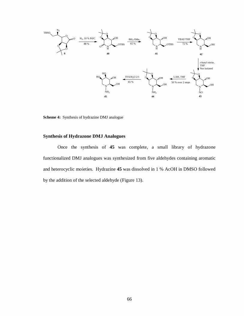
66
OO
OO
N3TBSO
6
NH
O
O
O
OH
OTBS
H2, 10 % Pd/C
NH
O
O OH
OTBS
BH3-SMe2 TBAF/THF
t-butyl nitrite,
THF
Not isolated
N
O
O OH
OH
NH2
LAH, THF
N
HO
HO OH
OH
NH2
TFA/H2O 2/1
40 41 42
434445
NH
O
O OH
OH
N
O
O OH
OH
NO
88 % 65 % 72 %
50 % over 2 steps65 %
Scheme 4: Synthesis of hydrazine DMJ analogue
Synthesis of Hydrazone DMJ Analogues
Once the synthesis of 45 was complete, a small library of hydrazone
functionalized DMJ analogues was synthesized from five aldehydes containing aromatic
and heterocyclic moieties. Hydrazine 45 was dissolved in 1 % AcOH in DMSO followed
by the addition of the selected aldehyde (Figure 13).

67
R
HNN
HO
HO
HO
OH
NH2N
HO
HO
HO
OH
1 % AcOH, DMSO
H R
O
H
N
NN
HO
HO
HO
OH
46
H
NN
H2N
N
HO
HO
HO
OH
47
N
H
NNH
N
HO
HO
HO
OH
48
H
S
NN
HO
HO
HO
OH
49
N
H
N
NH
N
HO
HO
HO
OH
50
Figure 13: Structures of DMJ analogues or initial library screening, 46-50
The reaction was monitored by HRMS-MALDI-TOF. Once complete, the
compounds were purified by HPLC and screened for inhibition and selectivity towards
ER Man I, and Golgi Man I. (Table 4).

68
Table 4: Ki data for DMJ analogues 46-50
Inhibitor ER Man I (M) Golgi Man I (M) Golgi/ER
DMJ 21.1 15.4 0.75
46 39.3 286.2 7.5
47 24.1 496.9 21
48 9.0 65.5 7.5
49 0.30 9.7 32.5
50 2.1 8.1 4
The parent substrate, DMJ, has a Ki of 21.1 M which is very poor along with
relatively no selectivity between the ER and Golgi enzymes. All five hydrazone DMJ
analogues demonstrated increased selectivity towards ER Man I over Golgi Man I.
Analogues 47 and 49 had the highest degree of selectivity with 21 and 32.5 fold
selectivity respectively. 3-methyl-thiophene analogue 49 not only had a very high degree
of selectivity but also had a remarkable increase in potency. The Ki for 49 is in the high
nanomolar range which results in over 100-fold higher potency than DMJ.
Conclusions
In summary, a novel hydrazine functionalized DMJ analogue was synthesized and
utilized as a lead compound in the synthesis of five hydrazone modified DMJ analogues.
A 3-methyl-thiophene functionalized DMJ analogue was prepared and found to have over
a 100 fold improvement in inhibition in respect to DMJ. Analogue 49 had a Ki of 0.30

69
M and 32.5 fold selectivity towards the ER -mannosidase, making it the first selective
and potent ER Man I inhibitor derived from DMJ.
Acknowledgments- I would like to thank the Moremen laboratory for performing all of
the Ki assays and cellular studies for our inhibitor molecules.
Experimental Section:
General Methods and Materials
All chemicals were commercially available and used without further purification.
Reactions were carried out using anhydrous solvents under an Argon atmosphere unless
otherwise stated. Anhydrous DCM was distilled from calcium hydride under a nitrogen
atmosphere. All column chromatography was carried out on Silica gel 60 (EM Science
70-230 mesh), and reactions were monitored using TLC (EM science, kiesel gel 60 F254
on aluminum) stained with cerium sulfate/ ammonium molybdate solution.
6-O-tert-butyldimethylsilyl-2,3-O-isopropylidene-D-mannono--lactam (40)
Azidolactone 6 (1.0 g, .0028 mol) was dissolved in MeOH (50 mL) and the reaction
vessel was flushed with argon. Pd/C (catalytic) was added to the reaction and the
solution was stirred overnight under a H2 atmosphere. Once complete, the reaction was
diluted in MeOH and filtered through a celite pad to remove the catalyst. The solution
was concentrated and purified by Normal silica gel chromatography (70 % Hexanes/#0 %
EtOAc to 100 % EtOAc), affording 6-O-tert-butyldimethylsilyl-2,3-O-isopropylidene-D-
mannono--lactam in 88 % yield as a white fluffy solid (0.8156 g, 0.0025 mol). 1H NMR

70
(300 MHz, CDCl3) δ 6.16 (s, 1H), 4.54 (d, J = 7.8 Hz, 1H), 4.21 (t, J = 7.9 Hz, 1H), 3.91
(dd, J = 10.2, 4.0 Hz, 1H), 3.52 (dt, J = 17.1, 8.5 Hz, 2H), 3.29 (td, J = 8.7, 4.0 Hz, 1H),
1.43 (s, 3H), 1.32 (s, 3H), 0.92 – 0.67 (m, 8H), -0.00 (s, 5H). Calculated MW = 375.34 ,
MALDI-TOF found [M+H]= 376.39.
6-O-tert-butyldimethylsilyl-2,3-O-isopropylidene-1-deoxy-D-mannono--lactam (41)
6-O-tert-Butyldimethylsilyl-2,3-O-isopropylidene-D-mannono--lactam (5.0 g, 0.0150
mol) was dissolved in anhydrous THF (60 mL). The solution was charged with borane
dimethyl sulfide complex (4.2 mL, 0.0450 mol) and stirred for 4 hrs. Once TLC showed
no starting material, the reaction was slowly quenched with MeOH until effervescence
stopped. The solvents were evaporated under vacuum anf the product was purified by
normal silica gel chromatography with a solvent gradient starting at hexanes/EtOAc (1/1)
and finished with EtOAc. 6-O-tert-butyldimethylsilyl-2,3-O-isopropylidene-1-deoxy-D-
mannono--lactam was obtained as a white fluff in 65 % yield (3.1 g, 0.0098 mol).
Calculated MW = 317.50, MALDI-TOF found [M+H] 318.48 .
2,3-O-isopropylidene-1-deoxy-D-mannono--lactam (42) 6-O-tert-Butyldimethylsilyl-
2,3-O-isopropylidene-1-deoxy-D-mannono--lactam (1.8 g, 0.0056 mol) was dissolved in
THF (25 mL) and cooled to -20 oC. TBAF (1 M in THF, 6.7 mL, 0.0067 mol) was added
to the cooled solution and the reaction was stirred at -20 oC for 2 hrs. Once no starting
material was observed on TLC, the reaction was warmed to rt, diluted in EtOAc and
poured into brine. The EtOAc layer was isolated and the brine layer was extracted in
EtOAc (2 x 100 mL). The combined organic layers were dried with MgSO4, filtered and

71
concentrated. The crude residue was purified by flash silica gel chromatography using
Hexanes/EtOAc (1/1) as an eluent. 2,3-O-isopropylidene-1-deoxy-D-mannono--lactam
(0.082g, 0.0040 mol) was obtained in 72 % yield. Calculated MW = 203.24, MALDI-
TOF found [M+H] 204.20.
1-deoxy -2,3-O-isopropylidene-N-amino-D-mannono--lactam (44) 2,3-O-
Isopropylidene-1-deoxy-D-mannono--lactam (0.30 g, 0.0015 mol) was dissolved in
anhydrous THF (5.0 mL) and t-butyl nitrite (0.36 mL, 0.0030 mol) was added to the
solution. The reaction was stirred at reflux in the dark for 18 hrs. Once complete the
solvent and excess t-butyl nitrite was removed under reduced pressure. The crude 1-
deoxy -2,3-O-isopropylidene-N-nitroso-D-mannono--lactam (43) was used in the next
step without further isolation. Lithium aluminum hydride (0.1139 g, 0.0030 mol) was
suspended in THF (10 mL) in a 2-neck flask. The suspension was heated to ~65 oC and a
solution of 43 (0.34 g, .0015 mol) in THF (5 mL) was added slowly. The reaction was
refluxed for 7 hrs. Once complete, the excess LAH was quenched with aqueous
ammonia followed by 1M LiOH. The solids were filtered off through a celite pad and the
filtrate was concentrated. 1-deoxy -2,3-O-isopropylidene-N-amino-D-mannono--lactam
(0.1635 g, 0.75 mmol) was obtained in 50 % yield over two steps. The crude hydrazine
was pure enough for the subsequent reactions. Calculated MW = 218.25, MALDI-TOF
found [M+H] 219.20.
1-deoxy-N-amino-D-mannono--lactam (45) 1-Deoxy -2,3-O-isopropylidene-N-amino-
D-mannono--lactam (0.1635 g, 0.75 mmol) was dissolved in TFA/H2O (2/1, 3 mL) and

72
stirred for 1 hr. Once no starting material remained, the solvents were removed by freeze
drying. The dried material was ready for use in the hydrazine coupling reactions.
Calculated MW = 178.19 MALDI-TOF found [M+H] 179. 17.
General Procedure for Hydrazine Coupling Reaction
45 (20 mg, 0.11 mmol ) was dissolved in 2 % AcOH in DMSO (1 mL) in a small glass
vial. The aldehyde was added (0.12 mmol) and the vials were stirred for 48 hrs. The
reaction progress was monitored by MALDI-TOF and once no starting material remained
the reaction was stopped. The solvents were frozen using liquid nitrogen and then the
samples were lyophilized. Once the DMSO was removed the samples were dissolved in
Water/ACN (1/1) and purified by HPLC. The samples were lyophilized and submitted
for inhibition studies.
General Procedure for Inhibition Study
Each kifunensine analogue was tested for inhibitory activity at three concentrations (0.05,
0.5, and 5 mM) using recombinant human ER mannosidase I or Golgi Man IA. ER Man
I assays were performed using 20 g of enzyme and Man9-GlcNAc2-PA (20 M) as
substrate in a final volume of 20 L containing 20 mM MES/NaOH, pH 7.0, 150 mM
NaCl, and 5 mM CaCl2. The enzyme reactions were allowed to proceed for 15 min at 37
oC and were stopped by addition of 20 L of 1.25 M Tris-HCl pH 7.6. The human ER
Man I product, Man8-GlcNac2-PA, was resolved from Man9GlcNac2-PA by HPLC on a
Hypersil APS-2 NH2 column. Man9GlcNac2-PA was prepared by reductive
pyridylamination of Man9GlcNac2 isolated from crude soybean agglutinin. Golgi Man IA

73
assays were performed using 10 g of enzyme in a reaction containing 20 mM potassium
phosphate, pH 6.0, 150 mM NaCl, 5mM Man--1,2-Man--O-CH3 in a total volume of
25 L. Enzyme reactions were allowed to proceed for 30 min at 37 oC and terminated by
the additionof 25 L of 1.25 Tris-HCl, pH 7.6. The amount of mannose released was
quantified using a glucose oxidase and peroxidase reagent. The recombinant enzymes
were expressed and purified. For determination of the compound inhibition, enzyme
activity was plotter as a percentage of residual activity (compared to no added
compound) versus compound concentration and the IC50 values were estimated from the
plots. All inhibitory assays and cellular studies were performed by the Dr Kelley
Moremen lab at the CCRC of the University of Georgia.
Pyridine-DMJ (46) 1H NMR (500 MHz, D2O) δ 8.35 (d, J = 6.8 Hz, 2H), 7.86 (d, J =
6.8 Hz, 3H), 7.55 (s, 2H), 4.24 (dd, J = 12.2, 2.8 Hz, 1H), 4.16 – 4.03 (m, 3H), 3.80 (t, J
= 8.7 Hz, 1H), 3.66 (dd, J = 8.7, 3.2 Hz, 1H), 3.33 (ddd, J = 8.5, 5.6, 2.8 Hz, 1H), 3.19
(dd, J = 13.9, 1.9 Hz, 1H). Calculated MW = 267.28, MALDI-TOF found [M+H] 268.32
.
4-Amino-pyridine-3-DMJ (47) Calculated MW =282.30, MALDI-TOF found [M+H] =
283.35.
4- Imidazole-DMJ (48) 1H NMR (500 MHz, d2o) δ 9.72 (s, 1H), 8.83 (s, 1H), 8.54 (d, J
= 12.3 Hz, 1H), 8.20 (s, 1H), 6.05 (s, 1H), 4.27 – 4.21 (m, 1H), 4.11 – 4.00 (m, 1H), 3.94
(dd, J = 12.1, 3.7 Hz, 1H), 3.78 (ddd, J = 16.7, 15.3, 6.0 Hz, 1H), 3.61 – 3.44 (m, 1H),

74
2.94 – 2.82 (m, 1H), 2.59 (s, 2H). Calculated MW = 256.26, MALDI-TOF found [M+H]
= 257.30.
3-Methyl-2-thiophene DMJ (49) Calculated MW =286.35, MALDI-TOF found [M+H]
= 287.42.
1H-pyrrole[3,2-b]-pyridine-6-DMJ (50) Calculated MW = 306.32, MALDI-TOF
found [M+H] = 307.40.

75
CHAPTER 4
THE EXTRACYCTOPLASMIC DOMAIN OF THE MYCOBACTERIUM
TUBERCULOSIS SER/THR KINASE PKnB BINDS SPECIFIC MUROPEPTIDES
AND IS REQUIRED FOR PKnB LOCALIZATION
Mushtaq Mir1, Jinkeng Asong
2, Xiuru Li
2, Jessica Cardot
2, Geert-Jan Boons
2, Robert N.
Husson1*
PLoS Pathogens July 2011
Reprinted here with permission from Publisher

76
Short title: Muropeptides bind and localize PknB
Mushtaq Mir1, Jinkeng Asong
2, Xiuru Li
2, Jessica Cardot
2, Geert-Jan Boons
2, Robert N.
Husson1*
1 Division of Infectious Diseases, Children’s Hospital Boston and Harvard Medical
School, Boston, MA 02115
2 Department of Chemistry and the Complex Carbohydrate Research Center, University
of Georgia, Athens, GA 30602
*Corresponding Author:
Robert N. Husson
Division of Infectious Diseases
Children’s Hospital Boston
300 Longwood Ave.
Boston, MA 02115
Tel: 617-919-2883
Fax: 617-730-0254
Email: [email protected]

77
Abstract
Mycobacterium tuberculosis Ser/Thr kinase PknB has been implicated in the
regulation of cell growth and morphology in this organism. The extracytoplasmic domain
of this membrane protein comprises four penicillin binding protein and Ser/Thr kinase
associated (PASTA) domains, which are predicted to bind stem peptides of
peptidoglycan. Using a comprehensive library of synthetic muropeptides, we demonstrate
that the extracytoplasmic domain of PknB binds muropeptides in a manner dependent on
the presence of specific amino acids at the second and third positions of the stem peptide,
and on the presence of the sugar moiety N-acetylmuramic acid linked to the peptide. We
further show that PknB localizes strongly to the mid-cell and also to the cell poles, and
that the extracytoplasmic domain is required for PknB localization. In contrast to strong
growth stimulation by conditioned medium, we observe no growth stimulation of M.
tuberculosis by a synthetic muropeptide with high affinity for the PknB PASTAs. We do
find a moderate effect of a high affinity peptide on resuscitation of dormant cells. While
the PASTA domains of PknB may play a role in stimulating growth by binding
exogenous peptidoglycan fragments, our data indicate that a major function of these
domains is for proper PknB localization, likely through binding of peptidoglycan
fragments produced locally at the mid-cell and the cell poles. These data suggest a model
in which PknB is targeted to the sites of peptidoglycan turnover to regulate cell growth
and cell division.

78
Author Summary
Regulation of growth by Mycobacterium tuberculosis is important in the
pathogenesis of tuberculosis (TB), including asymptomatic latent TB infection and active
TB disease. The M. tuberculosis kinase PknB regulates cell growth and cell division by
phosphorylating proteins involved in these processes to modify their function. The
activity of PknB is thought to respond to extracellular stimuli by binding specific
molecules with its extracytoplasmic domain. In this work we show that cell wall
fragments bind to this domain, and that strong binding requires that these interacting
molecules have specific molecular features. We demonstrate that a peptidoglycan
fragment that binds strongly can stimulate growth of dormant bacteria, but that it does not
affect growth of noN-dormant bacteria. We also show that PknB localizes to the site of
cell division and to the growing tip of the bacterium, where cell wall synthesis and
degradation occur, and that the extracytoplasmic domain is required for this localization.
These findings indicate that a major function of the extracytoplasmic domain of PknB is
to place it at the sites of cell wall turnover, and suggest a model by which PknB can
regulate growth and cell division, and thereby contribute to the pathogenesis of TB.
79
Introduction and Literature Review
Bacterial cell growth and cell division are highly regulated processes, requiring
the coordination of multiple activities within the cell. DNA replication and chromosome
segregation for example, must occur at the correct time and in the correct location, and be
coordinated with septum formation and cytokinesis. The molecules involved in septum
formation and the sequence in which they are recruited to the division site have been the
subject of intense investigation in the model organisms Bacillus subtilis and Escherichia
coli, and the identities and functions of many bacterial cell division proteins have been
elucidated [1], [2]. In addition to divisome assembly and DNA segregation, bacterial
growth and cell division require remodeling of the peptidoglycan (PGN) mesh that forms
the cell wall [3]. The enzymes and the sequence of reactions involved in cell wall
synthesis are relatively well understood as are the enzymatic activities of many of the
PGN hydrolases that can degrade this polymer [4], [5]. In the model organism B. subtilis,
the mechanisms by which cell wall hydrolases are regulated to achieve morphogenesis
are at least partially understood [6]. In other bacteria, including the slow growing
actinomycete Mycobacterium tuberculosis, less is known about the regulation of PGN
synthesis and hydrolysis, how these opposing processes are balanced, and how they are
coordinated with other cell processes in growing and dividing vs. noN-growing dormant
cells.
Because of the apparent ability of M. tuberculosis to become dormant in the
human host, leading to asymptomatic latent infection, there has been great interest in
understanding how cell growth and cell division are regulated in this organism [7]. A
longstanding observation that “spent” or “conditioned” medium, i.e. filter-sterilized

80
supernatant from bacterial cultures grown in liquid medium, is able to stimulate growth
of dormant cells, led to the identification of a resuscitation promoting factor (Rpf) by
purifying from spent medium a component that was able to stimulate growth of the
actinomycete Micrococus luteus [8]. Rpf is small protein that has homologues in other
actinobacteria, including M. tuberculosis, which has five rpf genes [9]. Functional studies
of these genes in M. tuberculosis have shown that individually they are not required for
resuscitation of dormant M. tuberculosis cells and single rpf mutant strains do not have
other growth or morphologic phenotypes. When two or more rpf genes are inactivated,
however, growth or resuscitation defects are observed [10], [11], [12]. The recent
demonstration that the Rpf's are PGN hydrolases suggests that growth stimulation of
dormant cells may result from the enzymatic activity of these secreted proteins, possibly
through alterations in PGN structure or through the interaction of PGN degradation
products with the bacterial cell surface [13].
A domain found to occur in the extracytoplasmic regions of penicillin binding
proteins and serine/threonine kinases (PASTA domain) was identified by bioinformatic
analysis and predicted to bind to the stem peptide of uN-crosslinked PGN precursors,
based on the structure of the PASTA-containing penicillin binding protein PBP2X of
Streptococcus pneumoniae bound to a cephalosporin antibiotic [14]. Recently the PASTA
domain of a Ser/Thr kinase of B. subtilis was shown to bind both intact and hydrolyzed
PGN [15]. Incubation of B. subtilis spores with PGN stimulated spore germination and
increased Ser/Thr phosphorylation. Some specificity with respect to the source of PGN
and these functional effects was observed, suggesting a preference for meso-

81
diaminopimelic acid (m-DAP)-containing PGN in stimulating spore germination in this
organism.
The M. tuberculosis genome encodes two proteins that contain PASTA domains,
the Ser/Thr protein kinase PknB (Rv0014c) whose extracytoplasmic region comprises
four PASTA domains, and the bifunctional penicillin binding protein PBP2 (PonA2,
Rv3682), which has a single PASTA domain at the extreme carboxy-terminus of the
protein distal to the extracytoplasmic transpeptidase and transglycosylase-containing
regions [16]. In this work we investigated the quantitative binding of a series of synthetic
muropeptides to the extracytoplasmic region of PknB. We identified specific features of
these molecules that are required for high affinity binding, and investigated the functional
effects of these compounds in vivo on mycobacterial growth, morphology and the
localization of PknB. We determined that PknB is strongly localized to septum and less
strongly to the cell poles, the sites of active PGN synthesis in mycobacteria, and that the
PASTA domains of PknB are required for its localization.
RESULTS
Binding of PGN fragments to the extracytoplasmic domain of PknB
The region of pknB that encodes the extracytoplasmic domain of PknB (ED-
PknB) was amplified by PCR, cloned and ED-PknB was expressed in Escherichia coli as
an N-terminal Glutathione-S-transferase (GST) fusion protein. The ED-PknB comprises 4
PASTA motifs that share limited sequence similarity aside from the key residues that
define the motif (Figure S1). Soluble recombinant GST-ED-PknB was affinity purified to

82
>95% purity and after removal of the GST tag was used in subsequent binding
experiments (Figure S2).
A series of PGN fragments (muropeptides) were synthesized as tri-, tetra- and
penta-peptides linked to N-acetylmuramic acid (MurNAc) or as unlinked peptides.
Amino acids characteristic of PGN stem peptides from Gram-positive bacteria, Gram-
negative bacteria or actinobacteria were incorporated into different compounds.
Modifications of amino acid side chains that correspond to PGN modifications that are
found in vivo were also included in the compound series (Figure 14).
Figure 14. Structures of synthetic muropeptides used in the binding and phenotypic assays. Lys-
containing compounds are typical of Gram-positive bacteria and DAP-containing compounds are typical of
Gram-negative bacteria and Actinomycetes, including mycobacteria. Variations in substituents are
indicated in red, and the specific variations are listed immediately below the
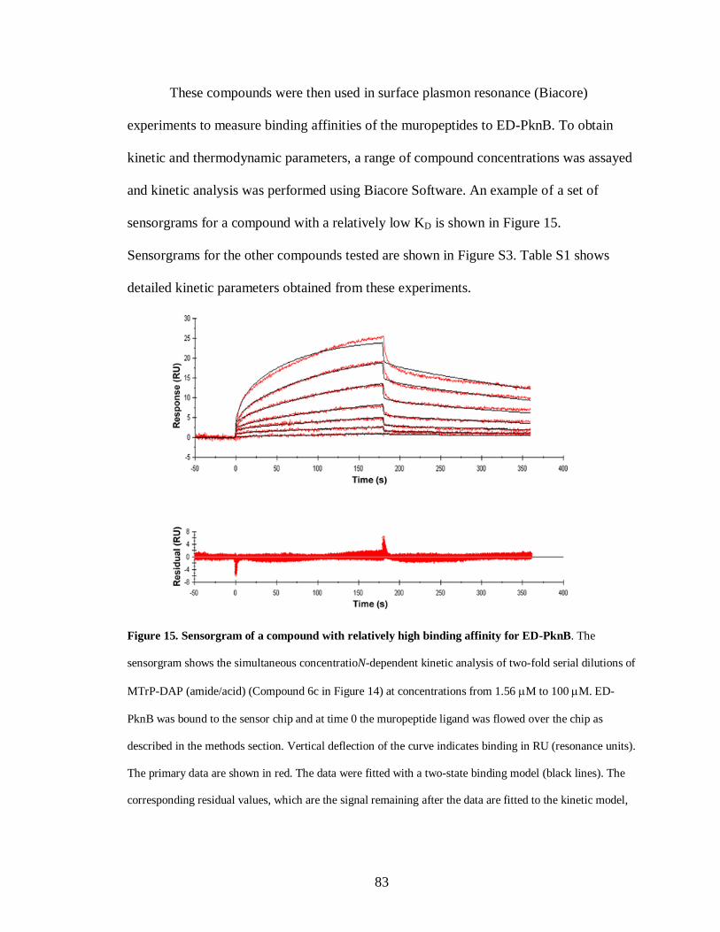
83
These compounds were then used in surface plasmon resonance (Biacore)
experiments to measure binding affinities of the muropeptides to ED-PknB. To obtain
kinetic and thermodynamic parameters, a range of compound concentrations was assayed
and kinetic analysis was performed using Biacore Software. An example of a set of
sensorgrams for a compound with a relatively low KD is shown in Figure 15.
Sensorgrams for the other compounds tested are shown in Figure S3. Table S1 shows
detailed kinetic parameters obtained from these experiments.
Figure 15. Sensorgram of a compound with relatively high binding affinity for ED-PknB. The
sensorgram shows the simultaneous concentratioN-dependent kinetic analysis of two-fold serial dilutions of
MTrP-DAP (amide/acid) (Compound 6c in Figure 14) at concentrations from 1.56 M to 100 M. ED-
PknB was bound to the sensor chip and at time 0 the muropeptide ligand was flowed over the chip as
described in the methods section. Vertical deflection of the curve indicates binding in RU (resonance units).
The primary data are shown in red. The data were fitted with a two-state binding model (black lines). The
corresponding residual values, which are the signal remaining after the data are fitted to the kinetic model,

84
are plotted below the sensorgrams. The kinetic parameters calculated from the model are shown below the
graphs.
As shown in Table 5, these experiments demonstrated moderately strong binding
of several PGN fragments that have DAP at the third position of the stem peptide.
Table 5. Affinity of synthetic muropeptides for the extracytoplasmic domain of M. tuberculosis PknB.

85
N-acetylation of the amino group of DAP as in compound 6e (MTrP-DAP
(amide/acid) NHAc), which is designed to mimic branching of the PGN subunits within
the PGN polymeric structure, resulted in a six-fold decrease in binding compared to
compound 6a. The MurNAc-pentapeptide, compound 7, corresponding to newly
synthesized PGN prior to remodeling, bound strongly though about two-fold less than the
corresponding MurNAc-tetrapeptide (6a).
In addition to preference for DAP at the third position of the stem peptide, another
clear result of these experiments is the requirement for amidation of D-isoglutamate (D-
iGlu) to D-isoglutamine (D-iGln) at the second position, in order to achieve high affinity
binding. Compound 6a, which contains both D-iGln and DAP at the second and third
positions, respectively, exhibited the highest affinity, while compound 6d, which is
identical except for D-iGlu at the second position, bound four-fold less strongly.
Similarly, compound 6c, which also bound with a relatively high affinity (KD = 15 µM),
contains D-iGln together with amidation of the carboxyl group of DAP. In contrast, a
similar compound, (6b), that has D-iGlu at the second position instead of D-iGln did not
show measurable binding. The importance of this residue is further underscored by the
finding that among the Lys-containing compounds, the only one that showed detectable
interaction was compound 2a, the muramyl tetrapeptide incorporating a D-iGln moiety.
While the data indicate a preference for DAP at the third position, the ε-carboxylic acid
group that is a major feature that distinguishes DAP from Lys is not an essential
requirement for binding. To determine whether the MurNAc moiety was important for
binding, compounds 4 and 8, pentapeptides not linked to MurNAc and containing either
Lys or DAP at the third position, respectively, were tested. Neither compound showed

86
significant interaction, indicating an important contribution of MurNAc in binding to the
PknB PASTA domains.
Muropeptides stimulate resuscitation of dormant M. tuberculosis cells
The Rpf's have been shown to have PGN hydrolytic activity, and are thought to
cleave the ß-1–4 glycosidic linkage between N-acetylmuramic acid and N-
acetylglucosamine [13]. This muralytic activity has been shown to be essential for the
resuscitation activity of these Rpf proteins, but the mechanism remains uncertain. To
determine whether muropeptides that bind to the PASTA domains of ED-PknB can
stimulate resuscitation of dormant M. tuberculosis cells, we utilized an established M.
tuberculosis dormancy and resuscitation model [17]. In this assay, M. tuberculosis cells
are incubated under hypoxic conditions for several months, at which point the number of
cells capable of resuming growth in liquid culture is markedly decreased. In this assay,
addition of sterile spent medium “resuscitates” dormant cells, leading to an increase in
the number of cells that can grow on solid or in liquid medium.
In two independent experiments, we took M. tuberculosis stationary phase
cultures that had been incubated under hypoxic conditions for 6 or 9 months, and
performed this resuscitation assay. In addition to cells incubated in Sauton's medium
alone, cells were incubated with a synthetic muropeptide with a high affinity for ED-
PknB (6c in Figure 14), a muropeptide with low affinity for ED-PknB (3b in Figure 14),
or with sterile conditioned medium as a positive control. The muropeptides were used at
a concentration of 10 times the KD of the high affinity compound as determined in the
SPR experiments. Using most probable number analysis [18], which has been used to

87
analyze results from this assay, we observed three and nine-fold increases in the viability
of cells that were incubated with the high affinity muropeptide in the two independent
experiments. No increase in viability was observed for cells incubated with the low
affinity peptide. The cells incubated with sterile spent medium showed a much stronger
resuscitation phenotype, with 14 and 100-fold increased viability relative to the cells
incubated in fresh medium alone (Table 6).
Table 6. Resuscitation of dormant M. tuberculosis cultures.
The original identification of Rpf in M. luteus was based on the observation that
stationary phase cells show decreased viability when plated or diluted to low density in
liquid medium, but that addition of sterile conditioned medium stimulates growth [8]. A
similar phenomenon is observed when mycobacteria are inoculated at low density. To
88
determine whether synthetic muropeptides stimulate growth when stationary phase cells
are inoculated at low density, cells from cultures of M. tuberculosis (O.D600 of 2.4–3.6)
were washed and diluted 10,000-fold in minimal medium with or without the addition of
the high or low affinity muropeptide. As shown in Figure 16, no growth stimulation by
either muropeptide was observed in this assay. In contrast, strong growth stimulation by
conditioned medium was observed.
Figure 16. Growth stimulation assay of low inoculum cultures of M. tuberculosis. M. tuberculosis cells
were grown in Sauton’s medium alone or in medium supplemented with a synthetic muropeptide at a
concentration 10 times the KD, or with 50% (v/v) sterile conditioned (spent) medium. Cells were grown to
stationary phase, diluted and inoculated into medium containing alamar blue and grown at 37o C, with
measurement of fluorescence at 595 nm at serial time points. A. Growth curves for MTrp-DAP
(Compound 6c in Figure 14). B. Growth curves for MPP-Lys (Compound 3b in Figure 14). ■, Sauton’s
medium. ▲, Sauton’s medium plus synthetic muropeptide. ▼, Sauton’s medium plus spent medium.

89
PknB is present in the cell envelope of M. tuberculosis
Based on its sequence, PknB is predicted to have a single transmembrane
segment, with an intracellular kinase domain and an extracytoplasmic region that
incorporates the four PASTA domains [16]. To determine whether PknB is a membrane
protein and in which subcellular fraction(s) PknB is located, we performed
immunoblotting with a PknB-specific monoclonal antibody. As a control, we probed
these subcellular fractions with an antibody to the membrane protein PknA, which like
PknB has a single transmembrane segment, but which has a small extracytoplasmic
region that is not known to interact with cell wall components. We found that PknB does,
as predicted, localize to the membrane fraction of the cell (Figure 17). An even stronger
signal was seen in the cell wall fraction, further confirming the association of PknB with
the cell envelope. The PknA antibody gave equally strong signals from the membrane
and cell wall fractions. These results demonstrate that PknB is a membrane protein and
that membrane is present in the cell wall fractions used in these experiments

90
Figure 17. Subcellular localization of M. tuberculosis PknB. Top panel: Immunoblot of subcellular
fractions of M. tuberculosis, probed with a mouse monoclonal antibody that recognizes M. tuberculosis
ED-PknB. Bottom panel: immunoblot in which the same fractions were probed with rabbit polyclonal sera
raised against M. tuberculosis membrane protein PknA. CM, membrane fraction; CY, cytoplasmic fraction;
CW, cell wall fraction; CF, culture filtrate fraction.

91
PknB localizes to the septum and poles, and the extracytoplasmic domain is
required for proper PknB localization
A construct designed to express a PknB-RFP fusion protein, in which RFP is
fused to the amino terminus of PknB, was introduced into wild type M. smegmatis.
Additional constructs, in which RFP is fused a) to the PknB kinase domain, intracellular
juxtamembrane sequence and transmembrane segment, but which lacks the
extracytoplasmic domain, and b) to the membrane and ED-PknB regions but which lacks
the intracellular linker and kinase domains, were also introduced into wild type M.
smegmatis. Cells were grown to early log phase, expression of the fusion protein was
induced, and the cells were examined using fluorescence microscopy (Figure 18). Cells
expressing the full-length PknB-RFP fusion showed strong localization of this protein to
the mid-cell and symmetrical, less intense localization to both cell poles. In contrast, in
cells expressing the fusion that lacks the extracytoplasmic domain containing the PASTA
domains, foci of fluorescence were visible at discrete sites along the length of the cell.
While in some cells there appears to be increased signal at the poles, we did not observe
clear mid-cell localization in cells expressing this construct. To confirm that these foci
are not cytoplasmic aggregates, we prepared subcellular fractions of these cells and
confirmed that the large majority of this protein is present in the cell membrane and cell
wall fractions (Figure S4). Cells expressing the ED-PknB-RFP fusion lacking the
intracellular linker and kinase domains showed clear localization to the mid-cell but
minimal signal from the poles. This result indicates that the extracytoplasmic PASTA
domains are required for proper localization of PknB to the mid-cell and likely to the cell
poles and suggests that the intracellular linker-kinase region makes a contribution to

92
localization at the cell poles. To verify these findings, we performed additional imaging
of live cells (Figure S5 and Protocol S1), which demonstrates the same localization
patterns observed with the fixed cell preparations.
Figure 18. Localization of PknB to sites of peptidoglycan turnover in the mycobacterial cell. A.
Schematic representation of domain organization of M. tuberculosis PknB, including the kinase domain
(residues 11-274; the juxtamembrane linker (residues 275-331), the transmembrane segment (residues 332-
354) and the extracytoplasmic PASTA domains (residues 355-626). B-I. Flourescence (B, D, F and H) and
DIC (C, E, G and I) images of M. smegmatis expressing RFP fused to full-length PknB (B and C), PknB
lacking the extracytoplasmic domain (D and E), PknB lacking the intracellular kinase domain and linker (F
and G) and unfused RFP (H and I). Arrowheads in panel D point to focal RFP signals along the length of
the bacillus.

93
To determine whether diffusible, noN-localized muropeptides might bind ED-
PknB and disrupt PknB localization, we incubated M. smegmatis with the high affinity
muropeptide used in the resuscitation experiments for 8 hours. No change in the
morphology of wild type bacteria were observed, and in the strain expressing the PknB-
RFP fusion protein the RFP signal remained localized to the septum and poles (not
shown).
Discussion
In this work we report three major findings. First, we demonstrated that
muropeptides bind to the extracytoplasmic region of PknB, which contains four PASTA
domains, and defined molecular requirements for ligand binding. These requirements
include both specific residues at the second and third positions in the stem peptide, and
the presence of the sugar moiety (MurNAc) linked to the amino-terminal residue of the
peptide. Using an extensive series of chemically synthesized compounds, we found
moderately high affinity binding by muropeptides that contain DAP at the third position
of the stem peptide, in which the D-iGlu at the second position is amidated to D-iGln.
The preference for DAP is consistent with the predominant structure of the stem peptide
of mycobacteria, where DAP is present at this position, in contrast to most Gram-positive
organisms in which Lys occurs at this position. D-iGln at the second position has been
reported to be predominant in M. tuberculosis PGN, however D-iGlu is present in a
minority of stem peptides [19], [20]. Whether synthesis of PGN incorporating D-iGlu vs.
D-iGln is site- or growth-stage specific in M. tuberculosis is not known. The markedly
stronger binding of compounds containing D-iGln suggests that variation in the structure
of PGN stem peptides may affect binding by ED-PknB in vivo, with potentially important

94
physiologic effects. A recent paper examining stimulation of B. subtilis spore
germination using synthetic muropeptides confirmed prior results using purified native
PGN in showing the importance of DAP at the third position of the stem peptide for this
phenotype in this species [21]. In this assay the presence of N-acetylglucosamine linked
to MurNAc was also required for potent activity.
A second finding of this work is that PknB localizes strongly to the mid-cell and
less strongly to the cell poles of mycobacteria, the sites of active PGN synthesis and
hydrolysis in these organisms [22]. Our results with RFP fusions to full-length PknB and
to separate domains of this protein in M. smegmatis demonstrate that the PASTA motif-
containing extracytoplasmic domain of PknB is required for its localization to the mid-
cell. We attempted to perform a similar experiment with full-length PknB-RFP in M.
tuberculosis, however we were unable to obtain consistent expression of the fusion
protein. We observed fluorescence in a minority of cells, which was highly variable from
cell to cell, and we observed markedly abnormal morphology of many cells, suggesting
severe toxicity of pknB overexpression, as previously described [23]. Despite these
limitations, we were able to see similar localization of full-length PknB in a minority of
rod-shaped cells expressing the pknB-rfp fusion (data not shown).
In the context of our in vitro binding results, these data suggest that binding of
PGN fragments by its extracytoplasmic domain is critical for PknB localization to the
mid-cell and possibly to the poles. This result, together with the finding that PknB is
found in the cell wall and membrane fractions of M. tuberculosis lysates, suggests that
the PASTA motifs of PknB bind endogenous cell wall or membrane-anchored PGN

95
precursors and/or PGN hydrolysis fragments produced at the septum and poles of the cell.
The finding that incubation of growing cells with a high affinity muropeptide had no
effect on PknB localization is consistent with this model, and suggests that exogenous
muropeptides may not be able to penetrate the complex, lipid-rich mycobacterial cell
envelope to reach the PknB PASTA domains at the surface of the cytoplasmic membrane.
Because both de novo synthesized PGN precursors and PGN hydrolysis products are
likely to be localized at the septum and the poles, our data do not indicate which of these
are the major PknB PASTA ligands in vivo.
The third important finding of this work is that, in contrast to spent medium,
which strongly stimulated both growth of noN-dormant M. tuberculosis cells and
resuscitation of dormant cells, a muropeptide with relatively high affinity for the PASTA
domains of ED-PknB did not stimulate M. tuberculosis growth and had only a modest
effect on resuscitation. This result suggests that while muropeptide binding may play a
role in resuscitation of M. tuberculosis, other factors present in spent medium may be
more important in stimulating M. tuberculosis growth. In this regard, D-amino acids
present in conditioned medium have recently been shown to be a potent growth stimulus
for Vibrio cholerae and to play a key role in biofilm disruption leading to resumption
planktonic growth in B. subtilis [24], [25]. Alternatively, PknB may require a different
muropeptide ligand, e.g. a disaccharide muropeptide or a multivalent muropeptide, or
higher concentrations of these ligands, which may be present in vivo, for greater
stimulation of growth or resuscitation.

96
The first structure of a PASTA domain was determined as part of the structure of
PBP2x from S. pneumoniae bound to a cephalosporin antibiotic [26]. In this structure two
PASTA domains interacted to form a compact globular domain. In recent work, the
structure of the PASTA motifs of M. tuberculosis ED-PknB was determined using NMR
and small angle X-ray scattering [27]. While the individual folds of each PASTA domain
were similar to those of the PBP2x PASTA domains, the four PASTA domains of PknB
are organized as a linear molecule, which is maintained with what the authors termed a
ß′/ß′′ brace that prevents interactions between the individual PASTA domains of a single
molecule of PknB. A previous structure of the PknB intracellular domain demonstrated
the presence of a highly flexible intracellular juxtamembrane segment linking the
transmembrane segment to the intracellular kinase domain, indicating that ligand binding
resulting in transmembrane propagation of conformational changes leading to PknB
activation is unlikely [28].
Based on the PknB PASTAs structure, a model was proposed in which binding of a
single ligand to two molecules of PknB would result in dimerization of the
extracytoplasmic domains, which would then cause dimerization of the intracellular
kinase domains, resulting in kinase activation [27]. Our data showing relatively high
affinity binding of muropeptide monomers, however, suggest an alternative model by
which muropeptide binding to ED-PknB could lead to localization and activation of this
kinase. In this model, at sites of active PGN hydrolysis and synthesis, i.e. the septum and
the cell poles, local concentrations of PGN precursors and PGN hydrolysis products will
be high, and binding of these ligands by ED-PknB would result in the septal and polar
localization of PknB that we observed. The recruitment of PknB to these sites will

97
consequently result in high concentrations of the intracellular kinase domain, leading to
the dimerization that results in kinase activation [28], [29], [30]. PknB activation will
then lead to phosphorylation of protein substrates, resulting in regulation of cell division
and cell wall synthesis (Figure 19). In this model, there is no requirement for binding of a
single muropeptide by PASTA domains from two PknB molecules, or for muropeptides
that diffuse from a distance, to achieve PknB localization and activation.
Figure 19. Model of PknB localization and activation by interaction of its extracytoplasmic domain
with peptidoglycan fragments. In this model, the extracytoplasmic domains of PknB bind PGN precursors
and/or hydrolysis products produced at the mid-cell and poles, the two sites where active PGN synthesis
and degradation occur in mycobacteria. This interaction leads to localization of PknB at the theses sites,
and to PknB activation by dimerization of the intracellular kinase domains. Both linear and noN-linear
forms of the extracytoplasmic domain are shown to allow for possible changes following ligand binding.
RPF, resuscitation promoting factor.

98
In summary, we have demonstrated sequence-specific binding of muropeptides to
the PASTA domaiN-containing extracytoplasmic region of M. tuberculosis PknB, and
that the presence of the PASTA domains is required for localization of PknB to sites of
PGN turnover. In the context of our phenotypic data and the finding that peptides that
bind with high affinity have peptide sequences characteristic of M. tuberculosis PGN, our
results suggest that in M. tuberculosis, the PknB PASTAs bind to PGN precursors or
fragments resulting from local PGN synthesis and/or hydrolysis at the mid-cell and poles.
This PASTA domaiN-mediated localization provides a mechanism by which PknB and
the co-regulated kinase PknA can regulate cell division and PGN turnover by reversible
phosphorylation of proteins involved in these processes, several of which have been
shown to be PknA or PknB substrates and to localize to these sites [22], [23], [31], [32],
[33].
Materials and Methods
Strains, media, and recombinant plasmid construction and protein production
Escherichia coli TOP10 (Invitrogen) was used for cloning and was grown in LB
broth. E. coli BL21 (DE3) (Stratagene) was used for expression of recombinant ED-
PknB. M. tuberculosis H37Rv or M. smegmatis mc2-155 were grown at 37°C in
Middlebrook 7H9 liquid medium (Difco) supplemented with albumiN-dextrose complex
(ADC), 0.2% Glycerol and 0.05% Tween 80, except for resuscitation experiments where
M. tuberculosis was grown in Sauton's medium (Difco). Kanamycin (50 µg ml−1
) or
ampicillin (100 µg ml−1
) was added to culture media or agar when appropriate.
99
For expression and purification of ED-PknB, the nucleotide sequence encoding
PknB from Gly354 to Gln626 was PCR-amplified from genomic DNA of M. tuberculosis
H37Rv and cloned in pGEX-4T-3 (GE Healthcare) for expression as a glutathione-S-
transferase (GS) fusion protein. Recombinant GST-ED- PknB was affinity purified to
>95% homogeneity using immobilized glutathione agarose (Pierce) (Figure S2). To
cleave the GST from the fusion protein, the thrombin CleanCleave kit (Sigma) was used.
In brief 900 µg of purified recombinant ED-PknB-GST was incubated with 100 µL of
50% (v/v) suspension of thrombin agarose for 1 hr at room temperature. After
centrifugation the supernatant containing ED-PknB and free GST was incubated with 500
µL of 50% (v/v) suspension of Glutathione-agarose for 15 min. After centrifugation the
supernatant containing ED-PknB was collected. For SPR analysis the supernatant was
dialyzed against phosphate buffered saline (PBS) pH 7.4 prior to use.
For localization of PknB or PknB lacking the extracytoplasmic domain
(PknBΔED) in wild type M. smegmatis, the full length pknB gene or the nucleotide
sequence encoding the region from Met1 to Gly354 of PknB (pknBΔED) respectively, were
PCR-amplified from genomic DNA of M. tuberculosis H37Rv. Overlap PCR
amplification of the above PCR products was performed with the PCR product of the red
fluorescent protein (rfp) gene using a forward primer annealing to the 5′ region of rfp and
a reverse primer annealing to the 3′ region of pknB or PknBΔED to obtain the PCR
products rfp-pknB or rfp-pknBΔED. A PacI site was introduced between rfp and pknB.
The fusion PCR products were cloned into the integrating vector pMV306-pacet
downstream of the inducible acetamide promoter at NdeI and XbaI sites to obtain
pMV306-pacet-rfp-pknB or pMV306-pacet-rfp- pknBΔED. To obtain recombinant clone

100
pMV306-pacet-rfp-pknBΔKD expressing RFP linked to transmembrane and extracellular
domains of PknB (ED-PknB-RFP) the pknB gene of the clone pMV306-pacet-rfp-pknB
was replaced with the nucleotide sequence encoding Ile326 - Gln626 using PacI and XbaI
sites. Cloned DNA was sequenced to verify the absence of mutations. A mycobacterial
replicating vector that constitutively expresses RFP was a gift from Eric Rubin.
Chemical synthesis of PGN fragments
Compounds were synthesized using classical fluorenylmethoxycarbonyl (Fmoc)
chemistry and standard manual solid-phase peptide synthesis techniques as previously
described [34], [35], [36], [37], [38], [39]. In the preparation of the peptide portion of the
compounds, Sieber Amide resin was swelled in dimethylformamide (DMF) for 45 min
and then treated with piperidine in DMF. After a reaction time of 30 min, the solvents
were removed by filtration and the resin washed with DMF, and then treated with Fmoc-
linked amino acid building blocks. This generated PGN partial structures with N-termini.
Compound 8 was newly synthesized for this work as follows. Rink amide AM LL
resin (0.1 mmol) was swelled in dichloromethane for 30 min and then rinsed with DMF
(3×5 mL). The resin was treated with piperidine in DMF (20%, 3×5 mL). After a reaction
time of 30 min, the solvents were removed by filtration and the resin was washed with
DMF (3×5 mL), followed by treatment with Fmoc-D-Ala-OH (155.7 mg, 0.5 mmol) in
DMF in the presence of HATU (mg, 0.5 mmol) and DIPEA (mL). The reaction progress
was monitored by a Kaiser test. After completion of the coupling, the resin was washed
with DMF (3×5 mL). The Fmoc protecting group was removed by treatment with
piperidine in DMF (20%, 3×5 mL, 3×10 min). The reaction cycle was repeated using

101
Fmoc-D-Ala-OH (155.7 mg, 0.5 mmol), Fmoc-DAP(BOC,tBu)-OH (113.7 mg, 0.2
mmol), Fmoc-D-iso-GlN-OH (73.7 mg, 0.2 mmol), Fmoc-L-Ala-OH (155.7 mg, 0.5
mmol). The final Fmoc protecting group was removed by treatment with piperidine in
DMF (20%, 3×5 mL, 3×10 min). The resin was washed with DMF (3×5 mL) and the
resin bound peptide was capped by treatment with acetic anhydride and (10%) and
DIPEA (5%) in DMF (3×5 mL, 3×10 min). The resin was washed with DMF (3×5 mL),
dichloromethane (7×5 mL), and methanol (3×5 mL) and dried in vacuo overnight. The
peptide was released from the resin by treatment with TFA/TIPS/Water
(95%/2.5%/2.5%) in DMF for 2 h. The resin was filtered and washed with TFA (1×10
mL). The filtrate was concentrated under reduced pressure and co-evaporated with
toluene. The crude peptide was dissolved in water/acetonitrile and purified by semi-
preparative HPLC (Eclipse XDB-C18 column, 5 mm, 9.4×250 mm, eluent:
water/acetonitrile/0.1%TFA) to afford, after lyophilization of the appropriate fraction, the
target compound 8. HRMS-MALDI-TOF calculated for C23H40N8O9Na [M + Na]:
595.29, experimental: 595.41.
Surface plasmon resonance binding assays and kinetic analysis
Binding interactions between ED-PknB and PGN analytes were examined using a
Biacore T100 biosensor system (Biacore Life Sciences - GE Healthcare). Soluble ED-
PknB was immobilized by standard amine coupling using an amine coupling kit
(Biacore). The surface was activated using freshly mixed N-hydroxysuccimide (NHS;
100 mM) and 1-(3-dimethylaminopropyl)-ethylcarbodiimide(EDC; 391 mM) (1/1, v/v) in
water. Next, ED-PknB (50 µg/ml) in aqueous NaOAc (10 mM, pH 4.5) was passed over

102
the chip surface until a ligand density of approximately 5,000 RU was achieved. The
remaining active esters were quenched by aqueous ethanolamine (1.0 M; pH 8.5). The
control flow cell was activated with NHS and EDC followed by immediate quenching
with ethanolamine. HBS-EP (0.01 M HEPES, 150 mM NaCl, 3 mM EDTA, 0.005%
polysorbate 20; pH 7.4) was used as the running buffer for the immobilization and kinetic
studies. Analytes were dissolved in running buffer and a flow rate of 20 µL/min was
employed for association and dissociation at a constant temperature of 25°C. A double
sequential 60 s injection of aqueous NaOH (50 mM; pH 11.0) at a flow rate of 50 µl/min
followed by 5 min stabilization with running buffer was used for regeneration and
achieved prior baseline status. The same experimental surface was used for
approximately 4 weeks and maintained under running buffer conditions. MTP-DAP
(amide/acid) (5) was used as a positive control in each experiment to check the stability
of the ED-PknB surface activity during the course of the experiments.
To minimize bulk refractive index changes, nonspecific binding and instrument
drift on the generated binding sensorgrams, a double referencing of the data was
performed. First, bulk refractive index change effects were minimized by preparing all
analytes in the HBS-EP buffer. Then, the binding responses over the reference surface
were subtracted from the active surface to correct for nonspecific binding. A blank
analyte run of running buffer alone was also subtracted from the specific binding
sensorgrams to minimize instrument noise. Using Biacore T100 evaluation software, the
response curves of various analyte concentrations were globally fitted to the two-state
binding model [40].

103
Conditioned medium preparation
Conditioned medium was prepared as previously described [41]. Briefly,
supernatant was obtained from M. tuberculosis H37Rv culture grown in ADC-
supplemented Sauton's medium containing 0.05% Tween 80 at 37°C with shaking to an
optical density at 600 nm (OD600) of 1.2. After centrifugation (4000 rpm, 10 min), the
supernatant was sterilized by passage through 0.22 µm filter, tested for sterility, and used
for the resuscitation experiments.
Dormancy and resuscitation assay
To obtain noN-culturable dormant bacilli, Mycobacterium tuberculosis H37Rv
was grown under long-term oxygen starvation in broth growth medium (Sauton's medium
containing 0.05% tween 80 and supplemented with ADC) as previously described [10].
In brief, M. tuberculosis was initially grown to late stationary phase at 37°C with
shaking. From this initial culture, 100 µL was subcultured into 20 ml of growth medium
and grown to an optical density at 600 nm (OD600) of 1.8 to 2. Finally 100 µL was
inoculated into 75 ml of growth medium containing 1.5 µg/ml methylene blue in a sealed
250-ml flask and grown with shaking at 37°C for 6 or 9 months. Methylene blue became
colorless by 10 days of incubation, indicating oxygen depletion.
For resuscitation experiments muropeptides were dissolved in sterile Sauton's
medium to a concentration of 20 times the binding constant (KD) of the high affinity
compound (6c) The dormant culture was serially diluted (10−1
to 10−6
) in growth
medium. From each dilution 4 sets of triplicate 100 µL culture were aliquoted in wells of

104
96 well plates (one set each for muropeptides (2 muropeptides tested), growth medium
and spent medium). 100 µL of growth medium, muropeptide, or spent medium was
added to each well of the corresponding set. The final concentration of muropeptide was
10 times the KD of the high affinity compound, and of the spent medium was 50%. Plates
were incubated at 37°C. Drying was prevented by maintaining sterile water in outer wells
of the plate. After 2 months the wells with visibly turbid growth were recorded and MPN
values were calculated as previously described [42].
Growth stimulation assay
To investigate the effect of muropeptides on growth initiation of low inoculum
cultures of M. tuberculosis, stationary phase (O.D600 of 2.4–3.6) cultures grown in
Sauton's medium supplemented with ADC and 0.05% Tween 80 were passed through
five micron pore filter (Millipore) to remove clumps. To obtain a single cell suspension,
the culture was passed five times through a 27½ gauge needle followed by washing three
times with Sauton's medium. 100 µl of a 10−4
dilution was inoculated into wells of a 96
well plate for a final volume of 200 µl. 1x alamar Blue was included in each well. As
above, the final concentration of the muropeptides was 10 times the KD of the high
affinity compound, and of the spent medium was 50%. Each condition was tested in
duplicate. Growth was monitored in each well by measuring fluorescence using
excitation of 550 nm and emission of 595 nm and plotted as fluorescence intensity units
versus time in days.

105
Microscopy
For cellular localization of RFP fusions to intact PknB or its domains, the
corresponding plasmid expressing the fusion under control of the acetamidase promoter
was electroporated into M. smegmatis cells. The resulting strains were grown in
Middlebrook 7H9 liquid medium supplemented with ADC, 0.2% Glycerol and 0.05%
Tween 80 to mid-log phase, followed by induction with 0.2% acetamide for 6 hrs. Cells
were fixed in 4% paraformaldehyde at 37°C for 30 min followed by incubation with 50
mM ammonium chloride for 5 min at room temperature. Cells were transferred onto a
glass slide, air-dried and one drop of Prolong Gold antifade reagent (Invitrogen) was
applied before covering with a coverslip. After 24 hrs of curing in the dark, cells were
observed using a Zeiss Axiophot microscope with a 63x differential interference contrast
(DIC) oil immersion objective and red fluorescence filter. Images were captured by a
Spot cooled CCD camera (Diagnostic Instruments), acquired with Spot software and
processed by Adobe Photoshop CS2.
Western blotting
For cellular localization of native PknB in wild type M. tuberculosis cells, 60 µg
total protein of cytosol, cell wall, cell membrane and culture filtrate fractions, prepared at
Colorado Statue University and obtained from the Biodefense and Emerging Infections
Research Resources Repository, was fractionated on 10% SDS-PAGE and transferred to
a PVDF membrane. The blot was blocked in Tris-buffered saline containing 0.1% Tween
20 (TBST) and 5% milk for 1 hr at room temperature. The blot was incubated overnight
at 4°C with 1:10,000 dilutions of either a mouse monoclonal antibody raised against

106
extracytoplasmic domain of PknB or with a rabbit polyclonal antibody against PknA.
After thorough washing with TBST, the blot was incubated with a 1:10,000 dilution of
HRP-conjugated secondary antibodies (Cell Signaling) for 3 hrs at room temperature.
Finally after 3 washes with TBST the blot was developed with Lumiglo (Cell Signaling)
and the blot image was obtained on a Kodak Image Station 4000.
Acknowledgments.
We thank and Michael Chao and Eric Rubin for the RFP expression vector,
Jessica Wagner for assistance with microscopy and Margreet Wolfert for help with the
figures. This work was supported by National Institutes of Health Grants R21AI062275
and R01AI059702 to RNH, and 2R01GM061761 to G-JB. M. tuberculosis subcellular
fractions were obtained through NIAID Contract No. HHSN266200400091C, entitled
"Tuberculosis Vaccine Testing and Research Materials," awarded to Colorado State
University. Antibodies to PknA were obtained from Vertex Pharmaceuticals
Incorporated.

107
REFERENCES
1. Goehring NW, Beckwith J (2005) Diverse paths to midcell: assembly of the bacterial
cell division machinery. Curr Biol 15: R514-526.
2. Errington J, Daniel RA, Scheffers DJ (2003) Cytokinesis in bacteria. Microbiol Mol
Biol Rev 67: 52-65.
3. Blackman SA, Smith TJ, Foster SJ (1998) The role of autolysins during vegetative
growth of Bacillus subtilis 168. Microbiology 144 ( Pt 1): 73-82.
4. Vollmer W, Blanot D, de Pedro MA (2008) Peptidoglycan structure and architecture.
FEMS Microbiol Rev 32: 149-167.
5. Vollmer W, Joris B, Charlier P, Foster S (2008) Bacterial peptidoglycan (murein)
hydrolases. FEMS Microbiol Rev 32: 259-286.
6. Morlot C, Uehara T, Marquis KA, Bernhardt TG, Rudner DZ (2010) A highly
coordinated cell wall degradation machine governs spore morphogenesis in Bacillus
subtilis. Genes Dev 24: 411-422.
7. Chao MC, Rubin EJ Letting sleeping dos lie: does dormancy play a role in
tuberculosis? Annu Rev Microbiol 64: 293-311.
8. Mukamolova GV, Kaprelyants AS, Young DI, Young M, Kell DB (1998) A bacterial
cytokine. Proc Natl Acad Sci U S A 95: 8916-8921.
9. Camus JC, Pryor MJ, Medigue C, Cole ST (2002) Re-annotation of the genome
sequence of Mycobacterium tuberculosis H37Rv. Microbiology 148: 2967-2973.
10. Downing KJ, Mischenko VV, Shleeva MO, Young DI, Young M, et al. (2005)
Mutants of Mycobacterium tuberculosis lacking three of the five rpf-like genes are
defective for growth in vivo and for resuscitation in vitro. Infect Immun 73: 3038-3043.

108
11. Kana BD, Gordhan BG, Downing KJ, Sung N, Vostroktunova G, et al. (2008) The
resuscitatioN-promoting factors of Mycobacterium tuberculosis are required for virulence
and resuscitation from dormancy but are collectively dispensable for growth in vitro. Mol
Microbiol 67: 672-684.
12. Russell-Goldman E, Xu J, Wang X, Chan J, Tufariello JM (2008) A Mycobacterium
tuberculosis Rpf double-knockout strain exhibits profound defects in reactivation from
chronic tuberculosis and innate immunity phenotypes. Infect Immun 76: 4269-4281.
13. Mukamolova GV, Murzin AG, Salina EG, Demina GR, Kell DB, et al. (2006)
Muralytic activity of Micrococcus luteus Rpf and its relationship to physiological activity
in promoting bacterial growth and resuscitation. Mol Microbiol 59: 84-98.
14. Yeats C, Finn RD, Bateman A (2002) The PASTA domain: a beta-lactam-binding
domain. Trends Biochem Sci 27: 438-440.
15. Shah IM, Laaberki MH, Popham DL, Dworkin J (2008) A eukaryotic-like Ser/Thr
kinase signals bacteria to exit dormancy in response to peptidoglycan fragments. Cell
135: 486-496.
16. Cole ST, Brosch R, Parkhill J, Garnier T, Churcher C, et al. (1998) Deciphering the
biology of Mycobacterium tuberculosis from the complete genome sequence. Nature 393:
537-544.
17. Mukamolova GV, Turapov OA, Young DI, Kaprelyants AS, Kell DB, et al. (2002) A
family of autocrine growth factors in Mycobacterium tuberculosis. Mol Microbiol 46:
623-635.

109
18. Mukamolova GV, Yanopolskaya ND, Kell DB, Kaprelyants AS (1998) On
resuscitation from the dormant state of Micrococcus luteus. Antonie Van Leeuwenhoek
73: 237-243.
19. Mahapatra S, Crick DC, McNeil MR, Brennan PJ (2008) Unique structural features
of the peptidoglycan of Mycobacterium leprae. J Bacteriol 190: 655-661.
20. Lavollay M, Arthur M, Fourgeaud M, Dubost L, Marie A, et al. (2008) The
peptidoglycan of stationary-phase Mycobacterium tuberculosis predominantly contains
cross-links generated by L,D-transpeptidation. J Bacteriol 190: 4360-4366.
21. Lee M, Hesek D, Shah IM, Oliver AG, Dworkin J, et al. (2010) Synthetic
peptidoglycan motifs for germination of bacterial spores. Chembiochem : a European
journal of chemical biology 11: 2525-2529.
22. Kang CM, Nyayapathy S, Lee JY, Suh JW, Husson RN (2008) Wag31, a homologue
of the cell division protein DivIVA, regulates growth, morphology and polar cell wall
synthesis in mycobacteria. Microbiology 154: 725-735.
23. Kang CM, Abbott DW, Park ST, Dascher CC, Cantley LC, et al. (2005) The
Mycobacterium tuberculosis serine/threonine kinases PknA and PknB: substrate
identification and regulation of cell shape. Genes Dev 19: 1692-1704.
24. Lam H, Oh DC, Cava F, Takacs CN, Clardy J, et al. (2009) D-amino acids govern
stationary phase cell wall remodeling in bacteria. Science 325: 1552-1555.
25. KolodkiN-Gal I, Romero D, Cao S, Clardy J, Kolter R, et al. (2010) D-amino acids
trigger biofilm disassembly. Science 328: 627-629.

110
26. Dessen A, Mouz N, Gordon E, Hopkins J, Dideberg O (2001) Crystal structure of
PBP2x from a highly penicilliN-resistant Streptococcus pneumoniae clinical isolate: a
mosaic framework containing 83 mutations. J Biol Chem 276: 45106-45112.
27. Barthe P, Mukamolova GV, Roumestand C, CoheN-Gonsaud M (2010) The structure
of PknB extracellular PASTA domain from Mycobacterium tuberculosis suggests a
ligand-dependent kinase activation. Structure 18: 606-615.
28. Young TA, Delagoutte B, Endrizzi JA, Falick AM, Alber T (2003) Structure of
Mycobacterium tuberculosis PknB supports a universal activation mechanism for Ser/Thr
protein kinases. Nat Struct Biol 10: 168-174.
29. Mieczkowski C, Iavarone AT, Alber T (2008) Auto-activation mechanism of the
Mycobacterium tuberculosis PknB receptor Ser/Thr kinase. Embo J 27: 3186-3197.
30. Ortiz-Lombardia M, Pompeo F, Boitel B, Alzari PM (2003) Crystal structure of the
catalytic domain of the PknB serine/threonine kinase from Mycobacterium tuberculosis. J
Biol Chem 278: 13094-13100.
31. Prisic S, Dankwa S, Schwartz D, Chou MF, Locasale JW, et al. (2010) Extensive
phosphorylation with overlapping specificity by Mycobacterium tuberculosis
serine/threonine protein kinases. Proc Natl Acad Sci U S A 107: 7521-7526.
32. Dasgupta A, Datta P, Kundu M, Basu J (2006) The serine/threonine kinase PknB of
Mycobacterium tuberculosis phosphorylates PBPA, a penicilliN-binding protein required
for cell division. Microbiology 152: 493-504.
33. Sureka K, Hossain T, Mukherjee P, Chatterjee P, Datta P, et al. (2010) Novel role of
phosphorylatioN-dependent interaction between FtsZ and FipA in mycobacterial cell
division. PLoS ONE 5: e8590.

111
34. Chowdhury A, Boons GJ (2005) The synthesis of diaminopimelic acid containing
peptidoglycan fragments using metathesis cross coupling. Tetrahedron Lett 46: 1675-
1678.
35. Chowdhury A, Siriwardena A, Boons GJ (2002) A highly convergent approach for
the synthesis of disaccharide repeating units of peptidoglycan. Tetrahedron Lett 43: 7805-
7807.
36. Guan R, Roychowdhury A, Ember B, Kumar S, Boons GJ, et al. (2004) Structural
basis for peptidoglycan binding by peptidoglycan recognition proteins. Proc Natl Acad
Sci U S A 101: 17168-17173.
37. Kumar S, Roychowdhury A, Ember B, Wang Q, Guan R, et al. (2005) Selective
recognition of synthetic lysine and meso-diaminopimelic acid-type peptidoglycan
fragments by human peptidoglycan recognit
37005-37012.
38. Roychowdhury A, Wolfert MA, Boons GJ (2005) Synthesis and proinflammatory
properties of muramyl tripeptides containing lysine and diaminopimelic acid moieties.
Chembiochem 6: 2088-2097.
39. Swaminathan CP, Brown PH, Roychowdhury A, Wang Q, Guan R, et al. (2006) Dual
strategies for peptidoglycan discrimination by peptidoglycan recognition proteins
(PGRPs). Proc Natl Acad Sci U S A 103: 684-689.
40. Asong J, Wolfert MA, Maiti KK, Miller D, Boons GJ (2009) Binding and cellular
activation studies reveal that Toll-like receptor 2 can differentially recognize
peptidoglycan from Gram-positive and Gram-negative bacteria. J Biol Chem 284: 8643-
8653.

112
41. Sun Z, Zhang Y (1999) Spent culture supernatant of Mycobacterium tuberculosis
H37Ra improves viability of aged cultures of this strain and allows small inocula to
initiate growth. J Bacteriol 181: 7626-7628.
42. de Man J (1975) The probability of most probable numbers. Eur J Appl Microbiol 1:
67-78.
113
Supporting Figures and Tables
Supporting Figure 1. T-Coffee alignment of the four PASTA domains of M. tuberculosis PknB and
the single PASTA domain of PBP2. Residues/positions corresponding to those that are conserved in
PASTA domains from multiple bacterial species, according to reference 14, are highlighted in blue.
114
Supporting Figure 2. SDS-PAGE gel showing expression and purification of ED-PknB. M, molecular
weight markers: UI, lysate from uninduced cultures; I, lysate from induced cultures, The purified protein
following removal of the GST tag, shown in the last lane on the right, was used in the binding experiments

115
Supporting Figure 3. Sensorgrams of compounds tested in the Biacore binding experiments. The
sensorgrams show the simultaneous concentratioN-dependent kinetic analysis of two-fold serial dilutions of
each compound. ED-PknB was bound to the sensor chip and at time 0 the muropeptide was flowed over the
chip as described in the Materials and Methods section. Positive deflection of the curve indicates binding in
RU (resonance units). The primary data are shown in red. For compounds that showed significant binding
the data were fitted with a two-state binding model (black lines) and the corresponding residual values,
which are the signal remaining after the data are fitted to the kinetic model, are plotted below the
sensorgrams. Sensorgrams for individual muropeptides are shown in A) MTP-Lys (amide) (1); B) MTrP-
Lys (amide) (2a); C) MTrP-Lys (amide) NHAc (2b); D) MTrP-Lys (Gly) (2c); E) MPP-Lys (D-Ala) (3a);
F) MPP-Lys (Gly) (3b); G) Peptide (amide) (4); H) MTP-DAP (amide/acid) (5); I) MTrP-DAP
(amide/acid) (6a); J) MTrP-DAP (acid/amide) (6b); K) MTrP-DAP (acid/acid) (6d); L) MTrP-
DAP(amide/acid)NHAc (6e); M) MPP-DAP (amide/acid) (7); N) Peptide (amide/amide) (8).

116
Supporting Figure 4. Immunoblot of subcellular fractions of M. smegmatis showing localization of the
RFP-PknB kinase domain fusion. M. smegmatis was grown to mid log phase, acetamide was added at a
concentration of 0.2% for 8 hours. Cells were harvested, lysed with a French Press and sub-cellular
fractions were isolated using the protocol developed by the TB Research Materials Contract at Colorado
State University (http://www.cvmbs.colostate.edu/mip/tb/pdf/scf.pdf). RH615, M. smegmatis expressing
the RFP-PknB kinase domain fusion under control of the inducible acetamidase promoter; CM,
cytoplasmic membrane fraction; CY, cytoplasm; CW, Cell wall fraction. Though some fusion protein is
present in the cytoplasm, the majority is in the cell wall and cell membrane fractions, as is native M.
smegmatis PknB.

117
Suppoting Figure 5. Live cell imaging of M. smegmatis. Cells expressing RFP fused to a) full-length
PknB, b) to the kinase domain, linker and transmembrane segment, or c) to ED-PknB and the
transmembrane segment. Fluorescence images are shown on the left and DIC images on the right. Bar = 1
µm.

118
Table S1. Kinetic binding parameters for the interaction of synthetic muropeptides with the PASTA
domains of M. tuberculosis PknB
Analyte
ka1
(10-3 s-1)
kd1
(10-3 s-1)
ka2
(10-3 s-1)
kd2
(10-3 s-1)
KD
2
(RU2)
MTP-Lys (amide) 1 - - - - >500 -
MTrP-Lys (amide) 2a 82.93 16 44.5 5.5 21.5 0.6
MTrP-Lys (amide) NHAc 2b - - - - >500 -
MTrP-Lys (Gly) 2c - - - - > 500 -
MPP-Lys (D-Ala) 3a - - - - > 500 -
MPP-Lys (Gly) 3b - - - - >500 -
Peptide 4 (amide) - - - - > 500 -
MTP-DAP (amide/acid) 5 5577--- 172.9 1.64 3.86 21.8 0.3
MTrP-DAP (amide/acid) 6a 3451 100.7 1.83 1.4 12.7 0.28
MTrP-DAP (acid/amide) 6b - - - - >100 -
MTrP-DAP (amide/amide) 6c 342.8 57.8 41.5 4.02 14.9 0.2
MTrP-DAP (acid/acid) 6d 1931 168.7 2.0 3.2 53.6 0.08
MTrP-DAP(amide/acid)NHAc 6e 354.2 106 6.6 2.2 73.8 0.09
MPP-DAP (amide/acid) 7 4316 161.5 1.4 2.8 25.1 0.23
Peptide 8 (amide/amide) - - - - >500 -
The recombinant extracellular domain of PknB was immobilized on NHS-activated groups of a CM-5
sensor chip surface (5,000 RU) and titration experiments were performed with the synthetic compounds 1-8
(Figure 1). The binding constants of all compounds were determined by fitting the data using a two-state
binding model. Sensorgrams for the kinetic analyses are presented in Figure S3. RU, resonance units; MTP,
muramyl-tripeptide; MTrP, muramyl-tetra peptide; MPP, muramyl-pentapeptide.
Protocol S1
To determine the localization of PknB in live M. smegmatis cells, strains of M.
smegmatis harboring fusions of rfp with full length or different domains of pknB,
expressed under the acetamidase promoter, were grown in 7H9 to an O.D600 of 0.6-0.8. A
1% agarose pad containing 7H9 medium and 0.5% acetamide was prepared in a
rectangular chamber made with Frame-Seal (Biorad) rectangular strips on a glass
microscope slide. The M. smegmatis cells were passed through a 5m pore filter and 3-4
l of the culture was spotted onto the agarose pad. After spotting the cells, the chamber
was sealed with a coverslip. The slide was incubated at 37o C for 12 hrs and observed

119
with a Nikon TE2000 microscope equipped with a 100x differential interference contrast
(DIC) oil immersion objective and a red fluorescence filter. Images were captured by an
OrcaER camera (Hamamatsu Photonics Inc.), acquired with SlideBook (Intelligent
Imaging Innovations Inc.) and processed with Adobe Photoshop CS5.
120
CHAPTER 5
CONCLUSIONS
In summary, the design and synthesis of a novel C2 hydrazone functionalized
Kifunensine analogue was achieved. A library of dihydrazone functionalized
Kifunensine analogues was prepared and tested for inhibitory activity against Human ER
Man I and Mouse Golgi Man I. A pyridine functionalized derivative, 21, was found to be
a potent and selective inhibitor of ER Man I. Analogue 21 had a Ki of 0.07 M for ER
Man I and 0.66 M for Golgi Man I indicating a 10-fold preference for the ER enzyme.
The selectivity was further demonstrated through an Endo F sensitivity assay in which
compound 21 was around 5-10 fold worse than Kif at inhibiting downstream N-glycan
processing. A novel hydrazine functionalized DMJ analogue was synthesized and
utilized as a lead compound in the synthesis of five hydrazone modified DMJ analogues.
A 3-methyl-thiophene functionalized DMJ analogue was prepared and found to have over
a 100 fold improvement in inhibition in respect to DMJ. Analogue 49 had a Ki of 0.30
M and 32.5 fold selectivity towards the ER -mannosidase, making it the first selective
and potent ER Man I inhibitor derived from DMJ.


![Selective serotonin reuptake inhibitors [SSRIs] and ... SSRIs SNRIs prevention... · Selective serotonin reuptake inhibitors (SSRIs) and serotonin-norepinephrine ... and tension-type](https://img.pdfslide.us/doc/110x75/5ce01be988c99399558de41a/selective-serotonin-reuptake-inhibitors-ssris-and-ssris-snris-prevention.jpg)