Embed Size (px)
Citation preview

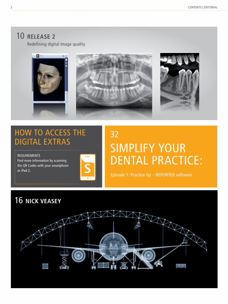
Release 2Redefining digital image quality
Nick VeaseyOn x-Ray images that aRe wORks Of aRt
HistoRy of iNNoVatioNwilhelm COnRad Röntgen as the fOunding fatheR Of the siROna x-Ray divisiOn
X-Visionthe dental x-Ray magaZine fROm siROna
2 COntents | editORial
RequiRementsfind more information by scanning the qR Codes with your smartphone or iPad 2.
32
simPlify yOuR dental PRaCtiCe: episode 1: Practice tip – RePORteR software
16 Nick Veasey
10 Release 2 Redefining digital image quality
hOw tO aCCess the digital extRas

3COntents | editORial
x-visiOn tOPiCs
6 state of the artimage quality then and now
8 PositionBest image quality with the lowest dose and a perfect work-flow
10 cover featurenew digital image quality with Release 2
16 interviewnick veasey talks about his x-ray art
x-visiOn PRaCtiCe
14 case reportendodontics with ORthOPhOs xg 3d
24 Dental practice reportdigital dentistry with dr. neal Patel
x-visiOn innOvatiOn
4 Historyfrom wilhelm Conrad Röntgen to sirona
22 Newsthe integrated facescanner for galileOs
27 WorkflowsiCat OPtiguide: the perfect implant in only two sessions
x-visiOn fORum
30 the futurewill the virtual patient become a reality?an interview with dr. wilhelm schneider
31 linkdental-users: the dental e-learning portal
32 Practice tipsimplify your dental practice: RePORteR software
34 offerstay up-to-date with the sirona software club
deaR ReadeR,with the discovery of x-rays, wilhelm Conrad Röntgen changed the world. his research was the starting point for a number of groundbreaking discoveries that have become firmly entrenched in modern life. the roots of the x-ray division of sirona dental systems can also be traced back to wilhelm Conrad Röntgen, making him the actual instigator of the company. now there are more than one hundred employees working everyday in the x-ray division of sirona dental systems on innovations for dentistry for today and tomorrow. this magazine is intended to be a witness to these innovations. we will use this magazine to present our developments and ideas as well as interesting and valuable information on the subject of x-rays. developers and users will be able to have their say and provide authentic reports about the background behind our innovations and successful use of our products.
we hope you enjoy reading the magazineand that the tips and tricks it contains are useful.
wilhelm schneider

4 x-visiOn innOvatiOn
1895
1895first x-ray image reveals the hand of Bertha Röntgen
1905first dental x-ray unit in the world: RekORd
1934x-ray sphere from siemens
1995first digital panoramic x-ray unit ORthOPhOs Plus ds
1962first panoramic x-ray unit in the world in collaboration with Palomex
1996ORthOPhOs Plus ds Ceph, until 2001 the world’s only digital Pan-Ceph x-ray unit
in the second half of the 19th century, medicine and science underwent a real boom. in the former institute of Physics at the university of würzburg on what is now the Rönt-genring science mile, wilhelm Conrad Röntgen discovered what he called “x-rays” in this room.
WHy tHe iNNoVatioN HistoRy of siRoNa staRts WitH WilHelm coNRaD RöNtgeN
On november 8, 1895 wilhelm Conrad Röntgen, Professor of Physics at the university of würzburg, experimented with a hittorf gas discharge tube. he happened to notice that coated paper continued to fluoresce even when he completely darkened the tube. wilhelm Conrad Röntgen was very quickly aware of the significance of his discovery. he called the rays “x-rays” and quickly sought an industrial partner to develop a medically useful device based on his discovery.Only three days after making his discovery the Professor made initial
contact with Julius max gotthard gebbert, a german mechanic and co-founder of the company Reiniger, gebbert & schall in erlangen. this business was a merger of karl friedrich schall’s workshop for electrical medical devices and erwin moritz Reiniger’s workshop for electrotechni-cal and physics equipment. a split shortly before Röntgen’s discovery meant that the former partner gebbert became the sole owner of the “united Physical-mechanical workshops of Reiniger, gebbert & schall (Rgs)”.

5histORy
2012
2004market launch of the newORthOPhOs xg range of pan-oramic x-ray units
2009a minor revolution in the field of panoramic x-rays: automatic patient positioning
2011Only one year after its market launch ORthOPhOs xg 3d be-comes the most sold 3d x-ray unit in dentistry
maRs software for metal arti-fact reduction
2005the first step towards 3d: transversal slices for ORthO-PhOs xgPlus
2007market launch of digital vol-ume tomography with the lowest radiation exposure (galileOs)
2009world first: introduction of the concept of integrated implan-tology with CeReC and galileOs
2012integrated facescanner for galileOs and the first fully digital process for producing drilling templates with Cad/Cam and Conebeam(siCat OPtiguide)
2006acquisition of the x-ray sensor manu-facturer schick technologies inc. leads to sirona being listed on the us tech-nology sector index nasdaq
in sirona’s “Center of innovation” in Bensheim, more than 200 engineers and scientists are working on integrated technologies for dental practices, laboratories and hospitals. the Center is the result of a systematic pursuit of innovation spanning more than 130 years which started in 1895 in würzburg.
max gebbert also recognized the significance of the “x-rays” and began to produce x-ray tubes only a few months later. focusing his business on the new technology secured him a rapid economic breakthrough. in 1905 he launched the world’s first dental x-ray unit, RekORd. after gebbert’s death siemens & halske purchased the majority of shares in Rgs in 1925 and took over the business completely in 1932. in 1934 siemens introduced the x-ray sphere, the smallest and soon to be the most widely sold x-ray unit in the world, and one that some of us are very likely familiar with from childhood visits to the dentist.
siemens dental became sirona dental systems in 1997. the spirit of innovation that started at siemens dental lives on with sirona dental systems - spanning more than 130 years. this can be seen in the ongoing history of innovation of the business’ x-ray division.

6 x-visiOn tOPiCs
image qualitythen
On december 22, 1895 wilhelm Conrad Rönt-gen successfully took the first x-ray image of the hand of his wife Bertha Röntgen.the first medically indicated images followed soon after. the duration of radiation was between one and a half and two hours at that time and the radiation exposure was also correspondingly high.

7state Of the aRt
and now“Best image quality with the lowest dose and a perfect workflow”is the guiding principle of theimaging systems division of sirona.the update package Release 2 for ORthOPhOs xg equip-ment, launched in January 2012, also adheres to this principle and delivers particu-larly clear and unambiguous images.

8 x-visiOn tOPiCs
no black margins around metal fillings and no excess contrast which may lead to misdiagnosis of caries, for example. this image was gener-ated using a reconstruction algorithm that cal-culates the contrast of anatomical structures without distorting them. astRa is an algorithm
derived from aeronautical technology which has already been used countless times to investigate the contents of luggage and for the first time can now be used in dentistry.* anatomically structured Reconstruction algorithm.
WitH astRa* WitHout astRa*
Best image quality WitH tHe loWest Dose aND a PeRfect WoRkfloWin balancing image quality, dose and workflow, sirona stands by responsible handling of
x-ray radiation.
the higher the radiation dose, the greater the contrast and therefore the clearer the image. although that may be true in principle, the current trend in dentistry to depart from the alaRa principle (as low as reason-ably achievable) when selecting radiation doses and to favor “esthetic” x-ray images is something that we at sirona consider more than worrying.
Of course, it also comes down to the intended use of the x-ray images when determining the radiation dose. if, for example, the images are to be used for planning surgical procedures, the safety of the operation has a high priority. in this case, it is safer to use a higher radiation dose than to run any risks. in routine clinical dentistry practice, however, the applied

9POsitiOn
WitHout astRa*
HD image
in addition to software that delivers artifact-free x-rays, the update package Release 2 for ORthOPhOs xg 3d also contains new sensor technology which now gives you the option of generating 3d images in high-definition mode as well. these images are constructed from a continuous exposures and provide a rapid overview and clear diagnoses, even in very difficult cases.
dose should be kept as low as possible regardless of whether it is for intraoral and panoramic x-rays or conebeam (CBCt).therefore, as part of Release 2, sirona ensures that dental practitioners have complete freedom of choice. innovations such as astRa for 2d imaging and maRs for reduction of metal artifacts in 3d imaging deliver substantially clearer images without increasing radiation. in par-ticularly difficult cases, owners of an ORthOPhOs xg 3d also have the option of generating hd images with higher resolution. the associated increase in dose can often be compensated for by selecting a smaller field of view.for this reason, the ORthOPhOs xg 3d now has a smaller second volume for taking CBCt images in hd or ld* quality. the hd images generated with the new “5 cm Ø x 5.5 cm height” volume are ideally suited to increase confidence in endodontics while at the same time keeping the radiation dose as low as possible.3d x-rays have shown to provide a significantly more reliable diagnosis in many cases than the traditional 2d imaging. the fact that the justifiable indications for conebeam are still being debated by scientists in some cases is unfortunately because their assumptions are based on equipment
using a radiation dose that almost equals the level of medical Ct scans. Our goal at sirona, on the other hand, is to achieve the best image quality using the lowest possible dose while at the same time perfecting the operation of equipment and software so that a reliable diagnosis can be made in the shortest possible time – because only then will CBCt benefit both patients and dental practices.
* low dose. ** alaRa: as low as Reasonably achievable.
alaRa** principlethe alaRa principle is legally enshrined in the x-ray regulations of most countries, although without stipulated limits. therefore, it is particularly important that both manufacturers and operators of x-ray facilities are aware of their specific local regulations.

10 x-visiOn tOPiCs
Release 2
ReDefiNiNg image qualityOnly x-ray films are or were true works of art? whoever still holds this opinion will change their mind after a tour through the “gallery of the new digital image quality.” the update package Release 2 for ORthOPhOs xg devices contains software and hardware features that now make digital x-ray images so meaningful that they have even earned the right to hang in a gallery.
By scanning this qR-code with your smartphone or iPad 2 you receive a movie featuring information on the new image quality by sirona.
11image quality
BefoRe afteR
tHe aRt of aRtifact-fRee imagiNg
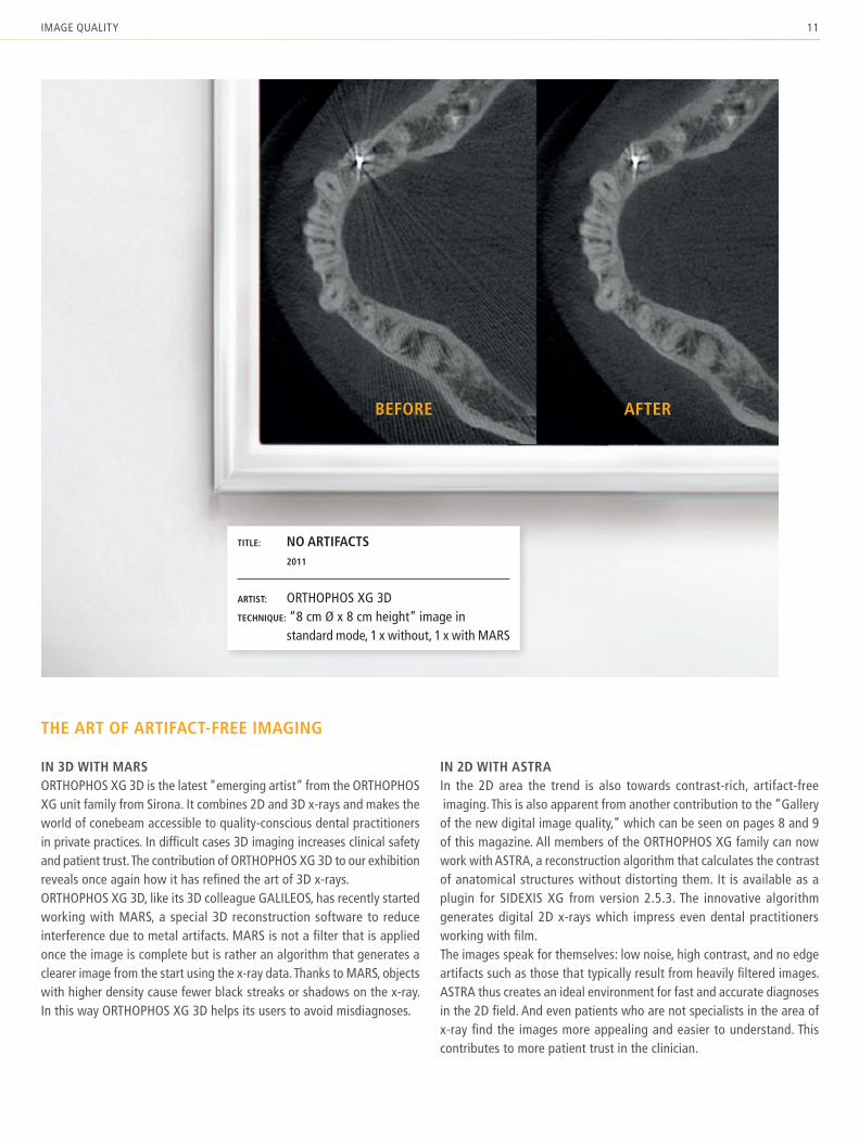
iN 3D WitH maRsORthOPhOs xg 3d is the latest “emerging artist” from the ORthOPhOs xg unit family from sirona. it combines 2d and 3d x-rays and makes the world of conebeam accessible to quality-conscious dental practitioners in private practices. in difficult cases 3d imaging increases clinical safety and patient trust. the contribution of ORthOPhOs xg 3d to our exhibition reveals once again how it has refined the art of 3d x-rays.ORthOPhOs xg 3d, like its 3d colleague galileOs, has recently started working with maRs, a special 3d reconstruction software to reduce interference due to metal artifacts. maRs is not a filter that is applied once the image is complete but is rather an algorithm that generates a clearer image from the start using the x-ray data. thanks to maRs, objects with higher density cause fewer black streaks or shadows on the x-ray. in this way ORthOPhOs xg 3d helps its users to avoid misdiagnoses.
iN 2D WitH astRain the 2d area the trend is also towards contrast-rich, artifact-free imaging. this is also apparent from another contribution to the “gallery of the new digital image quality,” which can be seen on pages 8 and 9 of this magazine. all members of the ORthOPhOs xg family can now work with astRa, a reconstruction algorithm that calculates the contrast of anatomical structures without distorting them. it is available as a plugin for sidexis xg from version 2.5.3. the innovative algorithm generates digital 2d x-rays which impress even dental practitioners working with film.the images speak for themselves: low noise, high contrast, and no edge artifacts such as those that typically result from heavily filtered images. astRa thus creates an ideal environment for fast and accurate diagnoses in the 2d field. and even patients who are not specialists in the area of x-ray find the images more appealing and easier to understand. this contributes to more patient trust in the clinician.
title: No aRtifacts 2011
aRtist: ORthOPhOs xg 3dtecHNique: “8 cm Ø x 8 cm height” image in standard mode, 1 x without, 1 x with maRs
12 x-visiOn tOPiCs
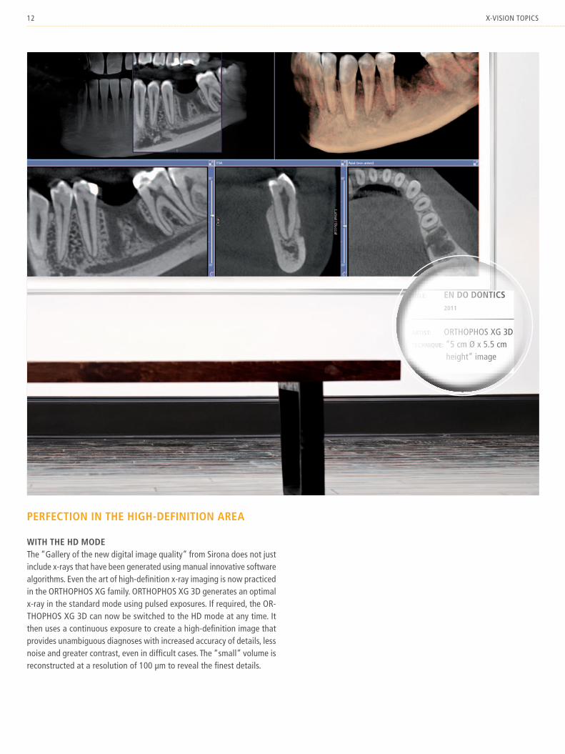
PeRfectioN iN tHe HigH-DefiNitioN aRea
WitH tHe HD moDethe “gallery of the new digital image quality” from sirona does not just include x-rays that have been generated using manual innovative software algorithms. even the art of high-definition x-ray imaging is now practiced in the ORthOPhOs xg family. ORthOPhOs xg 3d generates an optimal x-ray in the standard mode using pulsed exposures. if required, the OR-thOPhOs xg 3d can now be switched to the hd mode at any time. it then uses a continuous exposure to create a high-definition image that provides unambiguous diagnoses with increased accuracy of details, less noise and greater contrast, even in difficult cases. the “small” volume is reconstructed at a resolution of 100 µm to reveal the finest details.
title: eN Do DoNtics 2011
aRtist: ORthOPhOs xg 3dtecHNique: “5 cm Ø x 5.5 cm height” image

13image quality
NeW image quality
iN eNDoDoNticsthis very special contribution to the “gallery of the new digital image quality” was also provided by ORthOPhOs xg 3d. this multitalented machine now works with two different volumes: as well as the previous “8 cm Ø x 8 cm height” volume (vOl 1) it now also has a volume of 5 cm Ø x 5.5 cm height (vOl 2) which is particularly suitable for endodon-tics when combined with the hd mode. when the hd mode has been selected in “endo volume,” ORthOPhOs xg 3d generates extremely detailed images with a voxel size of 100 µm. this gives users greater confidence during root canal treatments and other cases and at the same time can also keep the radiation dose as low as possible thanks to the smaller field of view.
see more clearly. diagnose with greater confidence. treat faster and better. those who want to benefit from the advantages of Release 2 after this tour of the “gallery of the new digital image quality” can obtain more information from their authorized spe-cialist distributor.
title: DeeP 2011
aRtist: ORthOPhOs xg 3dtecHNique: “5 cm Ø x 5.5 cm height” image in hd mode, voxel size 100 µm

14 x-visiOn PRaCtiCe
Case RePORt
eNDoDoNtics WitH oRtHoPHos Xg 3DDR. sigRiD fRaNk, BesigHeim
with the new volume of 5 cm Ø x 5.5 cm height, ORthOPhOs xg 3d is now ideally suited for root canal treatments and surgical procedures in endodontics. high-precision 3d images ensure the greatest possible clinical safety while the radiation dose is kept as low as possible thanks to the reduced volume. dr. sigrid frank reports about a successful treatment on one of the maxillary second molars.
improving image quality. Reducing radiation dose: these are good reasons for using 3d conebeam imaging for endodontic diagnosis.a large percentage of incidental endodontic findings are detected in general dental prac-tices in CBCt images that have been prepared for other justifiable reasons such as planning for implants.
the desire for three-dimensional imaging of root anatomy – and specifically of the canal system with all macroscopically detectable pathological changes – remained unsatisfied for many years before the era of conebeam. Prior to conebeam, findings that could not be detected in two- dimensional x-rays were being ignored or classified as impossible or improbable. the moment when a 3d image provided the obvious explanation for a differential diagnostic finding that had remained unclear or resistant to treatment with two-dimensional imaging technology was then even more surprising.
in the present case the patient came to our practice with diffuse symptoms in the left max-illa and the desire for a “complete overhaul”, including implants.
15Case RePORt
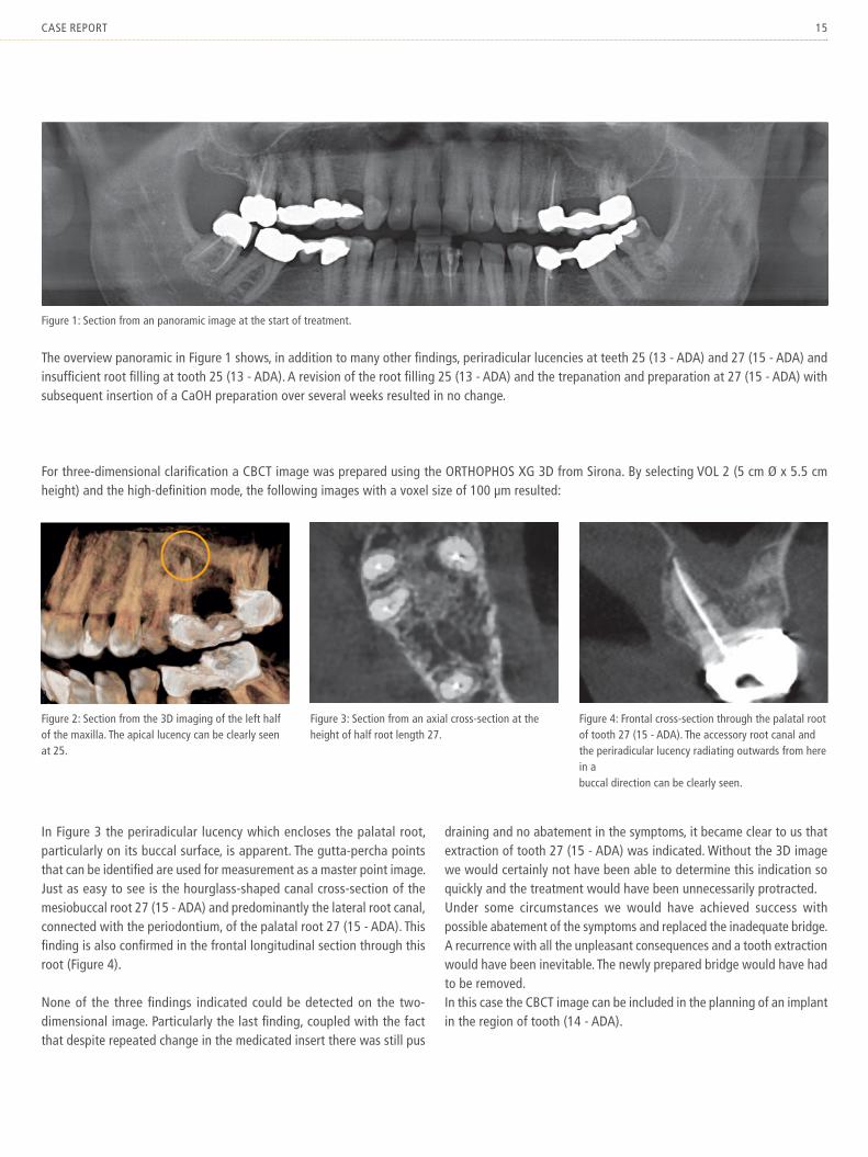
figure 1: section from an panoramic image at the start of treatment.
figure 2: section from the 3d imaging of the left half of the maxilla. the apical lucency can be clearly seen at 25.
figure 3: section from an axial cross-section at the height of half root length 27.
figure 4: frontal cross-section through the palatal root of tooth 27 (15 - ada). the accessory root canal and the periradicular lucency radiating outwards from here in a buccal direction can be clearly seen.
the overview panoramic in figure 1 shows, in addition to many other findings, periradicular lucencies at teeth 25 (13 - ada) and 27 (15 - ada) and insufficient root filling at tooth 25 (13 - ada). a revision of the root filling 25 (13 - ada) and the trepanation and preparation at 27 (15 - ada) with subsequent insertion of a CaOh preparation over several weeks resulted in no change.
for three-dimensional clarification a CBCt image was prepared using the ORthOPhOs xg 3d from sirona. By selecting vOl 2 (5 cm Ø x 5.5 cm height) and the high-definition mode, the following images with a voxel size of 100 µm resulted:
in figure 3 the periradicular lucency which encloses the palatal root, particularly on its buccal surface, is apparent. the gutta-percha points that can be identified are used for measurement as a master point image. Just as easy to see is the hourglass-shaped canal cross-section of the mesiobuccal root 27 (15 - ada) and predominantly the lateral root canal, connected with the periodontium, of the palatal root 27 (15 - ada). this finding is also confirmed in the frontal longitudinal section through this root (figure 4).
none of the three findings indicated could be detected on the two- dimensional image. Particularly the last finding, coupled with the fact that despite repeated change in the medicated insert there was still pus
draining and no abatement in the symptoms, it became clear to us that extraction of tooth 27 (15 - ada) was indicated. without the 3d image we would certainly not have been able to determine this indication so quickly and the treatment would have been unnecessarily protracted.under some circumstances we would have achieved success with possible abatement of the symptoms and replaced the inadequate bridge. a recurrence with all the unpleasant consequences and a tooth extraction would have been inevitable. the newly prepared bridge would have had to be removed.in this case the CBCt image can be included in the planning of an implant in the region of tooth (14 - ada).

16 x-visiOn tOPiCs
Nick Veasey | X-Ray aRtist

17x-Ray aRt
Nick Veasey | X-Ray aRtist

18 x-visiOn tOPiCs
JouRNey iNto tHe iNteRioRhis workspace is unusual. lead doors, lead-lined walls, underpants made
from lead. nick veasey journeys below the surface of objects. always
searching for hidden details. in an interview with the artist reveals what
excites him about x-ray photography, where his ideas come from, and why
he would love to work with museums.
fourteen years ago you decided to x-ray objects and people instead of continuing to take pho-tographs. x-ray is an art for you. would you say that true beauty comes from within?absolutely. Just ask yourself when you are at your happiest. when you have bought yourself a new dress or a new suit? Or perhaps a new car? Or is it instead when you’re with the peo-ple that you love? true contentment and true beauty come from within and not from mate-rial, superficial charms. i see this as the special significance of my work. it’s about peering in-side, looking beneath the surface, blocking out irrelevant information, and concentrating on the essential.
nick veasey was born in london in 1962 and now lives near maidstone in south east england. Before dedicating himself to x-ray photography, the adventurous photog-rapher worked for many years in advertising and the design industry. nowadays nick veasey is probably one of the best known x-ray photographers and he has re-ceived many awards.
as part of image processing you place semi-transparent images of the objects over x-ray images and then add color. in your opinion do pictures have to be in color to awaken feelings?not at all. i only have the impression that some of my pictures need color to achieve the desired effect. i prefer those pictures without color be-cause they appear pure and unadulterated. Purity and simplicity are becoming increasingly important to me as i get older.whether in color or in black and white – what does image quality mean to you and how pre-cisely do you ensure image quality?that’s a good question. On one hand, image quality is about depiction of detail, sharpness, contrast, and overall effect. i believe i can say that in this regard i can prepare a good x-ray image. the actual measure of quality, however, is that the image triggers something in the ob-server. a beautiful, detailed x-ray image is worth-less if it has no depth and does not move the observer.

19x-Ray aRt
when preparing an exhibition, you must also ensure that you combine pictures in such a way that there is a flow and a link between the sub-ject matter of the works. this is one of the areas that i still have to develop. while i am very proud of some of my creations, in the context of an exhibition i have to approach the relationship between the works with greater objectivity. sometimes what you leave out is more important than what is kept.
your most important tool are underpants made from lead to protect yourself from the radiation. what is different about your x-ray machines compared to those used in medicine?my machines are designed for x-ray imaging of steel constructions in industry. this sort of imag-ing requires a much higher radiation dose and a significantly longer exposure time than the x-ray images made by dentists. i have a bunker that has been specially set up for this but i still have to be careful. as sexy as my lead under-pants may be, there are more comfortable pieces of clothing.

20 x-visiOn tOPiCs
you reveal the inner life of the most diverse subjects – from tiny insects to a Boeing 777. where do you get your ideas from?i have been doing this for some time now and it should be expected that there is one or the other interesting result. i draw my inspiration from everyday life. i am open to stimulus from the most diverse sources. this keeps my mind alert and always gives me new ideas. the prob-lem, however, is that most of them are worthless.if my calendar allows it i like visiting exhibitions by other artists. i enjoy getting a little distance
from my own creations and engaging with the art of others. it helps me to sort out the worth-less ideas so that, i hope, only the valuable ideas are left over at the end.what’s hidden inside a t-shirt really couldn’t be that spectacular. what has been your most as-tonishing discovery?i find the inside of a t-shirt quite spectacular. i actually find that the texture of material can appear elegant and ethereal in x-rays. and when you look at a piece of clothing in isolation out-side the usual environment of a photo shoot (attractive models, sophisticated locations, …), you start to think how your own personality
undergoes a change when you wear different clothing. you feel different in a suit that you wear to an interview compared to your everyday clothes.that was my most surprising discovery: the most banal everyday object becomes interesting if we take the time to think about it. a successful x-ray image lends dignity to such an everyday object because it allows you to observe it from the inside. ultimately this object was designed and produced by someone who engaged in his work with love and attention – and precisely that is what i would like to celebrate.
“i Do ofteN DReam iN X-Ray images.”

21x-Ray aRt
you call yourself the “original x-ray nerd.” are you an x-ray photographer only at work or do these pictures not leave you alone even in your private life? do you dream of x-ray images?in a profession like mine it isn’t so easy to separate professional and private life. i try to take time for my family because that is really important for me. as an artist, however, you can’t always turn your brain off. i do often dream in x-ray images. i do have to add though, that i have already created so many x-ray pictures that i can easily imagine in my inner eye pre-cisely how a particular object would appear in x-ray radiation.
you don’t work with digital x-ray technology but rather with film. Once you have photo-graphed your subject, then the real work starts: exposure of the x-ray film, scanning of the im-ages, editing on the computer. does it annoy you that you can spend months working on a single picture? is your only motivation the thought of the final result?i don’t have any problem motivating myself to keep going. i love my work and pursue it with passion. maybe i’m even too productive. i don’t think much of sitting in front of a computer, though. i’m happy when i’m preparing the x-ray images, not in front of my mac.tell us what your favorite subject for an x-ray photograph has been. what would you like to x-ray in the future?my absolute favorite subject is called frog Crab. it looks like it has come from another planet. nature simply cannot be beaten – it contains an infinite number of possibilities for experi-menting. But nature always stays in charge and
i like that. humans have brought so much under their control and so many pictures – whether three-dimensional or airbrushed – are now syn-thetic. nature on the other hand is real and x-ray radiation offers an opportunity for undistorted representation of this reality, warts and all. in the future i would really like to x-ray a subma-rine and in collaboration with a museum reveal to the visitors what is normally hidden from them – that is, everything that happens inside the exhibits.
“X-Ray RaDiatioNoffeRs aN oPPoRtuNityfoR uNDistoRteDRePReseNtatioN.”
fROg CRaB

By scanning this qR-code with your smartphone or iPad 2 you receive a movie featuring infor-mation on galileOs facescanner.
22 x-visiOn innOvatiOn
integRated faCesCanneR
Welcome to tHe PRactice of tHe futuRe!
the face scan is displayed in an integrated viewer. the CBCt image and the face scan are automatically superimposed.

23news
with the facescanner integrated in galileOs x-ray units, the vision of the virtual patient is coming within reach: while taking the x-ray, this scanner plots a virtual face scan that is simultaneously superimposed with the 3d x-ray data. the face scan helps you to plan your therapy, makes treatment more comprehensible for your patients, and thus ensures greater understanding and trust.
as part of the vision of virtual patients, orthodontic and maxillofacial surgery applications are also being planned. it is absolutely plausible that dentists will be able to simulate changes to the facial surface resulting from surgical or orthodontic procedures using morphing software before starting any procedures, thus being able to clearly show patients the possible results of a therapy.
BeNefits
PatieNt maRketiNg WitH tHe galileos facescaNNeR:■■ Patients identify themselves more easily with a face scan than with
an x-ray image■■ this enables them to better understand therapy suggestions and to
more readily accept them■■ questions and misunderstandings can be more easily answered or
clarified through doctor/Patient codiagnosis■■ the perception of the dental practice improves thanks to word-of-
mouth recommendations by patients■■ no additional scan time is necessary, patients do not need to be
repositioned, and there is no tedious superimposition of the data thanks to simultaneous recording and automatic registration
tecHNical Data
aN oVeRVieW of tHe galileos facescaNNeR:■■ laser-free technology, gentle operation with white light■■ all galileOs devices can be retrofitted■■ easy data exchange using software from other manufacturers, e.g.,
for orthodontic measurements■■ integrated viewer software with easy operation■■ data superimposed with high precision
facescan-export in dolphin

24 x-visiOn PRaCtiCe
dental practitioners in private practices are often judged by the first impression they make on their patients.i know today that achieving success only three years after opening my own practice is the result of the best technology and equipment that modern dentistry has to offer. Patients leave my practice and share their great experience with friends and family. in today’s high tech environ-ment patients expect certain advantages during treatment. the opportunity to collect and store patient data digitally is considered advanced. i chose digital sensors by sirona that are seam-lessly integrated into my practice administration software.
“I rely onthree-dImensIonal dentIstry”
a PRaCtiCe RePORt fROm dR. neal Patel
the Patients aRe imPRessed By hOw shaRP the digital x-Ray images aPPeaR On the COmPuteR sCReen COmPaRed tO films On a light BOx.it is also very important to me to be able to offer comprehensive dental diagnostics. even the best trained dental practitioner can only make precise diagnoses with increasing experi-ence. my initial lack of clinical experience encouraged me to explore leveraging technology to improve my practice. this led me to choose a galileOs 3d x-ray unit from sirona. the ability to create 3d images clearly distinguished my practice from others. with a single scan i am able to consider all facets of dentistry when making a diagnosis: endodontics, periodontics,
orthodontics, maxillofacial surgery, implantology, airway analysis, and general dentistry.
sinCe i have Been exPlaining tReatment needs tO Patients using 3d images, they aCCePt my theRaPy suggestiOns muCh mORe Often.
the comprehensive display of data in a CBCt scan has also led to significant time savings during the initial examination of a new patient.in my practice i am now able to compile a comprehensive diagnostic and treatment plan. what was still missing was a flexible platform for restorative clinical treatments. i chose the Cad/Cam platform CeReC from sirona. with
dr. neal Patel has been the owner of “infinite smiles” in Powell, Ohio, since 2008. he is seen here with his galileOs 3d x-ray unit.

25manual
CeReC i can now offer prosthetic products in my practice such as crowns, inlays and onlays right up to veneers. the CeReC platform is flexible and functions as a digital impression system with digital lab communication and lab fabrication of prosthetics, to chairside milling for same appointment restorative care. what is most important is that i have full control over my results from start to finish. in addition to the interdisciplinary areas listed above i knew i needed a platform for growth in my practice. knowing the high demand for and excellent success rate of implants propelled me to want to learn how to offer them as the best replacement solution to take the place of a fixed bridge.
integRated imPlantOlOgy with CeReC and galileOs PROvided me with a teRRifiC wORkflOw, CliniCal safety, and eCOnOmiC suCCess.
Patients who are shown their treatment situation in three dimensions and recognize the corre-spondence between crown and implant and their natural teeth are more receptive and more read-ily choose the therapy suggestion. if, however, they have to create a picture of their mouth situation based on descriptions and models, the dentist needs considerably more time for the consultation.the combination of the 3d scan from galileOs with the data from the planned restorative treatment from the CeReC software enables me to simultaneously plan both surgery and prosthesis – without having to make esthetic compromises. with the help of drilling templates ordered directly from the software, i can then place implants exactly where they had been planned.
this gives me the assuRanCe and COnfi-denCe tO PlaCe and RestORe mORe imPlants myself and thus tO inCRease the PROduCtivity Of my PRaCtiCe.
the combination of these devices is my techno-logical recipe for excellent dentistry. i am ex-cited and really happy to work with this fabulous technology because i feel that i am able to offer every type of clinical therapy. the ultramodern features enabled me to very quickly make a name for myself after establishing my practice.
my Patients see in me a yOung and enthusiastiC dentist whO OffeRs dental tReatments at the highest level using Outstanding teChnOlOgy.

www.integrated-implantology.com
B-52
0-76
- v0
golf or implants: both are a question of absolute precision. this is why more and more dental practitioners are relying on CeReC and 3d x-rays from sirona. they have greater confidence and precision. and they save time. Patients understand immediately the therapy requirement and their acceptance of top-quality treatments increases. enjoy every day. With sirona.
integratedimplantology.more confidence,more opportunities,more implants.
when fractions of amillimeter make the difference.

By scanning this qR-code with your smartphone or iPad 2 you receive additional information on the integrated implantology by sirona.
27wORkflOw
Drilling templates from sicatthe business siCat was founded in 2004 as a joint venture with sirona. you can read more about siCat at www.siCat.com
sicat OptiGuide
fewer appointments necessary for treatment
treatment acceptance improves
highly precise and esthetic results
Complete control through the entire process
iNtegRateD imPlaNtology is NoW eVeN easieR: a PeRfect imPlaNt iN oNly tWo sessioNs!
thanks to the new siCat OPtiguide method for preparing surgical guides, integrated implantology for CeReC users is now even easier. to make the benefits clear, the standard siCat ClassiCguide method, which will still be offered for dentists who do not work with CeReC, is outlined here as a comparison.
tHe sicat classicguiDe as a comPaRisoN
with the siCat ClassiCguide method you take an impres-sion of the patient’s jaw in the first session. your dental technician then builds a wax-up of the proposed restoration on the model and prepares a stent with a proposed inte-grated crown which is fixed to the bite plate. alternatively, in simple cases bite register material can be directly applied to the bite plate. this forms an x-ray template with or without the prosthetic restoration which the patient then wears during the 3d x-ray imaging process. the reference balls on the bite plate enable perfect positioning later when preparing the drilling templates.

28 x-visiOn innOvatiOn
1st session
aN oVeRVieW of tHe sicat oPtiguiDe WoRkfloW
you take an optical impression of the bite using the CeReC Bluecam and plan the pros-thetic restoration using the CeReC software.your assistant creates a 3d x-ray image with galileOs or ORthOPhOs xg 3d.after the assessment of the 3d scan, you integrate the prosthetic restoration into the x-ray volume and plan the implant using the galileOs or siCat implant software. at the same time you can take the surgical
parameters as well as function and esthetics into consideration.you advise the patients and a decision can be made in the first session about the ther-apy.all the data required to manufacture a drilling template is transferred to siCat with a single click of the mouse.
By the seCOnd sessiOn you will receive a high-precision siCat surgical guide. Before the implantation in many cases you can prepare the long-term restoration or even an individual abut-ment with CeReC or inlab. you will receive the necessary model from infinident, you can mill it yourself or manufacture it conventionally based on a plastic impression.

SICAT GmbH & Co. KG. Brunnenallee 6. 53177 BonnTelf. +49 228 854697-0 . Fax +49 228 854697-99 . [email protected]
29wORkflOw
2nd session
thanks to the siCat OPtiguide, CeReC users can now completely avoid x-ray templates in many cases*. this also means no more waiting for the completed radiographic stent which meant in the past that the patient could only be x-rayed in the second session.
PossiBle iN a siNgle sessioN iN futuRe**
CeReC users who are equipped to manufacture ceramic restorations with a CeReC or inlab mC xl milling unit will soon be able to manufacture drilling templates themselves. the process includes several manual inter-mediate steps, enabling you to carry out the complete implantation from assessment to planning and insertion of the implant right through to temporary restoration in a single session if required.** this means you forego the safety check offered by the service department of drilling template manufacturer siCat, however.
* not for toothless jaws, extensive metal restorations, and gaps that cannot be dis-played using an optical impression.
** depends on the legal framework of the particular country.
the patient arrives for the implantation. you insert the implant with the help of the surgical guide. in the case of an immediately loadable implant, the screw retained abutment is mounted on the implant fixture and the provisional restoration (manufactured with CeReC) is cemented to the customized abutment.
Make every case count
Every case counts – use the simple and safe method for implant planning and implementation now.
■Simple data import of DICOM data set
■Exact implementation using SICAT drilling templates with guaranteed precision
■Cost-effective surgical guides
Intuitive software operation, exact and cost-effective drilling templates – imp-lant planning that makes sense Discover SICAT Implant now. Our sales team will provide live demonstrations in your practice or online.
www.sicat.com

30 x-visiOn fORum
Will tHe ViRtual PatieNt gaiN accePtaNce?
aN iNteRVieW WitH DR. WilHelm scHNeiDeR
dR. sChneideR, whiCh develOPments fROm siROna in the aRea Of imaging systems mean that we Can alReady talk aBOut a “viRtual Patient”?the first step towards creating virtual patients was the connection between Cad/Cam and 3d x-rays. using the unique process of integrated implantology, sirona made it possible to inte-grate a virtually constructed dental restoration into a 3d x-ray image. and now another step has been taken towards this vision of virtual patients: the integrated facescanner in galil-eOs.
at the mOment the galileOs faCesCanneR is mOstly a tOOl tO imPROve Patient COm-muniCatiOn. COuld it alsO Be used fOR diagnOstiC PuRPOses in futuRe?By combining CBCt scans, Cad/Cam data, and facial scans, completely new opportunities will be available in future, and not just for diagnos-tics.for example, this combination and visual display of the various data will enable therapy propos-als to be comprehensively communicated and the consequences for patients can be simulated in a very short time – with a large enough body of data, even long-term prognoses could be made. the next step in the development of virtual patients will be the integration of jaw movements in the CBCt scans. this is because visual analysis of the temporomandibular joints and the movements in the joints harbors a huge potential for well-founded diagnoses and new options for therapy. another option offered by the virtual patient will be to see the patient’s face smile naturally using their virtual face scan.
the provisions for this already exist. the smile could thus be used to perfect the smile design for CeReC applications.
many dental PRaCtitiOneRs aRe CautiOus when it COmes tO new develOPments. why dOes it make sense tO use an add-On like the integRated faCesCanneR Right nOw?Patients are increasingly at home in a digital world made up of social media and internet forums. Particularly for this reason, the market-ing aspect of the integrated facescanner should not be underestimated. Patients speak about their experience with the virtual face scan and go on to recommend the dental practice. and the more complex a dental procedure, the more important it is to clearly communicate with pa-tients. the three-dimensional display means that therapy proposals are more easily understood and this increases the acceptance of the patient.
hOw lOng dO yOu think it will take Be-fORe the viRtual Patient is standaRd in eveRy dental PRaCtiCe?it will happen quicker than many expect. digital dentistry has rapidly advanced in recent years. the virtual patient will put diagnosis, patient consultation, and therapy on a whole new level. the first few steps towards a virtual patient have already provided greater confidence, bet-ter acceptance of therapy proposals, time sav-ings, and greater pleasure when working in the practice.
Dr. Wilhelm schneider is the marketing Director of the sirona imaging systems division.

31the futuRe
DeNtal-useRs.comtHe DeNtal e-leaRNiNg PoRtal
modern dentistry and dental technology are developing rapidly. this gives rise to new work-flows which are both a challenge and motivation for the entire dental team.
dr. elmar frank, dr. sigrid frank, and gerhard werling are tackling this subject and have created an internet portal for dental continuing education and professional development: www.dental-users.com is the latest development in the area of dental e-learning. the portal is aimed at dental practitioners, dental technicians, dental assistants and dentistry students – in short, the whole dental “family.” with modern e-learning methods, the portal explains how to handle high-tech dental systems using relevant examples across systems.
the creators of dental-users.com have many years of experience with the use of 3d simulation and planning, Cad/Cam applications, digital
x-ray diagnostics, and dental computer applica-tions of all sorts. their many years of experience as authors, referees, developers, beta testers, technical advisers for colleagues and not least as enthusiastic users of dental high-tech in their own practices means that they know precisely where problems can develop and how and where the best help can be given.
unlike other online study programs in the medical and dental sectors, dental-users.com not only offers video presentations but also short multimedia videos (typical length: few minutes) that give concrete solutions for problems. all content is based on established workflows and strategies for daily use of modern techniques that require clarification – for example, in the area of digital x-rays, laser application, comput-er-assisted diagnostics, and dental technology.
as well as the short videos and recorded presentations, dental-users.com also provides
users with a discussion forum, calendar admin-istration for dental events with booking options, relevant downloads, and online tests. Only persons who can demonstrate their involvement in dental medicine can register on the portal. the content accessed is automatically recorded and can be retrieved to provide evidence of professional development in the form of a Cme points account statement according to the guidelines of the german dental association and the germany society for dentistry and Oral and maxillofacial surgery.
dental-users.com has also created an iPhone or iPad app. this enables non-members to access some sample material as well as summaries with the opportunity to make online bookings for dental events such as courses, presentations or conferences.
short videos on dental-users.com Record retrieval as evidence of professional
development
dental-users app for iPad or iPhone

32 x-visiOn fORum
simPlify yOuR dental PRaCtiCe:
RePoRteR softWaRe
HoW you caN cReate RePoRts fRom fiNDiNgs fast
if you operate a dental x-ray center or accept referrals as the owner of a CBCt device, one of the challenges you face is to create meaningful reports for your client based on your findings as quickly as possible.
with the RePORteR software this is all done quickly and easily – the software is directly linked to the 3d software galaxis. galaxis enables you to mark findings directly in the x-ray volume, add a short comment, and capture screen images complete with all settings and retrieve them again if needed. this saving procedure is also the reason why the data can be transferred at the click of a button to the RePORteR software.
PRaCtiCe tiP 33
geNeRate RePoRts iN oNly a feW stePs
when you want to create a report, first select the results that should be included in the documentation. the RePORteR software automatically fills one page with each result. the description of the results is automatically transferred and the relevant x-ray images are adapted to pre-defined image fields.you can, however, adjust the page layout at any time and change the layout separately for each page to optimally present your results. for this purpose you can choose between different page templates and adjust the sections and views for different tasks and for different page formats. even the
distances between individual sections as well as the layer thickness can be varied across a wide spectrum.
the completed x-ray report is saved as a sidexis document and can then be sent in a number of different ways to your client: printed on paper or film or as a Pdf or diCOm file. dental practitioners who work digitally receive a 3d viewer that is generated directly from galaxis at the click of a button. this enables you to meet the most diverse requirements of your clients quickly and easily.
tiP: individualiZatiOnthe templates can be individualized with your own practice logo. it is also possible to insert optional elements using suitable pro-grams such as adobe Professional in the Pdf format.

34 x-visiOn fORum
alWays uP to Date WitH tHe XWiN softWaRe cluB“it took 300,000 years for humanity to accumulate the first 12 exabytes (billion gigabytes) of information. we will only need two and a half years for the next 12 exabytes.”
university of California/Berkeley school of ims
Xwin silVeR
for dental practitioners without their own 3d x-ray unit but who still want to plan their implants in 3d.
Xwin golD
for dental practitioners who use their soft-ware for 2d diagnostics.
Xwin PlatiNum
for dental practitioners who want to take advantage of all the opportunities offered by sidexis, galaxis, and galileOs implant for diagnostics and therapy.

35OffeR
what is true for the development of knowledge of all of humankind can be readily applied to dentistry: ten years ago Cad/Cam was exotic, the use of laser light was still in its infancy, and three-dimensional x-rays were not even possible.today dental practitioners must keep pace not only with the development of products but also with the even more rapid development of software-controlled workflows in which the products are integrated.
with galaxis and galileOs implant, for example, there have gradually been significant improvements in image quality. new applica-tions such as the results-oriented workflow and integrated implantology (CeReC meets galileOs) have also been incorporated – and all within five years. Can it continue like this? the response is certainly yes. there is no reason for a reversal and no sign that this trend is reversing. what is important is staying on the ball. every additional development improves the opportunities for diagnostics or therapy, saves time, and promotes the practice. x-rays have not been a cost factor for a long time. three-dimensional x-rays in particular are fast becoming the basis for the success of a modern practice. wouldn’t it be great to simply get carried along? to expand without having to worry about the details? to take part, almost without effort?
the xwin software club now makes this possible – in three steps that are tailored to the various needs of a modern dental practice.
you can get more information from your specialist dealer.
Xwin silVeR*
■■ siCat implant Club version■■ all updates of the siCat implant Club version in the contractual period■■ Price offer for a Cad/Cam upgrade■■ internet-based training for implant planning
with the siCat implant Club version you can plan implants in 3d based on galileOs or ORthOPhOs xg 3d dvt images.
Xwin golD*
■■ sidexis■■ astRa ■■ sidexis easy Backup■■ all sidexis/astRa updates and upgrades in the contractual period■■ updates for an existing sidexis xg tse version■■ Price offer for sidexis ■■ internet-based training for sidexis
xwin gold contains all 2d-software needed for results. xwin gold already pays off thanks to astRa, the new reconstruction algorithm for panoramic x-ray images.
Xwin PlatiNum*
■■ all services of xwin gold■■ all galaxis/galileOs implant updates in the contractual period■■ RePORteR software■■ 3d reconstructionsoftware maRs facescan viewer updates diCOm
3rd Party export■■ internet-based training for sidexis/galaxis/galileOs implant/Cmg■■ voucher for six months membership for the e-learning platform
dental-users.com
with xwin platinum you always have the latest information at your fingertips.
* more information about prices, membership fees as part of a three-year contract, and additional current services is available from your participating authorized sirona x-ray dealer.

subj
ect t
o te
chni
cal m
odifi
catio
ns a
nd e
rror
s, or
der n
o. a
9110
0-m
47-B
512,
Prin
ted
in g
erm
any,
disp
o-n
o. 0
4602
, JP1
1-28
4 w
s 12
112.
5v0
sirona dental systems · fabrikstrasse 31 · 64625 Bensheim · germanyemail: [email protected] · www.sirona.com