Embed Size (px)
Citation preview
8/11/2019 The Cytoskeleton of Nerve Cells in Historic Perspective
http://slidepdf.com/reader/full/the-cytoskeleton-of-nerve-cells-in-historic-perspective 1/17
/15/12 Ev ernote Web
1
The Cytoskeleton of Nerve Cells in Historic
Perspective
Saturday, December 15 2012, 11:55 AM
The Cytoskeleton of Nerve Cells in Historic Perspective
Citation:Frixione, E (2006) History of Neuroscience: The Cytoskeleton of Nerve Cells in Historical Perspective, IBRO History of Neuroscience
[http://www.ibro.info/Pub/Pub_Main_Display.asp?LC_Docs_ID=3147]Accessed: date
Eugenio Frixione
Introduction
Neurons can be usually distinguished at once from most other cells by their peculiar multi-branched shapes, which allow them to maximize their spatial reach and surface-to-volume ratioswhile keeping metabolically manageable individual sizes. Each of them is capable of departing somuch from the basic round form of cells, and of growing and sustaining their characteristic ramifiedmorphology, mainly because of the development of an intracellular framework constituted of
filamentous structures collectively known as the "cytoskeleton". Nowhere else are the advantagesof possessing this highly complex and versatile scaffolding, common to nearly all eukaryotic cells,more apparent than in neurons. It is hardly surprising, therefore, that its existence, chief components and functions were commonly found or first studied in nerve tissue. The history of how the biologically important concept of a cytoskeleton emerged is, therefore, from the verybeginning, to a large extent a history of the neurocytoskeleton (for a survey of how the generalnotion of an internal cell skeleton originated and evolved, see Frixione, 2000). The followingparagraphs will attempt to summarize the main chapters of this Odyssey.
Precedents before the 19th century
Ever since Antiquity, nerves have been regarded as conduits for some sort of vehicle that rapidly
carries information between different parts of the animal body, typically communicatingimpressions from the sense organs to the brain as well as commands from the latter to themuscles. The carrier itself was originally conceived as a special mixture of air and fire called pneuma (see Solmsen, 1961; Temkin, 1977), a term later on Latinized as "spirits" (e.g. Descartes,1664), which would flow within the nerves just as the blood streams along arteries and veins, onlymuch faster despite the former being in general considerably narrower passages. Several varietiesand even doubts started to appear on this model since the Renaissance (reviewed by Clarke,1968, 1978), yet it was rather surprising that the first microscopical inspections of nerves failed toshow any clear ducts in them (Malpighi, 1666; van Leeuwenhoek, 1674). Fortunately, however, acloser look with presumptively improved optics revealed that the nerves are actually composed of "very minute vessels of an incredible thinness ... [i.e. axons] ... running along by the sides of eachother ... [and that] the cavity of each of these small vessels is about two thirds its diameter"
8/11/2019 The Cytoskeleton of Nerve Cells in Historic Perspective
http://slidepdf.com/reader/full/the-cytoskeleton-of-nerve-cells-in-historic-perspective 2/17
/15/12 Ev ernote Web
ps:// www.evernote.com/edit/c826182f -f cee-446a-b826-4b5024ca16ff #st=p&n=c826182f-f cee-446…
(van Leeuwenhoek, 1717; see also Van der Loos, 1967; Brazier, 1984). However, the first knownmicroscopical examination of the contents of such tiny tubes, carried out more than 60 yearslater, produced results that were deemed quite incompatible with the supposedly swift flow of "spirits" or any other thin fluid along the nerves. As judged from the soft dough that could beextruded from transversely cut nerves, the "primitive cylinders" were filled with "a glutinous,elastic, transparent material, which [...] seemed to be formed of granular filaments, tenacious andelastic, which the water could neither dissolve nor separate" (Fontana, 1782).
The Classic Period
The number of opinions about the fillings of the small cylinders and globules seen to constitute thebulk of all nervous tissues increased along with the expansion of microscopical research in the19th century. Yet the two main themes that were to dominate the scene can be found exemplifiedin descriptions provided already in the mid-1820s by two French authors: whereas René-JoachimDutrochet (1824) could only see a "fluide diaphane" within nerve fibers of the frog, Henri Milne-Edwards (1825) believed those of the rabbit to contain or be composed of "fibres élémentaires",themselves consisting of long chains of diminutive globules. Rapidly evolving technical skills andquality in optics - particularly the introduction of achromatic objectives - paved the way for morereliable observations and further controversy. The longitudinal "cavity" originally described in nervefibers (van Leeuwenhoek, 1717; see above) was now found clearly visible as a transparent"primitive band" coursing throughout the core of certain (myelinated) nerve fibers of vertebrates
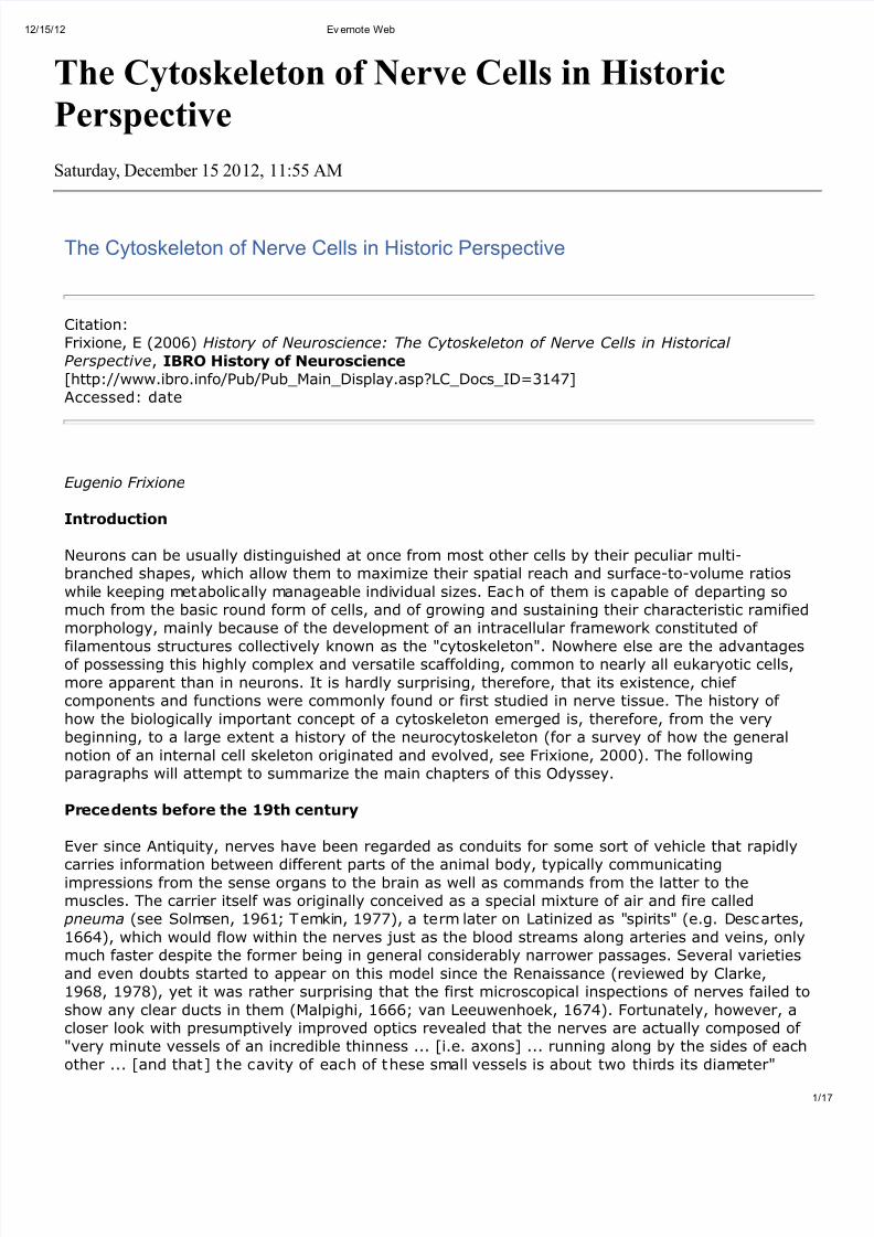
(Remak, 1838). But whereas such "cell-cavity" appeared just "to be filled by a firm substance"according to Theodor Schwann (1839), the corresponding axial region of the wider nerve fibers of some invertebrates was described by Robert Remak (1843) as containing an uninterrupted bundleof very delicate filaments or fibrils. Furthermore, when at least some of those nerve fibers weredemonstrated to be tubular processes anastomosed to, and therefore continuous with, nearbyglobules or nerve cells, the central fibrillar bundle within a given nerve fiber would occasionally beseen to reach into the body of an associated cell, and form therein concentric layers of fibrilsaround the nucleus (Remak, 1844; see Figure 1).
Figure 1: One of the first two known illustrations of the cytoskeleton shows a ganglion cell of thecrayfish nerve cord, in which concentric layers of delicate fibrils appear surrounding the nucleusand converging as they enter the axon. The fragile fibrils tend to break down as free grains near
the cut end of the axon (Figure 9 in Remak, 1844).
Inconsistencies in the descriptions of internal fine structure in nerve fibers and cells would occureven in the writings of a single author. The highly respected Albrecht von Kölliker, for example, atfirst interpreted the axial "primitive band" as "obviously quite solid, most generally homogeneous,

8/11/2019 The Cytoskeleton of Nerve Cells in Historic Perspective
http://slidepdf.com/reader/full/the-cytoskeleton-of-nerve-cells-in-historic-perspective 3/17
8/11/2019 The Cytoskeleton of Nerve Cells in Historic Perspective
http://slidepdf.com/reader/full/the-cytoskeleton-of-nerve-cells-in-historic-perspective 4/17
/15/12 Ev ernote Web
4ps:// www.evernote.com/edit/c826182f -f cee-446a-b826-4b5024ca16ff #st=p&n=c826182f-f cee-446…
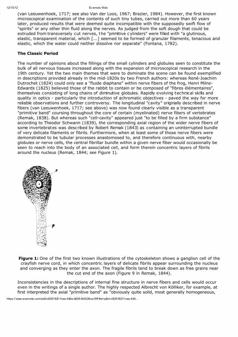
Figure 3: The young Sigmund Freud decided to inspect again the ganglion cells and fibers of thecrayfish nerve cord to determine, under the strictest conditions, the truth about the existence of
the controversial fibrils described by Remak more than 40 years earlier. His own observationsconfirmed Remak's findings (Figure 2 in Freud, 1882; cf. Figure 1 above).
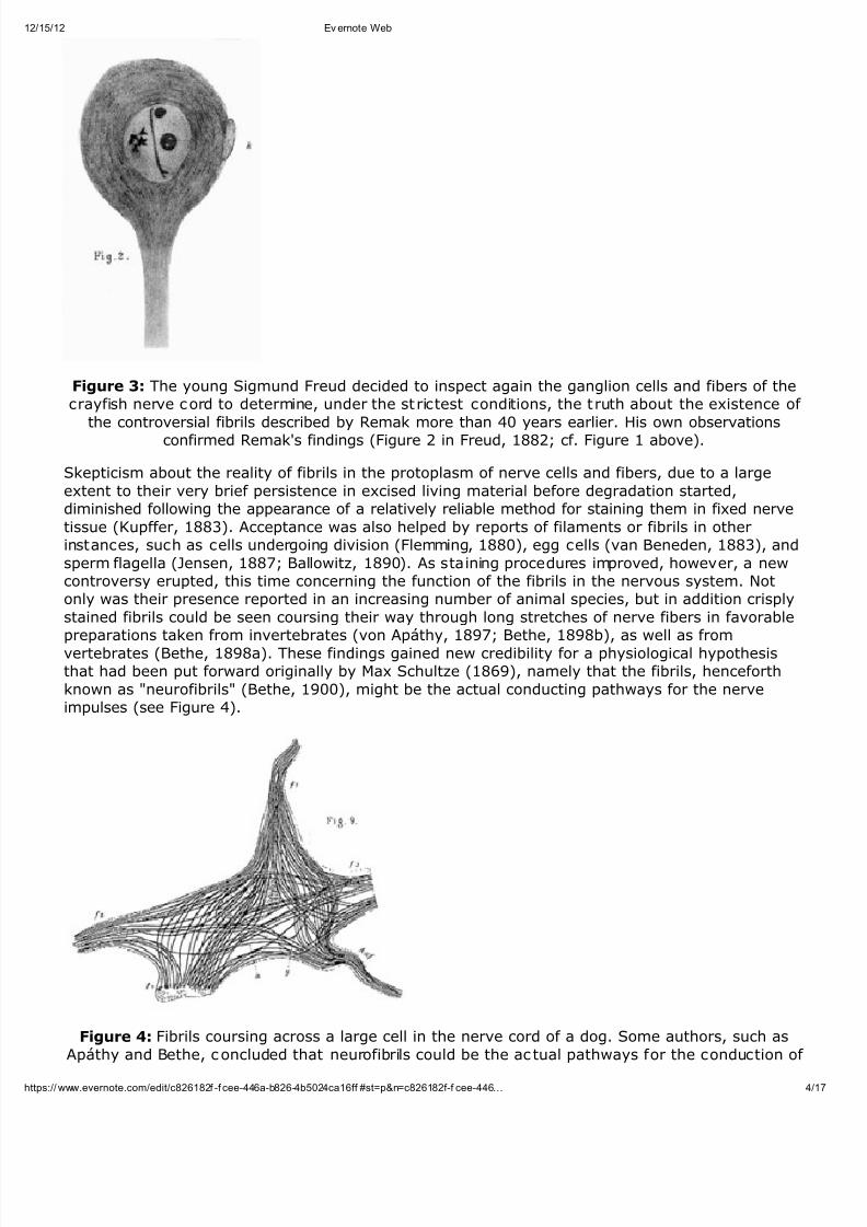
Skepticism about the reality of fibrils in the protoplasm of nerve cells and fibers, due to a largeextent to their very brief persistence in excised living material before degradation started,diminished following the appearance of a relatively reliable method for staining them in fixed nervetissue (Kupffer, 1883). Acceptance was also helped by reports of filaments or fibrils in otherinstances, such as cells undergoing division (Flemming, 1880), egg cells (van Beneden, 1883), andsperm flagella (Jensen, 1887; Ballowitz, 1890). As staining procedures improved, however, a newcontroversy erupted, this time concerning the function of the fibrils in the nervous system. Notonly was their presence reported in an increasing number of animal species, but in addition crisplystained fibrils could be seen coursing their way through long stretches of nerve fibers in favorablepreparations taken from invertebrates (von Apáthy, 1897; Bethe, 1898b), as well as fromvertebrates (Bethe, 1898a). These findings gained new credibility for a physiological hypothesisthat had been put forward originally by Max Schultze (1869), namely that the fibrils, henceforthknown as "neurofibrils" (Bethe, 1900), might be the actual conducting pathways for the nerveimpulses (see Figure 4).
Figure 4: Fibrils coursing across a large cell in the nerve cord of a dog. Some authors, such asApáthy and Bethe, concluded that neurofibrils could be the ac tual pathways for the conduction of

8/11/2019 The Cytoskeleton of Nerve Cells in Historic Perspective
http://slidepdf.com/reader/full/the-cytoskeleton-of-nerve-cells-in-historic-perspective 5/17
/15/12 Ev ernote Web
5ps:// www.evernote.com/edit/c826182f -f cee-446a-b826-4b5024ca16ff #st=p&n=c826182f-f cee-446…
nerve impulses, forming a continuous network in which the nerve cells would act just as nodes forthe convergence and divergence of neurofibrils (Figure 9 in Bethe, 1898a).
In this view the nerve cells and fibers acted as protective and insulating envelopes for theconducting neurofibrils, which would be wired in a virtually uninterrupted network across the wholenervous system, from sensory terminals to nerve centers to effector muscles. This ingenious modelhad the added merit of claiming the middle ground between the two currently rival interpretationsabout the general organization of the nervous system. It retained the basic concept of a
continuous reticular array of conducting elements, as inferred from the most advanced stainingtechniques of the day (von Gerlach, 1873; Golgi, 1886), while accommodating also the novel"neuron doctrine", according to which nerve cells are basically independent units, just like othercells, though extensively interconnected by means of numerous ramifications and long processes(Cajal, 1889; Waldeyer, 1891; see also Shepherd, 1991). For a thorough and conciliatorycontemporary review of the various opinions see Pugnat (1901).
The compromise suggested by von Apáthy and Bethe did not sit well with leading hard-coreneuronists like Cajal (see Frixione, 2002), who soon objected the idea of assigning a specificconducting function to the neurofibrils because: "The existence of intraprotoplasmic fibrils is ageneral anatomical law of the cell. More or less modified in their disposition, intracellular threadshave been found in skin epithelial cells, in the corpuscles of the lashes, in the egg cell [...] and
nobody will think of inferring from this fact that the above mentioned threads constitute theobliged pathway for the light, heat, electric or mechanical waves" (Ramón y Cajal, 1903a).
In fact, Cajal got so excited about the challenge posed by the neurofibril hypothesis to the neurondoctrine, that he promptly developed a superior and quite reproducible method for staining the tinyfilaments in nerve tissues of virtually any source (Ramón y Cajal, 1903b), which comparedfavorably with another excellent procedure (Bielschowsky, 1902, 1903). His own research on thisproblem produced ample evidence that neurofibrils are strictly intracellular structures, andtherefore could not bridge between contiguous nerve cells or their processes as required forconducting impulses (Ramón y Cajal, 1903a; 1908). In addition, he and one of his closecollaborators showed that the arrangement of the neurofibrils within nerve cells would changedepending upon certain factors affecting the animal, such as hibernation, acclimatization or a
pathological condition like infection with rabies virus (Ramón y Cajal, 1904a,b; Tello, 1904; seeFigure 5).
Figure 5: Neurofibrils in neurons from the nerve cord of rabbits kept at different temperatures,prepared by Cajal using his acclaimed staining method with reduced silver nitrate. Normal
appearance of the neurofibrils (left), and their altered morphologies after the animals were

8/11/2019 The Cytoskeleton of Nerve Cells in Historic Perspective
http://slidepdf.com/reader/full/the-cytoskeleton-of-nerve-cells-in-historic-perspective 6/17
/15/12 Ev ernote Web
6ps:// www.evernote.com/edit/c826182f -f cee-446a-b826-4b5024ca16ff #st=p&n=c826182f-f cee-446…
exposed to either heat (right, A) or cold (right, B and C) stress (Figures 2 and 5 in Ramón y Cajal,1903b and 1904b, respectively).
The subject of the neurofibrils became so important indeed for Cajal's concept of the neuron thatit occupies a fair portion and five figures of his Nobel Prize lecture (Ramón y Cajal, 1906). Abalanced assessment of the issue was also included in the French version of his monumentaltreatise on the histology of the nervous system (Ramón y Cajal, 1909). Continuing beyond thesefootsteps, another outstanding member of his school would later on contribute extensive studieson the fibrils characteristic of glial and epithelial cells (Río-Hortega, 1916, 1917).
The first decade of the 20th century was perhaps the zenith of research and vigorous contentionabout the neurofibrils. Incidentally, it was in the middle of this period when, using Bielschowsky'sstaining method, Alois Alzheimer first described "very strange changes of neurofibrils" in the brainof a female patient that had been afflicted with the disease now known by his name (Alzheimer,1906). The hour of neurofibrils soon came to an end, however, in the wake of newelectrophysiological findings hinting that the conduction of nerve impulses is a process takingplace on the surface of the nerve cells and fibers, rather than through a pathway in theirprotoplasm (Bernstein, 1902; see also Brazier, 1988). It did not help that neurofibrils, like otherintracellular filaments in general, had become suspect of being mere artifactual products of thehistological techniques used for the preparation of microscopical specimens (Hardy, 1899; Bayliss,1915).
A dark interlude
Thus, just after being placed for a short period at the very center of neuroscience theory, theneurofibrils went into relative obscurity. Nevertheless, debate persisted because even if theirexistence as real cellular components was tentatively granted, the question of their possiblefunctions remained wide open. Any role in the conduction of nerve impulses was fully ruled out asit was realized that these do not necessarily follow the intracellular course of neurofibrils. Thus,for example, crab neurons, in which the axon subdivides into one central and one peripheralbranch, would continue to conduct electrical impulses straight from one axonal branch to theother, even after the intermediate cell body and corresponding loop of neurofibrillar tract had beeneliminated (see Parker, 1929). An alternative option was that neurofibrils might constitute an
internal framework for supporting the shape of the nerve cell (Koltzoff, 1906, 1912; Goldschmidt,1910; von Lenhossék, 1910; von Szüts, 1914), but this idea was virtually discarded inconsideration that the fibrils seemed too flimsy or unstable for that purpose (Marinesco, 1914;Bozler, 1927). Still another possibility referred to the neurofibrils as being involved in thedistribution of metabolic influences exported from the cell body into and along its various branchesand processes (Parker, 1929; Rényi, 1932).
Persisting skepticism about the neurofibrils was largely the result of understandable reservationsabout the reliability of observations made on fixed and stained preparations, on one hand, and of the scarcity and contradictions of studies on living material, on the other. The findings wereindeed quite inconsistent even in fresh specimens taken from either vertebrate or invertebratesources (for a brief c ritical discussion of the state of the matter at the time, see Cowdry, 1928).
Distinct neurofibrils were found in living nerve fibers of jellyfishes (Bozler, 1927) and lobsters(Rényi, 1929, 1932), but just a faint longitudinal striation was reported in nerve fibers of frogtadpoles, and then only if these nerves were previously subject to irritation (Speidel, 1935). Ananalogous delicate longitudinal striation - suspected by the author to be perhaps an artifact ordamage caused by manipulation - was observed also in the squid giant axon (Young, 1936), soonto become a key protagonist in basic neuroscience (Hodgkin and Huxley, 1939). And yetbirefringence measurements of fresh squid axoplasm suggested that a fraction of its entire proteincontent was longitudinally oriented, as it would be expected from the presence of neurofibrils(Bear et al., 1937). Similarly, neurofibrils observed in cultured ganglion cells of the chick wereinterpreted as somehow reflecting the underlying molecular organization of the protoplasm (Weissand Wang, 1936). It was around this time that a totally new kind of microscopy was introduced inphysics and material science laboratories, and it soon began to be tested for use with biological
8/11/2019 The Cytoskeleton of Nerve Cells in Historic Perspective
http://slidepdf.com/reader/full/the-cytoskeleton-of-nerve-cells-in-historic-perspective 7/17
/15/12 Ev ernote Web
7ps:// www.evernote.com/edit/c826182f -f cee-446a-b826-4b5024ca16ff #st=p&n=c826182f-f cee-446…
specimens as well.
Electron microscopy arrives
The inspection of cells with electron microscopy posed significant problems not usually found withinorganic samples. The specimens had to be (1) fixed and fully dehydrated in order to stand in avacuum, (2) greatly reduced in thickness so that the electron beam could pass through them, and(3) preferably stained with a heavy metal in order to increase image contrast. A straightforwardearly attempt to inspect the contents of nerve fibers with this powerful instrument consisted of
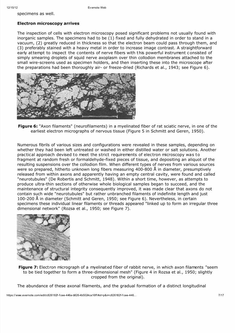
simply smearing droplets of squid nerve axoplasm over thin collodion membranes attached to thesmall wire-screens used as specimen holders, and then inserting these into the microscope afterthe preparations had been thoroughly air- or freeze-dried (Richards et al., 1943; see Figure 6).
Figure 6: "Axon filaments" (neurofilaments) in a myelinated fiber of rat sciatic nerve, in one of theearliest electron micrographs of nervous tissue (Figure 5 in Schmitt and Geren, 1950).
Numerous fibrils of various sizes and configurations were revealed in these samples, depending onwhether they had been left untreated or washed in either distilled water or salt solutions. Anotherpractical approach devised to meet the strict requirements of electron microscopy was tofragment at random fresh or formaldehyde-fixed pieces of tissue, and depositing an aliquot of theresulting suspensions over the collodion film. When different types of nerves from various sourceswere so prepared, hitherto unknown long fibers measuring 400-800 Å in diameter, presumptively
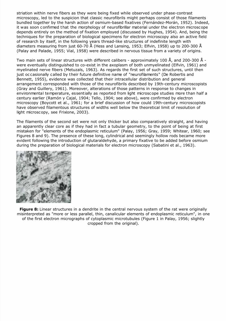
released from within axons and apparently having an empty central cavity, were found and called"neurotubules" (De Robertis and Schmitt, 1948). Within a short time, however, as attempts toproduce ultra-thin sections of otherwise whole biological samples began to succeed, and themaintenance of structural integrity consequently improved, it was made clear that axons do notcontain such wide "neurotubules" but rather unbranched filaments of indefinite length and just100-200 Å in diameter (Schmitt and Geren, 1950; see Figure 6). Nevertheless, in certainspecimens these individual linear filaments or threads appeared "linked up to form an irregular threedimensional network" (Rozsa et al., 1950; see Figure 7).
Figure 7: Electron micrograph of a myelinated fiber of rabbit nerve, in which axon filaments "seemto be tied together to form a three-dimensional mesh" (Figure 4 in Rozsa et al., 1950; slightly
cropped from the original).
The abundance of these axonal filaments, and the gradual formation of a distinct longitudinal
8/11/2019 The Cytoskeleton of Nerve Cells in Historic Perspective
http://slidepdf.com/reader/full/the-cytoskeleton-of-nerve-cells-in-historic-perspective 8/17
striation within nerve fibers as they were being fixed while observed under phase-contrastmicroscopy, led to the suspicion that classic neurofibrils might perhaps consist of those filamentsbundled together by the harsh action of osmium-based fixatives (Fernández-Morán, 1952). Indeed,it was soon confirmed that the morphology of neurofibrillar material under the electron microscopedepends entirely on the method of fixation employed (discussed by Hughes, 1954). And, being thetechniques for the preparation of biological specimens for electron microscopy also an active fieldof research by itself, in the following years thread-like structures of indefinite length withdiameters measuring from just 60-70 Å (Hess and Lansing, 1953; Elfvin, 1958) up to 200-300 Å(Palay and Palade, 1955; Vial, 1958) were described in nervous tissue from a variety of origins.
Two main sets of linear structures with different calibers - approximately 100 Å, and 200-300 Å -were eventually distinguished to co-exist in the axoplasm of both unmyelinated (Elfvin, 1961) andmyelinated nerve fibers (Metuzals, 1963). As regards the first set of such structures, until then just oc casionally called by their future definitive name of "neurofilaments" (De Robertis andBennett, 1955), evidence was collected that their intracellular distribution and generalarrangement corresponded with those of the neurofibrils described by 19th-century microscopists(Gray and Guillery, 1961). Moreover, alterations of those patterns in response to changes inenvironmental temperature, essentially as reported from light microscope studies more than half acentury earlier (Ramón y Cajal, 1904; Tello, 1904; see above), were confirmed by electronmicroscopy (Boycott et al., 1961; for a brief discussion of how could 19th-century microscopistshave observed filamentous structures of widths well below the theoretical limit of resolution of
light microscopy, see Frixione, 2003).
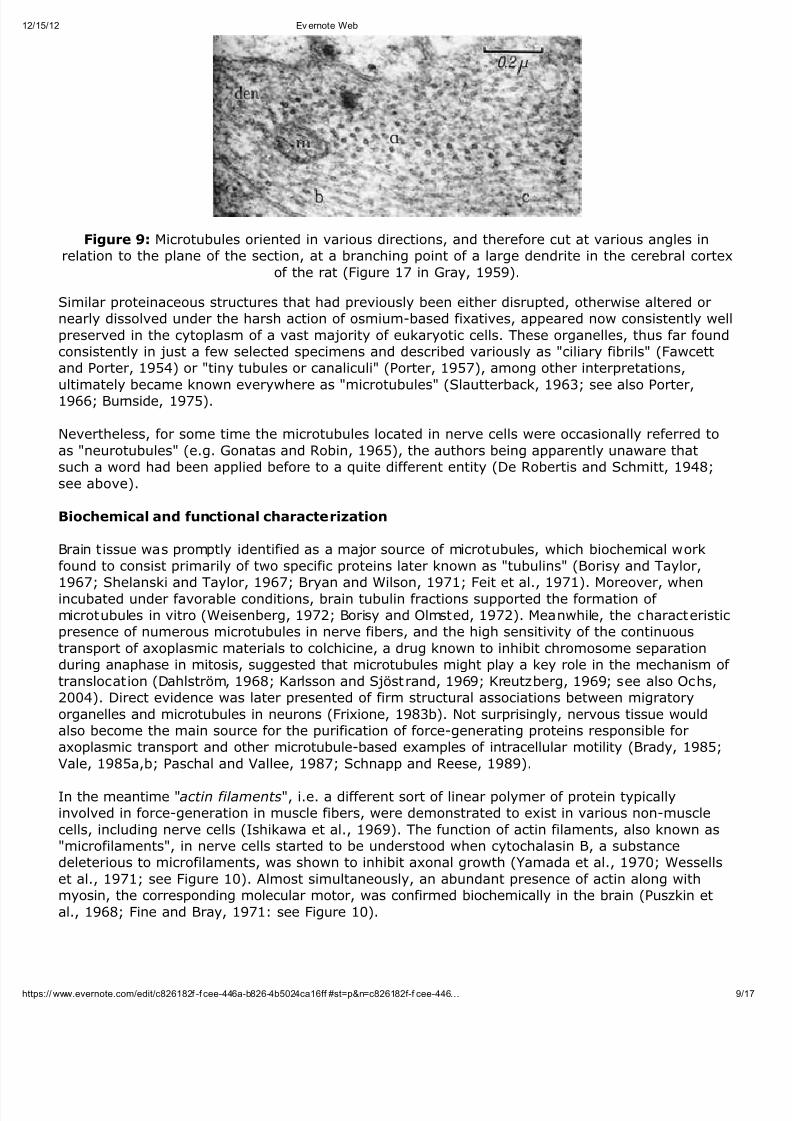
The filaments of the second set were not only thicker but also comparatively straight, and havingan apparently clear core as if they had in fact a tubular geometry, to the point of being at firstmistaken for "elements of the endoplasmic reticulum" (Palay, 1956; Gray, 1959; Whitear, 1960; seeFigures 8 and 9). The presence of these long, cylindrical and seemingly hollow rods became moreevident following the introduction of glutaraldehyde, a primary fixative to be added before osmiumduring the preparation of biological materials for electron microscopy (Sabatini et al., 1963).
Figure 8: Linear structures in a dendrite in the central nervous system of the rat were originallymisinterpreted as "more or less parallel, thin, canalicular elements of endoplasmic reticulum", in one
of the first electron micrographs of cytoplasmic microtubules (Figure 1 in Palay, 1956; slightly
cropped from the original).
8/11/2019 The Cytoskeleton of Nerve Cells in Historic Perspective
http://slidepdf.com/reader/full/the-cytoskeleton-of-nerve-cells-in-historic-perspective 9/17
/15/12 Ev ernote Web
9ps:// www.evernote.com/edit/c826182f -f cee-446a-b826-4b5024ca16ff #st=p&n=c826182f-f cee-446…
Figure 9: Microtubules oriented in various directions, and therefore cut at various angles inrelation to the plane of the section, at a branching point of a large dendrite in the cerebral cortex
of the rat (Figure 17 in Gray, 1959).
Similar proteinaceous structures that had previously been either disrupted, otherwise altered ornearly dissolved under the harsh action of osmium-based fixatives, appeared now consistently wellpreserved in the cytoplasm of a vast majority of eukaryotic cells. These organelles, thus far foundconsistently in just a few selected specimens and described variously as "ciliary fibrils" (Fawcettand Porter, 1954) or "tiny tubules or canaliculi" (Porter, 1957), among other interpretations,ultimately became known everywhere as "microtubules" (Slautterback, 1963; see also Porter,1966; Burnside, 1975).
Nevertheless, for some time the microtubules located in nerve cells were occasionally referred toas "neurotubules" (e.g. Gonatas and Robin, 1965), the authors being apparently unaware thatsuch a word had been applied before to a quite different entity (De Robertis and Schmitt, 1948;see above).
Biochemical and functional characterization
Brain tissue was promptly identified as a major source of microtubules, which biochemical workfound to consist primarily of two specific proteins later known as "tubulins" (Borisy and Taylor,
1967; Shelanski and Taylor, 1967; Bryan and Wilson, 1971; Feit et al., 1971). Moreover, whenincubated under favorable conditions, brain tubulin fractions supported the formation of microtubules in vitro (Weisenberg, 1972; Borisy and Olmsted, 1972). Meanwhile, the characteristicpresence of numerous microtubules in nerve fibers, and the high sensitivity of the continuoustransport of axoplasmic materials to colchicine, a drug known to inhibit chromosome separationduring anaphase in mitosis, suggested that microtubules might play a key role in the mechanism of translocation (Dahlström, 1968; Karlsson and Sjöstrand, 1969; Kreutzberg, 1969; see also Ochs,2004). Direct evidence was later presented of firm structural associations between migratoryorganelles and microtubules in neurons (Frixione, 1983b). Not surprisingly, nervous tissue wouldalso become the main source for the purification of force-generating proteins responsible foraxoplasmic transport and other microtubule-based examples of intracellular motility (Brady, 1985;Vale, 1985a,b; Paschal and Vallee, 1987; Schnapp and Reese, 1989).
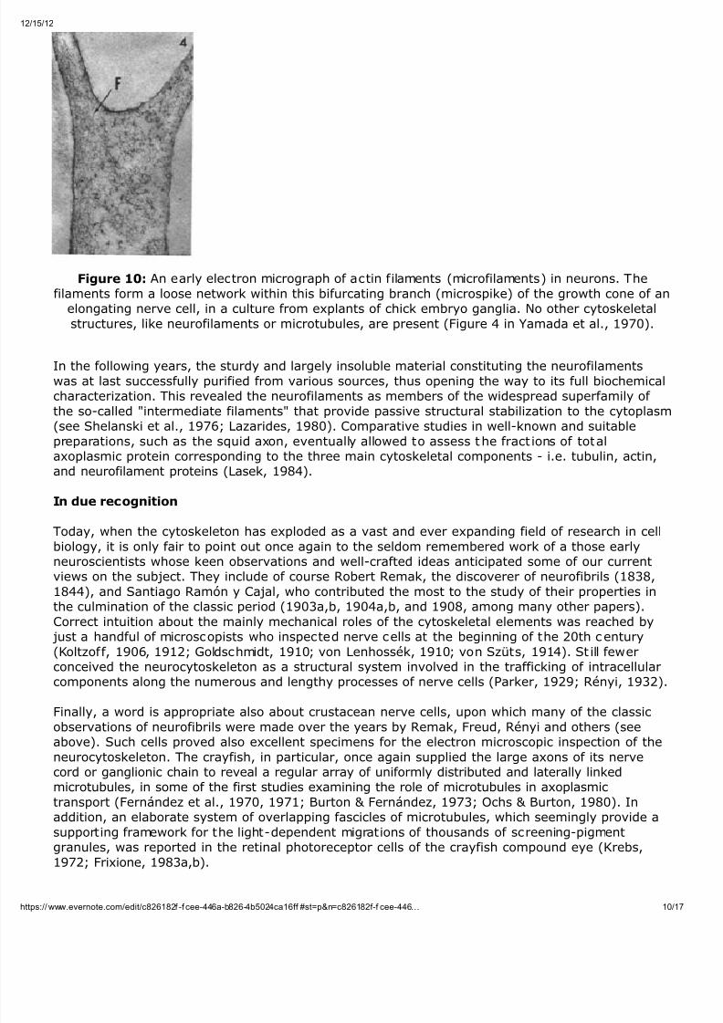
In the meantime "actin filaments", i.e. a different sort of linear polymer of protein typicallyinvolved in force-generation in muscle fibers, were demonstrated to exist in various non-musclecells, including nerve cells (Ishikawa et al., 1969). The function of actin filaments, also known as"microfilaments", in nerve cells started to be understood when cytochalasin B, a substancedeleterious to microfilaments, was shown to inhibit axonal growth (Yamada et al., 1970; Wessellset al., 1971; see Figure 10). Almost simultaneously, an abundant presence of actin along withmyosin, the corresponding molecular motor, was confirmed biochemically in the brain (Puszkin etal., 1968; Fine and Bray, 1971: see Figure 10).
8/11/2019 The Cytoskeleton of Nerve Cells in Historic Perspective
http://slidepdf.com/reader/full/the-cytoskeleton-of-nerve-cells-in-historic-perspective 10/17
/15/12
10/1ps:// www.evernote.com/edit/c826182f -f cee-446a-b826-4b5024ca16ff #st=p&n=c826182f-f cee-446…
Figure 10: An early electron micrograph of actin filaments (microfilaments) in neurons. Thefilaments form a loose network within this bifurcating branch (microspike) of the growth cone of an
elongating nerve cell, in a culture from explants of chick embryo ganglia. No other cytoskeletalstructures, like neurofilaments or microtubules, are present (Figure 4 in Yamada et al., 1970).
In the following years, the sturdy and largely insoluble material constituting the neurofilamentswas at last successfully purified from various sources, thus opening the way to its full biochemicalcharacterization. This revealed the neurofilaments as members of the widespread superfamily of the so-called "intermediate filaments" that provide passive structural stabilization to the cytoplasm(see Shelanski et al., 1976; Lazarides, 1980). Comparative studies in well-known and suitablepreparations, such as the squid axon, eventually allowed to assess the fract ions of totalaxoplasmic protein corresponding to the three main cytoskeletal components - i.e. tubulin, actin,and neurofilament proteins (Lasek, 1984).
In due recognition
Today, when the cytoskeleton has exploded as a vast and ever expanding field of research in cellbiology, it is only fair to point out once again to the seldom remembered work of a those earlyneuroscientists whose keen observations and well-crafted ideas anticipated some of our currentviews on the subject. They include of course Robert Remak, the discoverer of neurofibrils (1838,1844), and Santiago Ramón y Cajal, who contributed the most to the study of their properties inthe culmination of the classic period (1903a,b, 1904a,b, and 1908, among many other papers).Correct intuition about the mainly mechanical roles of the cytoskeletal elements was reached by just a handful of microscopists who inspected nerve cells at the beginning of the 20th century(Koltzoff, 1906, 1912; Goldschmidt, 1910; von Lenhossék, 1910; von Szüts, 1914). St ill fewerconceived the neurocytoskeleton as a structural system involved in the trafficking of intracellularcomponents along the numerous and lengthy processes of nerve cells (Parker, 1929; Rényi, 1932).
Finally, a word is appropriate also about crustacean nerve cells, upon which many of the classicobservations of neurofibrils were made over the years by Remak, Freud, Rényi and others (seeabove). Such cells proved also excellent specimens for the electron microscopic inspection of theneurocytoskeleton. The crayfish, in particular, once again supplied the large axons of its nervecord or ganglionic chain to reveal a regular array of uniformly distributed and laterally linkedmicrotubules, in some of the first studies examining the role of microtubules in axoplasmictransport (Fernández et al., 1970, 1971; Burton & Fernández, 1973; Ochs & Burton, 1980). Inaddition, an elaborate system of overlapping fascicles of microtubules, which seemingly provide asupporting framework for the light-dependent migrations of thousands of screening-pigmentgranules, was reported in the retinal photoreceptor cells of the crayfish compound eye (Krebs,1972; Frixione, 1983a,b).

8/11/2019 The Cytoskeleton of Nerve Cells in Historic Perspective
http://slidepdf.com/reader/full/the-cytoskeleton-of-nerve-cells-in-historic-perspective 11/17
/15/12 Ev ernote Web
11/ps:// www.evernote.com/edit/c826182f -f cee-446a-b826-4b5024ca16ff #st=p&n=c826182f-f cee-446…
Eugenio FrixioneMetodología y Teoría de la CienciaCenter for Research and Advanced Studies CINVESTAVApdo. Postal 14-740Mexico City [email protected]
References
Alzheimer, A. (1906) Über eine eigenartige Erkrankung der Hirnrinde. Allg. Zeitschr. Psychiatrie u.Psychisch-Gerichtliche Med. 64: 146-148.
Ballowitz, E. (1890) Fibrillare Struktur und Contraktilität. Pflügers Archiv ges. Physiol . 46: 433-464.
Bayliss, W. M. (1915) Principles of General Physiology . London: Longman, Green, and Co. See pp.396, 470, 491.
Bear, R. S., Schmitt, F. O., Young, J. Z. (1937) Investigations on the protein constituents of nerve axoplasm. Proc. Roy. Soc. Lond. Ser. B 123: 520-529.
Bernstein, J. (1902) Untersuchungen zur Thermodynamik der bioelektrischen Ströme. Pflügers Archiv ges. Physiol . 92: 521-562.
Bethe, A. (1898a) Ueber die Primitivfibrillen in den Ganglienzellen vom Menschen und anderenWirbelthieren. Morphol. Arbeit. 8: 95-116.
Bethe, A. (1898b) Ueber die Primitivfibrillen in den Ganglienzellen und Nervenfasern vonWirbelthieren und Wirbellosen. Verh. anat. Ges. 12: 37-38.
Bethe, A. (1900) Ueber die Neurofibrillen in den Ganglienzellen von Wirbelthieren und ihreBeziehungen zu Golginetzen. Arch. mikr. Anat. 55: 513-558.
Bielschowsky, M. (1902) Die Silberimprägnation der Axencylinder. Neurol. Centralbl . 21: 579-584.
Bielschowsky, M. (1903) Die Silberimprägnation der Neurofibrillen. Neurol. Centralbl. 22: 997-1006.
Borisy, G. G., and Olmsted, J. B. (1972) Nucleated assembly of microtubules in porcine brainextracts. Science 177 : 1196-1197.
Borisy, G. G., and Taylor, E.W. (1967) The mechanism of action of colchicine. Binding of colchicine-3H to cellular protein. J. Cell Biol. 34: 525-533.
Boycott, B. B., Gray, E. G., Guillery, R. W. (1961) Synaptic structure and its alteration withenvironmental temperature: a study by light and electron microscopy of the central nervous
system of lizards. Proc. Royal Soc. Lond. Ser. B 154: 151-172.
Bozler, E .(1927) Untersuchungen über das Nervensystem der Coelenteraten. II. Teil: Ueber dieStruktur der Ganglienzellen und die Funktion der Neurofibrillen nach Lebenduntersuchungen. Zeit.vergl. Physiol. 6: 255-263.
Brady, S. T. (1985) A novel brain ATPase with properties expected for the fast axonal transportmotor. Nature 317: 73-75.
Brazier, M. A. B. (1984) A History of Neurophysiology in the 17th and 18th Centuries. New York:Raven Press. [p. 36]
8/11/2019 The Cytoskeleton of Nerve Cells in Historic Perspective
http://slidepdf.com/reader/full/the-cytoskeleton-of-nerve-cells-in-historic-perspective 12/17
/15/12 Ev ernote Web
12/1ps:// www.evernote.com/edit/c826182f -f cee-446a-b826-4b5024ca16ff #st=p&n=c826182f-f cee-446…
Brazier, M. A. B. (1988) A History of Neurophysiology in the 19th Century . New York: Raven Press.
Bryan, J., and Wilson, L. (1971) Are cytoplasmic microtubules heteropolymers? Proc. Natl. Acad.Sci. USA 68:1762-1766.
Burnside, B. (1975) The form and arrangement of microtubules: an historical, primarilymorphological review. New York Acad. Sci. 253. 14-26.
Burton, P. R., Fernández, H. L. (1973) Delineation by lanthanum staining of filamentous elements
associated with the surfaces of axonal microtubules. J. Cell Sci. 12: 567-583.
Clarke, E. (1968) The doctrine of the hollow nerve in the Seventeenth and Eighteenth centuries.In: Stevenson, L. G., and Multhauf, R. P. (eds.) Medicine, Science and Culture: Historical Essaysin Honor of Owsei Temkin, Baltimore: The Johns Hopkins Press, pp. 123-141.
Clarke, E. (1978) The neural circulation. The use of analogy in medicine. Med. Hist. 22: 291-307.
Cowdry, E. V. (1928) The internal architecture of nerve cells. In: Cowdry, E. V. (ed.) Special Cytology: The Form and Function of the Cell in Health and Disease. A Textbook for Students of Biology and Medicine. New York: Paul B. Hoeber, Vol. II, pp. 965-987.
Dahlström, A. (1968) Effect of colchicine on transport of amine storage granules in sympatheticnerves of rat. Eur. J. Pharmacol. 5:111-113.
De Robertis, E., and Schmitt, F.O. (1948) An electron microscope analysis of certain nerve axonconstituents. J. Cell. Comp. Physiol. 31:1-23.
De Robertis, E., and Bennett, H. S. (1955) Some features of the submicroscopic morphology of synapses of frog and earthworm. J. Biophys. Biochem. Cytol. 1: 47-58.
Descartes, R. (1664) Traité de l'Homme, pp. 129ff. In: Oeuvres deDescartes, publiées par Charles Adam & Paul Tannery. Vol. IX. Paris: Vrin, 1986.
Dutrochet R-JH (1824) Recherches Anatomiques et Physiologiques sur la Structure Intime des Animaux et des Végétaux, et sur leur Motilité, Paris: Baillière, p. 168.
Elfvin, L.-G. (1958) The ultrastructure of unmyelinated fibers in the splenic nerve of the cat. J.Ultrastruct. Res. 1: 428-454.
Elfvin, L.-G. (1961) Electron-microscopic investigation of filament structures in unmyelinated fibersof cat splenic nerve. J. Ultrastruct. Res. 5: 51-64.
Fawcett , D. W., Porter, K. R. (1954) A study of the fine structure of c iliated epithelia. J. Morphol.94: 221-281.
Feit, H., Slusarek, L., and Shelanski, M.L. (1971) Heterogeneity of tubulin subunits. Proc. Natl. Acad. Sci. USA 68: 2028-2031.
Fernández HL, Burton PR, Samson FE (1971) Axoplasmic transport in the crayfish nerve cord. Therole of fibrillar constituents of neurons. J. Cell Biol. 51: 176-192.
Fernández HL, Huneeus FC, Davison PF (1970) Studies on the mechanism of axoplasmic transportin the crayfish nerve cord. J. Neurobiol. 1: 395-409.
Fernández-Morán, H. (1952) The submicroscopic organization of vertebrate nerve fibres. Anelectron microscope study of myelinated and unmyelinated nerve fibres. Exp. Cell Res. 3: 282-359.
8/11/2019 The Cytoskeleton of Nerve Cells in Historic Perspective
http://slidepdf.com/reader/full/the-cytoskeleton-of-nerve-cells-in-historic-perspective 13/17
/15/12 Ev ernote Web
13/1ps:// www.evernote.com/edit/c826182f -f cee-446a-b826-4b5024ca16ff #st=p&n=c826182f-f cee-446…
Fine, R. E., Bray, D. (1971) Actin in growing nerve cells. Nature - New Biology 234: 115-118.
Flemming, W. (1880) Beiträge zur Kenntniss der Zelle und ihrer Lebenserscheinungen, Theil II.Arch. für Mikros. Anat. 18: 151-259; transl. Piternick, L. (1965) Contributions to the knowledge of the cell and its vital processes. J. Cell Biol . 25-II: 1-69.
Fontana, F. (1782) Lettre de M. l'Abbé Fontana, à M. Gibelin, à Aix-en-Provence, datée deFlorence du 10 Juillet, 1782. J. Physique 24: 417-421, 1784. See: Hoff, H. E. (1959) A classic of microscopy: an early, if not the first, observation on the fluidity of the axoplasm,
micromanipulation, and the use of the cover-slip. Bull. Hist. Med . 32: 375-379.
Freud, S. (1882) Über den Bau der Nervenfasern und Nervenzellen beim Flusskrebs. Sitzungsb. d.kais. Akad. d. Wien., math. naturw. Classe 85 Abth. 3:9-46.
Frixione, E. (1983a) The microtubular system of crayfish retinula cells and its changes in relationto screening-pigment migration. Cell Tissue Res. 232: 335-348.
Frixione, E. (1983b) Firm structural associations between migratory pigment granules andmicrotubules in crayfish retinula cells. J. Cell Biol. 96: 1258-1265.
Frixione E (2000) Recurring views on the structure and function of the cytoskeleton: a 300-year
epic. Cell Motil. Cytoskel . 46: 73-94.
Frixione, E. (2002) Cajal's vision of a neuronal cytoskeleton. J. Hist. Neurosci. (Abs.) 11: 406-407.
Frixione, E. (2003) Sigmund Freud's contribution to the history of the neuronal cytoskeleton. J.Hist. Neurosci. 12: 11-24.
Goldschmidt, R. (1910) Das Nervensystem von Ascaris lumbricoides und megalocephala. Festschrift R. Hertwig 2: 253-354.
Golgi, C. (1886) Sulla Fina Anatomia degli Organi Centrali del Sistema Nervoso. Milano: Hoepli.
Gonatas, N. K., and Robbins, E. (1965) The homology of spindle tubules and neuro-tubules in thechick embryo retina. Protoplasma 59: 377-391.
Gray, E. G. (1959) Axo-somatic and axo-dendritic synapses of the cerebral cortex: an electronmicroscope study. J. Anat . 93: 420-433.
Gray, E. G., Guillery, R. W. (1961) The basis for silver staining of synapses of the mammalian spinalcord: A light and electron microscope study. J Physiol. (London) 157: 581-588.
Hardy, W. B. (1899) Structure of cell protoplasm. J. Physiol. (London) 24: 158-210.
Heitzmann, C. (1883) Microscopical Morphology of the Animal Body in Health and Disease. New
York: J. H. Vail, p. 288.
Hess, A., Lansing, A. (1953) The fine structure of peripheral nerve fibers. Anat. Rec. 117: 175-199.
Hodgkin, A. L., Huxley, A. F. (1939) Action potentials recorded from inside a nerve fibre. Nature144: 710-711.
Hughes, A. (1954) The effect of fixation on neurons of the chick. J. Anat. 88: 192-203.
Huxley, T.H. (1880) An Introduct ion to the Study of Zoology, Illustrated by the Crayfish. London:Kegan Paul, p. 189.
8/11/2019 The Cytoskeleton of Nerve Cells in Historic Perspective
http://slidepdf.com/reader/full/the-cytoskeleton-of-nerve-cells-in-historic-perspective 14/17
Ishikawa, H., Bischoff, R., and Holtzer, H. (1969) Formation of arrowhead complexes with heavymeromyosin in a variety of cell types. J. Cell Biol . 43: 312-328.
Jensen, O. (1887) Untersuchungen über die Samenkörper der Säugethiere, Vögel und Amphibien. Arch. Mikr. Anat. 30: 379-425.
Jones, E. (1953) The Life and Work of Sigmund Freud . New York: Basic Books, Vol. 1, p. 46.
Karlsson J.-O., and Sjöstrand, J. (1969) The effect of colchicine on the axonal transport of proteinin the optic nerve and tract of the rabbit. Brain Res. 13: 617-619.
Koltzoff, N. K. (1906) Studien über die Gestalt der Zelle. Arch. mikr. Anat . 67: 364-572.
Koltzoff, N. K. (1912) Zur Frage der Zellgestalt. Anat. Anz . 41: 183-207.
Krebs, W. (1972) The fine structure of the retinula of the compound eye of Astacus fluviatilis. Z.Zellforsch. Mikrosk. Anat. 133: 399-414.
Kreutzberg, G. W. (1969) Neuronal dynamics and axonal flow, IV. Blockage of intra-axonal enzymetransport by colchicine. Proc. Natl. Acad. Sci. USA 62:722-728.
Kupffer, C. (1883) Ueber den "Axencylinder" markhaltiger Nervenfasern. Sitzb. math.-phys. Cl. Akad. Wiss. Mûnchen 13: 466-475.
Lasek, R. J. (1984) The structure of axoplasm. In: Baker, P. F. (ed.) The Squid Axon. Curr. Top.Membr. Transp. 22: 39-53.
Lazarides, E. (1980) Intermediate filaments as mechanical integrators of cellular space. Nature283: 249-256.
Malpighi, M. (1666) De cerebri cortice. See: Meyer, A. (1967) Marcello Malpighi and the dawn of neurohistology. J. Neurol. Sci . 4: 185-193.
Marinesco, G. (1914) Sur la nature des neurofibrilles. Compt. Rend. Soc. Biol. Paris 77: 581-583.
Metuzals, J. (1963) Ultrastructure of myelinated nerve fibers in the central nervous system of thefrog. J. Ultrastruct. Res. 8: 30-47.
Milne-Edwards, H. (1825) Mémoire sur la Structure Élémentaire des Principaux Tissus Organiquesdes Animaux (Thèse). Paris: Didot Le Jeune, p. 19.
Ochs RL, Burton PR (1980) Distribution and selective extraction of filamentous componentsassociated with axonal microtubules of crayfish nerve cord. J. Ultrastruct. Res. 73: 169-182.
Ochs, Sidney (2004) A History of Nerve Functions: From Animal Spirits to Molecular Mechanisms.Cambridge: Cambridge University Press, Chapters 11-13, pp. 215-304.
Palay, S. L. (1956) Synapses in the central nervous system. J. Biophys. Biochem. Cytol. Suppl. 2:193-202.
Palay, S. L, Palade, G. E. (1955) The fine structure of neurons. J. Biophys. Biochem. Cytol. 1: 69-88.
Parker, G. H. (1929) The neurofibril hypothesis. Quart. Rev. Biol. 4: 155-178.
Paschal, B. M., and Vallee, R. B. (1987) Retrograde transport by the microtubule-associated
8/11/2019 The Cytoskeleton of Nerve Cells in Historic Perspective
http://slidepdf.com/reader/full/the-cytoskeleton-of-nerve-cells-in-historic-perspective 15/17
/15/12 Ev ernote Web
ps:// www.evernote.com/edit/c826182f -f cee-446a-b826-4b5024ca16ff #st=p&n=c826182f-f cee-446…
protein MAP 1C. Nature 330: 181-183.
Porter, K. R. (1957) The submicroscopic morphology of protoplasm. Harvey Lect. 51: 175-228.
Porter, K. R. (1966) Cytoplasmic microtubules and their functions. In: Principles of Biomolecular Organization ( Ciba Found. Symp.) Wolstenholme, G. E. W., and O'Connor, M. (eds.) London: J. & A. Churchill, pp. 308-356.
Pugnat, A. (1901) La biologie de la cellule nerveuse et la théorie des neurones. Bibliogr. Anatom.
9: 278-334.
Puszkin, S., Berl, S., Puszkin, E., and Clark, D. D. (1968) Actomyosin-like protein isolated frommammalian brain. Science 161: 170-171.
Ramón y Cajal, S. (1889) Conexión general de los elementos nerviosos. La Medicina Práctica 2:341-346.
Ramón y Cajal, S. (1903a/1904) Consideraciones críticas sobre la teoría de A. Bethe acerca de laestructura y conexiones de las células nerviosas. In: D. García Izcara (dir.) XIXè CongrésInternational de Médecine, Madrid, avril 23-30, 1903. Comptes Rendus [...] Section d'Anatomie.Madrid: Imprenta de J. Sastres y Cía., 1904, pp. 69-104 (also published in Trab. Lab. Invest. Biol.
Univ. Madrid 2:101-128, 1903).
Ramón y Cajal, S. (1903b) Un sencillo método de coloración selectiva del retículo protoplásmico ysus efectos en los diversos órganos nerviosos. Trab. Lab. Inv. Biol. Univ. Madrid 2: 129-221.
Ramón y Cajal, S. (1904a) Variaciones morfológicas, normales y patológicas del retículoneurofibrilar. Trab. Lab. Inv. Biol. Univ. Madrid 3: 9-15.
Ramón y Cajal, S. (1904b) Variaciones morfológicas del retículo nervioso de invertebrados yvertebrados sometidos a la acción de condiciones naturales. Trab. Lab. Inv. Biol. Univ. Madrid 3:287-297.
Ramón y Cajal, S. (1906) Les structures et les connexions des cellules nerveux. In: Les Prix Nobel 1904-1906. Stockholm: Imprimerie Royale. P.-A. Norstedt & fils, 1907.
Ramón y Cajal, S. (1908) L'hypothèse de la continuité d'Apathy. Réponse aux objections de cetauteur contre la doctrine neuronale. Trav. Lab. Rech. Biol. Univ. Madrid 6: 21-89 (published also in Anat. Anz. 33: 418-448, 468-493).
Ramón y Cajal, S. (1909) Histologie du Système Nerveux de l'Homme & des Vertébrés. (Éditionfrançaise revue & mise a jour par l'auteur. L. Azoulay, trad.) 2 Vols. Paris, Maloine, pp. 178-179.
Remak, R. (1838) Observationes anatomicae et microscopicae de systematis nervosi structura.Berlin: Reimer.
Remak, R. (1843) Ueber den Inhalt der Nervenprimitivröhren. Arch. Anat. Physiol. wiss. Med . 1843:197-201.
Remak, R. (1844) Neurologische Erläuterungen. Arch. Anat. Physiol. wiss. Med. 1844: 463-472.
Rényi, G. S. de (1929) The structure of cells in tissues as revealed by microdissection. IV.Observations of neurofibrils in the living nervous tissue of the lobster (Homarus americanus). J.Comp. Neurol. 48: 441-457.
Rényi, G. S. de (1932) Architecture of the nerve cell as revealed by microdissection. In: Cowdry E.V. (ed.) Special Cytology: The Form and Functions of the Cell in Health and Disease. New York:
8/11/2019 The Cytoskeleton of Nerve Cells in Historic Perspective
http://slidepdf.com/reader/full/the-cytoskeleton-of-nerve-cells-in-historic-perspective 16/17
/15/12 Ev ernote Web
16/1ps:// www.evernote.com/edit/c826182f -f cee-446a-b826-4b5024ca16ff #st=p&n=c826182f-f cee-446…
Paul B. Hoebber, Vol. III, pp. 1371-1402.
Richards, A. Glenn; Steinbach, H. Burr; Anderson, Thomas F. (1943) Electron microscope studiesof squid giant nerve axoplasm. J. Cell. Comp. Physiol. 21: 129-137.
Río-Hortega, P. (1916) Estructura fibrilar del protoplasma neuróglico y origen de las gliofibrillas.Trab. Lab. Inv. Biol. Univ. Madrid 14: 269-307.
Río-Hortega, P. (1917) Contribución al conocimiento de las epiteliofibrillas. Trab. Lab. Inv. Biol.
Univ. Madrid 15: 201-299.
Rozsa, G., Morgan, C., Szent-Györgyi, A., and Wyckoff, R.W. G. (1950) The electron microscopyof myelinated nerve. Biochim. Biophys. Acta 6: 13-27.
Sabatini, D. D., Bensch, K., Barrnett, R. J. (1963) Cytochemistry and electron microscopy. Thepreservation of cellular ultrastructure and enzymatic activity by aldehyde fixation. J. Cell Biol. 17:19-58.
Schmitt F. O., and Geren, B. B. (1950) The fibrous structure of the nerve axon in relation to thelocalization of "neurotubules". J. Exp. Med . 91: 499-504.
Schnapp, B. J., and Reese, T. S. (1989) Dynein is the motor for retrograde axonal transport of organelles. Proc. Nat. Acad. Sci. USA 86: 1548-1552.
Schultze, M. (1869) Allgemeines über die Structurelemente des Nervensystems. In: Stricker, S.(ed.) Handbuch der Lehre von den Geweben des Menschen und der Thiere. Leipzig: Engelmann,Vol. I, pp. 108-136. Translated by Power, H. (1870) as: The general characters of the structurescomposing the nervous system. In: Human Comparative Histology . London: Sydenham Society,Vol. I, pp. 180-182.
Schwann, T. (1839) Mikroskopische Untersuchungen über die Uebereinstimmung in der Struktur und dem Wachstum der Thiere und Pflanzen. Berlin: Reimer. Translated by Smith, H. (1847) as:Microscopical Researches into the Accordance in the Structure and Growth of Animals and Plants.
London: Sydenham Society, p. 148.
Shelanski, M.L., and Taylor, E.W. (1967) Isolation of a protein subunit from microtubules. J. Cell Biol . 34: 549-554.
Shelanski, M.L., Yen, S.-H., and Lee, V.M. (1976) Neurofilaments and glial filaments. In: Goldman,R., Pollard, T., and Rosenbaum, J. (eds.) Cell Motility . Cold Spring Harbor Conferences on CellProliferation. Cold Spring Harbor Laboratory, Book C, pp. 1007-1020.
Shepherd, Gordon M. (1991) Foundations of the Neuron Doctrine. New York/Oxford: OxfordUniversity Press.
Slautterback DB (1963) Cytoplasmic microtubules. I. Hydra. J Cell Biol 18: 367-388.
Solmsen, F. (1961) Greek philosophy and the discovery of the nerves. Museum Helveticum 18:150-197.
Speidel, C. C. (1935) Studies of living nerves. III. Phenomena of nerve irritation and recovery,regeneration and repair. J. Comp. Neurol. 61: 1-79.
Tello, Francisco (1904) Las neurofibrillas en los vertebrados inferiores. Trab. Lab. Inv. Biol. Univ.Madrid 3: 113-151.
Temkin, O. (1977) On Galen's pneumatology. In: Temkin, O. The Double Face of Janus and Other

8/11/2019 The Cytoskeleton of Nerve Cells in Historic Perspective
http://slidepdf.com/reader/full/the-cytoskeleton-of-nerve-cells-in-historic-perspective 17/17
Essays in the History of Medicine. Baltimore: The Johns Hopkins Press, Chapter 9, pp. 154-161.
Vale, R. D., Reese, T. S., and Sheetz, M.P. (1985a) Identification of a novel force-generatingprotein, kinesin, involved in microtubule-based motility. Cell 42: 39-50.
Vale, R. D., Schnapp, B. J., Mitchison, T., Steuer, E., Reese, T. S., and Sheetz, M. P. (1985b)Different axoplasmic proteins generate movement in opposite directions along microtubules in vitro.Cell 43: 623-632.
Van Beneden, E. (1883) Recherches sur la maturation de l'œuf et la fécondation. Arch. Biol . 4:265-638.
Van der Loos, H. (1967) The history of the neuron. In: Hydén H, ed., The Neuron. Amsterdam:Elsevier, pp. 1-47. [pp. 10-14]
Van Leeuwenhoek, A. (1674) Letter to Henry Oldenburg, Sept. 7th. In: Committee of DutchScientists (1939) The Collected Letters of Antoni van Leeuwenhoek. Amsterdam: Swets & Zeitlinger, Vol. I, pp. 139-167.
Van Leeuwenhoek, A. (1717) Letter to Dr. Abraham van Bleyswyk, March 2nd., subsequentlypublished as Epistolæ physiologicæ super compluribus naturæ arcanis (No. 32), Delft, Beman
(1719). See: Hoole, S. (1807) The Select Works of Antony van Leeuwenhoek, Containing HisMicroscopical Discoveries in Many of the Works of Nature. London: The Philanthropic Society, Vol.II, part 3, p. 303.
Vial, J. D. (1958) The early changes in the axoplasm during Wallerian degeneration. J. Biophys.Biochem. Cytol . 4: 551-556.
Von Apáthy, S. (1897) Das Leitende Element der Nervensystems und seine topographischenBeziehungen zu den Zellen. Mitth. zool. Station zu Neapel 12: 495-748.
Von Gerlach, J. (1873) The spinal cord. In: Stricker, S. (ed.) Manual of Human and ComparativeHistology (Stricker, S. ed.; Power, Henry, transl.) London: New Sydenham Society, Vol. II, pp.
327-366.
Von Kölliker, A. (1852) Handbuch der Gewebelehre des Menschen. Leipzig: Engelmann. Translatedby Busk, G., and Huxley, T.H. (1854) as Human Microscopical Anatomy . Philadelphia: Lippincott,Grambo & Co., p. 351.
Von Lenhossék, M. (1910) Ueber die physiologische Bedeutung der Neurofibrillen. Anat. Anz . 36:257-281, 321-346.