Embed Size (px)
Citation preview
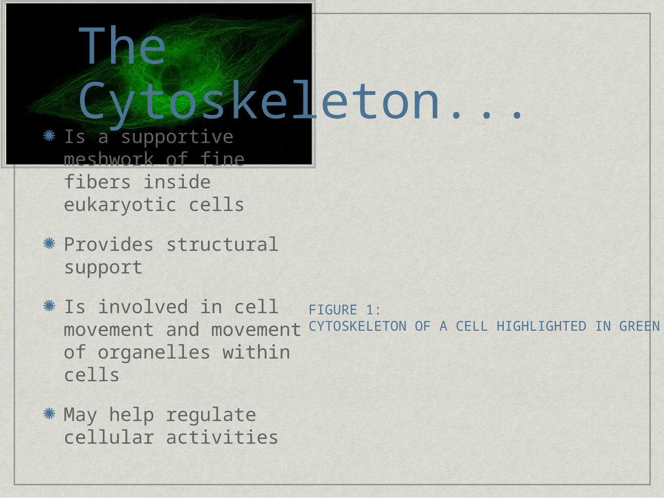
The Cytoskeleton...Is a supportive meshwork of fine fibers inside eukaryotic cells
Provides structural support
Is involved in cell movement and movement of organelles within cells
May help regulate cellular activities
FIGURE 1: CYTOSKELETON OF A CELL HIGHLIGHTED IN GREEN

Microfilaments
Fine, threaded protein fibers
Consists of Actin, a globular, contractile (can contract under stimuli-see next point) protein, one of the most abundant cellular proteins
Has myosin proteins (motor), activated by an ATP, causing movement along the actin fibers, causing contraction in the filament, and therefore muscle contraction
3-6 nanometers in diameter
Carry out cellular movement such as gliding, contraction, and cytokinesis (cytoplasm division of eukaryotic cells)
FIGURE 3: ATP ACTIVATED MYOSIN
MOTOR PROTEIN “WALKING” ON AN ACTIN MICROFIBER,
CAUSING IT TO CONTRACT
FIGURE 4: ARRANGEMENT OF ACTIN GLOBULAR
PROTEINS IN A MICROFILAMENT
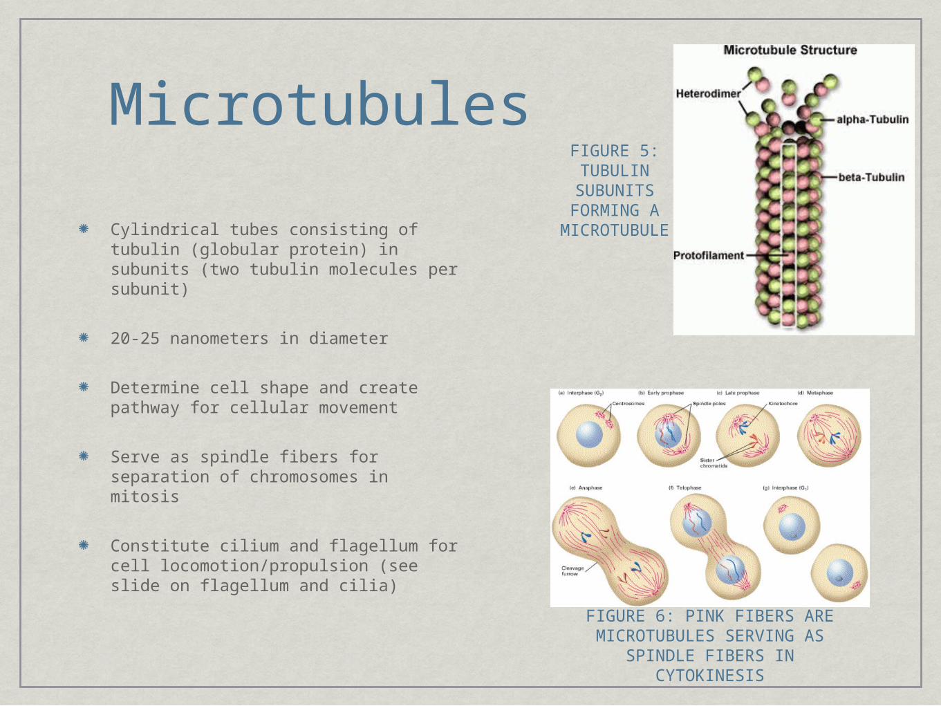
Microtubules
Cylindrical tubes consisting of tubulin (globular protein) in subunits (two tubulin molecules per subunit)
20-25 nanometers in diameter
Determine cell shape and create pathway for cellular movement
Serve as spindle fibers for separation of chromosomes in mitosis
Constitute cilium and flagellum for cell locomotion/propulsion (see slide on flagellum and cilia)
FIGURE 5: TUBULIN
SUBUNITS FORMING A MICROTUBU
LE
FIGURE 6: PINK FIBERS ARE MICROTUBULES SERVING AS
SPINDLE FIBERS IN CYTOKINESIS
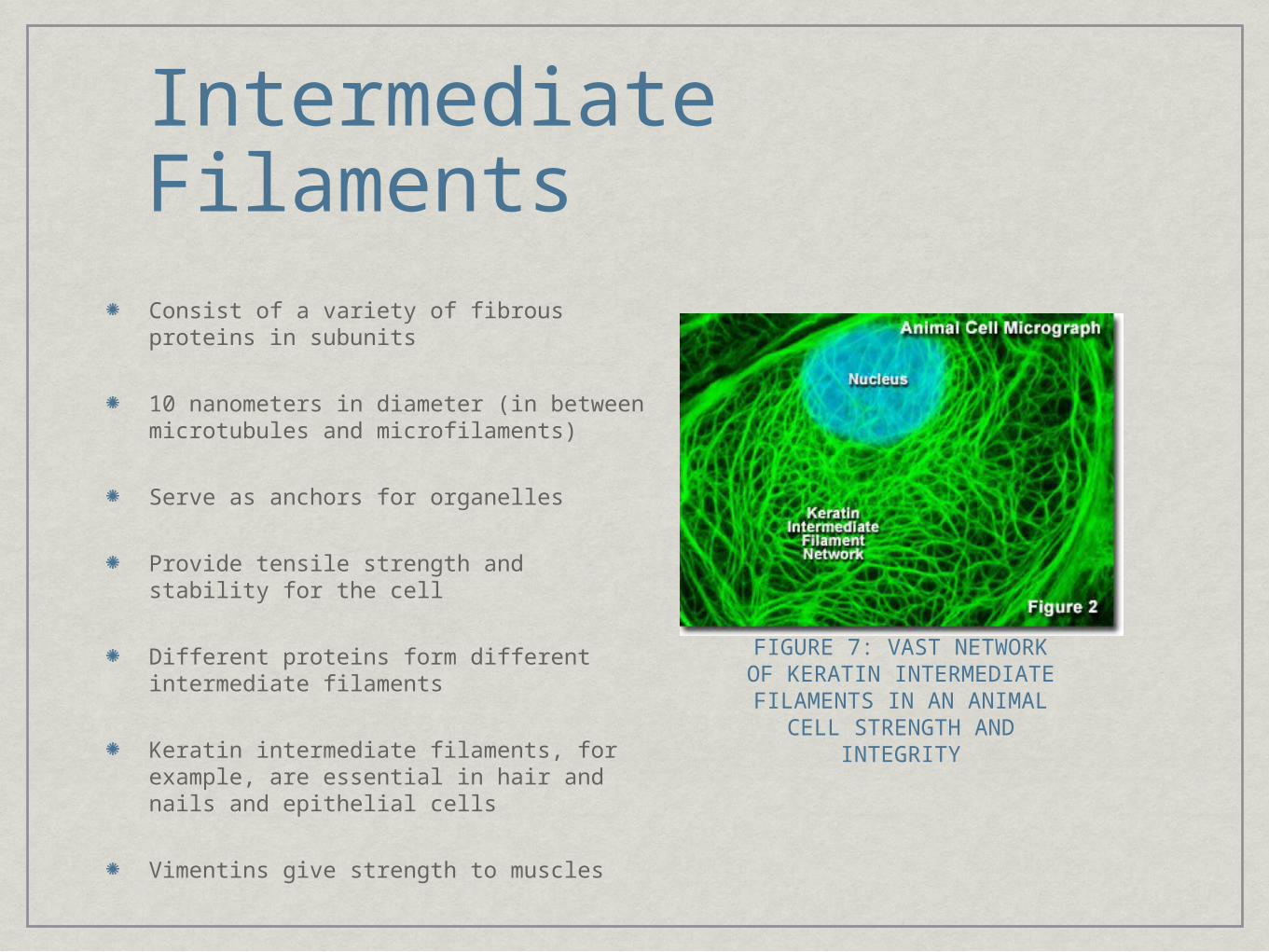
Intermediate Filaments
Consist of a variety of fibrous proteins in subunits
10 nanometers in diameter (in between microtubules and microfilaments)
Serve as anchors for organelles
Provide tensile strength and stability for the cell
Different proteins form different intermediate filaments
Keratin intermediate filaments, for example, are essential in hair and nails and epithelial cells
Vimentins give strength to muscles
FIGURE 7: VAST NETWORK OF KERATIN INTERMEDIATE FILAMENTS IN AN ANIMAL
CELL STRENGTH AND INTEGRITY
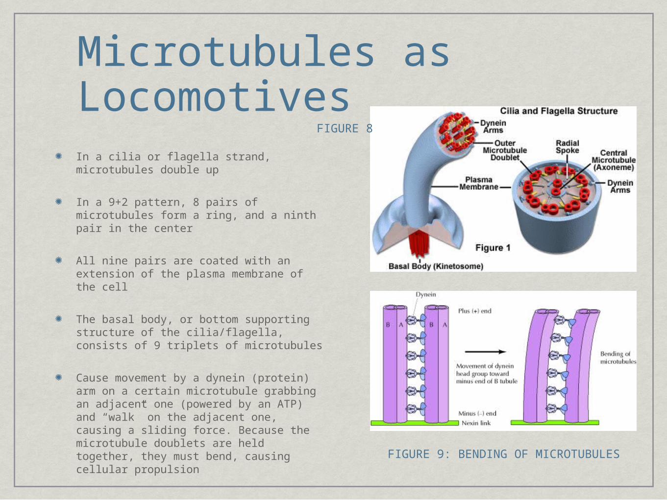
Microtubules as LocomotivesIn a cilia or flagella strand, microtubules double up
In a 9+2 pattern, 8 pairs of microtubules form a ring, and a ninth pair in the center
All nine pairs are coated with an extension of the plasma membrane of the cell
The basal body, or bottom supporting structure of the cilia/flagella, consists of 9 triplets of microtubules
Cause movement by a dynein (protein) arm on a certain microtubule grabbing an adjacent one (powered by an ATP) and “walk” on the adjacent one, causing a sliding force. Because the microtubule doublets are held together, they must bend, causing cellular propulsion
FIGURE 9: BENDING OF MICROTUBULES
FIGURE 8
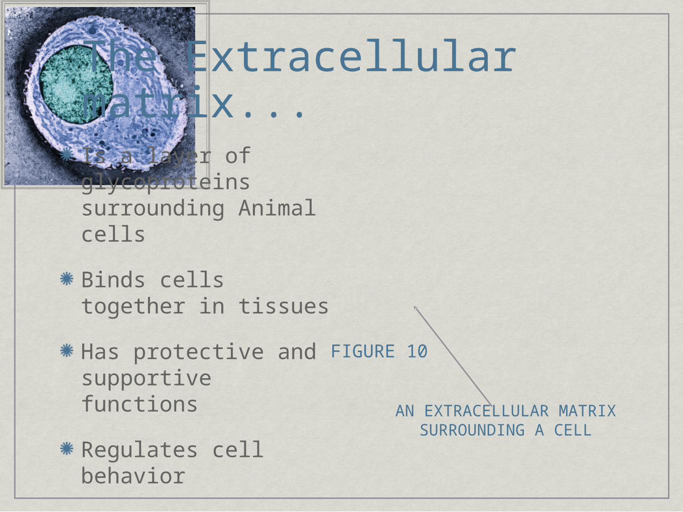
The Extracellular matrix...Is a layer of glycoproteins surrounding Animal cells
Binds cells together in tissues
Has protective and supportive functions
Regulates cell behavior
AN EXTRACELLULAR MATRIX SURROUNDING A CELL
FIGURE 10

CommunicationCommunication
Four main kinds of Four main kinds of communicationcommunication-ENDOCRINE: FROM FAR
AWAY
-PARACRINE: LOCALIZED
-AUTOCRINE: SELF
-JUXTACRINE: ADJACENT-Either a hydrophilic or hydrophobic signaling molecule is sent to a
receptor in/on the cell
-If hydrophilic, the molecule must find a receptor on the membrane
-If hydrophobic, the molecule can diffuse across the membrane
FIGURE 11
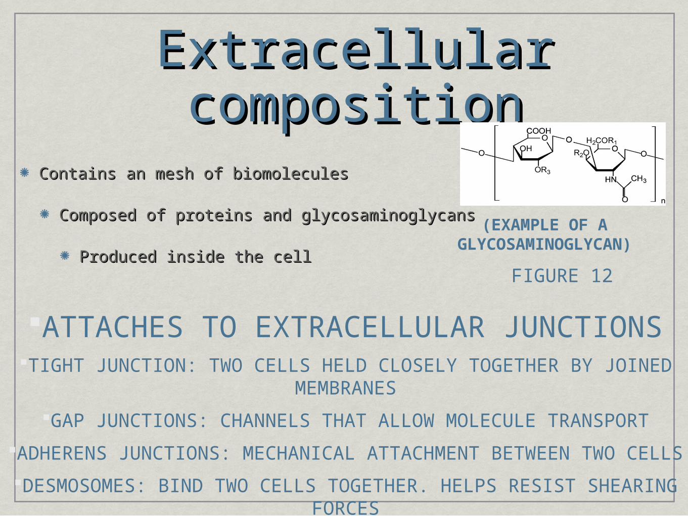
Extracellular Extracellular compositioncomposition
Contains an mesh of biomoleculesContains an mesh of biomolecules
Composed of proteins and glycosaminoglycansComposed of proteins and glycosaminoglycans
Produced inside the cellProduced inside the cell
(EXAMPLE OF A GLYCOSAMINOGLYCAN)
ATTACHES TO EXTRACELLULAR JUNCTIONSTIGHT JUNCTION: TWO CELLS HELD CLOSELY TOGETHER BY JOINED
MEMBRANES
GAP JUNCTIONS: CHANNELS THAT ALLOW MOLECULE TRANSPORT
ADHERENS JUNCTIONS: MECHANICAL ATTACHMENT BETWEEN TWO CELLS
DESMOSOMES: BIND TWO CELLS TOGETHER. HELPS RESIST SHEARING FORCES
FIGURE 12
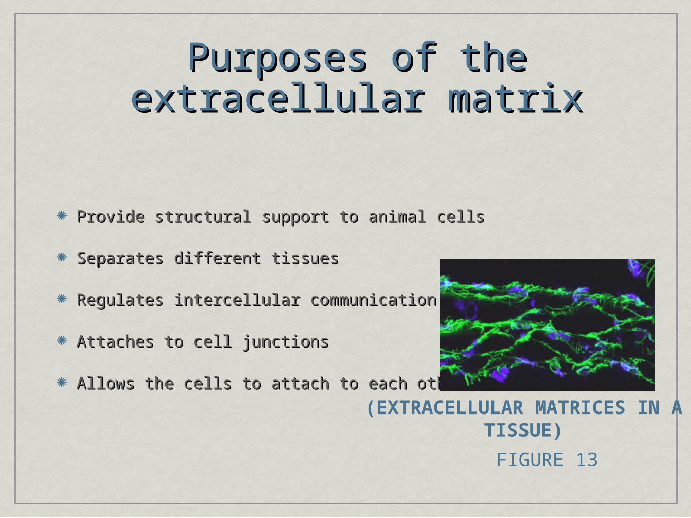
Purposes of the extracellular Purposes of the extracellular matrixmatrix
Provide structural support to animal cellsProvide structural support to animal cells
Separates different tissuesSeparates different tissues
Regulates intercellular communicationRegulates intercellular communication
Attaches to cell junctionsAttaches to cell junctions
Allows the cells to attach to each otherAllows the cells to attach to each other
(EXTRACELLULAR MATRICES IN A TISSUE)
FIGURE 13

Bibliography: (FOR PICTURES)
Figure 1: http://tensegrity.wikispaces.com/Mast
2: http://www.google.com/imgres?imgurl=http://i3.ytimg.com/vi/WRxsOMenNQM/0.jpg&imgrefurl=http://videowap.tv
3: http://www.google.com/imgres?imgurl=http://www.cartage.org
4. http://www.google.com/imgres?imgurl=http://vbaulin.front.ru
5: http://www.google.com/imgres?imgurl=http://tainano.com
6: http://www.google.com/imgres?imgurl=http://micro.magnet.fsu.edu
7:http://www.google.com/imgres?imgurl=http://micro.magnet.fsu.edu
8: http://www.google.com/imgres?imgurl=http://www.ncbi.nlm.nih.gov
9: http://www.google.com/imgres?imgurl=http://images.wellcome.ac.uk
10: http://www.nature.com/jcbfm/journal/v20/n10/images/9590992f1.jpg
11: http://www.statemaster.com/wikimir/images/upload.wikimedia.org/wikipedia
12: http://www.stevens.edu/ses/ccbbme/fileadmin/ccbbme/images/pic2.png