Embed Size (px)
Citation preview
COMPREHENSIVE INVITED REVIEW
The Cystine/Glutamate Antiporter System xc-
in Health and Disease: From Molecular Mechanismsto Novel Therapeutic Opportunities
Jan Lewerenz,1 Sandra J. Hewett,2 Ying Huang,3 Maria Lambros,3 Peter W. Gout,4 Peter W. Kalivas,5
Ann Massie,6 Ilse Smolders,7 Axel Methner,8 Mathias Pergande,8 Sylvia B. Smith,9
Vadivel Ganapathy,10 and Pamela Maher11
Abstract
The antiporter system xc- imports the amino acid cystine, the oxidized form of cysteine, into cells with a 1:1
counter-transport of glutamate. It is composed of a light chain, xCT, and a heavy chain, 4F2 heavy chain (4F2hc),and, thus, belongs to the family of heterodimeric amino acid transporters. Cysteine is the rate-limiting substratefor the important antioxidant glutathione (GSH) and, along with cystine, it also forms a key redox couple on itsown. Glutamate is a major neurotransmitter in the central nervous system (CNS). By phylogenetic analysis, weshow that system xc
- is a rather evolutionarily new amino acid transport system. In addition, we summarize thecurrent knowledge regarding the molecular mechanisms that regulate system xc
- , including the transcriptionalregulation of the xCT light chain, posttranscriptional mechanisms, and pharmacological inhibitors of system xc
- .Moreover, the roles of system xc
- in regulating GSH levels, the redox state of the extracellular cystine/cysteineredox couple, and extracellular glutamate levels are discussed. In vitro, glutamate-mediated system xc
- inhibitionleads to neuronal cell death, a paradigm called oxidative glutamate toxicity, which has successfully been used toidentify neuroprotective compounds. In vivo, xCT has a rather restricted expression pattern with the highestlevels in the CNS and parts of the immune system. System xc
- is also present in the eye. Moreover, an elevatedexpression of xCT has been reported in cancer. We highlight the diverse roles of system xc
- in the regulation ofthe immune response, in various aspects of cancer and in the eye and the CNS. Antioxid. Redox Signal. 00, 000–000.
I. IntroductionA. Oxidative stress and antioxidant defenseB. GSH metabolismC. Glutamate: neurotransmission and neurotoxicity
II. The Cystine/Glutamate Antiporter System xc-
A. Functional and pharmacological characteristics of system xc-
B. The molecular biology of system xc-
C. The phylogeny of xCT, the specific subunit of system xc-
D. Regulation of system xc- by transcriptional regulation of its specific subunit xCT
E. Regulation of system xc- activity by protein trafficking and protein modification
F. Regulation of system xc- activity by substrate availability
1Department of Neurology, University of Ulm, Ulm, Germany.2Program in Neuroscience, Department of Biology, Syracuse University, Syracuse, New York.3Center for the Advancement of Drug Research and Evaluation, College of Pharmacy, Western University of Health Sciences, Pomona,
California.4Department of Experimental Therapeutics, BC Cancer Agency, Vancouver, British Columbia, Canada.5Department of Neurosciences, Medical University of South Carolina, Charleston, South Carolina.Departments of 6Pharmaceutical Biotechnology and Molecular Biology and 7Pharmaceutical Chemistry and Drug Analysis, Center for
Neuroscience, Vrije Universiteit Brussel, Brussels, Belgium.8Department of Neurology, Heinrich-Heine-University Duesseldorf, Duesseldorf, Germany.Departments of 9Cellular Biology and Anatomy and 10Biochemistry and Molecular Biology, Medical College of Georgia, Augusta, Georgia.11Cellular Neurobiology Laboratory, Salk Institute for Biological Studies, La Jolla, California.
Reviewing Editors: Markus Conrad, Miriam Cortese-Krott, Giovanni Li Volti, Doug Lobner, Carlos Matute, Hugo Monteiro, Osamu Nagano,Chris Norris, Hideyo Sato, and Oren Tirosh
ANTIOXIDANTS & REDOX SIGNALINGVolume 00, Number 00, 2012ª Mary Ann Liebert, Inc.DOI: 10.1089/ars.2011.4391
1
III. Expression of System xc- In Vitro and In Vivo and Its Functional Consequences
A. In the absence of disease, system xc- shows a rather restricted expression pattern in vivo
B. System xc- is induced in most cultured cells
C. The role of system xc- in the regulation of GSH synthesis, the extracellular redox milieu,
and extracellular glutamate levelsD. Oxidative glutamate toxicity—an in vitro paradigm for neuronal death induced by system xc
- inhibition1. The cell death pathway in oxidative glutamate toxicity2. Using oxidative glutamate toxicity to identify neuroprotective pathways3. Using oxidative glutamate toxicity to screen for neuroprotective drugs4. Oxidative glutamate toxicity in vivo
IV. The Role of System xc- in Health and Disease
A. System xc- in vivo—lessons from xCT-deficient mice
B. The role of system xc- in the immune system and inflammation
C. The role of system xc- in cancer and resistance against anti-cancer drugs
1. System xc- is regulated by potentially oncogenic pathways
2. System xc- mediates the infection of cells by oncogenic Kaposi’s sarcoma herpesvirus
3. System xc- plays an important role in the multidrug resistance of cancers
4. Inhibition of system xc- reduces cancer cell replication, tissue invasion, and metastasis
5. System xc- expressed in tumor cells may be used as a target for anticancer drug delivery
6. Up-regulation of system xc- in normal cells provides protection against carcinogenesis—a possible
role in cancer prevention7. Synopsis of the role of system xc
- in cancer and resistance against anti-cancer drugsD. System xc
- and diseases of the eye1. Studies of system xc
- in the retina2. Studies of system xc
- in the lens and cornea3. Synopsis and future directions for system xc
- and diseases of the eyeE. The role of system xc
- in diseases of the CNSF. The role of system xc
- activity in memory and behaviorV. Conclusion
I. Introduction
A. Oxidative stress and antioxidant defense
Oxidative stress is defined as an imbalance between theproduction of free radicals, mostly reactive oxygen spe-
cies (ROS), and their removal by the antioxidant defensesystems present in tissues and body fluids (253) and, thus,results from an increase in ROS production and/or a decreasein antioxidant defense. Oxidative stress leads to the oxidativemodification of proteins, lipids, and DNA. Cells contain notonly small-molecule antioxidants such as vitamins C and Eand the tripeptide glutathione (GSH), which scavenge theROS produced during the cell’s metabolism, but also enzymeswhose specific role is the neutralization of ROS [reviewed in(241)]. These include the different isoforms of superoxidedismutase (SOD), which convert superoxide into hydrogenperoxide (H2O2), and catalase, which metabolises H2O2. GSHperoxidases (GPx) GSH-dependently catalyze the decompo-sition of H2O2 and of organic hydroperoxides while oxidizingGSH to GSH disulfide (GSSG).
Oxidative modification of proteins, lipids, and DNA hasbeen repeatedly shown to be associated with ageing, and ithas been frequently demonstrated that GSH levels are de-creased in diverse tissues in aged animals or elderly humans(149, 243, 300). Ageing is the major risk factor for many of themost important diseases in the Western World, including di-abetes, atherosclerosis, cancer, and neurodegenerative dis-eases such as Parkinson’s disease (PD), Alzheimer’s disease(AD), and ischemic stroke. Of note, oxidative stress is thoughtto play an important role in each of these diseases (88, 274).
B. GSH metabolism
The small-molecule antioxidant GSH is a tripeptide con-sisting of the amino acids glutamate, glycine, and cysteine.Cells contain approximately millimolar concentrations ofGSH. Thus, GSH is one of the most important small-moleculeantioxidants in somatic cells.
In most tissues, the rate-limiting amino acid for GSHsynthesis is the nonessential amino acid cysteine (160, 179).Cysteine can be imported into cells either directly or in itsoxidized form, cystine, via the cystine/glutamate antiportersystem xc
- (Fig. 1). Within the cell, cystine is immediatelyreduced to cysteine either by intracellular GSH via the for-mation of a mixed disulfide intermediate or by thioredoxinreductase 1 (TRR1) (172). Several amino acid transporters thatcan transport cysteine have been described. System alanine-serine-cysteine (ASC) transports cysteine as well as threonine,asparagine, alanine, serine, and, to some extent, glutamine(40). System A transports glycine, alanine, and proline muchmore efficiently than cysteine, and system L also transportsmethionine, valine, phenylalanine, leucine, and isoleucine. Inaddition, excitatory amino acid transporters (EAATs) havebeen proposed as playing a role in cysteine import into neu-rons (36). However, the affinity of EAATs for glutamate is 10times higher than for cysteine (122).
The first step in GSH synthesis, the generation of c-glutamylcysteine, is catalyzed by glutamate cysteine ligase (GCL) (182,reviewed in 82). c-Glutamyl cysteine and glycine then formGSH through the action of GSH synthase. GSH can bothnonenzymatically and enzymatically, in a reaction catalyzed
2 LEWERENZ ET AL.
by different GPx with distinct substrate specificities, reducediverse ROS. In scavenging ROS, GSH is oxidized to GSSG,which is either reduced by GSH reductase (GR), in a reactionthat requires reduced nicotinamide adenine dinucleotidephosphate (NADPH), or exported from the cell by multi-drugresistance proteins (MRPs). NADPH is generated via thehexose monophosphate shunt, an alternative pathway ofglucose metabolism. Moreover, the formation of GSH adductsby GSH S-transferases (GSTs) detoxifies endogenous a,b-unsaturated aldehydes, quinones, epoxides, and hydroper-oxides, which are formed as secondary metabolites during cellmetabolism and electrophilic xenobiotics, such as chemicalcarcinogens, environmental pollutants, and antitumor agents(90). The diverse GSH adducts are exported from the cell byMRPs. Both MRPs and/or organic anion transporter proteins(OATPs) release GSH into the extracellular space (65, 181).Extracellular GSH is metabolized by the ectoenzyme c-
glutamyl transferase (GGT), which transfers the c-glutamylresidue to different acceptor amino acids, leading to the for-mation of a c-glutamyl containing dipeptide and the dipep-tide cysteinyl glycine, which is either cleaved by extracellulardipeptidases to generate cysteine and glycine or directly takenup by cells (54). The neuroprotective function of GSH and/orGSH-related substrate export via MRP1 in the brain was re-cently demonstrated in an animal stroke model (200).
Generally, the GSH/GSSG redox couple determines thecell’s redox state with a more reducing environment associ-ated with cell proliferation, while a more oxidizing environ-ment is associated with differentiation (237). GSH is notuniformly distributed in cells, but rather different subcellularcompartments have distinct GSH levels and GSH/GSSG ra-tios (42). In the cytosol, the GSH-to-GSSG ratio is high, con-sistent with a reducing environment, whereas in theendoplasmic reticulum (ER), the GSH redox couple is in amuch more oxidized state, in line with the role of this or-ganelle in protein disulfide bond formation (269). The mito-chondria contain a separate pool of GSH that plays a key rolein maintaining mitochondrial function (173). Other roles forGSH include functioning as a cofactor for a variety of enzymessuch as glyoxalase 1, the rate-limiting enzyme for the removalof reactive dicarbonyls (213) and in the glutathionylation ofproteins, a potential signaling mechanism somewhat analo-gous to protein phosphorylation (180) and in the synthesis ofthe inflammatory mediators cysteinyl leukotrienes (130).
The major transcription factor that regulates GSH metab-olism is the ubiquitously expressed protein NF-E2-relatedfactor 2 (Nrf2), a member of the Cap ’n’ Collar family of bZIPproteins [for reviews see (113, 296)]. Under basal conditions,Nrf2-dependent transcription is repressed, because Nrf2 israpidly degraded by the proteasome, a process mediated byits interaction with Keap1, an Nrf2-specific adaptor proteinfor the Cul3 ubiquitin ligase complex. On exposure to a va-riety of different stimuli including oxidative stress, ER stress,nitric oxide (NO), 15d-PGJ2, phenolic compounds, Michaelacceptors, isothiocyanates, dithiolethiones, dimercaptans,heavy metals, peroxides and polyenes, Keap1-dependentNrf2 ubiquitination, and subsequent degradation are blocked,leading to the stabilization and nuclear accumulation of Nrf2,where it induces electrophile response element (EpRE)-dependent gene expression to re-establish cellular redoxhomeostasis (113). The EpRE is also known as the antioxidantresponse element (ARE) but since electrophiles rather thanantioxidants activate transcription mediated by this element,the term EpRE is preferable. Several key enzymes of GSHmetabolism are transcriptionally regulated by Nrf2, includingthe catalytic and regulatory subunits of GCL, GSH synthase,GPx2, GSTs, and GR (89, 113). Hence, Nrf2 is thought torepresent a key transcriptional regulator of GSH metabolism.
C. Glutamate: neurotransmission and neurotoxicity
Glutamate is the most important excitatory neurotrans-mitter in the brain. When released synaptically, glutamateactivates ionotropic glutamate receptors located in thepostsynaptic part of the synapse. Ionotropic glutamate re-ceptors are ligand-gated ion channels and include receptors ofthe a-amino-3-hydroxy-5-methyl-4-isoxazolepropionic acid(AMPA), kainite, and N-methyl-D-aspartic acid (NMDA)types [reviewed in (158)]. While AMPA and kainate receptors
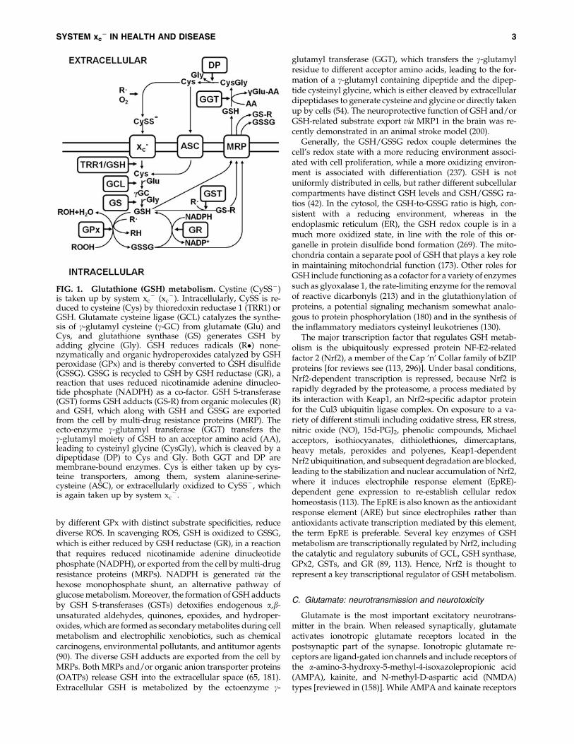
FIG. 1. Glutathione (GSH) metabolism. Cystine (CySS - )is taken up by system xc
- (xc- ). Intracellularly, CySS is re-
duced to cysteine (Cys) by thioredoxin reductase 1 (TRR1) orGSH. Glutamate cysteine ligase (GCL) catalyzes the synthe-sis of c-glutamyl cysteine (c-GC) from glutamate (Glu) andCys, and glutathione synthase (GS) generates GSH byadding glycine (Gly). GSH reduces radicals (R�) none-nzymatically and organic hydroperoxides catalyzed by GSHperoxidase (GPx) and is thereby converted to GSH disulfide(GSSG). GSSG is recycled to GSH by GSH reductase (GR), areaction that uses reduced nicotinamide adenine dinucleo-tide phosphate (NADPH) as a co-factor. GSH S-transferase(GST) forms GSH adducts (GS-R) from organic molecules (R)and GSH, which along with GSH and GSSG are exportedfrom the cell by multi-drug resistance proteins (MRP). Theecto-enzyme c-glutamyl transferase (GGT) transfers thec-glutamyl moiety of GSH to an acceptor amino acid (AA),leading to cysteinyl glycine (CysGly), which is cleaved by adipeptidase (DP) to Cys and Gly. Both GGT and DP aremembrane-bound enzymes. Cys is either taken up by cys-teine transporters, among them, system alanine-serine-cysteine (ASC), or extracellularly oxidized to CySS - , whichis again taken up by system xc
- .
SYSTEM xc2 IN HEALTH AND DISEASE 3
primarily mediate sodium influx, NMDA receptors havecalcium conductivity. Activation of NMDA receptors plays animportant role in synaptic plasticity and learning [reviewed in(183)]. In addition to ionotropic glutamate receptors, eightisoforms of metabotropic glutamate receptors exist, whichbelong to the family of G protein-coupled receptors andthat do not form ion channels but signal via various second-messenger systems [reviewed in (257)].
To ensure adequate neurotransmission, the extracellularglutamate concentration has to be tightly controlled. Whilethe mean glutamate concentration in the brain is 10 mM (127),the extracellular glutamate concentration in the brain is only2–9 lM (8). Thus, there is a steep concentration gradient withmuch higher intracellular glutamate concentrations. The ra-pid removal of released glutamate from the extracellularspace is brought about by EAATs, a family of high-affinityNa + /K + -dependent transporters for glutamate and aspar-tate, of which five different members, EAAT1-5, exist [re-viewed in (247)]. EAAT1 and - 2 are prominently expressedin astrocytes, especially within their processes surroundingglutamatergic synapses, where they are responsible for theimmediate uptake of synaptically released glutamate. How-ever, expression of both EAAT1 and - 2 has also been dem-onstrated in macrophages (218) and EAAT2 expression inmicroglia (204), indicating other roles for EAATs distinct fromneurotransmission. EAAT3 is mainly expressed not only inneurons, but also in the kidney and the intestinal mucosa (109,203). EAAT4 is most prominently expressed in cerebellarPurkinje cells, and EAAT5 is expressed in rod photoreceptorand bipolar cells of the retina. These transporters co-transport2 or 3 molecules of Na + and a proton with each molecule ofglutamate (or aspartate) and counter-transport of a K + ion.Thus, by using the electrochemical gradient of these ionsacross the plasma membrane as an energy source, they arecapable of effectively accumulating glutamate and aspartatein cells against the steep intra- to extracellular concentrationgradient of these amino acids. Any change in extracellularglutamate concentrations, within the synaptic cleft or extra-synaptically, can be expected to change the activity of meta-botropic or ionotropic glutamate receptors and therebyneuronal activation patterns and, finally, on the highest level,behavior. Overactivation of ionotropic glutamate receptorsinduces neuronal death, a pathway called excitotoxicity, aterm coined by Olney in 1969 (195). Calcium influx via NMDAreceptors is especially effective in inducing this cell deathpathway [reviewed in (38)]. Thus, any pathophysiologicalstate of the brain that is associated with increased extracellularglutamate concentrations has the propensity to lead to neu-ronal degeneration. Several cellular processes have been re-ported as contributing to a pathological rise in extracellularglutamate, including increased exocytotic vesicular release,reduced glutamate uptake via EAATs, and nonvesicularglutamate release via reversal of EAAT-mediated glutamateuptake or opening of astrocytic volume-sensitive organicanion channels (104, 244). Excess glutamate can also be re-moved by activation of glutamate oxaloacetate transaminase(GOT), which can metabolize glutamate into tricarboxylicacid cycle intermediates, as recently demonstrated by thecerebral glutamate lowering and neuroprotective action ofGOT in stroke (219).
Glutamate toxicity has been implicated in the pathogenesisof neuronal injury triggered by many central nervous system
(CNS) diseases, including cerebral ischemia, Alzheimer’s andHuntington’s disease, epilepsy, amyotrophic lateral sclerosis[reviewed in (247)], and multiple sclerosis (MS) [reviewedin (77)].
II. The Cystine/Glutamate Antiporter System xc2
A. Functional and pharmacological characteristicsof system xc
-
System xc- was first characterized in human fetal lung
fibroblasts in culture by Bannai and Kitamura in 1980 (17).Makowske and Christensen described a similar transportsystem in a transformed rat hepatoma cell line (171). Systemxc
- acts as a sodium-independent and chloride-dependentantiporter of the anionic forms of cystine and glutamate (Fig.2) (16, 76, 202). Although system xc
- can transport both aminoacids in both directions (11), since the intracellular pool ofcystine is negligibly small because intracellular cystine israpidly reduced and the intracellular glutamate concentrationis generally higher than in the extracellular space, system xc
-
generally imports cystine while exporting glutamate. Thistransport appears to be locked in a 1:1 ratio (11). Extracellularglutamate acts as a competitive inhibitor for cystine uptake viasystem xc
- . The reported Ki for glutamate inhibition of cystineuptake by system xc
- is 150 lM, while the Ki for cystine in-hibition of glutamate uptake is 33 lM (171). The Kms are78 lM for glutamate and 45 lM for cystine.
Though all naturally occurring proteinogenic amino acidsare neither transported by system xc
- nor inhibit glutamateor cystine transport, some structurally related, naturallyoccurring or artificial molecules do inhibit system xc
- . L-a-aminoadipate, an amino acid intermediate in chain lengthbetween glutamate and cystine, is an effective substrate in-hibitor of system xc
- (202). L-a-aminoadipate is a product oflysine metabolism and is present in the brain (32). Otherstructurally related molecules that inhibit system xc
- includeb-mercaptolactate-cysteine disulfide, L-homocysteate, L-homocysteine sulfinate, L-a-aminopimelate, and L-serine-O-sulphate (16, 171, 202). b-Mercaptolactate-cysteine disulfide isfound in the urine of healthy humans (294). L-homocysteate
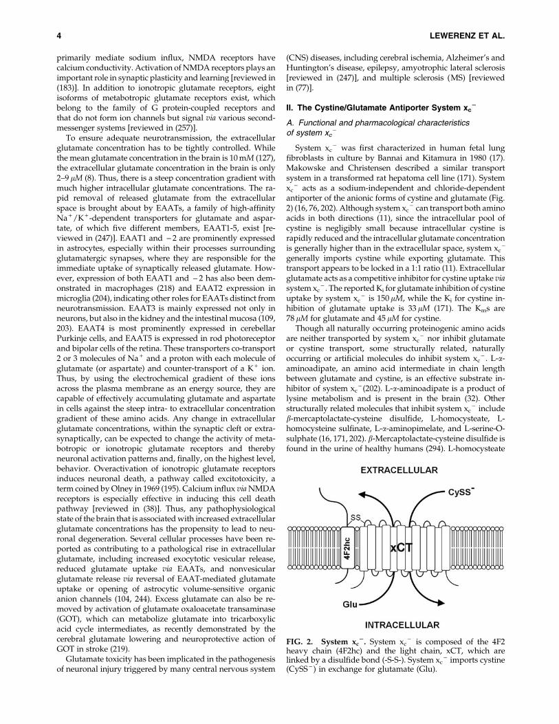
FIG. 2. System xc2. System xc
- is composed of the 4F2heavy chain (4F2hc) and the light chain, xCT, which arelinked by a disulfide bond (-S-S-). System xc
- imports cystine(CySS - ) in exchange for glutamate (Glu).
4 LEWERENZ ET AL.

and L-homocysteine sulfinate are acidic homocysteine deriv-atives that have been detected in astrocytes (51, 81). L-serine-O-sulfate and L-a-aminopimelate are artificial glutamateanalogs. Since L-homocysteate, L-a-aminopimelate, and L-a-aminoadipate activate cystine release from cells under oxi-dizing conditions, all of these are substrate inhibitors (11).Other inhibitors of system xc
- include the cyclic glutamateanalogues ibotenate, L-quisqualate, (RS)-4-bromo-homo-ibotenate, and (S)-4-carboxyphenylglycine [(S)-4-CPG] (202).Ibotenate is a compound that occurs in the mushroomsAmanita muscaria and Amanita pantherina (83), while quis-qualate naturally occurs in the seeds of the vine Quisqualisindica. The other compounds do not occur naturally. How-ever, although more potent at inhibiting system xc
- thannoncyclic glutamate analogs, both cyclic and noncyclic systemxc
- inhibitors have cross-reactivities, especially with iono-tropic and metabotropic glutamate receptors, due to theirstructural similarity to glutamate. L-homocysteate andL-ibotenate activate NMDA receptors (52, 58) and (RS)-4-bromohomoibotenate and L-quisqualate are AMPAreceptor agonists (43, 184). Moreover, L-serine-O-sulfate,L-homocysteate, L-homocysteine sulfinate, L-ibotenate, andL-quisqualate activate metabotropic glutamate receptors (26,119, 184, 248), and (S)-4-CPG is a Group I metabotropic glu-tamate receptor antagonist (22). L-serine-O-sulfate also acts asan inhibitor of serine racemase (199) and aspartate amino-transferase (273) as well as an EAAT substrate (278). While L-quisqualate and (S)-4-CPG are the most potent inhibitors ofsystem xc
- -mediated glutamate uptake, they are less welltransported by system xc
- than L-ibotenate and noncyclicglutamate analogs. Recently, AMPA analogues were de-scribed as new system xc
- inhibitors (201). However, theirspecificity has not been demonstrated yet. Moreover, theglutamate analogue (4S)-4-(3-[18F]fluoropropyl)-L-glutamate(BAY 94-9392) was shown to be efficiently taken up by systemxc
- into tumor cells as a possible positron emission tomog-raphy (PET) tracer for system xc
- activity (123).Early evidence suggested that nonsteroidal anti-inflammatory
drugs also inhibit system xc- (15). On this basis, the Gout lab
identified the FDA-approved drug sulfasalazine, commonlyused to treat chronic inflammatory diseases such as rheuma-toid arthritis, as a potent system xc
- inhibitor (79). However,this compound is also a potent inhibitor of nuclear factorkappa B (NF-jB) activation (283). In addition, glutamateuptake via system xc
- has been reported to be moderatelysensitive to the anion transport inhibitors 4,4¢-diisothiocya-natostilbene-2,20-disulfonic acid (DIDS), 4-acetamido-4¢-isothiocyanatostilbene-2,2¢-disulfonic acid (SITS), and4,4¢-dinitrostilbene-2,2¢-disulfonic acid (DNDS) (76).
Taken together, all pharmacological substances commonlyused to study system xc
- activity have off-target effects.Specific inhibitors of system xc
- have not yet been discovered.Moreover, some substrate and nonsubstrate inhibitors ofsystem xc
- might have different effects when used as probesto study the role of system xc
- , as substrate inhibitors willinduce glutamate release in addition to inhibiting cystineuptake. Of the known substrates of system xc
- not onlycystine and glutamate but also b-mercaptolactate-cysteinedisulfide, L-a-aminoadipate, L-homocysteate, and L-homo-cysteine sulfinate might act as endogenous substrates and/orcompetitive inhibitors for cystine uptake through system xc
-
in vivo.
B. The molecular biology of system xc-
Using expression cloning in oocytes and cDNA librariesfrom peritoneal macrophages where system xc
- expressionwas induced by the electrophilic agent diethyl maleate (DEM)and bacterial lipopolysaccharide (LPS), Sato and co-workersidentified the promiscuous 4F2 heavy chain (4F2hc/CD98/SLC3A2) as one subunit and a new 502 amino acid proteinnamed xCT or SLC7A11 as the specific light chain subunit ofsystem xc
- (Fig. 2) (232).The xCT protein shows significant homology with the light
chains of heterodimeric amino acid transporters (HATs), afamily of amino acid transporters consisting of a light chainand a heavy chain linked by a disulfide bridge [reviewed in(282)]. It was predicted to contain 12 transmembrane domainswith the N- and C-termini located inside the cell and a re-entrant loop within intracellular loops 2 and 3, a generalstructure shared with other HAT light chains (69). As shownfor other HAT family members, 4F2hc and xCT are linked by adisulfide bridge.
In mouse macrophages as well as in the murine hippo-campal cell line HT22, xCT transcripts of multiple lengthswere detected by Northern blotting, of which three of *2.5,3.5 and 12 kb predominate (138, 232). The largest transcriptwas also shown to be expressed in the cerebral cortex of mice(232) and rats and in cultured astrocytes (76).
Using 5¢ rapid amplification of 5¢ cDNA ends, Sasaki et al.identified the transcriptional start site in the murine xCT gene(227). This start site predicts that the xCT mRNA contains a 5¢untranslated region (5¢UTR) with a length of 329 bp. Thelongest mouse xCT cDNA (NM_011990.2) published online is9181 bp, which contains a remarkably long 3¢ untranslatedregion (3¢UTR) of 7351 bp (www.ncbi.nlm.nih.gov/sites/entrez?cmd = Retrieve&db = nucleotide&dopt = GenBank&RID = TU0CM91C016&log%24 = nucltop&blast_rank = 1&list_uids = 80861466). Cloning of human xCT from cDNA librariesof W126Va4 cells (233), the human retinal pigment epithelialcell line ARPE-19 by the Ganapathy lab (28), and the humanteratoma cell line NT2 (21) yielded putative transcripts of 1885and 6568 bp (233), 2482 bp (28), and 3144 bp (21), respectively,which all share a 231 bp 5¢UTR and a 1506 bp open readingframe (ORF), including the stop codon, but have divergent3¢UTRs. The protein encoded by these cDNAs has 501 aminoacids and shows 89% identity and 96% similarity with mousexCT (28, 233). The human reference cDNA (NM_014331.3)published online (www.ncbi.nlm.nih.gov/sites/entrez?cmd =Retrieve&db = nucleotide&dopt = GenBank&RID = TU4E6EH2014&log%24 = nucltop&blast_rank = 1&list_uids = 80861465)is 9648 bp long with a 280 bp 5¢UTR and a 7862 bp 3¢UTR. Itwas demonstrated by Northern blotting that a 12 kb transcriptof xCT mRNA predominates in human fibroblasts (233) and inthe human brain, which was the tissue with the highest xCTexpression among the organs studied (21). Interestingly, therelative expression of the different xCT mRNA transcriptsshows a tissue-specific distribution with the 12 kb form muchmore abundant in brain and meninges as compared withmacrophages, where the 2.5 and 3.5 kb forms are moreprominently expressed (234). A splice variant (hxCT1b) thatresults in a 495 amino acid protein with the 20 C-terminalamino acids of the 501 amino acid-long standard form(hxCTa) replaced by 13 divergent amino acids was found tobe expressed at high levels in human U87 glioma cells (115).
SYSTEM xc2 IN HEALTH AND DISEASE 5
The new sequence for the 3¢ end of the ORF and the subse-quent 3¢UTR is identical with the 3¢UTR of the referencesequence (NM_014331.3) starting at bp 9245. When expressedin oocytes, hxCTb cRNA induces system xc
- activity when co-injected with 4F2hc cRNA, indicating that the new C-terminuspreserves xCT function (115).
In summary, the molecular identity of the 12 kb form of xCTmRNA that seems to predominate in most tissues has notbeen identified so far. However, several 3¢UTRs of different,sometimes remarkable, lengths exist for xCT mRNA, andthere is even, at least in humans, a splice variant involving the3¢UTR and part of the xCT ORF. Whether these different xCTmRNAs are involved in the regulation of xCT expression iscurrently unknown.
C. The phylogeny of xCT, the specific subunitof system xc
-
The other members of the HAT family are the amino acidtransporters LAT1/SLC7A5, LAT2/SLC7A8, y +LAT1/SLC7A7, y +LAT2/SLC7A6, b0, +AT/SLC7A9, AGT-1/SLC7A13, asc-1/SLC7A10, and the orphan transporterSLC7A15. LAT1 and LAT2 correspond to the functionallydefined transporter system L, y + LAT1 and y + LAT2 to systemy +L and b0, +AT to system b0, +[reviewed in (282)]. SystemL mediates sodium-independent exchange of large, neutralamino acids (leucine, histidine, methionine, phenylalanine,and glutamine, LAT2 also alanine), whereas system y+L ex-changes extracellular neutral amino acids co-transported withNa + with cationic amino acids. System b0, + is a broadly activeamino acid transporter that accepts diamino acids, includingcationic amino acids and cystine from the outside and, withlower affinity, also large neutral amino acids and exportsneutral amino acids (282). AGT-1/SLC7A13 mediates sodi-um-independent aspartate and glutamate transport (178).Asc-1/SLC7A10 is a sodium-independent transporter forglycine, alanine, L- and D-serine, and cysteine (67). HATscluster with the members of the cationic transporter familycat-1/SCL7A1, cat-2/SCL7A2, cat-3/SCL7A3, SLC7A4, andthe more distantly related SLC7A14 and members of theSLC12 family, transporters for sodium, potassium, and chlo-ride, in the c-group of the SLC transporter family (66).
The fact that system xc- uses only two proteinogenic amino
acids as substrates raises the question of which evolutionaryconstraints might have required a rather specific cystinetransporter in addition to the broadly active system b0, + . Of thenonvertebrate HAT functionally characterized so far, noneexhibited system xc
- -like activity. When co-expressed with4F2hc, the SPRM1 protein from Schistosoma mansoni showed anuptake profile that shared similarities with system L and sys-tem y +L (177). Both AAT1 and AAT3, two of the three Cae-norhabditis elegans putative HAT light chains, AAT1-3, showeda transport activity that shared similarities with system L andasc-1 when expressed in oocytes in parallel with ATG-2, theheavy chain ortholog of C. elegans (281). AAT2 has not beenanalyzed so far. The HAT light chain encoded by the Drosophilamelanogaster gene JhI-21 showed system L activity when co-expressed with the D. melanogaster homolog of 4F2hc/CD98(216). Reynolds and colleagues also reported that a BLASTsearch with human LAT1 and LAT2 highlighted five fly aminoacid transporters that share a significant degree of homology(homology to human LAT1 and LAT2 in parentheses), CG1607
(47% and 46%), Minidisc (Mnd or CG3297; 49% and 44%),Genderblind (Gb or CG6070; 44% and 40%), JhI-21 (CG12317;49% and 44%), and CG9413 (38% and 34%). All these moleculesshare the conserved cysteine that covalently links the lightchain to the heavy chain (Cys164 in LAT1). Moreover, theyshowed that siRNA against CG2791, Mnd, and JhI-21 alsodown-regulated system L activity in D. melanogaster Schneidercells (216). Surprisingly, Augustin et al. identified the same fiveDrosophila proteins as putative xCT homologs with 36% to 45%amino acid similarity to murine and human xCT, respectively,and showed that Gb (CG6070) regulates hemolymphe gluta-mate levels, which was taken as supportive evidence that Gbacts as a system xc
- -like transporter in Drosophila (6).In order to clarify the exact distribution of xCT orthologs in
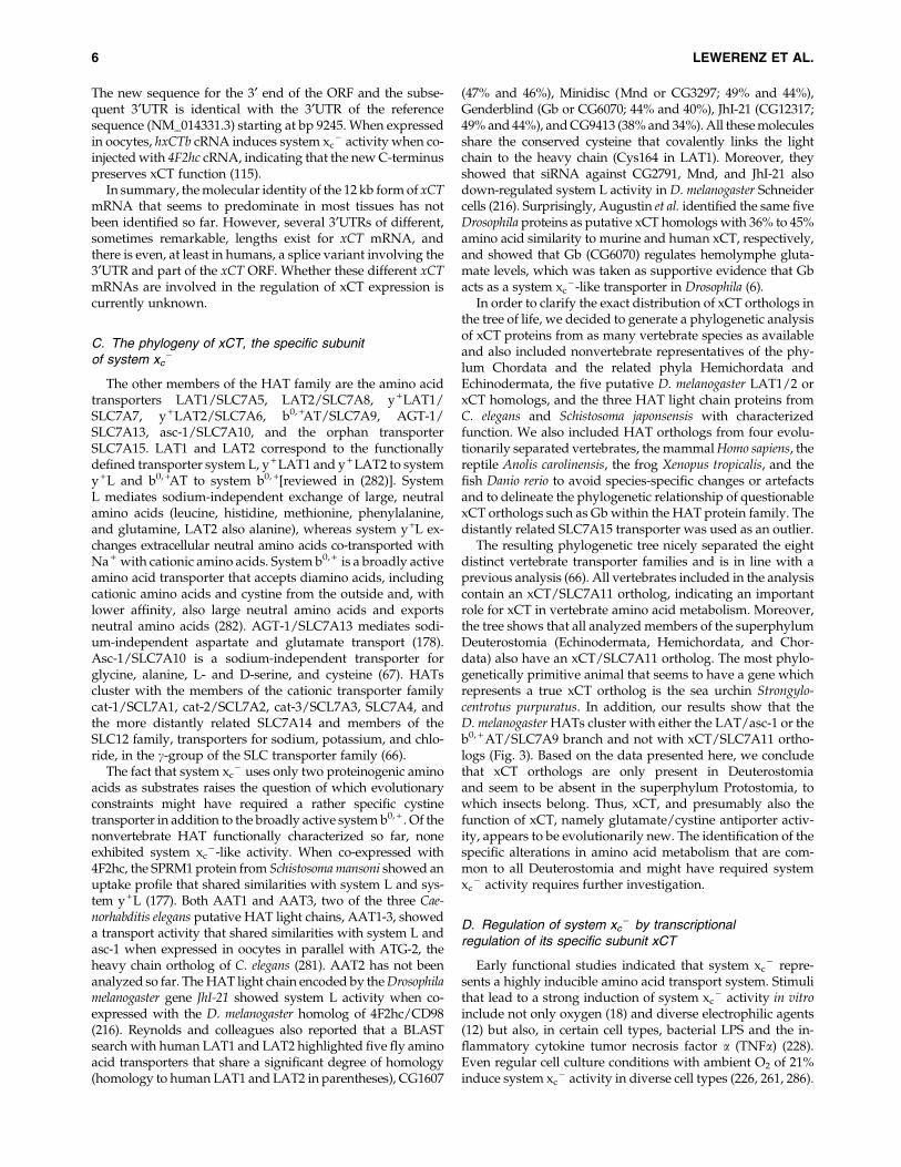
the tree of life, we decided to generate a phylogenetic analysisof xCT proteins from as many vertebrate species as availableand also included nonvertebrate representatives of the phy-lum Chordata and the related phyla Hemichordata andEchinodermata, the five putative D. melanogaster LAT1/2 orxCT homologs, and the three HAT light chain proteins fromC. elegans and Schistosoma japonsensis with characterizedfunction. We also included HAT orthologs from four evolu-tionarily separated vertebrates, the mammal Homo sapiens, thereptile Anolis carolinensis, the frog Xenopus tropicalis, and thefish Danio rerio to avoid species-specific changes or artefactsand to delineate the phylogenetic relationship of questionablexCT orthologs such as Gb within the HAT protein family. Thedistantly related SLC7A15 transporter was used as an outlier.
The resulting phylogenetic tree nicely separated the eightdistinct vertebrate transporter families and is in line with aprevious analysis (66). All vertebrates included in the analysiscontain an xCT/SLC7A11 ortholog, indicating an importantrole for xCT in vertebrate amino acid metabolism. Moreover,the tree shows that all analyzed members of the superphylumDeuterostomia (Echinodermata, Hemichordata, and Chor-data) also have an xCT/SLC7A11 ortholog. The most phylo-genetically primitive animal that seems to have a gene whichrepresents a true xCT ortholog is the sea urchin Strongylo-centrotus purpuratus. In addition, our results show that theD. melanogaster HATs cluster with either the LAT/asc-1 or theb0, + AT/SLC7A9 branch and not with xCT/SLC7A11 ortho-logs (Fig. 3). Based on the data presented here, we concludethat xCT orthologs are only present in Deuterostomiaand seem to be absent in the superphylum Protostomia, towhich insects belong. Thus, xCT, and presumably also thefunction of xCT, namely glutamate/cystine antiporter activ-ity, appears to be evolutionarily new. The identification of thespecific alterations in amino acid metabolism that are com-mon to all Deuterostomia and might have required systemxc
- activity requires further investigation.
D. Regulation of system xc- by transcriptional
regulation of its specific subunit xCT
Early functional studies indicated that system xc- repre-
sents a highly inducible amino acid transport system. Stimulithat lead to a strong induction of system xc
- activity in vitroinclude not only oxygen (18) and diverse electrophilic agents(12) but also, in certain cell types, bacterial LPS and the in-flammatory cytokine tumor necrosis factor a (TNFa) (228).Even regular cell culture conditions with ambient O2 of 21%induce system xc
- activity in diverse cell types (226, 261, 286).
6 LEWERENZ ET AL.
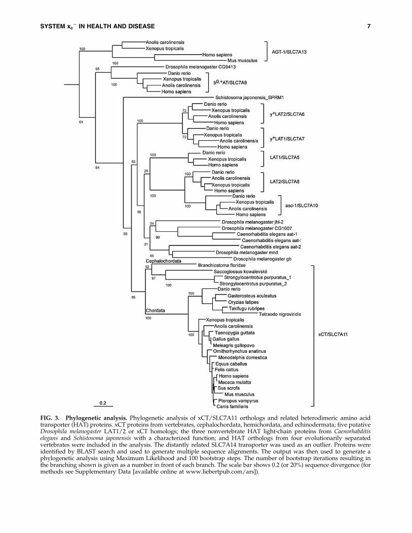
FIG. 3. Phylogenetic analysis. Phylogenetic analysis of xCT/SLC7A11 orthologs and related heterodimeric amino acidtransporter (HAT) proteins. xCT proteins from vertebrates, cephalochordata, hemichordata, and echinodermata; five putativeDrosophila melanogaster LAT1/2 or xCT homologs; the three nonvertebrate HAT light-chain proteins from Caenorhabditiselegans and Schistosoma japonensis with a characterized function; and HAT orthologs from four evolutionarily separatedvertebrates were included in the analysis. The distantly related SLC7A14 transporter was used as an outlier. Proteins wereidentified by BLAST search and used to generate multiple sequence alignments. The output was then used to generate aphylogenetic analysis using Maximum Likelihood and 100 bootstrap steps. The number of bootstrap iterations resulting inthe branching shown is given as a number in front of each branch. The scale bar shows 0.2 (or 20%) sequence divergence (formethods see Supplementary Data [available online at www.liebertpub.com/ars]).
SYSTEM xc2 IN HEALTH AND DISEASE 7
Experimental evidence strongly suggests that the tran-scriptional regulation of xCT expression is by far a much moreimportant determinant for system xc
- activity than the ex-pression of the heavy chain, 4F2hc, which is known to formheterodimers with at least five other specific amino acidtransporter light chains (282). Overexpression of the xCT lightchain alone robustly increases system xc
- activity in diversecell types, including NIH3T3 fibroblasts (284), HEK 293 cells(251), murine hippocampal HT22 cells (142), and astrocytes(250). Moreover, induction of system xc
- activity by treatmentwith the NO donor 3-nitroso-N-acetylpenicillamine in theretinal pigment epithelial cell line ARPE-19 (28) and by tert-butylhydroquinone in HT22 cells (142) was associated withinduction of xCT expression alone with no apparent regula-tion of 4F2hc. Thus, to shed light onto the transcriptionalregulation of system xc
- activity, most studies focussed onanalysis of the xCT promoter.
In 2002, Sasaki and co-workers identified four putativeEpREs in the proximal 5¢ flanking region of the mouse xCTgene. They showed that the DEM-dependent activation of thexCT promoter in BHK21 hamster kidney cells required themost 5¢ EpRE-like element in the xCT promoter. Consistentwith this observation, DEM induces system xc
- activity andthe activity of the xCT promoter, as judged by luciferase re-porter analysis, in wild-type but not in Nrf2 - / - fibroblasts(227). Moreover, in cultured murine peritoneal macrophages,induction of system xc
- activity by oxidative insults, includ-ing exposure to glucose oxidase (which generates H2O2),DEM, the superoxide generator paraquat, and the heavymetal cadmium depends on the presence of Nrf2, as inductionwas absent in macrophages derived from Nrf2 - / - mice (101).However, the induction of system xc
- either by in vitro culturefor 12 h or stimulation by LPS was diminished but not abol-ished, indicating that Nrf2-independent pathways are alsoinvolved (101). Nrf2 also regulates xCT expression in ratprimary cortical astrocytes (250). In addition, it was demon-strated that the neuroprotective antibiotic ceftriaxone inducesxCT expression and/or system xc
- activity in multiple celltypes, including hippocampal HT22 cells, cortical and spinalastrocytes, and stem cell-derived motor neurons (136). Cef-triaxone increases nuclear Nrf2 levels and the protective ac-tivity of ceftriaxone as well as the effect of ceftriaxone on GSHlevels was largely diminished in fibroblasts deficient in Nrf2.Hence, it was concluded that the ceftriaxone-mediated up-regulation of xCT is Nrf2 dependent (136). Thus, xCT andthereby system xc
- activity is connected to the network ofstress-inducible GSH metabolic enzymes that are coordi-nately regulated by Nrf2, which on induction by oxidativestress orchestrates the re-establishment of cellular redox ho-meostasis (see section I.B).
In early works on the regulation of system xc- activity,
Shiro Bannai reported that system xc- activity is induced not
only by electrophilic agents but also by cystine starvation (12).Of note, cystine starvation causes GSH depletion and therebyalso oxidative stress (263). In 2004, Sato et al. reported that theinduction of system xc
- activity and xCT expression is notonly specific for cystine starvation but also occurs when otheramino acids are depleted from the medium (230). The com-mon pathway that mediates transcriptional induction byamino acid limitation includes activation of general controlnon-derepressible-2 (GCN2) protein kinase by free tRNAsand subsequent phosphorylation of the translation initiation
factor eIF2a [reviewed in (114)]. eIF2a phosphorylation in-hibits cap-dependent translation, while several transcripts,including those of activating transcription factor 4 (ATF4), arepreferentially translated. ATF4 heterodimerizes with mem-bers of the CCAAT/enhancer-binding protein (C/EBP) andAP1 families and activates transcription on binding to theamino acid response element (AARE). Subsequently, it wasshown that the proximal promoter of xCT contains a tandemof two AAREs, the more 5¢ of which binds the transcriptionfactor ATF4, and both AAREs cooperatively mediate the ac-tivation of the xCT promoter after amino acid starvation (230).The two AAREs, with an intervening sequence of 9 bp ori-ented in the opposite direction, are completely conservedamong mouse, rat, bovine, and human xCT 5¢ flanking re-gions (140), indicating that this mechanism is functionallyimportant across species. While 4F2hc is also induced byamino acid limitation (230) and its expression is suppressed inATF4-deficient cells (87), the molecular mechanism underly-ing its regulation remains to be clarified.
At least in cell culture, the phosphorylation status of eIF2aplays a pivotal role in the basal activity of system xc
- , asembryonic fibroblasts derived from mice with a homozygousmutation of the eIF2a phosphorylation site, serine 51 to ala-nine, show very little ATF4 expression, xCT promoter activity,or system xc
- activity (140). Moreover, increased eIF2aphosphorylation brought about by the specific eIF2a phos-phatase inhibitor salubrinal induced ATF4 protein and sys-tem xc
- activity in hippocampal HT22 cells and the ratphaeochromocytoma cell line PC12 (140). Through inductionof ATF4 and xCT, eIF2a phosphorylation regulates cellularGSH concentrations and cellular sensitivity to oxidative stress(140). Interestingly, PC12 cells selected for resistance againstoxidative damage by the amyloid-b peptide, which is in-volved in the pathogenesis of AD, exhibit a strong activationof the phospho-eIF2a/ATF4/xCT signaling module (140).Since eIF2a phosphorylation and ATF4 expression were alsofound to be up-regulated in AD brains (140), this pathwaymight represent an adaptive response to oxidative stressin AD.
GCN2 is only one of four eIF2a kinases. The other three,protein kinase R (PKR), heme-regulated eIF2a kinase (HRI),and PKR-like kinase (PERK), are activated by diverse stimuli[reviewed in (287)]. While GCN2 is activated by not onlyamino acid deprivation, but also other stresses such as UVradiation and proteasome inhibition, PKR participates in ananti-viral defense mechanism that is mediated by interferon.HRI responds to haem deprivation and oxidative and heatstress in erythroid tissues, and PERK is responsive to ERstress. Thus, in theory, induction of xCT might occur after avariety of cellular insults, which are all relayed through eIF2aand ATF4. However, this hypothesis remains to be experi-mentally explored.
Inflammatory stimuli that strongly induce system xc- in
cells include LPS and TNFa (228). LPS binds to toll-like re-ceptor 4 and thereby activates multiple signaling cascades,including activation of NF-jB, an important mediator of in-flammation-induced gene transcription [reviewed in (291)]. Aputative binding site for NF-jB was identified in the murinexCT gene 5¢ flanking region (229). However, even LPS con-centrations too low to activate NF-jB strongly stimulated xCTexpression in macrophages (229). Thus, the signaling path-way through which LPS induces xCT expression remains to
8 LEWERENZ ET AL.

be clarified. Similarly, how TNFa, a proinflammatory cyto-kine that binds to two different receptors, which activatemultiple intracellular signaling pathways, including NF-jB[reviewed in (84)], activates system xc
- has not been clarified.Other extracellular ligands that have been reported to inducexCT expression, also without any detailed information aboutthe intracellular signaling pathways involved, include eryth-ropoietin and interleukin-1b (IL-1b). Erythropoietin has beenreported to up-regulate xCT expression in differentiated cor-tical neural stem cells and B104 neuroblastoma cells (255). Theinflammatory cytokine IL-1b specifically up-regulates systemxc
- activity in astrocytes but not in microglia and neurons byactivating the IL1-receptor via induction of xCT but not 4F2hcexpression (64, 105). In addition, fibroblast growth factor-2(FGF-2) induces system xc
- activity through induction of xCTexpression in astrocytes, but not in neurons or microglia,through activation of fibroblast growth factor receptor 1(FGFR1), a pathway sensitive to the combined inhibition ofthe MEK/ERK and phosphoinositide-3 kinase pathways(151). Interestingly, post-transcriptional mechanisms alsoregulate xCT mRNA levels. The microRNA-26b has beendemonstrated as directly targeting and down-regulating xCTtranscript expression (153). The pathways that regulate thelevels of the xCT transcript are summarized in Figure 4.
E. Regulation of system xc- activity
by protein trafficking and protein modification
Although the heavy chain, 4F2hc, is necessary for correctmembrane trafficking and insertion of a functionally active
system xc- , 4F2hc does not seem to be prominently involved
in the regulation of system xc- activity (see above). However,
the adhesion molecule CD44v, which is expressed in cancerstem cells, was shown to interact with the xCT-4F2hc het-erodimer and regulate its membrane insertion and therebyactivity (103). The laboratory of Sylvia Smith demonstratedthat a *40 kD band immunoreactive for xCT as detected byWestern blotting switched from an intracellular localization tothe plasma membrane compartment on exposure of retinalMuller glial cells to oxidative stress, whereas the more pre-dominant *50 kD band with constitutive membrane inser-tion did not show any regulation (191). This switch of the *40kD band to the membrane was associated with increasedsystem xc
- activity. However, the molecular basis of the dif-ferent sizes of xCT in Muller cells and whether this pathway isactive in other cell types has not been investigated.
Moreover, system xc- activity was found to be down-
regulated by signaling through metabotropic group II recep-tors, which suppress cyclic adenosine 3¢,5¢ monophosphate(cAMP) synthesis and thereby protein kinase A (PKA) acti-vation, in a cAMP-dependent manner in striatal slices (9).Human xCT contains two putative PKA phosphorylationsites (9). However, direct evidence for the regulation oftransport activity or membrane insertion by phosphorylationof xCT has not been published.
F. Regulation of system xc- activity
by substrate availability
Since extracellular glutamate is a competitive inhibitor ofcystine import via system xc
- while intracellular glutamatedrives cystine import, pathways that regulate the intra- aswell as the extracellular glutamate concentrations might bepotent, indirect regulators of cystine import via system xc
- . Infibroblasts, glutamine was shown to be taken up via systemASC and converted to glutamate to activate cystine import viasystem xc
- (13). In 2000, Rimaniol et al. reported that systemxc
- as well as EAAT1 and EAAT2 are expressed in humanmonocyte-derived macrophages (217). Glutamate amplifiedthe cystine-induced increase in GSH levels, an effect that wassensitive to EAAT inhibitors. This indicated that glutamateuptake via EAATs trans-stimulates cystine import via systemxc
- . Moreover, the same group reported that not only gluta-mate but also glutamine and aspartate stimulated GSH syn-thesis in these cells. These observations led to the hypothesisthat while glutamate uptake via EAATs directly transactivatessystem xc
- , the uptake of glutamine and L-aspartate, whichare then converted to glutamate by glutaminase and aspartateaminotransferase, respectively, also contribute to the intra-cellular glutamate pool that drives cystine import (218). Inaddition, some of the glutamate released by system xc
- is thesubject of immediate re-uptake by EAATs (138, 202). Inhippocampal HT22 cells, transient overexpression of theneuronal EAAT, EAAT3, increased intracellular GSH inthe presence of high glutamate concentrations andprotected HT22 cells from oxidative glutamate toxicity. Theseeffects were especially pronounced when EAAT3 was co-overexpressed with xCT (138). In summary, cystine uptake viasystem xc
- can be supported by multiple pathways thatincrease the intracellular concentration of the exported sub-strate glutamate. Glutamate uptake via EAATs, in theory,might be especially effective, because it causes a concomitant
FIG. 4. Transcriptional regulation of xCT expression. Avariety of stimuli (see section I.B), for example, electrophiles,heavy metals, and reactive oxygen species (ROS), lead to acti-vation of the nuclear factor NF-E2-related factor 2 (Nrf2), whichbinds to the electrophile response element (EpRE) within thexCT promoter region and activates transcription. Amino acid(AA) starvation leads to phosphorylation of eIF2a (peIF2a),which leads to the translational up-regulation of the transcrip-tion factor activating transcription factor 4 (ATF4). ATF4 acti-vates the transcription of xCT by binding to the amino acidresponse element (AARE) contained in the xCT promoter.Bacterial lipopolysaccharides (LPS), tumor necrosis factor a(TNFa), interleukin-1b (IL-1b), fibroblast growth factor-2 (FGF2),and erythropoietin (EPO) also increase the transcription of xCTthrough unknown or partially known signaling pathways. IL-1b acts via the IL-1 receptor (IL-1R), while FGF2 activates theFGF receptor 1 (FGFR1) and increases xCT transcription viaPI3K and MEK. MicroRNA-26b directly targets xCT mRNA.
SYSTEM xc2 IN HEALTH AND DISEASE 9

decrease in the extracellular glutamate that inhibits cystineuptake (Fig. 5).
Early reports about the substrate requirements of systemxc
- indicated that the exchanger only accepts the anionic formof cystine (16) (Fig. 5). The two amino groups of cystine havepK values of 7.48 and 9.02. As a result, at physiological pH,the neutral (cystine I) and the anionic (cystine II) forms ofcystine predominate, while at alkaline pH, cystine is predictedto exhibit two negative charges (cystine III). A decrease in pHreduces the concentration of cystine II, the substrate of systemxc
- . As a consequence, a shift to a more acidic pH decreasedcystine uptake, whereas the uptake of glutamate, which doesnot change its ionic state within this pH range, was hardlyaffected (16, 21, 137). The regulation of system xc
- by pHmight be of pathophysiogical relevance, as many diseasestates, including critical illness (4, 44) and diabetes (279), areassociated with both acidosis and oxidative stress. Inhibitionof system xc
- by lactate in rat cortical astrocytes has beenreported (124). However, Lewerenz and co-workers coulddemonstrate that in the hippocampal cell line HT22, systemxc
- is resistant to extracellular lactate in concentrations ofapproximately 20 mM (137). The reason for this discrepancy isnot known.
III. Expression of System xc2 In Vitro and In Vivo
and Its Functional Consequences
A. In the absence of disease, system xc- shows
a rather restricted expression pattern in vivo
Northern blot analysis revealed that xCT mRNA is prom-inently expressed in the brain in mice (232). Even higher ex-pression levels of xCT were found in the thymus and spleen,tissues that belong to the immune system (260). No xCT ex-pression or very low levels were found in lung, heart, liver,
and kidney. In human tissues, Kim et al. also detected highlevels of xCT expression in the brain and spinal cord. How-ever, very low or no expression of xCT was detected in pe-ripheral leucocytes, spleen, thymus, and lymph nodes (115).xCT mRNA was also demonstrated to be present in thepancreas (115).
The expression of xCT in the brain was confirmed in theprotein level by Western blotting and immunohistochemistry(30, 176, 250). Especially high levels were found in the me-ninges (250). Functionally, as judged by the uptake of L-aminoadipate in brain slices and the subsequent detection ofL-aminoadipate-like immunoreactivity, astrocytes seem toshow the highest system xc
- activity (209). Along with highexpression levels in the meninges, compatible with the resultsof Shih et al. (250), nonradioactive in situ hybridizationshowed expression of xCT only in restricted areas of themouse brain, including the area postrema, subfornical organ,habenular nucleus, hypothalamic area, and ependymal cellsof the lateral wall of the third ventricle (234). These results arein contrast to data that indicate the presence of xCT protein inthe cortex, hippocampus, cerebellum, and striatum (250) andfunctional system xc
- activity in diverse areas of the brain,including the nucleus accumbens, the striatum, and the hip-pocampus (8, 9, 48). Most probably, these differences areexplained by the limited sensitivity of nonradioactive in situhybridization. No or very low system xc
- activity has beenreported in freshly prepared hepatocytes (261), macrophages(286), and granulocytes (226).
Taken together, the expression of xCT and system xc-
seems to be rather restricted in vivo. Lymphoid organs,although the data in mice and humans are conflicting, and theCNS are the primary tissues of constitutive xCT expression.The detailed expression patterns of xCT in the eye, also a partof the CNS, are discussed next (see section IV.D). Clearly,more work needs to be done to fully characterize xCTexpression in vivo.
B. System xc- is induced in most cultured cells
As described in section II.D, system xc- is readily induced
in primary cells of diverse origin on culture in vitro (18, 101,226, 261, 286), most probably due to the high partial pressureof oxygen (18). In contrast, prolonged culture of fibroblastsunder conditions of reduced oxygen partial pressure de-creases the activity of system xc
- almost fivefold (18). Furtherresearch is needed to determine whether culture conditionssuch as these help to retain a pattern of system xc
- expressionthat more closely resembles the expression pattern in vivo.
The dependence of cultured cells on system xc- -mediated
cystine import is illustrated by the fact that cells derived fromxCT knock-out ( - / - ) mice or mice with a naturally occurringdeletion in the xCT gene fail to grow and finally die in vitrounless thiol-containing antioxidants are added to the cellculture medium (192, 231, 250). Thus, it is not surprising thatsystem xc
- was detected in numerous cell lines and culturedprimary cells, even though no xCT expression had beendemonstrated either in the tissue of origin or in freshly pre-pared cells (226, 261, 286). However, there are some notableexceptions. First, lymphocytes and T cell lines seem to be in-capable of inducing system xc
- in culture (73, 100, 102). Incontrast, the cysteine transporter system ASC is up-regulatedon activation (73, 102). As a consequence, these cells require
FIG. 5. Regulation of system xc- by substrate availability.
Glutamate released by system xc- is taken up by excitatory
amino acid transporters (EAAT). Intracellular glutamate,which also can be synthesized from aspartate (Asp) andglutamine (Gln), fuels import of the anionic form of cystine(CySS - ) by system xc
- . Extracellular protons (H + ) lead tothe formation of neutral cystine (CySS), which is not ac-cepted as a substrate by system xc
- .
10 LEWERENZ ET AL.

either b-mercaptoethanol in the culture medium or a feederlayer of fibroblasts that can provide the immune cells withcysteine (60, 100). Second, some reports indicate that matureneurons in culture cannot use cystine as a GSH precursorin vitro (125, 221). This is surprising, because robust systemxc
- activity was demonstrated in freshly dissociated braincells from rat E17 embryos (220), a developmental stage whereastrocytes have not been differentiated yet, and immatureneuronal cultures take up cystine and die on inhibition ofsystem xc
- (190). The existence of cystine/cysteine cyclingbetween astrocytes and neurons was hypothesized withastrocytes taking up cystine via system xc
- and releasingcysteine or GSH, which is then metabolized to cysteine, forneuronal GSH synthesis (55, 221). Thus, astrocytes in mixedneuronal/astrocytic cultures may play a similar role as thefibroblast feeder layer in lymphocyte cultures and compen-sate for insufficient system xc
- expression.
C. The role of system xc- in the regulation
of GSH synthesis, the extracellular redox milieu,and extracellular glutamate levels
The most obvious function of system xc- activity in vitro is
the delivery of cystine, which is intracellularly reduced tocysteine, for GSH synthesis. This is demonstrated by the factthat in many cells, including neuronal cell lines, in vitro inhi-bition of system xc
- activity induces GSH depletion and celldeath by oxidative stress, a cell death pathway termed oxi-dative glutamate toxicity [reviewed in (2)] (see section III.D).Furthermore, induction of system xc
- activity by eIF2aphosphorylation and subsequent ATF4 up-regulation pro-minently increased cellular GSH levels in PC12 cells (140).However, plasma levels of reduced GSH are lowered by morethan 50% in xCT - / - mice (231), whereas deletion of xCT doesnot decrease striatal or hippocampal GSH levels (48, 175),indicating that in vivo alternative mechanisms can compen-sate for system xc
- deficiency to maintain the intracellularGSH pool.
System xc- has also been implicated in the cellular regu-
lation of the extracellular cystine/cysteine redox couple viaseveral different mechanisms. GSH synthesized from cy-st(e)ine taken up by system xc
- is, in part, exported from cellsand thereby can regulate extracellular cysteine levels by dis-ulfide exchange with cystine, leading to the formation ofGSH-cysteinyl disulfide and cysteine or by catabolism of GSHthrough GGT and dipeptidases (55, 285). However, early ev-idence in fibroblasts suggested that cystine taken up by cellsvia system xc
- and immediately reduced to cysteine might bedirectly exported by system ASC, leading to an increase inextracellular cysteine concentrations without the intermediatestep of GSH synthesis (14). In line with this observation,Anderson et al. demonstrated that the regulation of the ex-tracellular cystine/cysteine redox couple occurs indepen-dently of GSH synthesis, export, and extracellular GSH redoxregulation (3). Interestingly, Banjac et al. showed that over-expression of xCT in the Burkitt lymphoma cell line HH514 BLdoes not change the intracellular GSH pool but rather in-creases extracellular cysteine concentrations. These findingssuggest that the import of cystine via system xc
- , its intra-cellular reduction, and the subsequent release of cysteinemight be the mechanism through which cells modify the re-dox state of the extracellular cystine/cysteine redox couple
(10). Taken together, these data suggest that system xc- might
not only provide intracellular cysteine for GSH synthesis butmay also comprise, along with TRR1 or intracellular GSH,which reduce imported cystine, and system ASC, which canact as a cysteine exporter, the third critical constituent of amolecular machinery through which cells adjust their extra-cellular environment to a more reduced state (Fig. 6). The im-portance of this system is supported by the observation thatxCT - / - mice show a more oxidized state of the cysteine/cystine redox couple in their plasma (231). In human plasma, amore oxidized state of the cysteine/cystine redox couple isassociated with risk factors for cardiovascular disease; for ex-ample, aging, smoking, and obesity and rodent and vascularcell studies show that the extracellular cysteine/cystine redoxstate can play a vital role in controlling cardiovascular diseasethrough proinflammatory signaling [for review see (75)].
Since system xc- exports one molecule of glutamate for
each molecule of cystine imported into the cell, it should alsoimpact extracellular glutamate concentrations with a plethoraof consequences (see section I.C). Indeed, cystine concentra-tions in the physiological range increased extracellular glu-tamate levels in hippocampal slices (185). Moreover, severalgroups demonstrated in microdialysis experiments in vivothat system xc
- inhibitors reduced extrasynaptic glutamateconcentrations in the striatum and hippocampus (9, 48). Therole of system xc
- in regulating extracellular glutamate levelsin the brain was independently substantiated by micro-dialysis experiments in xCT - / - mice, which showed reducedextracellular glutamate concentrations in the striatum andhippocampus (48, 175).
FIG. 6. System xc2 regulates the extracellular cystine/
cysteine redox couple. Cystine (CySS - ) imported by systemxc
- is intracellularly reduced to cysteine (Cys) by thioredoxinreductase 1 and/or GSH (TRR1/GSH). Cys can be directlyexported by system ASC; therefore, system xc
- changes theratio of the extracellular CySS/Cys redox couple in favor ofCys.
SYSTEM xc2 IN HEALTH AND DISEASE 11

In summary, cystine import via system xc- is involved in (i)
GSH synthesis and thereby the enhancement of the intracel-lular defenses against oxidative stress; (ii) the modification ofthe extracellular redox milieu; and (iii) the regulation ofextracellular glutamate levels.
D. Oxidative glutamate toxicity—an in vitro paradigmfor neuronal death induced by system xc
- inhibition
1. The cell death pathway in oxidative glutamate toxicity. In1989, Murphy and colleagues reported that in the N18-RE-105neuroblastoma X retina cell line, glutamate induced calcium-dependent cell death by inhibition of cystine import via sys-tem xc
- , resulting in GSH depletion and oxidative stress (189).This type of cytotoxicity has been named oxidative glutamatetoxicity or oxytosis (264) and has been extensively studied bythe Maher lab in the hippocampal cell line HT22, a glutamate-sensitive subclone of the hippocampal cell line HT4 (47).Oxidative glutamate toxicity is distinct from excitotoxicitywhere increased extracellular glutamate over-stimulates io-notropic glutamate receptors, thereby leading to a massivecalcium influx and rather rapid nerve cell death (38). Mostnotably, the sensitivity to oxidative glutamate toxicity is de-pendent on cell density, with higher densities rendering cellsmore resistant to system xc
- inhibition (Fig. 7A). GSH, which iscell impermeable, dose-dependently protects HT22 cellsagainst glutamate toxicity but not cystine-free medium(Fig. 7B). Since HT22 cells release GSH (137) and do not ex-press GGT (222), some of the protection by high cell densitymight be a consequence of GSH release followed by disulfideexchange reactions between GSH and cystine, leading to thegeneration of cysteine, which can then be taken up by trans-porters other than system xc
- .The series of events leading to cell death by oxytosis have
been quite well characterized, although some questions andcontroversies remain. Following the inhibition of system xc
-
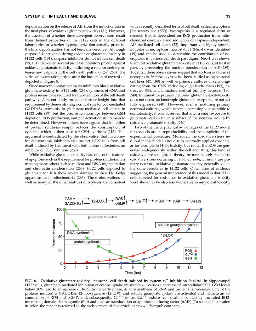
by glutamate, GSH levels drop in a time-dependent manner.When the GSH levels fall below *20% (about 6 h after glu-tamate treatment), ROS start to increase exponentially (263). Itis important to note that the ROS themselves do not kill the
cells but rather give rise to the activation of signaling path-ways which culminate in cell death. Thus, the accumulation oflarge amounts of intracellular ROS is not sufficient to causedeath, but it is a necessary step in the cell death process.Consistent with this idea, compounds that block signalingpathways downstream of ROS accumulation can be protec-tive even in the presence of elevated levels of ROS (e.g., 170,245). The major source of these ROS appears to be complex I ofthe mitochondrial electron transport chain (263). The impor-tance of mitochondrial ROS production is supported bythe observation that the mitochondrial uncoupler cyanidep-trifluoromethoxyphenylhydrazone (FCCP) and other mi-tochondrial inhibitors protect nerve cells from oxidativeglutamate toxicity (263). However, other sources of ROS, in-cluding the NADPH oxidase Nox4 (85) and lysosomes (126),may also contribute to the increase in ROS. GSH depletionalso results in the activation of 12/15 lipoxygenase (12/15-LOX) (144) most probably because of the resulting inhibitionof GSH peroxidase 4 (GPx4) (242), which depends on anadequate supply of GSH for activity. GPx4 is unique in itsability to reduce lipid hydroperoxides embedded in mem-branes (97). Activation of 12/15-LOX generates 12- and 15-hydroxyeicosatetraenoic acid. These eicosanoids activatesoluble guanylate cyclases, which then generate cGMP (145).Elevated cGMP eventually opens an uncharacterized calciumchannel, resulting in a detrimental influx of calcium (145).Activated 12/15-LOX may also have direct effects on mito-chondria, thereby further increasing ROS production (197).About 10–12 h after the induction of oxidative glutamatetoxicity, when both ROS and intracellular calcium levels havereached their maximum, the pro-apoptotic Bcl-2 familymember BH3-interacting domain death agonist (Bid) trans-locates to the mitochondria, and Bid-loaded mitochondriaaccumulate around the nucleus and lose their membrane in-tegrity (131). At this time, apoptosis-inducing factor (AIF)translocates from the mitochondria to the nucleus, where itrapidly induces caspase-independent cell death (131). TheMaher laboratory observed a transient hyperpolarizationof the mitochondria during the exponential increases inROS and intracellular calcium (264), whereas others reported
FIG. 7. Cell density and disulfide exchange of cystine with extracellular GSH regulate sensitivity of HT22 cells tosystem xc
2 inhibition by glutamate. (A) HT22 cells were seeded at the indicated densities per well in 96-well plates, andglutamate was added after 24 h. (B) After 24 h in culture, HT22 cells were treated with the indicated concentrations of GSH inthe absence of glutamate in a normal medium (Ctrl), along with 10 mM glutamate (10 mM Glu) or with a medium exchangedfor a cystine-free medium (-Cystine). (A/B) Survival was measured by the MTT assay after 24 h and normalized to cells nottreated with glutamate (A) or GSH (B). Graphs represent the means of three independent experiments.
12 LEWERENZ ET AL.
depolarization on the release of AIF from the mitochondria inthe final phase of oxidative glutamate toxicity (131). However,the question of whether these divergent observations resultfrom distinct properties of the HT22 cells in the differentlaboratories or whether hyperpolarization actually precedesthe final depolarization has not been answered yet. Althoughcaspase-3 is activated during oxidative glutamate toxicity inHT22 cells (131), caspase inhibitors do not inhibit cell death(59, 131). However, several protease inhibitors protect againstoxidative glutamate toxicity, indicating a role for serine pro-teases and calpains in the cell death pathway (59, 265). Theseries of events taking place after the induction of oxytosis isdepicted in Figure 8.
Since macromolecular synthesis inhibitors block oxidativeglutamate toxicity in HT22 cells (265), synthesis of RNA andprotein seems to be required for the execution of the cell deathpathway. A recent study provided further insight into thatrequirement by demonstrating a critical role for p53-mediatedGADD45a synthesis in glutamate-mediated cell death inHT22 cells (39), but the precise relationships between GSHdepletion, ROS production, and p53 activation still remain tobe determined. However, others have argued that inhibitionof protein synthesis simply reduces the consumption ofcysteine, which is then used for GSH synthesis (215). Thisargument is contradicted by the observation that macromo-lecular synthesis inhibitors also protect HT22 cells from celldeath induced by treatment with buthionine sulfoximine, aninhibitor of GSH synthesis (265).
While oxidative glutamate toxicity has some of the featuresof apoptosis such as the requirement for protein synthesis, it ismissing many others such as nuclear and DNA fragmentationand chromatin condensation (265). HT22 cells exposed toglutamate for 10 h show severe damage to their ER, Golgiapparatus, and mitochondria (265). These observations aswell as many of the other features of oxytosis are consistent
with a recently described form of cell death called necroptosis[for review see (277)]. Necroptosis is a regulated form ofnecrosis that is dependent on ROS production from mito-chondrial complex I and induction of caspase-independent,AIF-mediated cell death (23). Importantly, a highly specificinhibitor of necroptosis, necrostatin 1 (Nec-1), was identified(49) and can be used to determine the contribution of ne-croptosis in various cell death paradigms. Nec-1 was shownto inhibit oxidative glutamate toxicity in HT22 cells, at least inpart, by preventing the nuclear translocation of AIF (289).Together, these observations suggest that oxytosis is a form ofnecroptosis. In vitro, oxytosis has been studied using neuronalcell lines (47, 189) as well as primary cultures of cells origi-nating from the CNS, including oligodendrocytes (193), as-trocytes (33), and immature cortical primary neurons (190,240). In immature primary neurons, glutamate excitotoxicitydoes not occur, as ionotropic glutamate receptors are not yetfully expressed (240). However, even in maturing primaryneuronal cultures, which become increasingly susceptible toexcitotoxicity, it was observed that after a short exposure toglutamate, cell death in a subset of the neurons occurs byoxidative glutamate toxicity (240).
Two of the major practical advantages of the HT22 modelfor oxytosis are its reproducibility and the simplicity of theexperimental procedure. Moreover, the oxidative stress in-duced in this model is not due to externally applied oxidants,as for example in H2O2 toxicity, but rather the ROS are gen-erated endogenously within the cell and, thus, this kind ofoxidative stress might, in theory, be more closely related tooxidative stress occurring in vivo. Of note, in immature pri-mary neurons, oxidative glutamate toxicity generally yieldsthe same results as in HT22 cells. Other lines of evidencesuggesting the general importance of this model is that HT22cells selected for resistance to oxidative glutamate toxicitywere shown to be also less vulnerable to amyloid-b toxicity,
FIG. 8. Oxidative glutamate toxicity—neuronal cell death induced by system xc2 inhibition in vitro. In hippocampal
HT22 cells, glutamate-mediated inhibition of cystine uptake via system xc- causes a decrease of intracellular GSH. GSH levels
below 20% lead to an increase of ROS. In the early phase, de novo synthesis of RNA and proteins is necessary. One of theproteins induced is GADD45a. 12-lipoxygenase (12-LOX) and soluble guanylate cyclase are activated and mediate an ac-cumulation of ROS and cGMP, and, subsequently, Ca + + influx. Ca + + induces cell death mediated by truncated BH3-interacting domain death agonist (Bid) and nuclear translocation of apoptosis-inducing factor (nAIF) (To see this illustrationin color, the reader is referred to the web version of this article at www.liebertpub.com/ars).
SYSTEM xc2 IN HEALTH AND DISEASE 13
an in vitro model for AD (46), ER stress brought about by theglycosylation inhibitor tunicamycin, and over-expression ofthe pro-apoptotic protein Bax (50). Although, at least in part,the sensitivity of cells to system xc
- inhibition is an artifactof the increased dependence of cells in culture on cystineimport for GSH synthesis, together these data highlight thepathophysiological importance of oxytosis and the potentialtherapeutic use of compounds that protect against this par-ticular kind of cell death.
2. Using oxidative glutamate toxicity to identify neuropro-tective pathways. Due to its simplicity and reproducibility,the HT22 model of oxidative glutamate toxicity is an excellenttool to screen for and analyze pathways involved in bothneuroprotection and GSH metabolism. In particular, the roleof different G-protein coupled receptors (GPCRs) in oxidativeglutamate toxicity has been extensively studied. DopamineD4 receptor activation protects by inhibition of ROS produc-tion without affecting GSH depletion (98). Activation of groupI mGluRs protects by increasing GSH levels (224). Activationof stimulatory G proteins attenuated the glutamate-inducedaccumulation of ROS and calcium influx, at least in part, bycausing an increase in GSH due to improved uptake of cystinemediated by the induction of xCT or, additionally, by the up-regulation of the anti-apoptotic protein Bcl-2 (139).
Protein kinase C (PKC) is activated by Gq-coupled recep-tors via phospholipase C and diacylglycerol. Phorbol estersalso activate PKC and in a series of studies (47, 164), the Maherlab demonstrated that the phorbol ester-mediated activationof PKCa and PKCe combined with the phorbol ester-mediateddown-regulation of PKCd, activation of JNK, and inhibition ofp38 MAP kinase activation protected HT22 cells from oxida-tive glutamate toxicity by preventing the glutamate-inducedincrease in ROS without any effect on GSH levels (164). In acomplementary study, Aharoni-Simon et al. demonstrated thatthe phorbol ester TPA protects by increasing glutamate-in-duced AP-1 activity, which is downstream of JNK activation.ROS increased AP-1 activity and AP-1 contraintuitively stim-ulated ROS production, indicating that, at least in this context,ROS possibly serve protective functions (1).
To identify potentially neuroprotective GPCRs, the Meth-ner lab screened for the transcriptional up-regulation of alarge group of well-characterized GPCRs by quantitative real-time PCR in HT22 cells selected for resistance against gluta-mate and identified the receptor VPAC2, which is activatedby the vasoactive intestinal peptide VIP, as up-regulated inglutamate resistance. VPAC2 activation or overexpressionprotected from oxidative glutamate toxicity by increasinganti-apoptotic Bcl-2 (225). In a similar screen comparing theexpression of all known orphan GPCRs, GPCRs with noknown ligands, the constitutively active orphan GPCR GPR39was found most prominently up-regulated in glutamate-resistant cells (50). GPR39 protected against oxidative gluta-mate toxicity through coupling to Ga13. In a broader screen toidentify genes up-regulated in the same glutamate resistantHT22 cells, a new SOD motif-containing peroxisomal proteinthat has neuroprotective properties was identified (268). Weconclude that glutamate-resistant HT22 cells can serve as ascreening tool to identify novel neuroprotective genes.
The Maher lab (165) as well as another laboratory (276)found that inhibition of the proteasome using low doses ofseveral structurally distinct proteasome inhibitors could pre-
vent oxidative glutamate toxicity. While the proteasome in-hibitors had no effect on GSH levels, they did prevent theincrease in ROS (165). Surprisingly, the neuroprotective ef-fects of the proteasome inhibitors appeared to be at leastpartially mediated by the induction of the transcription factorNF-jB, as protection was significantly reduced in HT22 cellsexpressing a specific NF-jB repressor (165). This observationis consistent with the majority of the data on NF-jB whichsuggest that it is important for both normal nerve cell survivaland the survival of nerve cells exposed to oxidative stress(108). Moreover, the neuromodulator hydrogen sulfide (H2S)protects cortical neurons mainly by increasing GSH levels(118) and HT22 cells by both increasing GSH and activatingplasma membrane KATP and CFTR Cl - channels (116). H2Salso increases both cystine and cysteine uptake and enhancesGSH import into mitochondria (117).
3. Using oxidative glutamate toxicity to screen for neuro-protective drugs. We and others have used the model ofoxidative glutamate toxicity to identify a number of com-pounds that might be useful in the treatment of neurologicaldisorders. For example, the Maher lab identified tyrphostins,known inhibitors of protein tyrosine kinases, as compoundsthat block oxidative glutamate toxicity at different steps in thecell death pathway independent of their effects on tyrosinekinase activity (223). Some tyrphostins were inducers of GCL,whereas others acted by stabilizing the mitochondrial mem-brane potential or were direct antioxidants (223).
In a related study, flavonoids, natural plant compounds,were investigated (99). Different flavonoids also acted atdistinct steps in the cell death pathway, including mainte-nance of GSH levels, prevention of ROS accumulation, andinhibition of calcium influx. In addition, since many flavo-noids are inducers of Nrf2 (section I.B) (95), it is likely that atleast some of their neuroprotective effects are mediated viathis transcription factor. Interestingly, very small structuralchanges, such as the shifting of the placement of a single hy-droxyl group, resulted in very different activities for thisgroup of compounds. Together, these results are in agreementwith the emerging idea that the protective effects of flavo-noids result from their ability to modulate multiple signalingpathways rather than acting as direct antioxidants (239, 254).
Among the flavonoids tested, the flavonol fisetin, which isfound in strawberries, proved to be highly effective. Fisetinnot only has antioxidant activity, but is also able to maintainGSH levels. Consistent with this observation, fisetin wasshown to increase Nrf2 levels (163). The value of oxidativeglutamate toxicity for the identification of possible thera-peutics is highlighted by the observation that fisetin proved tobe protective in rabbit and mouse models of stroke (70, 169)and a mouse model of Huntington’s disease (166) and type 1diabetes (167). In another screen, cystine conjugates of cate-chin were tested for their ability to protect against oxidativeglutamate toxicity (267). This led to the identification of anovel mechanism by which one of these compounds, cyste-amine epicatechin, protects against oxidative glutamate tox-icity and increases GSH synthesis, which involves thegeneration of extracellular cysteine via a disulfide exchangemechanism (168).
Moreover, the HT22 cell model of oxidative glutamatetoxicity was used to identify novel neuroprotective deriva-tives of the curry spice turmeric (154). A derivative with a
14 LEWERENZ ET AL.

15-fold decrease in EC50 in this assay, CNB-001, was found tohave enhanced neuroprotective activity in a variety of otherassays, including protection against Ab toxicity (154). Furtherstudies showed that this novel curcumin derivative wasprotective in a rabbit model of stroke (133) and a rat model oftraumatic brain injury (288). Along with other in vitro para-digms of neuronal cell death, trophic factor withdrawal, andamyloid-b toxicity, oxidative glutamate toxicity was used toidentify another novel, curcumin-based, broadly neuropro-tective compound, J147, which prevented the loss of synapticproteins and cognitive decline in a transgenic AD mousemodel (34).
4. Oxidative glutamate toxicity in vivo. As indicatedearlier, the susceptibility of cultured cells to glutamate-mediated system xc
- inhibition might result from the en-hanced reliance of cells in culture on cystine import for GSHsynthesis. However, necroptosis and oxytosis have manyfeatures in common (23), and the necroptosis inhibitor Nec-1can protect HT-22 cells from oxidative glutamate toxicity(289). Nec-1 can also reduce lesion volume in mice aftertransient middle cerebral artery occlusion, a model of ische-mic stroke (49, 290), and in a model of traumatic brain injury(293). Very recently, Nec-1 was shown to exhibit protectiveactivity in cell culture and mouse models of Huntington’sdisease (299). Together, these data suggest that oxytosis, or atleast forms of cell death that share a very similar mechanism,may play an important role in a variety of neurologicaldisorders.
IV. The Role of System xc2 in Health and Disease
A. System xc- in vivo–lessons
from xCT-deficient mice
With the exception of a redox imbalance with the cystine/cysteine redox couple in the plasma shifted to - 89 mVcompared with - 100 mV in wild-type control animals andreduced plasma GSH, xCT - / - mice show no obvious phe-notype (231). Even in the brain, where xCT is constitutivelyexpressed, no accelerated hippocampal atrophy up to the ageof 18 months could be demonstrated by Massie and co-workers (48). These findings indicate that loss of xCT in thesemice is largely compensated for by unknown mechanisms.However, sut/sut mice, which harbor a spontaneous481,280 bp deletion that includes parts of the xCT gene, showchanges in fur color due to a deficiency in the cysteine-de-pendent yellow/red pigment, pheomelanin (37), and promi-nent brain atrophy, including in the hippocampus (250).Whether these divergent results can be explained by the dif-ferent genetic backgrounds, C57BL/6 for xCT - / - and C3H/HeSnJ for sut/sut mice, or are due to the different molecularbases for xCT deficiency remains to be determined.
B. The role of system xc- in the immune system
and inflammation
The immune system comprises cells of myeloid origin(monocytes, macrophages, dendritic cells, microglia, andgranulocytes) and lymphocytes. From a functional point ofview, two different systems can be distinguished. The innateimmune system responds to a limited set of conserved dangersignals such as double-stranded RNA or LPS that indicate the
presence of infectious agents or other harmful conditions. Theadaptive immune system generates a more specific response.Here, antigen-presenting cells (macrophages, dendritic cells,or stromal cells of the thymus) present specific antigens to thedifferent types of lymphocytes that specifically respond to thistype of antigen. These lymphocytes then proliferate, therebygenerating an antigen-specific immune response against avirtually unlimited number of target structures. Both systemsshow extensive interactions at all levels and are interdepen-dent.
Several lines of evidence suggest that system xc- might
play a role in the regulation of the innate immune response.First, activation of macrophages and granulocytes is associ-ated with excessive production of pathogen-toxic ROS, theso-called respiratory burst, leading to oxidative stress. Mac-rophages prominently up-regulate system xc
- expression onactivation with bacterial LPS or the pro-inflammatory cyto-kine TNFa (228). Similarly, in bleomycin-induced pneumo-nitis, infiltrating leukocytes prominently express xCT (192).Thus, induction of system xc
- might be an auto-protectiveresponse during activation of these cells within the innateimmune response. In line with this assumption, macrophagesfrom xCT-deficient mice die on activation with LPS (192).
System xc- may also modulate the adaptive immune re-
sponse. xCT is robustly expressed in vivo in the spleen, theorgan of B lymphocyte maturation, and the thymus, wherethe selection and maturation of T lymphocytes takes place. Onsystemic treatment with LPS, the expression of xCT is heavilyup-regulated in the cortex of the thymus and the white pulp ofthe spleen (260). However, lymphocytes are the only cell typewhere it has been consistently reported that system xc
- ex-pression cannot be induced (73, 100, 102). Nevertheless,spleen lymphocytes increase intracellular GSH levels on ac-tivation with LPS by up-regulation of cysteine uptake viasystem ASC (102). Cellular GSH levels have been reported tomodulate intracellular signaling after stimulation of T-cellreceptors or treatment with TNFa (259). Moreover, the avail-ability of cysteine for GSH synthesis during maturation ofstimulated lymphocytes in vitro has been shown to stronglyinfluence the generation of cytotoxic T-cell activity (72). Incontrast to lymphocytes, macrophages prominently up-regulate system xc
- activity and, as a consequence, releasecysteine into the extracellular space (72), probably through asystem L-like transporter (235). Therefore, it has been postu-lated that, in addition to the exposure to the specific antigenand cytokines, lymphocyte activation is modulated by cyste-ine released from antigen-presenting cells (Fig. 9).
Evidence from inflammatory bowel disease supports thisconcept (252). In the normal intestinal mucosa, the T-cell re-sponse is suppressed by low T-cell GSH. However, this doesnot result from altered properties of the mucosal T cellsthemselves but most probably is a consequence of very lowsystem xc
- activity and subsequently low cysteine releasefrom the resident macrophages. In contrast, in the inflamedgut, where the natural inhibition of the T cell response isknown to be abolished, the presence of cysteine-releasingmacrophages with high system xc
- activity was demon-strated. These results suggest that xCT expression in antigen-presenting cells might be a critical regulator of autoimmunity.
Further data indicate that xCT might also play a role in thetermination of an inflammatory response. In a model of focalchronic inflammation induced in mice by a subcutaneous
SYSTEM xc2 IN HEALTH AND DISEASE 15
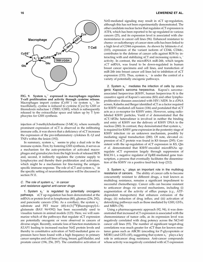
injection of 3-methylcholanthrene (3-MCA), where normallyprominent expression of xCT is observed in the infiltratingimmune cells, it was shown that a deficiency of xCT increasesthe expression of the pro-inflammatory cytokines IL-1b andTNFa within the lesion (192).
In summary, system xc- seems to play a dual role in the
immune system. First, by fostering GSH synthesis, it serves asa mechanism for the auto-protection of activated macro-phages and granulocytes from the high levels of released ROSand, second, it indirectly regulates the cysteine supply tolymphocytes and thereby their proliferation and activation,which might be a mechanism for fine-tuning the antigen-specific immune response. The role of xCT and system xc
- inthe specific setting of neuroinflammation will be discussed insection IV.E.
C. The role of system xc- in cancer
and resistance against anti-cancer drugs
1. System xc- is regulated by potentially oncogenic
pathways. xCT up-regulation has been demonstrated at themRNA or protein level in lymphomas (80), gliomas (236, 292),and pancreatic cancers (156). As a corollary, the system xc
-
substrate and PET tracer (4S)-4-(3-[18F]fluoropropyl)-L-glutamate (BAY 94-9392) has been successfully used tovisualize tumors in animal models (123). Here, we will sum-marize which of the pathways that regulate xCT expressionare potentially oncogenic or were observed in cancers ofdiverse origin. Recently, somatic loss of function mutations inKEAP1 leading to increased nuclear Nrf2 protein levels andthereby to constitutive activation of Nrf2-mediated gene ex-pression have been found with a high frequency in primarycancer samples and cell lines of lung, breast, gall bladder, andprostate cancer (196, 256, 297). The constitutive activation of
Nrf2-mediated signaling may result in xCT up-regulation,although this has not been experimentally demonstrated. Theother candidate nuclear factor that regulates xCT expression isATF4, which has been reported to be up-regulated in variouscancers (25), and its expression level is associated with che-moresistance in cancer cell lines (96). Enhanced resistance tochemo- or radiotherapy of cancer stem cells has been linked toa high level of CD44 expression. As shown by Ishimoto et al.(103), expression of the variant isoform of CD44, CD44v,contributes to the defense of cancer cells against ROS by in-teracting with and stabilizing xCT and increasing system xc
-
activity. In contrast, the microRNA miR-26b, which targetsxCT mRNA, was found to be down-regulated in humanbreast cancer specimens and cell lines, and transfection ofmiR-26b into breast cancer cell lines led to inhibition of xCTexpression (153). Thus, system xc
- is under the control of avariety of potentially oncogenic pathways.
2. System xc- mediates the infection of cells by onco-
genic Kaposi’s sarcoma herpesvirus. Kaposi’s sarcoma–associated herpesvirus (KSHV, human herpesvirus 8) is thecausative agent of Kaposi’s sarcoma (KS) and other lympho-proliferative diseases associated with HIV/AIDS. In a cDNAscreen, Kaleeba and Berger identified xCT as a factor requiredfor KSHV-mediated cell fusion (106) and postulated that xCTacts as a co-receptor for KSHV entry. However, using radio-labeled KSHV particles, Veetil et al demonstrated that thexCT/4F2hc heterodimer is involved in neither the bindingand entry of KSHV nor the delivery of KSHV DNA to thenucleus (280). In contrast, they demonstrated that system xc
-
is required for KHSV gene expression in the postentry stage ofKSHV infection via an unknown mechanism, possibly bymediating signal transduction (280). Of note, elevated ex-pression of xCT protein was found in KS tissues (295). Con-sistent with the up-regulation of xCT expression in KS, Qinet al. demonstrated that KSHV-encoded microRNAs up-regulate xCT expression largely through suppression ofBACH-1, a negative regulator of EpRE-mediated gene tran-scription, a process that eventually facilitates the dissemina-tion of the KSHV via a positive feed-back loop (212).
3. System xc- plays an important role in the multidrug
resistance of cancers. The ability of cancer cells to becomeconcurrently resistant to different drugs, a trait known asmultidrug resistance, remains a significant impediment tosuccessful chemotherapy. Cancer cells can become resistantto anticancer drugs via several mechanisms, including (i)augmentation of the activity of efflux pumps (e.g., ATP-dependent transporters) that enhances extrusion of thedrugs; (ii) reduction of drug influx; and (iii) activation ofdetoxifying pathways such as those mediated by GSH, GSTs,and MRPs (78).
Using a pharmacogenomic approach (92, 94), it was dem-onstrated that increased xCT expression is associated with thechemoresistance of tumor cells, as its expression level wasnegatively correlated with drug potency across the NCI-60cancer cell lines (93). The number of significant target-drugcorrelations was much greater for xCT than for known resis-tance genes such as ABCB1 (encoding for P-glycoprotein orMDR1) and GSTs (45), suggesting that system xc
- plays a keyrole in anticancer drug resistance. Anti-cancer compoundswhose activity was negatively correlated with xCT expression
FIG. 9. System xc2 expressed in macrophages regulates
T-cell proliferation and activity through cysteine release.Macrophages import cystine (CySS - ) via system xc
- . In-tracellularly, cystine is reduced to cysteine (Cys) by GSH orthioredoxin reductase 1 (TRR1/GSH), which is subsequentlyreleased to the extracellular space and taken up by T lym-phocytes for GSH synthesis.
16 LEWERENZ ET AL.
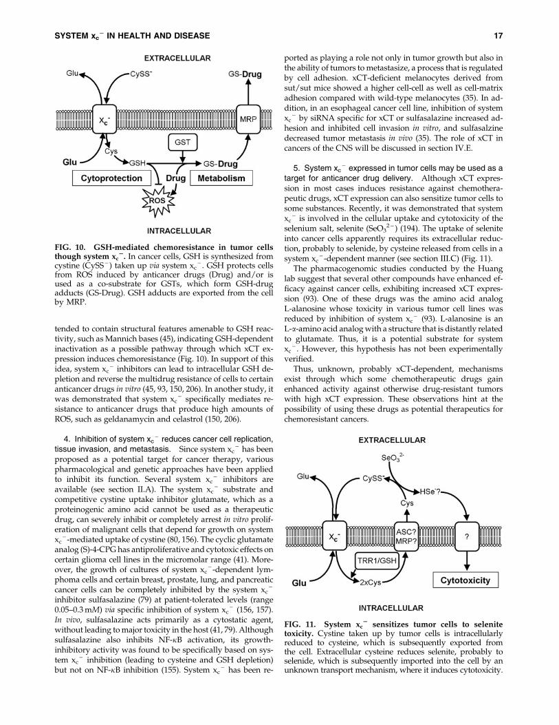
tended to contain structural features amenable to GSH reac-tivity, such as Mannich bases (45), indicating GSH-dependentinactivation as a possible pathway through which xCT ex-pression induces chemoresistance (Fig. 10). In support of thisidea, system xc
- inhibitors can lead to intracellular GSH de-pletion and reverse the multidrug resistance of cells to certainanticancer drugs in vitro (45, 93, 150, 206). In another study, itwas demonstrated that system xc
- specifically mediates re-sistance to anticancer drugs that produce high amounts ofROS, such as geldanamycin and celastrol (150, 206).
4. Inhibition of system xc- reduces cancer cell replication,
tissue invasion, and metastasis. Since system xc- has been
proposed as a potential target for cancer therapy, variouspharmacological and genetic approaches have been appliedto inhibit its function. Several system xc
- inhibitors areavailable (see section II.A). The system xc
- substrate andcompetitive cystine uptake inhibitor glutamate, which as aproteinogenic amino acid cannot be used as a therapeuticdrug, can severely inhibit or completely arrest in vitro prolif-eration of malignant cells that depend for growth on systemxc
- -mediated uptake of cystine (80, 156). The cyclic glutamateanalog (S)-4-CPG has antiproliferative and cytotoxic effects oncertain glioma cell lines in the micromolar range (41). More-over, the growth of cultures of system xc
- -dependent lym-phoma cells and certain breast, prostate, lung, and pancreaticcancer cells can be completely inhibited by the system xc
-
inhibitor sulfasalazine (79) at patient-tolerated levels (range0.05–0.3 mM) via specific inhibition of system xc
- (156, 157).In vivo, sulfasalazine acts primarily as a cytostatic agent,without leading to major toxicity in the host (41, 79). Althoughsulfasalazine also inhibits NF-jB activation, its growth-inhibitory activity was found to be specifically based on sys-tem xc
- inhibition (leading to cysteine and GSH depletion)but not on NF-jB inhibition (155). System xc
- has been re-
ported as playing a role not only in tumor growth but also inthe ability of tumors to metastasize, a process that is regulatedby cell adhesion. xCT-deficient melanocytes derived fromsut/sut mice showed a higher cell-cell as well as cell-matrixadhesion compared with wild-type melanocytes (35). In ad-dition, in an esophageal cancer cell line, inhibition of systemxc
- by siRNA specific for xCT or sulfasalazine increased ad-hesion and inhibited cell invasion in vitro, and sulfasalzinedecreased tumor metastasis in vivo (35). The role of xCT incancers of the CNS will be discussed in section IV.E.
5. System xc- expressed in tumor cells may be used as a
target for anticancer drug delivery. Although xCT expres-sion in most cases induces resistance against chemothera-peutic drugs, xCT expression can also sensitize tumor cells tosome substances. Recently, it was demonstrated that systemxc
- is involved in the cellular uptake and cytotoxicity of theselenium salt, selenite (SeO3
2 - ) (194). The uptake of seleniteinto cancer cells apparently requires its extracellular reduc-tion, probably to selenide, by cysteine released from cells in asystem xc
- -dependent manner (see section III.C) (Fig. 11).The pharmacogenomic studies conducted by the Huang
lab suggest that several other compounds have enhanced ef-ficacy against cancer cells, exhibiting increased xCT expres-sion (93). One of these drugs was the amino acid analogL-alanosine whose toxicity in various tumor cell lines wasreduced by inhibition of system xc
- (93). L-alanosine is anL-a-amino acid analog with a structure that is distantly relatedto glutamate. Thus, it is a potential substrate for systemxc
- . However, this hypothesis has not been experimentallyverified.
Thus, unknown, probably xCT-dependent, mechanismsexist through which some chemotherapeutic drugs gainenhanced activity against otherwise drug-resistant tumorswith high xCT expression. These observations hint at thepossibility of using these drugs as potential therapeutics forchemoresistant cancers.
FIG. 11. System xc2 sensitizes tumor cells to selenite
toxicity. Cystine taken up by tumor cells is intracellularlyreduced to cysteine, which is subsequently exported fromthe cell. Extracellular cysteine reduces selenite, probably toselenide, which is subsequently imported into the cell by anunknown transport mechanism, where it induces cytotoxicity.
FIG. 10. GSH-mediated chemoresistance in tumor cellsthough system xc
2. In cancer cells, GSH is synthesized fromcystine (CySS - ) taken up via system xc
- . GSH protects cellsfrom ROS induced by anticancer drugs (Drug) and/or isused as a co-substrate for GSTs, which form GSH-drugadducts (GS-Drug). GSH adducts are exported from the cellby MRP.
SYSTEM xc2 IN HEALTH AND DISEASE 17
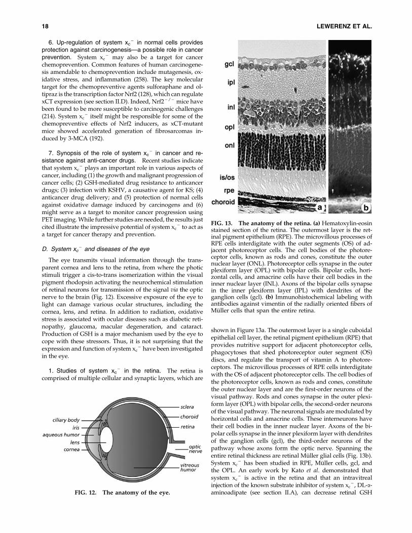
6. Up-regulation of system xc- in normal cells provides
protection against carcinogenesis—a possible role in cancerprevention. System xc
- may also be a target for cancerchemoprevention. Common features of human carcinogene-sis amendable to chemoprevention include mutagenesis, ox-idative stress, and inflammation (258). The key moleculartarget for the chemopreventive agents sulforaphane and ol-tipraz is the transcription factor Nrf2 (128), which can regulatexCT expression (see section II.D). Indeed, Nrf2 - / - mice havebeen found to be more susceptible to carcinogenic challenges(214). System xc
- itself might be responsible for some of thechemopreventive effects of Nrf2 inducers, as xCT-mutantmice showed accelerated generation of fibrosarcomas in-duced by 3-MCA (192).
7. Synopsis of the role of system xc- in cancer and re-
sistance against anti-cancer drugs. Recent studies indicatethat system xc
- plays an important role in various aspects ofcancer, including (1) the growth and malignant progression ofcancer cells; (2) GSH-mediated drug resistance to anticancerdrugs; (3) infection with KSHV, a causative agent for KS; (4)anticancer drug delivery; and (5) protection of normal cellsagainst oxidative damage induced by carcinogens and (6)might serve as a target to monitor cancer progression usingPET imaging. While further studies are needed, the results justcited illustrate the impressive potential of system xc
- to act asa target for cancer therapy and prevention.
D. System xc- and diseases of the eye
The eye transmits visual information through the trans-parent cornea and lens to the retina, from where the photicstimuli trigger a cis-to-trans isomerization within the visualpigment rhodopsin activating the neurochemical stimulationof retinal neurons for transmission of the signal via the opticnerve to the brain (Fig. 12). Excessive exposure of the eye tolight can damage various ocular structures, including thecornea, lens, and retina. In addition to radiation, oxidativestress is associated with ocular diseases such as diabetic reti-nopathy, glaucoma, macular degeneration, and cataract.Production of GSH is a major mechanism used by the eye tocope with these stressors. Thus, it is not surprising that theexpression and function of system xc
- have been investigatedin the eye.
1. Studies of system xc- in the retina. The retina is
comprised of multiple cellular and synaptic layers, which are
shown in Figure 13a. The outermost layer is a single cuboidalepithelial cell layer, the retinal pigment epithelium (RPE) thatprovides nutritive support for adjacent photoreceptor cells,phagocytoses that shed photoreceptor outer segment (OS)discs, and regulate the transport of vitamin A to photore-ceptors. The microvillous processes of RPE cells interdigitatewith the OS of adjacent photoreceptor cells. The cell bodies ofthe photoreceptor cells, known as rods and cones, constitutethe outer nuclear layer and are the first-order neurons of thevisual pathway. Rods and cones synapse in the outer plexi-form layer (OPL) with bipolar cells, the second-order neuronsof the visual pathway. The neuronal signals are modulated byhorizontal cells and amacrine cells. These interneurons havetheir cell bodies in the inner nuclear layer. Axons of the bi-polar cells synapse in the inner plexiform layer with dendritesof the ganglion cells (gcl), the third-order neurons of thepathway whose axons form the optic nerve. Spanning theentire retinal thickness are retinal Muller glial cells (Fig. 13b).System xc
- has been studied in RPE, Muller cells, gcl, andthe OPL. An early work by Kato et al. demonstrated thatsystem xc
- is active in the retina and that an intravitrealinjection of the known substrate inhibitor of system xc
- , DL-a-aminoadipate (see section II.A), can decrease retinal GSH
FIG. 13. The anatomy of the retina. (a) Hematoxylin-eosinstained section of the retina. The outermost layer is the ret-inal pigment epithelium (RPE). The microvillous processes ofRPE cells interdigitate with the outer segments (OS) of ad-jacent photoreceptor cells. The cell bodies of the photore-ceptor cells, known as rods and cones, constitute the outernuclear layer (ONL). Photoreceptor cells synapse in the outerplexiform layer (OPL) with bipolar cells. Bipolar cells, hori-zontal cells, and amacrine cells have their cell bodies in theinner nuclear layer (INL). Axons of the bipolar cells synapsein the inner plexiform layer (IPL) with dendrites of theganglion cells (gcl). (b) Immunohistochemical labeling withantibodies against vimentin of the radially oriented fibers ofMuller cells that span the entire retina.
FIG. 12. The anatomy of the eye.
18 LEWERENZ ET AL.

concentrations and induce retinal dysfunction in vivo (111), in-dicating that system xc
- is functionally important in the retina.Oxidative stress to the RPE is implicated in the devastating
disease age-related macular degeneration (AMD), and GSHprotects these cells from oxidant-induced apoptosis [reviewedin (31)]. The Ganapathy lab demonstrated system xc
- activityin the ARPE-19 human RPE cell line, from which human xCTwas subsequently cloned (28). Infection with HIV-1 in hu-mans is associated with decreased levels of GSH, which ismediated by Tat (56). The Ganapathy lab demonstrated thatHIV-Tat decreased GSH levels in ARPE-19 cells (27) and, mostpossibly as a consequence, induced system xc
- activity andxCT and 4F2hc expression at the mRNA and protein levelsthrough an unknown mechanism (27).
Excess levels of iron have been reported in patients withAMD (86). Investigators in the McGahan lab showed thatintracellular iron regulates the activity of system xc
- inRPE cells via cytosolic aconitase and possibly synthesis ofglutamate (129). The fact that iron plays a critical role in theregulation of xCT in RPE was supported by studies in HFEknock-out mice (HFE - / - ), a model for the human ironoverload disorder haemochromatosis. In the retina, HFE ispredominantly expressed in RPE (174). HFE - / - mice un-dergo RPE hypertrophy and hyperplasia (74). An analysis ofcultured HFE - / - primary RPE cells revealed increased ex-pression of xCT protein and mRNA and system xc
- activity(74). This finding is important given the documented criticalrole that xCT plays in cell proliferation (157). Thus, in RPE, thecell that nourishes and maintains photoreceptor cells, anumber of factors that increase oxidative stress (NO, ironoverload, and HIV-Tat), leads to up-regulation of system xc
- .A second retinal cell type in which system xc
- has beeninvestigated is the Muller glial cell. The GSH concentration inthe retina is relatively high (1.2 lmol/g wet weight in freshlyisolated rat retina) and, as demonstrated by immunohisto-chemistry with antibodies against GSH, much of the GSH ispresent in Muller cells (210). Indeed, Muller cells protect ad-jacent retinal neurons by providing GSH to neurons underischemic conditions. In early studies looking at uptake ofradiolabeled cystine in freshly dissected retinae, Kato et al.reported a pattern of cystine accumulation that is consistentwith high activity of system xc
- in Muller cells (111). Powobtained similar results after loading retinas with the systemxc
- substrate L-a-aminoadipate and visualizing intracellularL-a-aminoadipate accumulation by immunocytochemistrywith a specific antiserum (209). Taken together, these studiesindicate that Muller glia show system xc
- activity in situ.In vitro studies from the Hosoya lab showed that the con-
ditionally immortalized rat Muller cell line (TR-MUL) exhibitssystem xc
- activity and expresses xCT and 4F2hc. System xc- -
mediated L-cystine uptake and xCT expression could be in-duced by the GSH-depleting agent DEM (266). The laboratoryof Sylvia Smith obtained similar results in primary culturedMuller cells subjected to H2O2-mediated oxidative stressbrought about by xanthine/xanthine oxidase, which also in-creased xCT mRNA expression and system xc
- activity (191).Moreover, as mentioned earlier (see sectionII.E), on oxidativestress, a shift of a *40 kD isoform of xCT from the intracel-lular compartment to the plasma membrane was observed,whereas for the predominant form of xCT, with a molecularweight of *50 kD, no detectable change could be demon-strated (191).
In another disease of the retina, diabetic retinopathy, in-creased glutamate levels were found in the vitreous humor,indicating disturbed glutamate metabolism (159). However, 8days of hyperglycemia neither altered the uptake of gluta-mate by EAAT1 or system xc
- , nor altered the expression ofxCT mRNA or protein in primary cultured Muller cells (191).The regulation of system xc
- activity in the Muller glia byoxidative stress may be relevant, because, in theory, gluta-mate release by system xc
- might induce excitotoxic damageto retinal neurons.
Although earlier studies highlighted the prominent systemxc
- activity in Muller glia (111, 209), initial immunohisto-chemical studies using an antibody against xCT suggestedthat it was actually present in several neuronal layers in theretina (27). Studies from two laboratories working indepen-dently examined this comprehensively and concluded thatsystem xc
- was indeed present in retinal neurons (57, 91). Dunand co-workers showed that primary gcl from neonatal andadult mouse retina were positive for xCT and 4F2hc expres-sion and detected both xCT and 4F2hc as well as oxidative andnitrosative stress-inducible system xc
- activity in the retinalneuronal cell line RGC-5 (57). Electron microscopy im-munolocalization studies showed that xCT was present in theribbon synapses of rod and cone photoreceptor cells and thedendrites of rod bipolar cells in diverse species (91). It wasdemonstrated that developmentally the onset of xCT ex-pression in rat retina occurred at postnatal day 10, the timewhen photoreceptor cells make functional contact with bi-polar cells. Taken together, molecular, biochemical, andfunctional analyses indicate that system xc
- is present inseveral retinal cell types (RPE, Muller, ganglion, and photo-receptor/bipolar) and may play a key role in providing therate-limiting precursor, cyst(e)ine for the synthesis of GSH.
2. Studies of system xc- in lens and cornea. The lens is a
transparent convex disk positioned between the iris and thevitreous body; it is suspended within the globe by zonularfibers that extend from the ciliary body (Fig. 12). The lens iscomprised of (1) a capsule, which is a thickened basementmembrane that envelops the lens; (2) a simple, cuboidal epi-thelium, which lines the anterior lens surface; and (3) lensfibers, comprising the inner and outer cortex of the lens, whichare elongated cells that eventually lose their nuclei and arespecialized to allow passage of light. An opacity of the lens istermed a cataract. Excessive oxidative stress, which can leadto protein modification, is implicated in the formation ofcataracts [for review see (24)]. While young healthy eyesbenefit from an active ROS scavenging system largely com-prised of GSH, aging eyes have diminished capacity to copewith oxidative stress. Members of the Donaldson laboratoryfound that xCT is present at the mRNA and protein levels inlens fiber cells (143, 146, 148). Immunocytochemical studies ofthe lens showed a cytoplasmic distribution of xCT-like im-munoreactivity in outer cortical fibers and a membranousdistribution in the inner fiber cells (146). This team combinedimmunocytochemistry for cystine using antisera against glu-taraldehyde-cystine conjugates to map the distribution of freecystine and to determine its relationship to the expression ofxCT (143). In our opinion, the cystine immunoreactivityrepresents the distribution of both cysteine and cystine in vivoas cysteine should be oxidized to cystine during the process ofpreparation and fixation. They reported a bimodal distribution
SYSTEM xc2 IN HEALTH AND DISEASE 19
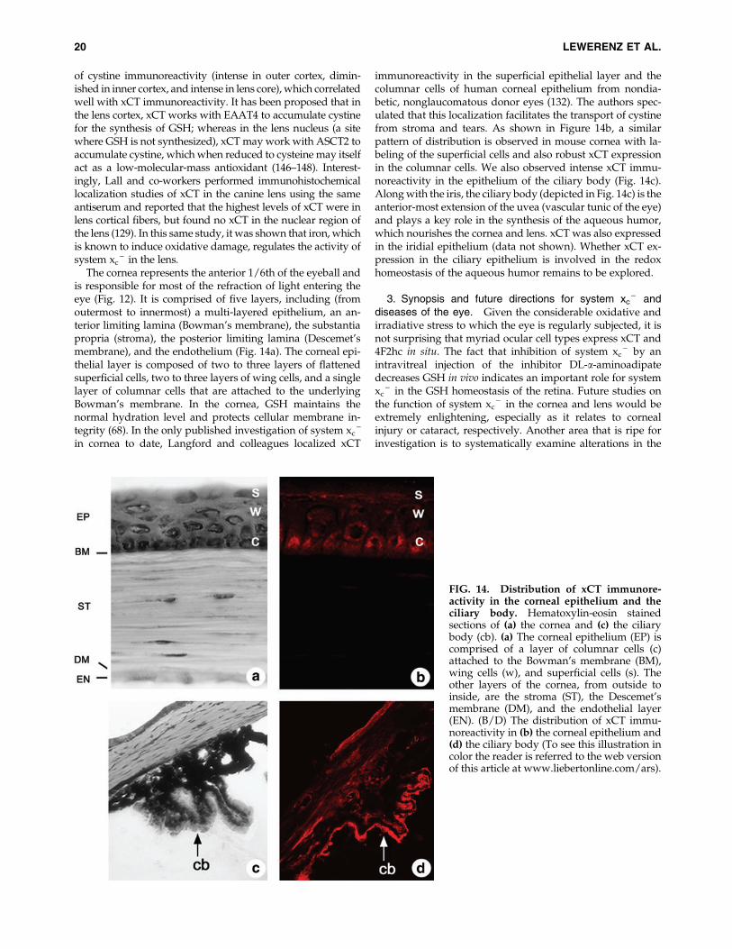
of cystine immunoreactivity (intense in outer cortex, dimin-ished in inner cortex, and intense in lens core), which correlatedwell with xCT immunoreactivity. It has been proposed that inthe lens cortex, xCT works with EAAT4 to accumulate cystinefor the synthesis of GSH; whereas in the lens nucleus (a sitewhere GSH is not synthesized), xCT may work with ASCT2 toaccumulate cystine, which when reduced to cysteine may itselfact as a low-molecular-mass antioxidant (146–148). Interest-ingly, Lall and co-workers performed immunohistochemicallocalization studies of xCT in the canine lens using the sameantiserum and reported that the highest levels of xCT were inlens cortical fibers, but found no xCT in the nuclear region ofthe lens (129). In this same study, it was shown that iron, whichis known to induce oxidative damage, regulates the activity ofsystem xc
- in the lens.The cornea represents the anterior 1/6th of the eyeball and
is responsible for most of the refraction of light entering theeye (Fig. 12). It is comprised of five layers, including (fromoutermost to innermost) a multi-layered epithelium, an an-terior limiting lamina (Bowman’s membrane), the substantiapropria (stroma), the posterior limiting lamina (Descemet’smembrane), and the endothelium (Fig. 14a). The corneal epi-thelial layer is composed of two to three layers of flattenedsuperficial cells, two to three layers of wing cells, and a singlelayer of columnar cells that are attached to the underlyingBowman’s membrane. In the cornea, GSH maintains thenormal hydration level and protects cellular membrane in-tegrity (68). In the only published investigation of system xc
-
in cornea to date, Langford and colleagues localized xCT
immunoreactivity in the superficial epithelial layer and thecolumnar cells of human corneal epithelium from nondia-betic, nonglaucomatous donor eyes (132). The authors spec-ulated that this localization facilitates the transport of cystinefrom stroma and tears. As shown in Figure 14b, a similarpattern of distribution is observed in mouse cornea with la-beling of the superficial cells and also robust xCT expressionin the columnar cells. We also observed intense xCT immu-noreactivity in the epithelium of the ciliary body (Fig. 14c).Along with the iris, the ciliary body (depicted in Fig. 14c) is theanterior-most extension of the uvea (vascular tunic of the eye)and plays a key role in the synthesis of the aqueous humor,which nourishes the cornea and lens. xCT was also expressedin the iridial epithelium (data not shown). Whether xCT ex-pression in the ciliary epithelium is involved in the redoxhomeostasis of the aqueous humor remains to be explored.
3. Synopsis and future directions for system xc- and
diseases of the eye. Given the considerable oxidative andirradiative stress to which the eye is regularly subjected, it isnot surprising that myriad ocular cell types express xCT and4F2hc in situ. The fact that inhibition of system xc
- by anintravitreal injection of the inhibitor DL-a-aminoadipatedecreases GSH in vivo indicates an important role for systemxc
- in the GSH homeostasis of the retina. Future studies onthe function of system xc
- in the cornea and lens would beextremely enlightening, especially as it relates to cornealinjury or cataract, respectively. Another area that is ripe forinvestigation is to systematically examine alterations in the
FIG. 14. Distribution of xCT immunore-activity in the corneal epithelium and theciliary body. Hematoxylin-eosin stainedsections of (a) the cornea and (c) the ciliarybody (cb). (a) The corneal epithelium (EP) iscomprised of a layer of columnar cells (c)attached to the Bowman’s membrane (BM),wing cells (w), and superficial cells (s). Theother layers of the cornea, from outside toinside, are the stroma (ST), the Descemet’smembrane (DM), and the endothelial layer(EN). (B/D) The distribution of xCT immu-noreactivity in (b) the corneal epithelium and(d) the ciliary body (To see this illustration incolor the reader is referred to the web versionof this article at www.liebertonline.com/ars).
20 LEWERENZ ET AL.
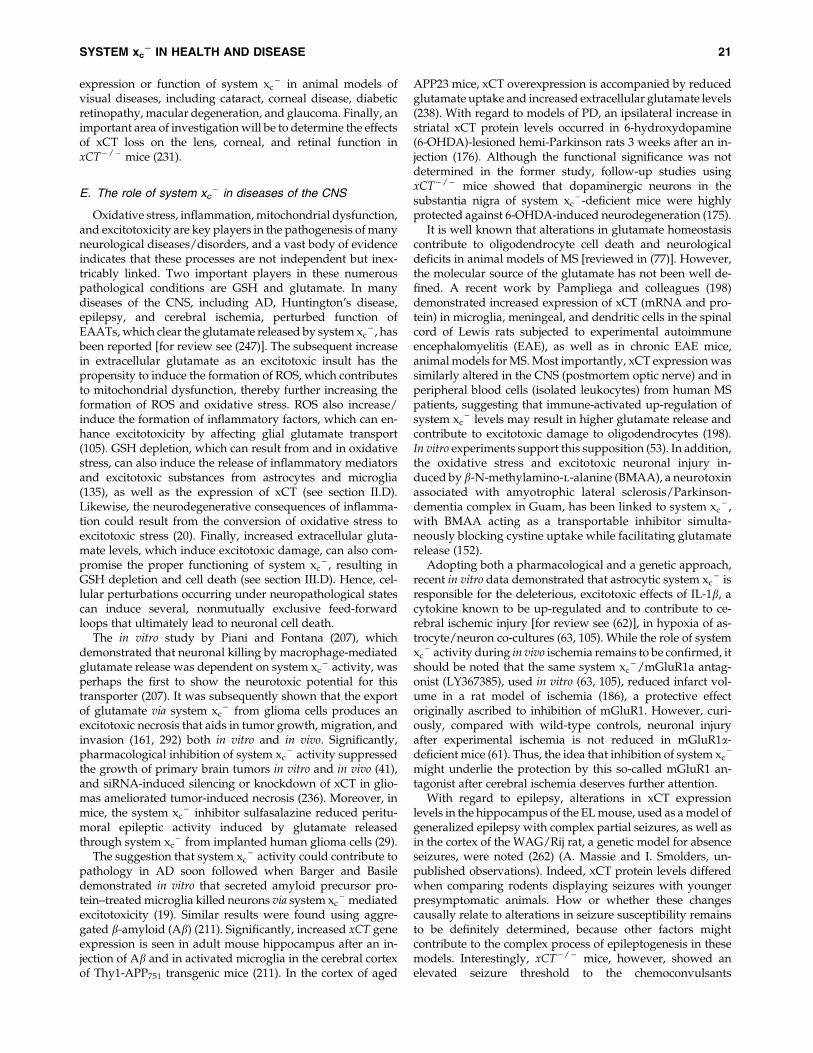
expression or function of system xc- in animal models of
visual diseases, including cataract, corneal disease, diabeticretinopathy, macular degeneration, and glaucoma. Finally, animportant area of investigation will be to determine the effectsof xCT loss on the lens, corneal, and retinal function inxCT - / - mice (231).
E. The role of system xc- in diseases of the CNS
Oxidative stress, inflammation, mitochondrial dysfunction,and excitotoxicity are key players in the pathogenesis of manyneurological diseases/disorders, and a vast body of evidenceindicates that these processes are not independent but inex-tricably linked. Two important players in these numerouspathological conditions are GSH and glutamate. In manydiseases of the CNS, including AD, Huntington’s disease,epilepsy, and cerebral ischemia, perturbed function ofEAATs, which clear the glutamate released by system xc
- , hasbeen reported [for review see (247)]. The subsequent increasein extracellular glutamate as an excitotoxic insult has thepropensity to induce the formation of ROS, which contributesto mitochondrial dysfunction, thereby further increasing theformation of ROS and oxidative stress. ROS also increase/induce the formation of inflammatory factors, which can en-hance excitotoxicity by affecting glial glutamate transport(105). GSH depletion, which can result from and in oxidativestress, can also induce the release of inflammatory mediatorsand excitotoxic substances from astrocytes and microglia(135), as well as the expression of xCT (see section II.D).Likewise, the neurodegenerative consequences of inflamma-tion could result from the conversion of oxidative stress toexcitotoxic stress (20). Finally, increased extracellular gluta-mate levels, which induce excitotoxic damage, can also com-promise the proper functioning of system xc
- , resulting inGSH depletion and cell death (see section III.D). Hence, cel-lular perturbations occurring under neuropathological statescan induce several, nonmutually exclusive feed-forwardloops that ultimately lead to neuronal cell death.
The in vitro study by Piani and Fontana (207), whichdemonstrated that neuronal killing by macrophage-mediatedglutamate release was dependent on system xc
- activity, wasperhaps the first to show the neurotoxic potential for thistransporter (207). It was subsequently shown that the exportof glutamate via system xc
- from glioma cells produces anexcitotoxic necrosis that aids in tumor growth, migration, andinvasion (161, 292) both in vitro and in vivo. Significantly,pharmacological inhibition of system xc
- activity suppressedthe growth of primary brain tumors in vitro and in vivo (41),and siRNA-induced silencing or knockdown of xCT in glio-mas ameliorated tumor-induced necrosis (236). Moreover, inmice, the system xc
- inhibitor sulfasalazine reduced peritu-moral epileptic activity induced by glutamate releasedthrough system xc
- from implanted human glioma cells (29).The suggestion that system xc
- activity could contribute topathology in AD soon followed when Barger and Basiledemonstrated in vitro that secreted amyloid precursor pro-tein–treated microglia killed neurons via system xc
- mediatedexcitotoxicity (19). Similar results were found using aggre-gated b-amyloid (Ab) (211). Significantly, increased xCT geneexpression is seen in adult mouse hippocampus after an in-jection of Ab and in activated microglia in the cerebral cortexof Thy1-APP751 transgenic mice (211). In the cortex of aged
APP23 mice, xCT overexpression is accompanied by reducedglutamate uptake and increased extracellular glutamate levels(238). With regard to models of PD, an ipsilateral increase instriatal xCT protein levels occurred in 6-hydroxydopamine(6-OHDA)-lesioned hemi-Parkinson rats 3 weeks after an in-jection (176). Although the functional significance was notdetermined in the former study, follow-up studies usingxCT - / - mice showed that dopaminergic neurons in thesubstantia nigra of system xc
- -deficient mice were highlyprotected against 6-OHDA-induced neurodegeneration (175).
It is well known that alterations in glutamate homeostasiscontribute to oligodendrocyte cell death and neurologicaldeficits in animal models of MS [reviewed in (77)]. However,the molecular source of the glutamate has not been well de-fined. A recent work by Pampliega and colleagues (198)demonstrated increased expression of xCT (mRNA and pro-tein) in microglia, meningeal, and dendritic cells in the spinalcord of Lewis rats subjected to experimental autoimmuneencephalomyelitis (EAE), as well as in chronic EAE mice,animal models for MS. Most importantly, xCT expression wassimilarly altered in the CNS (postmortem optic nerve) and inperipheral blood cells (isolated leukocytes) from human MSpatients, suggesting that immune-activated up-regulation ofsystem xc
- levels may result in higher glutamate release andcontribute to excitotoxic damage to oligodendrocytes (198).In vitro experiments support this supposition (53). In addition,the oxidative stress and excitotoxic neuronal injury in-duced by b-N-methylamino-l-alanine (BMAA), a neurotoxinassociated with amyotrophic lateral sclerosis/Parkinson-dementia complex in Guam, has been linked to system xc
- ,with BMAA acting as a transportable inhibitor simulta-neously blocking cystine uptake while facilitating glutamaterelease (152).
Adopting both a pharmacological and a genetic approach,recent in vitro data demonstrated that astrocytic system xc
- isresponsible for the deleterious, excitotoxic effects of IL-1b, acytokine known to be up-regulated and to contribute to ce-rebral ischemic injury [for review see (62)], in hypoxia of as-trocyte/neuron co-cultures (63, 105). While the role of systemxc
- activity during in vivo ischemia remains to be confirmed, itshould be noted that the same system xc
- /mGluR1a antag-onist (LY367385), used in vitro (63, 105), reduced infarct vol-ume in a rat model of ischemia (186), a protective effectoriginally ascribed to inhibition of mGluR1. However, curi-ously, compared with wild-type controls, neuronal injuryafter experimental ischemia is not reduced in mGluR1a-deficient mice (61). Thus, the idea that inhibition of system xc
-
might underlie the protection by this so-called mGluR1 an-tagonist after cerebral ischemia deserves further attention.
With regard to epilepsy, alterations in xCT expressionlevels in the hippocampus of the EL mouse, used as a model ofgeneralized epilepsy with complex partial seizures, as well asin the cortex of the WAG/Rij rat, a genetic model for absenceseizures, were noted (262) (A. Massie and I. Smolders, un-published observations). Indeed, xCT protein levels differedwhen comparing rodents displaying seizures with youngerpresymptomatic animals. How or whether these changescausally relate to alterations in seizure susceptibility remainsto be definitely determined, because other factors mightcontribute to the complex process of epileptogenesis in thesemodels. Interestingly, xCT - / - mice, however, showed anelevated seizure threshold to the chemoconvulsants
SYSTEM xc2 IN HEALTH AND DISEASE 21
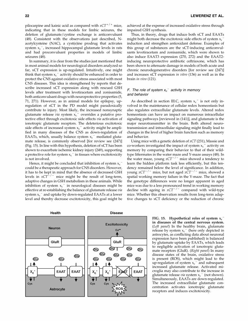
pilocarpine and kainic acid as compared with xCT + / + mice,indicating that in these models for limbic seizures, thedeletion of glutamate/cystine exchange is anticonvulsant(48). Consistent with the observations just described, N-acetylcysteine (NAC), a cyst(e)ine prodrug that activatessystem xc
- , increased hippocampal glutamate levels in ratsand had proconvulsant properties in models of limbicseizures (48).
In summary, it is clear from the studies just mentioned thatin most animal models for neurological disorders analyzed sofar, xCT expression is up-regulated. Intuitively, one mightthink that system xc
- activity should be enhanced in order toprotect the CNS against oxidative stress associated with mostCNS diseases. This idea is strengthened by reports that de-scribe increased xCT expression along with rescued GSHlevels after treatment with levetiracetam and zonisamide,both anticonvulsant drugs with neuroprotective properties (5,71, 271). However, as in animal models for epilepsy, up-regulation of xCT in the PD model might paradoxicallycontribute to injury. Most likely, the increased nonsynapticglutamate release via system xc
- overrides a putative pro-tective effect through excitotoxic side effects via activation ofionotropic glutamate receptors. The deleterious excitotoxicside effects of increased system xc
- activity might be ampli-fied in many diseases of the CNS as down-regulation ofEAATs, which, usually balance system xc
- mediated gluta-mate release, is commonly observed [for review see (247)](Fig. 15). In line with this hypothesis, deletion of xCT has beenshown to exacerbate ischemic kidney injury (249), supportinga protective role for system xc
- in tissues where excitotoxicityis not involved.
Hence, it might be concluded that inhibition of system xc-
could be a therapeutic approach for CNS disorders. However,it has to be kept in mind that the absence of decreased GSHlevels in xCT - / - mice might be the result of long-term,adaptive changes in GSH metabolism in these animals. Whileinhibition of system xc
- in neurological diseases might beeffective at re-establishing the balance of glutamate release viasystem xc
- and uptake by down-regulated EAATs at a lowerlevel and thereby decrease excitotoxicity, this goal might be
achieved at the expense of increased oxidative stress throughimpaired GSH synthesis.
Thus, in theory, drugs that induce both xCT and EAATsmight both decrease the excitotoxic side effects of system xc
-
induction and strengthen antioxidant defense. Examples ofthis group of substances are the xCT-inducing anticonvul-sants levetiracetam and zonisamide, which were shown toalso induce EAAT3 expression (270, 272) and the EAAT2-inducing neuroprotective antibiotic ceftriaxone, which hasbeen shown to attenuate damage in models of both acute andchronic neurodegenerative disorders [for review see (247)]and increases xCT expression in vitro (136) as well as in thebrain in vivo (121).
F. The role of system xc- activity in memory
and behavior
As described in section III.C, system xc- is not only in-
volved in the maintenance of cellular redox homeostasis butalso regulates extracellular glutamate levels. Altered redoxhomeostasis can have an impact on numerous intracellularsignaling pathways [reviewed in (141)], and glutamate is themajor neurotransmitter in the brain. Both altered neuro-transmission and intracellular signaling might finally lead tochanges in the level of higher brain function such as memoryand behavior.
Using mice with a genetic deletion of xCT (231), Massie andco-workers investigated the impact of system xc
- activity onmemory by comparing their behavior to that of their wild-type littermates in the water maze and Y-maze assays (48). Inthe water maze, young xCT - / - mice showed a tendency tolearn the hidden platform task less efficiently, but this ten-dency remained below the level of significance. In addition,young xCT - / - mice, but not aged xCT - / - mice, showed aspatial working memory failure in the Y-maze. The fact thatthe genotype differences were no longer apparent in agedmice was due to a less pronounced trend in working memorydecline with ageing in xCT - / - compared with wild-typemice. Whether this observation results from long-term adap-tive changes to xCT deficiency or the reduction of chronic
FIG. 15. Hypothetical roles of system xc-
in diseases of the central nervous system.(Left panel) In the healthy brain, glutamaterelease by system xc
- (here only depicted inastrocytes, as conflicting data about neuronalexpression have been published) is balancedby glutamate uptake by EAATs, which leadsto negligible activation of ionotropic gluta-mate receptors (GluR). (Right panel) In manydisease states of the brain, oxidative stressis present (ROS), which might lead to theup-regulation of system xc
- and subsequentincreased glutamate release. Activated mi-croglia may also contribute to the increase inglutamate release via system xc
- (not shown).Simultaneously, EAATs are down-regulated.The increased extracellular glutamate con-centration activates ionotropic glutamatereceptors and induces excitotoxicity.
22 LEWERENZ ET AL.
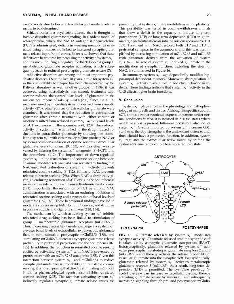
excitotoxicity due to lower extracellular glutamate levels re-mains to be determined.
Schizophrenia is a psychiatric disease that is thought toinvolve disturbed glutamate signaling. In a rodent model ofschizophrenia, where the NMDA antagonist phencyclidine(PCP) is administered, deficits in working memory, as eval-uated using a t-maze, are linked to increased synaptic gluta-mate release in prefrontal cortex. Baker et al. showed that thesedeficits can be restored by increasing the activity of system xc
-
and, as such, inducing a negative feedback loop via group IImetabotropic glutamate receptor activation, which subse-quently leads to reduced presynaptic glutamate release (7).
Addictive disorders are among the most important psy-chiatric diseases. Over the last 10 years, a role for system xc
-
in the vulnerability to relapse has been characterized by theKalivas laboratory as well as other groups. In 1996, it wasobserved using microdialysis that chronic treatment withcocaine reduced the extracellular levels of glutamate in thenucleus accumbens of rats by *50% (208). Since the gluta-mate measured by microdialysis is not derived from synapticactivity (275), other sources of extracellular glutamate wereexamined. It was found that the reduction in extracellularglutamate after chronic treatment with either cocaine ornicotine resulted from reduced system xc
- activity and levelsof xCT expression in the accumbens (8, 120). The reducedactivity of system xc
- was linked to the drug-induced re-ductions in extracellular glutamate by showing that stimu-lating system xc
- with either the cyst(e)ine prodrug NAC orby intra-accumbens infusion of cystine restores extracellularglutamate levels to normal (8, 162), and this effect was re-versed by infusing the system xc
- antagonist (S)-4-CPG intothe accumbens (112). The importance of down-regulatedsystem xc
- in the reinstatement of cocaine-seeking behavior,an animal model of relapse (246), was revealed by finding thatNAC-mediated restoration of system xc
- activity preventsreinstated cocaine seeking (8, 112). Similarly, NAC preventsrelapse to heroin seeking (298). When NAC is chronically gi-ven, an enduring restoration of xCT levels in the accumbens ismeasured in rats withdrawn from self-administered cocaine(121). Importantly, the restoration of xCT by chronic NACadministration is associated with an enduring inhibition ofreinstated cocaine seeking and a restoration of basal levels ofglutamate (162, 188). These behavioural findings have led tomoderate success using NAC to inhibit craving and drug usein cocaine addicts and cigarette smokers (120, 134).
The mechanism by which activating system xc- inhibits
reinstated drug seeking has been linked to stimulation ofgroup II metabotropic glutamate receptors (mGluR2/3).Thus, increasing cystine/glutamate exchange via system xc
-
elevates basal levels of extracellular extrasynaptic glutamatethat, in turn, stimulate presynaptic mGluR2/3 (188), andstimulating mGluR2/3 decreases synaptic glutamate releaseprobability in prefrontal projections into the accumbens (107,185). In addition, the reduction in reinstated cocaine seekingelicited by activating system xc
- with NAC is prevented bypretreatment with an mGluR2/3 antagonist (185). Given thisinteraction between system xc
- and mGluR2/3 to reducesynaptic glutamate release probability and reinstated cocaineseeking, it is not surprising that directly stimulating mGluR2/3 with a pharmacological agonist also inhibits reinstatedcocaine seeking (205). The fact that activating system xc
-
indirectly regulates synaptic glutamate release raises the
possibility that system xc- may modulate synaptic plasticity.
This possibility was tested in cocaine-withdrawn animalsthat show a deficit in the capacity to induce long-termpotentiation (LTP) or long-term depression (LTD) in gluta-matergic prefrontal afferents into the nucleus accumbens (110,187). Treatment with NAC restored both LTP and LTD inprefrontal synapses in the accumbens, and this was accom-plished by increasing stimulation of mGluR2/3 and mGluR5with glutamate derived from the activation of systemxc
- (187). The role of system xc- derived glutamate in the
modification of synaptic function, including the effect ofNAC, is summarized in Figure 16.
In summary, system xc- age-dependently modifies hip-
pocampal-dependent memory. Moreover, dysregulation ofsystem xc
- activity plays a role in addictive behavior in ro-dents. These findings indicate that system xc
- activity in theCNS affects higher brain functions.
V. Conclusion
System xc- plays a role in the physiology and pathophys-
iology of many cells and tissues. Although its specific subunit,xCT, shows a rather restricted expression pattern under nor-mal conditions in vivo, it is induced in disease states whereoxidative stress is present. Inflammatory stimuli also inducesystem xc
- . Cystine imported by system xc- increases GSH
synthesis, thereby strengthens the antioxidant defense, and,thus, should have a protective function. In addition, systemxc
- regulates the extracellular redox milieu by shifting thecystine/cysteine redox couple to a more reduced state.
FIG. 16. Glutamate released by system xc2 modulates
synaptic activity. Glutamate released into the synaptic cleftis taken up by astrocytic glutamate transporters (EAAT)Extrasynaptically, glutamate released by system xc
- acti-vates presynaptic metabotropic glutamate receptors 2 and 3(mGluR2/3) and thereby reduces the release probability ofvesicular glutamate into the synaptic cleft. Postsynaptically,glutamate released by system xc
- activates metabotropicglutamate receptor 5 (mGluR5). As a result, long-term de-pression (LTD) is permitted. The cyst(e)ine pro-drug N-acetyl cysteine can increase extracellular cystine, therebyactivating glutamate release by system xc
- and subsequentlyincreasing signaling through pre- and postsynaptic mGluRs.
SYSTEM xc2 IN HEALTH AND DISEASE 23
Transcriptional regulation through the nuclear factors Nrf2and ATF4 is the best characterized molecular mechanism bywhich xCT expression is induced. Other pathways, includingits induction in certain cell types through inflammatorystimuli such as LPS, TNFa, and IL-1b, remain to be charac-terized in detail. On the functional level system, xc
- activity isregulated by substrate availability and pH. Most importantly,glutamate transporters, EAATs, might regulate system xc
-
activity by increasing intracellular glutamate levels as thedriving force for cystine uptake and reducing the inhibitoryaction of extracellular glutamate.
We show that the specific subunit of system xc- , xCT, and
thus most probably system xc- -like activity, is an evolution-
arily rather new member of the HAT light chain family, asorthologs of xCT are restricted to Deuterostomia and are ab-sent in the Protostomia. Which evolutionary contraints re-quired system xc
- activity remain to be defined. However,since xCT-deficient mice are viable and show an inconsistentphenotype, loss of system xc
- can be compensated for byunknown mechanisms.
In vivo xCT is most prominently expressed in the CNS andin parts of the immune system. Experimental evidence sug-gests that system xc
- activity plays a role in regulating theimmune response. In the CNS, the fact that the amino acidglutamate, which is exported by system xc
- , also acts as aneurotransmitter explains why system xc
- has the propensityto modulate memory and behavior. However, since higherlevels of glutamate induce excitotoxic neuronal death, up-regulation of system xc
- in disease states of the brain mightrepresent a double-edged sword by inducing excitotoxicity.This could explain why xCT deficiency is protective in animalmodels of PD and epilepsy. Whether induction of system xc
-
activity is beneficial or detrimental might depend on thepathway of induction and whether increased glutamate re-lease can be compensated for by glutamate uptake throughEAATs, which are frequently down-regulated in diseases ofthe brain. Whether inhibition of system xc
- or dual inductionof xCT and EAAT is the more promising neuroprotectivestrategy remains to be explored. In addition, similar to thebrain, system xc
- activity might also play a role in bothantioxidant defense and neurotransmission in the eye. Up-regulation of xCT also has been frequently observed in can-cers. Here, xCT expression correlates with resistance againstmany chemotherapeutic drugs, most likely through GSH-mediated drug resistance. However, xCT-overexpressing re-sistant cancer cells are uniquely sensitive to some specificcompounds, for example, selenite.
In contrast to the in vivo situation, system xc- activity is
indispensable to the growth and survival of most cell types incell culture. Nerve cell death induced by glutamate-mediatedinhibition of system xc
- activity, a pathway called oxidativeglutamate toxicity, has been successfully used to characterizeneuroprotective signaling pathways, genes, and putativetherapeutic substances.
Future research designed to (i) characterize the functionalrelevance of system xc
- activity in disease states with oxida-tive stress and inflammation; (ii) understand the regulation ofxCT in diverse cells types in greater detail; and (iii) developmore specific pharmacological tools that inhibit and inducesystem xc
- might pave the way for treating disorders asdiverse as autoimmunity, neurodegeneration, and addiction.With regard to clinical trials, in our opinion, the most
important question to be addressed is whether xCT expres-sion in tumours correlates with disease progression and re-sistance to chemotherapy in humans in vivo. Moreover, largerclinical trials are warranted to substantiate the efficacy of theindirect system xc
- activator NAC to treat addiction.
Acknowledgments
J. Lewerenz. and A. Methner were supported by the theFritz-Thyssen-Stiftung (Az10.08.1.174); S.B. Smith and V.Ganapathy, by the National Institutes of Health NEI (2R01EY012830 and 2R01 EY014560); S. Hewett, by the NationalInstitutes of Health NINDS (NS051445); A. Massie and I.Smolders, by the Medical Foundation Queen Elisabeth andthe Charcot Foundation; and P. Maher, by the Alzheimer’sAssociation (IRG-07-58874). S.B.S and V.G. thank Mrs. L.McKie for preparing Figure 12 and Dr. A. Tawfik for im-munolocalization data shown in Figure 14.
References
1. Aharoni-Simon M, Reifen R, and Tirosh O. ROS-production-mediated activation of AP-1 but not NFkappaB inhibitsglutamate-induced HT4 neuronal cell death. Antioxid RedoxSignal 8: 1339–1349, 2006.
2. Albrecht P, Lewerenz J, Dittmer S, Noack R, Maher P, andMethner A. Mechanisms of oxidative glutamate toxicity:the glutamate/cystine antiporter system xc
- as a neuro-protective drug target. CNS Neurol Disord Drug Targets 9:373–382, 2010.
3. Anderson CL, Iyer SS, Ziegler TR, and Jones DP. Control ofextracellular cysteine/cystine redox state by HT-29 cells isindependent of cellular glutathione. Am J Physiol Regul In-tegr Comp Physiol 293: R1069–R1075, 2007.
4. Antonini B, Piva S, Paltenghi M, Candiani A, and LatronicoN. The early phase of critical illness is a progressive acidicstate due to unmeasured anions. Eur J Anaesthesiol 25: 566–571, 2008.
5. Asanuma M, Miyazaki I, Diaz-Corrales FJ, Kimoto N,Kikkawa Y, Takeshima M, Miyoshi K, and Murata M.Neuroprotective effects of zonisamide target astrocyte. AnnNeurol 67: 239–249, 2010.
6. Augustin H, Grosjean Y, Chen K, Sheng Q, and Feath-erstone DE. Nonvesicular release of glutamate by glial xCTtransporters suppresses glutamate receptor clustering invivo. J Neurosci 27: 111–123, 2007.
7. Baker DA, Madayag A, Kristiansen LV, Meador-WoodruffJH, Haroutunian V, and Raju I. Contribution of cystine-glutamate antiporters to the psychotomimetic effects ofphencyclidine. Neuropsychopharmacology 33: 1760–1772, 2008.
8. Baker DA, McFarland K, Lake RW, Shen H, Tang XC, TodaS, and Kalivas PW. Neuroadaptations in cystine-glutamateexchange underlie cocaine relapse. Nat Neurosci 6: 743–749,2003.
9. Baker DA, Xi ZX, Shen H, Swanson CJ, and Kalivas PW.The origin and neuronal function of in vivo nonsynapticglutamate. J Neurosci 22: 9134–9141, 2002.
10. Banjac A, Perisic T, Sato H, Seiler A, Bannai S, Weiss N,Kolle P, Tschoep K, Issels RD, Daniel PT, Conrad M, andBornkamm GW. The cystine/cysteine cycle: a redox cycleregulating susceptibility versus resistance to cell death.Oncogene 27: 1618–1628, 2008.
11. Bannai S. Exchange of cystine and glutamate across plasmamembrane of human fibroblasts. J Biol Chem 261: 2256–2263, 1986.
24 LEWERENZ ET AL.
12. Bannai S. Induction of cystine and glutamate transportactivity in human fibroblasts by diethyl maleate and otherelectrophilic agents. J Biol Chem 259: 2435–2440, 1984.
13. Bannai S and Ishii T. A novel function of glutamine in cellculture: utilization of glutamine for the uptake of cystine inhuman fibroblasts. J Cell Physiol 137: 360–366, 1988.
14. Bannai S and Ishii T. Transport of cystine and cysteine andcell growth in cultured human diploid fibroblasts: effect ofglutamate and homocysteate. J Cell Physiol 112: 265–272,1982.
15. Bannai S and Kasuga H. Anti-inflammatory drug inhibitionof transport of cystine and glutamate in cultured humanfibroblasts. Biochem Pharmacol 34: 1852–1854, 1985.
16. Bannai S and Kitamura E. Role of proton dissociation in thetransport of cystine and glutamate in human diploid fi-broblasts in culture. J Biol Chem 256: 5770–5772, 1981.
17. Bannai S and Kitamura E. Transport interaction of L-cystine and L-glutamate in human diploid fibroblasts inculture. J Biol Chem 255: 2372–2376, 1980.
18. Bannai S, Sato H, Ishii T, and Sugita Y. Induction of cystinetransport activity in human fibroblasts by oxygen. J BiolChem 264: 18480–18484, 1989.
19. Barger SW and Basile AS. Activation of microglia bysecreted amyloid precursor protein evokes release ofglutamate by cystine exchange and attenuates synapticfunction. J Neurochem 76: 846–854, 2001.
20. Barger SW, Goodwin ME, Porter MM, and Beggs ML.Glutamate release from activated microglia requires theoxidative burst and lipid peroxidation. J Neurochem 101:1205–1213, 2007.
21. Bassi MT, Gasol E, Manzoni M, Pineda M, Riboni M,Martin R, Zorzano A, Borsani G, and Palacin M. Identifi-cation and characterisation of human xCT that co-expresses, with 4F2 heavy chain, the amino acid transportactivity system xc
- . Pflugers Arch 442: 286–296, 2001.22. Bedingfield JS, Kemp MC, Jane DE, Tse HW, Roberts PJ,
and Watkins JC. Structure-activity relationships for a seriesof phenylglycine derivatives acting at metabotropic gluta-mate receptors (mGluRs). Br J Pharmacol 116: 3323–3329,1995.
23. Berghe TV, Vanlangenakker N, Parthoens E, Deckers W,Devos M, Festjens N, Guerin CJ, Brunk UT, Declercq W,and Vandenabeele P. Necroptosis, necrosis and secondarynecrosis converge on similar cellular disintegration fea-tures. Cell Death Differ 17: 922–930, 2010.
24. Berthoud VM and Beyer EC. Oxidative stress, lens gapjunctions, and cataracts. Antioxid Redox Signal 11: 339–353,2009.
25. Bi M, Naczki C, Koritzinsky M, Fels D, Blais J, Hu N,Harding H, Novoa I, Varia M, Raleigh J, Scheuner D,Kaufman RJ, Bell J, Ron D, Wouters BG, and Koumenis C.ER stress-regulated translation increases tolerance to ex-treme hypoxia and promotes tumor growth. EMBO J 24:3470–3481, 2005.
26. Brauner-Osborne H, Nielsen B, and Krogsgaard-Larsen P.Molecular pharmacology of homologues of ibotenic acid atcloned metabotropic glutamic acid receptors. Eur J Phar-macol 350: 311–316, 1998.
27. Bridges CC, Hu H, Miyauchi S, Siddaramappa UN,Ganapathy ME, Ignatowicz L, Maddox DM, Smith SB, andGanapathy V. Induction of cystine-glutamate transporterxc
- by human immunodeficiency virus type 1 transacti-vator protein tat in retinal pigment epithelium. InvestOphthalmol Vis Sci 45: 2906–2914, 2004.
28. Bridges CC, Kekuda R, Wang H, Prasad PD, Mehta P,Huang W, Smith SB, and Ganapathy V. Structure, function,and regulation of human cystine/glutamate transporter inretinal pigment epithelial cells. Invest Ophthalmol Vis Sci 42:47–54, 2001.
29. Buckingham SC, Campbell SL, Haas BR, Montana V, RobelS, Ogunrinu T, and Sontheimer H. Glutamate release byprimary brain tumors induces epileptic activity. Nat Med17: 1269–1274, 2011.
30. Burdo J, Dargusch R, and Schubert D. Distribution of thecystine/glutamate antiporter system xc
- in the brain, kid-ney, and duodenum. J Histochem Cytochem 54: 549–557,2006.
31. Cai J, Nelson KC, Wu M, Sternberg P, Jr., and Jones DP.Oxidative damage and protection of the RPE. Prog RetinEye Res 19: 205–221, 2000.
32. Chang YF. Lysine metabolism in the human and themonkey: demonstration of pipecolic acid formation in thebrain and other organs. Neurochem Res 7: 577–588, 1982.
33. Chen CJ, Liao SL, and Kuo JS. Gliotoxic action of glutamateon cultured astrocytes. J Neurochem 75: 1557–1565, 2000.
34. Chen Q, Prior M, Dargusch R, Roberts A, Riek R, EichmannC, Chiruta C, Akaishi T, Abe K, Maher P, and Schubert D.A novel neurotrophic drug for cognitive enhancement andAlzheimer’s disease. PLoS One 6: e27865, 2011.
35. Chen RS, Song YM, Zhou ZY, Tong T, Li Y, Fu M, Guo XL,Dong LJ, He X, Qiao HX, Zhan QM, and Li W. Disruptionof xCT inhibits cancer cell metastasis via the caveolin-1/beta-catenin pathway. Oncogene 28: 599–609, 2009.
36. Chen Y and Swanson RA. The glutamate transportersEAAT2 and EAAT3 mediate cysteine uptake in corticalneuron cultures. J Neurochem 84: 1332–1339, 2003.
37. Chintala S, Li W, Lamoreux ML, Ito S, Wakamatsu K,Sviderskaya EV, Bennett DC, Park YM, Gahl WA, HuizingM, Spritz RA, Ben S, Novak EK, Tan J, and Swank RT.Slc7a11 gene controls production of pheomelanin pigmentand proliferation of cultured cells. Proc Natl Acad Sci U S A102: 10964–10969, 2005.
38. Choi DW. Glutamate neurotoxicity and diseases of thenervous system. Neuron 1: 623–634, 1988.
39. Choi HJ, Kang KS, Fukui M, and Zhu BT. Critical role of theJNK-p53-GADD45alpha apoptotic cascade in mediatingoxidative cytotoxicity in hippocampal neurons. Br J Phar-macol 162: 175–192, 2011.
40. Christensen HN. Role of amino acid transport and coun-tertransport in nutrition and metabolism. Physiol Rev 70:43–77, 1990.
41. Chung WJ, Lyons SA, Nelson GM, Hamza H, Gladson CL,Gillespie GY, and Sontheimer H. Inhibition of cystine up-take disrupts the growth of primary brain tumors. J Neu-rosci 25: 7101–7110, 2005.
42. Circu ML and Aw TY. Reactive oxygen species, cellularredox systems, and apoptosis. Free Radic Biol Med 48: 749–762, 2010.
43. Coquelle T, Christensen JK, Banke TG, Madsen U,Schousboe A, and Pickering DS. Agonist discriminationbetween AMPA receptor subtypes. Neuroreport 11: 2643–2648, 2000.
44. Crimi E, Sica V, Williams-Ignarro S, Zhang H, Slutsky AS,Ignarro LJ, and Napoli C. The role of oxidative stress inadult critical care. Free Radic Biol Med 40: 398–406, 2006.
45. Dai Z, Huang Y, Sadee W, and Blower P. Chemoinfor-matics analysis identifies cytotoxic compounds susceptibleto chemoresistance mediated by glutathione and cystine/
SYSTEM xc2 IN HEALTH AND DISEASE 25
glutamate transport system xc- . J Med Chem 50: 1896–1906,
2007.46. Dargusch R and Schubert D. Specificity of resistance to
oxidative stress. J Neurochem 81: 1394–1400, 2002.47. Davis JB and Maher P. Protein kinase C activation inhibits
glutamate-induced cytotoxicity in a neuronal cell line. BrainRes 652: 169–173, 1994.
48. De Bundel D, Schallier A, Loyens E, Fernando R, MiyashitaH, Van Liefferinge J, Vermoesen K, Bannai S, Sato H,Michotte Y, Smolders I, and Massie A. Loss of system xc
-
formula does not induce oxidative stress but decreasesextracellular glutamate in hippocampus and influencesspatial working memory and limbic seizure susceptibility. JNeurosci 31: 5792–5803, 2011.
49. Degterev A, Huang Z, Boyce M, Li Y, Jagtap P, MizushimaN, Cuny GD, Mitchison TJ, Moskowitz MA, and Yuan J.Chemical inhibitor of nonapoptotic cell death with thera-peutic potential for ischemic brain injury. Nat Chem Biol 1:112–119, 2005.
50. Dittmer S, Sahin M, Pantlen A, Saxena A, Toutzaris D, PinaAL, Geerts A, Golz S, and Methner A. The constitutivelyactive orphan G-protein-coupled receptor GPR39 protectsfrom cell death by increasing secretion of pigment epithe-lium-derived growth factor. J Biol Chem 283: 7074–7081,2008.
51. Do KQ, Benz B, Sorg O, Pellerin L, and Magistretti PJ. beta-Adrenergic stimulation promotes homocysteic acid releasefrom astrocyte cultures: evidence for a role of astrocytes inthe modulation of synaptic transmission. J Neurochem 68:2386–2394, 1997.
52. Do KQ, Herrling PL, Streit P, and Cuenod M. Release ofneuroactive substances: homocysteic acid as an endoge-nous agonist of the NMDA receptor. J Neural Transm 72:185–190, 1988.
53. Domercq M, Sanchez-Gomez MV, Sherwin C, Etxebarria E,Fern R, and Matute C. System xc
- and glutamate trans-porter inhibition mediates microglial toxicity to oligoden-drocytes. J Immunol 178: 6549–6556, 2007.
54. Dringen R. Metabolism and functions of glutathione inbrain. Prog Neurobiol 62: 649–671, 2000.
55. Dringen R and Hirrlinger J. Glutathione pathways in thebrain. Biol Chem 384: 505–516, 2003.
56. Droge W. Cysteine and glutathione deficiency in AIDSpatients: a rationale for the treatment with N-acetyl-cysteine.Pharmacology 46: 61–65, 1993.
57. Dun Y, Mysona B, Van Ells T, Amarnath L, Ola MS,Ganapathy V, and Smith SB. Expression of the cystine-glutamate exchanger xc
- in retinal ganglion cells and reg-ulation by nitric oxide and oxidative stress. Cell Tissue Res324: 189–202, 2006.
58. Ebert B, Madsen U, Johansen TN, and Krogsgaard-LarsenP. NMDA receptor agonists: relationships between struc-ture and biological activity. Adv Exp Med Biol 287: 483–487,1991.
59. Elphick LM, Hawat M, Toms NJ, Meinander A, MikhailovA, Eriksson JE, and Kass GE. Opposing roles for caspaseand calpain death proteases in L-glutamate-induced oxi-dative neurotoxicity. Toxicol Appl Pharmacol 232: 258–267,2008.
60. Falk MH, Hultner L, Milner A, Gregory CD, and Born-kamm GW. Irradiated fibroblasts protect Burkitt lym-phoma cells from apoptosis by a mechanism independentof bcl-2. Int J Cancer 55: 485–491, 1993.
61. Ferraguti F, Pietra C, Valerio E, Corti C, Chiamulera C, andConquet F. Evidence against a permissive role of the me-tabotropic glutamate receptor 1 in acute excitotoxicity.Neuroscience 79: 1–5, 1997.
62. Fogal B and Hewett SJ. Interleukin-1beta: a bridge betweeninflammation and excitotoxicity? J Neurochem 106: 1–23, 2008.
63. Fogal B, Li J, Lobner D, McCullough LD, and Hewett SJ.System xc
- activity and astrocytes are necessary for inter-leukin-1 beta-mediated hypoxic neuronal injury. J Neurosci27: 10094–10105, 2007.
64. Fogal B, Trettel J, Uliasz TF, Levine ES, and Hewett SJ.Changes in secondary glutamate release underlie the de-velopmental regulation of excitotoxic neuronal cell death.Neuroscience 132: 929–942, 2005.
65. Franco R and Cidlowski JA. SLCO/OATP-like transport ofglutathione in FasL-induced apoptosis: glutathione efflux iscoupled to an organic anion exchange and is necessary forthe progression of the execution phase of apoptosis. J BiolChem 281: 29542–29557, 2006.
66. Fredriksson R, Nordstrom KJ, Stephansson O, HagglundMG, and Schioth HB. The solute carrier (SLC) complementof the human genome: phylogenetic classification revealsfour major families. FEBS Lett 582: 3811–3816, 2008.
67. Fukasawa Y, Segawa H, Kim JY, Chairoungdua A, KimDK, Matsuo H, Cha SH, Endou H, and Kanai Y. Identifi-cation and characterization of a Na + -independent neutralamino acid transporter that associates with the 4F2 heavychain and exhibits substrate selectivity for small neutralD- and L-amino acids. J Biol Chem 275: 9690–9698, 2000.
68. Ganea E and Harding JJ. Glutathione-related enzymes andthe eye. Curr Eye Res 31: 1–11, 2006.
69. Gasol E, Jimenez-Vidal M, Chillaron J, Zorzano A, andPalacin M. Membrane topology of system xc
- light subunitreveals a re-entrant loop with substrate-restricted accessi-bility. J Biol Chem 279: 31228–31236, 2004.
70. Gelderblom M, Leypoldt F, Lewerenz J, Birkenmayer G,Orozco D, Ludewig P, Thundyil J, Arumugam TV, GerloffC, Tolosa E, Maher P, and Magnus T. The flavonoid fisetinattenuates postischemic immune cell infiltration, activationand infarct size after transient cerebral middle artery oc-clusion in mice. J Cereb Blood Flow Metab 32: 835–843, 2012.
71. Gibbs JE, Walker MC, and Cock HR. Levetiracetam: anti-epileptic properties and protective effects on mitochondrialdysfunction in experimental status epilepticus. Epilepsia 47:469–478, 2006.
72. Gmunder H, Eck HP, Benninghoff B, Roth S, and Droge W.Macrophages regulate intracellular glutathione levels oflymphocytes. Evidence for an immunoregulatory role ofcysteine. Cell Immunol 129: 32–46, 1990.
73. Gmunder H, Eck HP, and Droge W. Low membranetransport activity for cystine in resting and mitogenicallystimulated human lymphocyte preparations and human Tcell clones. Eur J Biochem 201: 113–117, 1991.
74. Gnana-Prakasam JP, Thangaraju M, Liu K, Ha Y, MartinPM, Smith SB, and Ganapathy V. Absence of iron-regula-tory protein Hfe results in hyperproliferation of retinalpigment epithelium: role of cystine/glutamate exchanger.Biochem J 424: 243–252, 2009.
75. Go YM and Jones DP. Cysteine/cystine redox signaling incardiovascular disease. Free Radic Biol Med 50: 495–509,2011.
76. Gochenauer GE and Robinson MB. Dibutyryl-cAMP(dbcAMP) up-regulates astrocytic chloride-dependent L-
26 LEWERENZ ET AL.
[3H]glutamate transport and expression of both system xc-
subunits. J Neurochem 78: 276–286, 2001.77. Gonsette RE. Neurodegeneration in multiple sclerosis: the
role of oxidative stress and excitotoxicity. J Neurol Sci 274:48–53, 2008.
78. Gottesman MM, Fojo T, and Bates SE. Multidrug resistancein cancer: role of ATP-dependent transporters. Nat RevCancer 2: 48–58, 2002.
79. Gout PW, Buckley AR, Simms CR, and Bruchovsky N.Sulfasalazine, a potent suppressor of lymphoma growth byinhibition of the xc
- cystine transporter: a new action for anold drug. Leukemia 15: 1633–1640, 2001.
80. Gout PW, Kang YJ, Buckley DJ, Bruchovsky N, and Buck-ley AR. Increased cystine uptake capability associated withmalignant progression of Nb2 lymphoma cells. Leukemia 11:1329–1337, 1997.
81. Grieve A and Griffiths R. Simultaneous measurement byHPLC of the excitatory amino acid transmitter candidateshomocysteate and homocysteine sulphinate supports apredominant astrocytic localisation. Neurosci Lett 145: 1–5,1992.
82. Griffith OW. Biologic and pharmacologic regulation ofmammalian glutathione synthesis. Free Radic Biol Med 27:922–935, 1999.
83. Guldin WO and Markowitsch HJ. Epidural kainate, but notibotenate, produces lesions in local and distant regions ofthe brain. A comparison of the intracerebral actions ofkainic acid and ibotenic acid. J Neurosci Methods 5: 83–93,1982.
84. Gupta S. A decision between life and death duringTNF-alpha-induced signaling. J Clin Immunol 22: 185–194,2002.
85. Ha JS, Lim HM, and Park SS. Extracellular hydrogen per-oxide contributes to oxidative glutamate toxicity. Brain Res1359: 291–297, 2010.
86. Hahn P, Milam AH, and Dunaief JL. Maculas affected byage-related macular degeneration contain increased che-latable iron in the retinal pigment epithelium and Bruch’smembrane. Arch Ophthalmol 121: 1099–1105, 2003.
87. Harding HP, Zhang Y, Zeng H, Novoa I, Lu PD, Calfon M,Sadri N, Yun C, Popko B, Paules R, Stojdl DF, Bell JC,Hettmann T, Leiden JM, and Ron D. An integrated stressresponse regulates amino acid metabolism and resistanceto oxidative stress. Mol Cell 11: 619–633, 2003.
88. Harris RA and Amor S. Sweet and sour—oxidative andcarbonyl stress in neurological disorders. CNS Neurol Dis-ord Drug Targets 10: 82–107, 2011.
89. Harvey CJ, Thimmulappa RK, Singh A, Blake DJ, Ling G,Wakabayashi N, Fujii J, Myers A, and Biswal S. Nrf2-regulated glutathione recycling independent of biosynthe-sis is critical for cell survival during oxidative stress. FreeRadic Biol Med 46: 443–453, 2009.
90. Hayes JD, Flanagan JU, and Jowsey IR. Glutathione trans-ferases. Annu Rev Pharmacol Toxicol 45: 51–88, 2005.
91. Hu RG, Lim J, Donaldson PJ, and Kalloniatis M. Char-acterization of the cystine/glutamate transporter in theouter plexiform layer of the vertebrate retina. Eur J Neurosci28: 1491–1502, 2008.
92. Huang Y. Pharmacogenetics/genomics of membranetransporters in cancer chemotherapy. Cancer Metastasis Rev26: 183–201, 2007.
93. Huang Y, Dai Z, Barbacioru C, and Sadee W. Cystine-glutamate transporter SLC7A11 in cancer chemosensitivityand chemoresistance. Cancer Res 65: 7446–7454, 2005.
94. Huang Y, Penchala S, Pham AN, and Wang J. Geneticvariations and gene expression of transporters in drugdisposition and response. Expert Opin Drug Metab Toxicol 4:237–254, 2008.
95. Hur W and Gray NS. Small molecule modulators of anti-oxidant response pathway. Curr Opin Chem Biol 15: 162–173, 2011.
96. Igarashi T, Izumi H, Uchiumi T, Nishio K, Arao T, TanabeM, Uramoto H, Sugio K, Yasumoto K, Sasaguri Y, WangKY, Otsuji Y, and Kohno K. Clock and ATF4 transcriptionsystem regulates drug resistance in human cancer cell lines.Oncogene 26: 4749–4760, 2007.
97. Imai H and Nakagawa Y. Biological significance of phos-pholipid hydroperoxide glutathione peroxidase (PHGPx,GPx4) in mammalian cells. Free Radic Biol Med 34: 145–169,2003.
98. Ishige K, Chen Q, Sagara Y, and Schubert D. The activationof dopamine D4 receptors inhibits oxidative stress-inducednerve cell death. J Neurosci 21: 6069–6076, 2001.
99. Ishige K, Schubert D, and Sagara Y. Flavonoids protectneuronal cells from oxidative stress by three distinctmechanisms. Free Radic Biol Med 30: 433–446, 2001.
100. Ishii T, Hishinuma I, Bannai S, and Sugita Y. Mechanism ofgrowth promotion of mouse lymphoma L1210 cells in vitroby feeder layer or 2-mercaptoethanol. J Cell Physiol 107:283–293, 1981.
101. Ishii T, Itoh K, Takahashi S, Sato H, Yanagawa T, Katoh Y,Bannai S, and Yamamoto M. Transcription factor Nrf2 co-ordinately regulates a group of oxidative stress-induciblegenes in macrophages. J Biol Chem 275: 16023–16029, 2000.
102. Ishii T, Sugita Y, and Bannai S. Regulation of glutathionelevels in mouse spleen lymphocytes by transport of cyste-ine. J Cell Physiol 133: 330–336, 1987.
103. Ishimoto T, Nagano O, Yae T, Tamada M, Motohara T,Oshima H, Oshima M, Ikeda T, Asaba R, Yagi H, MasukoT, Shimizu T, Ishikawa T, Kai K, Takahashi E, Imamura Y,Baba Y, Ohmura M, Suematsu M, Baba H, and Saya H.CD44 variant regulates redox status in cancer cells by sta-bilizing the xCT subunit of system xc
- and thereby pro-motes tumor growth. Cancer Cell 19: 387–400, 2011.
104. Jabaudon D, Scanziani M, Gahwiler BH, and Gerber U.Acute decrease in net glutamate uptake during energydeprivation. Proc Natl Acad Sci U S A 97: 5610–5615, 2000.
105. Jackman NA, Uliasz TF, Hewett JA, and Hewett SJ. Reg-ulation of system xc
- activity and expression in astrocytesby interleukin-1beta: implications for hypoxic neuronalinjury. Glia 58: 1806–1815, 2010.
106. Kaleeba JA and Berger EA. Kaposi’s sarcoma-associatedherpesvirus fusion-entry receptor: cystine transporter xCT.Science 311: 1921–1924, 2006.
107. Kalivas PW. The glutamate homeostasis hypothesis of ad-diction. Nat Rev Neurosci 10: 561–572, 2009.
108. Kaltschmidt B, Widera D, and Kaltschmidt C. Signaling viaNF-kappaB in the nervous system. Biochim Biophys Acta1745: 287–299, 2005.
109. Kanai Y and Hediger MA. Primary structure and functionalcharacterization of a high-affinity glutamate transporter.Nature 360: 467–471, 1992.
110. Kasanetz F, Deroche-Gamonet V, Berson N, Balado E,Lafourcade M, Manzoni O, and Piazza PV. Transition toaddiction is associated with a persistent impairment insynaptic plasticity. Science 328: 1709–1712, 2010.
111. Kato S, Ishita S, Sugawara K, and Mawatari K. Cystine/glutamate antiporter expression in retinal Muller glial cells:
SYSTEM xc2 IN HEALTH AND DISEASE 27
implications for DL-alpha-aminoadipate toxicity. Neu-roscience 57: 473–482, 1993.
112. Kau KS, Madayag A, Mantsch JR, Grier MD, AbdulhameedO, and Baker DA. Blunted cystine-glutamate antiporterfunction in the nucleus accumbens promotes cocaine-induced drug seeking. Neuroscience 155: 530–537, 2008.
113. Kensler TW, Wakabayashi N, and Biswal S. Cell survivalresponses to environmental stresses via the Keap1-Nrf2-ARE pathway. Annu Rev Pharmacol Toxicol 47: 89–116, 2007.
114. Kilberg MS, Shan J, and Su N. ATF4-dependent transcrip-tion mediates signaling of amino acid limitation. TrendsEndocrinol Metab 20: 436–443, 2009.
115. Kim JY, Kanai Y, Chairoungdua A, Cha SH, Matsuo H,Kim DK, Inatomi J, Sawa H, Ida Y, and Endou H. Humancystine/glutamate transporter: cDNA cloning and upre-gulation by oxidative stress in glioma cells. Biochim BiophysActa 1512: 335–344, 2001.
116. Kimura Y, Dargusch R, Schubert D, and Kimura H. Hy-drogen sulfide protects HT22 neuronal cells from oxidativestress. Antioxid Redox Signal 8: 661–670, 2006.
117. Kimura Y, Goto Y, and Kimura H. Hydrogen sulfide in-creases glutathione production and suppresses oxidativestress in mitochondria. Antioxid Redox Signal 12: 1–13, 2010.
118. Kimura Y and Kimura H. Hydrogen sulfide protects neu-rons from oxidative stress. FASEB J 18: 1165–1167, 2004.
119. Kingston AE, Lowndes J, Evans N, Clark B, Tomlinson R,Burnett JP, Mayne NG, Cockerham SL, and Lodge D.Sulphur-containing amino acids are agonists for group 1metabotropic receptors expressed in clonal RGT cell lines.Neuropharmacology 37: 277–287, 1998.
120. Knackstedt LA, LaRowe S, Mardikian P, Malcolm R,Upadhyaya H, Hedden S, Markou A, and Kalivas PW. Therole of cystine-glutamate exchange in nicotine dependencein rats and humans. Biol Psychiatry 65: 841–845, 2009.
121. Knackstedt LA, Melendez RI, and Kalivas PW. CeftriaxoneRestores Glutamate Homeostasis and Prevents Relapse toCocaine Seeking. Biol Psychiatry 67: 81–84, 2009.
122. Knickelbein RG, Seres T, Lam G, Johnston RB, Jr., andWarshaw JB. Characterization of multiple cysteine andcystine transporters in rat alveolar type II cells. Am J Physiol273: L1147–L1155, 1997.
123. Koglin N, Mueller A, Berndt M, Schmitt-Willich H, ToschiL, Stephens AW, Gekeler V, Friebe M, and Dinkelborg LM.Specific PET imaging of xc
- transporter activity using a18F-labeled glutamate derivative reveals a dominant path-way in tumor metabolism. Clin Cancer Res 17: 6000–6011,2011.
124. Koyama Y, Kimura Y, Hashimoto H, Matsuda T, and BabaA. L-lactate inhibits L-cystine/L-glutamate exchangetransport and decreases glutathione content in rat culturedastrocytes. J Neurosci Res 59: 685–691, 2000.
125. Kranich O, Hamprecht B, and Dringen R. Different pref-erences in the utilization of amino acids for glutathionesynthesis in cultured neurons and astroglial cells derivedfrom rat brain. Neurosci Lett 219: 211–214, 1996.
126. Kubota C, Torii S, Hou N, Saito N, Yoshimoto Y, Imai H,and Takeuchi T. Constitutive reactive oxygen species gen-eration from autophagosome/lysosome in neuronal oxi-dative toxicity. J Biol Chem 285: 667–674, 2010.
127. Kvamme E, Schousboe A, Hertz L, Torgner IA, andSvenneby G. Developmental change of endogenous gluta-mate and gamma-glutamyl transferase in cultured cerebralcortical interneurons and cerebellar granule cells, and in
mouse cerebral cortex and cerebellum in vivo. NeurochemRes 10: 993–1008, 1985.
128. Kwak MK and Kensler TW. Targeting NRF2 signaling forcancer chemoprevention. Toxicol Appl Pharmacol 244: 66–76,2010.
129. Lall MM, Ferrell J, Nagar S, Fleisher LN, and McGahanMC. Iron regulates L-cystine uptake and glutathione levelsin lens epithelial and retinal pigment epithelial cells by itseffect on cytosolic aconitase. Invest Ophthalmol Vis Sci 49:310–319, 2008.
130. Lam BK. Leukotriene C4 synthase: a critical enzyme for thebiosynthesis of SRS-A. Front Biosci 2: d380–d386, 1997.
131. Landshamer S, Hoehn M, Barth N, Duvezin-Caubet S,Schwake G, Tobaben S, Kazhdan I, Becattini B, Zahler S,Vollmar A, Pellecchia M, Reichert A, Plesnila N, Wagner E,and Culmsee C. Bid-induced release of AIF from mito-chondria causes immediate neuronal cell death. Cell DeathDiffer 15: 1553–1563, 2008.
132. Langford MP, Redmond P, Chanis R, Misra RP, andRedens TB. Glutamate, excitatory amino acid transport-ers, xc
- antiporter, glutamine synthetase, and gamma-glutamyltranspeptidase in human corneal epithelium. CurrEye Res 35: 202–211, 2010.
133. Lapchak PA, Schubert DR, and Maher PA. Delayed treat-ment with a novel neurotrophic compound reduces be-havioral deficits in rabbit ischemic stroke. J Neurochem 116:122–131, 2011.
134. LaRowe SD, Myrick H, Hedden S, Mardikian P, Saladin M,McRae A, Brady K, Kalivas PW, and Malcolm R. Is cocainedesire reduced by N-acetylcysteine? Am J Psychiatry 164:1115–1117, 2007.
135. Lee M, Cho T, Jantaratnotai N, Wang YT, McGeer E, andMcGeer PL. Depletion of GSH in glial cells induces neu-rotoxicity: relevance to aging and degenerative neurologi-cal diseases. FASEB J 24: 2533–2545, 2010.
136. Lewerenz J, Albrecht P, Tien ML, Henke N, Karumbayar-am S, Kornblum HI, Wiedau-Pazos M, Schubert D, MaherP, and Methner A. Induction of Nrf2 and xCT are involvedin the action of the neuroprotective antibiotic ceftriaxonein vitro. J Neurochem 111: 332–343, 2009.
137. Lewerenz J, Dargusch R, and Maher P. Lactacidosis Mod-ulates Glutathione Metabolism and Oxidative GlutamateToxicity. J Neurochem 113: 502–514, 2010.
138. Lewerenz J, Klein M, and Methner A. Cooperative action ofglutamate transporters and cystine/glutamate antiportersystem xc
- protects from oxidative glutamate toxicity. JNeurochem 98: 916–925, 2006.
139. Lewerenz J, Letz J, and Methner A. Activation of stimula-tory heterotrimeric G proteins increases glutathione andprotects neuronal cells against oxidative stress. J Neurochem87: 522–531, 2003.
140. Lewerenz J and Maher P. Basal levels of eIF2alpha phos-phorylation determine cellular antioxidant status byregulating ATF4 and xCT expression. J Biol Chem 284: 1106–1115, 2009.
141. Lewerenz J and Maher P. Control of redox state and redoxsignaling by neural antioxidant systems. Antioxid RedoxSignal 14: 1449–1465, 2011.
142. Lewerenz J, Maher P, and Methner A. Regulation of xCTexpression and system xc
- function in neuronal cells.Amino Acids 42: 171–179, 2012.
143. Li L, Lim J, Jacobs MD, Kistler J, and Donaldson PJ. Re-gional differences in cystine accumulation point to a sutural
28 LEWERENZ ET AL.

delivery pathway to the lens core. Invest Ophthalmol Vis Sci48: 1253–1260, 2007.
144. Li Y, Maher P, and Schubert D. A role for 12-lipoxygenasein nerve cell death caused by glutathione depletion. Neuron19: 453–463, 1997.
145. Li Y, Maher P, and Schubert D. Requirement for cGMP innerve cell death caused by glutathione depletion. J Cell Biol139: 1317–1324, 1997.
146. Lim J, Lam YC, Kistler J, and Donaldson PJ. Molecularcharacterization of the cystine/glutamate exchanger andthe excitatory amino acid transporters in the rat lens. InvestOphthalmol Vis Sci 46: 2869–2877, 2005.
147. Lim J, Lorentzen KA, Kistler J, and Donaldson PJ. Mole-cular identification and characterisation of the glycinetransporter (GLYT1) and the glutamine/glutamate trans-porter (ASCT2) in the rat lens. Exp Eye Res 83: 447–455,2006.
148. Lim JC and Donaldson PJ. Focus on molecules: the cystine/glutamate exchanger (System xc
- ). Exp Eye Res 92: 162–163,2011.
149. Liu H, Wang H, Shenvi S, Hagen TM, and Liu RM. Glu-tathione metabolism during aging and in Alzheimer dis-ease. Ann N Y Acad Sci 1019: 346–349, 2004.
150. Liu R, Blower PE, Pham AN, Fang J, Dai Z, Wise C, GreenB, Teitel CH, Ning B, Ling W, Lyn-Cook BD, Kadlubar FF,Sadee W, and Huang Y. Cystine-glutamate transporterSLC7A11 mediates resistance to geldanamycin but not to17-(allylamino)-17-demethoxygeldanamycin. Mol Pharma-col 72: 1637–1646, 2007.
151. Liu X, Resch J, Rush T, and Lobner D. Functional upregu-lation of system xc
- by fibroblast growth factor-2. Neuro-pharmacology 62: 901–906, 2012.
152. Liu X, Rush T, Zapata J, and Lobner D. beta-N-methylamino-l-alanine induces oxidative stress and glutamate releasethrough action on system xc
- . Exp Neurol 217: 429–433,2009.
153. Liu XX, Li XJ, Zhang B, Liang YJ, Zhou CX, Cao DX, He M,Chen GQ, He JR, and Zhao Q. MicroRNA-26b is under-expressed in human breast cancer and induces cell apo-ptosis by targeting SLC7A11. FEBS Lett 585: 1363–1367,2011.
154. Liu Y, Dargusch R, Maher P, and Schubert D. A broadlyneuroprotective derivative of curcumin. J Neurochem 105:1336–1345, 2008.
155. Lo M, Ling V, Low C, Wang YZ, and Gout PW. Potentialuse of the anti-inflammatory drug, sulfasalazine, for tar-geted therapy of pancreatic cancer. Curr Oncol 17: 9–16,2010.
156. Lo M, Ling V, Wang YZ, and Gout PW. The xc - cystine/glutamate antiporter: a mediator of pancreatic cancergrowth with a role in drug resistance. Br J Cancer 99: 464–472, 2008.
157. Lo M, Wang YZ, and Gout PW. The xc- cystine/glutamate
antiporter: a potential target for therapy of cancer and otherdiseases. J Cell Physiol 215: 593–602, 2008.
158. Lodge D. The history of the pharmacology and cloning ofionotropic glutamate receptors and the development ofidiosyncratic nomenclature. Neuropharmacology 56: 6–21,2009.
159. Lu MJ, Pulido JS, McCannel CA, Pulido JE, Hatfield RM,Dundervill RF, 3rd, and Shippy SA. Detection of elevatedsignaling amino acids in human diabetic vitreous by rapidcapillary electrophoresis. Exp Diabetes Res 2007: 39765,2007.
160. Lu SC. Regulation of glutathione synthesis. Mol AspectsMed 30: 42–59, 2009.
161. Lyons SA, Chung WJ, Weaver AK, Ogunrinu T, andSontheimer H. Autocrine glutamate signaling promotesglioma cell invasion. Cancer Res 67: 9463–9471, 2007.
162. Madayag A, Lobner D, Kau KS, Mantsch JR, AbdulhameedO, Hearing M, Grier MD, and Baker DA. Repeated N-acetylcysteine administration alters plasticity-dependenteffects of cocaine. J Neurosci 27: 13968–13976, 2007.
163. Maher P. A comparison of the neurotrophic activities of theflavonoid fisetin and some of its derivatives. Free Radic Res40: 1105–1111, 2006.
164. Maher P. How protein kinase C activation protects nervecells from oxidative stress-induced cell death. J Neurosci 21:2929–2938, 2001.
165. Maher P. Proteasome inhibitors prevent oxidative stress-induced nerve cell death by a novel mechanism. BiochemPharmacol 75: 1994–2006, 2008.
166. Maher P, Dargusch R, Bodai L, Gerard PE, Purcell JM, andMarsh JL. ERK activation by the polyphenols fisetin andresveratrol provides neuroprotection in multiple models ofHuntington’s disease. Hum Mol Genet 20: 261–270, 2011.
167. Maher P, Dargusch R, Ehren JL, Okada S, Sharma K, andSchubert D. Fisetin lowers methylglyoxal dependent pro-tein glycation and limits the complications of diabetes.PLoS One 6: e21226, 2011.
168. Maher P, Lewerenz J, Lozano C, and Torres JL. A novelapproach to enhancing cellular glutathione levels. J Neu-rochem 107: 690–700, 2008.
169. Maher P, Salgado KF, Zivin JA, and Lapchak PA. A novelapproach to screening for new neuroprotective compoundsfor the treatment of stroke. Brain Res 1173: 117–125, 2007.
170. Maher P and Schubert D. Signaling by reactive oxygenspecies in the nervous system. Cell Mol Life Sci 57: 1287–1305, 2000.
171. Makowske M and Christensen HN. Contrasts in transportsystems for anionic amino acids in hepatocytes and ahepatoma cell line HTC. J Biol Chem 257: 5663–5670, 1982.
172. Mandal PK, Seiler A, Perisic T, Kolle P, Banjac Canak A,Forster H, Weiss N, Kremmer E, Lieberman MW, Bannai S,Kuhlencordt P, Sato H, Bornkamm GW, and Conrad M.System xc
- and thioredoxin reductase 1 cooperativelyrescue glutathione deficiency. J Biol Chem 285: 22244–22253,2010.
173. Mari M, Morales A, Colell A, Garcia-Ruiz C, and Fernan-dez-Checa JC. Mitochondrial glutathione, a key survivalantioxidant. Antioxid Redox Signal 11: 2685–2700, 2009.
174. Martin PM, Gnana-Prakasam JP, Roon P, Smith RG, SmithSB, and Ganapathy V. Expression and polarized localiza-tion of the hemochromatosis gene product HFE in retinalpigment epithelium. Invest Ophthalmol Vis Sci 47: 4238–4244, 2006.
175. Massie A, Schallier A, Kim SW, Fernando R, Kobayashi S,Beck H, De Bundel D, Vermoesen K, Bannai S, Smolders I,Conrad M, Plesnila N, Sato H, and Michotte Y. Dopami-nergic neurons of system xc
- deficient mice are highlyprotected against 6-hydroxydopamine-induced toxicity.FASEB J 25: 1359–1369, 2011.
176. Massie A, Schallier A, Mertens B, Vermoesen K, Bannai S,Sato H, Smolders I, and Michotte Y. Time-dependentchanges in striatal xCT protein expression in hemi-Parkin-son rats. Neuroreport 19: 1589–1592, 2008.
177. Mastroberardino L, Spindler B, Pfeiffer R, Skelly PJ, LoffingJ, Shoemaker CB, and Verrey F. Amino-acid transport by
SYSTEM xc2 IN HEALTH AND DISEASE 29
heterodimers of 4F2hc/CD98 and members of a permeasefamily. Nature 395: 288–291, 1998.
178. Matsuo H, Kanai Y, Kim JY, Chairoungdua A, Kim DK,Inatomi J, Shigeta Y, Ishimine H, Chaekuntode S,Tachampa K, Choi HW, Babu E, Fukuda J, and Endou H.Identification of a novel Na + -independent acidic aminoacid transporter with structural similarity to the member ofa heterodimeric amino acid transporter family associatedwith unknown heavy chains. J Biol Chem 277: 21017–21026,2002.
179. Meister A. Metabolism and function of glutathione. In:Glutathione: Chemical, Biochemical and Medical Aspects, editedby Dolphin D, Poulsen R, and Avramovic O. New York:John Wiley & Sons, 1989, pp. 367–474.
180. Mieyal JJ and Chock PB. Posttranslational modification ofcysteine in redox signaling and oxidative stress: focus ons-glutathionylation. Antioxid Redox Signal 16: 471–475, 2012.
181. Minich T, Riemer J, Schulz JB, Wielinga P, Wijnholds J, andDringen R. The multidrug resistance protein 1 (Mrp1), butnot Mrp5, mediates export of glutathione and glutathionedisulfide from brain astrocytes. J Neurochem 97: 373–384,2006.
182. Misra I and Griffith OW. Expression and purification ofhuman gamma-glutamylcysteine synthetase. Protein ExprPurif 13: 268–276, 1998.
183. Miyamoto E. Molecular mechanism of neuronal plasticity:induction and maintenance of long-term potentiation in thehippocampus. J Pharmacol Sci 100: 433–442, 2006.
184. Monaghan DT, Howson PA, Jane DE, and Bridges RJ. Theexcitatory amino acid system. In: Biological Psychiatry,edited by D’haenen H, den Boer JA, Westenberg H andWillner P. Chichester: John Wiley and Sons Ltd., 2002,pp. 76–84.
185. Moran MM, McFarland K, Melendez RI, Kalivas PW, andSeamans JK. Cystine/glutamate exchange regulates meta-botropic glutamate receptor presynaptic inhibition of ex-citatory transmission and vulnerability to cocaine seeking. JNeurosci 25: 6389–6393, 2005.
186. Moroni F, Attucci S, Cozzi A, Meli E, Picca R, ScheidelerMA, Pellicciari R, Noe C, Sarichelou I, and Pellegrini-Giampietro DE. The novel and systemically active metabo-tropic glutamate 1 (mGlu1) receptor antagonist 3-MATIDAreduces post-ischemic neuronal death. Neuropharmacology42: 741–751, 2002.
187. Moussawi K, Pacchioni A, Moran M, Olive MF, Gass JT,Lavin A, and Kalivas PW. N-Acetylcysteine reversescocaine-induced metaplasticity. Nat Neurosci 12: 182–189,2009.
188. Moussawi K, Zhou W, Shen H, Reichel CM, See RE, CarrDB, and Kalivas PW. Reversing cocaine-induced synapticpotentiation provides enduring protection from relapse.Proc Natl Acad Sci U S A 108: 385–390, 2011.
189. Murphy TH, Miyamoto M, Sastre A, Schnaar RL, andCoyle JT. Glutamate toxicity in a neuronal cell line involvesinhibition of cystine transport leading to oxidative stress.Neuron 2: 1547–1558, 1989.
190. Murphy TH, Schnaar RL, and Coyle JT. Immature corticalneurons are uniquely sensitive to glutamate toxicity byinhibition of cystine uptake. FASEB J 4: 1624–1633, 1990.
191. Mysona B, Dun Y, Duplantier J, Ganapathy V, and SmithSB. Effects of hyperglycemia and oxidative stress on theglutamate transporters GLAST and system xc
- in mouseretinal Muller glial cells. Cell Tissue Res 335: 477–488, 2009.
192. Nabeyama A, Kurita A, Asano K, Miyake Y, Yasuda T,Miura I, Nishitai G, Arakawa S, Shimizu S, Wakana S,Yoshida H, and Tanaka M. xCT deficiency accelerateschemically induced tumorigenesis. Proc Natl Acad Sci U S A107: 6436–6441, 2010.
193. Oka A, Belliveau MJ, Rosenberg PA, and Volpe JJ. Vul-nerability of oligodendroglia to glutamate: pharmacology,mechanisms, and prevention. J Neurosci 13: 1441–1453,1993.
194. Olm E, Fernandes AP, Hebert C, Rundlof AK, Larsen EH,Danielsson O, and Bjornstedt M. Extracellular thiol-assistedselenium uptake dependent on the xc
- cystine transporterexplains the cancer-specific cytotoxicity of selenite. ProcNatl Acad Sci U S A 106: 11400–11405, 2009.
195. Olney JW. Brain lesions, obesity, and other disturbances inmice treated with monosodium glutamate. Science 164:719–721, 1969.
196. Padmanabhan B, Tong KI, Ohta T, Nakamura Y, ScharlockM, Ohtsuji M, Kang MI, Kobayashi A, Yokoyama S, andYamamoto M. Structural basis for defects of Keap1 activityprovoked by its point mutations in lung cancer. Mol Cell 21:689–700, 2006.
197. Pallast S, Arai K, Wang X, Lo EH, and van Leyen K. 12/15-Lipoxygenase targets neuronal mitochondria under ox-idative stress. J Neurochem 111: 882–889, 2009.
198. Pampliega O, Domercq M, Soria FN, Villoslada P,Rodriguez-Antiguedad A, and Matute C. Increased ex-pression of cystine/glutamate antiporter in multiple scle-rosis. J Neuroinflammation 8: 63, 2011.
199. Panizzutti R, De Miranda J, Ribeiro CS, Engelender S, andWolosker H. A new strategy to decrease N-methyl-D-aspartate (NMDA) receptor coactivation: inhibition of D-serine synthesis by converting serine racemase into aneliminase. Proc Natl Acad Sci U S A 98: 5294–5299, 2001.
200. Park HA, Kubicki N, Gnyawali S, Chan YC, Roy S, KhannaS, and Sen CK. Natural vitamin E alpha-tocotrienol protectsagainst ischemic stroke by induction of multidrug resis-tance-associated protein 1. Stroke; J Cereb Circ 42: 2308–2314, 2011.
201. Patel SA, Rajale T, O’Brien E, Burkhart DJ, Nelson JK,Twamley B, Blumenfeld A, Szabon-Watola MI, Gerdes JM,Bridges RJ, and Natale NR. Isoxazole analogues bind thesystem xc
- transporter: structure-activity relationship andpharmacophore model. Bioorg Med Chem 18: 202–213, 2010.
202. Patel SA, Warren BA, Rhoderick JF, and Bridges RJ. Dif-ferentiation of substrate and non-substrate inhibitors oftransport system xc
- : an obligate exchanger of L-glutamateand L-cystine. Neuropharmacology 46: 273–284, 2004.
203. Peghini P, Janzen J, and Stoffel W. Glutamate transporterEAAC-1-deficient mice develop dicarboxylic aminoacid-uria and behavioral abnormalities but no neurodegenera-tion. EMBO J 16: 3822–3832, 1997.
204. Persson M, Brantefjord M, Hansson E, and Ronnback L.Lipopolysaccharide increases microglial GLT-1 expressionand glutamate uptake capacity in vitro by a mechanismdependent on TNF-alpha. Glia 51: 111–120, 2005.
205. Peters J and Kalivas PW. The group II metabotropic glu-tamate receptor agonist, LY379268, inhibits both cocaine-and food-seeking behavior in rats. Psychopharmacology(Berl) 186: 143–149, 2006.
206. Pham AN, Blower PE, Alvarado O, Ravula R, Gout PW,and Huang Y. Pharmacogenomic approach reveals a rolefor the xc
- cystine/glutamate antiporter in growth and
30 LEWERENZ ET AL.
celastrol resistance of glioma cell lines. J Pharmacol Exp Ther332: 949–958, 2010.
207. Piani D and Fontana A. Involvement of the cystinetransport system xc
- in the macrophage-induced glutamate-dependent cytotoxicity to neurons. J Immunol 152: 3578–3585,1994.
208. Pierce RC, Bell K, Duffy P, and Kalivas PW. Repeated co-caine augments excitatory amino acid transmission in thenucleus accumbens only in rats having developed behav-ioral sensitization. J Neurosci 16: 1550–1560, 1996.
209. Pow DV. Visualising the activity of the cystine-glutamateantiporter in glial cells using antibodies to aminoadipicacid, a selectively transported substrate. Glia 34: 27–38,2001.
210. Pow DV and Crook DK. Immunocytochemical evidence forthe presence of high levels of reduced glutathione in radialglial cells and horizontal cells in the rabbit retina. NeurosciLett 193: 25–28, 1995.
211. Qin S, Colin C, Hinners I, Gervais A, Cheret C, and MallatM. System xc
- and apolipoprotein E expressed by micro-glia have opposite effects on the neurotoxicity of amyloid-beta peptide 1–40. J Neurosci 26: 3345–3356, 2006.
212. Qin Z, Freitas E, Sullivan R, Mohan S, Bacelieri R, BranchD, Romano M, Kearney P, Oates J, Plaisance K, Renne R,Kaleeba J, and Parsons C. Upregulation of xCT by KSHV-encoded microRNAs facilitates KSHV dissemination andpersistence in an environment of oxidative stress. PLoSPathog 6: e1000742, 2010.
213. Rabbani N and Thornalley PJ. Methylglyoxal, glyoxalase 1and the dicarbonyl proteome. Amino Acids 42: 1133–1142,2012.
214. Ramos-Gomez M, Kwak MK, Dolan PM, Itoh K,Yamamoto M, Talalay P, and Kensler TW. Sensitivity tocarcinogenesis is increased and chemoprotective efficacy ofenzyme inducers is lost in nrf2 transcription factor-deficientmice. Proc Natl Acad Sci U S A 98: 3410–3415, 2001.
215. Ratan RR, Murphy TH, and Baraban JM. Macromolecularsynthesis inhibitors prevent oxidative stress-induced apo-ptosis in embryonic cortical neurons by shunting cysteinefrom protein synthesis to glutathione. J Neurosci 14: 4385–4392, 1994.
216. Reynolds B, Roversi P, Laynes R, Kazi S, Boyd CA, andGoberdhan DC. Drosophila expresses a CD98 transporterwith an evolutionarily conserved structure and amino acid-transport properties. Biochem J 420: 363–372, 2009.
217. Rimaniol AC, Haik S, Martin M, Le Grand R, Boussin FD,Dereuddre-Bosquet N, Gras G, and Dormont D. Na + -dependent high-affinity glutamate transport in macro-phages. J Immunol 164: 5430–5438, 2000.
218. Rimaniol AC, Mialocq P, Clayette P, Dormont D, and GrasG. Role of glutamate transporters in the regulation of glu-tathione levels in human macrophages. Am J Physiol CellPhysiol 281: C1964–C1970, 2001.
219. Rink C, Gnyawali S, Peterson L, and Khanna S. Oxygen-inducible glutamate oxaloacetate transaminase as protec-tive switch transforming neurotoxic glutamate to metabolicfuel during acute ischemic stroke. Antioxid Redox Signal 14:1777–1785, 2011.
220. Sagara J, Miura K, and Bannai S. Cystine uptake and glu-tathione level in fetal brain cells in primary culture and insuspension. J Neurochem 61: 1667–1671, 1993.
221. Sagara JI, Miura K, and Bannai S. Maintenance of neuronalglutathione by glial cells. J Neurochem 61: 1672–1676, 1993.
222. Sagara Y, Dargusch R, Chambers D, Davis J, Schubert D,and Maher P. Cellular mechanisms of resistance to chronicoxidative stress. Free Radic Biol Med 24: 1375–1389, 1998.
223. Sagara Y, Ishige K, Tsai C, and Maher P. Tyrphostinsprotect neuronal cells from oxidative stress. J Biol Chem 277:36204–36215, 2002.
224. Sagara Y and Schubert D. The activation of metabotropicglutamate receptors protects nerve cells from oxidativestress. J Neurosci 18: 6662–6671, 1998.
225. Sahin M, Saxena A, Joost P, Lewerenz J, and Methner A.Induction of Bcl-2 by functional regulation of G-proteincoupled receptors protects from oxidative glutamate tox-icity by increasing glutathione. Free Radic Res 40: 1113–1123, 2006.
226. Sakakura Y, Sato H, Shiiya A, Tamba M, Sagara J, MatsudaM, Okamura N, Makino N, and Bannai S. Expression andfunction of cystine/glutamate transporter in neutrophils. JLeukoc Biol 81: 974–982, 2007.
227. Sasaki H, Sato H, Kuriyama-Matsumura K, Sato K,Maebara K, Wang H, Tamba M, Itoh K, Yamamoto M, andBannai S. Electrophile response element-mediated induc-tion of the cystine/glutamate exchange transporter geneexpression. J Biol Chem 277: 44765–44771, 2002.
228. Sato H, Fujiwara K, Sagara J, and Bannai S. Induction ofcystine transport activity in mouse peritoneal macrophagesby bacterial lipopolysaccharide. Biochem J 310 (Pt 2): 547–551, 1995.
229. Sato H, Kuriyama-Matsumura K, Hashimoto T, Sasaki H,Wang H, Ishii T, Mann GE, and Bannai S. Effect of oxygenon induction of the cystine transporter by bacterial lipo-polysaccharide in mouse peritoneal macrophages. J BiolChem 276: 10407–10412, 2001.
230. Sato H, Nomura S, Maebara K, Sato K, Tamba M, andBannai S. Transcriptional control of cystine/glutamatetransporter gene by amino acid deprivation. Biochem Bio-phys Res Commun 325: 109–116, 2004.
231. Sato H, Shiiya A, Kimata M, Maebara K, Tamba M, Saka-kura Y, Makino N, Sugiyama F, Yagami K, Moriguchi T,Takahashi S, and Bannai S. Redox imbalance in cystine/glutamate transporter-deficient mice. J Biol Chem 280:37423–37429, 2005.
232. Sato H, Tamba M, Ishii T, and Bannai S. Cloning and ex-pression of a plasma membrane cystine/glutamate ex-change transporter composed of two distinct proteins. J BiolChem 274: 11455–11458, 1999.
233. Sato H, Tamba M, Kuriyama-Matsumura K, Okuno S, andBannai S. Molecular cloning and expression of human xCT,the light chain of amino acid transport system xc
- . AntioxidRedox Signal 2: 665–671, 2000.
234. Sato H, Tamba M, Okuno S, Sato K, Keino-Masu K, MasuM, and Bannai S. Distribution of cystine/glutamate ex-change transporter, system xc
- , in the mouse brain. JNeurosci 22: 8028–8033, 2002.
235. Sato H, Watanabe H, Ishii T, and Bannai S. Neutral aminoacid transport in mouse peritoneal macrophages. J BiolChem 262: 13015–13019, 1987.
236. Savaskan NE, Heckel A, Hahnen E, Engelhorn T, DoerflerA, Ganslandt O, Nimsky C, Buchfelder M, and EyupogluIY. Small interfering RNA-mediated xCT silencing in glio-mas inhibits neurodegeneration and alleviates brain ede-ma. Nat Med 14: 629–632, 2008.
237. Schafer FQ and Buettner GR. Redox environment of thecell as viewed through the redox state of the glutathione
SYSTEM xc2 IN HEALTH AND DISEASE 31

disulfide/glutathione couple. Free Radic Biol Med 30: 1191–1212, 2001.
238. Schallier A, Smolders I, Van Dam D, Loyens E, De DeynPP, Michotte A, Michotte Y, and Massie A. Region- andage-specific changes in glutamate transport in the Abe-taPP23 mouse model for Alzheimer’s disease. J AlzheimersDis 24: 287–300, 2011.
239. Schewe T, Steffen Y, and Sies H. How do dietary flavanolsimprove vascular function? A position paper. Arch BiochemBiophys 476: 102–106, 2008.
240. Schubert D and Piasecki D. Oxidative glutamate toxicitycan be a component of the excitotoxicity cascade. J Neurosci21: 7455–7462, 2001.
241. Schulz JB, Lindenau J, Seyfried J, and Dichgans J. Glu-tathione, oxidative stress and neurodegeneration. Eur JBiochem 267: 4904–4911, 2000.
242. Seiler A, Schneider M, Forster H, Roth S, Wirth EK,Culmsee C, Plesnila N, Kremmer E, Radmark O, Wurst W,Bornkamm GW, Schweizer U, and Conrad M. Glutathioneperoxidase 4 senses and translates oxidative stress into 12/15-lipoxygenase dependent- and AIF-mediated cell death.Cell Metab 8: 237–248, 2008.
243. Sekhar RV, Patel SG, Guthikonda AP, Reid M, Balasu-bramanyam A, Taffet GE, and Jahoor F. Deficient synthesisof glutathione underlies oxidative stress in aging and canbe corrected by dietary cysteine and glycine supplementa-tion. Am J Clin Nutr 94: 847–853, 2011.
244. Seki Y, Feustel PJ, Keller RW, Jr., Tranmer BI, and Kimel-berg HK. Inhibition of ischemia-induced glutamate releasein rat striatum by dihydrokinate and an anion channelblocker. Stroke; J Cereb Circ 30: 433–440, 1999.
245. Sen CK, Khanna S, Roy S, and Packer L. Molecular basis ofvitamin E action. Tocotrienol potently inhibits glutamate-induced pp60c - Src kinase activation and death of HT4neuronal cells. J Biol Chem 275: 13049–13055, 2000.
246. Shalev U, Grimm JW, and Shaham Y. Neurobiology of re-lapse to heroin and cocaine seeking: a review. PharmacolRev 54: 1–42, 2002.
247. Sheldon AL and Robinson MB. The role of glutamatetransporters in neurodegenerative diseases and potentialopportunities for intervention. Neurochem Int 51: 333–355,2007.
248. Shi Q, Savage JE, Hufeisen SJ, Rauser L, Grajkowska E,Ernsberger P, Wroblewski JT, Nadeau JH, and Roth BL. L-homocysteine sulfinic acid and other acidic homocysteinederivatives are potent and selective metabotropic gluta-mate receptor agonists. J Pharmacol Exp Ther 305: 131–142,2003.
249. Shibasaki T, Iuchi Y, Okada F, Kuwata K, Yamanobe T,Bannai S, Tomita Y, Sato H, and Fujii J. Aggravation ofischemia-reperfusion-triggered acute renal failure in xCT-deficient mice. Arch Biochem Biophys 490: 63–69, 2009.
250. Shih AY, Erb H, Sun X, Toda S, Kalivas PW, and MurphyTH. Cystine/glutamate exchange modulates glutathionesupply for neuroprotection from oxidative stress and cellproliferation. J Neurosci 26: 10514–10523, 2006.
251. Shih AY and Murphy TH. xCt cystine transporter expres-sion in HEK293 cells: pharmacology and localization. Bio-chem Biophys Res Commun 282: 1132–1137, 2001.
252. Sido B, Lasitschka F, Giese T, Gassler N, Funke B, Schroder-Braunstein J, Brunnemer U, Meuer SC, and Autschbach F.A prominent role for mucosal cystine/cysteine metabolismin intestinal immunoregulation. Gastroenterology 134: 179–191, 2008.
253. Sies H. Oxidative stress: oxidants and antioxidants. ExpPhysiol 82: 291–295, 1997.
254. Sies H. Polyphenols and health: update and perspectives.Arch Biochem Biophys 501: 2–5, 2010.
255. Sims B, Clarke M, Njah W, Hopkins ES, and Sontheimer H.Erythropoietin-induced neuroprotection requires cystineglutamate exchanger activity. Brain Res 1321: 88–95, 2010.
256. Singh A, Misra V, Thimmulappa RK, Lee H, Ames S,Hoque MO, Herman JG, Baylin SB, Sidransky D,Gabrielson E, Brock MV, and Biswal S. DysfunctionalKEAP1-NRF2 interaction in non-small-cell lung cancer.PLoS Med 3: e420, 2006.
257. Spooren W, Lesage A, Lavreysen H, Gasparini F, and StecklerT. Metabotropic glutamate receptors: their therapeutic po-tential in anxiety. Curr Top Behav Neurosci 2: 391–413, 2010.
258. Sporn MB and Liby KT. Cancer chemoprevention: scientificpromise, clinical uncertainty. Nat Clin Pract Oncol 2: 518–525, 2005.
259. Staal FJ, Anderson MT, Staal GE, Herzenberg LA, andGitler C. Redox regulation of signal transduction: tyrosinephosphorylation and calcium influx. Proc Natl Acad Sci U SA 91: 3619–3622, 1994.
260. Taguchi K, Tamba M, Bannai S, and Sato H. Induction ofcystine/glutamate transporter in bacterial lipopolysaccha-ride induced endotoxemia in mice. J Inflamm (Lond) 4: 20,2007.
261. Takada A and Bannai S. Transport of cystine in isolated rathepatocytes in primary culture. J Biol Chem 259: 2441–2445,1984.
262. Takaki M, Ueda Y, Doi T, Nagatomo K, Murashima YL,and Kannan H. Molecular regulation of antioxidant abilityin the hippocampus of EL mice. Brain Res 1228: 1–5, 2008.
263. Tan S, Sagara Y, Liu Y, Maher P, and Schubert D. Theregulation of reactive oxygen species production duringprogrammed cell death. J Cell Biol 141: 1423–1432, 1998.
264. Tan S, Schubert D, and Maher P. Oxytosis: a novel form ofprogrammed cell death. Curr Top Med Chem 1: 497–506, 2001.
265. Tan S, Wood M, and Maher P. Oxidative stress induces aform of programmed cell death with characteristics of bothapoptosis and necrosis in neuronal cells. J Neurochem 71:95–105, 1998.
266. Tomi M, Funaki T, Abukawa H, Katayama K, Kondo T,Ohtsuki S, Ueda M, Obinata M, Terasaki T, and Hosoya K.Expression and regulation of L-cystine transporter, systemxc
- , in the newly developed rat retinal Muller cell line (TR-MUL). Glia 43: 208–217, 2003.
267. Torres JL, Lozano C, and Maher P. Conjugation of catechinswith cysteine generates antioxidant compounds with en-hanced neuroprotective activity. Phytochemistry 66: 2032–2037, 2005.
268. Toutzaris D, Lewerenz J, Albrecht P, Jensen LT, Letz J,Geerts A, Golz S, and Methner A. A novel giant peroxi-somal superoxide dismutase motif-containing protein. FreeRadic Biol Med 48: 811–820, 2010.
269. Tu BP and Weissman JS. Oxidative protein folding ineukaryotes: mechanisms and consequences. J Cell Biol 164:341–346, 2004.
270. Ueda Y, Doi T, Nagatomo K, Tokumaru J, Takaki M, andWillmore LJ. Effect of levetiracetam on molecular regula-tion of hippocampal glutamate and GABA transporters inrats with chronic seizures induced by amygdalar FeCl3injection. Brain Res 1151: 55–61, 2007.
271. Ueda Y, Doi T, Takaki M, Nagatomo K, Nakajima A, andWillmore LJ. Levetiracetam enhances endogenous antioxi-
32 LEWERENZ ET AL.

dant in the hippocampus of rats: in vivo evaluation by brainmicrodialysis combined with ESR spectroscopy. Brain Res1266: 1–7, 2009.
272. Ueda Y, Doi T, Tokumaru J, and Willmore LJ. Effect ofzonisamide on molecular regulation of glutamate andGABA transporter proteins during epileptogenesis in ratswith hippocampal seizures. Brain Res 116: 1–6, 2003.
273. Ueno H, Likos JJ, and Metzler DE. Chemistry of the inac-tivation of cytosolic aspartate aminotransferase by serineO-sulfate. Biochemistry 21: 4387–4393, 1982.
274. Valko M, Leibfritz D, Moncol J, Cronin MT, Mazur M, andTelser J. Free radicals and antioxidants in normal physio-logical functions and human disease. Int J Biochem Cell Biol39: 44–84, 2007.
275. van der Zeyden M, Oldenziel WH, Rea K, Cremers TI, andWesterink BH. Microdialysis of GABA and glutamate:analysis, interpretation and comparison with microsensors.Pharmacol Biochem Behav 90: 135–147, 2008.
276. van Leyen K, Siddiq A, Ratan RR, and Lo EH. Proteasomeinhibition protects HT22 neuronal cells from oxidativeglutamate toxicity. J Neurochem 92: 824–830, 2005.
277. Vandenabeele P, Galluzzi L, Vanden Berghe T, and Kroe-mer G. Molecular mechanisms of necroptosis: an orderedcellular explosion. Nat Rev Mol Cell Biol 11: 700–714, 2010.
278. Vandenberg RJ, Mitrovic AD, and Johnston GA. Serine-O-sulphate transport by the human glutamate transporter,EAAT2. Br J Pharmacol 123: 1593–1600, 1998.
279. Vantyghem MC, Balduyck M, Zerimech F, Martin A,Douillard C, Bans S, Degand PM, and Lefebvre J. Oxidativemarkers in diabetic ketoacidosis. J Endocrinol Invest 23: 732–736, 2000.
280. Veettil MV, Sadagopan S, Sharma-Walia N, Wang FZ,Raghu H, Varga L, and Chandran B. Kaposi’s sarcoma-associated herpesvirus forms a multimolecular complex ofintegrins (alphaVbeta5, alphaVbeta3, and alpha3beta1)and CD98-xCT during infection of human dermal micro-vascular endothelial cells, and CD98-xCT is essential forthe postentry stage of infection. J Virol 82: 12126–12144,2008.
281. Veljkovic E, Stasiuk S, Skelly PJ, Shoemaker CB, and VerreyF. Functional characterization of Caenorhabditis elegans het-eromeric amino acid transporters. J Biol Chem 279: 7655–7662, 2004.
282. Verrey F, Closs EI, Wagner CA, Palacin M, Endou H, andKanai Y. CATs and HATs: the SLC7 family of amino acidtransporters. Pflugers Arch 447: 532–542, 2004.
283. Wahl C, Liptay S, Adler G, and Schmid RM. Sulfasalazine:a potent and specific inhibitor of nuclear factor kappa B. JClin Invest 101: 1163–1174, 1998.
284. Wang H, Tamba M, Kimata M, Sakamoto K, Bannai S, andSato H. Expression of the activity of cystine/glutamateexchange transporter, system xc
- , by xCT and rBAT. Bio-chem Biophys Res Commun 305: 611–618, 2003.
285. Wang XF and Cynader MS. Astrocytes provide cysteine toneurons by releasing glutathione. J Neurochem 74: 1434–1442, 2000.
286. Watanabe H and Bannai S. Induction of cystine transportactivity in mouse peritoneal macrophages. J Exp Med 165:628–640, 1987.
287. Wek RC, Jiang HY, and Anthony TG. Coping with stress:eIF2 kinases and translational control. Biochem Soc Trans 34:7–11, 2006.
288. Wu A, Ying Z, Schubert D, and Gomez-Pinilla F. Brain andspinal cord interaction: a dietary curcumin derivative
counteracts locomotor and cognitive deficits after braintrauma. Neurorehabil Neural Repair 25: 332–342, 2011.
289. Xu X, Chua CC, Kong J, Kostrzewa RM, Kumaraguru U,Hamdy RC, and Chua BH. Necrostatin-1 protects againstglutamate-induced glutathione depletion and caspase-in-dependent cell death in HT-22 cells. J Neurochem 103: 2004–2014, 2007.
290. Xu X, Chua KW, Chua CC, Liu CF, Hamdy RC, and ChuaBH. Synergistic protective effects of humanin and necros-tatin-1 on hypoxia and ischemia/reperfusion injury. BrainRes 1355: 189–194, 2010.
291. Yamamoto M and Takeda K. Current views of toll-likereceptor signaling pathways. Gastroenterol Res Pract 2010:240365, 2010.
292. Ye ZC, Rothstein JD, and Sontheimer H. Compromisedglutamate transport in human glioma cells: reduction-mislocalization of sodium-dependent glutamate transport-ers and enhanced activity of cystine-glutamate exchange. JNeurosci 19: 10767–10777, 1999.
293. You Z, Savitz SI, Yang J, Degterev A, Yuan J, Cuny GD,Moskowitz MA, and Whalen MJ. Necrostatin-1 reduceshistopathology and improves functional outcome aftercontrolled cortical impact in mice. J Cereb Blood Flow Metab28: 1564–1573, 2008.
294. Yuasa S, Akagi R, Ubuka T, Masuoka N, and Yao K. Ex-cretion of 3-mercaptolactate-cysteine mixed disulfide, sul-fate and taurine in human urine before and after oraladministration of sulfur-containing amino acids. Acta MedOkayama 44: 117–122, 1990.
295. Zeng Y, Li Y, Chen RS, He X, Yang L, and Li W. Over-expression of xCT induces up-regulation of 14–3-3beta inKaposi’s sarcoma. Biosci Rep 30: 277–283, 2010.
296. Zhang DD. Mechanistic studies of the Nrf2-Keap1 signal-ing pathway. Drug Metab Rev 38: 769–789, 2006.
297. Zhang P, Singh A, Yegnasubramanian S, Esopi D, Kom-bairaju P, Bodas M, Wu H, Bova SG, and Biswal S. Loss ofKelch-like ECH-associated protein 1 function in prostatecancer cells causes chemoresistance and radioresistanceand promotes tumor growth. Mol Cancer Ther 9: 336–346,2010.
298. Zhou W and Kalivas PW. N-acetylcysteine reduces ex-tinction responding and induces enduring reductions incue- and heroin-induced drug-seeking. Biol Psychiatry 63:338–340, 2008.
299. Zhu S, Zhang Y, Bai G, and Li H. Necrostatin-1 amelioratessymptoms in R6/2 transgenic mouse model of Hunting-ton’s disease. Cell Death Dis 2: e115, 2011.
300. Zhu Y, Carvey PM, and Ling Z. Age-related changes inglutathione and glutathione-related enzymes in rat brain.Brain Res 1090: 35–44, 2006.
Address correspondence to:Dr. Jan Lewerenz
Department of NeurologyUniversity of Ulm
Oberer Eselsberg 4589081 Ulm
Germany
E-mail: [email protected]
Date of first submission to ARS Central, November 2, 2011;date of final revised submission, May 19, 2012; date of ac-ceptance, June 5, 2012.
SYSTEM xc2 IN HEALTH AND DISEASE 33
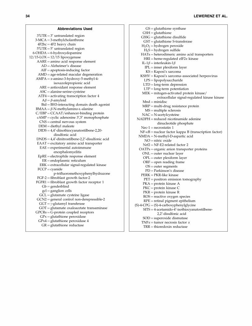
Abbreviations Used
3¢UTR¼ 3¢ untranslated region3-MCA¼ 3-methylcholanthrene
4F2hc¼ 4F2 heavy chain5¢UTR¼ 5¢ untranslated region
6-OHDA¼ 6-hydroxydopamine12/15-LOX¼ 12/15 lipoxygenase
AARE¼ amino acid response elementAD¼Alzheimer’s diseaseAIF¼ apoptosis-inducing factor
AMD¼ age-related macular degenerationAMPA¼ a-amino-3-hydroxy-5-methyl-4-
isoxazolepropionic acidARE¼ antioxidant response elementASC¼ alanine-serine-cysteine
ATF4¼ activating transcription factor 4Ab¼ b-amyloidBid¼BH3-interacting domain death agonist
BMAA¼ b-N-methylamino-l-alanineC/EBP¼CCAAT/enhancer-binding proteincAMP¼ cyclic adenosine 3¢,5¢ monophosphate
CNS¼ central nervous systemDEM¼diethyl maleateDIDS¼ 4,4¢-diisothiocyanatostilbene-2,20-
disulfonic acidDNDS¼ 4,4¢-dinitrostilbene-2,2¢-disulfonic acidEAAT¼ excitatory amino acid transporter
EAE¼ experimental autoimmuneencephalomyelitis
EpRE¼ electrophile response elementER¼ endoplasmic reticulum
ERK¼ extracellular signal-regulated kinaseFCCP¼ cyanide
p-trifluoromethoxyphenylhydrazoneFGF-2¼fibroblast growth factor-2
FGFR1¼fibroblast growth factor receptor 1Gb¼ genderblindgcl¼ ganglion cells
GCL¼ glutamate cysteine ligaseGCN2¼ general control non-derepressible-2
GGT¼ c-glutamyl transferaseGOT¼ glutamate oxaloacetate transaminase
GPCRs¼G-protein coupled receptorsGPx¼ glutathione peroxidase
GPx4¼ glutathione peroxidase 4GR¼ glutathione reductase
GS¼ glutathione synthaseGSH¼ glutathione
GSSG¼ glutathione disulfideGST¼ glutathione S-transferase
H2O2¼hydrogen peroxideH2S¼hydrogen sulfide
HATs¼heterodimeric amino acid transportersHRI¼heme-regulated eIF2a kinase
IL-1b¼ interleukin-1bIPL¼ inner plexiform layerKS¼Kaposi’s sarcoma
KSHV¼Kaposi’s sarcoma–associated herpesvirusLPS¼ lipopolysaccharide
LTD¼ long-term depressionLTP¼ long-term potentiation
MEK¼mitogen-activated protein kinase/extracellular signal-regulated kinase kinase
Mnd¼minidiscMRP¼multi-drug resistance protein
MS¼multiple sclerosisNAC¼N-acetylcysteine
NADPH¼ reduced nicotinamide adeninedinucleotide phosphate
Nec-1¼necrostatin 1NF-jB¼nuclear factor kappa B (transcription factor)
NMDA¼N-methyl-D-aspartic acidNO¼nitric oxide
Nrf2¼NF-E2-related factor 2OATPs¼ organic anion transporter proteins
ONL¼ outer nuclear layerOPL¼ outer plexiform layerORF¼ open reading frame
OS¼ outer segmentsPD¼Parkinson’s disease
PERK¼PKR-like kinasePET¼positron emission tomography
PKA¼protein kinase APKC¼protein kinase CPKR¼protein kinase RROS¼ reactive oxygen speciesRPE¼ retinal pigment epithelium
(S)-4-CPG¼ (S)-4-carboxyphenylglycineSITS¼ 4-acetamido-4¢-isothiocyanatostilbene-
2,2¢-disulfonic acidSOD¼ superoxide dismutase
TNFa¼ tumor necrosis factor aTRR¼ thioredoxin reductase
34 LEWERENZ ET AL.