Embed Size (px)
Citation preview
The cusp of evolution and development: a model of cichlid
tooth shape diversity
J. T. Streelman,a,� J. F. Webb,b R. C. Albertson,a,1 and T. D. Kocher a
aHubbard Center for Genome Studies, 4th Floor, Environmental Technology Building, University of New Hampshire,
35 Colovos Road, Durham, NH 03824, USAbDepartment of Biology, Villanova University, Villanova, PA 19085, USA�Author for correspondence (e-mail: [email protected])
1Present address: Department of Cytokine Biology, The Forsyth Institute, 140 The Fenway, Boston, MA 02115, USA.
SUMMARY Tooth shape is a hallmark of repeated evolu-tionary radiations among cichlid fishes from East Africa. Cuspshape and number vary both within populations and amongclosely related species with different feeding behaviors andecologies. Here, we use histology and scanning electronmicroscopy to chart the developmental trajectory of toothshape differences in fishes from Lake Malawi. We demon-strate that species with bi- or tricuspid adult (replacement) teethinitially possess a first-generation unicuspid dentition. Notably,the timing of turnover from first-generation to replacement teeth
differs among species and is correlated with feeding ecology.Next, we use field data for cichlid species with adult unicuspid,bicuspid, and tricuspid teeth to demonstrate a strong andpositive relationship between the number of teeth in a row andtooth shape.We discuss cichlid tooth ontogeny in the context ofmorphogenetic models designed to explain the developmentalbasis of tooth shape variation in mammals.We suggest that thedramatic differences in cichlid dentitions can be explained byvariation in the expression of common activators and inhibitorsacting at multiple stages of odontogenesis.
INTRODUCTION
The shape of teeth occupies a central position in various
disciplines, from paleontology to ecology to molecular
biology. Comparative odontology is used to identify species
or describe new fossils (Carpenter et al. 1998; Sereno et al.
1999), to test biogeographic hypotheses (Krause et al. 1997),
and to decipher ancient (MacFadden et al. 1999; Dean et al.
2001) as well as recent (MacLeod 2000) ecologies. Similarly,
molecular and developmental biologists consider tooth
development a classic example of tissue (Lumsden 1988), cell
(Chai et al. 2000), and gene interaction (Peters and Balling
1999). Despite a long tradition of study in mammalian model
systems, important questions remain regarding the genetic
and developmental basis of differences in tooth shape. Here,
we take a different approach to the study of tooth shape. We
concentrate on an evolutionary model system that exhibits
tremendous tooth shape diversity. We ask how patterns of
development observed in the laboratory, coupled with data
from natural populations, can help to fill the gaps in our
understanding of vertebrate odontogenesis.
East African cichlids represent a striking example of
adaptive radiation and concomitant divergence in trophic
ecomorphology (Fryer and Iles 1972). Each of the three rift
lakes, Tanganyika, Malawi, and Victoria, houses a mono-
phyletic group of cichlids that has evolved convergent feeding
morphologies in a short period of evolutionary time (Kocher
et al. 1993). For instance, each lake has piscivorous species
with long snouts and gracile jaws used to engulf prey, as well
as species characterized by short and firmly reinforced jaws
used to scrape algae from rock surfaces.
Cichlid teeth are as diverse as their jaws and are key
components of the trophic machinery. Tooth morphology
ranges from widely spaced sharply pointed unicuspids in
zooplanktivorous and insectivorous species (e.g., Cyanoti-
lapia afra) to closely packed tricuspids in algal scrapers (e.g.,
Labeotropheus fuelleborni). The shape of cichlid teeth may
respond rapidly to selection; tooth shape varies among
individuals within populations (Streelman, unpublished
data), characterizes diverging morphs (Tichy and Seegers
1999), and evolves replicatively (Ruber et al. 1999).
Recently, we demonstrated that shape differences in the
first tooth row (bicuspid vs. tricuspid) between Metriaclima
zebra and Labeotropheus fuelleborni are controlled by
changes in a small number of genes (Albertson et al.
2003).
A great deal is known about the development of cichlid
teeth (Huysseune 1990; Huysseune and Sire 1992a,b). Like
most teleost fishes, adult cichlids have (a) multiple rows of
teeth on two sets of jaws (oral and pharyngeal), (b) similarly
EVOLUTION & DEVELOPMENT 5:6, 600–608 (2003)
& BLACKWELL PUBLISHING, INC. 600
shaped teeth within a row (homodonty), and (c) tooth
replacement throughout life via de novo formation of tooth
germs (polyphyodonty). In addition, cichlids (and probably
most teleosts) possess a set of first-generation teeth, which can
be distinguished from replacement teeth by their small size
and rudimentary organization (Huysseune and Sire 1997).
Cichlid teeth pass through developmental stages that are
similar to those described in mammals (e.g., initiation, bud,
cap, bell; Huysseune and Sire 1997; Stock et al. 1997). The
stages of mammalian tooth development are elicited by
specific combinations of targeted gene expression (Stock
et al. 1997; Peters and Balling 1999). It is not known
whether the same signaling molecules choreograph tooth
development in fishes.
A lot has been learned from mammalian models about the
genetic control of tooth initiation. By contrast, less is known
about the genes responsible for differences in tooth shape. In
the words of Peters and Balling (1999), we know ‘‘where and
how to make them,’’ but we are not sure how to explain
patterns of variation within a jaw, within a species, or between
related species. Few gene knockouts alter tooth shape
phenotypes (Pispa et al. 1999), and traditional model
organisms (zebrafish, frog, chick, and mouse) exhibit derived
dentition patterns characterized by the loss or gross
modification of teeth in the oral jaws.
However, recent work allows us to guess the identity of
genes that specify differences in tooth shape. Tucker et al.
(1998b) engineered a functional knockdown of bone mor-
phogenetic proteins (BMPs) in mice by implanting NOGGIN
beads in dental explants. Presumptive incisors treated with
NOGGIN acquired cusps and developed as molars; this
transformation was accompanied by expanded expression of
Barx1 in dental mesenchyme. Jernvall et al. (2000) showed
that correlated gene expression patterns of Fgf4, Lef1, p21,
and Shh could predict differences in molar cusp shapes
between mice and voles. Finally, Jernvall (2000) and Salazar-
Ciudad and Jernvall (2002) presented developmental and
morphogenetic models to explain the ‘‘evolvability’’ of cusps
in mammalian evolution. The models can accurately repro-
duce the diversity of mammalian tooth shape within and
between individuals by varying the concentration of mole-
cular ‘‘activators’’ and ‘‘inhibitors’’ expressed from singular or
multiple signaling centers. Taken together, the results of this
work are consistent with the hypothesis that tooth shape (i.e.,
cusp number and morphology) is controlled by antagonistic
actions of extracellular signaling ligands (e.g., fibroblast
growth factors [FGFs] and BMPs) secreted from transitory
enamel knots (EKs) (Jernvall and Thesleff 2000).
In fact, multiple stages of mammalian tooth development
can be characterized as the balance between the opponent
signaling molecules FGF and BMP. In tooth initiation, the
expression of Bmp4 and Fgf8 in the epithelium control the
mesenchymal expression of Pax9 and Msx1, which direct
tooth formation and position (Neubuser et al. 1997; Tucker
et al. 1998a; Peters and Balling 1999). Later, Bmp4 expression
in the mesenchyme may promote formation of the primary
EK (Jernvall et al. 1998). Finally, BMPs and FGFs secreted
from the EK are candidate activators and inhibitors of cusp
development. The expression of Bmp4 from the primary EK
may inhibit secondary EKs from forming (Jernvall and Jung
2000) and/or regulate (induce) the development of subsequent
EKs (Salazar-Ciudad and Jernvall 2002). Control of EK
number and spacing ultimately determines cusp number and
the sharpness of teeth (Jernvall 2000; Salazar-Ciudad and
Jernvall 2002). The interplay between BMPs and FGFs as
regulators of both tooth initiation and tooth morphogenesis
means that, in homodont species, there may be a relationship
between the number of teeth per row and the number of cusps
per tooth.
We use histology and scanning electron microscopy (SEM)
to characterize the developmental trajectory of oral jaw teeth
in two cichlid species from Lake Malawi, East Africa. We
demonstrate that individuals of both species replace unicuspid
first-generation teeth with a multicusped adult dentition.
Interestingly, the timing of turnover from first-generation to
replacement teeth differs among species. Next, we use field
data for cichlid species with unicuspid, bicuspid, and tricuspid
teeth to demonstrate a strong and positive relationship
between the number of teeth in a row and tooth shape.
Finally, we integrate these results with studies of vertebrate
odontogenesis and propose a model to account for cichlid
evolutionary and developmental tooth shape variation.
MATERIALS AND METHODS
Tooth development in two study speciesOur work on the anatomy, genetics, and development of cichlid
craniofacial differences features two species with divergent feeding
morphologies (Albertson and Kocher 2001; Albertson et al.
2003a,b). Metriaclima zebra (MZ) and Labeotropheus fuelleborni
(LF) are members of Lake Malawi’s rock dwelling ‘‘mbuna’’ and
shared a common ancestor 50,000 to 500,000 years ago (Meyer
et al. 1990). These species represent points along a continuum, from
ram feeding to suction feeding to biting, which likely reflects early
morphological divergence in the rock-dwelling clade (Albertson
et al. 1999; Danley and Kocher 2001). MZ is a widespread species
characterized as a generalist feeder (McKaye and Marsh 1983). It
has a terminal mouth, which it uses to brush diatoms from
attached algae and to suck plankton from the water column
(Reinthal 1990). By contrast, LF feeds on attached material in the
shallows of the surge zone where it uses its inferior-subterminal
mouth to bite or scrape algae from rocks (Reinthal 1990).
Both MZ and LF are maternal mouthbrooders that hold
embryos and posthatching (3–4 days post fertilization) fishes in the
mouth for 21–25 days before release. Newly hatched fry were taken
from brooding females at 4 dpf, cultured in 200-ml flasks at 25–
261C, and moved to small aquaria on day 22. Individuals were
Cichlid tooth shape 601Streelman et al.
fixed on days 7, 10–12, 16–22, 42, 56, and 70 and prepared for SEM
or histology. This sampling regime was chosen because we had
reason to believe that replacement teeth would not appear until
about 6 weeks postfertilization (Huysseune and Sire 1997). There
were no significant differences in growth rate between species over
this period (data not shown).
SEMs of adult teeth were prepared using skeletonized material.
Embryos and juvenile fishes were fixed in 10% formalin in
phosphate-buffered saline, dehydrated in an ascending ethanol
series, and then critical point-dried out of liquid CO2. Specimens
were sputter coated with Au-Pd alloy, photographed using 4� 5
Polaroid film, or digitally captured and postprocessed using Adobe
Photoshop 4.0 (Adobe Systems, Inc., San Jose, CA, USA).
Histological material was prepared by decalcifying whole or
partially dissected fish heads in Cal-Ex (Fisher, Hampton, NH,
USA), rinsing in fresh water, dehydrating in an ascending ethanol-
butyl alcohol series, and then infiltrating and embedding in
Paraplast (Fisher). Blocks were sectioned at 8mm and stained for
bone, cartilage, and connective tissue using the HBQ stain (Hall
1986). Material was digitally photographed at 100–600� and
images postprocessed using Adobe Photoshop 4.0.
Tooth shape and tooth number in natureTo test for an association between cusp number and number of
teeth per tooth row, 10–15 individuals of species from natural
populations, sampled in 2001, were assayed. Species included
Cyanotilapia afra (unicuspid teeth in the first tooth row), MZ
(bicuspid), and LF (tricuspid). The number of teeth in the first
tooth row was determined for both the upper and lower jaws
(premaxillae and dentaries). Here, we concentrate on counts for the
dentaries (premaxillae counts exhibited the same trend). Because
jaw size differs among these species, we measured jaw width (to the
nearest 0.01mm) for all individuals and express number of teeth in
the first tooth row as the number of teeth per millimeter of jaw
width. Analysis of variance (ANOVA) and regression were
performed in Microsoft Excel.
RESULTS
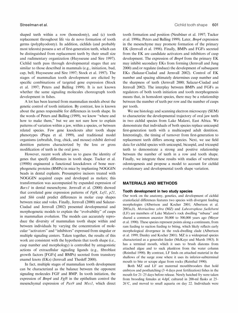
The adult teeth of MZ are bicuspid (with a larger medial
cusp) in the outer-most row and tricuspid (even cusp heights)
in two to three posterior rows on both the dentaries and
premaxillae (Stauffer et al. 1997). LF adults are characterized
by three to five rows of closely spaced tricuspid teeth (with
even cusp heights) on the oral jaws. Notably, F1 of LF and
MZ have teeth intermediate in morphology (Fig. 1, D–F).
By 7dpf, MZ and LF both possess a single row of first-
generation unicuspid teeth (Figs. 1A and 2, A and E). In LF,
Fig. 1. SEM of first-generation and replacement teeth in the lower jaw of Metriaclima zebra (MZ) and Labeotropheus fuelleborni (LF). (A)Lingual view of unicuspid first-generation teeth in LF, 11dpf (scale bar57.5mm). (B) Lingual view of spatulate (transitional) and tricuspidreplacement teeth in LF, 17dpf (scale bar57.5mm). (C) Lingual view of multiple rows of replacement tricuspid teeth in LF, 70dpf (scalebar525mm). D, E, and F are facial views of adult teeth in LF, the F1 hybrid of LF and MZ, and MZ, respectively (scale bars in D, E, andF5100mm).
602 EVOLUTION & DEVELOPMENT Vol. 5, No. 6, November^December 2003

first-generation teeth are replaced by tricuspids beginning
at day 17 (Figs. 1B and 2, B–D). However, in MZ, bicuspid
replacement teeth are not apparent until day 42, approxi-
mately 3 weeks later than in LF (Fig. 2F). In both species, the
complete adult dentition appears as waves of replacement
teeth. For instance, in LF, unicuspids are first replaced by
tricuspids with three sharply pointed cusps, followed by wider
spatulate teeth with rounded cusps. Replacement teeth erupt
first at the midline of the first tooth row and fill in laterally.
By 70 dpf in LF, tricuspid teeth appear in multiple rows
(Fig. 1C).
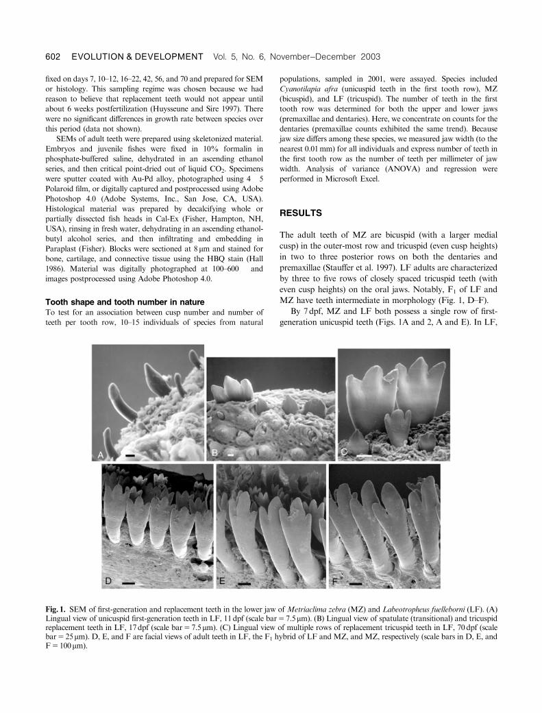
In adult fishes collected in the field, there is a significant
difference among species in the mean number of teeth per
millimeter of jaw width (Fig. 3; ANOVA Po0.0001). This
difference is positively correlated with tooth shape (i.e., cusp
number; r250.995, ANOVA Po0.0001). The mean number
of unicuspid teeth in the first tooth row, per millimeter of jaw
width, for C. afra specimens was 1.370.20 (means7SD). The
mean number of bicuspid teeth, per millimeter of jaw width,
in the first row of MZ individuals was 3.370.52; LF
individuals averaged 4.870.74 tricuspid teeth per millimeter
of jaw width, in the first tooth row. This trend is probably
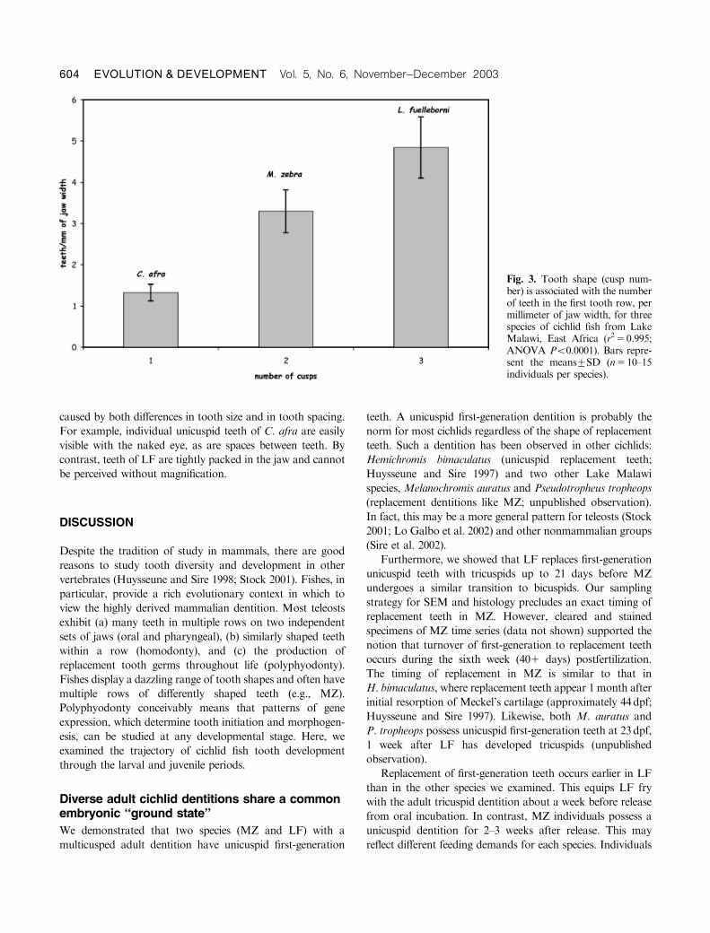
Fig. 2. Histological sections of de-veloping teeth in the upper jawof Metriaclima zebra (MZ) andLabeotropheus fuelleborni (LF). (A)Unicuspid first generation tooth(arrow) in LF, 11 dpf (scale bar510mm). (B) Tricuspid replacementtooth in LF, 17 dpf (scale bar510mm). (C) Oblique cross-sectionthrough tricuspid tooth, showingsurrounding odontoblasts (arrow)in LF, 20 dpf (scale bar510mm).(D) Tricuspid tooth in LF, 42dpf(scale bar520mm). (E) Cross-sec-tion through unicuspid first-genera-tion tooth (arrow) in MZ, 22dpf(LF already has tricuspid teeth atthis age) (scale bar515mm). (F)Bicuspid teeth in MZ, 70dpf (scalebar540mm).
Cichlid tooth shape 603Streelman et al.
caused by both differences in tooth size and in tooth spacing.
For example, individual unicuspid teeth of C. afra are easily
visible with the naked eye, as are spaces between teeth. By
contrast, teeth of LF are tightly packed in the jaw and cannot
be perceived without magnification.
DISCUSSION
Despite the tradition of study in mammals, there are good
reasons to study tooth diversity and development in other
vertebrates (Huysseune and Sire 1998; Stock 2001). Fishes, in
particular, provide a rich evolutionary context in which to
view the highly derived mammalian dentition. Most teleosts
exhibit (a) many teeth in multiple rows on two independent
sets of jaws (oral and pharyngeal), (b) similarly shaped teeth
within a row (homodonty), and (c) the production of
replacement tooth germs throughout life (polyphyodonty).
Fishes display a dazzling range of tooth shapes and often have
multiple rows of differently shaped teeth (e.g., MZ).
Polyphyodonty conceivably means that patterns of gene
expression, which determine tooth initiation and morphogen-
esis, can be studied at any developmental stage. Here, we
examined the trajectory of cichlid fish tooth development
through the larval and juvenile periods.
Diverse adult cichlid dentitions share a commonembryonic ‘‘ground state’’
We demonstrated that two species (MZ and LF) with a
multicusped adult dentition have unicuspid first-generation
teeth. A unicuspid first-generation dentition is probably the
norm for most cichlids regardless of the shape of replacement
teeth. Such a dentition has been observed in other cichlids:
Hemichromis bimaculatus (unicuspid replacement teeth;
Huysseune and Sire 1997) and two other Lake Malawi
species, Melanochromis auratus and Pseudotropheus tropheops
(replacement dentitions like MZ; unpublished observation).
In fact, this may be a more general pattern for teleosts (Stock
2001; Lo Galbo et al. 2002) and other nonmammalian groups
(Sire et al. 2002).
Furthermore, we showed that LF replaces first-generation
unicuspid teeth with tricuspids up to 21 days before MZ
undergoes a similar transition to bicuspids. Our sampling
strategy for SEM and histology precludes an exact timing of
replacement teeth in MZ. However, cleared and stained
specimens of MZ time series (data not shown) supported the
notion that turnover of first-generation to replacement teeth
occurs during the sixth week (401 days) postfertilization.
The timing of replacement in MZ is similar to that in
H. bimaculatus, where replacement teeth appear 1 month after
initial resorption of Meckel’s cartilage (approximately 44dpf;
Huysseune and Sire 1997). Likewise, both M. auratus and
P. tropheops possess unicuspid first-generation teeth at 23dpf,
1 week after LF has developed tricuspids (unpublished
observation).
Replacement of first-generation teeth occurs earlier in LF
than in the other species we examined. This equips LF fry
with the adult tricuspid dentition about a week before release
from oral incubation. In contrast, MZ individuals possess a
unicuspid dentition for 2–3 weeks after release. This may
reflect different feeding demands for each species. Individuals
Fig. 3. Tooth shape (cusp num-ber) is associated with the numberof teeth in the first tooth row, permillimeter of jaw width, for threespecies of cichlid fish from LakeMalawi, East Africa (r250.995;ANOVA Po0.0001). Bars repre-sent the means7SD (n510–15individuals per species).
604 EVOLUTION & DEVELOPMENT Vol. 5, No. 6, November^December 2003
of LF have a subterminal mouth with a strongly overslung
upper jaw and may be morphologically incapable of feeding
by suction. Alternatively, MZ juveniles probably feed on
plankton from the water column, a predominant mode of
feeding for larval and juvenile fishes (Liem 1991) and a
preferred mode of MZ adults given an abundance of plankton
(Reinthal 1990).
Both MZ and LF replace a unicuspid first-generation
dentition (one row) with multiple rows of multicusped adult
teeth. This suggests that, for these species, patterns of gene
expression governing both tooth initiation and morphogenesis
vary through ontogeny. Moreover, once the pattern of gene
expression corresponding to the adult dentition is generated,
it must be replicated with each wave of replacement to
faithfully maintain adult dental patterns. It has been
estimated that the functional life of an adult tooth is 101
days in the cichlid Tilapia mariae (Tuisku and Hildebrand
1994). Thus, a 5-year-old individual will have replaced its
adult dentition at least 18 times. Additional changes in gene
expression during adult tooth replacement should result in
ontogenetic variation in tooth shape and patterning.
Ontogenetic shifts in tooth morphology are known for a
few cichlids for both oral (Chilotilapia rhoadesii) and phary-
ngeal (Haplochromis incola) teeth (Fryer and Iles 1972). These
changes are usually associated with differing feeding demands
for juveniles versus adults (e.g., soft vs. hard prey). Since the
1960s, biologists have known that certain cichlid species (e.g.,
Astatoreochromis alluaudi, Cichlasoma citrinellum and mana-
guense) have distinct adult morphs with divergent pharyngeal
jaw shapes and dentition (Greenwood 1965; Meyer 1987).
This phenomenon likely has a strong environmental compo-
nent and may be induced by compressive forces on the
pharyngeal bones themselves (Huysseune et al. 1994).
It is not known whether phenotypic plasticity plays a role
in generating different oral jaw tooth shapes. Tuisku and
Hildebrand (1994) demonstrated that the development of
replacement tooth germs in the cichlid lower jaw is dependent
on mandibular innervation. It is possible that such neural
input directs not only the process of replacement but, in
certain cases, the shape of new teeth as well. This might result
in rapid changes in dentition given changing feeding
behaviors. Polyphyodonty, coupled with the potential to
modulate gene expression over ontogeny, may provide
cichlids with the means to alter tooth shape in the face of
fluctuating environmental conditions.
A model of cichlid tooth shape diversity
The developmental trajectories and evolutionary diversity of
cichlid teeth raises several questions. How are the shapes of
first-generation versus replacement teeth specified? How do
polyphyodont species maintain the fidelity of multiple
generations of replacement teeth? How do we explain the
presence of multiple rows of differently shaped teeth within
the same jaw? In what follows, we offer a model to account
for the diversity of cichlid tooth shapes. The model assumes
that the molecules governing tooth development in mammals
have similar functions in fishes. We acknowledge that there is
no direct evidence of this. However, despite the differences
discussed above, the structural basis of tooth organ develop-
ment is similar between fishes and mammals (Huysseune and
Sire 1998). The developmental blueprint of other structures,
involving the same molecular players (e.g., FGFs, BMPs,
Wnts, Shh) is conserved across vertebrate classes (Jung
et al. 1999; Poss et al. 2000; Capdevila and Izpisua Belmonte
2001). Our model borrows heavily from other conceptions
of reaction-diffusion systems to describe periodic patterning
of feather primordia (Jung et al. 1998), tongue papillae
(Jung et al. 1999), and primate molars (Jernvall and Jung
2000).
The simplicity of our model is motivated by the positive
relationship between number of teeth in the first tooth row
and tooth shape, maintained in natural cichlid populations
(Fig. 3). This association, coupled with the iterative role of
certain signaling molecules (e.g., BMPs, FGFs) in mammalian
tooth development, suggests that variation in a common set
of these factors might explain differences in both aspects of
the cichlid dentition.
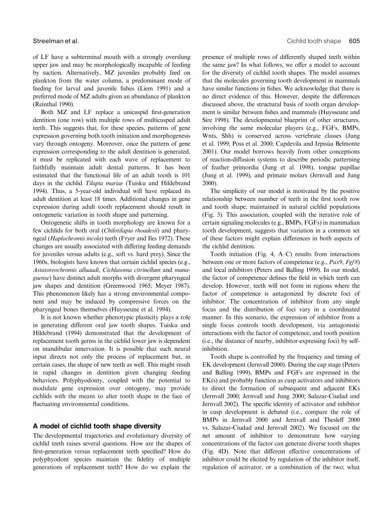
Tooth initiation (Fig. 4, A–C) results from interactions
between one or more factors of competence (e.g., Pax9, Fgf8)
and local inhibitors (Peters and Balling 1999). In our model,
the factor of competence defines the field in which teeth can
develop. However, teeth will not form in regions where the
factor of competence is antagonized by discrete foci of
inhibitor. The concentration of inhibitor from any single
focus and the distribution of foci vary in a coordinated
manner. In this scenario, the expression of inhibitor from a
single focus controls tooth development, via antagonistic
interactions with the factor of competence, and tooth position
(i.e., the distance of nearby, inhibitor-expressing foci) by self-
inhibition.
Tooth shape is controlled by the frequency and timing of
EK development (Jernvall 2000). During the cap stage (Peters
and Balling 1999), BMPs and FGFs are expressed in the
EK(s) and probably function as cusp activators and inhibitors
to direct the formation of subsequent and adjacent EKs
(Jernvall 2000; Jernvall and Jung 2000; Salazar-Ciudad and
Jernvall 2002). The specific identity of activator and inhibitor
in cusp development is debated (i.e., compare the role of
BMPs in Jernvall 2000 and Jernvall and Thesleff 2000
vs. Salazar-Ciudad and Jernvall 2002). We focused on the
net amount of inhibitor to demonstrate how varying
concentrations of the factor can generate diverse tooth shapes
(Fig. 4D). Note that different effective concentrations of
inhibitor could be elicited by regulation of the inhibitor itself,
regulation of activator, or a combination of the two; what
Cichlid tooth shape 605Streelman et al.
Fig. 4. Variation in cichlid toothnumber and shape can be explainedby gradients in the net concentrationof a single inhibitory factor duringtwo stages of tooth development.(A–C) Tooth development at theinitiation stage; (D) tooth develop-ment at the time of morphogenesis.(A–C) Cross-section through thefirst tooth row with the tip of thejaw toward the top of the page. (D)Inhibitory signal from the primaryenamel knot (EK) and resultingtooth morphologies. (A–C) Place-ment of teeth is determined by afactor(s) of competence that definesthe area in which teeth can develop(in orange), as well as localized fociof inhibitor (blue). Teeth develop inareas of orange that are not affectedby areas of blue. (D) Likewise, thedevelopment of secondary EKs, andthus cusps, is dependent on theinhibitory signal from the primaryEK. In all panels, the size of bluecircles represents the concentration(strength) of inhibitor. The first-generation dentition of all cichlids(A) is characterized by a single rowof widely spaced uni-cuspid teeth,resulting from high concentrationsof inhibitor from distantly spacedfoci and a high concentration ofinhibitor from the EK (D, top). Inspecies like C. afra, with a unicuspidadult dentition, the concentration ofinhibitor does not change with toothreplacement. A second row of uni-cuspid teeth is initiated by a repli-cated stripe of competence andinhibitory factors. In species withmulticusped replacement teeth, theconcentration of inhibitor decreaseswith tooth turnover (B and C). Thisdecrease is greater in posterior toothrows for MZ, setting the stage for anouter row of bicuspid teeth withinner rows of greater numbers oftricuspids. Note that the adult bi-cuspid tooth of MZ is asymmetric(D, middle), implying an asymmetricfield of inhibition (stronger at themidline). In LF adults (C and D,bottom) the decrease in concentra-tion is strong and uniform, generat-ing multiple rows of tightly packedtricuspid teeth.
606 EVOLUTION & DEVELOPMENT Vol. 5, No. 6, November^December 2003
matters in the model is that the ratio of activator to inhibitor
is dynamic.
All cichlids examined to date have widely spaced,
unicuspid, first-generation teeth (Huysseune and Sire 1997).
According to our model, this ground state is produced by
relatively few foci expressing inhibitor at relatively high levels.
For species with unicuspid adult teeth (C. afra), the
concentration of inhibitor does not change with tooth
replacement. Alternatively, in species replacing unicuspid
with multicusped teeth, (e.g., MZ and LF, Fig. 4, B and C),
the concentration of inhibitor from single foci, and likewise
the number of foci, should change with tooth turnover. A
decrease in the concentration of inhibitor from any single
focus will remove the self-inhibitory field toward neighboring
foci and reduce the inhibitory signal toward the factor(s) of
competence. In cichlids and most teleosts, teeth near the
midline develop before those in lateral and posterior
positions. In the context of our model, this means that the
position of any tooth is most likely regulated by foci located
medial to the site of tooth development.
A similar decrease in the concentration of inhibitor from
the primary EK removes the inhibitory field toward
subsequent EKs (Fig. 4D). Therefore, lower concentrations
of inhibitor during both tooth initiation and morphogenesis
would result in more replacement teeth per row with more
cusps. A direct prediction of the model, then, is that the
number of first-row teeth per millimeter of jaw width should
increase through ontogeny in species like MZ and LF. A
second related prediction is that the cusps of tricuspid teeth
should be closer to one another than the cusps of bicuspid
teeth. In fact, this seems to be borne out by inspection of the
distance between cusps in the adult dentition of LF vs. MZ
(Fig. 1, D and F).
Most adult cichlids have teeth in multiple rows. This
means that a second (and sometimes third, fourth, or fifth)
region of the jaw gains odontogenic potential. In species
like C. afra, this second row is patterned like the first (e.g.,
Fig. 4A); the result of another wave of inhibitor in relatively
high concentration, which generates well-spaced foci. In
species like MZ, with adult bicuspids in the first row and
multiple rows of tightly packed tricuspids behind, a gradient
of inhibitor along the facial-lingual axis can explain the
variation in tooth shape and spacing among rows. Similarly in
LF, our model would predict that multiple rows of closely
spaced tricuspid teeth are generated by a consistent decrease
in levels of inhibitor across each tooth row.
Given the role of Bmp4 in both tooth initiation and tooth
morphogenesis, coupled with the inhibitory action of this
signaling ligand in other reaction-diffusion models (Jung et al.
1998, 1999; Jernvall and Jung 2000), it is tempting to speculate
that this is the molecule responsible for the coordinated
changes (i.e., tooth placement and tooth shape) in the cichlid
dentition. If so, our model would help to resolve an apparent
paradox in vertebrate tooth development. Stock (2001)
observed that in the mouse, Bmp4 has the opposing functions
of inhibiting tooth initiation (Neubuser et al. 1997) and
specifying incisor identity (Tucker et al. 1998b). We suggest a
different interpretation: BMP4 plays an ancestral inhibitory
role in both tooth initiation and tooth morphogenesis. The
molecule does not ‘‘specify’’ incisor identity per se but rather
inhibits secondary cusp development. Secondary cusps do not
develop either because Bmp4 is an inhibitor as modeled by
some (Jung et al. 1998; Jernvall and Thesleff 2000; Jernvall
and Jung 2000) or because the molecule is an activator
(Salazar-Ciudad and Jernvall 2002) that induces a particularly
large primary EK. Testing the role of putative inhibitors and
activators, during various stages of tooth development, may
be informative in a system in which more than two tooth
types occur.
Our model should direct research aimed to explain the
genetic basis of differences in cichlid dentitions. For instance,
we demonstrated that tooth shape differences have a simple
genetic basis (Albertson et al. 2003a,b). Tooth shape in
LF�MZ F2 does not associate with RFLP variation in the
Bmp4 gene (Albertson et al. 2003). This suggests that the
genetic factor(s), which hypothetically modulate BMP4
concentrations in the developing dentition, operate in trans
(i.e., the hypothesized differences in BMP4 concentration are
not caused by mutations in the Bmp4 gene). Despite evidence
for genetic control of tooth shape, we need a better
understanding of the heritability of dentition patterns in
cichlids and the environmental conditions that might induce
plasticity. Finally, the ability to culture cichlid dental explants
(Koumans and Sire 1996) provides a means to directly
evaluate the role of candidate factors in cichlid tooth initiation
and morphogenesis.
AcknowledgmentsWe thank members of the Kocher laboratory and two anonymousreviewers for comments on previous versions of this manuscript. Thiswork was supported by grants from the NIH (R03 DE 14446-01 toJ. T. S.) and NSF (IBN 9905127 to T. D. K.).
REFERENCES
Albertson, R. C., and Kocher, T. D. 2001. Assessing morphologicaldifferences in an adaptive trait: a landmark-based morphometricapproach. J. Exp. Zool. 289: 385–403.
Albertson, R. C., Markert, J. A., Danley, P. D., and Kocher, T. D. 1999.Phylogeny of a rapidly evolving clade: the cichlid fishes of Lake Malawi,East Africa. Proc. Natl. Acad. Sci. USA 96: 5107–5110.
Albertson, R. C., Streelman, J. T., and Kocher, T. D. 2003a. Directionalselection has shaped the oral jaws of Lake Malawi cichlid fishes. Proc.Natl. Acad. Sci. USA 100: 5252–5257.
Albertson, R. C., Streelman, J. T., and Kocher, T. D. 2003b. Genetic basisof adaptive differences in the cichlid head. J. Hered. 94: 291–301.
Carpenter, K., Miles, C., and Cloward, K. 1998. Skull of a Jurassicankylosaur. Nature 393: 782–783.
Cichlid tooth shape 607Streelman et al.
Capdevila, J., and Izpisua Belmonte, J. C. 2001. Patterning mechanismscontrolling vertebrate limb development. Annu. Rev. Cell. Dev. Biol. 17:87–132.
Chai, Y., et al. 2000. Fate of the mammalian cranial neural crest duringtooth and mandibular morphogenesis. Development 127: 1671–1679.
Danley, P. D., and Kocher, T. D. 2001. Speciation in rapidly divergingsystems: lessons from Lake Malawi. Mol. Ecol. 10: 1075–1086.
Dean, C., et al. 2001. Growth processes in teeth distinguish humans fromHomo erectus and earlier hominins. Nature 414: 628–631.
Fryer, G., and Iles, T. D. 1972. The Cichlid Fishes of the Great Lakes ofAfrica: Their Biology and Evolution. Oliver and Boyd, Edinburgh.
Greenwood, P. H. 1965. Environmental effects on the pharyngeal mill of acichlid fish, Astatoreochromis alluaudi, and their taxonomic implications.Proc. Linn. Soc. Lond. 176: 1–10.
Hall, B. K. 1986. The role of movement and tissue interactions in thedevelopment and growth of bone and secondary cartilage in the clavicleof the embryonic chick. J. Embryol. Exp. Morph. 93: 133–152.
Huysseune, A. 1990. Development of the anterior part of the mandible andthe mandibular dentition in two species of Cichlidae (Teleostei). Cybium14: 327–344.
Huysseune, A., and Sire, J.-Y. 1992a. Development of cartilage and bonetissues of the anterior part of the mandible in cichlid fish: a light andTEM study. Anat. Rec. 233: 357–375.
Huysseune, A., and Sire, J.-Y. 1992b. Bone and cartilage resorption inrelation to tooth development in the anterior part of the mandible incichlid fish: a light and TEM study. Anat. Rec. 234: 1–14.
Huysseune, A., and Sire, J.-Y. 1997. Structure and development of first-generation teeth in the cichlid Hemichromis bimaculatus (Teleostei,Cichlidae). Tissue Cell 29: 679–697.
Huysseune, A., and Sire, J.-Y. 1998. Evolution of patterns and processes inteeth and tooth-related tissues in non-mammalian vertebrates. Eur. J.Oral. Sci. 106 (suppl. 1): 437–481.
Huysseune, A., Sire, J.-Y., and Meunier, F. J. 1994. Comparative study oflower pharyngeal jaw structure in two phenotypes of Astatoreochromisalluaudi (Teleostei:Cichlidae). J. Morph. 221: 25–43.
Jernvall, J. 2000. Linking development with generation of novelty inmammalian teeth. Proc. Natl. Acad. Sci. USA 97: 2641–2645.
Jernvall, J., Aberg, T., Kettunen, P., Keranen, S., and Theslef, I. 1998. Thelife history of an embryonic signaling center: BMP-4 induces p21 and isassociated with apoptosis in the mouse tooth enamel knot. Development125: 161–169.
Jernvall, J., Keranen, S. V. E., and Theslef, I. 2000. Evolutionary modi-fication of development in mammalian teeth: quantifying gene expressionpatterns and topography. Proc. Natl. Acad. Sci. USA 97: 14444–14448.
Jernvall, J., and Jung, H.-S. 2000. Genotype, phenotype and thedevelopmental biology of molar tooth characters. Y. Phys. Anthropol.48: 171–190.
Jernvall, J., and Thesleff, I. 2000. Reiterative signaling and patterningduring mammalian tooth morphogenesis. Mech. Dev. 92: 19–29.
Jung, H. S., et al. 1998. Local inhibitory action of BMPs and theirrelationships with activators in feather formation: implications forperiodic patterning. Dev. Biol. 196: 11–23.
Jung, H. S., Oropeza, V., and Thesleff, I. 1999. Shh, Bmp-2, Bmp-4 andFgf-8 are associated with initiation and patterning of mouse tonguepapillae. Mech. Dev. 81: 179–182.
Kocher, T. D., Conroy, J. A., McKaye, K. R., and Stauffer, J. R. 1993.Similar morphologies of cichlids in lakes Tanganyika and Malawi aredue to convergence. Mol. Phylogen. Evol. 2: 158–165.
Koumans, J. T. M., and Sire, J.-Y. 1996. An in vitro, serum-free organculture technique for the study of development and growth of the dermalskeleton in fish. In Vitro Cell. Dev. Biol. Anim. 32: 612–626.
Krause, D. W., Prasad, G. V. R., von Koenigswald, W., Sahni, A., andGrine, F. E. 1997. Cosmopolitanism among Gondwanan Late Cretac-eous mammals. Nature 390: 504–507.
Liem, K. F. 1991. A functional approach to the development of the head ofteleosts: implications on constructional morphology and constraints. InN. Schmidt-Kittler and K. Vogel (eds.). Constructional Morphology andEvolution. Springer-Verlag, Berlin, pp. 231–249.
Lo Galbo, A. M., Carpenter, K. E., and Reed, D. L. 2002. Evolution oftrophic types in emperor fishes (Lethrinus, Letherinidae, Percoidei) basedon cytochrome b gene sequence variation. J. Mol. Evol. 54: 754–762.
Lumsden, A. G. S. 1988. Spatial organization of the epithelium and the roleof neural crest cells in the initiation of the mammalian tooth germ.Development 103 (suppl.): 155–169.
MacFadden, B. J., Solounias, N., and Cerling, T. E. 1999. Ancient diets,ecology, and extinction of 5-million-year-old horses from Florida.Science 283: 824–827.
MacLeod, C. D. 2000. Species recognition as a possible function for thevariations in position and shape of the sexually dimorphic tusks ofMesoplodon whales. Evolution 54: 2171–2173.
McKaye, K. R., and Marsh, A. 1983. Food switching by two specializedalgae-scraping cichlid fishes in Lake Malawi, Africa. Oecologia 56: 245–248.
Meyer, A. 1987. Phenotypic plasticity and heterochrony in Cichlasomamanaguense (Pisces, Cichlidae) and their implications for speciation incichlid fishes. Evolution 41: 1357–1369.
Meyer, A., Kocher, T. D., Basasibwaki, P., and Wilson, A. C. 1990.Monophyletic origin of Lake Victoria cichlid fishes suggested bymitochondrial DNA sequences. Nature 347: 550–553.
Neubuser, A., Peters, H., Balling, R., and Martin, G. R. 1997. Antagonisticinteractions between FGF and BMP signaling pathways: a mechanismfor positioning the sites of tooth formation. Cell 90: 247–255.
Peters, H., and Balling, R. 1999. Teeth: where and how to make them.Trends Genet. 15: 59–65.
Pispa, J., et al. 1999. Cusp patterning defects in Tabby mouse teeth and itspartial rescue by FGF. Dev. Biol. 216: 521–534.
Poss, K. D., et al. 2000. Roles for Fgf signaling during zebrafish finregeneration. Dev. Biol. 222: 347–358.
Reinthal, P. N. 1990. The feeding habits of a group of tropical herbivorousrock-dwelling cichlid fishes (Cichlidae: Perciformes) from Lake Malawi,Africa. Env. Biol. Fish. 27: 215–223.
Ruber, L., Verheyen, E., and Meyer, A. 1999. Replicated evolution oftrophic specializations in an endemic cichlid fish lineage from LakeTanganyika. Proc. Natl. Acad. Sci. USA 31: 10230–10235.
Salazar-Ciudad, I., and Jernvall, J. 2002. A gene network model accountingfor development and evolution of mammalian teeth. Proc. Natl. Acad.Sci. USA 99: 8116–8120.
Sereno, P. C., et al. 1999. Cretaceous sauropods from the Sahara and theuneven rate of skeletal evolution among dinosaurs. Science 286: 1342–1347.
Sire, J.-Y., Davit-Beal, T., Delgato, S., Van der Hayden, C., andHuysseune, A. 2002. First-generation teeth in non-mammalian lineages:evidence for a conserved ancestral character. Microsc. Res. Tech. 59:408–434.
Stauffer, J. R., Bowers, N. J., Kellogg, K. A., and McKaye, K. R. 1997. Arevision of the blue-black Pseudotropheus zebra (Teleostei: Cichlidae)complex from Lake Malawi, Africa, with a description of a new genusand ten new species. Proc. Acad. Natl. Sci. Philos. 148: 189–230.
Stock, D. W. 2001. The genetic basis of modularity in the development andevolution of the vertebrate dentition. Philos. Trans. R. Soc. Lond. B 356:1633–1653.
Stock, D. W., Weiss, K. M., and Zhao, Z. 1997. Patterning of themammalian dentition in development and evolution. Bioessays 19: 481–490.
Tichy, H., and Seegers, L. 1999. The Oreochromis alcalicus flock (Teleostei:Cichlidae) from lakes Natron and Magadi, Tanzania and Kenya: amodel for the evolution of ‘‘new’’ species flocks in historical times?Ichthyol. Explor. Freshw. 10: 147–174.
Tucker, A. S., Al Khamis, A., and Sharpe, P. T. 1998a. Interactionsbetween Bmp-4 and Msx-1 act to restrict gene expression to odontogenicmesenchyme. Dev. Dyn. 212: 533–539.
Tucker, A. S., Matthews, K. L., and Sharpe, P. T. 1998b. Transformationof tooth type induced by inhibition of BMP signaling. Science 282: 1136–1138.
Tuisku, F., and Hildebrand, C. 1994. Evidence for a neural influence ontooth germ generation in a polyphyodont species. Dev. Biol. 165: 1–9.
608 EVOLUTION & DEVELOPMENT Vol. 5, No. 6, November^December 2003