Embed Size (px)
Citation preview
of Author^
The Cure of Vesico-Vaginal Fistula by theFree Dissection of the Bladder from its
Vaginal Attachments and Closurewith the Buried Continuous
Tendon Suture.
Presented in the Section on Obstetrics and Diseases of Women, at theForty-eighth Annual Meeting of the American Medical Association,
held at Philadelphia, Pa., June 1-4,1897.
HENRY O. MARCY, A.M., M.D., LL.D.BOSTON, MASS.
REPRINTED FROMTHE JOURNAL OF THE AMERICAN MEDICAL ASSOCIATION.
NOVEMBER 20, 1897.
CHICAGO:Ameeican Medical Association Press.
1897.

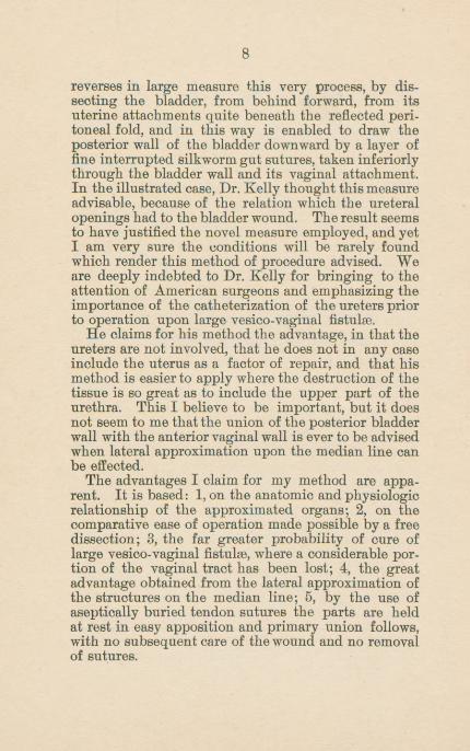
THE CURE OF VESICO-VAGINAL FISTULABY THE FREE DISSECTION OF THE
BLADDER FROM ITS VAGINALATTACHMENTS AND CLOS-
URE WITH THE BURIEDCONTINUOUS TEN-
DON SUTURE.
HENRY O. MARCY, A.M., M.D., LL.D.
In 1887 my attention was first called to the easewith which the bladder wall will reunite after injury,under the most adverse circumstances, when the por-tion involved is free within the peritoneal cavity.The case upon which I operated at this time was thatof a child eighteen months old, where an abscess inthe vicinity of the appendix had resulted in a vesico-intestinal fistulous opening in the appendiceal region.
Laparotomy disclosed two fistulse of the small intes-tine, complicated with an opening into the bladder tothe right of the fundus. The intestines were freedfrom adhesions and drawn out of the wound. Therefreshed edges of the openings were closed by doublelines of fine continuous tendon sutures, w Thich inturn were intrafolded by a second layer of continu-ous tendon Lembert sutures. After the intestine hadbeen returned, the bladder wall was freed from itsadhesions, the opening into it refreshed and suturedin double lines of continuous tendon sutures, in pre-cisely the same manner as I had closed the intestinalwounds.
The child made an easy and perfect recovery, withprimary union of the openings, and is today a strong,vigorous girl. The age of the child rendered drain-
2
age of the bladder unsuccessful, but from the first thefunction of the organ seemed little impaired.
The case was instructive in many ways. Perhapsthe most important lesson was theprimary restorationof the bladder wall without the supposed necessaryphysiologic rest of the organ. The method of apply-ing the first row of sutures in the approximation of therefreshed edges, held in even support by means of thecontinuous sutures, inverted the mucous membrane.
The insertion of the suture in both intestinal andbladder walls is in the following manner; The needleis a fine one, curved upon the flat, with eye near thepoint, and set in a firm handle, made to penetrate thecoats of the bladder or the intestine also, but notthrough the mucous membrane. It emerges aboutone-quarter of an inch from the line of the opening,and is introduced upon the opposite side at a like dis-tance. The needle is then unthreaded, rethreadedwith the opposite end of the suture and withdrawn.This makes the needle a suture carrier like a shuttle,and holds in even coaptation the included structuresin a double loop from side to side (shoemaker’s stitch).One stitch follows another in an even continuousseam, until the opening is completely closed. Bythis side to side coaptation, the parts enclosed areheld in juxtaposition the breadth of the enclosingloop, while the interrupted suture holds only thewidth of the suture material. The submucous pene-tration of the needle buries the suture in healthyvitalized structures and the line of stitches, taken aquarter of an inch away from the edges of the wound,inverts the mucous membrane into the bladder. Thefinal fixation of the suture is by one knot, a manifestadvantage over leaving as many knots as stitches,while the continuous suture permits of an even ad-justment of the enclosing force. It mast be remem-bered that no undue force is used in the tightening ofthe stitches, since readjustment, retention and restwith a minimum of devitalization, are the factors inthe relation of the sutured parts as important as inthe treatment of a fractured bone.
3
The exterior line of continuous Lembert suturesintrafolds the peritoneum so as to re-enforce andgreatly strengthen the structures over the wound.When taken properly they are themselves buriedwithout puckering of the peritoneum (parallel con-tinuous suture).
From a variety of causes, quite a number of timeswithin the last ten years, I have closed wounds in thebladder from within the peritoneal cavity as aboveoutlined, and in every instance primary restorationhas occurred. But in the entire series I have beenenabled to drain the bladder for a number of daysthrough the urethra.
During this period, in the larger number of casesof vesico-vaginal fistula, I have closed the woundthrough the vaginal opening by lines of buried ten-don sutures, with varying results. Little by little Ihave found that both ease of operation and certitudeof outcome seemed dependent on the freedom ofdissection and the amount of tissue held in fixation.Especially has it appeared dependent, in considerabledegree, on the extent of the dissection of the bladderfrom its vaginal attachments. Then the importanceof the lesson taught me by the safe repair of thebladder wounds from within the abdominal cavitybecame apparent. From this standpoint I reviewedwith renewed interest the entire history of surgicaloperations undertaken for the cure of vesico-vaginalfistula, from the days of Sims and Bozemann in thiscountry and Jobert and Simon of Europe.
The various modifications in technic and detail ofthese great masters, save the introduction of anti-septic measures, has been that, little by little, withoutthe recognition of the underlying anatomic reason,the successful cure of large vaginal fistulse has beendependent on the greater freedom of dissection bywhich, the bladder has been separated from its vaginalattachments.
I can not myself question that the primary causeof failure, the surgical technic having been satisfac-tory, lies in the anatomic conditions, the importance
4
of which, a study of the physiologic functions ofthese different organs makes easily apparent. Thecontraction of the circular fibers of the vaginal mus-cle tends constantly to pull upon the base of thebladder, and these muscular contractions are necessa-rily called into special activity following the irritationincident to the repair of a vesico-vaginal fistula. Whenboth the bladder wall and the vaginal muscle areunited by sutures, holding them in approximation,the contraction of the vaginal muscle not alone pullsupon the suture, causing it to cut through its ownstructure, but at the same time acts with equallydestructive force upon the enclosed bladder wall. Ibelieve that it is on this account that the great major-ity of failures to cure vesico-vaginal fistulse ensue.
Reflection on the diverse physiologic relationshipof the parts involved must lead to the conclusionthat, at least, this is an hitherto overlooked importantfactor for surgical consideration, and I now invite theprofession to give it due consideration in order toprofit, as far as possible, from its better understanding,by the adaptation of improved surgical methods.
Every one who is familiar with anterior colporrhaphyfor cystocele knows the comparative ease and safetywith which a large portion of the vaginal muscle maybe dissected from the dependent posterior bladderwall, and the almost sure complete primary unionwhich ensues upon its fixation and closure.
It is safe, and not very difficult, having split theedges of a refreshed vesico-vaginal fistula, to separatewidely the bladder from the vagina. When this hasbeen effected, but not until the dissection is complete,the opening in the bladder wall is refreshed and closedby a double line of continuous sutures, as alreadydescribed,carefully avoiding penetration of the mucousmembrane, using for suture material a fine tendon.
By the use of a needle with the eye near the point,the suture is carried back and forth through thesame stitch opening, precisely as the shoemaker sewsleather.
The opening in the viscus is then closed by an even,
5
uniform support, coaptating but not unduly compress-ing the enclosed structures A single knot fixes thesuture, a fact of considerable importance when thematerial used is to be left buried. The free dissectionmakes the union of the bladder wall a comparativelyeasy operation, and even when a large portion of thevaginal vault has been lost, there is little difficulty ineffecting an easy uniform closure without tension ofthe bladder wall. As every operator knows, in largefistulse, when thevaginal structures are enclosed withthe bladder wall, this part of the operation is exceed-ingly difficult, and the tension upon the parts whichfollows is the usual cause of failure.
Therefore, again I emphasize the necessity of freedissection which, to the timid operator, at the outsetseems at least to border on the verge of rashness.
The bladder wall having been closed, we have re-maining an operation not unlike that ordinarily under-taken for the cure of cystocele, with the exceptionthat oftentimes the remaining vaginal structures areunduly minimized. How these structures shall beclosed, so far as the cure of the fistula is concerned,is comparatively of minor importance. It is wise,however, to restore them as nearly as possible to theirprimal condition. I think that this is best effectedin a general way, a method which for many years Ihave continuously practiced, by the use of a singletendon suture applied with an Hagedorn needle, using alacing stitch, the sutures being deeply imbeddedfromside to side. In this way the vaginal wall is coaptatedin two or three layers and the mucous surfaces ofthe vagina are carefully approximated. This resultsin an entirely closed wound, the antero-posterior diam-eter of which has been considerably increased; in thefirst place, by the inversion of the mucous membraneof the bladder; in the second place, the approxima-tion of the widely denuded structures upon the medianline; and in the third place, by the inversion of theapproximated vaginal surfaces of the wound.
Fistula in which the cervical portion of the uterusis involved comes easily under the same general plan
6
of operative measures. The orgau must be freelyseparated from its attachments.
Recto-vaginal fistulse are operated upon by essen-tially the same method. From below through the peri-neum the vagina is freely separated from the bowel.The opening in each is closed precisely as the open-ing in the bladder wall; then the perineum is restoredby layers of buried tendon sutures. I have long prac-ticed this method almost without failure, a descriptionof which in careful detail published years ago.
Suprapubic cystotomy in the male furnishes itscorroborative testimony of importance in this direc-tion, Here a large wound of the bladder is made withseeming impunity for a variety of reasons, the bladderwall independently closed, the superadjacent structuresunited in layers, the skin rejoined by a buried sutureand the wound sealed without drainage. The supra-pubic attachment of the bladder is by loose elasticstructures which furnish the conditions favorable foreasy retention at rest of the rejoined bladder wall.This is the factor usually wanting in vesico-vaginalfistula, and it is on this account that the wide dissec-tion which I have before advocated is advised. Iadvocated this method of operation in a paper pub-lished in 1893.1
Dittel,2 apparently without the recognition of theconditions which I have emphasized, in 1898 at-tempted a new operation for the closure of vesico-vaginal fistula, based probably on the experiences insuprapubic cystotomy. He opened the abdomen,freed the uterus and the vagina, sutured the fistulaand then closed the peritoneal incision through thevesioo-uterine space. This operation has its merit, inthat it accomplishes the purpose above outlined. Iffor any other reason a laparotomy is necessary thismethod of closure of the vesico-vaginal fistula mightbe adopted, but hardly otherwise.i “ The Reconstruction of the Pelvic Structures in Woman. The Advan-
tage Derived from the Use of the Buried Tendon Suture.” Reprintfrom the Transactions of the Amebican Medical Association, Sec-tion of Obstetrics and Gynecology, 1892.a “Ahdom. Blasenscheidenflstel Operation,” Wien. Woch., 1893.
7
Mackenrodt of Berlin, in 1894, advocated a freedissection not unlike that which I have abovedescribed, after which he united the wound in thebladder with fine silkworm gut sutures. Then heclosed the vaginal wound by drawing the body of theuterus forward so as to give the parts as far as possi-ble a support from this organ.
Schauta 3 in adherent vesico-vaginal fistula advises avertical incision, lateral to the left labium majus, dis-secting down to the descending, ramus of the pubes.Separation of the cicatricial tissue, walls of the vaginaand fistula from the bowel by means of a periostealelevation as far as the obturator foramen.
Ferguson 4 advocates the following method of clos-ure: The fistulous opening being exposed, an inci-sion is made through the mucosa of the vagina at thedistance of a full eighth of an inch from the openingof the margin of the fistula. This incision is extendeduntil it completely encircles the opening. The lineof the incision is carefully deepened until the liningmembrane of thebladder is reached, and great cautionis exercised in retaining the integrity of that mem-brane. In this manner a circumfrontal flap, hingedby the mucosa of the bladder, is obtained. This flapis inverted into the bladder and held in position by acontinuous catgut suture. There is no loss of tissueand a very broad raw surface is obtained for apposition.
Waloher advocated cutting away all tissue and,without making it quite apparent as to the purpose,he evidently frees the bladder freely from its vaginalattachments and unites thebladder wT ound with catgutsutures taken one-fourth of an inch from the edge ofthe fistula. After these have all been inserted theyare tied. The bladder having been thus closed, thevaginal flaps are united by a line of silk sutures.
Howard A. Kelly, M.D. of Baltimore, has made avaluable contribution on this subject,5 in which he3 Monatsch. f. Geburtsh. u. Gyntik.,Bd. 1, No. 6,1895.* British Medical Journal, Feb. 24, 1896.
5 “The Treatment of Large Vesico-vaginal Fistulse,” Johns HopkinsHospital Bulletin, February and March, 1896.
8
reverses in large measure this very process, by dis-secting the bladder, from behind forward, from itsuterine attachments quite beneath the reflected peri-toneal fold, and in this way is enabled to draw theposterior wall of the bladder downward by a layer offine interrupted silkworm gut sutures, taken interiorlythrough the bladder wall and its vaginal attachment.In the illustrated case, Dr. Kelly thought this measureadvisable, because of the relation which the ureteralopenings had to the bladder wound. The result seemsto have justified the novel measure employed, and yetI am very sure the conditions will be rarely foundwhich render this method of procedure advised. Weare deeply indebted to Dr. Kelly for bringing to theattention of American surgeons and emphasizing theimportance of the catheterization of the ureters priorto operation upon large vesico-vaginal fistulse.
He claims for his method the advantage, in that theureters are not involved, that he does not in any caseinclude the uterus as a factor of repair, and that hismethod is easier to apply where the destruction of thetissue is so great as to include the upper part of theurethra. This I believe to be important, but it doesnot seem to me thatthe union of the posterior bladderwall with the anterior vaginal wall is ever to be advisedwhen lateral approximation upon the median line canbe effected.
The advantages I claim for my method are appa-rent. It is based: 1,on the anatomic and physiologicrelationship of the approximated organs; 2, on thecomparative ease of operation made possible by a freedissection; 3, the far greater probability of cure oflarge vesico-vaginal fistulas, where a considerable por-tion of the vaginal tract has been lost; 4, the greatadvantage obtained from the lateral approximation ofthe structures on the median line; 5, by the use ofaseptically buried tendon sutures the parts are heldat rest in easy apposition and primary union follows,with no subsequent care of the wound and no removalof sutures.


![Aetiology of Vesico Vaginal Fistula …P. Majinge et al. 650 der, ureter and/or the uterus, cervix and/or vagina [2]. The physical result of urinary incontinence has profound social](https://img.pdfslide.us/doc/110x75/5e52cfd79cb8e03fca620d87/aetiology-of-vesico-vaginal-fistula-p-majinge-et-al-650-der-ureter-andor-the.jpg)

![Placenta percreta with a vaginal fistula after successful ...€¦ · A study reported previous cesarean delivery as a risk factor for placenta accreta [1]. Several surgical techniques](https://img.pdfslide.us/doc/110x75/5eab7527173d071a15069e7e/placenta-percreta-with-a-vaginal-fistula-after-successful-a-study-reported-previous.jpg)