Embed Size (px)
Citation preview
THE CORRELATIONS OF 3D PSEUDO-CONTINUOUS ARTERIAL SPIN LABELING AND DYNAMIC SUSCEPTIBILITY
CONTRAST PERFUSION MRI IN BRAIN TUMORS
Delgerdalai Khashbat, Takashi Abe, Ganbold Mungunbagana,
Saho Irahara, Seiji Iwamoto, Naoto Uyama, Youichi Otomi, Masafumi Harada
Department of Radiology, Institute of Health Biosciences,
Tokushima University Graduate School

☑ The author has no conflict of interest to disclose with respect to this presentation.

Background Background Arterial sipin labeling (ASL) is non contrast perfusion MR method
for quantitativily measuring cerebral blood flow (CBF) and its an alternative method to Dynamic susceptibility contrast enhanced perfusion (DSC) MRI to estimate brain tumor perfusion (1).
DSC MRI is frequently used perfusion method to evaluate various neurological diseases that technique relies on the first passage of contrast agent through to the brain tissue (2).
Recently DSC Time to maximum (Tmax) is introduced as a reliable parameter for evaluation of cerebral hemodynamics (3). However, the findings of Tmax in brain tumors and correlation between ASL and Tmax in brain tumors are not studied yet.
1. Hanna Jarnum et al, Neuroradiology (2010) 52:307-3172. Wolf RL et al, Stroke. 2007 Jul;4(3):346-59.3. 3. Fernando Calamante et al; Stroke. 2010;41:1169-1174
PurposePurpose
• The purpose of the study was to compare 3D ASL with DSC MRI parameters including Tmax in patients with brain tumors
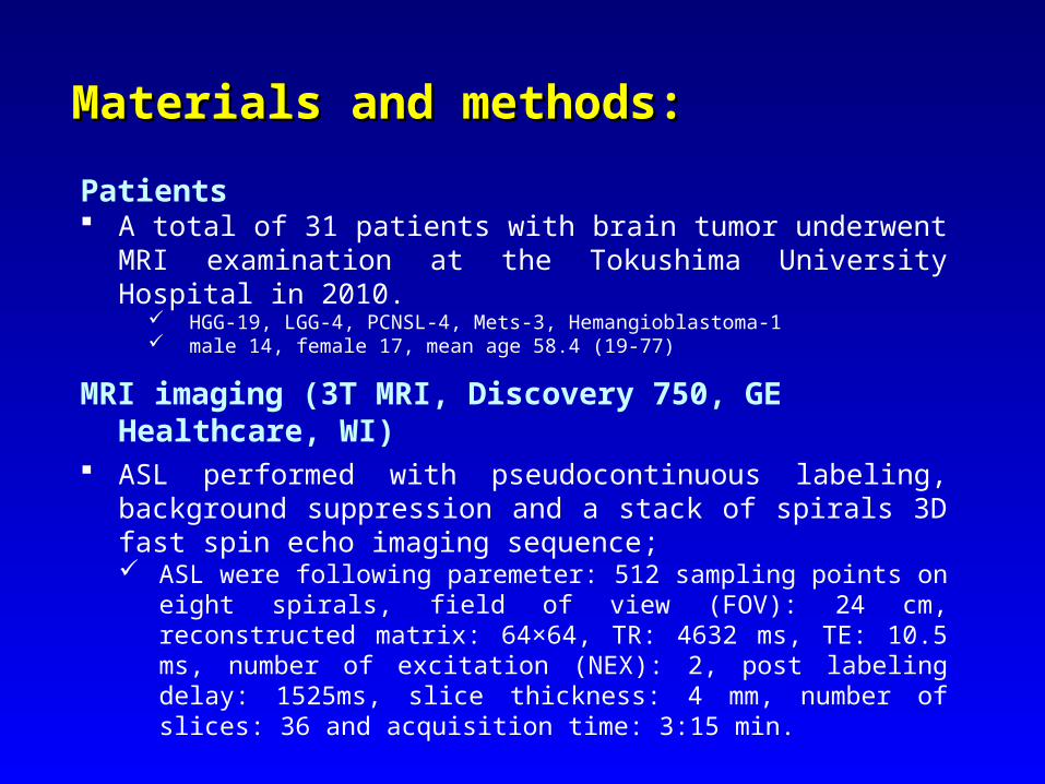
Materials and methods:Materials and methods:
Patients A total of 31 patients with brain tumor underwent MRI
examination at the Tokushima University Hospital in 2010. HGG-19, LGG-4, PCNSL-4, Mets-3, Hemangioblastoma-1 male 14, female 17, mean age 58.4 (19-77)
MRI imaging (3T MRI, Discovery 750, GE Healthcare, WI) ASL performed with pseudocontinuous labeling, background
suppression and a stack of spirals 3D fast spin echo imaging sequence; ASL were following paremeter: 512 sampling points on eight spirals,
field of view (FOV): 24 cm, reconstructed matrix: 64×64, TR: 4632 ms, TE: 10.5 ms, number of excitation (NEX): 2, post labeling delay: 1525ms, slice thickness: 4 mm, number of slices: 36 and acquisition time: 3:15 min.
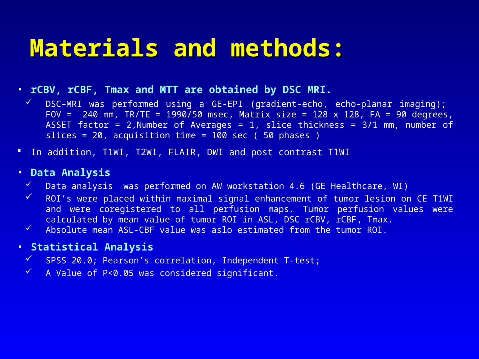
MaterialMaterialss and method and methods:s:
• rCBV, rCBF, Tmax and MTT are obtained by DSC MRI. DSC–MRI was performed using a GE-EPI (gradient-echo, echo-planar imaging); FOV = 240 mm,
TR/TE = 1990/50 msec, Matrix size = 128 x 128, FA = 90 degrees, ASSET factor = 2,Number of Averages = 1, slice thickness = 3/1 mm, number of slices = 20, acquisition time = 100 sec ( 50 phases )
In addition, T1WI, T2WI, FLAIR, DWI and post contrast T1WI
• Data Analysis Data analysis was performed on AW workstation 4.6 (GE Healthcare, WI) ROI’s were placed within maximal signal enhancement of tumor lesion on CE T1WI and were
coregistered to all perfusion maps. Tumor perfusion values were calculated by mean value of tumor ROI in ASL, DSC rCBV, rCBF, Tmax.
Absolute mean ASL-CBF value was aslo estimated from the tumor ROI.
• Statistical Analysis SPSS 20.0; Pearson’s correlation, Independent T-test; A Value of P<0.05 was considered significant.
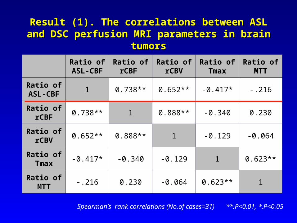
ResultResult (1). (1). The cThe correlationorrelationss between between ASL ASL and DSC and DSC perfusion MRI parameters in brain tumorsperfusion MRI parameters in brain tumors
Ratio of Ratio of ASL-CBF
Ratio of Ratio of rCBF
Ratio of Ratio of rCBV
Ratio of Ratio of Tmax
Ratio of Ratio of MTT
Ratio of Ratio of ASLASL-CBF-CBF 1 0.738** 0.652** -0.417* -.216
Ratio of Ratio of rrCBCBFF
0.738** 1 0.888** -0.340 0.230
Ratio of Ratio of rrCBCBVV
0.652** 0.888** 1 -0.129 -0.064
Ratio of Ratio of TmaxTmax -0.417* -0.340 -0.129 1 0.623**
Ratio of Ratio of MTTMTT -.216 0.230 -0.064 0.623** 1
Spearman’s rank correlations (No.of cases=31) **.P<0.01, *.P<0.05
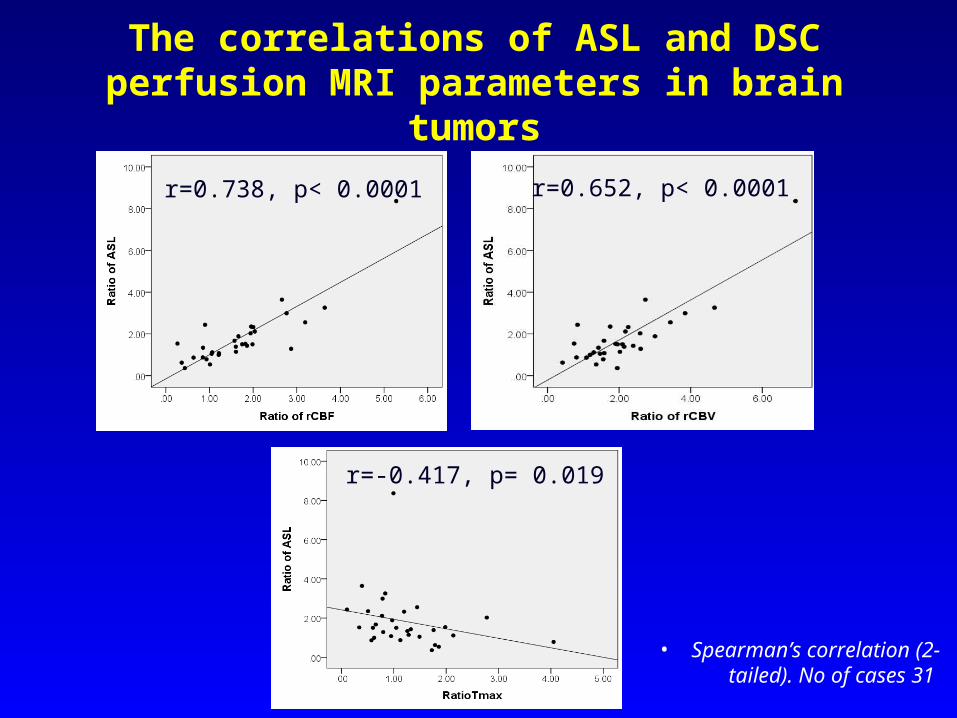
The correlations of ASL and DSC perfusion MRI parameters in brain tumors
• Spearman’s correlation (2-tailed). No of cases 31
r=0.652, p< 0.0001r=0.738, p< 0.0001
r=-0.417, p= 0.019
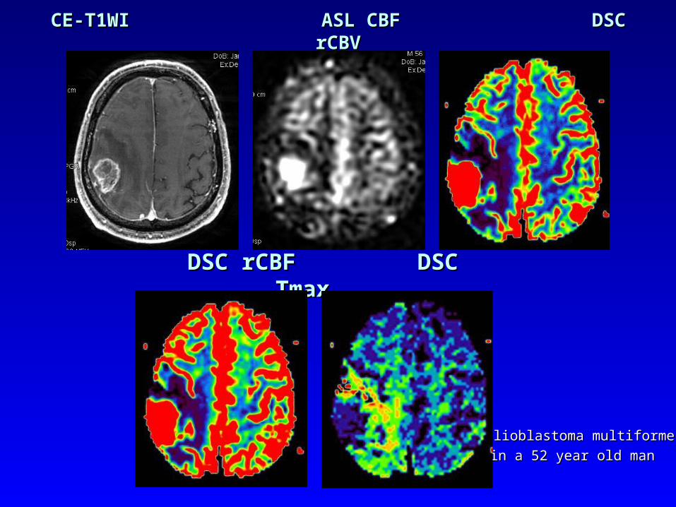
CE-CE-T1T1WIWI ASL CBF ASL CBF DSC rCBV DSC rCBV
Glioblastoma multiforme Glioblastoma multiforme
in a 52 year old manin a 52 year old man
DSC rCBF DSC rCBF DSC TmaxDSC Tmax
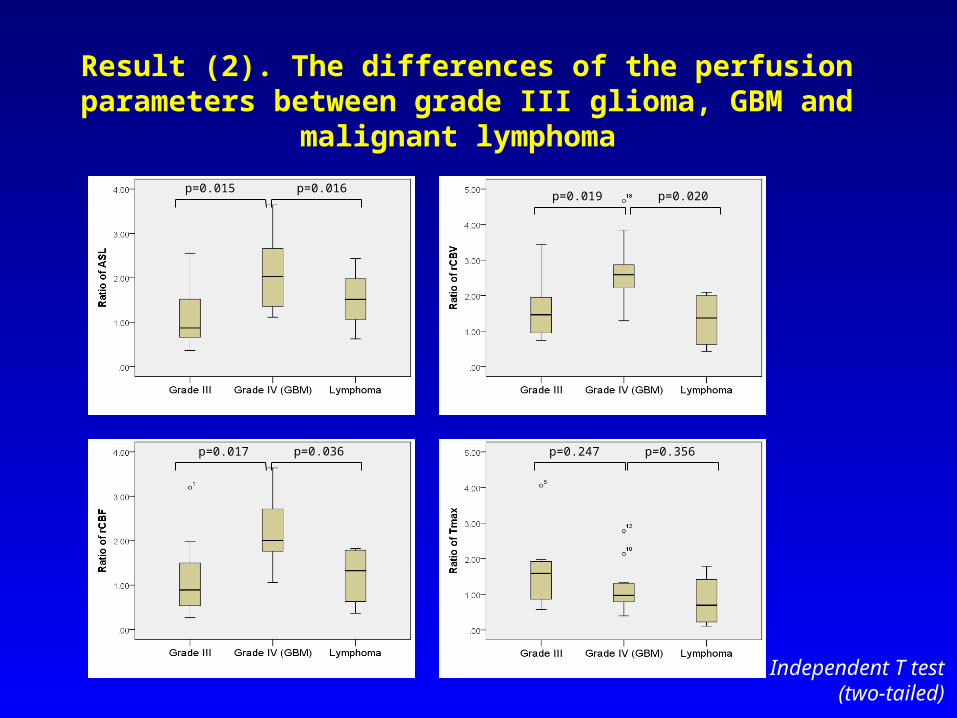
Result (2). The differences of the perfusion parameters between grade III glioma, GBM and malignant lymphoma
Independent T test(two-tailed)
p=0.015 p=0.016p=0.019 p=0.020
p=0.017 p=0.036 p=0.247 p=0.356
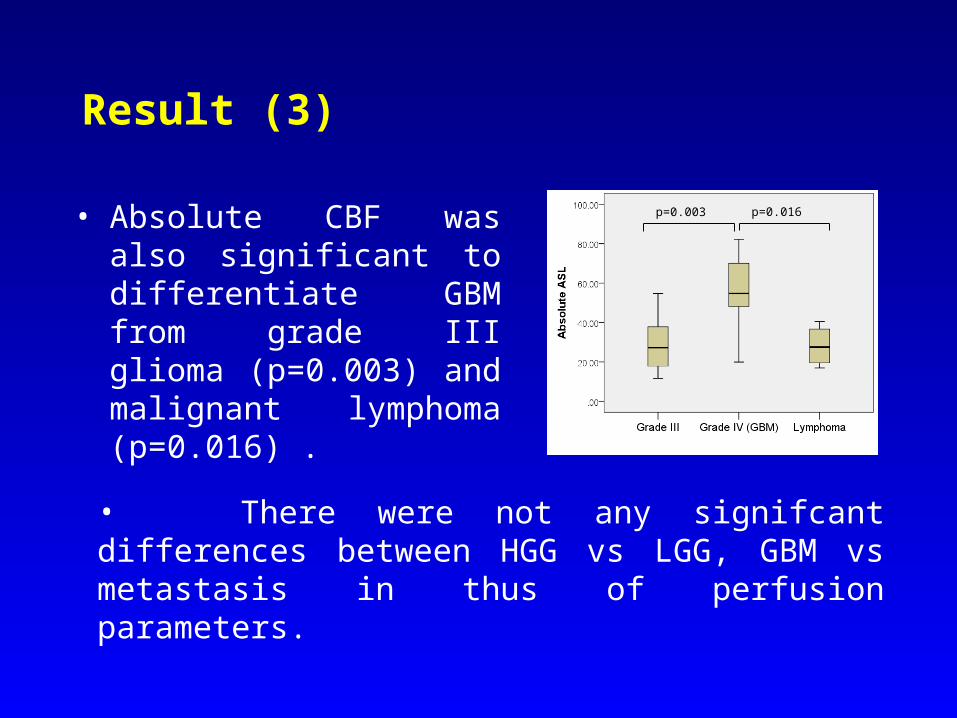
Result (3)
• Absolute CBF was also significant to differentiate GBM from grade III glioma (p=0.003) and malignant lymphoma (p=0.016) .
• There were not any signifcant differences between HGG vs LGG, GBM vs metastasis in thus of perfusion parameters.
p=0.003 p=0.016
DiscussionDiscussion
• ASL is strongly correlated with rCBF and rCBV in this study. In addition, ASL is correlated with Tmax in brain tumors, but not correlated with MTT.
• ASL and Tmax parameter may reflects by transit delay, dispersion that is contrary to rCBF and rCBV. The calculated Tmax value is found to reflect a combination of delay, dispersion and a lesser degree mean transit time (MTT) (3).
• Absolute ASL-CBF and ratio of ASL-CBF both can differentiate GBM from grade III glioma and malignant lymphoma as well as rCBV, rCBF.
• Absolute ASL-CBF is may conveniece perfusion method in clinical practice by spending less time compared with normalized relative perfusion values which derrived from DSC perfusion MRI.
3. Fernando Calamante et al; Stroke. 2010;41:1169-1174
ConclusionConclusion
• 3D ASL could be used an alternative perfusion method to the DSC MRI for evaluation of brain tumors. But ASL may be influenced by vascular delay and dispersion which are not contained in rCBV and rCBF derived DSC MRI.

THANK YOU FOR THANK YOU FOR YOUR ATTENTIONYOUR ATTENTION
FIREWORKS (HANABI), TOKUSHIMA, JAPAN