Embed Size (px)
Citation preview

The Convenience of Camel Milk Proteins for the Nutrition of Cow Milk Allergic Children
J. Saudi Soc. for Food and Nutrition., Vol. 3, No. 1; 2008
42
The Convenience of Camel Milk Proteins for the Nutrition of Cow Milk Allergic Children
El-Agamy, E. I.1; Nawar, M. A.1; Shamsia, Sh.2 and Awad, S.1
1Department of Dairy Science, Faculty of Agriculture, Alexandria University, El-Shatby, Alexandria, Egypt
2Department of Food & Dairy Science and Technology, Faculty of Agriculture (Damanhour), Alexandria University, Egypt
Abstract: Camel, cow and human milk proteins were prepared and analyzed by two different gel electrophoretic techniques. The immunological cross-reactivity between camel milk proteins and their counterparts in cow milk was tested using prepared antisera to camel milk proteins as well as sera from some allergic children to cow’s milk. The tests included immunoelectrophoretic and immunoblotting techniques.
Camel milk proteins have unique electrophoretic patterns, which differ completely from those of cow and human milk. Immunoelectrophoretic and immunoblotting (Western blot) analyses showed the absence of immunological cross-reactivity between camel and cow milk proteins, when specific antisera to camel milk proteins were applied. When sera from some allergic children to cows’ milk were tested for the specificity of immunoglobulin E (IgE) to camel milk proteins using enzyme linked immunosorbent assay (ELISA) technique, the same results as that of immunoelectrophoresis were obtained.
The study concluded that the absence of immunological similarity between camel and cow milk proteins can be considered an important criterion from nutritional and clinical points of view, since camel milk may be suggested a new protein source for nutrition of cow’s milk allergic children who their percentage reached 3–7% worldwide. Key words: Camel milk proteins, milk protein allergy, cow milk proteins, human milk proteins, antigenicity, immunogenicity, immunoblotting (Western blot), ELISA, immunoglobulin E (IgE) , Polyacrylamide gel electrophoresis (PAGE).
INTRODUCTION The best nutritional option for newborn infants is mother’s milk; however, some infants may not be exclusively breast fed during the first months of life. In that case, another substitute or alternative must be provided as cows’ milk. This substitution results in an allergic disease known as cows’ milk allergy (CMA) in 2 -6 % of children. Nowadays, most common alternatives are soy and extensively hydrolyzed milk proteins formulae. However, there is an evidence that 10-20 % of cows’ milk allergic children do not tolerate soy derivatives (Ament and Rubin, 1972; Whitington and Gibson, 1977; Perkkio et al.,1981; Businco et al., 1992) and some cases of high immunological reaction to extensively hydrolysed formulae have been reported (Businco et al., 1989; Saylor and Bahna, 1991; Sampson et al., 1992). Meanwhile, several studies revealed that goat (Webber et al., 1989;Ellis et al., 1991; El-Agamy 2007a), sheep (Bernard et al., 1992; Spuergin et al., 1997), buffalo (Gjesing et al., 1986; El-Agamy 2007 b), ass and mare (El-Agamy et al., 1997;Muraro et al., 2002). Cow milk proteins are not suitable for nutrition of cows’ milk allergic children due to the presence of positive immunological cross-reaction with their counterparts in cows’ milk. On these bases, the identification of a suitable protein source
El-Agamy, E. I.1; Nawar, M. A.1; Shamsia, Sh.2 and Awad, S.
J. Saudi Soc. for Food and Nutrition., Vol. 3, No. 1; 2008
43
for cows’ milk allergic children represents an important goal for both nutritionists and paediatricians.
The present study was undertaken to evaluate the suitability of camel milk for the nutrition of cows’ milk allergic children by studying the antigenic characteristics of camel milk proteins and the immunological cross-reactivity with their counterparts of cows’ milk.
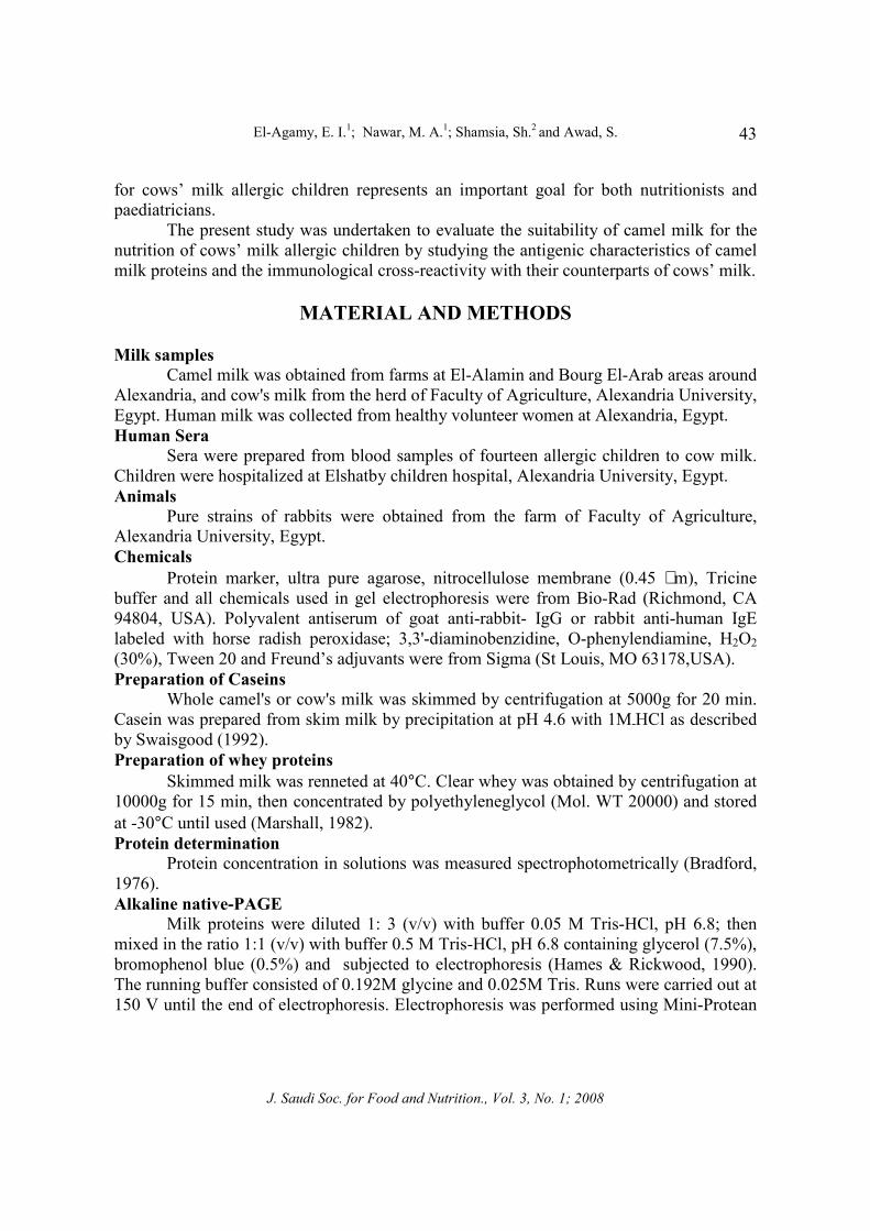
MATERIAL AND METHODS
Milk samples
Camel milk was obtained from farms at El-Alamin and Bourg El-Arab areas around Alexandria, and cow's milk from the herd of Faculty of Agriculture, Alexandria University, Egypt. Human milk was collected from healthy volunteer women at Alexandria, Egypt. Human Sera
Sera were prepared from blood samples of fourteen allergic children to cow milk. Children were hospitalized at Elshatby children hospital, Alexandria University, Egypt. Animals
Pure strains of rabbits were obtained from the farm of Faculty of Agriculture, Alexandria University, Egypt. Chemicals
Protein marker, ultra pure agarose, nitrocellulose membrane (0.45 µm), Tricine buffer and all chemicals used in gel electrophoresis were from Bio-Rad (Richmond, CA 94804, USA). Polyvalent antiserum of goat anti-rabbit- IgG or rabbit anti-human IgE labeled with horse radish peroxidase; 3,3'-diaminobenzidine, O-phenylendiamine, H2O2(30%), Tween 20 and Freund’s adjuvants were from Sigma (St Louis, MO 63178,USA). Preparation of Caseins
Whole camel's or cow's milk was skimmed by centrifugation at 5000g for 20 min. Casein was prepared from skim milk by precipitation at pH 4.6 with 1M-HCl as described by Swaisgood (1992). Preparation of whey proteins
Skimmed milk was renneted at 40°C. Clear whey was obtained by centrifugation at 10000g for 15 min, then concentrated by polyethyleneglycol (Mol. WT 20000) and stored at -30°C until used (Marshall, 1982). Protein determination
Protein concentration in solutions was measured spectrophotometrically (Bradford, 1976). Alkaline native-PAGE
Milk proteins were diluted 1: 3 (v/v) with buffer 0.05 M Tris-HCl, pH 6.8; then mixed in the ratio 1:1 (v/v) with buffer 0.5 M Tris-HCl, pH 6.8 containing glycerol (7.5%), bromophenol blue (0.5%) and subjected to electrophoresis (Hames & Rickwood, 1990). The running buffer consisted of 0.192M glycine and 0.025M Tris. Runs were carried out at 150 V until the end of electrophoresis. Electrophoresis was performed using Mini-Protean
The Convenience of Camel Milk Proteins for the Nutrition of Cow Milk Allergic Children
J. Saudi Soc. for Food and Nutrition., Vol. 3, No. 1; 2008
44
II cell (Bio-Rad) and protein bands were localized in the gels using Coomassie blue R-250 (0.1%). Sodium dodecyl sulphate polyacrylamide gel electrophoresis (SDS-PAGE)
Milk proteins were diluted in the same manner as that of native-PAGE and diluted samples were mixed in the ratio 1:1 (v/v) with sample buffer 0.5 M Tris-HCl, pH 6.8 containing glycerol (7.5%), SDS (2%), β-mercaptoethanol (5%) and bromophenol blue (0.5%) and subjected to heat in a water bath at 100°C for 10 min. Samples were cooled at room temperature, centrifuged at 10000g for 10 min to remove any insoluble material then loaded onto the gel using the discontinous buffer system (Laemmli, 1970). The running buffer consisted of 0.192 M glycine, 0.025M Tris and SDS (0.1%). Protein molecular weights determination
Molecular masses (kDa) of separated proteins on SDS-PAGE were determined according to the method described by Weber and Osborn (1969) using the standard protein marker. Antisera production (immunization)
Polyvalent antisera to camel milk proteins were prepared according to the procedure described by Clausen (1988). Rabbits were firstly immunized, with 0.5 ml of antigen (5 mg/ml sterile NaCl, 0.9%) in suspension with 0.5 ml complete Freund's adjuvant by intramuscularly injection in several sites at week 1. In weeks 3 and 5, each animal was injected intradermaly with booster dose 0.5 ml of antigen (1mg/ml) in suspension with 0.5 ml incomplete Freund's adjuvant. The sera were tested for antibody production before the third immunization. The animals were bled about 14 days after the last immunization. Blood was taken from rabbits and the antiserum titre was measured. Antisera were stored at -30 °C until used. Immunoelectrophoresis
It was carried out at 0.8 V mm-1 for 1-2 h using standard low-Mr agarose dissolved at 10g/l in 0.1M Tricine buffer, pH 8.6 as described by Mayer and Walker (1990). Immunoblotting (Western blot)
After SDS-PAGE, transfer of separated proteins from the gel onto the nitrocellulose membrane (0.45µm, Bio-Rad) was achieved by electrophoretic elution using 0.025M Tris, 0.192 M glycine and methanol (200ml/l) at 100 V for 1h with Mini Trans-Blot Electrophoretic Transfer Cell (Bio-Rad). To verify the protein transfer, the gels were stained by Coomassie blue R-250. The blotted membranes were blocked with gelatin (10g/l) and washed three times with Tris-buffered saline (0.05M Tris, 0.15M NaCl). Membranes were then incubated overnight at room temperature with polyvalent antiserum to camel casein or whey proteins diluted 1:10 with Tris- buffered saline. To detect the antigen–IgG complex, a second incubation was made with polyclonal antiserum of goat anti-rabbit-IgG peroxidase conjugate diluted 1: 1000 with Tris-buffered saline. Colour was developed in the presence of H2O2 with 3,3'-diaminobenzidine as a substrate. (Holen et al., 2001). All the washing steps used Tris-buffered saline containing Tween 20 (2ml/l) (Holen et al., 2001).
El-Agamy, E. I.1; Nawar, M. A.1; Shamsia, Sh.2 and Awad, S.
J. Saudi Soc. for Food and Nutrition., Vol. 3, No. 1; 2008
45
Enzyme linked immunosorbent assay (ELISA) inhibition IgE-ELISA was performed using ninety-six wells; flexible round bottom, microtitre
plates (Falcon Laboratory ware, CA 93030, USA). Plates were precoated with 50 µl per well of 20 µg/ml of cow casein or whey protein in 0.05 M carbonate buffer, pH 9.6. After overnight incubation at 4°C, wells were washed three times with phosphate-buffered saline (PBS) with Tween and blocked with PBS/BSA (50g/l) in PBS for 3 h at 37°C, rinsed with PBS-Tween 20 at 0.5ml/l, and then filled with 50 µl serum sample dilutions (1/25) and incubated for 1h at 37°C. To detect specific IgE, plates were washed again and incubated with polyclonal antiserum of rabbit anti-human IgE (Fc specific) horseradish-peroxidase-conjugate (Sigma) for 1h at 37°C. The reaction was developed with O-phenylendiamine-H2O2 (Sigma). Absorbance was measured at 490 nm in a Titertek Multiskan (Motrich et al., 2003).
RESULTS AND DISCUSSION For all newborn infants, mothers’ milk will always be the ideal nutrition, because it best ensures healthy short- and long-term development as well as enhances the immune functions and hypoallergenic (Wold and Adlerberth, 1998). However, some infants may not be exclusively breastfed during the first months of life, potentially leading to a reduction in overall health status and the early onset of allergic diseases in some infants (Exl, 2001). Gel electrophoresis of milk casein
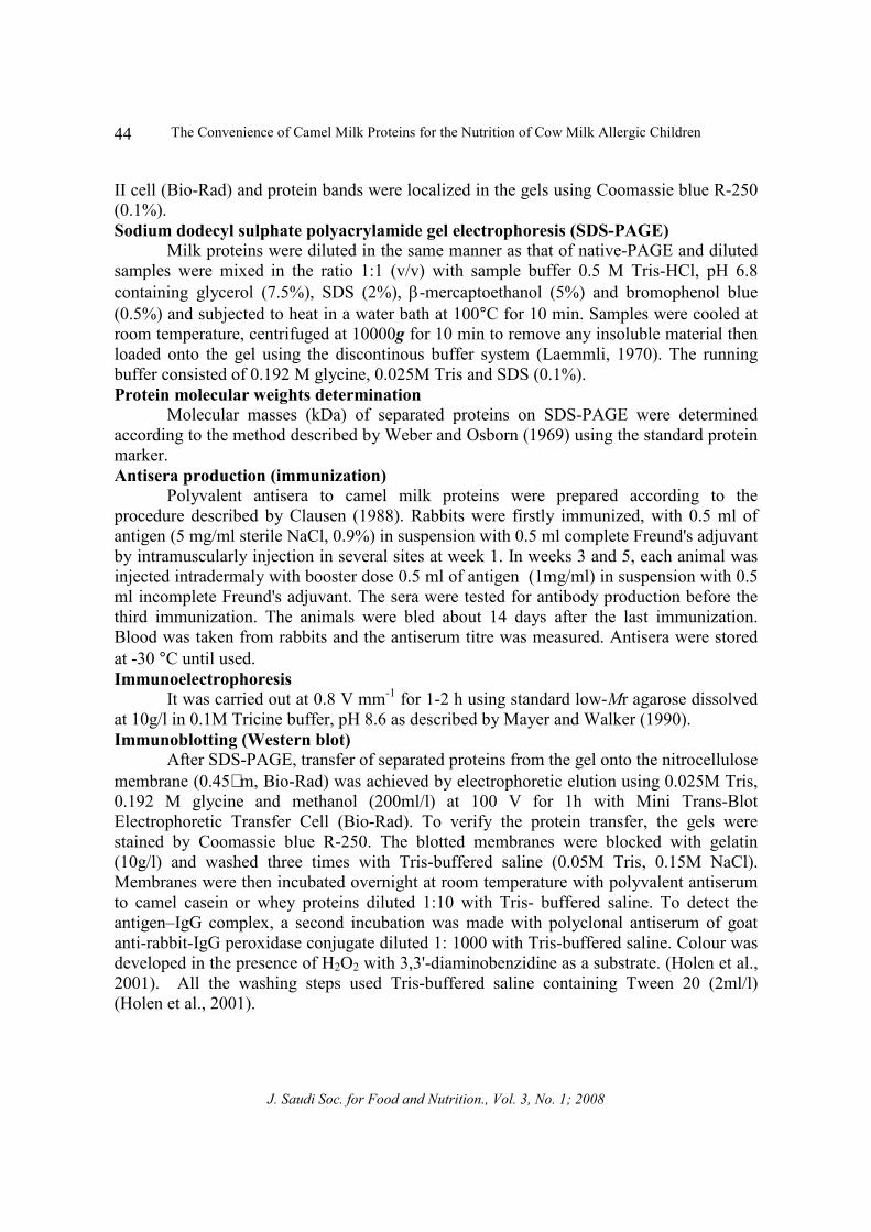
Fig. (1) shows the alkaline native-PAGE of camel, cow and human milk caseins. Each type of casein has a unique electrophoretic pattern due to the appearance of distinguished differences in migration positions. In camel milk casein, β- and αs-CN differ markedly in their migration positions on the gel comparing with those of cow and human milk. These patterns revealed that each type of casein has its unique profile. This feature indicates the differences in charges of such casein fractions among the three types of milk. On the bases of this behavior it is expected that the amino acids composition and structure will be different. On the other hand, although camel and cow milk caseins showed the appearance of equal peptide fractions (3 peptides) on the gel, the variations in migration behavior of all peptides were observed. This mainly reflects the charge differences between both types of caseins. On the other side, human casein was separated into only two fractions differ in migration positions than those of either camel or cow casein. Farah and Farah-Riesen (1985) reported that camel casein was separated into three main fractions on polyacrylamide gel in the absence of non denaturing agents and these fractions are similar in number to those of cow milk casein but different in migration positions. It is interesting that the electrophoretic patterns of camel and cow caseins showed the quite equality of β- and αs-casein fractions in their intensities. On the contrary, human casein pattern revealed that β-CN was dominant. The study of Kroening et al. (1998) showed that human casein is mainly β-CN and αs-CN is present in very low concentration. On the bases of these findings it is clear now that the high concentration of β-CN and its
The Convenience of Camel Milk Proteins for the Nutrition of Cow Milk Allergic Children
J. Saudi Soc. for Food and Nutrition., Vol. 3, No. 1; 2008
46
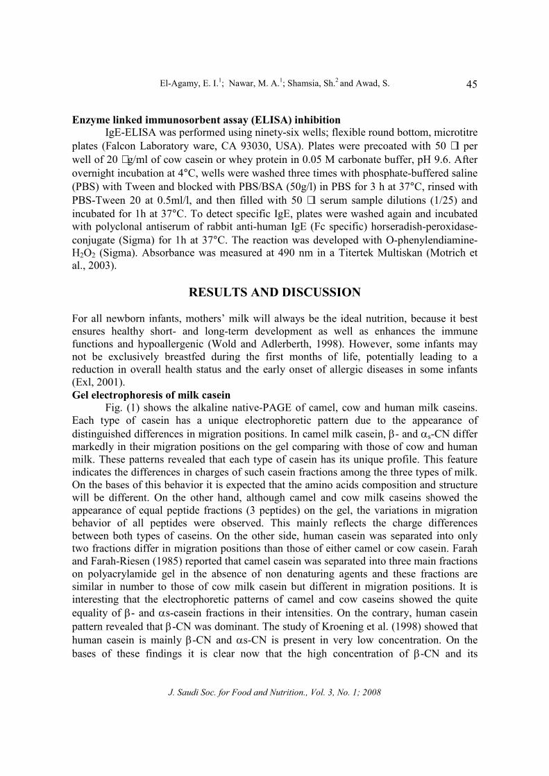
dominance in human casein reflects the higher digestibility rate of human milk in the infant’s gut. Because human milk β-CN is more sensitive to peptic hydrolysis than αs-CN (Abou-Soliman, 2005). Meanwhile, it should be taken into account that the higher the concentration of αs-CN in cow milk, the higher the incidence of hypersensitivity reaction (allergy) in children (Taylor, 1986). Therefore, the hypoallergenicity of human milk is due to the high concentration of β-CN and limited level of αs-CN. The molecular masses of β-and αs-CN were estimated at 29, 33 and 28, 32 kDa for camel and cow milk caseins, respectively. For human β-CN, the molecular weight was 27 kDa (Fig.2).
β-CN
αs1-CN
αs2-CN
Camel Cow Human
Fig.1
Figure 1. Alkaline native-PAGE (12.5% T) of acid casein from
camel, cow and human milk. Anode is toward bottom of photo.
Figure 2. SDS-PAGE of acid camel, cow and human milk caseins.
Std: Standard protein marker. Anode is toward bottom of photo.
αs1 -CN?-CN
αs2 -CN
18
21
31
43
Std CamelHuman Cow
97
67
Mr

El-Agamy, E. I.1; Nawar, M. A.1; Shamsia, Sh.2 and Awad, S.
J. Saudi Soc. for Food and Nutrition., Vol. 3, No. 1; 2008
47
Concerning molecular and immunological characterization of whey proteins of the three types of milk, different techniques were applied. Firstly alkaline native-PAGE (Fig. 3) showed that the electrophoretic patterns of whey proteins were also different. Camel α-la was faster in migration than cow and human α-la. But both α-la of cow and human milks had almost the same migration position. β-lg was not present in patterns of camel and human milk but it was dominant in cow milk whey proteins and separated into two different variants (β-lg B & β-lg A). This feature must be taken into account because β-lg is one of the major proteins of cow milk responsible for the incidence of hypersensitivity reactions (allergy) in infants (Lara-Villoslada et al., 2005).
Figure 3. .Alkaline native-PAGE (12.5% T) of camel, cow and
human milk whey proteins. Anode is toward bottom of photo.
Human whey proteins were characterized by the presence of α-la and lactoferrin in high ratio, whereas in camel milk immunoglobulin G, α-la and serum albumin were dominant. Reiter (1985) reported that lactoferrin is abundant in human milk whey proteins. Meanwhile, the high ratio of α-la in human milk may due to its vital physiological role in lactose synthesis in the mammary gland. The evidence is that human milk has high level of lactose comparing with milks of other dairy animals (El-Agamy et al., 1998).
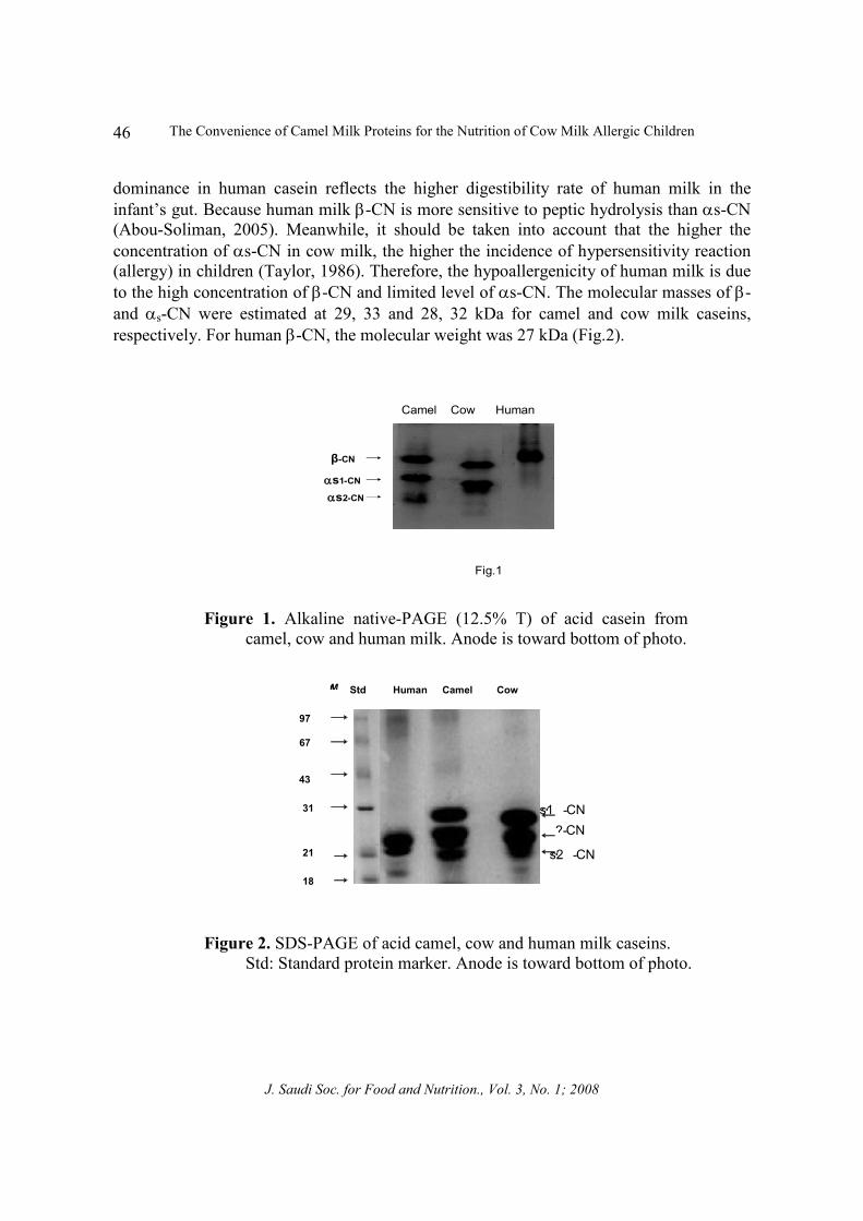
The molecular masses of whey proteins were estimated after separation on SDS-PAGE (Fig. 4). Camel α-la (15 KDa) was higher than those of cow and human milk (14.4 kDa). Serum albumin of cow milk was equal to that of blood serum (66.2 kDa), (El-Agamy et al., 1996). Camel, cow and human serum albumins were estimated at 66.2 kDa. The study of Farah (1993) showed that camel serum albumin (CSA) has a molecular mass of 66 kDa.
Generally, according to the molecular characterization of all proteins of the three types of milk, we conclude that each type of milk has unique proteins having their own composition and structure to be the fit proteins for the requirements of the corresponding newborn nutrition.
Camel
β - lgα - lac
Cow Human
α - lacBSA
IgG
The Convenience of Camel Milk Proteins for the Nutrition of Cow Milk Allergic Children
J. Saudi Soc. for Food and Nutrition., Vol. 3, No. 1; 2008
48
Fig.(4)
97826643
31
1814.4
BSA α-lac Std Mr
LF
HumanCamel Cow
Figure 4. SDS-PAGE (7.5–15% T, gradient gel) of camel, cow and human milk whey proteins .LF; lactoferrin; α -la : α-lactalbumin; BSA: bovine serum albumin. Std: Standard protein marker, Anode is toward bottom of photo.
Immunoelectrophoretic analysis
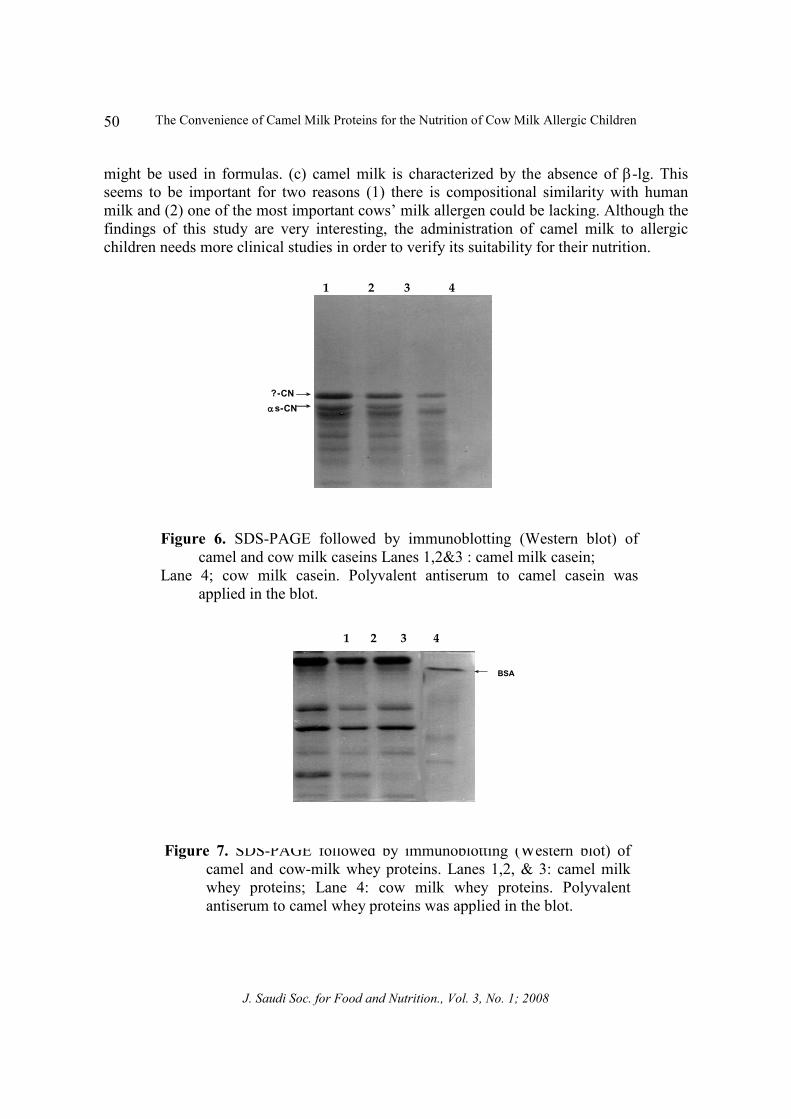
The present study was further pursued in order to confirm such findings by applying another immunological criterion for the characterization of such proteins. In order to evaluate camel milk proteins to be substitute of human milk ones and to compare them to those of cow milk, which were well evaluated as positive-allergy causing proteins, different immunological techniques were applied using specific antisera to camel milk or cow milk proteins. In (Fig. 5A), immunoelectrophoretic analysis of camel and cow milk caseins showed the absence of similar antigenic properties between them. Because there was no positive immunological reaction between specific antiserum to camel milk casein with cow milk casein (as an antigen). This result indicates that each casein has its own antigenic determinants, which formed its unique structure. By the same technique, the immunological relationship between camel and cow milk whey proteins was also studied (Fig. 5B). Results showed that very limited immunological similarity between camel and cow milk whey protein. Because of the appearance of only one positive precipitin arc in cow milk whey proteins (CMWP) when specific antiserum to camel milk whey proteins were applied in the test. The position of that precipitin arc of CMWP refers to serum albumin. It is noted that the intensity of the arc was light, means that its reaction with camel milk antiserum was weak. This reveals that BSA shares a limited part in its primary structure similar to that of CSA and they are not identical in structure. The immunological similarity between serum albumins in both camel and cow milk protein may due to that serum albumin is derived from blood and not synthesized in mammary gland, i.e., not organ-specific protein (Fox and McSweeney, 1998). To confirm these results, another immunological analysis (Western blot) was applied (Fig. 6). When antiserum to camel milk casein was used, no detection of any fraction of cow milk casein was observed. It was clear that all fractions of camel milk casein were detected on the nitrocellulose

El-Agamy, E. I.1; Nawar, M. A.1; Shamsia, Sh.2 and Awad, S.
J. Saudi Soc. for Food and Nutrition., Vol. 3, No. 1; 2008
49
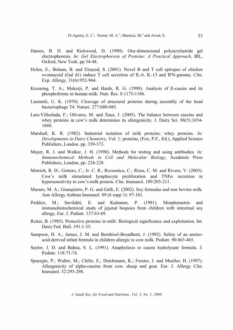
membrane. This result confirms also that the antigenic similarities between both types of casein are not present and they are completely different than each other. Therefore, it is expected that the allergenicity mode of casein is different between cow and camel milk. Western blot was also applied for examination the similarities between both types of milk (Fig. 7). The majority of camel whey proteins showed no similar immunological cross-reaction with those of cow milk except BSA which showed little antigenic similarity with CSA. Although, the existence of such immunological similarity, it means nothing from the allergy point of view. Because BSA has no a significant role in the hypersensitivity reaction comparing with other whey proteins like β-lg and α-la (El-Agamy, 2007c). In order to have highly confirmation of the antigenic dissimilarity between camel and cow milk proteins, we examined serum samples, from allergic children to cows’ milk proteins, for the specificity of IgE (Specific immunoglobulin to cow proteins) to camel milk proteins. Ig E- ELISA assays for both camel and cow milk proteins were presented in (Fig. 8). The results showed that cow casein was recognized at high binding affinity as shown by inhibition rate of 65.8 % versus zero % for camel casein. For whey proteins, the Ig E inhibition rates were 39.8 % and zero % for cow and camel milk, respectively.
Figure 5. Immunoelectrophoretic analysis of camel and cow milk proteins. (A): CM Cas ; Camel milk casein; BV Cas: bovine casein;
1: Rabbit antiserum to camel milk casein. (B): CM WP: camel milk whey proteins; BV WP: bovine milk whey proteins.
2: Rabbit antiserum to camel milk whey proteins.
The positive results of such diagnostic test with cow milk proteins are expected because the serum samples were obtained from allergic children to cows’ milk. But on the other side the results were very interesting, since no IgE recognition at all to the epitope of any one of camel milk proteins.
On the bases of these findings we can conclude that: (a) the epitopes of proteins of cows’ milk which cause the hypersensitivity are completely different than those of camel milk. (b) camel milk might be a promising new protein source for allergic children and it
-
(B)
CM Cas
BV Cas
1
CM WP
BV WP
2
+
(A)
The Convenience of Camel Milk Proteins for the Nutrition of Cow Milk Allergic Children
J. Saudi Soc. for Food and Nutrition., Vol. 3, No. 1; 2008
50
might be used in formulas. (c) camel milk is characterized by the absence of β-lg. This seems to be important for two reasons (1) there is compositional similarity with human milk and (2) one of the most important cows’ milk allergen could be lacking. Although the findings of this study are very interesting, the administration of camel milk to allergic children needs more clinical studies in order to verify its suitability for their nutrition.
Figure 6. SDS-PAGE followed by immunoblotting (Western blot) of camel and cow milk caseins Lanes 1,2&3 : camel milk casein;
Lane 4; cow milk casein. Polyvalent antiserum to camel casein was applied in the blot.
Figure 7. SDS-PAGE followed by immunoblotting (Western blot) of camel and cow-milk whey proteins. Lanes 1,2, & 3: camel milk whey proteins; Lane 4: cow milk whey proteins. Polyvalent antiserum to camel whey proteins was applied in the blot.
1 2 3 4
?-CNαs-CN
BSA
1 2 3 4

El-Agamy, E. I.1; Nawar, M. A.1; Shamsia, Sh.2 and Awad, S.
J. Saudi Soc. for Food and Nutrition., Vol. 3, No. 1; 2008
51
Figure 8. IgE-ELISA inhibition of cow and camel milk proteins.
REFERENCES
Abou-Soliman, N. H. (2005). Studies on goat milk proteins: Molecular and immunological characterization with respect to human health and nutrition. Ph.D. Thesis, Alexandria University, Egypt.
Ament, M. E. and Rubin, C. E. (1972). Soy-protein-another cause of the flat intestinal lesion. Gastroenterol. 62:227-234.
Bernard, H.; Wal, J. M.; Greminon, C.; Grassi, J. Yvon, M. and Houdebine, L. M. (1992). Sensitivities of cow’s milk allergic patients to casein fraction of milks from different species. Allergy. 47:306 (Abstr.).
Bradford, M. M. (1976). A rapid and sensitive method for the quantitation of microgram quantities of protein utilizing the principle of protein–dye binding. Anal. Biochem. 72:248 -254.
Businco, L.; Bruno, G.; Giampietro, P. G. and Cantoni, A. (1992). Allergenicity and nutritional adequacy of soy protein formulas. J. Pediatr. 121:S21-S28.

The Convenience of Camel Milk Proteins for the Nutrition of Cow Milk Allergic Children
J. Saudi Soc. for Food and Nutrition., Vol. 3, No. 1; 2008
52
Businco, L.; Cantoni, A.; Longhi, A. and Giampietro, P. G. (1989). Anaphylactic reactions to a cow milk whey protein hydrolysate (Alfa Re’, Nestle’) in infants with cow’s milk allergy. Ann. Allergy. 62:333-335.
Clausen, J. (1988). Antisera. In: Laboratory techniques in biochemistry and molecular biology: Immunochemical techniques for the identification and estimation of macromolecules, 3rd edition, vol. 1 (part3). Elsevier, Amsterdam, pp. 88-102.
El-Agamy, E. I. (2007a). Goat Milk Proteins. VI. Immunochemical characterization (Submitted to Small Ruminant. Res.).
El-Agamy, E. I. (2007b). Buffalo milk proteins: molecular and immunological characterization (Submitted to J. Dairy Res.).
El-Agamy, E. I. (2007c). The challenge of cow milk allergy. Small Ruminant Res. 68:64-72.
El-Agamy, E. I.; Ruppanner, R.; Ismail, A.; Champagne, C. P. and Assaf, R. (1996). Purification and characterization of lactoferrin, lactoperoxidase, lysozyme and immunoglobulins from camel’s milk. Inter. Dairy J. 6:129-145.
El-Agamy, E. I.; Abou-Shloue, Z. I. and Abdel-Kader, Y. I. (1997). A comparative study of milk proteins from different species. II. Electrophoretic patterns, molecular characterization, amino acid composition and immunological relationships. Third Alexandria Conference on Food Science and Technology, Alexandria, Egypt, March, 1-3.
El-Agamy, E. I.; Abou-Shloue, Z. I. and Abdel-Kader, Y. I. (1998). Gel electrophoresis of proteins, physicochemical characterization and vitamin C content of milk of different species. Alex. J. Agric. Res. 43(2):57-70.
Ellis, M. H.; Short, J. and Heiner, D. C. (1991). Anaphylaxis after ingestion of a recently introduced hydrolysed whey protein formula. J. Pediatr. 118:74-77.
Exl, B. M. (2001). A review of recent developments in the use of moderately hydrolyzed whey formulae in infant nutrition. Nutr. Res. 21:355-379.
Farah, Z. (1993). Composition and characteristics of camel milk. J. Dairy Res. 60:603-626.
Farah, Z. and Farah-Riesen, M. (1985). Separation and characterization of major components of camel milk casein. Milchwissenchaft. 40(11):669-671.
Fox, P. F. and McSweeney, P. L. H. (1998). Milk proteins. In: Dairy Chemistry and Biochemistry, Blackie Academic & Professional, London, p. 146.
Gjesing, B.; Osterballe, O.; Schwartz, B.; Wahn, U. and Lowenstein, H. (1986). Allergen-Specific IgE antibodies against antigenic components in cow milk and milk substitutes. Allergy. 41:51-56.
El-Agamy, E. I.1; Nawar, M. A.1; Shamsia, Sh.2 and Awad, S.
J. Saudi Soc. for Food and Nutrition., Vol. 3, No. 1; 2008
53
Hames, B. D. and Rickwood, D. (1990). One-dimensional polyacrylamide gel electrophoresis. In: Gel Electrophoresis of Proteins: A Practical Approach, IRL, Oxford, New York. pp 34-48.
Holen, E.; Bolann, B. and Elsayed, S. (2001). Novel B and T cell epitopes of chicken ovomucoid (Gal d1) induce T cell secretion of IL-6, IL-13 and IFN-gamma. Clin. Exp. Allergy. 31(6):952-964.
Kroening, T. A.; Mukerji, P. and Hards, R. G. (1998). Analysis of β-casein and its phosphoforms in human milk. Nutr. Res. 8:1175-1186.
Laemmli, U. K. (1970). Cleavage of structural proteins during assembly of the head bacteriophage T4. Nature. 277:680-685.
Lara-Villoslada, F.; Olivares, M. and Xaus, J. (2005). The balance between caseins and whey proteins in cow’s milk determines its allergenicity. J. Dairy Sci. 88(5):1654-1660.
Marshall, K. R. (1982). Industrial isolation of milk proteins: whey proteins. In: Developments in Dairy Chemistry, Vol. 1: proteins, (Fox, P.F., Ed.), Applied Science Publishers, London. pp. 339-373.
Mayer, R. J. and Walker, J. H. (1990). Methods for testing and using antibodies. In: Immunochemical Methods in Cell and Molecular Biology, Academic Press Publishers, London, pp. 224-228.
Motrich, R. D.; Gottero, C.; Jr. C. R.; Rezzonico, C.; Riera, C. M. and Rivero, V. (2003). Cow’s milk stimulated lymphocyte proliferation and TNFα secretion in hypersensitivity to cow’s milk protein. Clin. Immunol. 109:203-211.
Muraro, M. A.; Giampietro, P. G. and Galli, E. (2002). Soy formulas and non bovine milk. Ann Allergy Asthma Immunol. 89 (6 supp 1): 97-101.
Perkkio, M.; Savilahti, E. and Kuitunen, P. (1981). Morphometric and immunohistochemical study of jejunal biopsies from children with intestinal soy allergy. Eur. J. Pediatr. 137:63-69.
Reiter, B. (1985). Protective proteins in milk: Biological significance and exploitation. Int. Dairy Fed. Bull. 191:1-35.
Sampson, H. A.; James, J. M. and Bernhisel-Broadbent, J. (1992). Safety of an amino- acid-derived infant formula in children allergic to cow milk. Pediatr. 90:463-465.
Saylor, J. D. and Bahna, S. L. (1991). Anaphylaxis to casein hydrolysate formula. J. Pediatr. 118:71-74.
Spuergin, P.; Walter, M.; Chiltz, E.; Deichmann, K.; Forster, J. and Mueller, H. (1997). Allergenicity of alpha-caseins from cow, sheep and goat. Eur. J. Allergy Clin. Immunol. 52:293-298.

The Convenience of Camel Milk Proteins for the Nutrition of Cow Milk Allergic Children
J. Saudi Soc. for Food and Nutrition., Vol. 3, No. 1; 2008
54
Swaisgood, H. E. (1992). Chemistry of casein. In: Advanced Dairy Chemistry-1: Proteins (Fox, P.F., Ed.), Elsevier Science Publishers LTD, England. p. 67.
Taylor, S. L. (1986). Immunological and allergic properties of cows’ milk proteins in humans. J. Food Prot. 49(3):239-250.
Webber, S. A.; Graham-Brown, R. A. C.; Hutchinson, P. E. and Burns, D. A. (1989). Dietary manipulation in childhood atopic dermatitis. Br. J. Dermatol. 121:91-98.
Weber, K. and Osborn, M. C. (1969). The reliability of molecular weight determinations by dodecyl sulfate polyacryamide gel electrophoresis. J. Biol. Chem. 244:4405-4412.
Whitington, P. F. and Gibson, R. (1977). Soy protein intolerance: four patients with intestinal soy. Allergy. 59:730-732.
Wold, A. E. and Adlerberth, L. (1998). Does breast-feeding affect the infant’s immune responsiveness ?. Acta Paediatr. 87:19-22.
El-Agamy, E. I.1; Nawar, M. A.1; Shamsia, Sh.2 and Awad, S.
J. Saudi Soc. for Food and Nutrition., Vol. 3, No. 1; 2008
55
���� ������ �������� ��� ������ ���� �� ����� ������� ������� ��������������
!����"�#$�� %�&���'(��� !��� ���� '(!. ���* +��*,�- .���'
��� ���� ���� �� ���� ������ ��������� ���� ����������������� ��� ������ ���� ��� �� ��� ���� �� ���� ������ �)������(��������� ���� ������� ��� ������
/0����:#������� ��$%� �� &��%'���� �� ��(��������� )���(���)�*��� +��(��� ��� ����)� �� ���(��*, �+���� '�, �� ���-��)� �������� .���� �� /��� 0��1��� ������ ����� ����� ��� 2� ��*�)���� ��� ��3�*%4
#�������� ��$� &��%'���� �#�������� ��$� �)5� &��% � ��(��� ��*�� ���*� �*� 0��� 6��%�� �� ��)�������� 6�-,�� 7���&��% #������� �$ �����%�� 7��� ����(��� .
2�� �� 89����� #%$�4 �(� �:;�;#�������� �������� +���� ,���4 #-��)� &��%����'� ���� ��(���� <�=>�) <����� 4 ?�� 6�� ��� �#�*������
�� @�� 6� &��%����� + ����� ���9��) ���.�;'���� &��% 0��1� 2= #����� &��%� ������ ?�� ���9���%� ��� β-lactoglobulin A��� #��������� �� �����
��%�� ������� ����9����(��� &��% B�� ?�� 6�-,�� ��� ���0�� ����� ���� ?�� A��%� A��� �.C;2������ 2������ 6��%��� 89��� #%$�4 Immunoelectrophoresis)� /���� 2*�������� D�*��� ���� 2��*����
Immunoblotting 0��1� A4 ��� ��� #������� ��� 2���� &��%'���� &��%� ��(��� ���)�*�� �*�� /��� 4&��% #�������� +��$� ������ ���'����.
23��� ����)�#���� +���� #�������� �����%��� �������� 6�-,�� �� ���� �� E�%� �� ��(��� &��% ,�*���F G�)���� ������� ������ ��� Immunoglobulin E (IgE)������ ��������� �#������� �� ������ A4 H�
&��%'�������� 2������ 6��%��� �(��, ���)��� ��� /��2�� ELISA )� ����(.�2�F ������� #��) �� 0��1� A4 ��� ��� �4 #������� ��� 2���� &��%'�����&��% �*�� ��*(���
<������ <���� � ����I��� ����%���� �� ����>� � ���� E�% J���� &��%'���� <����� <>��� ���I�� �������� 6�-,�������%�� 7���K� #�������� &��%����(� ������ 6�� ������ ��C–M%������ N���� ?�� .