Embed Size (px)
Citation preview

17The Confocal Tonal Shift
Timothy J. Bennett, CRA, OCT-C, FOPS
Penn State Hershey Eye Center500 University Drive, HU19Hershey, PA 17033717/[email protected]
CASE REPORT
IntroductIon
he Heidelberg Spectralis confocal scanning laser ophthalmoscope (cSLO) is a commonly used diagnostic imaging device that uses monochro-
matic laser illumination to image the eye. It can be used for several retinal imaging modalities including infrared reflectance (IR), blue reflectance, fluorescein angiography, ICG angiography and fundus autofluorescence (FAF). The confocal capability of the cSLO allows it to capture high-contrast, finely detailed images. By definition, the word confocal simply means “having the same focus”. In this case it refers to the confocal pinhole or aperture that is located at the conjugate focal plane of the subject. The cSLO utilizes a focused laser to scan the subject point-by-point. The reflected light is captured after it passes through a confocal pinhole as long as it is in focus. The pinhole suppresses out-of-focus light from reach-ing the image detector resulting in very sharp images.1-2 The confocal pinhole is especially effective at eliminat-ing unwanted scatter from cataracts or corneal opacities since these structures fall far outside the plane of focus, and light reflected from these media opacities is blocked from reaching the sensor (Figure 1).
With a traditional fundus camera, focus and brightness are independent of one another. Adjusting one does not significantly affect the other. The same principle does not hold true with the cSLO because of the confo-cal aperture. Adjusting the focus will affect brightness. The instrument is most light efficient at the plane of focus.3 You can see the confocal effect as you adjust the focus on the retina. The image will get brightest just as you come into sharpest focus. It is most noticeable in instruments
set for manual exposure control. It is easy to demonstrate this link between focus and brightness with pathology that exhibits exaggerated depth. For example, vitreous floaters from asteroid hyalosis appear as dark shadows when focus is set on the surface of the retina. As focus is shifted up into the vitreous, the floaters begin to brighten and the retina fades to dark (Figure 2).
clInIcal observatIons
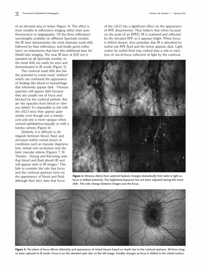
A secondary result of the confocal aperture is how it affects the appearance of elevated or out-of-focus retinal structures. Because of the confocal aperture, as well as the inherently shallow depth of focus of the cSLO, some ocular findings may appear dark simply because they are slightly out of focus. Elevated pathologies such as serous detachments, macular edema, or papilledema are exam-ples of this phenomenon that I refer to as the confocal tonal shift (Figure 3).
In some cases the confocal tonal shift can enhance diagnostic information by clearly outlining the borders
The Confocal Tonal Shift
T
Figure 1: Left: Patient with a cataract obscuring the view of the retina and optic nerve through a fundus camera. Right: The confocal pinhole of the cSLO suppresses light scatter from the cataract improving the view of the fundus.
The Journal of Ophthalmic Photography Volume 38, Number 1 • Spring 201618
of an elevated area or lesion (Figure 4). The effect is most notable in reflectance imaging rather than auto-fluorescence or angiography. Of the three reflectance wavelengths available on different Spectralis models, the IR laser demonstrates the most dramatic tonal shift, followed by blue reflectance, and finally green reflec-tance on instruments that have this additional laser for MultiColor imaging. The near IR laser at 820 nm is standard on all Spectralis models, so the tonal shift can easily be seen and demonstrated in IR mode (Figure 5).
The confocal tonal shift also has the potential to create tonal “artifacts” which can confound the appearance of findings like blood or hemorrhage that inherently appear dark. Vitreous opacities will appear dark because they are usually out of focus and blocked by the confocal pinhole. But are the opacities from blood or vitre-ous debris? It’s impossible to tell with the cSLO since they appear quite similar even though one is translu-cent and one is more opaque when viewed ophthalmoscopically or with a fundus camera (Figure 6).
Similarly, it is difficult to dis-tinguish between blood, fluid, and elevation within retinal tissues in conditions such as macular degenera-tion, retinal vein occlusions and dia-betic macular edema (Figures 7, 8). Theelen , Hoyng and Klevering state that blood and fluid absorb IR and will appear dark in IR images.4 This fails to consider the role that focus and the confocal aperture have on the appearance of blood and fluid, although they later state that focus
of the cSLO has a significant effect on the appearance of RPE detachments. They believe that when focused on the peak of an RPED, IR is scattered and reflected by the elevated RPE so it appears bright. When focus is shifted deeper, they postulate that IR is absorbed by turbid sub RPE fluid and the lesion appears dark. Light scatter by turbid fluid may indeed play a role in rejec-tion of out-of-focus reflection of light by the confocal
Figure 2: Vitreous debris from asteroid hyalosis changes dramatically from dark to light as focus is shifted anteriorly. The brightness/exposure has not been adjusted during this tonal shift. The only change between images was the focus.
Figure 3: The plane of focus affects reflectivity and appearance of retinal tissues based on depth due to the confocal aperture. All three imag-es were captured in IR mode. Focus is on the elevated optic disc on the left image. Tonality changes as focus is shifted to the retinal surface.

19The Confocal Tonal Shift
Figure 5: The confocal tonal shift in three different modalities. From left to right: IR, blue reflectance, fundus autofluorescence. The effect seems to be most pronounced in IR mode.
Figure 4: A case of central serous chorioreti-nopathy with a classic serous detachment that can be seen ophthalmoscopically. The cSLO image is dramatic in its appearance due to the confocal tonal shift. The dark area is elevated and filled with clear fluid (not blood).
Figure 6: Vitreous debris can appear very dark in cSLO images even though it is almost completely transparent. Without an addi-tional frame of reference, it is impossible to know if these dark areas represent vitreous debris or hemorrhage.
Figure 7: Patient with bilateral diabetic mac-ular edema (DME). There are some associ-ated dot and blot hemorrhages present, but the dark patches are a result of elevation from the DME. Each of these dark areas cor-respond to elevation on OCT.

The Journal of Ophthalmic Photography Volume 38, Number 1 • Spring 201620
pinhole, but plane of focus likely has a more significant effect.
It is also important to note that elevated lesions can appear dark regardless of the pathologic location in the fundus. Confocal imaging alone can’t always differentiate the anatomic location within the layers of the retina and choroid (Figure 9). OCT imaging or angiography may be necessary to further investigate the location of the pathology. In some cases, the confocal pinhole seems to suppress light that is reflected from the actual plane of focus, but is slightly blurred because of scat-tering from a lesion in tissue that is normally clear. Retinal whitening from artery occlusions, paracentral acute middle maculopathy (PAMM) and acute macular neuroretinopa-thy (AMN) can all appear as dark patches on confocal IR imaging. These conditions occur in different layers of the neuroretina but all can be identified by SD-OCT and IR confocal imaging. The dark patchy areas correspond well with increased thickening on SD-OCT (Figure 10). For this reason, several authors men-tion the value of confocal IR imaging in AMN and PAMM.5-8 Fawzi, et al suggest that the origin of character-istic hyporeflective lesions in AMN may be related to melanin and dis-ruption of the OS/RPE junction.8 Again this overlooks the simpler explanation that these lesions may be dark because of suppression of reflectance by the confocal aperture. The fact that more anterior ischemic conditions such as PAMM and retinal artery occlusions exhibit similar dark lesion on IR supports the confocal tonal shift as a likely cause of the hyporeflectance in AMN.
Although originally designed to image the retina, the cSLO can also
Figure 8: Left: Comparison of elevation from blood and fluid. Left: Branch retinal vein occlu-sion (BRVO). The dramatic dark lesion is a result of both hemorrhage and elevation. Right: The hyporeflectance is from an elevated area of intraretinal fluid in cystoid macular edema.
Figure 9: Two very different disease processes with elevated lesions found in different retinal lay-ers. Despite these differences their appearance is quite similar because of the confocal shift. Left: Choroidal osteoma. Right: Idiopathic central serous chorioretinopathy with serous detachment.
Figure 10: IR imaging with the cSLO can help diagnose paracentral acute middle maculopa-
thy (PAMM). Although these lesions aren’t elevated, light scatter from the slight thickening
in the middle retinal layers is suppressed by the confocal pinhole making the lesion appear
dark. The findings are far more dramatic that what is visible in the color fundus photographs

21The Confocal Tonal Shift
be used to photograph the front of the eye. The normal monochromatic tonal appearance of anterior segment findings is altered in IR imaging. The iris reflects IR and will appear bright regardless of eye color when the instrument is focused on the iris. The cornea will normal-ly be clear with a dark ring seen at the limbus. The con-
focal tonal shift can also affect the appearance of some anterior segment findings. Iris atrophy appears darker than the surrounding normal iris either from the slight change in focal plane or reduced reflection of IR from the thinning iris (Figure 11). In addition to the confocal shift in focal planes, scattered light from some corneal
lesions may also be suppressed by the pinhole, contributing to the dark appearance of the lesion (Figure 12).
dIscussIon
The literature suggests that dark areas on cSLO images are related to the absorption of IR wavelengths by blood, pigment, melanin, or tur-bid fluid. The question is whether the tonal shift we see in confocal images is a direct result of relative reflectance and absorption as sug-gested by several authors, or simply confocal suppression of poorly focused areas due to elevation or scatter. Many of the conditions that cause elevation indeed consist
Figure 11: Patient with iris atrophy. The cSLO is focused on the surface of the iris which causes these dark brown irides to appear light at the plane of focus. The dark areas repre-sent absence or thinning of the anterior iris surface. The deep, out-of-focus layers appear dark and may represent the confocal tonal shift or reduced IR reflectance from thinning.
Figure 13: This case represents a rare exception to the tonal shift in an elevated lesion. Adjusting the focus up and down had little effect on the tonal density, except at the borders of the lesion. Note the very high reflec-tivity of the inner retina on OCT. Presumably the highly reflective surface of the elevated area was bright enough to attenuate the normal confocal shift.
Figure 12: Corneal opacities shown with diffuse illumination and sclerotic scatter at the slit lamp. The cSLO image on the right more clearly delineates the extent of the lesion. Focus is at the level of the iris with the cornea being out-of-focus. Where the cornea is clear, there is no blocking effect from the confocal pinhole. But where there is scatter and reflectivity from the (out-of-focus) corneal lesion, this light is reject-ed by the confocal pinhole causing the dark appearance.

The Journal of Ophthalmic Photography Volume 38, Number 1 • Spring 201622
of blood or fluid: macular edema, retinal vein occlusion, papilledema, serous detachments, RD, etc. So it may seem logical that absorption of IR by blood and fluid may be the cause of the hyporeflectance. But we also see these dark areas in blue and green reflectance cSLO images where fluid coincides with eleva-tion. Blood will absorb these visible wavelengths but fluid does not. This suggests that confocal rejection of focal differences may be the better explanation. We can also see hypo-reflectance in elevated pathology that is not directly associated with fluid such as traction from epiretinal membranes, or fibrovascular prolif-eration in diabetic retinopathy.
I believe that what we see in confocal images is a combination of pure reflectance from the plane of focus, plus attenuated reflectance from out-of-focus areas. Abnormal elevation of retinal morphology is one cause of the confocal tonal shift. A secondary cause may be the confocal rejection of focal areas of tissue that scatter reflected light or IR. The neuroretina is normally transparent to IR wavelengths. When these layers are disrupted or contain fluid, they scatter IR and will appear dark because of rejection of poorly focused IR reflection by the confocal pinhole. To a lesser extent, the same is true of blue and green reflectance as well. There are a few exceptions to the confocal tonal shift in elevated retinal pathology. Vitelliform lesions, astrocytic hamartomas, dense fibro-vascular strands, and other elevated, but highly reflec-tive structures may not demonstrate the confocal tonal shift (Figure 13).
It is important to understand the confocal density shift when capturing or interpreting cSLO images and differentiate between structures that truly are dark in tonality, from those that are simply out-of-focus (Figure 14). In some cases the tonal shift will enhance areas of interest that may not be easily identified by other means. In others it may confound the documentation of findings that may contain blood or hemorrhage. A second imag-ing modality such as color fundus photography, OCT, or angiography is often needed to present a more complete diagnostic imaging study.
references
1. Woon WH, Fitzke FW, Chester GH, et al. The scanning laser ophthalmoscope: basic principles and applications. J Ophthalmic
Photography 12:17-23, 1990.2. Clark TM. Scanning laser ophthalmoscopes. In: Saine PJ, Tyler
ME, eds. Ophthalmic Photography: Retinal Photography, Angiography
and Electronic Imaging. 2nd ed. Boston, Butterworth-Heinemann, 2002:306-321.
3. Steffens T. Optimizing IR and OCT imaging with the Spectralis. http://www.heidelbergengineering.com/us/academy/optimizing-ir-and-oct-imaging-with-the-spectralis/
4. Theelen T, Hoyng CB, and Klevering JB. Near-infrared subretinal imaging in choroidal neovascularization. In: Holtz FG and Spaide R eds. Medical Retina, Focus on Retinal Imaging, Heidelberg, Springer 2010:77-93.
5. Sarraf D, Rahimy E, Fawzi AA, et al. Paracentral acute middle maculopathy: a new variant of acute macular neuroretinopa-thy associated with retinal capillary ischemia. JAMA Ophthalmol 131(10):1275-1287, 2013.
6. Rahimy E and Sarraf D. Paracentral acute middle maculopathy spectral-domain feature of deep capillary ischemia. Curr Opin
Ophthalmol. 25(3):207-12, 2014.7. Rahimy E, Sarraf D, Dollin ML, et al. Paracentral acute middle
maculopathy in nonischemic central retinal vein occlusion. Am J
Ophthalmol. 158(2):372-380, 2014.8. Fawzi AA, Pappuru RR, Sarraf D, et al. Acute macular neuro-
retinopathy: long-term insights revealed by multimodal imaging. Retina 32:1500-1513, 2012.
Figure 8: Dark lesions on two separate patients diagnosed with a different phakomatosis. Left: A patient with tuberous sclerosis and multiple ill-defined lesions that appear dark from elevation and scatter. Right: A retinal hemangioma in von Hippel-Lindau disease. Although elevated, the hemangioma is dark from blood itself, much like the dilated feeder vessel.