Embed Size (px)
Citation preview
POSEIDO. 2013;1(2) Screw-‐Guided Bone Regeneration (S-‐GBR). Part 3
93
ISSN 2307-5295, Published by the POSEIDO Organization & Foundation
under a Creative Commons Attribution-NonCommercial-NoDerivs 3.0 Unported (CC BY-NC-ND 3.0) License.
Clinical case letter The concept of Screw-Guided Bone Regeneration (S-GBR). Part 3: Fast Screw-Guided Bone Regeneration (FS-GBR) in the severely resorbed preimplant posterior mandible using allograft and Leukocyte- and Platelet-Rich Fibrin (L-PRF): a 4-year follow-up Roland Toeroek,1,* and David M. Dohan Ehrenfest.2 1 Private Practice, Nuremberg, Germany. 2 LoB5 unit, Research Center for Biomineralization Disorders, School of Dentistry, Chonnam National University, Gwangju, South Korea. Department of Stomatology, Oral Surgery, and Dental and MaxilloFacial Radiology, School of Dental Medecine, University of Geneva, Geneva, Switzerland. *Corresponding author: Roland Toeroek, [email protected] Submitted June 5th, 2013; accepted after minor corrections on June 25th, 2013.
1. Introduction The rehabilitation of the severely resorbed posterior mandible remains a challenge. Even if many techniques of bone regeneration were tested with success in this area, they remain difficult surgeries and no consensus or standard have been raised yet [1]. Because of the thick cortical bone of the mandible body, the alveolar bone regeneration or integration of a grafting material is often compromised. Moreover, the management of a regenerative compartment in this area is always difficult due to mechanical constraints and risk of soft tissue dehiscence.
In this series of article, we developed and illustrated the concept of Screw-Guided Bone Regeneration (S-GBR), with excellent results in the posterior mandible. In this form of GBR, the barrier between the bone and gingival compartment is supported and protected through the presence of screws, serving both as tent pegs to maintain the regenerative chamber space and as bone growth pillars. Many combinations of bone materials and membranes are possible to get adequate results with various healing times [2,3], but the use of Leukocyte- and Platelet-Rich Fibrin (L-PRF)[4] membranes as interposition, healing and maturation material became a common standard for us [5,6]. L-PRF (Intra-Spin system and Xpression kit, Intra-Lock, Boca-Raton, FL, USA) is an optimized blood clot or membrane, which concentrates most of the platelets and half of the leukocytes of a blood sample [7,8]. Through the release of growth factors and the effect of fibrin [9,10], this material promotes - among other effects - quick soft tissue healing and maturation [5] and is considered as a form of barrier for Guided Bone Regeneration [11].
In this article, we describe a modification of the S-GBR protocol termed Fast Screw-Guided Bone Regeneration (FS-GBR), where the severely resorbed posterior mandible was treated mostly with screws, allograft material and L-PRF membranes in order to reduce significantly the healing and regeneration times of the alveolar ridges.
94 Clinical case letter: Toeroek R, et al. (2013)
ISSN 2307-5295, Published by the POSEIDO Organization & Foundation
under a Creative Commons Attribution-NonCommercial-NoDerivs 3.0 Unported (CC BY-NC-ND 3.0) License.
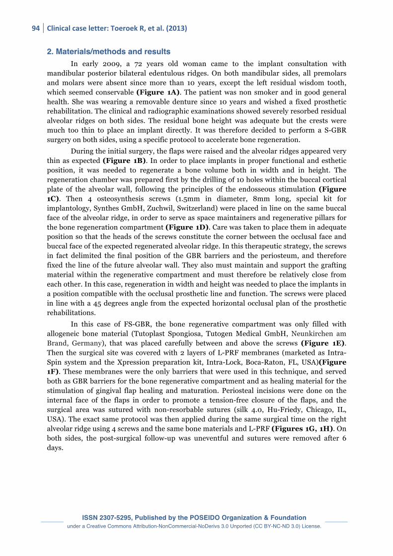
2. Materials/methods and results In early 2009, a 72 years old woman came to the implant consultation with
mandibular posterior bilateral edentulous ridges. On both mandibular sides, all premolars and molars were absent since more than 10 years, except the left residual wisdom tooth, which seemed conservable (Figure 1A). The patient was non smoker and in good general health. She was wearing a removable denture since 10 years and wished a fixed prosthetic rehabilitation. The clinical and radiographic examinations showed severely resorbed residual alveolar ridges on both sides. The residual bone height was adequate but the crests were much too thin to place an implant directly. It was therefore decided to perform a S-GBR surgery on both sides, using a specific protocol to accelerate bone regeneration.
During the initial surgery, the flaps were raised and the alveolar ridges appeared very thin as expected (Figure 1B). In order to place implants in proper functional and esthetic position, it was needed to regenerate a bone volume both in width and in height. The regeneration chamber was prepared first by the drilling of 10 holes within the buccal cortical plate of the alveolar wall, following the principles of the endosseous stimulation (Figure 1C). Then 4 osteosynthesis screws (1.5mm in diameter, 8mm long, special kit for implantology, Synthes GmbH, Zuchwil, Switzerland) were placed in line on the same buccal face of the alveolar ridge, in order to serve as space maintainers and regenerative pillars for the bone regeneration compartment (Figure 1D). Care was taken to place them in adequate position so that the heads of the screws constitute the corner between the occlusal face and buccal face of the expected regenerated alveolar ridge. In this therapeutic strategy, the screws in fact delimited the final position of the GBR barriers and the periosteum, and therefore fixed the line of the future alveolar wall. They also must maintain and support the grafting material within the regenerative compartment and must therefore be relatively close from each other. In this case, regeneration in width and height was needed to place the implants in a position compatible with the occlusal prosthetic line and function. The screws were placed in line with a 45 degrees angle from the expected horizontal occlusal plan of the prosthetic rehabilitations.
In this case of FS-GBR, the bone regenerative compartment was only filled with allogeneic bone material (Tutoplast Spongiosa, Tutogen Medical GmbH, Neunkirchen am Brand, Germany), that was placed carefully between and above the screws (Figure 1E). Then the surgical site was covered with 2 layers of L-PRF membranes (marketed as Intra-Spin system and the Xpression preparation kit, Intra-Lock, Boca-Raton, FL, USA)(Figure 1F). These membranes were the only barriers that were used in this technique, and served both as GBR barriers for the bone regenerative compartment and as healing material for the stimulation of gingival flap healing and maturation. Periosteal incisions were done on the internal face of the flaps in order to promote a tension-free closure of the flaps, and the surgical area was sutured with non-resorbable sutures (silk 4.0, Hu-Friedy, Chicago, IL, USA). The exact same protocol was then applied during the same surgical time on the right alveolar ridge using 4 screws and the same bone materials and L-PRF (Figures 1G, 1H). On both sides, the post-surgical follow-up was uneventful and sutures were removed after 6 days.
POSEIDO. 2013;1(2) Screw-‐Guided Bone Regeneration (S-‐GBR). Part 3
95
ISSN 2307-5295, Published by the POSEIDO Organization & Foundation
under a Creative Commons Attribution-NonCommercial-NoDerivs 3.0 Unported (CC BY-NC-ND 3.0) License.
Figure 1. Bilateral FS-GBR surgery in the severely resorbed posterior mandible. (A) Initial situation. (B) The left surgical site was opened and revealed a severely resorbed crest. (C) Ten holes of endosseous stimulation were done to prepare the bone regeneration compartment. (D) Four osteosynthesis screws were placed in line on the residual alveolar ridge, with a 45 degrees angle from the expected occlusal plan. (E) The space between and above the screws was filled with an allograft bone material. (F) The whole surgical site was covered with 2 layers of L-PRF membranes, and then sutured tension-free after periosteal incisions on the flaps. (G, H) The right surgical site was similarly resorbed, and was treated exactly like the left side.
96 Clinical case letter: Toeroek R, et al. (2013)
ISSN 2307-5295, Published by the POSEIDO Organization & Foundation
under a Creative Commons Attribution-NonCommercial-NoDerivs 3.0 Unported (CC BY-NC-ND 3.0) License.
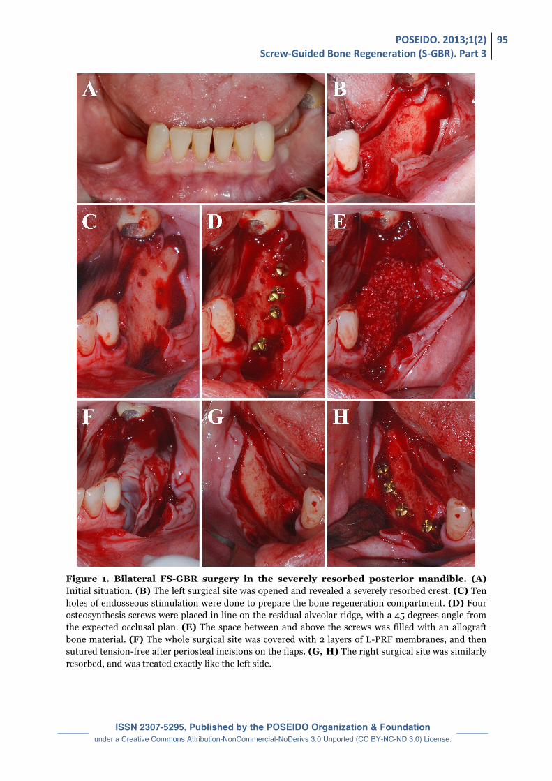
Figure 2. Implantation phase, 2 months after FS-GBR surgery in the severely resorbed posterior mandible. (A, B) Two months after FS-GBR, the surgical sites were reopened, revealing a dense, lightly bleeding and regular external aspect of the regenerated volumes. The screws were partially covered with bone. (C) Screws were removed, and a light bleeding appeared from the holes. (D) Three implants were placed on the right side, and 2 implants on the left side. (E) A collagen membrane was placed in position. (F) The whole regenerated area was covered with a supplementary layer of bovine xenograft bone material, to stabilize the regenerated alveolar ridge. (G) Two layers of L-PRF membranes were placed all over the surgical site, to maintain the grafted material and to stimulate soft tissue healing and maturation. (H) The sites were sutured tension-free using again periosteal incisions.
POSEIDO. 2013;1(2) Screw-‐Guided Bone Regeneration (S-‐GBR). Part 3
97
ISSN 2307-5295, Published by the POSEIDO Organization & Foundation
under a Creative Commons Attribution-NonCommercial-NoDerivs 3.0 Unported (CC BY-NC-ND 3.0) License.
Eight weeks after the bone regeneration surgery, the sites appeared well healed and were reopened. The bone volumes seemed well regenerated and homogeneous (Figures 2A, 2B). The regenerated bone was dense but relatively soft (D3 quality). The osteosynthesis screws were still covered with some regenerated bone and were then removed (Figure 2C). Implant osteotomies were performed carefully and the bone volume seemed compact and homogeneous during drilling. Three implants were placed on the right side (Figure 2D) and 2 implants of the left side (Ankylos C/X, Dentsply implants, Mannheim, Germany), all of them 11mm long and 4.5mm in diameter, except the mesial right implant (3.5mm in diameter).
After implantation, it was decided to use a combination of bovine bone and collagen membrane to help the long-term stability of the allograft material. On both sides, a resorbable collagen membrane made from porcine pericardial tissue (now marketed as BoneProtect Membrane, Dentegris GmbH, Duisburg, Germany) was placed to delimit each regenerative compartment (Figure 2E). This membrane had a long-term resorption evaluated between 3 and 4 months. Then the regenerated alveolar ridges were covered with bone particulate material (now marketed as CompactBone B, Dentegris GmbH, Duisburg, Germany)(Figure 2F) and the collagen membranes were closed on it respectively. The surgical sites were finally covered with 2 layers of L-PRF membranes (marketed as Intra-Spin system and the Xpression preparation kit, Intra-Lock, Boca-Raton, FL, USA) on each side (Figure 2G), in order to help gingival healing and maturation and to prevent eventual gingival dehiscence. Periosteal incisions were performed again on the flaps in order to suture the sites without tensions, using non-resorbable sutures (silk 4.0, Hu-Friedy, Chicago, IL, USA)(Figure 2H). Sutures were removed after 6 days.
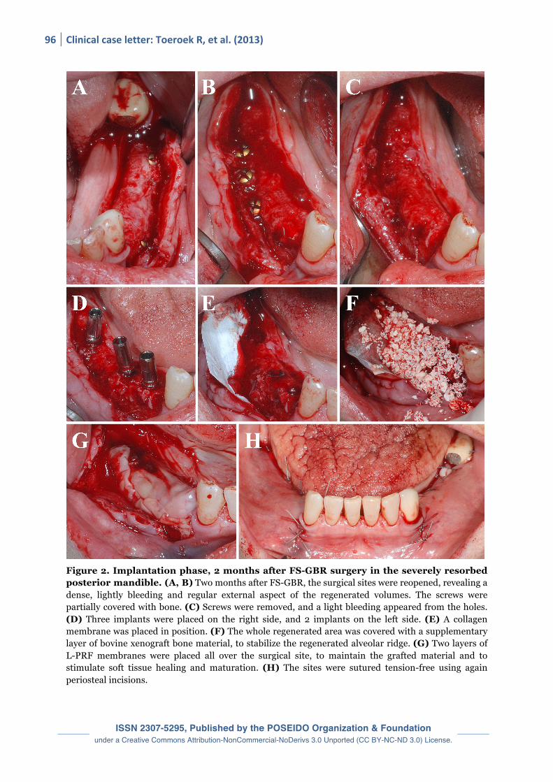
Figure 3. Prosthetic phase and follow-up. (A) Three months after implantation, trans-gingival screws were placed and the sites were sutured using split-thickness apically repositioned flap to improve the peri-implant gingival shape. (B, C) After 4 weeks of healing, the gingival tissue appeared healed and strong. (D) The implant abutments were placed.
98 Clinical case letter: Toeroek R, et al. (2013)
ISSN 2307-5295, Published by the POSEIDO Organization & Foundation
under a Creative Commons Attribution-NonCommercial-NoDerivs 3.0 Unported (CC BY-NC-ND 3.0) License.
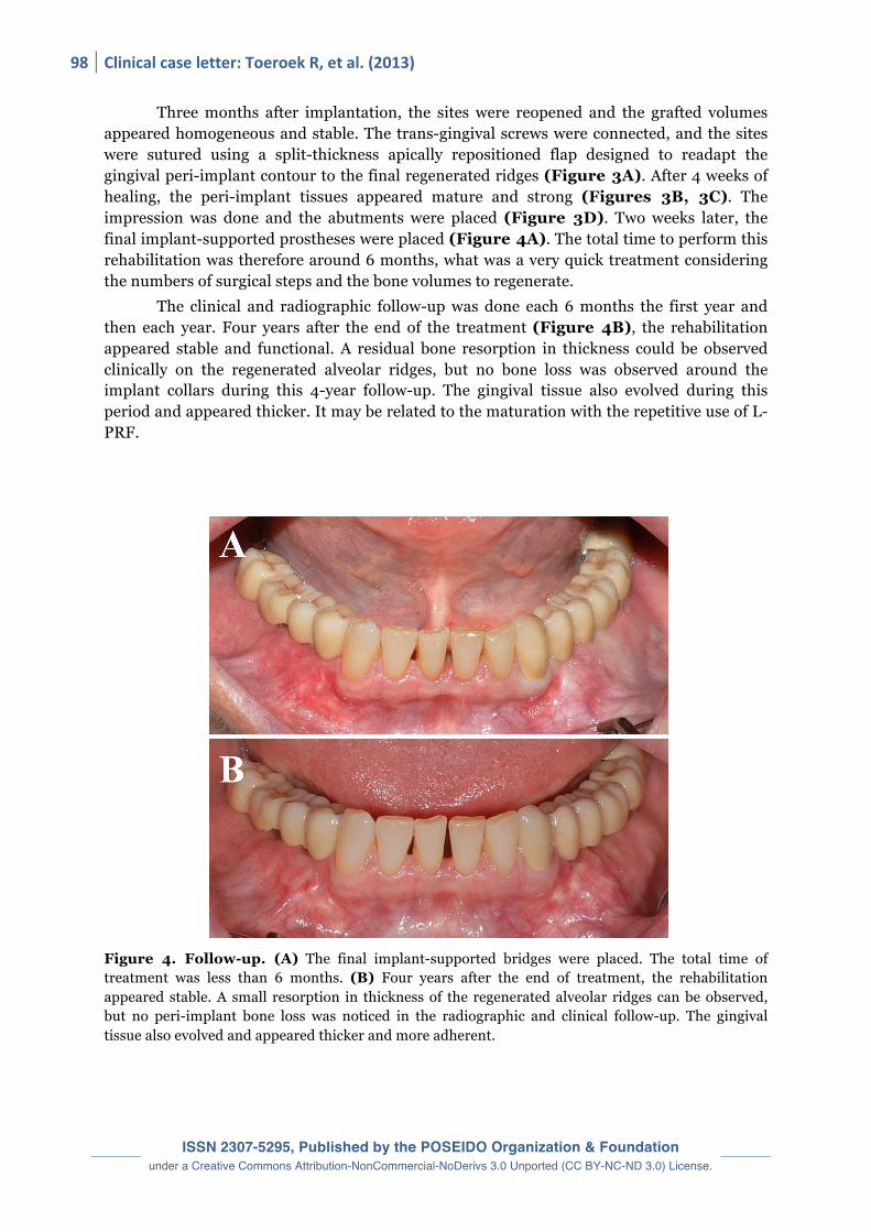
Three months after implantation, the sites were reopened and the grafted volumes appeared homogeneous and stable. The trans-gingival screws were connected, and the sites were sutured using a split-thickness apically repositioned flap designed to readapt the gingival peri-implant contour to the final regenerated ridges (Figure 3A). After 4 weeks of healing, the peri-implant tissues appeared mature and strong (Figures 3B, 3C). The impression was done and the abutments were placed (Figure 3D). Two weeks later, the final implant-supported prostheses were placed (Figure 4A). The total time to perform this rehabilitation was therefore around 6 months, what was a very quick treatment considering the numbers of surgical steps and the bone volumes to regenerate.
The clinical and radiographic follow-up was done each 6 months the first year and then each year. Four years after the end of the treatment (Figure 4B), the rehabilitation appeared stable and functional. A residual bone resorption in thickness could be observed clinically on the regenerated alveolar ridges, but no bone loss was observed around the implant collars during this 4-year follow-up. The gingival tissue also evolved during this period and appeared thicker. It may be related to the maturation with the repetitive use of L-PRF.
Figure 4. Follow-up. (A) The final implant-supported bridges were placed. The total time of treatment was less than 6 months. (B) Four years after the end of treatment, the rehabilitation appeared stable. A small resorption in thickness of the regenerated alveolar ridges can be observed, but no peri-implant bone loss was noticed in the radiographic and clinical follow-up. The gingival tissue also evolved and appeared thicker and more adherent.
POSEIDO. 2013;1(2) Screw-‐Guided Bone Regeneration (S-‐GBR). Part 3
99
ISSN 2307-5295, Published by the POSEIDO Organization & Foundation
under a Creative Commons Attribution-NonCommercial-NoDerivs 3.0 Unported (CC BY-NC-ND 3.0) License.
3. Discussion As explained in the previous parts of this series of articles, the S-GBR approach
requires different healing times depending on the combination of materials and membranes [3], in order to obtain bone ridges ready for implantation. With bovine xenograft, collagen membrane and non-resorbable membrane, the regeneration time was of 7-8 months. With a mix of xenograft and allograft and the cover with collagen and L-PRF membranes, the regeneration time was around 4 months. In this article, only allograft and L-PRF were used [12,13] and the regeneration time felt to 2 months only, what is a very fast regeneration strategy, particularly in the posterior mandible.
Even if the literature is not very clear on this matter [14], it is commonly known that collagenated allograft materials have a quicker bone integration than bovine xenograft, but also that allograft have tendency to resorb slowly on the long-term while bovine xenograft are very stable with time [15]. For this reason, both materials were mixed in the S-GBR technique we described in the part 2. In the FS-GBR approach, the regeneration surgery was done only with allograft and L-PRF in order to stabilize quickly a regenerated alveolar ridge, and some bovine xenograft (always associated with a collagen membrane)[15] was added during the second surgery in order to stabilize the regenerated ridge and to control or stop the resorption of the allograft material. This final cover with bovine xenograft should not be neglected, this is a key element of this therapeutic strategy for the long-term stability of the rehabilitation. However in the FS-GBR, all steps require the use of L-PRF membranes as healing and maturation material [5,6].
Finally, in this case the regeneration was done with L-PRF as sole barrier above the allograft and with only 2 layers of L-PRF (in comparison to the 4 layers described in the previous articles). It raised the question if L-PRF can be used as a GBR barrier alone [11], as no collagen membrane was needed in this case. From our experience and the literature [12,13], L-PRF functions as a competitive barrier when associated with the adequate bone materials and clinical situations [5,6]. In the case of FS-GBR, the concept of association of L-PRF with some forms of allografts (already collagenated materials) gave adequate results, but it must be investigated carefully in the future to determine the best indications and relevant combinations.
Disclosure of interests
The authors have no conflict of interest to report.
Acknowledgement This work for the definition of international standards in implantable materials and
techniques is supported by a grant from the National Research Foundation of Korea (NRF) funded by the Korean government-MEST (No. 2011-0030121) and by the LoB5 Foundation for Research, France.
References [1] Buser D, Dula K, Belser UC, Hirt HP, Berthold H. Localized ridge augmentation using guided bone regeneration. II. Surgical procedure in the mandible. Int J Periodontics Restorative Dent. 1995;15(1):10-29. [2] Schliephake H, Dard M, Planck H, Hierlemann H, Jakob A. Guided bone regeneration around endosseous implants using a resorbable membrane vs a PTFE membrane. Clin Oral Implants Res. 2000;11(3):230-41. [3] Browaeys H, Bouvry P, De Bruyn H. A literature review on biomaterials in sinus augmentation procedures. Clin Implant Dent Relat Res. 2007;9(3):166-77.
100 Clinical case letter: Toeroek R, et al. (2013)
ISSN 2307-5295, Published by the POSEIDO Organization & Foundation
under a Creative Commons Attribution-NonCommercial-NoDerivs 3.0 Unported (CC BY-NC-ND 3.0) License.
[4] Dohan Ehrenfest DM, Bielecki T, Mishra A, Borzini P, Inchingolo F, Sammartino G, Rasmusson L, Evert PA. In search of a consensus terminology in the field of platelet concentrates for surgical use: platelet-rich plasma (PRP), platelet-rich fibrin (PRF), fibrin gel polymerization and leukocytes. Curr Pharm Biotechnol. 2012;13(7):1131-7. [5] Del Corso M, Vervelle A, Simonpieri A, Jimbo R, Inchingolo F, Sammartino G, Dohan Ehrenfest DM. Current knowledge and perspectives for the use of platelet-rich plasma (PRP) and platelet-rich fibrin (PRF) in oral and maxillofacial surgery part 1: Periodontal and dentoalveolar surgery. Curr Pharm Biotechnol. 2012;13(7):1207-30. [6] Simonpieri A, Del Corso M, Vervelle A, Jimbo R, Inchingolo F, Sammartino G, Dohan Ehrenfest DM. Current knowledge and perspectives for the use of platelet-rich plasma (PRP) and platelet-rich fibrin (PRF) in oral and maxillofacial surgery part 2: Bone graft, implant and reconstructive surgery. Curr Pharm Biotechnol. 2012;13(7):1231-56. [7] Dohan Ehrenfest DM, Del Corso M, Diss A, Mouhyi J, Charrier JB. Three-dimensional architecture and cell composition of a Choukroun's platelet-rich fibrin clot and membrane. J Periodontol. 2010;81(4):546-55. [8] Dohan Ehrenfest DM. How to optimize the preparation of leukocyte- and platelet-rich fibrin (L-PRF, Choukroun's technique) clots and membranes: introducing the PRF Box. Oral Surg Oral Med Oral Pathol Oral Radiol Endod. 2010;110(3):275-8. [9] Dohan Ehrenfest DM, Diss A, Odin G, Doglioli P, Hippolyte MP, Charrier JB. In vitro effects of Choukroun's PRF (platelet-rich fibrin) on human gingival fibroblasts, dermal prekeratinocytes, preadipocytes, and maxillofacial osteoblasts in primary cultures. Oral Surg Oral Med Oral Pathol Oral Radiol Endod. 2009;108(3):341-52. [10] Clark RA. Fibrin and wound healing. Ann N Y Acad Sci. 2001;936:355-67. [11] Gassling V, Douglas T, Warnke PH, Acil Y, Wiltfang J, Becker ST. Platelet-rich fibrin membranes as scaffolds for periosteal tissue engineering. Clin Oral Implants Res. 2010;21(5):543-9. [12] Simonpieri A, Del Corso M, Sammartino G, Dohan Ehrenfest DM. The relevance of Choukroun's platelet-rich fibrin and metronidazole during complex maxillary rehabilitations using bone allograft. Part I: a new grafting protocol. Implant Dent. 2009;18(2):102-11. [13] Simonpieri A, Del Corso M, Sammartino G, Dohan Ehrenfest DM. The relevance of Choukroun's platelet-rich fibrin and metronidazole during complex maxillary rehabilitations using bone allograft. Part II: implant surgery, prosthodontics, and survival. Implant Dent. 2009;18(3):220-9. [14] Froum SJ, Wallace SS, Elian N, Cho SC, Tarnow DP. Comparison of mineralized cancellous bone allograft (Puros) and anorganic bovine bone matrix (Bio-Oss) for sinus augmentation: histomorphometry at 26 to 32 weeks after grafting. Int J Periodontics Restorative Dent. 2006;26(6):543-51. [15] Zitzmann NU, Naef R, Scharer P. Resorbable versus nonresorbable membranes in combination with Bio-Oss for guided bone regeneration. Int J Oral Maxillofac Implants. 1997;12(6):844-52. This article can be cited as: Toeroek R, Dohan Ehrenfest DM. The concept of Screw-Guided Bone Regeneration (S-GBR). Part 3: Fast Screw-Guided Bone Regeneration (FS-GBR) in the severely resorbed preimplant posterior mandible using allograft and Leukocyte- and Platelet-Rich Fibrin (L-PRF): a 4-year follow-up. POSEIDO. 2013;1(2):93-100.
![Evaluations of guided bone regeneration in canine …...guided bone regeneration (GBR) [5, 6] have been succes-sively applied in clinical settings. Both GTR and GBR use a barrier membrane](https://img.pdfslide.us/doc/110x75/5ed6c28ed397173fdb727e96/evaluations-of-guided-bone-regeneration-in-canine-guided-bone-regeneration-gbr.jpg)