Embed Size (px)
Citation preview
1
The composition of endolithic communities in gypcrete is determined
by the specific microhabitat architecture
María Cristina Casero1*, Victoria Meslier2$, Jocelyne DiRuggiero2, Antonio Quesada3, Carmen Ascaso1,
Octavio Artieda4, Tomasz Kowaluk5, Jacek Wierzchos1**
1Departamento Biogeoquímica y Ecología Microbiana, Museo Nacional de Ciencias Naturales, CSIC, Madrid, 28006, Spain 5 2Department of Biology, and Department of Earth and Planetary Sciences, Johns Hopkins University, Baltimore, MD, 21218.
USA 3Departamento de Biología, Universidad Autónoma de Madrid, Madrid, 28014, Spain 4Departamento de Biología Vegetal, Ecología y Ciencias de la Tierra, Universidad de Extremadura, Plasencia, 06006, Spain 5Institute of Metrology and Biomedical Engineering, Faculty of Mechatronics, Warsaw University of Technology, 02-525 10
Warsaw, Poland
$ now at: MetaGenoPolis, Jouy-en-Josas, France
Correspondence to: María Cristina Casero* ([email protected]) and Jacek Wierzchos**
Abstract. Endolithic microhabitats have been described as the last refuge for life in arid and hyper-arid deserts where life has 15
to deal with harsh environmental conditions. A number of rock substrates from the hyper-arid Atacama Desert, colonized by
endolithic microbial communities, such as halite, gypsum crusts, gypcrete, calcite, granite and ignimbrite, have been
characterized and compared using different approaches. In this work, three different endolithic microhabitats are described,
each one with a particular origin and architecture, found within a lithic substrate known as gypcrete. Gypcrete, an evaporitic
rock mainly composed of gypsum (CaSO4·2H2O) and collected in the Cordón de Lila area of the desert (Preandean Atacama 20
Desert), was found to harbour cryptoendolithic (within pore spaces in the rock), chasmoendolithic (within cracks and fissures)
and hypoendolithic (within microcave-like pores in the rock-bottom layer) microhabitats. A combination of microscopy
investigation and high-throughput sequencing approaches were used to characterize the endolithic communities and their
habitats at the microscale within the same piece of gypcrete. Microscopy techniques revealed differences in the architecture of
the endolithic microhabitats and the distribution of the microorganisms within those microhabitats. Cyanobacteria and 25
Proteobacteria were dominant in the endolithic communities, of which the hypoendolithic community was the least diverse
and hosted unique taxa, as a result of lower access to sun radiation. These results show, for the first time, that the differences
in the architecture of a microhabitat, even within the same piece of a lithic substrate, plays an essential role in shaping the
diversity and composition of endolithic microbial communities.
30
2
1. Introduction 35
The statement developed by Professor Lourens Gerhard Marinus Baas-Becking (1934) “everything is everywhere but the
environment selects” which established the most referred principle for microbial biogeography remains in discussion regarding
the first half of the statement (everything is everywhere) (de Wit and Bouvier 2006, O’Malley 2008, Bass and Boenigk 2011,
Fontaneto and Hortal 2012, van der Gast 2015). Regarding the second half of the statement (but, the environment selects)
extreme environments present some of the most plausible scenarios since they are inhabited only by microorganisms that can 40
survive and/or thrive in their respective physical or geochemical extremes such as temperature, solar radiation, pressure,
desiccation, pH (Rothschild and Mancinelli 2001).
Hyper-arid deserts, where aridity index is lower than 0.05 (Nienow, 2009) constitute the most extreme deserts on Earth, and
usually combine a series of simultaneous stress conditions such as water limitation, extreme high and low temperatures,
scarcity of organic carbon, high solar radiation and osmotic stress (Pointing and Belnap 2012). While these environments are 45
considered polyextreme, they are inhabited by microbiota able to survive under such conditions. Hence, polyextreme
environments are excellent microbial ecosystem models to study adaptive mechanisms to environmental stress. The Atacama
Desert (North Chile) is perhaps the most challenging polyextreme environment on Earth and the most barren region
imaginable, with scarce precipitation events (McKay et al. 2003; Wierzchos et al. 2012a) and extremely low mean annual
relative humidity (RH) (Azua-Bustos et al. 2015). Further, this desert holds another world record: the highest surface ultraviolet 50
radiation (UV), photosynthetic active radiation (PAR) and annual mean surface solar radiation (Cordero et al. 2018) in the
Coastal Cordillera and Andes sites.
In this inhospitable polyextreme desert, microbial life has been found in different lithic habitats as epilithic (on rocks,
hypolithic (under rocks) (Azua-Bustos et al., 2012) and endolithic (inside rocks) microhabitats (rev. by Wierzchos et al. 2018;
Wierzchos et al. 2012b). Three different locations of these endolithic habitats have been described within rocks of the Atacama 55
Desert: cryptoendolithic (occupying pore spaces in the rock), chasmoendolithic (living within cracks and fissures in the rock),
and hypoendolithic (living inside pores in the bottom part of the rock). Endolithic colonization can be viewed as a stress
avoidance strategy whereby the overlying mineral substrate provides protection from incident lethal UV and PAR radiation
levels and also offers enhanced moisture availability (Walker and Pace 2007; Wierzchos et al. 2012b). These microbial
communities, regardless of the position they occupy in the rock, or the type of rock, are supported by oxygenic phototrophic 60
primary producers supporting a diversity of heterotrophic microorganisms (rev. in Wierzchos et al. 2018). Molecular and
microscopy characterization of these endolithic microbial communities shows that, overall, these communities are dominated
by Cyanobacteria, mostly from the extremely resistant to ionizing radiation and desiccation genus Chroococcidiopsis (Meslier
et al. 2018, Crits-Christoph et al. 2016, Billi et al. 2000, Cockell et al. 2005) as well as members from genera Gloeocapsa
(Crits-Christoph et al. 2016) and Halothece (de los Ríos et al. 2010; Robinson et al. 2015 and Uristkyi et al 2019), and phyla 65
Actinobacteria, Proteobacteria, Chloroflexi, Bacterioidetes and Euryarchaeota (Meslier et al. 2018). In gypcrete and gypsum
crust from Preandean Atacama Desert (Casero et al., 2020), previous studies reported endolithic communities dominated by
3
the phyla Cyanobacteria (36-83%), Actinobacteria (10-25%) and Proteobacteria (13-30%) (Wierzchos et al. 2015; Dong et al.
2007; and Meslier et al. 2018; rev. in Casero et al. 2020), however, these studies did not differentiate between microhabitats,
even though the occurrence of different endolithic microhabitats in gypcrete had already been described (Wierzchos et al. 70
2015).
This work addresses the impact of microhabitat architecture on the diversity and composition of gypcrete endolithic microbial
communities (EMCs). The concept of rock architecture was introduced by Wierzchos et al. (2015) for colonized gypcrete
substrate and encompasses the internal structures of rock with all elements that are essential for microbial life. Microhabitat
architecture allows perceiving the rock interior from the existence of porous spaces of different sizes and also the solid 75
structures that divide and support these spaces. All these components and elements are interrelated and influence one another,
thus fulfilling a requisite: they might shape a suitable architecture to hold microbial life.
The study is based on the hypothesis that the different architecture of endolithic microhabitats involves small-scale differences
in the micro-environmental conditions, which in turn determine the distribution of organisms in each community. The
hypothesis is tested here for the first time by using a multidisciplinary approach combining microscopy and molecular tools 80
for their characterization. The microscale dimensions and differential diversity distribution in this unique environment have
led us to coin the new term “microbiogeography”.
2. Experimental procedures
2.1 Site description and sampling 85
Colonized rocks were collected in the Atacama Desert in December 2015 from the Monturaqui area (MTQ) (GPS coordinates
23°57’S; 068°10’W; 2868 m.a.s.l.) located in an N-S trending depression of the Cordón de Lila Range. The area experiences
a pronounced rain shadow effect by the western slope of the central Andes from 15° to 23°S (DiRuggiero et al. 2013; Wierzchos
et al. 2015). In order to study endolithic communities that inhabit the same piece of a lithic substrate, we sampled gypcrete
pieces that harboured at least two of the three endolithic microhabitats of interest, that were collected within a 50 m2 area. All 90
samples were packed in sterile bags and stored at room temperature in dry and dark conditions before further processing.
2.2 Microclimate data
Microclimate data (Meslier et al. 2018) were recorded using an Onset HOBO® Microweather Station Data Logger (H21-
USB), as previously described by Wierzchos et al. (2015). Air temperature (T), air relative humidity (RH in %) and
Photosynthetic Active Radiation (PAR in μmol photons m-2 s-1) were recorded from January 2011 to February 2013 (22 95
months) (Wierzchos et al. 2015). Rainfall data were obtained from DiRuggiero et al. (2013). Thermal measurements of the
gypcrete surface were acquired at zenith time at 20 cm distance from the substrate. Thermal images were taken using a thermal
infrared camera (FLIR® E6, FLIR Systems, Oregon, USA) whose technical specifications are: ±2°C or ±2% of reading; <
4
0.06°C pixel sensitivity with a resolution of 160 × 120 pixels. Calibration of the FLIR camera for measurements of gypcrete
surface temperature was performed introducing the emissivity value of 0.92. 100
2.3 Microscopy analyses
Colonized gypcrete samples were processed for scanning electron microscopy in backscattered detection mode (SEM-BSE)
according to methods described by Wierzchos and Ascaso (1994) and Wierzchos et al. (2011). Light microscopy (LM) in
differential interference contrast mode (DIC) was used to examine cell aggregates gently isolated from the cryptoendolithic,
chasmoendolithic and hypoendolithic microhabitats and on cyanobacterial isolates cultured from those microhabitats. The 105
samples were examined using a microscope AxioImager M2, Carl Zeiss, Germany in DIC mode equipped with Apochrome
63x n=1.4 oil immersion objective.
2.4 CT-Scan analysis
Micro-CT scans were run on pieces of gypcrete with an X-Ray Computed Tomography system (CT-scan) — HMXST 225
micro-CT system (Nikon Metrology, Tring, UK) to observe volume, bulk density, and variations in internal density. For 110
volume and bulk density measurements a Nikon X-Tek CT-Scan device was used, with an X-ray peak voltage of 146 kV and
current of 65 mA, collecting 1583 sections at 1000 microseconds on average from four frames. The system operates with an
X-ray tube and added filtration (0.875 mm Cu) to reduce beam hardening. Three-dimensional viewing and analyses of the
obtained X-ray sections were performed by software VG Studio Max Version 2.2. software. The auto-threshold feature
determined the grey-scale intensity for 3-D surface segmentation and subsequent analysis. 115
2.5 Cyanobacteria isolation and characterization and DNA extraction procedures from isolates.
Biological material removed from each endolithic colonization zones of gypcrete was transferred to different BG11 1.5%-agar
plates (Purified agar, Condalab, Spain). All samples were incubated in a growth chamber at 28±2°C with illumination of 20
μmol photons m-2 s-1 by cool white 40W fluorescent tubes (Philips). After incubation for 15 days, visible cyanobacterial growth
appeared. Colonies were isolated by repeated plating on 0.8%-agar with BG11 medium (Rippka et al. 1979), and successfully 120
isolated colonies were transferred to liquid BG11 medium. Culture material from each strain (2 mL) was harvested during
exponential growth and centrifuged (10,000 g, 5 min). Genomic DNA was extracted from the cell pellet using the UltraClean
DNA isolation kit (MoBio Laboratories, Solana Beach, CA, USA). 16S rRNA was amplified using primers PA (Edwards et
al. 1989) and B23S (Lepère et al. 2000), PCR reaction and sequencing were performed as described in Casero et al. (2014).
2.6 DNA extraction procedures from natural samples, 16S rRNA gene libraries preparation and sequencing 125
Three individual rocks harbouring at least two of the three endolithic microhabitats were processed, which resulted in 11
samples, including technical replicates: cryptoendolithic (n=2), chasmoendolithic (n=6) and hypoendolithic (n=3).
Colonization zones were scraped and ground for DNA extraction. To avoid contamination between samples from different
5
microhabitats, the scraping of material was carried out in the following way: due to the possible proximity of both
chasmoendolithic and cryptoendolithic microhabitats, on the top of the rock, chasmoendolithic colonization zones more distant 130
from cryptoendolithic colonization zones were selected. In addition, material from each of them was scraped avoiding the
edges, so that material from different microhabitats could not be mixed. In the case of the samples coming from hypoendolithic
samples, the distance from the other two microhabitats allowed their full scraping.
This DNA extraction was performed using 0.3 g of samples and the UltraClean DNA isolation kit (MoBio Laboratories, Solana
Beach, CA, USA) including a three-cycle step of freezing 0.3mL aliquots of sample suspended in buffer, breaking them down 135
by using an adapted drill and melting in 60ºC water bath, as described in Loza et al. (2013) and Becerra-Absalón et al. (2019).
A two-step PCR strategy was used to prepare the sequencing libraries of endolithic microbial communities, as previously
described (Robinson et al. 2015). DNA was amplified using primers 338F and 806R (V3-V4 hypervariable region) barcoded
for multiplexing; amplicons from 2 PCR reactions were pooled after the first step. Illumina paired-end sequencing (2 x 250bp)
was performed using the MiSeq platform at the Johns Hopkins Genetic Resources Core Facility (GRCF). 140
2.7 Computational analysis
After demultiplexing and barcode removal, sequence reads with Phred score <20 and length <100bp were discarded using
sickle (Joshi and Fass, 2011), representing only 2% of the initial reads count. The Qiime package (v1.6.0) was used to further
process the sequences (Caporaso et al. 2010) and diversity metrics were calculated based on Operational Taxonomic Units
(OTUs) at the 0.03% cutoff against the Ribosomal Database Project (RDP) database release 11 (Cole et al. 2014). The resulting 145
OTUs table was filtered of the rare OTUs (total abundance across all samples below 1%), representing 40% of the initial count
(1511 OTUs).
2.8 Phylogenetic analysis
Sequences of 16S rRNA genes from Cyanobacterial OTUs that showed significant differences in their relative abundance
between endolithic microhabitats and 16S rRNA gene sequences from cyanobacterial isolates were aligned with sequences 150
obtained from the NCBI GenBank using the Clustal W 1.4 software (Thompson et al. 1994). 16S rRNA gene sequences from
GenBank were selected using the NCBI MegaBlast tool (http://blast.ncbi.nlm.nih.gov/ Blast.cgi, accessed 28.08.18). The final
alignment length was 400 bp. Phylogenetic trees of each of the genes were constructed in MEGA 7.0 using the Maximum
Likelihood (ML) method (Kumar et al. 2016). The best-fitting evolutionary model, chosen following the BIC (Bayesian
Inference Criterion) in MEGA 7.0, was the Kimura 2-parameter model (Kimura 1980) for 16S rRNA genes. 1000 bootstrap 155
replicates were performed for all trees.

6
3. Results
We combined microclimate measurements, microscopy analyses and high throughput culture-independent molecular data to
identify the effect of micro-biogeography and the factors underlying the structure and composition of microbial assemblages
of gypcrete endoliths from the hyper-arid Atacama Desert. 160
3.1 Sampling site
Gypcrete samples were collected from the Monturaqui area (MTQ), located in the Preandean Depression of the Atacama Desert
(Casero et al., 2020) (Fig. 1) in December 2015. Climate data recorded over a period of 22 months described a mean air
temperature about 15°C, with strong amplitude between minima and maxima (from -4.7°C to 49.3°C), average diurnal PAR ~
1000 μmol photons m-2 s-1 with a maximum of 2553.7 μmol photons m-2 s-1, providing evidence for the extremely intense solar 165
irradiance found in this region (Cordero et al. 2014). This area experiences extremely dry conditions, with an average air RH
of about 20% with frequent lows of 1% and precipitations extremely scarce with mean annual values of 24.5 mm (Wierzchos
et al. 2015). Gypcrete surface temperature examined with a thermal infrared camera revealed a maximum temperature of 68°C.
3.2 Micromorphology of gypcrete
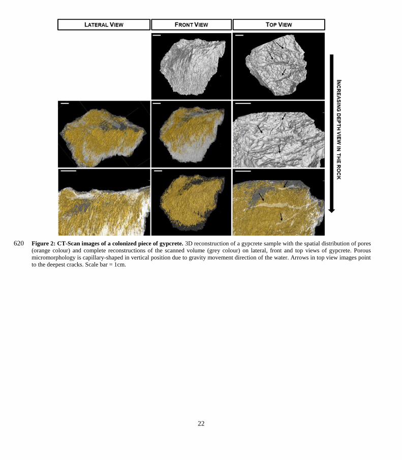
CT-Scan images provided a 3D spatial representation of pore shapes and their distribution inside the gypcrete rock (Figs. 2). 170
The pores revealed capillary-like micromorphology exhibit a vertical orientation as is shown in both top and lateral views.
Detailed 3D images pointed to the apparent absence of connectivity with the surface of most of the pores (Figs. 2). However,
the presence of this connectivity cannot be discarded due to the limited resolution of the CT-Scan technique and the conditions
of acquisition. Moreover, CT-scan images of the gypcrete surface reveal microrills weathering features (DiRuggiero et al.
2013) due to the dissolution of gypsum after scarce rains (Video S1). 175
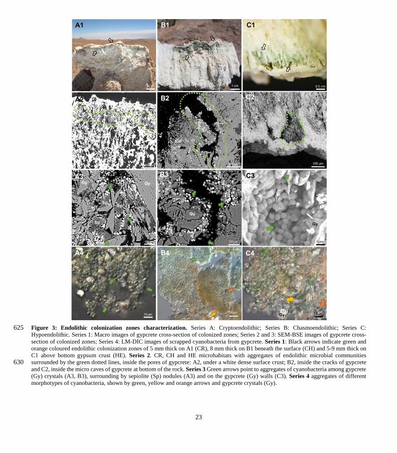
3.3 Endolithic microhabitats
Cross-sections of the gypcrete rocks reveal the presence of three clearly differentiated microhabitats where a significant
heterogeneity in micromorphology and structure was found (Figs. 3). The cryptoendolithic colonization zone is close to the
compact gypcrete surface layer (up to 5mm depth). Within cryptoendolithic microbial communities, two characteristic
pigmented layers are distinguished. The observed orange colour belongs to microorganisms with high carotenoids content 180
closest to the gypcrete surface. The green colour layer beneath the orange layer belongs to microorganisms with chlorophyll
and phycobiliproteins content. The presence of these pigments was previously reported by Wierzchos et al. (2015) and Vítek
et al. (2016) (Fig. 3, A1). The chasmoendolithic colonization zone reaches a deeper (up to 8mm depth) position in the substrate
and is directly connected to the surface (Fig. 3, B1). Finally, the hypoendolithic colonization zone is located close to the
compact bottom gypcrete crust, shaped like micro-caves (Fig. 3, C1). 185
7
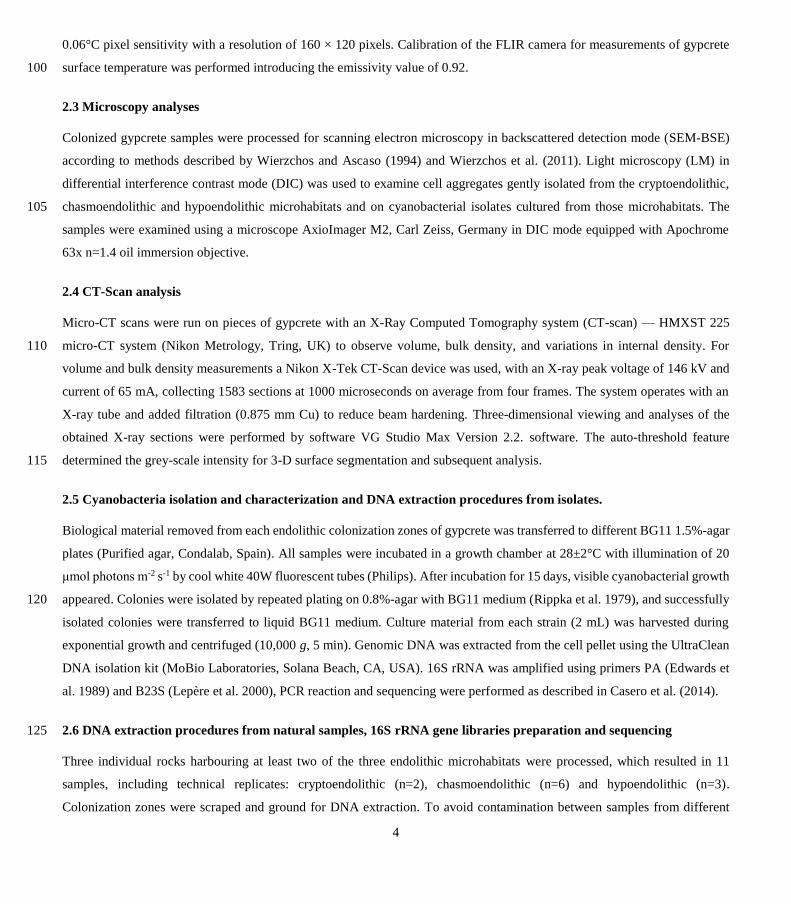
Cyanobacteria were found in the cryptoendolithic habitat among lenticular gypcrete crystals, filling up vertically elongated
pores, and aggregated around sepiolite nodules (Figs. 3, A2-A3), a clay mineral with high water retention capacity, previously
identified in gypcrete by Wierzchos et al. (2015). SEM-BSE also revealed dense arrangements of cyanobacterial cells
embedded in concentric sheets of EPSs (Figs. 3, A3). By contrast, the microbial assemblages inhabiting the chasmoendolithic
and hypoendolithic microhabitats were coating the walls of the cracks and caves previously described (Figs. 3, B2, B3, C2, 190
C3). Detailed SEM-BSE (Figs. 3, A3-C3) and LM images (Figs. 3, A4-C4) of each microhabitat showed mainly Cyanobacteria
with different size and morphology accompanied by heterotrophic bacteria.
3.4 Cyanobacterial isolates from endolithic microhabitats
A total of 12 cyanobacterial strains were isolated from the three different endolithic microhabitats (Table S1): 3 from
cryptoendolithic, 3 from chasmoendolithic and 6 from hypoendolithic. The cyanobacterial strains were identified, following 195
Komárek et al. (2014), as Chroococcidiopsis sp. (UAM800, UAM801, UAM802, UAM805, UAM808, UAM809, UAM810,
UAM811), Gloeocapsa sp. (UAM803, UAM804) and Gloeocapsopsis sp. (UAM806, UAM807).
3.5 Structure and composition of endolithic communities
High throughput sequencing of 16S rRNA gene amplicons across 11 samples and 3 microhabitats resulted in a total of 385,440
V3-V4 SSU rDNA reads, with an average number of paired-end reads per sample of 35,040 ± 6,288 and an average length of 200
456 ± 11 bp. Diversity metrics, calculated from OTUs clustered at 97%, revealed no significant differences between
microhabitats in terms of alpha-diversity (Table 1).
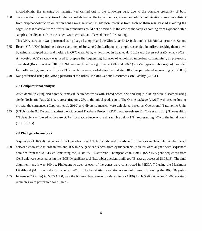
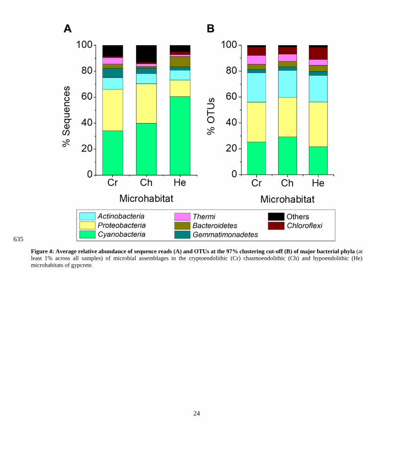
A total of 11 bacterial phyla with a relative abundance > 0.1% were found across all microhabitats. Of these only 7 had a
relative abundance over 1% of sequences across the different microhabitats (Figs. 4). Cyanobacteria, Proteobacteria,
Actinobacteria and Gemmatimonadetes were the most abundant phyla, representing 82%–83% of the total community (Fig. 4, 205
A). Cyanobacteria dominated the communities inhabiting all endolithic microhabitats; in the cryptoendolithic and
chasmoendolithic communities, Cyanobacteria did not exceed 40% of the sequences, while in the hypoendolithic community
they reached a relative abundance of 60% (Fig. 4, A). Proteobacteria were the second most abundant phylum, contributing
~30% of the sequences in the cryptoendolithic and chasmoendolithic communities, and less than a half in the hypoendolithic
community (13%). The relative abundance of Actinobacteria was even across all microhabitats, never exceeding 10% of the 210
sequence reads. Gemmatimonadetes relative abundance showed differences across microhabitats representing 7%, 4.4% and
2.3% of sequences in the cryptoendolithic, chasmoendolithic and hypoendolithic communities, respectively (Fig. 4, A). The
phyla Bacteroidetes and Thermi also exhibited variation between the different endolithic communities, showing the higher
relative abundance in the hypoendolithic (8.2%) and cryptoendolithic (4.9%) microhabitats. Firmicutes and Planctomycetes
were also found in all three microhabitats at very low relative abundance (0.003% and 0.002%). No archaeal OTUs were 215
detected before or after the quality filtering of sequences during the processing of the samples.
8
The four main phyla constituted ~ 80% of OTUs, clustered at 97% identity, across all microhabitats, which was quite different
from the distribution of sequence reads (Fig. 4, B). The three major phyla, Cyanobacteria, Proteobacteria and Actinobacteria,
has similar OTUs relative abundances across all three microhabitats (25%, 32% and 21% respectively). The greatest difference
between the distribution of the relative abundance of sequences and that of OTUs is observed for Cyanobacteria in the 220
hypoendolithic community.
Compared to other microhabitats this phylum showed the highest relative abundance in terms of sequences (60.4%) but the
lowest in terms of OTUs (21.9%), thus revealing the high abundance of a very low number of cyanobacterial OTUs. Average
Bray Curtis distance confirmed that dissimilarity between microhabitats (CR-CH= 0.36, CR-HE= 0.44, CH-HE= 0.44) was
higher than between replicates of the same microhabitat (CR=0.36, CH= 0.29, HE=0.32). Adonis and ANOSIM tests, 225
performed with the 3 microhabitats categories (cryptoendolithic, chasmoendolithic and hypoendolithic), confirmed the
statistical significance of the grouping (R2 =0.38, p- value=0.014 and R2=0.48, p-value=0.003 for adonis and ANOSIM,
respectively).
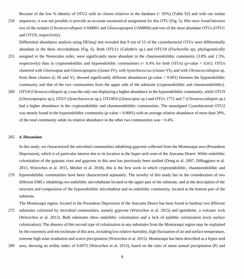
3.6 Cyanobacterial composition
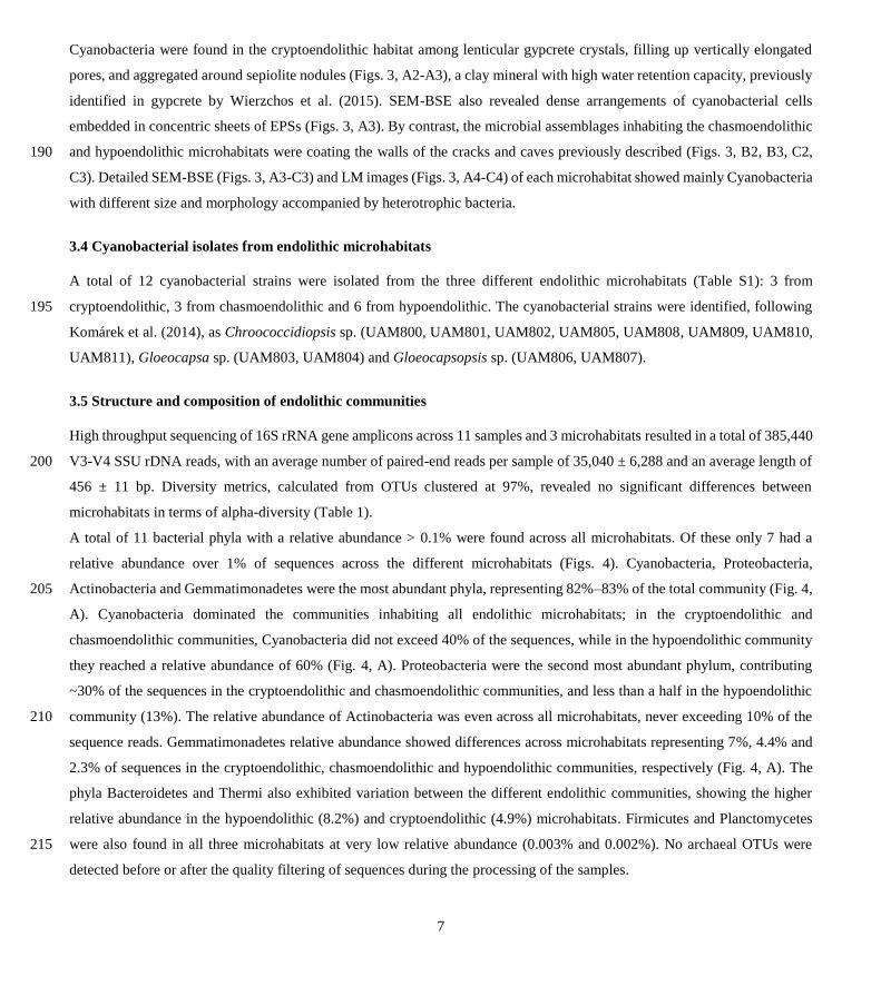
As the major component of the endolithic communities from the 3 described microhabitats, Cyanobacteria OTUs and isolates 230
were studied in detail. A phylogenetic analysis of the 15 major cyanobacterial OTUs (relative abundance > 1%) and 12 isolates
revealed 6 main clusters supported by high bootstrap values (Fig. 5).
Most of the OTUs (9 out of 15) and isolates (8 out of 12) were assigned to the genus Chroococcidiopsis and were distributed
in three clusters (I, III and V), each with representatives of Chroococcidiopsis isolates and clones’ sequences from various
deserts. Cluster I had the highest number of sequences from this study: six of the Chroococcidiopsis strains (UAM801, 235
UAM810, UAM802, UAM809, UAM800, UAM808) and four of the cyanobacterial OTUs (OTU1, OTU497, OTU8,
OTU112). This cluster also included two reference Chroococcidiopsis sp. sequences of soils from the Atacama Desert (Patzelt
et al. 2014). Cluster III included only one Chroococcidiopsis isolate (UAM805), three OTUs sequences (OTU1772, OTU420
and OTU4), and reference sequences of Chroococcidiopsis sp. strains isolated from quartz hypolithic communities from
hyperarid Chinese deserts (Pointing et al. 2007) and from University Valley (Antarctica)(Cumbers and Rothschild, 2014). The 240
last Chroococcidiopsis sp. cluster, number V, had no sequences from isolates, two OTUs sequences (OTU7 and OTU98),
sequences from cloning libraries from two deserts, Atacama and Jordan (Dong et al. 2007), and one Chroococcidiopsis sp.
sequence from a Mediterranean biocrust (Muñoz-Martín et al. 2019).
Cluster II comprised cyanobacterial sequences belonging to the Nostocales order from the genera Fischerella and Calothrix
to which OTU18 and OTU11 were respectively assigned. A total of 6 Cyanobacteria of this study were clustered with members 245
of the genera Gloeocapsa and Gloeocapsopsis (order Chroococcales), four isolates (UAM806, UAM807, UAM804, UAM803)
and two OTUs (OTU9, OTU854), forming cluster IV. Two reference sequences of Synechococcus together with OTU5
constituted Cluster VI.
9
Because of the low % identity of OTU2 with its closest relatives in the database (< 95%) (Table S2) and with our isolate
sequences, it was not possible to provide an accurate taxonomical assignment for this OTU (Fig. 5). Hits were found between 250
two of the isolates (Chroococcidiopsis UAM801 and Gloeocapsopsis UAM806) and two of the most abundant OTUs (OTU1
and OTU9, respectively).
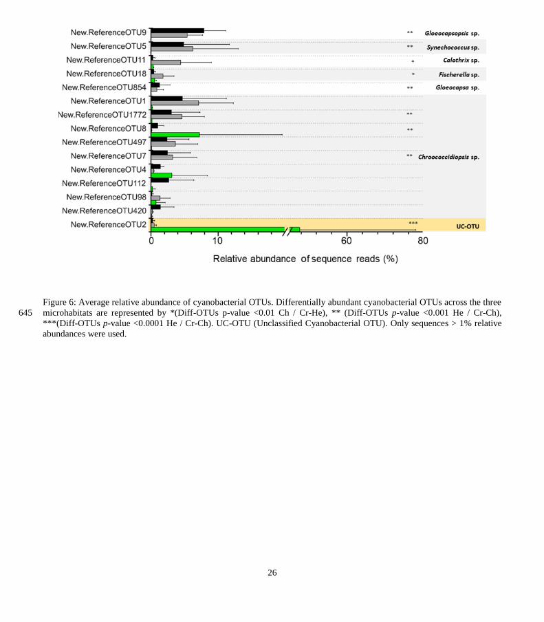
Differential abundance analysis using DESeq2 test revealed that 9 out of 15 of the cyanobacterial OTUs were differentially
abundant in the three microhabitats (Fig. 6). Both OTU11 (Calothrix sp.) and OTU18 (Fischerella sp), phylogenetically
assigned to the Nostocales order, were significantly more abundant in the chasmoendolithic community (3.8% and 1.5%, 255
respectively) than in cryptoendolithic and hypoendolithic communities (< 0.4% for both OTUs) (p-value < 0.01). OTUs
clustered with Gloeocapsa and Gloeocapsopsis (cluster IV), with Synechococcus (cluster VI), and with Chroococcidiopsis sp.
from three clusters (I, III and V), showed significantly different abundances (p-value < 0.001) between the hypoendolithic
community and that of the two communities from the upper side of the substrate (cryptoendolithic and chasmoendolithic).
OTU8 (Chroococcidiopsis sp.) was the only one displaying a higher abundance in the hypoendolithic community, while OTU9 260
(Gloeocapsopsis sp.), OTU5 (Synechococcus sp.), OTU854 (Gloeocapsa sp.) and OTUs 1772 and 7 (Chroococcidiopsis sp.)
had a higher abundance in the cryptoendolithic and chasmoendolithic communities. The unassigned Cyanobacterial OTU2
was mostly found in the hypoendolithic community (p-value < 0.0001) with an average relative abundance of more than 39%,
of the total community while its relative abundance in the other two communities was ~ 0.4%.
4. Discussion 265
In this study, we characterized the microbial communities inhabiting gypcrete collected from the Monturaqui area (Preandean
Depression), which is of particular interest due to its location in the hyper-arid zone of the Atacama Desert. While endolithic
colonization of the gypsum crust and gypcrete in this area has previously been studied (Dong et al. 2007, DiRuggiero et al.
2013, Wierzchos et al. 2015, Meslier et al. 2018), this is the first work in which cryptoendolithic, chasmoendolithic and
hypoendolithic communities have been characterized separately. The novelty of this study lies in the consideration of two 270
different EMCs inhabiting two endolithic microhabitats located in the upper part of the substrate, and in the description of the
structure and composition of the hypoendolithic microhabitat and its endolithic community, located at the bottom part of the
substrate.
The Monturaqui region, located in the Preandean Depression of the Atacama Desert has been found to harbour two different
substrates colonized by microbial communities, namely gypcrete (Wierzchos et al. 2015) and ignimbrite, a volcanic rock 275
(Wierzchos et al. 2013). Both substrates show endolithic colonization and a lack of epilithic colonization (rock surface
colonization). The absence of this second type of colonization in any substrates from the Monturaqui region may be explained
by the extremely arid microclimate of this area, including low relative humidity, high fluctuation of air and surface temperature,
extreme high solar irradiation and scarce precipitation (Wierzchos et al. 2015). Monturaqui has been described as a hyper-arid
area, showing an aridity index of 0.0075 (Wierzchos et al. 2013), based on the ratio of mean annual precipitation (P) and 280
10
potential evapotranspiration rate (PET) (P/PET), up to one order of magnitude lower than the established limit (Nienow 2009)
for the classification of hyper-arid regions (0.05). Specific measurements of surface temperature for gypcrete revealed values
close to 70°C. This value was detected at the zenith, when microbial communities are desiccated and metabolically inactive
(Cockell et al., 2008). The temperature within the endolithic habitats is expected to be close to that in the rock surface as shown
by Wierzchos et al. (2012a) for halite endoliths. The combination of these environmental conditions has led to the avoidance 285
of epilithic colonization in favour of endolithic colonization.
Potential endolithic habitability is tightly linked to the porosity of a lithic substrate because the distribution and size of pores
are often directly related to the substrate’s water retention capacity (Cámara et al. 2014; Herrera et al. 2009; Matthes et al.
2001; Omelon 2008; Pointing et al. 2009; Meslier et al. 2018). Porosity in gypcrete allows microbial communities to survive
in different microhabitats, providing sufficient space for the communities, while receiving enough light and having enough 290
water to metabolise and grow. The porous network of gypcrete restricts water loss by rapid evaporation and helps its retention
by capillary forces acting in small capillary-like shape pores. The inner architecture of gypcrete allows the habitability of three
different locations inside the substrate. The CT-Scan and SEM-BSE images from this work showed that all three types of
microhabitats shared a vertical axis of morphology with vertical cracks constituting the chasmoendolithic (CH) microhabitat
and capillary-like pores constitute the cryptoendolithic (CR) and hypoendolithic (HE) microhabitats. This capillary-like pore 295
architecture found in the CR microhabitat could be explained by the progressive substrate dissolution due to scarce rains and
by the water retained and condensed within the micropores, as it occurs in halite endolithic microhabitats (Wierzchos et al.
2012a). The observed HE microhabitat architecture supports the proposal of Wierzchos et al. (2015), in which the authors
described the presence of a dense crust delimiting the bottom part of the HE microhabitat. This structure reveals different
dissolving and crystallization processes of the gypsum following the water displacement from the surface to the bottom of the 300
rock (gravity flow). This water gravity flow giving rise to the cave-shaped pores, thus providing this HE microhabitat with a
hard permeable bottom gypsum layer.
The larger distance between the HE microhabitat and the top surface microhabitats CR and CH might be thought as a limiting
factor for the development of HE communities, especially in terms of water availability. However, the location of the HE
microhabitat at the bottom of the rock could reduce water losses due to evaporation processes. Thus, the micro-cave structures 305
we observed in the HE microhabitat might retain liquid water for longer times, leading to cyanobacterial growth.
The structural characteristics of the crypto- and chasmoendolithic microhabitats, located at the top of the substrate, also allow
access to water for the EMCs. Within the CR microhabitat, the labyrinth of pores directly or indirectly connected to the surface
may act as cavities where water might be retained, condensed, and also be present in form of saturated water vapour (high RH)
through the substrate, and be available to the microbial communities. Additionally, the presence of sepiolite inclusions 310
improves water retention in those pores, as previously described (Wierzchos et al. 2015, Meslier et al. 2018), leading to lower
rates of water loses by evaporation and gravitational forces. In contrast, the CH microhabitat provides direct access to rainfall
liquid water for its community, via its fissure and cracks, while at the same time lowering water retention capacity by higher
evaporation rates and losing liquid water by percolation through the rock.

11
Microbial communities inhabiting all three microhabitats were found in the form of large aggregates and were often embedded 315
in an EPS matrix. These characteristics are closely linked to survival strategies under harsh environmental conditions related
to low water and nutrient availability (Billi 2009, Wright et al. 2005). Since water is the most limiting factor for the
development of microbial communities inhabiting endolithic microhabitats of gypcrete, it is the component on which adaptive
strategies are primarily focused. EPS, because of their role in hydration and dehydration processes in lithobiontic communities
from Antarctic deserts (de los Ríos et al 2007) and the Atacama Desert (Dong et al. 2007; Wierzchos et al. 2011; Wierzchos 320
et al. 2015; Crits-Christoph et al. 2016) are an essential adaptation strategy against hyper-aridity. The aggregate-like structure
of these communities composed by cyanobacteria and heterotrophic bacteria also helps their survival against drought, since
dead cells could provide physical protection against desiccation processes (Postgate 1967; Roszak and Colwell 1987; Billi
2009; de los Ríos et al. 2004). In the case of the CR community, a special strategy against dryness was observed in this work,
since microorganisms were located close to the sepiolite, as previously reported concerning gypcrete endolithic communities 325
(Wierzchos et al. 2015, Meslier et al. 2018). EPS and dead cells taking part in the aggregates can also act as a nutrient reservoir
in such an oligotrophic environment; since low amounts of water-soluble ions were previously detected in the MTQ gypcrete
(Meslier et al. 2018).
The absence of significant differences in diversity metrics between the three EMCs of gypcrete is in accordance with the
diversity values of previously reported EMCs in the Atacama Desert (rev. in Casero et al. 2020). At a phylum level, the 330
community was composited of three main dominant phyla, Cyanobacteria, Proteobacteria and Actinobacteria (Fig. 4) as in
other EMCs of the Atacama Desert (Wierzchos et al. 2015, Meslier et al. 2018, Dong et al. 2007). However, a switch in the
Proteobacteria and Actinobacteria relative abundances was found compared to gypcrete cryptoendolithic communities
previously described (Meslier et al. 2018). That difference is presumably associated with different DNA extraction methods
and the inherent associated biases. While the three types of gypcrete microhabitats are exposed to the same climatic conditions, 335
we suggest that differences in micro-architectures resulted in drastically different sets of characteristics for water retention
discussed previously: CR counts on water capillary porous condensation and sepiolite water absorption properties, CH has
easier access to liquid water, and HE suffers less water loss.
While the communities from the 3 microhabitats had similar alpha diversity metrics, we found the composition of these
communities was statistically different, which is supported by the relative abundance of the main phyla, Cyanobacteria, 340
Proteobacteria and Actinobacteria, across the microhabitats distributed differentially, exhibiting differences between the CR
and CH communities as compared to the HE community, especially regarding cyanobacterial OTUs. This notable difference
in the relative abundance of cyanobacteria could be related to the particular resources of the phototrophic community. The
differential access to solar irradiance could explain the contrast between cyanobacterial proportions on both sides, at the top
(CR and CH) and bottom (HE) of the substrate. Thus, an update to the proposal by Wierzchos et al. (2018) is here suggested, 345
in which a causal link is evoked to explain the higher abundances of phototrophs as opposed to heterotrophs in EMCs, which
has been observed previously (Robinson et al. 2015, DiRuggiero et al. 2013, Wierzchos et al. 2015, Meslier et al. 2018).
According to that work, the scarcity of water was suggested to cause a lower metabolic activity in phototrophs, thus leading
12
to lower support of the heterotrophic community. However, in this scenario, light intensity should also be considered a crucial
factor in understanding the differences between the composition of top and bottom EMCs, since the HE community has notably 350
lower access to sun radiation. Recently the light intensity, as a driving factor of spatial heterogeneity within halite endolithic
microbial communities was reported by Uritskiy et al. (2020). Thus, for EMCs communities based on phototrophic
microorganisms, a limitation to one of those resources essential for photosynthesis would further lead to low rates of CO2
fixation and, consequently, to a smaller heterotrophic community.
In contrast with results of Wierzchos et al. (2015) in gypcrete endolithic communities, no eukaryotic algae were found in 355
neither microscopy nor molecular analyses, being Cyanobacteria the phototrophic phylum observed in all gypcrete endolithic
microhabitats. While we found multiple phylotypes of cyanobacteria among the gypcrete microhabitats, most of them belonged
to the genus Chroococcidiopsis. Several strains of this genus have previously been described in EMCs from both hot and cold
deserts (Friedmann 1980) as a result of their capacity to cope with extreme environmental conditions (Billi et al. 2011; Verseux
et al. 2017). Further supporting the different micro-environmental conditions and community composition between the top CR 360
and CH habitats and the bottom HE habitat, was the discovery of an unclassified cyanobacterial OTU (UC-OTU, New
Reference OTU2), which was almost exclusive to the HE microhabitat and the phylogenetic distance of the hypoendolithic
Chroococcidiopsis UAM811 strain with the different Chrooococcidiopsis clusters. Regarding the so-called UC-OTU, although
the low percentage of sequence similarity did not allow for an accurate taxonomical assignment, its closest relatives (~94%
sequence identity for 450 nt of the 16S rRNA gene) were from habitats where light is the limiting factor for photosynthesis 365
such as a pinnacle mat at 10 m depth from a sinkhole (Hamilton et al. 2017) and groundwater sample from a tectonically-
formed cavern (Table S2). Both observations, the inability to identify the UC-OTU and the phylogenetic position of the
UAM811 strain, highlight the importance of greater efforts in terms of isolation and characterization of cyanobacteria,
especially from these environments.
The differential distribution of key members of these EMCs among microhabitats in the same lithic substrate and the same 370
piece of rock, as their primary producers, reveals an “environmental filtering” process (Kraft et al. 2015). This concept focuses
on the relationship between an organism and the environment, recognizing that not all organisms will be able to establish
themselves successfully and persist in all abiotic conditions. Thus, in this scenario, the abiotic conditions linked to the
architecture and location of the endolithic microhabitat would force the development of community assemblages highly
specialized to small scale differences, thereby exhibiting a microbiogeographical behaviour. 375
Our work answers that there are certain differences in endolithic microbial communities’ structure among crypto-, chasmo-
and hypoendolithic habitats. Considering that the external climatic regime was the same for studied pieces of rock, our results
have shown that the structure of these microbial communities was different among endolithic habitats. Following the definition
of microhabitat architecture by Wierzchos et al. (2015) we can distinguish different architecture of the substrate within different
endolithic microhabitats. In this context, our work suggests that distinct features of microhabitat architecture that have an 380
influence on microenvironmental variables at the microscale would shape microbial communities' structure.
13
However, we are aware that more “microbiogeographic” studies should be done with other endolithic microhabitats from the
Atacama Desert and elsewhere showing that gypcrete is not only a peculiar case where differences in the architecture of a
microhabitat play an essential role in shaping the diversity and composition of endolithic microbial communities.
5. Conclusions 385
This study is the first to address differences between microbial communities inhabiting three differentiated endolithic
microhabitats within the same lithic substrate. In this study, liquid water availability was proposed to be a driver of community
composition because the specific architectural features of each microhabitat facilitated water input and retention in different
ways. Water, light, and CO2, are indispensable resources for photosynthetic activity. Thus, we support the cause and effect
relationship where the restriction of these factors may affect the proportion of phototrophic and heterotrophic components in 390
the EMC communities as proposed by previous works (Robinson et al. 2015, Wierzchos et al. 2018 and Meslier et al. 2018).
The genus Chroococcidiopsis displayed a variety of strains distributed among all microhabitats, proving its high capacity to
colonize effectively endolithic microhabitats under polyextreme conditions. Nevertheless, the presence of a singular
cyanobacterial OTU stresses the need for additional efforts in cyanobacterial characterization from these extreme
environments. 395
Findings from this work reveal the importance of using an appropriate scale for the study of microbial communities. Indeed,
we found that the microstructural and microarchitectural features of the endolithic habitats were key factors in determining the
composition of endolithic microbial communities. Thus, this study suggests a cautious use of “macroenvironmental”
parameters in characterizing differences between endolithic communities from different deserts or substrates. Our results point
to the need for a more thorough description of the micro-environmental conditions that directly exert an effect on microbial 400
assemblages: light, water and CO2. Therefore, once the relationship between factors affecting the absence and/or presence of
certain taxa, the actual environmental filtering in these microhabitats could be described in more detail, it will be possible to
draw conclusions on the interactions and specific roles of the different members in the community and their
microbiogeography.
405
Data availability
All the sequencing data sets generated in this study have been submitted to the National Center for Biotechnology Information
(NCBI) SRA database and can be found under the BioProject ID PRJNA637482.
14
Author contributions
MCC and JW designed and performed the research. JW conceived the original project. MCC, JW and OA carried out the 410
sampling. MCC, JW and AQ wrote the manuscript; MCC, JW and CA performed the microscopy; TK contributed to CT-
SCAN analysis; MCC, VMA and JDR contributed to the molecular data, analysis, and performed the sequencing. All authors
contributed to editing and revising the manuscript and approved this version for submission.
Competing interest 415
The authors declare that they have no conflict of interest.
Acknowledgements
This study was supported by grant PGC2018-094076-B-I00 from MCIU/AEI (Spain) and FEDER (UE) to MCC and JW, by
NSF grant DEB1556574 and NASA grant NNX15AP18G to JDR. The work of MCC was supported by grant BES 2014-
069106 from the Spanish Ministry of Science and Innovation (MCINN). The MNCN-CSIC, Madrid, Spain is acknowledged 420
for microscopy services.
References
Azua-Bustos, A., Caro-Lara, L. and Vicuña, R.: Discovery and microbial content of the driest site of the hyperarid Atacama
Desert, Chile., Environ. Microbiol. Rep., 7(3), 388–394, doi:10.1111/1758-2229.12261, 2015. 425
Azua-Bustos, A., Urrejola, C. and Vicuña, R.: Life at the dry edge: microorganisms of the Atacama Desert., FEBS Lett.,
586(18), 2939–2945, doi:10.1016/j.febslet.2012.07.025, 2012.
Baas-Becking, L.G.M.: Geobiologie; of inleiding tot de milieukunde, edited by WP Van Stockum & Zoon NV, The Hague,
Netherlands., 1934.
Bass, D. and Boenigk, J.: Everything is everywhere: a twenty-first century de-/reconstruction with respect to protists, Biogeogr. 430
Microsc. Org. Is everything small everywhere, 88–110, https://doi.org/10.1017/CBO9780511974878.007, 2011.
Becerra-Absalón, I., Muñoz-Martín, M. Á., Montejano, G. and Mateo, P.: Differences in the cyanobacterial community
composition of biocrusts from the drylands of Central Mexico. Are there endemic species? Front. Microbiol, 10, 937, https://
10.3389/fmicb.2019.00937, 2019
Billi, D.: Subcellular integrities in Chroococcidiopsis sp. CCMEE 029 survivors after prolonged desiccation revealed by 435
molecular probes and genome stability assays, Extremophiles, 13(1), 49-57, https://doi.org/10.1007/s00792-008-0196-0, 2009.
15
Billi, D., Friedmann, E. I., Hofer, K. G., Caiola, M. G. and Ocampo-Friedmann, R.: Ionizing-radiation resistance in the
desiccation-tolerant cyanobacterium Chroococcidiopsis, Appl. Environ. Microbiol., 66(4), 1489–1492,
https://doi.org/10.1128/aem.66.4.1489-1492.2000, 2000.
Billi, D., Viaggiu, E., Cockell, C. S., Rabbow, E., Horneck, G. and Onofri, S.: Damage escape and repair in dried 440
Chroococcidiopsis spp. from hot and cold deserts exposed to simulated space and Martian conditions, Astrobiology, 11(1),
65–73, https://doi.org/10.1089/ast.2009.0430, 2011.
Cámara, B., Suzuki, S., Nealson, K. H., Wierzchos, J., Ascaso, C., Artieda, O. and de los Ríos, A.: Ignimbrite textural
properties as determinants of endolithic colonization patterns from hyper-arid Atacama Desert, Int. Microbiol, 17(4), 235–247,
https://doi.org/10.2436/20.1501.01.226, 2014. 445
Caporaso, J. G., Kuczynski, J., Stombaugh, J., Bittinger, K., Bushman, F. D., Costello, E. K., Fierer, N., Pena, A. G., Goodrich,
J. K. and Gordon, J. I.: QIIME allows analysis of high-throughput community sequencing data, Nat. Methods, 7(5), 335-336,
https://doi.org/10.1038/nmeth.f.303, 2010.
Casero, M. C., Ballot, A., Agha, R., Quesada, A. and Cirés, S.: Characterization of saxitoxin production and release and
phylogeny of sxt genes in paralytic shellfish poisoning toxin-producing Aphanizomenon gracile, Harmful Algae, 37, 28-37, 450
https://doi.org/10.1016/j.hal.2014.05.006, 2014.
Casero, M. C., Meslier, V., Wierzchos, J. and DiRuggiero, J.: Preandean Atacama Desert Endolithic Microbiology, in
Microbial Ecosystems in Central Andes Extreme Environments, edited by M. Farías, pp. 51–71, Springer International
Publishing, Cham., https://doi.org/10.1007/978-3-030-36192-1_4, 2020.
Castenholz, R. W., Wilmotte, A., Herdman, M., Rippka, R., Waterbury, J. B., Iteman, I. and Hoffmann, L.: Phylum BX. 455
cyanobacteria, in Bergey’s manual® of systematic bacteriology, pp. 473–599, Springer., https://doi.org/10.1007/978-0-387-
21609-6_27, 2001.
Cockell, C. S., McKay, C. P., Warren-Rhodes, K. and Horneck, G.: Ultraviolet radiation-induced limitation to epilithic
microbial growth in arid deserts – Dosimetric experiments in the hyperarid core of the Atacama Desert, J. Photochem.
Photobiol. B Biol., 90(2), 79–87, doi:https://doi.org/10.1016/j.jphotobiol.2007.11.009, 2008. 460
Cockell, C. S., Schuerger, A. C., Billi, D., Friedmann, E. I. and Panitz, C.: Effects of a simulated martian UV flux on the
cyanobacterium, Chroococcidiopsis sp. 029, Astrobiology, 5(2), 127–140, https://doi.org/10.1089/ast.2005.5.127, 2005.
Cole, J. R., Wang, Q., Fish, J. A., Chai, B., McGarrell, D. M., Sun, Y., Brown, C. T., Porras-Alfaro, A., Kuske, C. R. and
Tiedje, J. M.: Ribosomal Database Project: data and tools for high throughput rRNA analysis, Nucleic Acids Res., 42(1), 633–
642, https://doi.org/10.1093/nar/gkt1244, 2014. 465
Cordero, R. R., Damiani, A., Jorquera, J., Sepúlveda, E., Caballero, M., Fernandez, S., Feron, S., Llanillo, P. J., Carrasco, J.
and Laroze, D.: Ultraviolet radiation in the Atacama Desert, Antonie Van Leeuwenhoek, 111(8), 1301–1313,
https://doi.org/10.1007/s10482-018-1075-z, 2018.
Cordero, R. R., Seckmeyer, G., Damiani, A., Riechelmann, S., Rayas, J., Labbe, F. and Laroze, D.: The world’s highest levels
of surface UV, Photochem. Photobiol. Sci., 13(1), 70–81, https://doi.org/10.1039/C3PP50221J, 2014. 470
16
Crits-Christoph, A., Robinson, C. K., Ma, B., Ravel, J., Wierzchos, J., Ascaso, C., Artieda, O., Souza-Egipsy, V., Casero, M.
C. and DiRuggiero, J.: Phylogenetic and functional substrate specificity for endolithic microbial communities in hyper-arid
environments, Front. Microbiol., 7, 301, https://doi.org/10.3389/fmicb.2016.00301, 2016.
Cumbers, J. and Rothschild, L. J.: Salt tolerance and polyphyly in the cyanobacterium Chroococcidiopsis (Pleurocapsales).,
J. Phycol., 50(3), 472–482, doi:10.1111/jpy.12169, 2014. 475
de los Ríos, A., Grube, M., Sancho, L. G. and Ascaso, C.: Ultrastructural and genetic characteristics of endolithic
cyanobacterial biofilms colonizing Antarctic granite rocks, FEMS Microbiol. Ecol., 59(2), 386–395,
https://doi.org/10.1111/j.1574-6941.2006.00256.x, 2007.
de los Ríos, A., Valea, S., Ascaso, C., Davila, A., Kastovsky, J., McKay, C. P., Gómez-Silva, B. and Wierzchos, J.:
Comparative analysis of the microbial communities inhabiting halite evaporites of the Atacama Desert., Int. Microbiol. Off. 480
J. Spanish Soc. Microbiol., 13(2), 79–89, doi:10.2436/20.1501.01.113, 2010.
de los Ríos, A., Wierzchos, J., Sancho, L. G. and Ascaso, C.: Exploring the physiological state of continental Antarctic
endolithic microorganisms by microscopy, FEMS Microbiol. Ecol., 50(3), 143–152,
https://doi.org.10.1016/j.femsec.2004.06.010, 2004.
de Wit, R. and Bouvier, T.: “Everything is everywhere, but, the environment selects”; what did Baas Becking and Beijerinck 485
really say?, Environ. Microbiol., 8(4), 755–758, https://doi.org/10.1111/j.1462-2920.2006.01017.x, 2006.
DiRuggiero, J., Wierzchos, J., Robinson, C.K., Souterre, T., Ravel, J., Artieda, O., Souza-Egipsy, V. and Ascaso, C.: Microbial
colonisation of chasmoendolithic habitats in the hyper-arid zone of the Atacama Desert. Biogeosci. 10, 2439-2450. doi:
10.5194/bg-10-2439-2013, 2013.
Dong, H., Rech, J. A., Jiang, H., Sun, H. and Buck, B. J.: Endolithic cyanobacteria in soil gypsum: Occurrences in Atacama 490
(Chile), Mojave (United States), and Al‐Jafr Basin (Jordan) Deserts, J. Geophys. Res. Biogeosciences, 112(G2),
https://doi.org/10.1029/2006JG000385, 2007.
Edwards, U., Rogall, T., Blöcker, H., Emde, M. and Böttger, E. C.: Isolation and direct complete nucleotide determination of
entire genes. Characterization of a gene coding for 16S ribosomal RNA, Nucleic Acids Res., 17(19), 7843–7853,
https://doi.org/10.1093/nar/17.19.784, 1989. 495
Fontaneto, D. and Hortal, J.: Microbial biogeography: is everything small everywhere, Microb. Ecol. Theory Curr. Perspect.
Caister Acad. Press. Norfolk, 87-98, 2012.
Friedmann, E. I.: Endolithic microbial life in hot and cold deserts, in Limits of life, pp. 33–45, Springer.,
https://doi.org/10.1007/978-94-009-9085-2_3, 1980.
Hamilton, T. L., Welander, P. V, Albrecht, H. L., Fulton, J. M., Schaperdoth, I., Bird, L. R., Summons, R. E., Freeman, K. H. 500
and Macalady, J. L.: Microbial communities and organic biomarkers in a Proterozoic‐analog sinkhole, Geobiology, 15(6),
784–797, https://doi.org/10.1111/gbi.12252, 2017.
Herrera, A., Cockell, C. S., Self, S., Blaxter, M., Reitner, J., Thorsteinsson, T., Arp, G., Dröse, W. and Tindle, A. G.: A
cryptoendolithic community in volcanic glass, Astrobiology, 9(4), 369–381, https://doi.org/10.1089/ast.2008.0278, 2009.
17
Joshi, N. A. and Fass, J. N.: Sickle: A sliding-window, adaptive, quality-based trimming tool for FastQ files (Version 1.33) 505
[Software], 2011.
Kimura, M.: A simple method for estimating evolutionary rates of base substitutions through comparative studies of nucleotide
sequences, J. Mol. Evol., 16(2), 111–120, https://doi.org/10.1007/BF01731581, 1980.
Komárek, J., Kaštovský, J., Mareš, J. and Johansen, J. R.: Taxonomic classification of cyanoprokaryotes (cyanobacterial
genera) 2014, using a polyphasic approach, Preslia, 86(4), 295–335, 2014. 510
Kraft, N. J. B., Adler, P. B., Godoy, O., James, E. C., Fuller, S. and Levine, J. M.: Community assembly, coexistence and the
environmental filtering metaphor, Funct. Ecol., 29(5), 592–599, https://doi.org/10.1111/1365-2435.12345, 2015.
Kumar, S., Stecher, G. and Tamura, K.: MEGA7: molecular evolutionary genetics analysis version 7.0 for bigger datasets,
Mol. Biol. Evol., 33(7), 1870–1874, https://doi.org/10.1093/molbev/msw054, 2016.
Lepère, C., Wilmotte, A. and Meyer, B.: Molecular diversity of Microcystis strains (Cyanophyceae, Chroococcales) based on 515
16S rDNA sequences, Syst. Geogr. Plants, 275–283, https://doi.org/10.2307/3668646, 2000.
Loza, V., Perona, E. and Mateo, P.: Molecular fingerprinting of cyanobacteria from river biofilms as a water quality monitoring
tool, Appl. Environ. Microbiol., 79(5), 1459–1472, https://doi.org/ 10.1128/AEM.03351-12, 2013.
Matthes, U., Turner, S. J. and Larson, D. W.: Light attenuation by limestone rock and its constraint on the depth distribution
of endolithic algae and cyanobacteria, Int. J. Plant Sci., 162(2), 263–270, https://doi.org/10.1086/319570, 2001. 520
McKay, C. P., Friedmann, E. I., Gómez-Silva, B., Cáceres-Villanueva, L., Andersen, D. T. and Landheim, R.: Temperature
and moisture conditions for life in the extreme arid region of the Atacama Desert: four years of observations including the El
Nino of 1997–1998, Astrobiology, 3(2), 393–406, https://doi.org/10.1089/153110703769016460, 2003.
Meslier, V., Casero, M. C., Dailey, M., Wierzchos, J., Ascaso, C., Artieda, O., McCullough, P. R. and DiRuggiero, J.:
Fundamental drivers for endolithic microbial community assemblies in the hyperarid Atacama Desert, Environ. Microbiol., 525
20(5), https://doi.org/10.1111/1462-2920.14106, 2018.
Muñoz‐Martín, M. Á., Becerra‐Absalón, I., Perona, E., Fernández‐Valbuena, L., Garcia‐Pichel, F. and Mateo, P.:
Cyanobacterial biocrust diversity in Mediterranean ecosystems along a latitudinal and climatic gradient, New Phytol., 221(1),
123–141, https://doi.org/10.1111/nph.15355, 2019.
Nienow, J. A.: Extremophiles: Dry Environments (including Cryptoendoliths), edited by M. B. T.-E. of M. (Third E. 530
Schaechter, pp. 159–173, Academic Press, Oxford., https://doi.org/10.1016/B978-012373944-5.00277-7, 2009.
O’Malley, M. A.: “Everything is everywhere: but the environment selects”: ubiquitous distribution and ecological determinism
in microbial biogeography., Stud. Hist. Philos. Biol. Biomed. Sci., 39(3), 314–325,
https://doi.org/10.1016/j.shpsc.2008.06.005, 2008.
Omelon, C. R.: Endolithic microbial communities in polar desert habitats, Geomicrobiol. J., 25(7–8), 404–414, 2008. 535
Patzelt, D. J., Hodač, L., Friedl, T., Pietrasiak, N. and Johansen, J. R.: Biodiversity of soil cyanobacteria in the hyper‐arid
Atacama Desert, Chile, J. Phycol., 50(4), 698–710, https://doi.org/10.1111/jpy.12196, 2014.
18
Pointing, S. B. and Belnap, J.: Microbial colonization and controls in dryland systems, Nat. Rev. Microbiol., 10(8), 551–562,
https://doi.org/10.1038/nrmicro2831, 2012.
Pointing, S. B., Chan, Y., Lacap, D. C., Lau, M. C. Y., Jurgens, J. A. and Farrell, R. L.: Highly specialized microbial diversity 540
in hyper-arid polar desert, Proc. Natl. Acad. Sci., 106(47), 19964–19969, https://doi.org/10.1073/pnas.0908274106, 2009.
Pointing SB, Warren-Rhodes KA, Lacap DC, Rhodes KL, McKay CP. Hypolithic community shifts occur as a result of liquid
water availability along environmental gradients in China's hot and cold hyperarid deserts. Environ Microbiol. 9(2):414-424.
doi:10.1111/j.1462-2920.2006.01153.x, 2007
Postgate, J. R.: Viability measurements and the survival of microbes under minimum stress, in Advances in microbial 545
physiology, vol. 1, pp. 1–23, Elsevier., https://doi.org/10.1016/S0065-2911(08)60248-9, 1967.
Rippka, R., Deruelles, J., Waterbury, J. B., Herdman, M. and Stanier, R. Y.: Generic assignments, strain histories and properties
of pure cultures of cyanobacteria, Microbiology, 111(1), 1–61, https://doi.org/10.1099/00221287-111-1-1, 1979.
Robinson, C. K., Wierzchos, J., Black, C., Crits‐Christoph, A., Ma, B., Ravel, J., Ascaso, C., Artieda, O., Valea, S. and Roldán,
M.: Microbial diversity and the presence of algae in halite endolithic communities are correlated to atmospheric moisture in 550
the hyper‐arid zone of the Atacama Desert, Environ. Microbiol., 17(2), 299–315, https://doi.org/10.1111/1462-2920.12364,
2015.
Roszak, D. B. and Colwell, R. R.: Survival strategies of bacteria in the natural environment., Microbiol. Rev., 51(3), 365,
1987.
Rothschild, L. J. and Mancinelli, R. L.: Life in extreme environments, Nature, 409(6823), 1092–1101, 555
https://doi.org/10.1038/35059215, 2001.
Thompson, J. D., Higgins, D. G. and Gibson, T. J.: CLUSTAL W: improving the sensitivity of progressive multiple sequence
alignment through sequence weighting, position-specific gap penalties and weight matrix choice, Nucleic Acids Res., 22(22),
4673–4680, https://doi.org/10.1093/nar/22.22.4673, 1994.
van der Gast, C. J.: Microbial biogeography: the end of the ubiquitous dispersal hypothesis?, Environ. Microbiol., 17(3), 544–560
546, https://doi.org/10.1111/1462-2920.12635, 2015.
Uritskiy, G., Getsin, S., Munn, A., Gomez-Silva, B., Davila, A., Glass, B., Taylor, J. and DiRuggiero, J.: Halophilic microbial
community compositional shift after a rare rainfall in the Atacama Desert, ISME J., 13(11), 2737–2749, doi:10.1038/s41396-
019-0468-y, 2019.
Uritskiy G, Munn A, Dailey M, Gelsinger DR, Getsin S, Davila A, McCullough PR, Taylor J and DiRuggiero J.: Environmental 565
factors driving spatial heterogeneity in desert halophile microbial communities. Front. Microbiol.
11.,doi.org/10.3389/fmicb.2020.578669, 2020.
Verseux, C., Baqué, M., Cifariello, R., Fagliarone, C., Raguse, M., Moeller, R. and Billi, D.: Evaluation of the resistance of
Chroococcidiopsis spp. to sparsely and densely ionizing irradiation, Astrobiology, 17(2), 118–125,
https://doi.org/10.1089/ast.2015.1450, 2017. 570
19
Vítek, P., Ascaso, C., Artieda, O. and Wierzchos, J.: Raman imaging in geomicrobiology: endolithic phototrophic
microorganisms in gypsum from the extreme sun irradiation area in the Atacama Desert, Anal. Bioanal. Chem., 408(15), 4083–
4092, https://doi.org/10.1007/s00216-016-9497-9, 2016.
Walker, J. J. and Pace, N. R.: Endolithic microbial ecosystems, Annu. Rev. Microbiol., 61, 331–347,
https://doi.org/10.1146/annurev.micro.61.080706.093302, 2007. 575
Wierzchos, J., Casero, M. C., Artieda, O. and Ascaso, C.: Endolithic microbial habitats as refuges for life in polyextreme
environment of the Atacama Desert, Curr. Opin. Microbiol., 43, https://doi.org/10.1016/j.mib.2018.01.003, 2018.
Wierzchos, J. and Ascaso, C.: Application of back‐scattered electron imaging to the study of the lichen‐rock interface, J.
Microsc., 175(1), 54–59, https://doi.org/10.1111/j.1365-2818.1994.tb04787.x, 1994.
Wierzchos, J., Cámara, B., de Los Rios, A., Davila, A. F., Sánchez Almazo, I. M., Artieda, O., Wierzchos, K., Gomez‐Silva, 580
B., McKay, C. and Ascaso, C.: Microbial colonization of Ca‐sulfate crusts in the hyperarid core of the Atacama Desert:
implications for the search for life on Mars, Geobiology, 9(1), 44–60, https://doi.org/10.1111/j.1472-4669.2010.00254.x, 2011.
Wierzchos, J., Davila, A. F., Sánchez-Almazo, I. M., Hajnos, M., Swieboda, R. and Ascaso, C.: Novel water source for
endolithic life in the hyperarid core of the Atacama Desert, Biogeosciences, 9(6), 2275-2286, https://doi.org/10.5194/bg-9-
2275-2012, 2012 a 585
Wierzchos, J., Davila, A. F., Artieda, O., Cámara-Gallego, B., de los Ríos, A., Nealson, K. H., Valea, S., García-González, M.
T. and Ascaso, C.: Ignimbrite as a substrate for endolithic life in the hyper-arid Atacama Desert: implications for the search
for life on Mars, Icarus, 224(2), 334–346, https://doi.org/10.1016/j.icarus.2012.06.009, 2013.
Wierzchos, J., DiRuggiero, J., Vítek, P., Artieda, O., Souza-Egipsy, V., Skaloud, P., Tisza, M., Davila, A. F., Vílchez, C. and
Garbayo, I.: Adaptation strategies of endolithic chlorophototrophs to survive the hyperarid and extreme solar radiation 590
environment of the Atacama Desert, Front. Microbiol., 6, 934, https://doi.org/10.3389/fmicb.2015.00934, 2015.
Wierzchos J, de los Ríos A, Ascaso C. Microorganisms in desert rocks: the edge of life on Earth. Int Microbiol.,15(4):173‐
183. https://doi.org/10.2436/20.1501.01.170, 2012, b.
Wright DJ, C Smith S, Joardar V, Scherer S, Jervis J, Warren A, Helm R, Potts M. UV Irradiation and Desiccation Modulate
the Three-dimensional Extracellular Matrix of Nostoc commune (Cyanobacteria), J Biol Chem, 280 (48), 40271-40281. doi: 595
10.1074/jbc.M505961200, 2005
600
605
20
610
Table 1: Diversity estimates of microbial communities in the endolithic microhabitats of gypcrete.
Microhabitats Chao OTU Richness Shannon
Cryptoendolithic Avg 583.8 430 6.3
SD 43.2 38 0.2
Chasmoendolithic Avg 574.9 419 6.1
SD 46.0 29 0.1
Hypoendolithic Avg 564.9 409 4.6
SD 31.7 32 1.0
21
615
Figure 1: Sampling location in the Atacama Desert. Monturaqui area: MTQ (black diamond). (© Google Earth, image providers:
Landsat /Copernicus)
22
Figure 2: CT-Scan images of a colonized piece of gypcrete. 3D reconstruction of a gypcrete sample with the spatial distribution of pores 620 (orange colour) and complete reconstructions of the scanned volume (grey colour) on lateral, front and top views of gypcrete. Porous
micromorphology is capillary-shaped in vertical position due to gravity movement direction of the water. Arrows in top view images point
to the deepest cracks. Scale bar = 1cm.
23
Figure 3: Endolithic colonization zones characterization. Series A: Cryptoendolithic; Series B: Chasmoendolithic; Series C: 625 Hypoendolithic. Series 1: Macro images of gypcrete cross-section of colonized zones; Series 2 and 3: SEM-BSE images of gypcrete cross-
section of colonized zones; Series 4: LM-DIC images of scrapped cyanobacteria from gypcrete. Series 1: Black arrows indicate green and
orange coloured endolithic colonization zones of 5 mm thick on A1 (CR), 8 mm thick on B1 beneath the surface (CH) and 5-9 mm thick on
C1 above bottom gypsum crust (HE). Series 2. CR, CH and HE microhabitats with aggregates of endolithic microbial communities
surrounded by the green dotted lines, inside the pores of gypcrete: A2, under a white dense surface crust; B2, inside the cracks of gypcrete 630 and C2, inside the micro caves of gypcrete at bottom of the rock. Series 3 Green arrows point to aggregates of cyanobacteria among gypcrete
(Gy) crystals (A3, B3), surrounding by sepiolite (Sp) nodules (A3) and on the gypcrete (Gy) walls (C3). Series 4 aggregates of different
morphotypes of cyanobacteria, shown by green, yellow and orange arrows and gypcrete crystals (Gy).
24
635
Figure 4: Average relative abundance of sequence reads (A) and OTUs at the 97% clustering cut-off (B) of major bacterial phyla (at
least 1% across all samples) of microbial assemblages in the cryptoendolithic (Cr) chasmoendolithic (Ch) and hypoendolithic (He)
microhabitats of gypcrete.

25
Figure 5: Maximum likelihood tree based on partial 16S rRNA sequences of Cyanobacteria OTUs above 1% relative abundance 640 and cyanobacterial strains isolated from the three gypcrete microhabitats. Bold indicates sequences from this study. Scale bar indicates
5% sequence divergence.
26
Figure 6: Average relative abundance of cyanobacterial OTUs. Differentially abundant cyanobacterial OTUs across the three
microhabitats are represented by *(Diff-OTUs p-value <0.01 Ch / Cr-He), ** (Diff-OTUs p-value <0.001 He / Cr-Ch), 645
***(Diff-OTUs p-value <0.0001 He / Cr-Ch). UC-OTU (Unclassified Cyanobacterial OTU). Only sequences > 1% relative
abundances were used.


![Changes in composition of epigeic invertebrate communities (with accent on spiders) in Carpathian alpine meadows influenced by added nitrogen and phosphorus [Peter Gajdos]](https://img.pdfslide.us/doc/110x75/54c0656d4a79596e0b8b45ea/changes-in-composition-of-epigeic-invertebrate-communities-with-accent-on-spiders-in-carpathian-alpine-meadows-influenced-by-added-nitrogen-and-phosphorus-peter-gajdos.jpg)