Embed Size (px)
Citation preview
The Complexities of theDiagnosis and
Management of Kawasaki DiseaseAnne H. Rowley, MD
KEYWORDS
� Coronary artery aneurysm � Prolonged fever � Systemic inflammation� Myocardial infarction � Acquired pediatric heart disease
KEY POINTS
� The diagnosis of Kawasaki disease (KD) requires a high index of suspicion. Infants andchildren may present with “soft” or incomplete clinical features, yet still develop significantcoronary artery abnormalities.
� Asian children have the highest incidence of KD of all ethnic/racial groups. Siblings andchildren of KD patients are at increased risk.
� Laboratory and echocardiographic findings can help establish the diagnosis in nonclassiccases. In particular, a child with prolonged fever, laboratory evidence of systemicinflammation, and a coronary artery z score of 2.5 or higher has a very high probabilityof having KD.
� Prompt treatment with intravenous gammaglobulin and aspirin can be life-saving. Chil-dren who do not have resolution of fever with this primary therapy are at increased riskof developing coronary artery abnormalities, and additional anti-inflammatory therapiesshould be administered.
INTRODUCTION
Although Kawasaki disease (KD) has been recognized in Japan and the United Statesfor decades, the etiology and pathogenesis of the illness remain major pediatric mys-teries. The abrupt onset of clinical signs such as fever, exanthem, and enanthem inpreviously healthy children, the rarity of recurrence, the very young age groupaffected, and the well-documented epidemics and outbreaks of illness are stronglysuggestive of infectious etiology, as well described in a classic epidemiologic studyfrom the 1980s.1 The very high incidence of the illness in Japan, where approximately
Disclosure statement: The author has nothing to disclose.Department of Pediatrics, Northwestern University Feinberg School of Medicine, 310 East Supe-rior Street, Morton 4-685B, Chicago, IL 60611, USAE-mail address: [email protected]
Infect Dis Clin N Am - (2015) -–-http://dx.doi.org/10.1016/j.idc.2015.05.006 id.theclinics.com0891-5520/15/$ – see front matter � 2015 Elsevier Inc. All rights reserved.
Rowley2
1 in 90 children develop KD by age 5 years,2 is highly suggestive of a ubiquitous infec-tious agent affecting young susceptible children who are genetically predisposed. Keyfeatures of KD are presented in Box 1. It is important to understand the major patho-logic features of KD arteriopathy, briefly summarized in Box 1, because these featurespredict the adverse clinical outcomes observed in KD patients who develop coronaryartery abnormalities.3 Moreover, knowledge of the many organs and tissues involvedin the systemic inflammation of KD assists in understanding the many possible clinicalmanifestations of the illness (Box 2).4 Risk factors for KD and the development of cor-onary artery abnormalities are summarized in Box 3.
INCIDENCE AND MORTALITY RATES
The incidence of KD varies in different countries throughout the world (Table 1) andremains unknown in many regions, especially in those that continue to have a highprevalence of measles, which shares many clinical features with KD. In Japan, thereis high recognition and early treatment of the condition, and mortality rates have fallenfrom 1.4% in 1970 to 0.01% in recent years; fatality rates began to decrease markedlyafter the introduction of intravenous gammaglobulin therapy in the late 1980s.8 In theUnited States, fatality rates are also very low; fatal cases are often associated withdelayed or missed diagnoses.3 Peak months of KD incidence vary somewhat by coun-try, but a consistent theme seems to be a peak during the winter in nontemperateclimates.9
PATIENT HISTORY
The history is particularly important in KD, as some clinical features of the illness maybegin and abate before the patient’s presentation. It is recommended that the parentor guardian is asked nonleading questions about symptoms to avoid introducing recallbias. Box 4 lists common features in the history of children with KD. Excessive irrita-bility, refusal to bear weight, redness and swelling of the hands and feet, and an
Box 1
Key features of Kawasaki disease (KD)
An acute onset of prolonged febrile illness in previously healthy children.
The leading cause of acquired heart disease in children in developed nations.
A systemic inflammatory illness affecting many organs and tissues, but leading to long-termconsequences almost exclusively confined to medium-sized muscular arteries, particularly thecoronary arteries.
KD arteriopathy is characterized by 3 linked pathologic processes: necrotizing arteritis,subacute/chronic arteritis, and luminal myofibroblastic proliferation. Necrotizing arteritisoccurs in the first 2 weeks after onset and can result in necrosis of the coronary arteries; if thenecrosis is extensive, giant coronary artery aneurysms can form, which are associated withsevere outcomes. Subacute/chronic arteritis begins in the first 2 weeks and can continue formonths to years after onset. It is closely associated with luminal myofibroblastic proliferation,an active proliferative process of smooth muscle cell–derived myofibroblasts and their matrixproducts, which can lead to progressive arterial stenosis.
The etiology remains unknown, but clinical and epidemiologic features support an infectiouscause.
Genetic factors play a role in susceptibility, with Asian children at highest risk. However,children of all racial and ethnic groups can develop KD.
Box 2
Systemic pathologic abnormalities reported in KD
� Cardiovascular: vasculitis, endocarditis, myocarditis, pericarditis
� Gastrointestinal: sialoductitis, enteritis, hepatitis, cholangitis, pancreatitis, pancreaticductitis
� Respiratory: bronchitis, segmental interstitial pneumonia
� Genitourinary: cystitis, focal interstitial nephritis, prostatitis
� Nervous system: aseptic leptomeningitis, choriomeningitis, ganglionitis, neuritis
� Hematopoietic: lymphadenitis, splenitis, thymitis
Data from Amano S, Hazama F, Kubagawa H, et al. General pathology of Kawasaki disease. Onthe morphological alterations corresponding to the clinical manifestations. Acta Pathol Jpn1980;30(5):681–94.
Diagnosis and Management of KD 3
erythematous, peeling groin rashmay be helpful in establishing the diagnosis, as theseare not common features of most other diseases in the differential diagnosis. In a childwho has received Bacillus Calmette-Guerin vaccine, redness at the site should promptconsideration of KD21; the mechanism of this response is unknown.
PHYSICAL EXAMINATION
Physical findings in KD can be very striking in classic cases (Box 5, Figs. 1 and 2).However, young infants in particular can present with incomplete clinical signs (feverwith fewer than 4 of the other findings) or signs that are relatively mild. The findings ofclassic KD are listed in Box 5. Although some clinicians refer to incomplete KD asatypical KD, it is important to recognize that these terms indicate a lack of full clinicalfeatures, not to the presence of unexpected signs not listed in Box 5. An alternativediagnosis should be strongly considered in a child who has signs or symptoms notgenerally associated with KD. Because hydrops of the gallbladder occurs commonlyin KD, right upper quadrant pain may be present on abdominal examination. Markedirritability, greater than that observed in other routine childhood febrile illnesses, is alsocharacteristic and commonly observed during physical examination.
Box 3
Risk factors for KD
Factors associated with an increased risk of KD
� Asian ethnicity
� Age less than 5 years
� Parent or sibling with prior history of KD5,6
Factors associated with higher risk of coronary artery abnormalities in children with KD7
� Age less than or equal to 12 months or greater than or equal to 8 years
� Male gender
� Longer interval from disease onset to treatment with intravenous gammaglobulin
� Failure to respond to initial intravenous gammaglobulin therapy
� Laboratory features (albumin <3.0 mg/dL, anemia for age, elevated alanineaminotransferase, hyponatremia, thrombocytopenia)
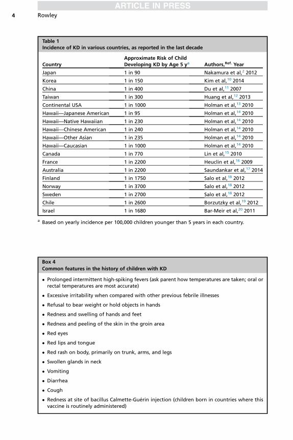
Table 1Incidence of KD in various countries, as reported in the last decade
CountryApproximate Risk of ChildDeveloping KD by Age 5 ya Authors,Ref. Year
Japan 1 in 90 Nakamura et al,2 2012
Korea 1 in 150 Kim et al,10 2014
China 1 in 400 Du et al,11 2007
Taiwan 1 in 300 Huang et al,12 2013
Continental USA 1 in 1000 Holman et al,13 2010
Hawaii—Japanese American 1 in 95 Holman et al,14 2010
Hawaii—Native Hawaiian 1 in 230 Holman et al,14 2010
Hawaii—Chinese American 1 in 240 Holman et al,14 2010
Hawaii—Other Asian 1 in 235 Holman et al,14 2010
Hawaii—Caucasian 1 in 1000 Holman et al,14 2010
Canada 1 in 770 Lin et al,15 2010
France 1 in 2200 Heuclin et al,16 2009
Australia 1 in 2200 Saundankar et al,17 2014
Finland 1 in 1750 Salo et al,18 2012
Norway 1 in 3700 Salo et al,18 2012
Sweden 1 in 2700 Salo et al,18 2012
Chile 1 in 2600 Borzutzky et al,19 2012
Israel 1 in 1680 Bar-Meir et al,20 2011
a Based on yearly incidence per 100,000 children younger than 5 years in each country.
Box 4
Common features in the history of children with KD
� Prolonged intermittent high-spiking fevers (ask parent how temperatures are taken; oral orrectal temperatures are most accurate)
� Excessive irritability when compared with other previous febrile illnesses
� Refusal to bear weight or hold objects in hands
� Redness and swelling of hands and feet
� Redness and peeling of the skin in the groin area
� Red eyes
� Red lips and tongue
� Red rash on body, primarily on trunk, arms, and legs
� Swollen glands in neck
� Vomiting
� Diarrhea
� Cough
� Redness at site of bacillus Calmette-Guerin injection (children born in countries where thisvaccine is routinely administered)
Rowley4
Box 5
Physical examination findings (classic diagnostic criteria)
� Intermittent high fever
� Plus 4 of the following 5 features:
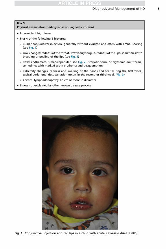
� Bulbar conjunctival injection, generally without exudate and often with limbal sparing(see Fig. 1)
� Oral changes: redness of the throat, strawberry tongue, redness of the lips, sometimes withbleeding or peeling of the lips (see Fig. 1)
� Rash: erythematous maculopapular (see Fig. 2), scarlatiniform, or erythema multiforme,sometimes with marked groin erythema and desquamation
� Extremity changes: redness and swelling of the hands and feet during the first week;typical periungual desquamation occurs in the second or third week (Fig. 3)
� Cervical lymphadenopathy 1.5 cm or more in diameter
� Illness not explained by other known disease process
Fig. 1. Conjunctival injection and red lips in a child with acute Kawasaki disease (KD).
Diagnosis and Management of KD 5
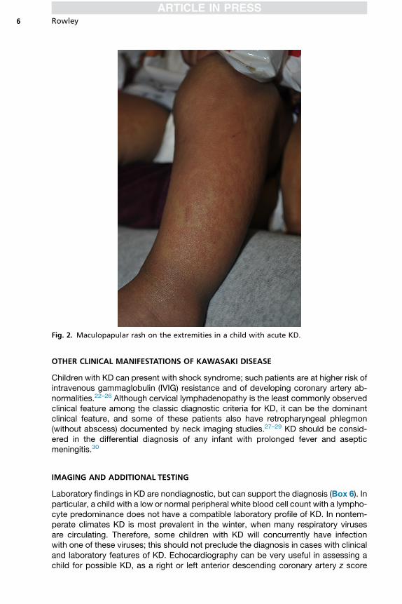
Fig. 2. Maculopapular rash on the extremities in a child with acute KD.
Rowley6
OTHER CLINICAL MANIFESTATIONS OF KAWASAKI DISEASE
Children with KD can present with shock syndrome; such patients are at higher risk ofintravenous gammaglobulin (IVIG) resistance and of developing coronary artery ab-normalities.22–26 Although cervical lymphadenopathy is the least commonly observedclinical feature among the classic diagnostic criteria for KD, it can be the dominantclinical feature, and some of these patients also have retropharyngeal phlegmon(without abscess) documented by neck imaging studies.27–29 KD should be consid-ered in the differential diagnosis of any infant with prolonged fever and asepticmeningitis.30
IMAGING AND ADDITIONAL TESTING
Laboratory findings in KD are nondiagnostic, but can support the diagnosis (Box 6). Inparticular, a child with a low or normal peripheral white blood cell count with a lympho-cyte predominance does not have a compatible laboratory profile of KD. In nontem-perate climates KD is most prevalent in the winter, when many respiratory virusesare circulating. Therefore, some children with KD will concurrently have infectionwith one of these viruses; this should not preclude the diagnosis in cases with clinicaland laboratory features of KD. Echocardiography can be very useful in assessing achild for possible KD, as a right or left anterior descending coronary artery z score
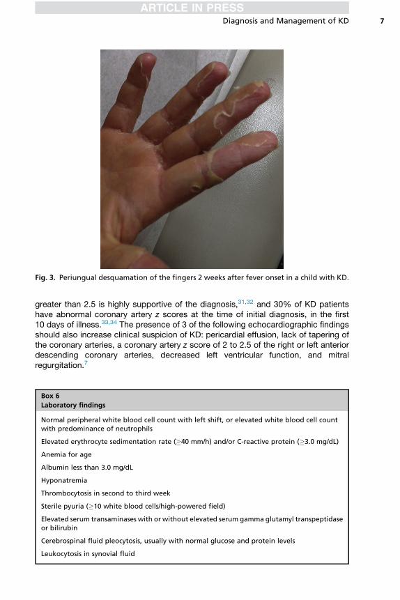
Fig. 3. Periungual desquamation of the fingers 2 weeks after fever onset in a child with KD.
Diagnosis and Management of KD 7
greater than 2.5 is highly supportive of the diagnosis,31,32 and 30% of KD patientshave abnormal coronary artery z scores at the time of initial diagnosis, in the first10 days of illness.33,34 The presence of 3 of the following echocardiographic findingsshould also increase clinical suspicion of KD: pericardial effusion, lack of tapering ofthe coronary arteries, a coronary artery z score of 2 to 2.5 of the right or left anteriordescending coronary arteries, decreased left ventricular function, and mitralregurgitation.7
Box 6
Laboratory findings
Normal peripheral white blood cell count with left shift, or elevated white blood cell countwith predominance of neutrophils
Elevated erythrocyte sedimentation rate (�40 mm/h) and/or C-reactive protein (�3.0 mg/dL)
Anemia for age
Albumin less than 3.0 mg/dL
Hyponatremia
Thrombocytosis in second to third week
Sterile pyuria (�10 white blood cells/high-powered field)
Elevated serum transaminases with or without elevated serum gamma glutamyl transpeptidaseor bilirubin
Cerebrospinal fluid pleocytosis, usually with normal glucose and protein levels
Leukocytosis in synovial fluid
Rowley8
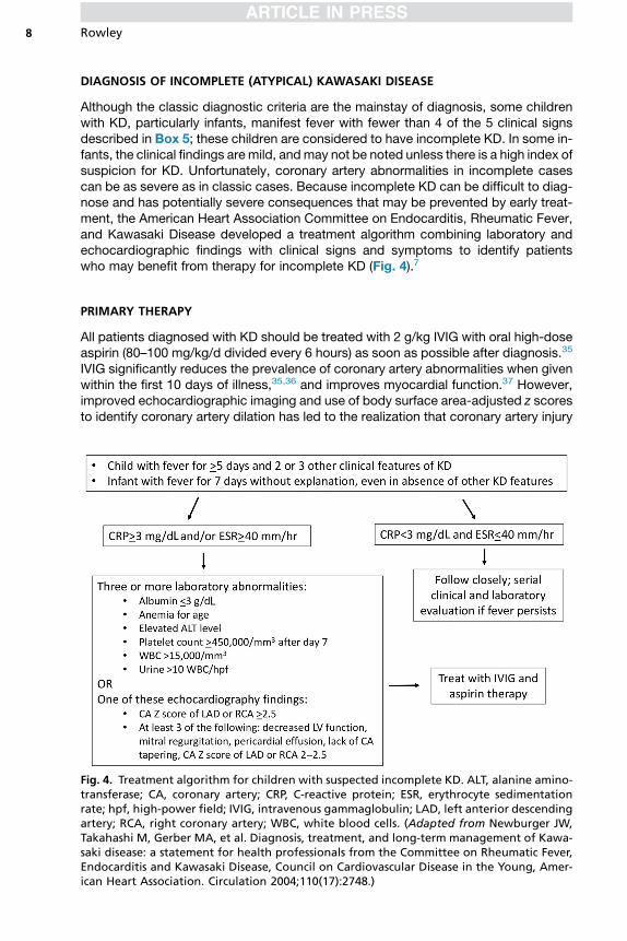
DIAGNOSIS OF INCOMPLETE (ATYPICAL) KAWASAKI DISEASE
Although the classic diagnostic criteria are the mainstay of diagnosis, some childrenwith KD, particularly infants, manifest fever with fewer than 4 of the 5 clinical signsdescribed in Box 5; these children are considered to have incomplete KD. In some in-fants, the clinical findings are mild, andmay not be noted unless there is a high index ofsuspicion for KD. Unfortunately, coronary artery abnormalities in incomplete casescan be as severe as in classic cases. Because incomplete KD can be difficult to diag-nose and has potentially severe consequences that may be prevented by early treat-ment, the American Heart Association Committee on Endocarditis, Rheumatic Fever,and Kawasaki Disease developed a treatment algorithm combining laboratory andechocardiographic findings with clinical signs and symptoms to identify patientswho may benefit from therapy for incomplete KD (Fig. 4).7
PRIMARY THERAPY
All patients diagnosed with KD should be treated with 2 g/kg IVIG with oral high-doseaspirin (80–100 mg/kg/d divided every 6 hours) as soon as possible after diagnosis.35
IVIG significantly reduces the prevalence of coronary artery abnormalities when givenwithin the first 10 days of illness,35,36 and improves myocardial function.37 However,improved echocardiographic imaging and use of body surface area-adjusted z scoresto identify coronary artery dilation has led to the realization that coronary artery injury
Fig. 4. Treatment algorithm for children with suspected incomplete KD. ALT, alanine amino-transferase; CA, coronary artery; CRP, C-reactive protein; ESR, erythrocyte sedimentationrate; hpf, high-power field; IVIG, intravenous gammaglobulin; LAD, left anterior descendingartery; RCA, right coronary artery; WBC, white blood cells. (Adapted from Newburger JW,Takahashi M, Gerber MA, et al. Diagnosis, treatment, and long-term management of Kawa-saki disease: a statement for health professionals from the Committee on Rheumatic Fever,Endocarditis and Kawasaki Disease, Council on Cardiovascular Disease in the Young, Amer-ican Heart Association. Circulation 2004;110(17):2748.)
Diagnosis and Management of KD 9
likely occurs in many patients in the first week of illness,33,34 and that coronary artery zscores greater than 2 can be observed in 18% of KD children at about 6 weeks afteronset even with treatment within the first 10 days.34 This finding emphasizes the needfor early diagnosis and treatment. At least 80% of KD patients respond to initial ther-apy with IVIG and aspirin with resolution of fever, improvement in clinical signs andsymptoms, and decreased laboratory markers of inflammation. Aspirin is maintainedat high doses for anti-inflammatory effect until the patient is afebrile for 2 to 3 days; atsome centers, it is continued until the 14th day of illness. Aspirin is then reduced toantiplatelet doses of 3 to 5 mg/kg/d in a single daily dose, and continued untilechocardiography at 6 to 8 weeks after the onset remains normal and acute-phasereactants have normalized. In patients who develop coronary artery abnormalities,low-dose aspirin is continued indefinitely. In patients with severe coronary artery ab-normalities, clopidogrel and/or anticoagulation therapy with warfarin or low molecularweight heparin may be indicated, and consultation with a pediatric cardiologist isadvised.7
RESEARCH STUDIES ON ADJUNCTIVE PRIMARY THERAPY
Unfortunately, approximately 15% to 20% of children with KD do not respond to initialIVIG therapy, with persistence of fever 36 hours after completion of IVIG infusion, andthese patients are at increased risk of developing coronary artery abnormalities. SomeKD patients, especially infants, can develop coronary artery abnormalities despiteapparent clinical response to IVIG treatment given in the first 10 days of illness. There-fore, recent research has focused on the study of combination immunomodulatorytherapies given with IVIG as primary therapy for KD. A randomized study of a single30 mg/kg dose of methylprednisolone administered with IVIG did not reveal a signifi-cant improvement in outcomes.38 A randomized, double-blind, placebo-controlledtrial of infliximab (a tumor necrosis factor a inhibitor) for intensification of primary ther-apy for KD did not show a reduction in treatment resistance nor a reduction in theoverall prevalence of coronary artery abnormalities when infliximab was administeredwith IVIG, although the addition of infliximab did result in lower levels of C-reactive pro-tein and absolute neutrophil counts 24 hours after the infusion.39 More promising wasthe Randomized controlled trial to Assess Immunoglobulin plus Steroid Efficacy forKawasaki disease (RAISE study), which demonstrated improvement in coronary arteryoutcomes in Japanese patients with high-risk KD when prednisolone was given withIVIG and continued for 15 days after normalization of the C-reactive protein level.40
A randomized trial of cyclosporin with IVIG for Japanese children with high-risk KDis presently under way in Japan, and the results of this study will also be of interest.Because the identification of risk scoring systems with high sensitivity for the predic-tion of coronary artery abnormalities in mixed ethnic populations has proved elusive,application of the RAISE study protocol or other high-risk protocols to KD children incountries such as the United States and Canada is not presently feasible.41
REFRACTORY KAWASAKI DISEASE
Refractory KD generally refers to the persistence of fever for 36 hours or longer aftercompletion of initial IVIG infusion. Patients with IVIG resistance have a higher preva-lence of coronary artery abnormalities.42 Most of these patients respond to a second2 g/kg dose of IVIG. For those patients who do not respond to a second dose of IVIG,several options for treatment exist, although controlled data are lacking (Box 7).
Box 7
Options for treatment of refractory KD
� Additional dose(s) of 2 g/kg of intravenous gammaglobulin43
� Intravenous methylprednisolone 30 mg/kg/d for 1 to 3 days44
� Infliximab 5 mg/kg � 145
� Other possible therapies: cyclosporine,46,47 methotrexate48
� Therapies sometimes used in Japan: plasmapheresis, neutrophil elastase inhibitor
� Possible future therapies: statins, interleukin-1 inhibitors
Rowley10
CLINICAL OUTCOMES AND COMPLICATIONS
Most children with KD respond to IVIG, and those who do not develop coronary arteryabnormalities by 4 to 6 weeks after the onset of fever have no known adverse out-comes. In patients who develop coronary artery dilation or aneurysm formation, out-comes depend on the severity of coronary artery disease. In severe cases giantcoronary artery aneurysms can form, which can rarely rupture, and virtually alwaysthrombose to a varying extent. Patients with this severe complication of KD are gener-ally maintained on antiplatelet and anticoagulation therapy, and are at the highest riskfor thrombotic occlusion and myocardial infarction, in some cases requiring catheterinterventions or coronary artery bypass surgery. However, coronary artery stenosis inKD patients can also be caused by luminal myofibroblastic proliferation (LMP) with orwithout thromboses. LMP is an active proliferative process of smooth muscle cell–derived myofibroblasts and their matrix products that can result in progressive arterialstenosis.3 In rare cases, LMP or thrombosis can result in such significant stenoses ofmultiple coronary arteries that heart transplantation is required.3,49 KD can affect allmedium-sized muscular arteries outside of the central nervous system, but peripheralarterial aneurysms seem to occur only in children with severe coronary artery disease.The most commonly affected arteries are the axillary, brachial, and inguinal arteries;aneurysms in these arteries rarely result in morbidity or mortality.3,50
REFERENCES
1. Yanagawa H, Nakamura Y, Yashiro M, et al. A nationwide incidence survey of Ka-wasaki disease in 1985-1986 in Japan. J Infect Dis 1988;158(6):1296–301.
2. Nakamura Y, Yashiro M, Uehara R, et al. Epidemiologic features of Kawasaki dis-ease in Japan: results of the 2009-2010 nationwide survey. J Epidemiol 2012;22(3):216–21.
3. Orenstein JM, Shulman ST, Fox LM, et al. Three linked vasculopathic processescharacterize Kawasaki disease: a light and transmission electron microscopicstudy. PLoS One 2012;7(6):e38998.
4. Amano S, Hazama F, Kubagawa H, et al. General pathology of Kawasaki disease.On the morphological alterations corresponding to the clinical manifestations.Acta Pathol Jpn 1980;30(5):681–94.
5. Fujita Y, Nakamura Y, Sakata K, et al. Kawasaki disease in families. Pediatrics1989;84(4):666–9.
6. Uehara R, Yashiro M, Nakamura Y, et al. Kawasaki disease in parents and chil-dren. Acta Paediatr 2003;92(6):694–7.
7. Newburger JW, Takahashi M, Gerber MA, et al. Diagnosis, treatment, and long-term management of Kawasaki disease: a statement for health professionals
Diagnosis and Management of KD 11
from the Committee on Rheumatic Fever, Endocarditis and Kawasaki Disease,Council on Cardiovascular Disease in the Young, American Heart Association.Circulation 2004;110(17):2747–71.
8. Takahashi K, Oharaseki T, Yokouchi Y, et al. A half-century of autopsy results–inci-dence of pediatric vasculitis syndromes, especially Kawasaki disease. Circ J2012;76(4):964–70.
9. Uehara R, Belay ED. Epidemiology of Kawasaki disease in Asia, Europe, and theUnited States. J Epidemiol 2012;22(2):79–85.
10. Kim GB, Han JW, Park YW, et al. Epidemiologic features of Kawasaki disease inSouth Korea: data from nationwide survey, 2009-2011. Pediatr Infect Dis J 2014;33(1):24–7.
11. Du ZD, Zhao D, Du J, et al. Epidemiologic study on Kawasaki disease in Beijingfrom 2000 through 2004. Pediatr Infect Dis J 2007;26(5):449–51.
12. Huang SK, Lin MT, Chen HC, et al. Epidemiology of Kawasaki disease: preva-lence from national database and future trends projection by system dynamicsmodeling. J Pediatr 2013;163(1):126–31.e1.
13. Holman RC, Belay ED, Christensen KY, et al. Hospitalizations for Kawasaki syn-drome among children in the United States, 1997-2007. Pediatr Infect Dis J2010;29(6):483–8.
14. Holman RC, Christensen KY, Belay ED, et al. Racial/ethnic differences in the inci-dence of Kawasaki syndrome among children in Hawaii. Hawaii Med J 2010;69(8):194–7.
15. Lin YT, Manlhiot C, Ching JC, et al. Repeated systematic surveillance of Kawasakidisease in Ontario from 1995 to 2006. Pediatr Int 2010;52(5):699–706.
16. Heuclin T, Dubos F, Hue V, et al. Increased detection rate of Kawasaki diseaseusing new diagnostic algorithm, including early use of echocardiography.J Pediatr 2009;155(5):695–699 e1.
17. Saundankar J, Yim D, Itotoh B, et al. The epidemiology and clinical features ofKawasaki disease in Australia. Pediatrics 2014;133(4):e1009–14.
18. Salo E, Griffiths EP, Farstad T, et al. Incidence of Kawasaki disease in northernEuropean countries. Pediatr Int 2012;54(6):770–2.
19. Borzutzky A, Hoyos-Bachiloglu R, Cerda J, et al. Rising hospitalization rates ofKawasaki disease in Chile between 2001 and 2007. Rheumatol Int 2012;32(8):2491–5.
20. Bar-Meir M, Haklai Z, Dor M. Kawasaki disease in Israel. Pediatr Infect Dis J 2011;30(7):589–92.
21. Lai CC, Lee PC, Wang CC, et al. Reaction at the Bacillus Calmette-Guerin inoc-ulation site in patients with Kawasaki disease. Pediatr Neonatol 2013;54(1):43–8.
22. Gatterre P, Oualha M, Dupic L, et al. Kawasaki disease: an unexpected etiologyof shock and multiple organ dysfunction syndrome. Intensive Care Med 2012;38(5):872–8.
23. Gamez-Gonzalez LB, Murata C, Munoz-Ramirez M, et al. Clinical manifestationsassociated with Kawasaki disease shock syndrome in Mexican children. Eur JPediatr 2013;172(3):337–42.
24. Kanegaye JT, Wilder MS, Molkara D, et al. Recognition of a Kawasaki diseaseshock syndrome. Pediatrics 2009;123(5):e783–9.
25. Lin MT, Fu CM, Huang SK, et al. Population-based study of Kawasaki diseaseshock syndrome in Taiwan. Pediatr Infect Dis J 2013;32(12):1384–6.
26. Dominguez SR, Friedman K, Seewald R, et al. Kawasaki disease in a pediatricintensive care unit: a case-control study. Pediatrics 2008;122(4):e786–90.
Rowley12
27. Kanegaye JT, Van Cott E, Tremoulet AH, et al. Lymph-node-first presentation ofKawasaki disease compared with bacterial cervical adenitis and typical Kawa-saki disease. J Pediatr 2013;162(6):1259–63, 1263.e1–2.
28. Stamos JK, Corydon K, Donaldson J, et al. Lymphadenitis as the dominant mani-festation of Kawasaki disease. Pediatrics 1994;93(3):525–8.
29. Nomura O, Hashimoto N, Ishiguro A, et al. Comparison of patients with Kawasakidisease with retropharyngeal edema and patients with retropharyngeal abscess.Eur J Pediatr 2014;173(3):381–6.
30. Yeom JS, Park JS, Seo JH, et al. Initial characteristics of Kawasaki disease withcerebrospinal fluid pleocytosis in febrile infants. Pediatr Neurol 2012;47(4):259–62.
31. Bratincsak A, Reddy VD, Purohit PJ, et al. Coronary artery dilation in acute Kawa-saki disease and acute illnesses associated with fever. Pediatr Infect Dis J 2012;31(9):924–6.
32. Muniz JC, Dummer K, Gauvreau K, et al. Coronary artery dimensions in febrilechildren without Kawasaki disease. Circ Cardiovasc Imaging 2013;6(2):239–44.
33. Dominguez SR, Anderson MS, Eladawy M, et al. Preventing coronary artery ab-normalities: a need for earlier diagnosis and treatment of Kawasaki disease. Pe-diatr Infect Dis J 2012;31(12):1217–20.
34. Printz BF, Sleeper LA, Newburger JW, et al. Noncoronary cardiac abnormalitiesare associated with coronary artery dilation and with laboratory inflammatorymarkers in acute Kawasaki disease. J Am Coll Cardiol 2011;57(1):86–92.
35. Newburger JW, Takahashi M, Beiser AS, et al. A single intravenous infusion ofgamma globulin as compared with four infusions in the treatment of acute Kawa-saki syndrome. N Engl J Med 1991;324(23):1633–9.
36. Newburger JW, Takahashi M, Burns JC, et al. The treatment of Kawasaki syn-drome with intravenous gamma globulin. N Engl J Med 1986;315(6):341–7.
37. Newburger JW, Sanders SP, Burns JC, et al. Left ventricular contractility and func-tion in Kawasaki syndrome. Effect of intravenous gamma-globulin. Circulation1989;79(6):1237–46.
38. Newburger JW, Sleeper LA, McCrindle BW, et al. Randomized trial of pulsedcorticosteroid therapy for primary treatment of Kawasaki disease. N Engl JMed 2007;356(7):663–75.
39. Tremoulet AH, Jain S, Jaggi P, et al. Infliximab for intensification of primary ther-apy for Kawasaki disease: a phase 3 randomised, double-blind, placebo-controlled trial. Lancet 2014;383(9930):1731–8.
40. Kobayashi T, Saji T, Otani T, et al. Efficacy of immunoglobulin plus prednisolonefor prevention of coronary artery abnormalities in severe Kawasaki disease(RAISE study): a randomised, open-label, blinded-endpoint trial. Lancet 2012;379(9826):1613–20.
41. Son MB, Newburger JW. Management of Kawasaki disease: corticosteroids re-visited. Lancet 2012;379(9826):1571–2.
42. Wallace CA, French JW, Kahn SJ, et al. Initial intravenous gammaglobulin treat-ment failure in Kawasaki disease. Pediatrics 2000;105(6):E78.
43. Sundel RP, Burns JC, Baker A, et al. Gamma globulin re-treatment in Kawasakidisease. J Pediatr 1993;123(4):657–9.
44. Wright DA, Newburger JW, Baker A, et al. Treatment of immune globulin-resistantKawasaki disease with pulsed doses of corticosteroids. J Pediatr 1996;128(1):146–9.
45. Burns JC, Best BM, Mejias A, et al. Infliximab treatment of intravenousimmunoglobulin-resistant Kawasaki disease. J Pediatr 2008;153(6):833–8.
Diagnosis and Management of KD 13
46. Tremoulet AH, Pancoast P, Franco A, et al. Calcineurin inhibitor treatment of intra-venous immunoglobulin-resistant Kawasaki disease. J Pediatr 2012;161(3):506–512 e1.
47. Suzuki H, Terai M, Hamada H, et al. Cyclosporin a treatment for Kawasaki diseaserefractory to initial and additional intravenous immunoglobulin. Pediatr Infect DisJ 2011;30(10):871–6.
48. Lee TJ, Kim KH, Chun JK, et al. Low-dose methotrexate therapy for intravenousimmunoglobulin-resistant Kawasaki disease. Yonsei Med J 2008;49(5):714–8.
49. Checchia PA, Pahl E, Shaddy RE, et al. Cardiac transplantation for Kawasaki dis-ease. Pediatrics 1997;100(4):695–9.
50. Suda K, Tahara N, Honda A, et al. Persistent peripheral arteritis long after Kawa-saki disease - another documentation of ongoing vascular inflammation. Int J Car-diol 2015;180:88–90.