Embed Size (px)
Citation preview
168 CHAPTER 4 Systemic diseases
The classifi cation of paediatric vasculitis Background to classifi cation criteria • Classifi cation criteria are often described for diseases where the
pathogenesis and/or molecular mechanisms are poorly understood. • They are used to facilitate clinical trials and improve epidemiological
descriptions by providing a set of agreed criteria that can be used by investigators anywhere in the world.
• Classifi cation criteria for vasculitis are designed to differentiate one form of vasculitis from another once the diagnosis of vasculitis has been secured. They are not the same as diagnostic criteria (such as those described for Kawasaki disease), but are often misused as such.
• Thus, classifi cation criteria aim to: • Identify a set of clinical fi ndings (criteria) that recognize a high
proportion of patients with the particular disease (sensitivity), and • Exclude a high proportion of patients with other diseases
(specifi city). • Classifi cation criteria typically include manifestations that are
characteristics of the disease in question that occur with less frequency or are absent in other conditions.
• Symptoms or fi ndings that might be typical or common but may also be present in other diseases tend to be excluded.
• An important limitation to these criteria is that they are not based on a robust understanding of the pathogenesis and as such are relatively crude tools that are likely to be modifi ed as scientifi c understanding of these diseases progresses.
Paediatric vasculitis classifi cation 2010 • New paediatric classifi cation criteria are described, and validated on
> 1300 cases worldwide (Table 4.1 ). • These criteria do not include Kawasaki disease (see b Kawasaki
disease, p 183); nor do they include defi nitions for microscopic polyangiitis (too few cases included in dataset).
• For Takayasu arteritis, care must be taken to exclude fi bromuscular dysplasia (or other cause of non-infl ammatory large- and medium-vessel arteriopathy) since undoubtedly there could be scope for overlap in the clinical presentation between these 2 entities, although the pathogenesis and treatment for these are clearly distinct.
General scheme for the classifi cation of paediatric vasculitides • This is based on the size of the vessel predominantly involved in the
vasculitic syndrome and is summarized as follows. • It should be noted, however, that most vasculitides exhibit a signifi cant
degree of ‘polyangiitis overlap’: e.g. Wegener’s granulomatosis can affect the aorta and its major branches, and small vessel vasculitis can occur in polyarteritis nodosa.
04-Foster-04.indd 16804-Foster-04.indd 168 5/9/2012 4:39:39 PM5/9/2012 4:39:39 PM

169THE CLASSIFICATION OF PAEDIATRIC VASCULITIS
1. Predominantly large-vessel vasculitis • Takayasu arteritis.
2. Predominantly medium-sized vessel vasculitis • Childhood polyarteritis nodosa • Cutaneous polyarteritis • Kawasaki disease.
3. Predominantly small-vessel vasculitis • Granulomatous:
• Wegener’s granulomatosis • Churg–Strauss syndrome
• Non-granulomatous: • Microscopic polyangiitis • Henoch–Schönlein purpura • Isolated cutaneous leucocytoclastic vasculitis • Hypocomplementemic urticarial vasculitis.
4. Other vasculitides • Behçet’s disease • Vasculitis s to infection (including hepatitis B-associated PAN),
malignancies and drugs, including hypersensitivity vasculitis • Vasculitis associated with other connective tissue diseases • Isolated vasculitis of the CNS (childhood p angiitis of the central
nervous system: cPACNS) • Cogan’s syndrome • Unclassifi ed.
04-Foster-04.indd 16904-Foster-04.indd 169 5/9/2012 4:39:39 PM5/9/2012 4:39:39 PM

170 CHAPTER 4 Systemic diseases
Table 4.1 Classifi cation criteria for specifi c vasculitic syndromes 1
Vasculitis Classifi cation criteria Sensitivity 2 Specifi city *
HSP Purpura, predominantly lower limb or diffuse * (mandatory) plus 1 out of 4 of: • Abdo pain • IgA on biopsy • Haematuria/proteinuria • Arthritis/arthralgia
* If diffuse (i.e. atypical distribution) then IgA deposition on biopsy required
100 % 87 %
WG At least 3 out of 6 of the following criteria: • Histopathology • Upper airway involvement • Laryngo-tracheobronchial stenoses • Pulmonary involvement • ANCA positivity • Renal involvement
93 % 99 %
PAN Histopathology or angiographic abnormalities (mandatory) plus 1 out of 5 of the following criteria: • Skin involvement • Myalgia/muscle tenderness • Hypertension • Peripheral neuropathy • Renal involvement
89 % 99 %
TA Angiographic abnormalities of the aorta or its main branches (also pulmonary arteries) showing aneurysm/dilatation (mandatory criterion), plus 1 out of 5 of the following criteria: • Pulse defi cit or claudication, • 4 limb BP discrepancy • Bruits • Hypertension • Acute phase response
100 % 99 %
1 Adapted from Ozen S, Pistorio A, Iusan SM, et al . Paediatric Rheumatology International Trials Organisation (PRINTO). EULAR/PRINTO/PRES criteria for Henoch–Schönlein purpura, childhood polyarteritis nodosa, childhood Wegener granulomatosis and childhood Takayasu arteritis: Ankara 2008. Part II: Final classifi cation criteria. Ann Rheum Dis 2010; 69 :798–806.
2 Based on 1347 children with miscellaneous vasculitides. Ruperto N, Ozen S, Pistorio A, et al .; for the Paediatric Rheumatology International Trials Organisation (PRINTO). EULAR/PRINTO/PRES criteria for Henoch–Schönlein purpura, childhood polyarteritis nodosa, childhood Wegener granulomatosis and childhood Takayasu arteritis: Ankara 2008. Part I: Overall methodology and clinical characterisation. Ann Rheum Dis 2010; 69 :790–7.
04-Foster-04.indd 17004-Foster-04.indd 170 5/9/2012 4:39:39 PM5/9/2012 4:39:39 PM

171THE EPIDEMIOLOGY OF PAEDIATRIC VASCULITIS
The epidemiology of paediatric vasculitis • Childhood vasculitis is rare and the incidence and prevalence are not
accurately described. • Henoch–Schönlein purpura (HSP) and Kawasaki disease (KD) are the
2 commonest childhood vasculitides and as such those with the most epidemiological information. • There is undoubtedly some ethnic variation for these diseases (see
individual section). • In paediatric populations other systemic vasculitides including
polyarteritis nodosa (PAN), Wegener’s granulomatosis (and other ANCA-associated vasculitides), Behçet’s disease, and Takayasu arteritis are rare and epidemiology diffi cult to assess. • Some ethnic variation has been noted: Takayasu’s being more
common in Asians than North Americans and Europeans.
Henoch–Schönlein purpura The estimated annual incidence in the West Midlands, UK has been reported as 20.4 per 100,000 and was highest in the 4–7yr age group. This is comparable to reported fi gures from the Czech Republic of 10.2 per 100,000 and Taiwan of 12.9 per 100,000.
Kawasaki disease • KD has the highest incidence and prevalence in Asian populations,
particularly Japan. • Nationwide surveys conducted in Japan show the incidence of KD in
children aged 0–4yr continues to rise with the average annual incidence in 2007 being 184.6 per 100,000.
• This compares to an estimated annual incidence rate of 5.5–8.1 per 100,000 (0–5yr) in the UK and an estimated annual incidence rate of 1.6 per 100,000 (0–5yr) in the Czech Republic.
• There are limitations to these studies as they were all done by survey or questionnaire reporting.
Further reading Dolezalova P , Telekesova P , Nemcova D , et al . Incidence of vasculitis in children in the Czech
Republic: 2-year prospective epidemiology survey . J Rheumatol 2004 ; 31 : 2295 – 9 . Gardner-Medwin JMM , Dolezalova P , Cummins C , et al . Incidence of Henoch-Scholein purpura,
Kawasaki disease, and rare vasculitides in children of different ethnic origins . Lancet 2002 ; 360 : 1197 – 202 .
Yang YH , Hung CF , Hsu CR , et al . A nationwide survey on epidemiological characteristics of childhood Henoch-Scholein purpura in Tiawan . Rheumatology 2005 ; 44 : 618 – 22 .
04-Foster-04.indd 17104-Foster-04.indd 171 5/9/2012 4:39:39 PM5/9/2012 4:39:39 PM
172 CHAPTER 4 Systemic diseases
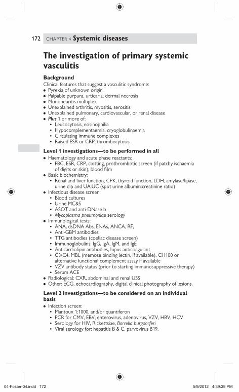
The investigation of primary systemic vasculitis Background Clinical features that suggest a vasculitic syndrome: • Pyrexia of unknown origin • Palpable purpura, urticaria, dermal necrosis • Mononeuritis multiplex • Unexplained arthritis, myositis, serositis • Unexplained pulmonary, cardiovascular, or renal disease • Plus 1 or more of:
• Leucocytosis, eosinophilia • Hypocomplementaemia, cryoglobulinaemia • Circulating immune complexes • Raised ESR or CRP, thrombocytosis.
Level 1 investigations — to be performed in all • Haematology and acute phase reactants:
• FBC, ESR, CRP, clotting, prothrombotic screen (if patchy ischaemia of digits or skin), blood fi lm
• Basic biochemistry: • Renal and liver function, CPK, thyroid function, LDH, amylase/lipase,
urine dip and UA:UC (spot urine albumin:creatinine ratio) • Infectious disease screen:
• Blood cultures • Urine MC&S • ASOT and anti-DNase b • Mycoplasma pneumoniae serology
• Immunological tests: • ANA, dsDNA Abs, ENAs, ANCA, RF, • Anti-GBM antibodies • TTG antibodies (coeliac disease screen) • Immunoglobulins: IgG, IgA, IgM, and IgE • Anticardiolipin antibodies, lupus anticoagulant • C3/C4, MBL (memose binding lectin, if available), CH100 or
alternative functional complement assay if available • VZV antibody status (prior to starting immunosuppressive therapy) • Serum ACE
• Radiological: CXR, abdominal and renal USS • Other: ECG, echocardiography, digital clinical photography of lesions.
Level 2 investigations — to be considered on an individual basis • Infection screen:
• Mantoux 1:1000, and/or quantiferon • PCR for CMV, EBV, enterovirus, adenovirus, VZV, HBV, HCV • Serology for HIV, Rickettsiae, Borrelia burgdorferi • Viral serology for: hepatitis B & C, parvovirus B19.
04-Foster-04.indd 17204-Foster-04.indd 172 5/9/2012 4:39:39 PM5/9/2012 4:39:39 PM

173THE INVESTIGATION OF PRIMARY SYSTEMIC VASCULITIS
• Imaging: • Radiograph of bones and joints. • Selective contrast visceral angiography. • DMSA scan. • MRI/MRA of brain (for suspected cerebral vasculitis). • CT abdomen, thorax, brain, sinus X ray (for Wegener’s). • Labelled white cell scan (for extent and location of infl ammation). • Cerebral contrast angiography (for suspected cerebral vasculitis). • PET-CT: for differential of malignancy or Castleman’s disease. • DEXA scan. • V/Q scan. • USS Doppler of peripheral arteries. • Thermography and nail fold capillaroscopy.
• Tissue biopsy: skin, nasal or sinus, kidney, sural nerve, lung, liver, gut, temporal artery, brain, other.
• Bone marrow analysis and/or lymph node excision biopsy (for suspected malignancy).
• Biochemistry, immunology and immunogenetics: • Serum amyloid A. • Formal GFR. • Organ specifi c autoantibodies. • IgD. • B 2 Glycoprotein 1 antibodies. • Urinary catecholamines (consider plasma catecholamines as well),
and urine VMA, HVA (for phaeochromocytoma, or neuroblastoma). • Cryoglobulins (if there is a history of cold sensitivity/vasculitis mainly
present in exposed areas of the body). • Basic lymphocyte panel and CD19 count if monitoring post rituximab. • Mitochondrial DNA mutations. • DNA analysis for periodic fever syndromes that can mimic vasculitis:
MEFV (familial Mediterranean fever, TNFRSF1A (TNF A receptor associated periodic fever syndrome, TRAPS), MVK (hyper IgD syndrome, HIDS), NLRP3 (cryopyrin associated periodic syndrome, CAPS), and NOD2 (Crohn’s/Blau’s/juvenile sarcoid mutations).
• Nitroblue tertrazolium test if granulomatous infl ammation found on biopsy.
• Nerve conduction studies (PAN, WG, Behçet’s [before starting thalidomide]).
• Ophthalmology screen. • Ambulatory 24h BP/4-limb BP.
04-Foster-04.indd 17304-Foster-04.indd 173 5/9/2012 4:39:39 PM5/9/2012 4:39:39 PM
174 CHAPTER 4 Systemic diseases
The standard treatment of childhood vasculitis Guidelines for the use and monitoring of cytotoxic drugs in non-malignant disease are shown in Table 4.2 . Standard vasculitis therapy ( excluding crescentic glomerulonephritis) is described in Fig. 4.1 . Prior to using this approach, remember there should be: • A well-established diagnosis. • Severe, potentially life-threatening disease. • Inadequate response to less toxic therapy — milder cases of vasculitis
(e.g. isolated cutaneous forms) may respond to less toxic agents such as colchicine. Therapy should always be tailored for each individual.
• No known infection or neoplasm. • No pregnancy or possibility thereof. • Informed consent obtained and documented in notes.
Other points of note • Although the use of oral cyclophosphamide is highlighted in Table
4.3 , increasingly IV cyclophosphamide is favoured over the oral route in children and adults because of reduced side effects and lower cumulative dose, but comparable effi cacy as suggested by a number of studies in adults with ANCA vasculitis (e.g. the ‘CYCLOPS’ trial).
• IV cyclophosphamide has the added advantage of ensuring adherence to therapy, of particular relevance in adolescents with vasculitis.
Use of biologic therapy in systemic vasculitis of the young • Whilst the therapeutic approach and drugs used as suggested in
Figs. 4.1 and 4.2 undoubtedly have improved survival and long-term outlook for children with severe vasculitis, concerns relating to toxicity particularly with cyclophosphamide, and relapses despite this conventional therapeutic approach have led to the increasing use of biologic therapy such as rituximab, anti-TNF alpha, or other biologic therapy.
• Evidence to support the use of rituximab as a p induction agent in place of cyclophosphamide for the treatment of ANCA-associated vasculitis is now available for adults with this group of diseases (RITUXIVAS, and RAVE trials).
• Evidence to support this approach in children remains anecdotal, but undoubtedly rituximab is being increasingly used for children with ANCA vasculitis that is not adequately controlled using the conventional cyclophosphamide followed by azathioprine therapeutic regimen outlined in Fig. 4.1 .
• Evidence for the use of anti-TNF A or other biologic agents such as anakinra remains anecdotal for children and adults with vasculitis.
• Whilst there is not enough evidence to recommend specifi c biologic therapy for specifi c vasculitic syndromes, a general approach favoured by the author is given in Table 4.3
04-Foster-04.indd 17404-Foster-04.indd 174 5/9/2012 4:39:39 PM5/9/2012 4:39:39 PM

Tab
le 4
.2 D
oses
, sid
e ef
fect
s, an
d cl
inic
al m
onito
ring
of c
omm
only
use
d im
mun
osup
pres
sant
and
cyt
otox
ic im
mun
osup
pres
sant
dru
gs u
sed
for
the
trea
tmen
t of v
ascu
litis
C
yclo
phos
pham
ide
(CY
C)
Aza
thio
prin
e M
ycop
heno
late
mof
etil
(MM
F)
Cic
losp
orin
M
etho
trex
ate
Dos
e 2–
3mg/
kg o
nce
a da
y PO
2–3
m
onth
s; 0.
5–1.
0g/m
2 IV
mon
thly
w
ith m
esna
to
prev
ent
cyst
itis
(see
b
p 4
27 C
hapt
er 9
for
mes
na d
ose
and
IV C
YC
adm
inis
trat
ion
prot
ocol
) 0.5–
2.5m
g/kg
onc
e a
day
PO
for
1yr
or m
ore
(600
mg/
m 2
twic
e a
day)
3–
5mg/
kg/d
ay P
O in
2
divi
ded
dose
s 10
–15m
g/m
2 /w
eek
PO o
r SC
(s
ingl
e do
se)
Side
ef
fect
s Le
ucop
enia
; hae
mor
rhag
ic c
ystit
is;
reve
rsib
le a
lope
cia;
infe
rtili
ty;
leuk
aem
ia, l
ymph
oma,
tra
nsiti
onal
ce
ll ca
rcin
oma
of b
ladd
er
GI t
oxic
ity; h
epat
otox
icity
; ra
sh; l
euco
peni
a; te
rato
geni
city
; no
incr
ease
in m
alig
nanc
y in
ad
ults
with
RA
; no
conc
lusi
ve
data
for
canc
er r
isk
in c
hild
ren
Bone
mar
row
sup
pres
sion
; se
vere
dia
rrho
ea;
pulm
onar
y fi b
rosi
s
Ren
al im
pairm
ent,
hype
rten
sion
, he
pato
toxi
city
, tre
mor
, gi
ngiv
al h
yper
plas
ia,
hype
rtric
hosis
, lym
phom
a
Bone
mar
row
sup
pres
sion
and
in
ters
titia
l pne
umon
itis
( d r
isk
with
folic
aci
d), r
ever
sibl
e el
evat
ion
of t
rans
amin
ases
, he
patic
fi br
osis
Cum
ulat
ive
toxi
c do
se
Not
des
crib
ed fo
r m
alig
nanc
y;
500m
g/kg
for
azoo
sper
mia
N
ot d
escr
ibed
N
ot d
escr
ibed
N
ot d
escr
ibed
N
ot d
escr
ibed
Clin
ical
m
onito
ring
Wee
kly
FBC
for
dura
tion
of t
hera
py
(usu
ally
2–3
mon
ths)
; bas
elin
e an
d m
onth
ly r
enal
and
live
r fu
nctio
n
Tem
pora
rily
disc
ontin
ue a
nd/o
r re
duce
dos
e if
neut
rope
nia
<1.
5 ×
10
9 /L,
pla
tele
ts <
150
× 1
0 9 /L,
or
hae
mat
uria
Day
10
FBC
if IV
. Red
uce
dose
if
rena
l or
hepa
tic fa
ilure
e.g
. to
250–
300m
g/m
2 .
Wee
kly
FBC
for
1 m
onth
, th
en 3
-mon
thly
Tem
pora
rily
disc
ontin
ue a
nd/o
r re
duce
dos
e if
neut
rope
nia
<1.
5 ×
10 9 /
L, p
late
lets
<15
0 ×
10 9 /
L,
and
chec
k T
PMT
enz
yme-
pa
tient
s de
fi cie
nt in
TPM
T
requ
ire r
educ
ed d
oses
(or
m
ay n
ot to
lera
te)
azat
hiop
rine
beca
use
of i
mar
row
toxi
city
Fort
nigh
tly F
BC fo
r 2
mon
ths,
then
mon
thly
for
2/12
. 3-
mon
thly
whe
n st
able
. Ba
selin
e m
onth
ly r
enal
and
liv
er fu
nctio
n un
til s
tabl
e
Dis
cont
inue
tem
pora
rily
and/
or r
educ
e do
se if
ne
utro
peni
a <
1.5
× 1
0 9 /L,
pl
atel
ets
<15
0 ×
10 9 /
L, o
r si
gnifi
cant
GI s
ide
effe
cts
Wee
kly
mea
sure
men
t of
BP;
bas
elin
e th
en
mon
thly
ren
al a
nd
liver
func
tion;
mai
ntai
n 12
h tr
ough
leve
l at
50–1
00ng
/mL.
6–
12-m
onth
ly G
FR.
Con
side
r re
nal b
iops
y ev
ery
2 ye
ars
Base
line
CX
R, F
BC, a
nd L
FTs,
then
FBC
and
LFT
s fo
rtni
ghtly
unt
il do
se s
tabl
e, th
en m
onth
ly to
eve
ry
6 w
eeks
(afte
r 6
mon
ths)
. Red
uce
or d
iscon
tinue
if h
epat
ic e
nzym
es
> 3 ×
upp
er li
mit
of n
orm
al,
neut
rope
nia
<1.
5 × 1
0 9 /L,
new
or
wor
seni
ng c
ough
, sev
ere
naus
ea,
vom
iting
, or
diar
rhoe
a, p
late
lets
<
150
× 1
0 9 /L
or fa
lling
rap
idly
04-Foster-04.indd 17504-Foster-04.indd 175 5/9/2012 4:39:40 PM5/9/2012 4:39:40 PM

176 CHAPTER 4 Systemic diseases
Fig. 4.1 Standard vasculitis therapy ( excluding crescentic glomerulonephritis).
Induction therapy
• Prednisolone 30–60 mg/m2 once a
day (1–2 mg/kg) for 4 weeks,
weaning over next 6–8 weeks
(depending on response to Rx) to
0.3–0.7 mg/kg on alternate days;
or IV methylprednisolone 30 mg/kg
(max. 1g) for 3 consecutive days
followed by oral prednisolone as
above
• CYC 2–3 mg/kg PO once a day for
2–3/12 or 500–1000 mg/m2 IV
(max. 1.2 g) once a month for
6 months (reduce dose if renal or
hepatic failure). Aspirin 1–2 mg/kg
once a day (or dipyridamole
2.5 mg/kg BD if aspirin
contraindicated)
Failed
induction
Consider
• Single dose of IV CYC
750–1000 mg/m2 if previously
given oral CYC for induction
of remission
• Methylprednisolone
30 mg/kg (max. 1 g) IV x3
• 5- or 10-day course of daily
2-volume plasma exchange
with 4.5% HAS
• 2nd course of oral CYC
2 mg/kg once a day for
2 weeks
• IVIG 2 g/kg
• Biologic agent:
• Anti-TNF therapy
• Rituximab
Post induction (maintenance) phase:
18 months to 3 yr for PAN; may
require prolonged Rx in some
vasculitic syndromes
• Azathioprine 2–3 mg/kg PO once
a day (start 3–5 days after stopping
PO CYC; 10 days after IV CYC):
consider measuring TPMT first
• Prednisolone 0.2–0.5 mg/kg
alternate days (daily if ongoing
disease activity)
• Aspirin 1–2 mg/kg once a day or
dipyridamole 2.5 mg/kg twice a
day
• Consider ranitidine or proton
pump inhibitor
Major relapse
whilst on
maintenance
therapy
• Minor relapse: i oral
prednisolone
• Recurrent minor relapses or
‘grumbling vasculitis’: consider
IV pulsed methylprednisolone
and/or switch to 2nd
-line
maintenance therapy
Notes:
1. 2nd
-line maintenance agents
1. MMF
2. Ciclosporin
3. MTX
4. Colchicine
2. Consider sperm
cryopreservation for all
post-pubertal males receiving
CYC
3. For monitoring of
complications of therapy
refer to Table 4.2
4. Beware neutropenia as
prednisolone dose is weaned
during maintenance phase of
therapy
5. Miscellaneous vasculitides
such as Behçet’s may require
colchicine and/or thalidomide
6. Treatment with biological
agents in select individuals
who fail to respond to
standard induction therapy
(see separate guidelines).
7. Epoprostenol (prostacyclin)
1–20 ng/kg/min IV for
incipient gangrene
8. Other agents with as yet
unproven efficacy in
childhood vasculitis:
leflunamide; DSG
Stopping treatment
• Usually withdrawn slowly over
6 months if no relapse for
12 months
• Recommend stopping azathioprine
first over 3 months, followed by
gradual taper of prednisolone over
next 3 months
04-Foster-04.indd 17604-Foster-04.indd 176 5/9/2012 4:39:40 PM5/9/2012 4:39:40 PM

177THE STANDARD TREATMENT OF CHILDHOOD VASCULITIS
Fig. 4.2 Guidelines for treatment of crescentic glomerulonephritis (note early diagnosis and starting therapy is of major importance).
Linear IgG staining on
immunofluorescence
Anti GBM +
• Plasma exchange for
10–14 days (2 volume,
4.5% HAS)—or until
anti GBM Ab disappears
• Pulsed IV methyl
prednisolone 30 mg/kg
(max. 1 g) x3, then oral
prednisolone 2 mg/kg
once a day (weaned over
2 months then stop)
• CYC 2 mg/kg PO once a
day for 2 months, or IV at
500 mg/m2 monthly
(reduced dose because of
renal failure), possibly
increasing by 250 mg/m2
per
month (response dependent)
to maximum of 1000 mg/m2
(max. 1.2 g) for 6 months
Consider prolonging therapy if
Anti GBM still detectable
Pauci-immune on
immunofluorescence
—consider
microscopic
polyangiitis or ‘renal-
limited vasculitis’—
may be ANCA +
Treatment as per
vasculitis algorithm
above, but consider
using plasma
exchange as 1st
-line
induction therapy
in conjunction
with steroid
and CYC
Granular deposits on
immunofluorescence—
indicative of immune
complex disease
Electron microscopy
Treat specific disorders (refer
to relevant protocols):
• HSP
• IgA nephropathy
• Post streptococcal
• SLE
• Membranoproliferative GN
• Membranous GN
Consideration of biologic
therapy:
• Rituximab
• Anti-TNF therapy
Refer to specific protocols
Failed
remission
Crescentic GN on
renal biopsy
04-Foster-04.indd 17704-Foster-04.indd 177 5/9/2012 4:39:40 PM5/9/2012 4:39:40 PM

178 CHAPTER 4 Systemic diseases
Further reading Eleftheriou D , Melo M , Marks SD , et al . Biologic therapy in primary systemic vasculitis of the
young . Rheumatology 2009 ; 48 : 978 – 86 .
Table 4.3 Recommendations for indication and choice of biologic therapy for primary systemic vasculitis of the young based on published experience 1
Vasculitis type
Indication for biologic agent Proposed fi rst choice of biologic agent
ANCA-associated vasculitis
Critical organ or life-threatening disease which has failed to respond to standard vasculitis therapy or concerns regarding cumulative CYC dose.
Rituximab or other B cell depleting monoclonal antibody
Polyarteritis nodosa
Failed therapy with standard agents or concern regarding cumulative CYC dose
Rituximab or anti-TNF A * †
Behçet’s disease
Recalcitrant and severe disease; alternative to thalidomide
Anti-TNF A (infl iximab, adalimumab or etanercept)
CYC, cyclophosphamide; * Authors have more experience with infl iximab than etanercept for PAN, although etanercept has been used in individual cases of childhood PAN; †no fi rm recommendation is made regarding fi rst choice of biologic for PAN.
1 Adapted from Eleftheriou D, Melo M, Marks SD, et al . Biologic therapy in primary systemic vasculitis of the young. Rheumatology 2009; 48 :978–86.
04-Foster-04.indd 17804-Foster-04.indd 178 5/9/2012 4:39:40 PM5/9/2012 4:39:40 PM

179HENOCH–SCHÖNLEIN PURPURA
Henoch–Schönlein purpura Introduction • Henoch-Schönlein purpura (HSP), the commonest systemic vasculitis
in childhood is predominantly a small-vessel, non-granulomatous leucocytoclastic vasculitis of unknown aetiology. • Reported by Heberden (1801), Willan (1808), Schönlein (1832), and
Henoch (1874). • Also called anaphylactoid purpura (1948).
• Classifi cation criteria are palpable purpura in a predominantly lower limb distribution with at least 1 of 4 of: • Diffuse abdominal pain. • Any biopsy showing IgA deposition (mandatory criterion if rash is
atypical). • Arthritis and/or arthralgia. • Haematuria and/or proteinuria.
• Variable and often relapsing course without specifi c laboratory fi ndings with 1/3 of children having symptoms up to a fortnight, another 1/3 up to 1 month and recurrence of symptoms within 4 months of resolution in 1/3.
• Henoch–Schönlein nephritis (HSN) accounts for 1.6–3 % of all childhood cases of end-stage renal failure (ESRF) in the UK.
Epidemiology • Commoner in Caucasian and Asian populations and boys:
• 4:5 ratio of 1.5–2:1. • 50 % present before the age of 5yr, 75 % present before the age
of 10yr. • Incidence of 10–20.4 (mean of 13.5) per 100,000 children.
• 22.1 per 100,000 children under 14yr of age. • 70.3 per 100,000 children aged 4–7yr.
• Almost 20 × rarer in adults. • 0.8 cases per 100,000 adults. • More severe in adults.
• Seasonal variation (commoner in winter) with infectious triggers. • Associations with bacteria (e.g. Group A beta-haemolytic
streptococci) and viruses (hepatitis, CMV, HSV, human parvovirus B19, coxsackie, and adenovirus), and some cases after vaccination described.
Immunopathology • Type III hypersensitivity reaction with immune complex.
• IgA deposition: galactose defi cient IgA may contribute to this. • Alternate pathway complement activation. • Associated C2 defi ciency.
• Vasculitis of small blood vessels with diffuse angiitis. • Perivascular exudate of leucocytes.
• Polygenic inheritance with renal involvement associated with HLA-B35, IL-1 B (–511) T allele, and IL-8 allele A.
04-Foster-04.indd 17904-Foster-04.indd 179 5/9/2012 4:39:40 PM5/9/2012 4:39:40 PM

180 CHAPTER 4 Systemic diseases
Systemic involvement Dermatological • All patients have purpura but skin involvement may not be present at
time of initial presentation. • Generally symmetrical purpura involving lower limbs and buttocks but
can spread to upper limbs (rarely abdomen, chest or face). • Urticaria and angio-oedema can occur.
Gastrointestinal • Commonly occurs (68 % of patients). • Abdominal pain precedes rash by 1–14 days in 43 % of patients. • Presents with intermittent colicky abdominal pain, vomiting with
or without haematemesis or melaena (faecal occult blood may be positive) as a result of haemorrhage into gut wall.
• Involvement may result in intussusception, appendicitis, cholecystitis, pancreatitis, GI haemorrhage, ulceration, infarction, or perforation.
Rheumatological 60 % of patients will have joint involvement with arthritis and/or arthralgia usually affecting the knees and ankles resulting in pain, swelling and d range of movement.
Renal • 25–60 % of patients will have renal involvement with HSN:
• 76 % will develop within 4 weeks of disease onset. • 97 % will develop within 3 months of disease onset.
• Most cases are usually asymptomatic which necessitates screening up to 6 months after last recrudescence of rash or HSP symptoms with <10 % having signifi cant involvement requiring referral for consideration of renal biopsy (Fig. 4.3 ). • Microscopic haematuria without proteinuria is benign. • 82 % have normal renal function after 23yr. • Good prognosis with isolated haematuria and mild proteinuria with
mild histological changes as less than 5 % will develop chronic kidney disease (CKD) within 10–25yr.
• Improved renal prognosis in children <7yr. • Severe disease indicated by increasing proteinuria, development of
nephrotic syndrome and/or renal failure. • 20 % of patients with acute mixed nephritic and nephrotic syndrome
progress to ESRF. • 44–50 % develop hypertension or CKD. • Histological pattern is identical to IgA nephropathy and includes
focal segmental proliferative glomerulonephritis and rapidly progressive crescentic glomerulonephritis (Table 4.4 ).
Other Patients may present with orchitis, severe pulmonary haemorrhage, and/or cerebral vasculitis (which may respond to immunosuppression combined with plasmapheresis).
04-Foster-04.indd 18004-Foster-04.indd 180 5/9/2012 4:39:40 PM5/9/2012 4:39:40 PM

181HENOCH–SCHÖNLEIN PURPURA
Treatment • Symptomatic treatment with rest and analgesia. • No role of antibiotics unless suspected or proven infection. • Prophylactic corticosteroid therapy at commencement of HSP does
not prevent renal or GI involvement. • However, corticosteroids do seem to be effective in treating these
complications and severe facial and/or scrotal haemorrhagic oedema. • Patients with severe renal involvement may require other
immunosuppressive agents, antiproteinuric and antihypertensive agents.
Table 4.4 ISKDC classifi cation of Henoch–Schönlein nephritis
ISKDC grade Pathoanatomical fi ndings
I Minimal alterations
II Mesangial proliferation
III A Focal proliferation or sclerosis with <50 % crescents
III B Diffuse proliferation or sclerosis with <50 % crescents
IV A Focal proliferation or sclerosis with 50–75 % crescents
IV B Diffuse proliferation or sclerosis with 50–75 % crescents
V A Focal proliferation or sclerosis with >75 % crescents
V B Diffuse proliferation or sclerosis with >75 % crescents
VI Membranoproliferative glomerulonephritis
ISKDC=International Study of Kidney Diseases in Children.
04-Foster-04.indd 18104-Foster-04.indd 181 5/9/2012 4:39:40 PM5/9/2012 4:39:40 PM

182 CHAPTER 4 Systemic diseases
Fig.
4.3
Clin
ical
pat
hway
for
inve
stig
atio
n an
d re
ferr
al fo
r re
nal b
iops
y in
HSN
: Rep
rodu
ced
with
per
mis
sion
from
Tiz
ard
EJ, H
amilt
on-A
yres
MJJ.
H
enoc
h–Sc
hönl
ein
purp
ura.
Arc
h D
is Ch
ild E
duc
Prac
t Ed
2008
; 93 :
1–8.
AIP
, aut
o-im
mun
e pr
ofi le
; AN
CA
, ant
i-neu
trop
hil c
ytop
lasm
ic A
bs; A
RF,
acu
te r
enal
fa
ilure
; ASO
T, a
nti-s
trep
toly
sin
O ti
tre;
BP,
blo
od p
ress
ure;
C3,
C4,
com
plem
ent C
3 an
d C
4; E
MU
, ear
ly m
orni
ng u
rine;
GP,
gen
eral
pra
ctiti
oner
; Ht,
heig
ht;
U&
E, u
rea
and
elec
trol
ytes
; Wt,
wei
ght;
UP:
UC
, urin
e pr
otei
n:cr
eatin
ine
ratio
.
W1–W
4: w
eekly
GP* r
evie
w
(B
P, EM
U d
ipstic
k)
General paedia
tric
ian
Baseline:
Dis
cuss w
ith n
ephro
logis
t if:
• A
cute n
ephritic
syndro
me:
• P
ersis
tent p
ro
tein
uria
:
• U
P:U
C>
250 m
g/m
mo
l fo
r 4
weeks
• U
P:U
C>
100 m
g/m
mo
l fo
r 3
mo
nths
• U
P:U
C>
50 m
g/m
mo
l fo
r 6
mo
nths
Indic
atio
ns fo
r c
onsid
eratio
n o
f renal bio
psy:
1. A
cute n
ephritic
syndro
me/A
RF
2. N
ephro
tic
syndro
me/n
ephro
tic
range p
ro
tein
uria
(U
P:U
C>
250 m
g/m
mo
l)
fo
r 4
–6 w
eeks
Haem
aturia
/pro
tein
uria
/oedem
a, hypertensio
n/o
liguria
Or if persis
tin
g a
bno
rm
alitie
s w
ith:
UP:U
C>
250 m
g/m
mo
l pla
sm
a a
lbum
in<
25 g
/l o
edem
a
• H
ypertensio
n
• A
bno
rm
al renal fu
nctio
n
• M
acro
sco
pic
haem
aturia
—5 d
ays
• N
ephro
tic
syndro
me:
• W
t, H
t, B
P, urin
e d
ipstic
k
• E
MU
UP:U
C
• U
&E, creat, alb
um
in
• F
BC
, clo
ttin
g
• A
SO
T a
ntiD
NA
se B
Co
nsid
er
• C
3, C
4 A
IP, A
NC
A, lg
s
• T
hereaft
er (
pendin
g r
esult
s),
weekly
:
• E
MU
UP:U
C
• B
P a
nd w
eig
ht m
onito
rin
g
• C
linic
al assessm
ent
W5–W
12: fo
rtnig
htly
GP* r
evie
w (
BP, EM
U d
ipstic
k)
6–12: m
onth G
P* r
evie
w
(B
P, EM
U d
ipstic
k)
No
renal
invo
lvem
ent
Dis
charge
Mic
ro
sco
pic
haem
aturia
Annual G
P r
evie
w (
BP, EM
U d
ipstic
k)
* R
evie
w in p
rim
ary c
are/w
ith lo
cal paedia
tric
ian a
cco
rdin
g t
o lo
cal arrangem
ents N
B c
hildren w
ith r
ecurrent e
pis
odes s
ho
uld
mo
nito
red a
s fo
r t
he fir
st e
pis
ode
Hypertensio
n/m
acro
sco
pic
haem
aturia
/pro
tein
uria
Hypertensio
n/m
acro
sco
pic
haem
aturia
/pro
tein
uria
Hypertensio
n/m
acro
sco
pic
haem
aturia
/pro
tein
uria
Hypertensio
n/m
acro
sco
pic
haem
aturia
/pro
tein
uria
04-Foster-04.indd 18204-Foster-04.indd 182 5/9/2012 4:39:40 PM5/9/2012 4:39:40 PM

183KAWASAKI DISEASE
Kawasaki disease Kawasaki disease (KD) is a self-limiting vasculitic syndrome that predomi-nantly affects medium- and small-sized arteries. It is the 2 nd commonest vasculitic illness of childhood (the commonest being HSP) and it is the leading cause of childhood acquired heart disease in developed countries.
Pathogenesis and epidemiology • Pronounced seasonality and clustering of KD cases have led to the hunt
for infectious agents as a cause. However, so far no single agent has been identifi ed.
• The aetiology of KD remains unknown but it is currently felt that one or more widely distributed infectious agents evoke an abnormal immunological response in genetically susceptible individuals, leading to the characteristic clinical presentation of the disease.
• KD has a world-wide distribution with a 4 preponderance, an ethnic bias towards Asian and in particular Japanese or Chinese children, some seasonality, and occasional epidemics.
• The incidence of KD is rising world-wide, including the UK. The current reported incidence in the UK is 8.1/100,000 children aged <5yr. This may refl ect a truly rising incidence or i clinician awareness.
Clinical features
• For the diagnosis of KD to be established 5 of the 6 clinical features should be present.
• Patients with <5 or 6 principal features can be diagnosed with KD when coronary aneurysm or dilatation is recognized by two-dimensional (2D) echocardiography or coronary angiography.
• The cardiovascular features are the most important manifestations of the condition with widespread vasculitis affecting predominantly medium-size muscular arteries, especially the coronary arteries. Coronary artery involvement occurs in 15–25 % of untreated cases with additional cardiac features in a signifi cant proportion of these including pericardial effusion, electrocardiographic abnormalities, pericarditis, myocarditis, valvular incompetence, cardiac failure, and myocardial infarction.
• Of note, irritability is an important sign, which is virtually universally present although not included in the diagnostic criteria.
• Another clinical sign that may be relatively specifi c to KD is the development of erythema and induration at sites of BCG inoculations. The mechanism of this sign is thought to be cross reactivity of T cells
The principal clinical features of KD are:
• Fever persisting for 5 days or more. • Peripheral extremity changes (reddening of the palms and soles,
indurative oedema, and subsequent desquamation). • A polymorphous exanthema. • Bilateral conjunctival injection/congestion. • Lips and oral cavity changes (reddening/cracking of lips, strawberry
tongue, oral and pharyngeal injection), and • Acute non-purulent cervical lymphadenopathy.
04-Foster-04.indd 18304-Foster-04.indd 183 5/9/2012 4:39:41 PM5/9/2012 4:39:41 PM

184 CHAPTER 4 Systemic diseases
in KD patients between specifi c epitopes of mycobacterial and human heat shock proteins.
• An important point worthy of emphasis is that the principal symptoms and signs may present sequentially such that the full set of criteria may not be present at any one time. Awareness of other non-principal signs (such as BCG scar reactivation) may improve the diagnostic pick-up rate of KD.
• Other clinical features include: arthritis, aseptic meningitis, pneumonitis, uveitis, gastroenteritis, meatitis and dysuria, and otitis.
• Relatively uncommon abnormalities include hydrops of the gallbladder, GI ischaemia, jaundice, petechial rash, febrile convulsions, and encephalopathy or ataxia, macrophage activation syndrome, and syndrome of inappropriate anti-diuretic hormone secretion (SIADH).
Differential diagnosis Conditions that can cause similar symptoms to KD and must be considered in the differential diagnosis include: • Scarlet fever • Rheumatic fever • Streptococcal or staphylococcal toxic shock syndrome • Staphylococcal scalded skin syndrome • Systemic JIA • Infantile PAN • SLE • Adenovirus, enterovirus. Epstein–Barr virus, CMV, parvovirus, infl uenza
virus infection • Mycoplasma pneumoniae infection • Measles • Leptospirosis • Rickettsiae infection • Adverse drug reaction • Mercury toxicity (acrodynia) • Lymphoma — particularly for IVIg resistant cases.
Investigations In cases of suspected KD the following investigations should be considered: • FBC and blood fi lm. • ESR. • CRP. • Blood cultures. • ASOT and anti-DNase B. • Nose and throat swab, and stool sample for culture (superantigen
toxin typing if Staphylococcus aureus and/or beta-haemolytic streptococci detected).
• Renal and liver function tests. • Coagulation screen. • Autoantibody profi le (ANA, ENA, RF, ANCA). • Serology (IgG and IgM) for Mycoplasma pneumoniae , enterovirus,
adenovirus, measles, parvovirus, Epstein–Barr virus, cytomegalovirus. • Urine MC&S.
04-Foster-04.indd 18404-Foster-04.indd 184 5/9/2012 4:39:41 PM5/9/2012 4:39:41 PM

185KAWASAKI DISEASE
• Dip test of urine for blood and protein. • Consider serology for rickettsiae and leptospirosis if history suggestive. • Consider CXR. • ECG. • 2D echocardiography to identify coronary artery involvement acutely
and monitoring changes long term. • Coronary arteriography has an important role for delineating detailed
anatomical injury, particularly for children with giant coronary artery aneurysms ( > 8mm), where stenoses adjacent to the inlet/outlet of the aneurysms are a concern. Note that the procedure may need to be delayed until at least 6 months after disease onset since there could be a risk of myocardial infarction if performed in children with ongoing severe coronary artery infl ammation.
Treatment (Fig. 4.4 ) The treatment of KD comprises of: • IVIg at a dose of 2g/kg as a single infusion over 12h (consider splitting
the dose over 2–4 days in infants with cardiac failure). • IVIg should be started early preferably within the fi rst 10 days of the
illness. However, clinicians should not hesitate to give IVIg to patients who present after 10 days if there are signs of persisting infl ammation.
• Aspirin 30–50mg/kg/day in 4 divided doses. • The dose of aspirin can be reduced to 2–5mg/kg/day when the
fever settles (disease defervescence). Aspirin at antiplatelet doses is continued for a minimum of 6 weeks.
• If the symptoms persist within 48h or there is disease recrudescence within 2 weeks a 2 nd dose of IVIg at 2g/kg over 12h should be considered.
• However, IVIg resistance occurs up to 20 % of cases. • When a patient fails to respond to a 2 nd dose of IVIg, consider IV
pulsed methylprednisolone at 15–30mg/kg daily for 3 days to be followed by oral prednisolone 2mg/kg/day once a day weaning over 6 weeks. Some clinicians are increasingly using corticosteroids after disease recrudescence following one dose of IVIg based on the results of a recent study. This remains an area of controversy, but seems rational since this is associated in most cases with rapid resolution of infl ammation.
• In refractory cases infl iximab, a human chimeric anti-TNF A monoclonal antibody, given IV at a single dose of 6mg/kg has been reported to be effective, and is increasingly used for IVIg resistant cases. Considering that rapid and effective interruption of infl ammation is a p target of KD therapy, TNF A blockade may be a logical step following one failed dose of IVIg, particularly in very active disease with evidence of early coronary artery dilatation.
• Echocardiography should be repeated at 2 weeks and 6 weeks from initiation of treatment (refer to paediatric cardiology).
• If the repeat echocardiogram shows no coronary artery abnormalities (CAAs) at 6 weeks, aspirin can be discontinued and lifelong follow-up at least every 2yr should be considered.
04-Foster-04.indd 18504-Foster-04.indd 185 5/9/2012 4:39:41 PM5/9/2012 4:39:41 PM

186 CHAPTER 4 Systemic diseases
• In cases of CAA <8mm with no stenoses present, aspirin should be continued until aneurysms resolve.
• If CAA > 8mm and/or stenoses is present, aspirin at a dose of 2–5mg/kg/day should be continued lifelong. The combination of aspirin and warfarin therapy in these patients with giant aneurysms has been shown to d the risk of myocardial infarction.
• In patients who develop CAA, echocardiography and ECG should be repeated at 6-monthly intervals and an exercise stress test considered.
• Other specifi c interventions such as PET scanning, addition of calcium channel blocker therapy, and coronary angioplasty should be organized at the discretion of the paediatric cardiologist.
Outcome • Treatment with IVIg and aspirin reduces CAA from 25 % for untreated
cases to 4–9 % . • IVIg resistance occurs in approximately 20 % , and is associated with a
higher risk of CAA. • The overall outlook of children with KD is good, with the acute
mortality rate due to myocardial infarction having been reduced to <1 % by i alertness of the clinicians to the diagnosis and prompt treatment.
• Nonetheless the disease may contribute to the burden of adult cardiovascular disease and cause premature atherosclerosis, an area of active ongoing research.
04-Foster-04.indd 18604-Foster-04.indd 186 5/9/2012 4:39:41 PM5/9/2012 4:39:41 PM

187KAWASAKI DISEASE
Fig.
4.4
Gui
delin
e fo
r th
e m
anag
emen
t of K
awas
aki d
isea
se.
Estab
lish
dia
gn
osis
of K
aw
asaki d
isease
a
• IV
IG 2
g/k
g a
s a
sin
gle
infu
sio
n o
ver 1
2 h
(co
nsid
er
splittin
g t
he d
ose o
ver 2
–4 d
ays in infa
nts w
ith
cardia
c failure)
• A
spir
in 3
0–50 m
g/k
g /
day in 4
div
ided d
oses
• P
erfo
rm
echo
cardio
graphy, and E
CG
• A
spir
in 2
–5 m
g/k
g/d
ay w
hen fever s
ettle
d (
dis
ease
d
efe
rvescence) c
ontin
uin
g fo
r a
min
imum
of 6 w
eeks
• b
Repeat e
cho
cardio
graphy a
t 2
weeks a
nd 6
weeks
Dis
ease d
efervescen
ce
Seek e
xp
ert a
dvic
e t
o
co
nsid
er:
• 2nd d
ose o
f IV
IG a
t 2
g/k
g o
ver 1
2 h
• P
uls
ed IV
methyl prednis
olo
ne
at 1
5–30 m
g/k
g d
aily fo
r 3
days t
o
be fo
llo
wed b
y o
ral prednis
olo
ne
2 m
g/k
g/d
ay o
nce a
day w
eanin
g o
ver
6 w
eeks—
seek e
xpert a
dvic
e
• In
flix
imab (
6 m
g/k
g) fo
r r
efr
acto
ry
cases-seek e
xpert a
dvic
e
No
dis
ease d
efervescen
ce
wit
hin
48 h
, o
r d
isease
recru
descen
ce w
ith
in 2
weeks
No
co
ro
nary a
rtery
ab
no
rm
alitie
s (
CA
As)
• S
to
p a
spir
in a
t 6
weeks
• C
onsid
er lifelo
ng fo
llo
w-
up a
t least e
very 2
years
CA
A <
8 m
m, n
o s
ten
oses
• C
ontin
ue a
spir
in u
ntil a
neurysm
s r
eso
lve
• R
epeat e
cho
cardio
graphy &
EC
G a
t
6-m
onthly
intervals
• D
isco
ntin
ue a
spir
in if aneurysm
s r
eso
lve
• C
onsid
er e
xercis
e s
tress t
est if m
ultip
le
aneurysm
s
• Specific
advic
e r
e: m
inim
izin
g a
thero
ma
ris
k facto
rs, and c
onsid
er lifelo
ng fo
llo
w-up
cC
AA
>8 m
m, an
d/o
r s
ten
oses
• Lifelo
ng a
spir
in 2
–5 m
g/k
g/d
ay
• W
arfa
rin
(w
ith initia
l fu
ll h
eparin
izatio
n
to
prevent p
arado
xic
al thro
mbo
sis
)
• C
onsid
er c
oro
nary a
ngio
graphy
(aft
er a
t least 6
mo
nths fro
m d
isease
o
nset) a
nd e
xercis
e s
tress t
estin
g
• R
epeat e
cho
cardio
graphy &
EC
G a
t
6-m
onthly
intervals
• Specific
advic
e r
e: m
inim
izin
g a
thero
ma
ris
k facto
rs
• Lifelo
ng fo
llo
w-up
No
tes:
aT
reatm
ent c
an b
e c
om
menced b
efo
re full
5 d
ays o
f fe
ver if sepsis
exclu
ded; treatm
ent
sho
uld
als
o b
e g
iven if the p
resentatio
n is
>10 d
ays fro
m fever o
nset
bR
efe
r t
o p
aedia
tric
cardio
logis
t
cO
ther s
pecific
interventio
ns s
uch a
s P
ET
scannin
g, additio
n o
f calc
ium
channel
blo
cker t
herapy, and c
oro
nary a
ngio
pla
sty a
t
dis
cretio
n o
f paedia
tric
cardio
logis
t.
04-Foster-04.indd 18704-Foster-04.indd 187 5/9/2012 4:39:41 PM5/9/2012 4:39:41 PM

188 CHAPTER 4 Systemic diseases
The anti-neutrophil cytoplasmic antibody (ANCA)-associated vasculitides Background • The ANCA-associated vasculitides (AAV) are:
• Wegener’s granulomatosis (WG) • Microscopic polyangiitis (MPA) • Churg–Strauss syndrome (CSS) and • Renal limited vasculitis (previously referred to as idiopathic
crescentic glomerulonephritis). • Although rare, the AAV do occur in childhood.
Defi nitions of AAV Defi nitions for each of the AAV describing the salient major clinical and laboratory features are given here. These are not the same as clas-sifi cation criteria, which (for WG) are provided in a separate section on classifi cation. • WG: granulomatous infl ammation involving the respiratory tract and
necrotizing vasculitis affecting small- to medium-size vessels • MPA: necrotizing vasculitis, with few or no immune deposits, affecting
small vessels; necrotizing arteritis involving small- and medium-sized arteries may be present; pulmonary capillaritis often occurs. Clinically, it often presents with rapidly progressive pauci-immune glomerulonephritis, in association with perinuclear ANCA (pANCA, MPO-ANCA) positivity.
• CSS: an eosinophil-rich and granulomatous infl ammation involving the respiratory tract and necrotizing vasculitis affecting small- to medium-sized vessels; there is an association with asthma and eosinophilia.
• Renal limited: rapidly progressive glomerulonephritis, often with ANCA positivity (usually MPO-ANCA) but without other organ involvement.
Pathogenesis • It is not known why patients develop ANCA in the fi rst instance. • When ANCA are present, the most accepted current model
of pathogenesis proposes that ANCA activate cytokine-primed neutrophils, leading to bystander damage of endothelial cells and an escalation of infl ammation with recruitment of mononuclear cells.
• However, other concomitant exogenous factors and genetic susceptibility appear to be necessary for disease expression.
Clinical features of WG From a clinical perspective WG may be broadly considered as having 2 forms: • Predominantly granulomatous form with mainly localized disease, and • Florid, acute small vessel vasculitic form characterized by severe
pulmonary haemorrhage and/or rapidly progressive vasculitis or other severe vasculitic manifestation.
These 2 broad presentations may coexist or present sequentially in individual patients.
04-Foster-04.indd 18804-Foster-04.indd 188 5/9/2012 4:39:41 PM5/9/2012 4:39:41 PM

189ANCA-ASSOCIATED VASCULITIDES
Organ specifi c involvement includes: • Upper respiratory tract:
• Epistaxis. • Otalgia, and hearing loss (conductive and/or sensorineural); chronic
otitis media; mastoiditis. • Nasal septal involvement with cartilaginous collapse results in the
characteristic saddle nose deformity (Fig. 4.5 ). • Chronic sinusitis. • Glottic and subglottic polyps and/or large- and medium-sized airway
stenosis. • Lower respiratory tract manifestations include (singly or in
combination): • Granulomatous pulmonary nodules with or without central
cavitation. • Pulmonary haemorrhage with respiratory distress, frank
haemoptysis, and/or evanescent pulmonary shadows (CXR). • Interstitial pneumonitis.
• Renal involvement: typically a focal segmental necrotizing glomerulonephritis, with pauci-immune crescentic glomerular changes. The clinical manifestations associated with this lesion are: • Hypertension. • Signifi cant proteinuria. • Nephritic and nephrotic syndrome. • Other protean manifestations of renal failure.
• Ophthalmological disease: retinal vasculitis, conjunctivitis, episcleritis, uveitis, optic neuritis. Unilateral or bilateral proptosis may be caused by granulomatous infl ammation affecting the orbit (pseudotumour) (Fig. 4.5 ).
• Malaise, fever, weight loss or growth failure, arthralgia, and arthritis. • Other manifestations include: peripheral gangrene with tissue loss, and
vasculitis of the skin, gut (including appendicitis), heart, central nervous system and/or peripheral nerves (mononeuritis multiplex), salivary glands, gonads, and breast.
Investigations (also see b Vasculitis investigation, p 172) • WG is commonly associated with a cytoplasmic staining pattern of
ANCA by IIF, and ELISA reveals specifi city against PR3 (PR3-ANCA). • MPA and renal limited AAV are typically associated with pANCA by IIF
and with MPO-ANCA specifi city on ELISA. • ANCA-negative forms of WG, MPA, renal limited vasculitis, and CSS
are well described in children. • While the diagnostic value of ANCA is without question important,
the value of ANCA for the longitudinal monitoring of disease activity is probably unreliable in many patients with WG.
• Tissue diagnosis, in particular renal biopsy but also biopsy of skin, nasal septum, or other tissue, can be important diagnostically for diagnosing all of the AAV and can help stage the disease for therapeutic decision-making.
04-Foster-04.indd 18904-Foster-04.indd 189 5/9/2012 4:39:41 PM5/9/2012 4:39:41 PM

190 CHAPTER 4 Systemic diseases
• Other commonly observed non-specifi c fi ndings include: • Mild normochromic normocytic anaemia together with a
leucocytosis and thrombocytosis. • Elevated ESR and CRP. • Raised immunoglobulins (polyclonal IgG).
• Laboratory manifestations relating to renal involvement include: • Dipstick haematuria and proteinuria positive. • Raised urinary spot protein creatinine ratio. • Raised serum creatinine and other associated laboratory features of
renal failure. • Chest radiography may be abnormal but high resolution CT chest has
better sensitivity for demonstrating pulmonary infi ltrates or discrete nodular and/or cavitating lesions
• Plain x-ray or CT sinuses for sinusitis
Fig. 4.5 Right orbital and characteristic saddle nose deformity in Wegener’s granu-lomatosis.
(a) (b)
04-Foster-04.indd 19004-Foster-04.indd 190 5/9/2012 4:39:41 PM5/9/2012 4:39:41 PM

191ANCA-ASSOCIATED VASCULITIDES
Treatment of AAV (See b BSPAR guidelines for treatments used in paediatric rheumatology, p 415.)
When considering therapy, it is useful to remember that most evidence for treatment is derived from adult trials. It is also useful to consider the different phases of the therapeutic journey for AAV: • The pre-diagnostic phase: occasionally lasting years. Signifi cant organ
damage can accrue in this phase, or even death. • Induction of remission phase: typically 3–6 months. • Maintenance of remission phase: usually 18–24 months. • Therapy withdrawal phase: not all patients achieve this.
The following general points are worthy of note: • The key to successful treatment is early diagnosis to limit organ damage. • Treatment for paediatric AAV is broadly similar to the approach used
in adults and involves corticosteroids, cyclophosphamide, and in some individuals plasma exchange (particularly for pulmonary capillaritis and/or rapidly progressive glomerulonephritis — ’pulmonary-renal syndrome’) to induce remission; followed by low-dose corticosteroids and azathioprine to maintain remission.
• Antiplatelet doses of aspirin can also be considered empirically on the basis of the i risk of thrombosis associated with the disease process.
• MTX in combination with corticosteroids may have a role for inducing remission in patients with limited WG.
• Co-trimoxazole is commonly added to therapeutic programmes for the treatment of WG, particularly in those with upper respiratory tract involvement, serving both as prophylaxis against opportunistic infection and as a possible disease-modifying agent.
• Newer immunosuppressive agents and immunomodulatory strategies such as MMF and rituximab (see the ‘RAVE’ and ‘RITUXIVAS’ trials in adults), have been reported to be effective at inducing or maintaining remission in adults with AAV and are increasingly used in children for recalcitrant disease.
• Anti-TNF therapy is less effective for the treatment of AAV, although has been used anecdotally in this context with some success in select patients.
Outcome of AAV • The AAV still carry considerable disease-related morbidity and mortality,
particularly due to progressive renal failure or aggressive respiratory involvement, and therapy-related complications, such as sepsis.
• The mortality for WG from one recent paediatric series was 12 % over a 17yr period of study inclusion. The largest paediatric series of patients with WG reported 40 % of cases with chronic renal impairment at 33 months of follow-up despite therapy.
• Mortality in paediatric patients with MPA during follow-up has been reported to be 0–14 % .
• For CSS in children, the most recent series quotes a related mortality of 18 %
Further reading Brogan P , Eleftheriou D , Dillon M . Small vessel vasculitis . Pediatr Nephrol 2010 ; 25 : 1025 – 35 .
04-Foster-04.indd 19104-Foster-04.indd 191 5/9/2012 4:39:42 PM5/9/2012 4:39:42 PM

192 CHAPTER 4 Systemic diseases
Polyarteritis nodosa (PAN) Background • PAN is a necrotizing vasculitis associated with aneurysmal nodules
along the walls of medium-sized muscular arteries. • Despite some overlap with smaller-vessel disease, PAN appears to be
a distinct entity and, in adults in Europe and the USA, has an estimated annual incidence of 2.0–9.0/million.
• Although comparatively rare in childhood, it is the most common form of systemic vasculitis after HSP and KD.
• Peak age of onset in childhood is 7–11yr, often with a 4 preponderance. • Classifi cation criteria for PAN are not diagnostic criteria, and meeting
classifi cation criteria is not equivalent to making a diagnosis in an individual patient — see rest of section and b Vasculitis classifi cation, p 168.
Aetiology • Unknown: possible interaction between infection and aberrant host
response. • There may be genetic factors that make individuals vulnerable to PAN
and other vasculitides, but these are not yet defi ned. • There are reports of PAN occurring in siblings that add weight to
this hypothesis, but there are no detailed genetic studies. • There is a well-recognized association of PAN and familial
Mediterranean fever in parts of the world where this is common. • There are data to support roles for hepatitis B and reports of a higher
frequency of exposure to parvovirus B19 and cytomegalovirus in PAN patients compared with control populations.
• HIV has also been implicated, and PAN-like illnesses have been reported in association with cancers and haematological malignancies. However, in childhood, associations between PAN and these infections or other conditions are rare.
• Bacterial superantigens may play a role in some cases. • Occasional reports suggest immunization as a cause, but this is not
proven.
Clinical features of PAN A diagnosis is made by considering all clinical features in a patient, only some of which may be classifi cation criteria. Clinical manifestations (and investigation fi ndings) can be very confusing, especially in the early phase of the disease with absence of conclusive diagnostic evidence. • The main systemic clinical features of PAN are malaise, fever, weight
loss, skin rash, myalgia, abdominal pain, and arthropathy. • Skin lesions are variable, and may masquerade as those of HSP or
erythema multiforma. The cutaneous features described in a recent international classifi cation exercise for PAN in children occurred commonly and were defi ned as follows: • Livedo reticularis — purplish reticular pattern usually irregularly
distributed around subcutaneous fat lobules, often more prominent with cooling.
• Skin nodules — tender subcutaneous nodules.
04-Foster-04.indd 19204-Foster-04.indd 192 5/9/2012 4:39:42 PM5/9/2012 4:39:42 PM

193POLYARTERITIS NODOSA (PAN)
• Superfi cial skin infarctions — superfi cial skin ulcers (involving skin and superfi cial subcutaneous tissue) or other minor ischaemic changes (nailbed infarctions, splinter haemorrhages, digital pulp necrosis).
• Deep skin infarctions — deep skin ulcers (involving deep subcutaneous tissue and underlying structures), digital phalanx or other peripheral tissue (nose and ear tips) necrosis/gangrene.
• Renal manifestations such as haematuria, proteinuria, and hypertension.
• GI features and abdominal pain are relatively common and include: • Indeterminate intestinal infl ammation: intestinal infl ammation
without characteristic histological features of either ulcerative colitis or Crohn’s disease. NB: routine mucosal gut biopsies rarely detect overt vasculitis since the small- and medium-sized arteries lie below the mucosa.
• GI haemorrhage (upper and lower). • Intestinal perforation. • Panreatitis.
• Neurological features such as focal defects, hemiplegia, visual loss, mononeuritis multiplex; and organic psychosis may be present.
• Other important clinical features include: ischaemic heart and testicular pain. Rupture of arterial aneurysms can cause retroperitoneal and peritoneal bleeding, with perirenal haematomata being a recognized manifestation of this phenomenon, although this is rare.
Differential diagnosis • Other p vasculitides: HSP, WG, MPA, KD. See b relevant chapters,
HSP p 179, WG p 188, MPA p 188, KD p 183. • Autoimmune or autoinfl ammatory diseases:
• JIA — particularly the systemic form. • JDM. • SLE. • Undifferentiated connective tissue disease. • Sarcoidosis. • Behçet’s disease.
• Infections: • Bacterial, particularly streptococcal infections, and sub-acute
bacterial endocarditis. • Viral — many: specifi cally look for hepatitis B/C, CMV, EBV,
parvovirus B19 and consider HIV. • Malignancy: lymphoma, leukaemia, and other malignancies can
mimic PAN.
Diagnostic laboratory and radiological investigation Blood tests • Anaemia, polymorphonuclear leucocytosis, thrombocytosis, i ESR and
CRP. • Platelets are hyperaggregable. • Circulating immune complexes or cryoglobulins may be present. • Positive hepatitis B serology in children is unusual in association with
PAN but can occur.
04-Foster-04.indd 19304-Foster-04.indd 193 5/9/2012 4:39:42 PM5/9/2012 4:39:42 PM

194 CHAPTER 4 Systemic diseases
• ANCA are not thought to play a major part in the causality of PAN, but there are reports demonstrating their presence in some adults and children with PAN. • The presence of cytoplasmic ANCA (C-ANCA) with antibodies
to proteinase 3 in a patient suspected of having PAN makes it mandatory to eliminate WG as a diagnosis.
• Likewise, a signifi cant titre of perinuclear ANCA (P-ANCA) with antibodies to myeloperoxidase would necessitate steps to eliminate microscopic polyangiitis (MPA) as the diagnosis.
Tissue biopsy • Biopsy material is diagnostically important, especially skin or muscle,
although tissue biopsy has overall low diagnostic sensitivity since the disease is patchy and vasculitis can be easily missed.
• The characteristic histopathological changes of PAN are fi brinoid necrosis of the walls of medium or small arteries, with a marked infl ammatory response within or surrounding the vessel (Fig. 4.6 ).
• However, absence of such changes would not exclude the diagnosis, as the vasculitic features are variable and affected tissue may not have been sampled.
• Renal biopsy is usually not helpful and carries a greater risk than usual of bleeding and the formation of arteriovenous fi stulae.
Radiological tests • The most valuable investigative procedure is catheter-selective visceral
digital subtraction arteriography to include fl ush aortogram and selective renal, hepatic, and mesenteric arteriography. This should be performed and interpreted only by those with expertise in this test in paediatric patients. • Arteriography fi ndings include aneurysms, segmental narrowing,
and variations in the calibre of arteries, together with pruning of the peripheral renal vascular tree (Fig. 4.7 ).
• Treatment with prior corticosteroids will alter the arteriography and can result in false negatives.
• Non-invasive arteriography such as CT or MR angiography (CTA/MRA) are not as sensitive as catheter arteriography for the detection of medium-sized vessel vasculitis such as PAN (discussed later in this list).
• Consider formal cerebral arteriography if clinical and MRI features suggest cerebral vasculitis (see b Cerebral vasculitis, p 209).
• Indirect evidence of the presence of medium-size artery vasculitis affecting renal arteries may be obtained by demonstrating patchy areas within the renal parenchyma of d isotope uptake on Tc-99m dimercaptosuccinic acid (DMSA) scanning of the kidneys.
• Magnetic resonance angiography (MRA) usually fails to detect aneurysms of small- and medium-sized muscular arteries, although it may demonstrate large intra- and extrarenal aneurysms and stenoses/occlusions of the main renal arteries, and areas of ischaemia and infarction. • A caveat is that MRA may overestimate vascular stenotic lesions —
CTA may also reveal larger aneurysms and arterial occlusive lesions
04-Foster-04.indd 19404-Foster-04.indd 194 5/9/2012 4:39:42 PM5/9/2012 4:39:42 PM

195POLYARTERITIS NODOSA (PAN)
and demonstrate areas of renal cortical ischaemia and infarction, but at the expense of high ionizing radiation exposure with less sensitivity than catheter arteriography.
• Echocardiography can be useful for the identifi cation of pericarditis, valve insuffi ciency, myocarditis, or coronary artery abnormalities.
Fig. 4.6 (See also Colour plate 1.) PAN — skin biopsy.
Fig. 4.7 PAN — renal arteriogram.
Large aneurysm
Perfusion defect Arterial cut-off
Small aneurysms
Pruning of peripheralarteries
04-Foster-04.indd 19504-Foster-04.indd 195 5/9/2012 4:39:42 PM5/9/2012 4:39:42 PM

196 CHAPTER 4 Systemic diseases
Treatment (See b The standard treatment of childhood vasculitis, p 174, for specifi c drug doses and protocols, and b BSPAR guidelines for treatments used in paediatric rheumatology, p 415.) • In most patients, it is appropriate to treat aggressively to induce
remission (typically 3–6 months), followed by less aggressive therapy to maintain remission (typically 18–24 months).
• In those presenting with mild predominantly cutaneous disease (see b Cutaneous PAN, p 198), corticosteroid alone may be appropriate, with careful monitoring of clinical and laboratory parameters as this is weaned.
• Induction therapy: high-dose corticosteroid with an additional cytotoxic agent such as cyclophosphamide: • Cyclophosphamide is usually given as pulsed monthly IV injections
for up to 6 months or for shorter periods in children if remission is achieved.
• Oral cyclophosphamide 2mg/kg per day for 2–3 months is an alternative, although for other vasculitides the IV regimen has been shown to have a more favourable therapeutic index.
• Aspirin 1–5mg/kg/day as an antiplatelet agent may be considered. • Maintenance therapy: once remission is achieved, therapy with daily
low-dose prednisolone and oral azathioprine is frequently used for up to 18–24 months. • Other maintenance agents include MTX, MMF, and ciclosporin. • Some advocate alternate day low-dose prednisolone in the
maintenance phase with the intention of limiting steroid toxicity such as growth impairment although data to support this approach are limited.
• Adjunctive plasma exchange can be used in life-threatening situations (see b Vasculitis treatment, p 174).
• Biologic agents such as infl iximab or rituximab have been used for those unresponsive to conventional therapy.
• Treatment response can be assessed using a modifi ed Birmingham Vasculitis Activity Score (BVAS); the paediatric version of BVAS, or ‘PVAS’, is currently still being validated; and by monitoring of conventional acute phase reactants, urinary sediment, BP, and growth.
Outcome • PAN, unlike some other vasculitides such as WG, appears to be a
condition in which permanent remission can be achieved. Relapses can occur, but despite these, a real possibility of cure can be anticipated.
• However, if treatment is delayed or inadequate, life-threatening complications can occur due to the vasculitic process.
• Severe complications, especially infections, can occur from immunosuppressive treatment.
• In comparison with the almost 100 % mortality rate in the pre-steroid era, mortality rates as low as 1.1 % were reported in a recent retrospective multicentre analysis. However, this may not truly refl ect mortality in circumstances of severe disease because 30 % of patients in that series were considered to have predominantly cutaneous PAN.
• A mortality rate of 10 % was recently recorded from a major tertiary referral centre seeing predominantly children with aggressive advanced disease.
04-Foster-04.indd 19604-Foster-04.indd 196 5/9/2012 4:39:43 PM5/9/2012 4:39:43 PM

197POLYARTERITIS NODOSA (PAN)
• Late morbidity can occur years after childhood PAN from chronic vascular injury, possibly resulting in premature atherosclerosis. This remains a cause for concern and an area of ongoing research.
Further reading Dillon MJ , Eleftheriou D , Brogan PA . Medium-size-vessel vasculitis . Pediatr Nephrol 2010 ;
25 ( 9 ): 1641 – 52 . Ozen S , Anton J , Arisoy N , et al . Juvenile polyarteritis: results of a multicenter survey of 110 children .
J Pediatr 2004 ; 145 : 517 – 22 .
04-Foster-04.indd 19704-Foster-04.indd 197 5/9/2012 4:39:43 PM5/9/2012 4:39:43 PM

198 CHAPTER 4 Systemic diseases
Cutaneous polyarteritis nodosa (cPAN) Background and clinical features • cPAN is a form of vasculitis affecting small- and medium-sized vessels
limited to the skin. • It is characterized by the presence of fever; subcutaneous nodular,
painful, non-purpuric lesions with or without livedo reticularis occurring predominantly in the lower extremities; with no systemic involvement (except for myalgia, arthralgia, and non-erosive arthritis).
• In a recent international survey of childhood vasculitis, approximately 1/3 of children identifi ed as having PAN were categorized as cPAN.
• The clinical course is characterized by periodic exacerbations and remissions that may persist for many years.
• Skin biopsy shows features identical to systemic PAN. • ANCA are usually negative and the condition is often associated with
serological or microbiological evidence of streptococcal infection. • There is debate as to whether the condition should be classed as a
separate entity or as a part of the spectrum of PAN since a proportion of cases appear to evolve into full-blown PAN.
Treatment of cPAN • NSAIDs may suffi ce. • Some require moderate doses of oral steroids. • When streptococcal infection is implicated, penicillin may be effective.
• Some recommend continuing prophylactic penicillin throughout childhood, as relapses are common and occur in up to 25 % of cases in association with further streptococcal infections.
• When there is a lack of response to the above, or concerns about possible steroid toxicity, other agents may be considered: • IVIg has been successfully used. • Alternatives with anecdotal success for cPAN therapy include
colchicine, hydroxychloroquine, azathioprine, MTX, dapsone (beware haemolytic anaemia as a relatively common and severe side effect of this agent), cyclophosphamide, and pentoxifylline.
Outcome of cPAN • A minority of patients experience a persistence of cutaneous lesions
through childhood. • Overall it is uncommon for the condition to progress to PAN. • However, it is mandatory for such patients to remain under surveillance
to detect any evidence of developing systemic disease that would be an indication for intensifi cation of treatment as per that of PAN.
Further reading Dillon MJ , Eleftheriou D , Brogan PA . Medium-size-vessel vasculitis . Pediatr Nephrol 2010 ;
25 ( 9 ): 1641 – 52 . Ozen S , Anton J , Arisoy N , et al . Juvenile polyarteritis: results of a multicenter survey
of 110 children . J Pediatr 2004 ; 145 : 517 – 22 .
04-Foster-04.indd 19804-Foster-04.indd 198 5/9/2012 4:39:43 PM5/9/2012 4:39:43 PM
199TAKAYASU ARTERITIS
Takayasu arteritis Background • Takayasu arteritis (TA) is an idiopathic, chronic infl ammation of the
large vessels, affecting the aorta and its major branches. • The disease is named after Mikito Takayasu, a Japanese
ophthalmologist, who fi rst described an association between retinal peri-papillary arterio-venous anastomoses and absent radial pulses.
• Other names include ‘pulseless disease’, aortic arch syndrome, or idiopathic aortoarteritis.
• Classifi cation criteria are provided in the b Vasculitis classifi cation, p 168.
Epidemiology • TA is more prevalent in Asian and African populations, and is rarer
in Europe and North America. Most studies report an incidence of 1–3 per million/year in Caucasian populations — in Japan the estimated incidence is up to 100 times higher: 1 per 3000/year.
• In adult studies there is a 9:1 5 predominance. In children however gender ratios vary amongst different studies. A recent study from Southeast Asia and Africa report a 5:4 ratio of 2:1.
• TA is a rare vasculitis in children. Age of onset may range from infancy to middle age. The peak period of onset is in the 3 rd decade of life.
Aetiopathogenesis • The cause remains unknown. • Genetic factors may play a role, and there are several reports of
familial TA including in identical twins. • HLA associations include: HLA-A10, HLA-B5, HLA-Bw52,
HLA-DR2, and HLADR4 in Japan and Korea; HLA B22 association has been described in the US population.
• The presence of HLA Bw52 has been associated with coronary artery and myocardial involvement and worse prognosis.
• TA is described in association with RA, ulcerative colitis, and other auto-immune diseases suggesting an autoimmune mechanism for the pathogenesis of the disease.
• Circulating anti-aortic endothelial cell antibodies in patients with TA have been reported; their exact role however is yet to be determined.
Histopathology • TA is characterized by granulomatous infl ammation of all layers of the
arterial vessels (panarteritis). • Infl ammation of the tunica intima is followed by intimal hyperplasia
leading to stenoses or occlusions. • Destruction of tunica elastica and muscularis cause dilatation and
aneurysms. • Endothelial cell damage leads to a prothrombotic tendency. • The lesions have a patchy distribution. • The initial fi nding is neutrophil infi ltration of the adventitia and
cuffi ng of the vasa vasorum with proliferation and penetration of the latter within the tunica intima.
04-Foster-04.indd 19904-Foster-04.indd 199 5/9/2012 4:39:43 PM5/9/2012 4:39:43 PM

200 CHAPTER 4 Systemic diseases
• Various mixed chronic infl ammatory cells including T cells contribute to granuloma formation in the tunica media and adventitia mediated by the release of interferon- G and TNF A .
• Later, the adventitia and media are replaced by fi brous sclerotic tissue and the intima undergoes acellular thickening, thus narrowing the vessel’s lumen and contributing to ischaemia.
• In paediatric series: • Occlusions and stenoses were present in 98 % of the patients while
aneurysms were only seen in 15.6 % of the patients. • Post-stenotic dilatations were present in 34 % of cases. • Lesions are most commonly seen in the subclavian arteries (90 % ),
the common carotids (60 % ), the abdominal aorta (45 % ), the aortic arch (35 % ), and the renal arteries (35 % ); pulmonary arteries are involved in 25 % of the cases.
Clinical features Acute phase Non-specifi c features of systemic infl ammation (systemic, pre-stenotic phase). In children, up to 65 % of TA present abruptly with systemic features: • Pyrexia, malaise, weight loss, headache, arthralgias and/or myalgias. • Rash (erythema nodosum, pyoderma gangrenosum). • Arthritis. • Myocarditis causing congestive heart failure ( ± hypertension) or valvular
involvement (aortic valve most commonly affected followed by mitral valve).
• Myocardial infarction. • Hypertension. • Hypercoagulable state: thrombotic tendency.
Chronic phase Features and signs s to vessel occlusion and ischaemia (stenotic phase): • Asymmetric or absent pulses; a measured difference of > 10mmHg on
4-limb BP monitoring is likely to indicate arterial occlusion. • Systemic hypertension: commonest fi nding. • Arterial bruits. • Congestive heart failure s to hypertension and/or aortic regurgitation
when the valve is affected. • Angiodynia: localized tenderness on palpation of the affected arteries. • Claudication. • Coronary angina. • Mesenteric angina presenting with abdominal pain and diarrhoea from
malabsorption. • Recurrent chest pain from chronic dissection of the thoracic aorta or
pulmonary arteritis. • Pulmonary hypertension.
CNS involvement May be attributed to ischaemia ± hypertension: dizziness, or headache; seizures; transient ischaemic attacks, stroke.
04-Foster-04.indd 20004-Foster-04.indd 200 5/9/2012 4:39:43 PM5/9/2012 4:39:43 PM

201TAKAYASU ARTERITIS
Eye involvement • Diplopia, blurry vision, amaurosis, visual fi eld defect. Fundoscopy
fi ndings include: • Retinal haemorrhage • Micro aneurysms of the peripheral retina • Optic atrophy.
Renal involvement • Renal hypertension s to renal artery stenosis with s glomerular
damage. • Chronic renal failure. • Amyloidosis. • Glomerulonephritis (GN) has been described in association with
TA: IgA nephropathy; membranoproliferative GN; crescentic GN; mesangioproliferative GN.
Differential diagnoses • Other vasculitides including medium- and small-vessel vasculitis:
Kawasaki disease; polyarteritis nodosa; Wegener’s granulomatosis is also recognized cause of aortitis.
• Infections: • Bacterial endocarditis • Septicaemia without true endocarditis • TB • Syphilis • HIV • Borelliosis (Lyme disease) • Brucellosis (very rare).
• Other autoimmune or autoinfl ammatory diseases: SLE; rheumatic fever; sarcoidosis; Blau’s syndrome.
• Non-infl ammatory large vessel vasculopathy of congenital cause. Treatment with immunosuppression will be ineffective and could be harmful: • Fibromuscular dysplasia • William’s syndrome • Congenital coartctation of the aorta • Congenital mid-aortic syndrome • Ehler–Danlos type IV • Marfan syndrome • Neurofi bromatosis type I.
• Other: post radiation therapy.
Laboratory investigations (also see b Vasculitis investigation, p 172) • Normochromic normocytic anaemia, leucocytosis, thrombocytosis;
raised ESR, raised CRP — may not be present in chronic (stenotic) phase of illness.
• Elevated transaminases and hypoalbuminaemia. • Deranged renal function tests in cases of renal involvement. • Poyclonal hyperglobulinaemia.
04-Foster-04.indd 20104-Foster-04.indd 201 5/9/2012 4:39:43 PM5/9/2012 4:39:43 PM

202 CHAPTER 4 Systemic diseases
Further tests required to exclude other causes mimicking TA or for disease monitoring • Regular 4-limb BP measurement (preferably with a manual
sphygnomanometer). • In cases of signifi cant peripheral artery stenosis, central BP
measurements may be required. • Renal function tests, urinalysis. • Auto-immune screen. • Baseline immunology tests including lymphocyte subsets, nitroblue
tetrazolium (NBT) test. • Blood cultures (acute phase) . • Mantoux test or interferon gamma releasing assays (IGRA). • Syphilis serology. • Tissue biopsy, rarely performed but should include microbiological
culture, 16S and 18S ribosomal PCR if available to exclude bacterial and fungal infection respectively.
Imaging • An echocardiogram (and ECG) and a CXR are simple 1 st -line imaging
tests and should be performed in all cases where TA is suspected. • Conventional digital subtraction catheter arteriography is the method
used routinely for obtaining a generalized arterial survey when TA is suspected, but essentially only provides ‘lumenography’ with no imaging of arterial wall pathology.
• MRI and MRA, and CTA, or a combination of these may help accurately diagnose TA and monitor disease activity, and (for MRA and CTA) provide cross-sectional aortic wall images allowing detection of arterial wall thickness and intramural infl ammation. • MRI and MRA are gradually replacing conventional angiography in
most centres and are useful for diagnosis and follow-up. • However, MR lacks sensitivity in evaluation of the distal aortic
branches, and may overestimate the degree of arterial stenosis, especially in small children.
• Cardiac MRI is increasingly employed to look for valvular involvement and/or myocarditis.
• Angiographic fi ndings form the basis of one classifi cation for TA (Takayasu Conference, 1994): • Type I. Classic pulseless type that affects blood vessels of aortic arch;
involving the brachiocephalic trunk, carotid, and subclavian arteries. • Type II. Affects middle aorta (thoracic and abdominal aorta). • Type III. Affects aortic arch and abdominal aorta. • Type IV. Affects pulmonary artery in addition to any of the above
types. • Type V. Includes patients with involvement of the coronary arteries.
• Doppler USS: • High resolution duplex US technology is a valuable tool in
evaluation and follow-up of TA. • This modality offers high-resolution imaging of the vascular wall and
can be useful for the detection of i wall thickness.
04-Foster-04.indd 20204-Foster-04.indd 202 5/9/2012 4:39:43 PM5/9/2012 4:39:43 PM

203TAKAYASU ARTERITIS
• 18 F-FDG-PET co-registered with CTA can be a powerful technique combining information relating to the metabolic activity of the arterial wall ( 18 F-FDG uptake detected using PET) with detailed lumenography (CTA) thus providing information on disease activity and anatomy. This technique is not available in all centres, and carries a high radiation exposure limiting its use for routine follow-up of disease activity.
Diagnosis The diagnosis of TA is based on clinical and laboratory fi ndings of systemic infl ammation and/or of large-vessel ischaemia, raised and angiographic demonstration of lesions in the aorta or its major branches, with exclu-sion of other causes listed in the differential diagnosis.
Treatment (also see b Vasculitis treatment, p 174) • Early diagnosis and aggressive treatment is fundamental for the
outcome of the disease, although new lesions can continue to develop even in the presence of clinical remission in 60 % of cases.
• Vascular damage already established in some patients will usually not respond to medical treatment.
• Medical management of TA includes: high-dose corticosteroids, usually in combination with MTX or cyclophosphamide for induction of remission. Maintenance agents include MTX, azathioprine, or more recently MMF. There are case reports of benefi t with the use of infl iximab.
Hypertension • At least 40 % of TA patients are hypertensive. • Optimal control of hypertension is essential in the longer term since it
is a major contributor to long-term morbidity. • Medical treatment of hypertension in TA may be challenging since
renovascular hypertension may not respond to medical therapy alone. Seek specialist advice from a paediatric nephrologist.
• Revascularization procedures may be required.
Revascularization and other surgical procedures Techniques include: • Angioplasty (including percutaneous transluminal angioplasty; or patch
angioplasty), arterial bypass procedures, endarterectomy, arterial stenting, cardiac valve repair/replacement
• Surgery during the acute phase of the disease carries signifi cant risk of re-occlusion and procedural complication, so should be deferred until the acute phase is treated
• These techniques should only be undertaken in centres with expertise.
Indications for revascularization include: • Hypertension from stenotic coarctation of the aorta or renovascular
disease. • End-organ ischaemia or peripheral limb ischaemia. • Cerebral ischaemia. • Aortic or other arterial aneurysms, or aortic regurgitation.
04-Foster-04.indd 20304-Foster-04.indd 203 5/9/2012 4:39:44 PM5/9/2012 4:39:44 PM

204 CHAPTER 4 Systemic diseases
Prognosis • There is usually a signifi cant time lag (approximately 18 months,
occasionally much longer) between initial presentation and diagnosis of TA in children. Arterial damage accrues during this pre-diagnostic phase, and infl uences prognosis.
• The course of the disease is variable, but most patients experience new lesions over time. Typically vascular infl ammation persists even in patients thought to be clinically in remission.
• Aortic valve insuffi ciency and congestive heart failure are reported in 25 % . • Vascular claudication limiting activities occurs in up to 40 % . • Long-term mortality ranges from 10–30 % :
• The main causes of death include congestive cardiac failure, myocardial infarction, aneurysm rupture, or renal failure.
• After commencement of treatment approximately 60 % will respond to corticosteroids while 40 % will relapse when these are tapered off.
• Poor prognostic factors are severe aortic regurgitation, severe hypertension, cardiac failure, and aneurysms.
Further reading Gulati A , Bagga A . Large vessel vasculitis . Paed Nephrol 2010 25 : 1037 – 48 .
04-Foster-04.indd 20404-Foster-04.indd 204 5/9/2012 4:39:44 PM5/9/2012 4:39:44 PM

205BEHÇET’S DISEASE
Behçet’s disease Behçet’s disease (BD) is a multisystem disease with a classic triad of recurrent oral aphthous ulcers, genital ulcers, and uveitis, which may also affect the skin, joints, GI tract, and CNS. Both small- and large-vessel vasculitis occurs, and in some patients there is a propensity to arterial and venous thromboses. Many now regard BD within the spectrum of autoinfl ammatory diseases.
Pathophysiology • Unknown. • BD has geographical variability, being more commonly found in the
Mediterranean, Middle East, and Japan than in the USA and the UK. • Likely auto infl ammatory condition with possible environmental
triggers in genetically susceptible individuals. • HLA-B51 is signifi cantly associated with BD. • Recent studies also implicate the familial Mediterranean fever gene
( MEFV ) as an additional genetic susceptibility factor in some patients. • Other genetic associations including MICA and TNF genes are thought
to be the result of linkage disequilibrium with the HLA-B 51 gene, and are thus not causally associated.
• Pathergy: the skin hyper-reactivity response or pathergy test is not pathognomic of BD but may be an important diagnostic indicator. Early or pathergy-induced cutaneous lesions in BD show a neutrophilic vascular reaction. This may be vascular or perivascular with a diffuse dermal neutrophilic infi ltrate. Longstanding lesions may show a lymphocytic vasculitis. A typical pathergy test protocol is described later in this section.
Diagnostic criteria The presence of recurrent oral aphthae ( > 3 times in one year), plus 2 of the following in the absence of other systemic diseases: • Recurrent genital aphthae • Eye lesions (uveitis or retinal vasculitis) • Skin lesions • Positive pathergy test.
Clinical features Almost any organ can be involved due to the vasculitis affecting both arteries and veins of all sizes: • Oral ulcers — painful, occur singly or in crops, affect lips, gingivae,
buccal mucosa and tongue, last 1–2 weeks. • Genital ulcers — affect scrotum in 4, vulva in 5, may result in scarring. • Cutaneous lesions may be erythema nodosum-like, papulopustular,
vesicular, abscesses, erythema multiforme-like eruptions, folliculitis, thrombophlebitis. Less commonly are pyoderma gangrenosum-type lesions, palpable purpura, purulent bullae, bullous necrotizing vasculitis and Sweet syndrome-like lesions.
• Ocular — iridocyclitis affecting the anterior segment or chorioretinitis, optic papillitis, retinal thrombophlebitis, arteritis.
• Arthralgia and arthritis usually affecting knees and ankles.
04-Foster-04.indd 20504-Foster-04.indd 205 5/9/2012 4:39:44 PM5/9/2012 4:39:44 PM

206 CHAPTER 4 Systemic diseases
• GI — nausea, vomiting, diarrhoea, weight loss. May mimic infl ammatory bowel disease. Ulcers may occur anywhere but usually affect ileum and caecum.
• Neurological — headaches, paralysis, hyperparaesthesia, dementia, behavioural disorders, psychiatric problems, cerebellar signs, peripheral nerve palsies. Underlying pathology includes meningoencephalitis, cerebral venous thrombosis and ‘benign intracranial hypertension’, parenchymal infl ammatory brain disease without obvious vasculitis (‘neuro-Behçet’s’), and true arterial vasculitis.
• Cardiovascular — myocarditis, pericarditis. • Vascular — arterial or venous occlusions, varices, aneurysms.
• Pulmonary arterial aneurysms are of particular concern in adults with BD, and can present with haemoptysis mimicking pulmonary embolism, but anticoagulation can be fatal in this situation so beware this diagnostic pitfall.
• Respiratory — pulmonary infi ltrates may be associated with pulmonary haemorrhage.
• Nephro-urological — haematuria and proteinuria (which may cause nephrotic syndrome), urethritis, orchitis, and epididymitis.
• Haematological — thrombocytopenia, neutropenia. • Systemic — malaise, anorexia, weight loss.
Investigations • FBC, CRP, ESR, routine clinical chemistry, thrombophilia screen (to
exclude other causes of thrombosis), urinalysis. • IgG, A, and M to exclude common variable immunodefi ciency or other
cause of low immunoglobulins (can present with oral ulcers). • ANA, ds-DNA antibodies, RF, C3 and C4, ANCA, anticardiolipin
antibodies, and antiphospholipid antibody are negative or normal but should be performed to exclude other autoimmune disease.
• Consider IgD and genetic testing for hyper IgD syndrome in atypical cases of suspected BD.
• Skin pathergy test — A 20-gauge needle is inserted obliquely 5mm into the skin of the anterior forearm. The presence of erythema, papules, erythematous papules, or pustules at 24–48h indicate a positive test result.
• MRI of the brain including MR arteriography and venography should be considered early for those with headaches to exclude neuro-BD or cerebral venous thrombosis.
Treatment Evidence in children is limited. EULAR recommendations for treatment in adults are based on limited evidence and were not designed with children in mind, but provide a framework for the management of children. See Table 4.5 .
General principles of treatment of BD in the young. • Tailored to the individual and refl ecting organ involvement and severity. • Least toxic therapies should be tried fi rst. • Oral ulcers should be treated with topical agents before considering
systemic drugs. • Anti-TNF A agents (etanercept, infl iximab, and adalimumab) are
increasingly used before thalidomide in children.
04-Foster-04.indd 20604-Foster-04.indd 206 5/9/2012 4:39:44 PM5/9/2012 4:39:44 PM

207BEHÇET’S DISEASE
• Thalidomide is still used on a named-patient basis for children and adolescents with BD, typically for severe and resistant oral and/or genital ulceration, although it may also benefi t other systemic symptoms of BD. Peripheral neuropathy and teratogenicity limit its use. • Suggested dosing regimen: 0.5–1mg/kg (maximum 100mg) orally
once a week at nighttime (to avoid daytime drowsiness). i dose by adding another daily dose every week until symptoms controlled or until dose of 1mg/kg daily (whichever achieved fi rst).
• Exclude pregnancy, and check peripheral nerve conduction studies prior to starting thereafter repeating nerve conduction 3–6-monthly (or after every 10g of total accumulative dose). Stop immediately if symptoms of neuropathy (e.g. numbness, tingling) develop.
• Remember that teratogenic risk also applies to mothers/female carers who handle the drug- full precautions always must be advised.
• The L isomer lenalidomide may be less toxic but have greater (or comparable) effi cacy, but experience in paediatric BD is limited.
Prognosis — variable • Eye disease can result in long-term visual impairment. • Course may be worse in children than adults. • 4 tend to be more severely affected. • Reported mortality of 3 % , usually due to vascular complications.
Patient and parental support, including information sheets for children and adults can be found at: M http://www.behcets.org.uk .
Table 4.5 EULAR 2008 recommendations for the treatment of Behçet’s disease *
Recommendation Category of evidence
1. Eye involvement: any patient with BD and infl ammatory eye disease affecting the posterior segment should be on a treatment regimen that includes azathioprine and systemic corticosteroids
Ib
2. Refractory eye involvement: if the patient has severe eye disease defi ned as > 2 lines of drop in visual acuity on a 10/10 scale and/or retinal disease it is recommended that either ciclosporin or infl iximab be used in combination with azathioprine and corticosteroids; alternatively interferon- A with or without corticosteroids could be used instead
Ib/IIb
3. Major vessel disease: there is no fi rm evidence to guide the management of major vessel disease in BD. For the management of acute deep vein thrombosis immunosuppressive agents such as corticosteroids, azathioprine, cyclophosphamide, or ciclosporin are recommended. For the management of pulmonary and peripheral arterial aneurysms, cyclophosphamide and corticosteroids are recommended
III
(continued )
04-Foster-04.indd 20704-Foster-04.indd 207 5/9/2012 4:39:44 PM5/9/2012 4:39:44 PM
208 CHAPTER 4 Systemic diseases
Table 4.5 (Contd.)
Recommendation Category of evidence
4. Anticoagulation: there are no controlled data on, or evidence of benefi t from uncontrolled experience with anticoagulants, antiplatelet,or anti-fi brinolytic agents and management of deep vein thrombosis or for the use of anticoagulation for the arterial lesions of BD
IV
5. Gastrointestinal involvement: there is no evidence-based treatment that can be recommended for the management of GI involvement of BD. Agents such as sulfasalazine, corticosteroids, azathioprine, TNF A antagonists and thalidomide should be tried fi rst before surgery, except in emergencies
III
6. Joint involvement: in most patients with BD, arthritis can be managed with colchicine
Ib
7. Neurological involvement: there are no controlled data to guide the management of CNS involvement in BD. For parenchymal involvement agents to be tried may include corticosteroids, interferon- A , azathioprine, cyclophosphamide, MTX, and TNF A antagonists. For dural sinus thrombosis corticosteroids are recommended
III
8. Ciclosporin neurotoxicity: ciclosporin should not be used in BD patients with CNS involvement unless necessary for intra ocular infl ammation
III
9. Mucocutaneous involvement: the decision to treat skin and mucosal involvement will depend on the perceived severity by the doctor and patient. Mucocutaneous involvement should be treated according to the dominant or codominant lesions present.
Topical measures (i.e. topical corticosteroids) should be the 1 st -line treatment for isolated oral and genital ulcers
Acne like lesions are usually of cosmetic concern only. Thus, topical measures as used in acne vulgaris are suffi cient
Colchicine should be preferred when the dominant lesion is erythema nodosum
Leg ulcers in BD might have different causes. Treatment should be planned accordingly
Azathioprine, IFN A and TNF A antagonists may be considered in resistant cases
Ib
CNS, central nervous system; IFN, interferon; TNF, tumour necrosis factor.
Categories of evidence: Ia: meta-analysis of randomized controlled trials; Ib: randomized controlled trial; IIa: controlled study without randomization; IIb: quasi-experimental study; III: non-experimental descriptive studies such as comparative, correlation and case-control studies; IV: expert committee reports or opinion or clinical experience of respected authorities or both.
* Adapted from: Hatemi G, Silman A, Bang D, et al . EULAR recommendations for the management of Behcet disease. Ann Rheum Dis 2008; 67 :1656–62.
04-Foster-04.indd 20804-Foster-04.indd 208 5/9/2012 4:39:44 PM5/9/2012 4:39:44 PM

209CENTRAL NERVOUS SYSTEM VASCULITIS IN CHILDREN
Central nervous system vasculitis in children Background • Central nervous system (CNS) vasculitis in children is an increasingly
recognized infl ammatory brain disease that continues to pose great diagnostic and therapeutic challenges. CNS vasculitis may occur as a p disease that is isolated to the CNS (primary angiitis of the CNS, PACNS) or as a s manifestation of an underlying systemic condition.
• The most common systemic infl ammatory diseases and infections that may cause s CNS vasculitis are summarized in Box 4.1 .
Diagnostic criteria Diagnostic criteria for PACNS in adults were proposed by Calabrese et al . in 1992 and they include the following: • An acquired neurological defi cit. • Angiographic and/or histopathological features of angiitis within the
CNS, and • No evidence of systemic condition associated with these fi ndings.
Although a paediatric case defi nition of PACNS in children (cPACNS) has not been proposed, most reported cases fi t the Calabrese et al . criteria.
cPACNS is broadly subdivided into two forms of the disease: • Large–medium-vessel vasculitis (further divided into progressive
and non-progressive, according to angiographic evidence of disease progression 3 months after diagnosis).
• Small-vessel vasculitis.
Epidemiology The true incidence of cPACNS is diffi cult to establish as the condition is rare and there is lack of consensus on diagnostic criteria.
Clinical features The clinical presentation of cPACNS is heterogeneous, with some chil-dren presenting with a rapidly progressive neurological defi cit, whereas others have a slowly evolving disease course over weeks or months. Most common presenting features (in isolation or in various combinations) include the following: • Acute severe headache. • Focal neurological defi cit. • Gross motor defi cit, hemiparesis. • Cranial nerve involvement and optic neuritis. • Concentration and cognitive defi cits, behaviour, and personality changes. • New onset of seizures. • Acute loss of consciousness and symptoms of i intracranial pressure
caused by either intracerebral or subarachnoid haemorrhage, or a CNS mass lesion.
• Movement abnormalities. • Constitutional symptoms (fever, fatigue, weight loss) are uncommon
but can present in a minority of patients with small vessel vasculitis.
04-Foster-04.indd 20904-Foster-04.indd 209 5/9/2012 4:39:44 PM5/9/2012 4:39:44 PM

210 CHAPTER 4 Systemic diseases
Box 4.1 Secondary central nervous system vasculitis in children
Infl ammatory disorders • Systemic lupus erythematosus • Behçet’s disease • Sjögren’s syndrome • Juvenile dermatomyositis • Scleroderma • Infl ammatory bowel disease.
Systemic vasculitides • Polyarteritis nodosa • Kawasaki disease • Henoch–Schönlein purpura • ANCA-associated vasculitides: Wegener’s granulomatosis; microscopic
polyangiitis; Churg–Strauss syndrome.
Infectious/post-infectious • Bacterial:
• Streptococcus pneumoniae • Salmonella spp. • Mycoplasma pneumonia • Mycobacterium tuberculosis • Treponema pallidum
• Viral: • Hepatitis C virus • Cytomegalovirus • Epstein–Barr virus • HIV • Parvovirus B19 • Varicella zoster virus • Enterovirus • Spirochete • Borrelia burgdorferi.
• Fungal: • Candida albicans • Aspergillus.
Other • Malignancy • Graft-versus-host disease.
04-Foster-04.indd 21004-Foster-04.indd 210 5/9/2012 4:39:44 PM5/9/2012 4:39:44 PM

211CENTRAL NERVOUS SYSTEM VASCULITIS IN CHILDREN
Differential diagnosis CNS vasculitis can be mimicked in both its clinical presentation and radio-logical manifestations by a number of infl ammatory and non-infl ammatory disorders, summarized in Box 4.2 .
Investigations The aim of the diagnostic work-up in patients with suspected cPACNS is to exclude causes of s CNS vasculitis or other neuro-infl ammatory condi-tions that can present in the same way. In general there are no consistent or reliable laboratory abnormalities in children with cPACNS, and normal infl ammatory markers by no means exclude an active vasculitic process in the CNS. The following investigations should be considered:
Laboratory investigations • FBC and blood fi lm, haemoglobin electrophoresis/sickle cell screen if
patient is black or of Mediterranean ethnicity. • ESR and CRP. • ANA/dsDNA, ENA. • ANCA. • C3 and C4 levels. • IgG, IgA, IgM. • Clotting screen. • Full thrombophilia screen including:
• Lupus anticoagulant and anticardiolipin antibodies • Protein C • Protein S • Antithrombin • APC resistance ratio • Factor V Leiden • Methylenetetrahydrofolate reductase (MTHFR) and prothrombin
G20210A gene mutations • Von Willebrand antigen levels may be elevated • CSF studies to establish cell count, protein levels, and exclude
infection. CSF opening pressure should also be measured; CSF oligoclonal bands (simultaneous serum testing for total IgG required)
• Plasma amino-acids and plasma homocysteine • Plasma lactate and ammonia • Alpha galactosidase A (to exclude Fabry’s disease) • Serology for mycoplasma, Borrelia burgdorferi , VZV.
• Ophthalmology assessment to look for retinal vasculitis, infection, or other infl ammatory disease.
• Echocardiogram and ECG. • Imaging studies:
• MRI brain/spine and MRA. • MRI in cPACNS reveals areas of acute ischemia in a vascular
distribution when large–medium vessels are affected. In cases of small-vessel disease, the lesions may be multifocal and not necessarily conform to a specifi c vascular distribution.
• The parenchymal lesions may involve both grey and white matter, and meningeal enhancement has also been described.
04-Foster-04.indd 21104-Foster-04.indd 211 5/9/2012 4:39:44 PM5/9/2012 4:39:44 PM

212 CHAPTER 4 Systemic diseases
• Diffusion weighted imaging (DWI) identifi es areas of ischaemia in large-vessel disease.
• MRA provides an assessment of the vasculature and may reveal beading, tortuosity, stenosis, and occlusion of the vessels.
• Conventional catheter arteriography (CA) continues to be the radiological gold-standard for identifying cerebrovascular changes in patients with suspected CNS vasculitis and is more sensitive than MRA at detecting distal lesions that affect small calibre vessels, and for lesions in the posterior part of the brain.
• Brain biopsy for confi rmation of vasculitis should be considered in diffi cult cases, but is rarely performed in children due to the invasiveness of the procedure. It should however be strongly considered in cases of high clinical suspicion but with negative arteriography fi ndings or in cases of poor response to therapy. • Biopsy fi ndings characteristically reveal segmental, non-
granulomatous, intramural infi ltration of arteries, arterioles, capillaries, or venules.
• Non-lesional biopsy may be considered when lesions identifi ed on imaging are not easily accessible.
Treatment There are no randomized control trials (RCTs) to guide therapy. • Current therapeutic recommendations are based on those for systemic
vasculitis (see b Vasculitis therapy, p 174) and include: • 6 months’ induction therapy with IV cyclophosphamide:
500–1000mg/m 2 (max 1.2g) every 3–4 weeks (usually 7 doses); corticosteroids and antiplatelet doses of aspirin, followed by
• 1–2yr maintenance therapy with azathioprine (1.5–3mg/kg/day), low dose daily or alternate day corticosteroid, and continuation of aspirin.
• MMF has also been reported to be effective in some cases to maintain remission.
• Full anticoagulation may need to be considered on an individual patient basis.
• Treatment of large-vessel non-progressive disease remains controversial. There may be a role for steroids and aspirin without cytotoxic immunosuppression.
Outcome • A recent study of 62 children with cPACNS suggested a poorer
prognosis for patients presenting with: 1) a neurocognitive dysfunction, 2) multifocal parenchymal lesions on MRI, or 3) evidence of distal stenoses on arteriography.
• Further long-term follow-up studies are necessary to accurately defi ne the prognosis of this condition in children.
04-Foster-04.indd 21204-Foster-04.indd 212 5/9/2012 4:39:45 PM5/9/2012 4:39:45 PM

213CENTRAL NERVOUS SYSTEM VASCULITIS IN CHILDREN
Further reading Elbers J , Benseler SM . Central nervous system vasculitis in children . Curr Opin Rheumatol 2008 ;
20 : 47 – 54 .
Box 4.2 Mimics of CNS vasculitis in children
• Arterial dissection • Thromboembolic disease (congenital heart disease, inherited
thrombophilia) • Antiphospholipid syndrome • Sickle cell disease • Moyamoya disease (cerebral arteriopathy characterized by
progressive steno-occlusive changes at the terminal portions of the bilateral internal carotid arteries with arterial collateral vessels at the base of the brain)
• Fibromuscular dysplasia • Fabry’s disease • Sneddon’s syndrome (morphologically fi xed livedo reticularis and
cerebrovascular accidents) • CADASIL (cerebral autosomal dominant arteriopathy with
subcortical infarcts and leukoencephalopathy) • Susac’s syndrome (acute encephalopathy, branch retinal artery
occlusions and sensorineural hearing loss) • Amyloid angiopathy • Neurofi bromatosis type I • Hyperhomocysteinaemia • Drug-exposure (cocaine, amphetamine, methylphenidate) • Metabolic diseases:
• Mitochondrial diseases • Leucodystrophies • Mucopolysaccharidoses
• Multiple sclerosis • Acute disseminated encephalomyelitis (ADEM) • Devic’s disease/neuromyelitis optica (CNS demyelinating condition
affecting predominantly the spinal cord and optic nerves characterized by the presence of aquaporin-4 water channel IgG antibodies)
• Vitamin B12 defi ciency • Rasmussen syndrome (neurological disorder characterized intractable
focal seizures, progressive hemiplegia and increasing cognitive impairement)
• Sarcoidosis • Coeliac disease • 1 ° and 2 ° haemophagocytic lymphohistiocytosis (HLH) • Progressive multifocal leukoencephalopathy (JC virus) • Lymphoma • Glioma • Migraine/vasospasm • Post radiation therapy for CNS tumour.
04-Foster-04.indd 21304-Foster-04.indd 213 5/9/2012 4:39:45 PM5/9/2012 4:39:45 PM

214 CHAPTER 4 Systemic diseases
Other vasculitides Background The EULAR/PReS endorsed consensus criteria for the classifi cation of childhood vasculitides include a category of “other vasculitides’’ for vas-culitides where an aetiological process was defi ned, or in which no other classifi cation category was appropriate (see b p 168). These include: • Behçet’s disease • Vasculitis s to infection (including hepatitis B-associated PAN),
malignancies, and drugs, including hypersensitivity vasculitis • Vasculitis associated with connective tissue diseases • Cogan’s syndrome • cPACNS • Unclassifi ed vasculitides.
Isolated cutaneous leucocytoclastic vasculitis, and hypocomplementic urticarial vasculitis are also described in this chapter. Behçet’s disease and cPACNS are described in their respective chapters.
Vasculitis secondary to infection, malignancies, and drugs, including hypersensitivity vasculitis Vasculitis secondary to infection • Many viruses (HIV, parvovirus B19, cytomegalovirus, varicella-zoster
virus, and human T-cell lymphotropic virus- HTLV1) can be responsible for systemic vasculitis, the most frequent being hepatitis B virus-related polyarteritis nodosa (HBV-PAN), even though its incidence has d over the past few decades.
• Mixed cryoglobulinemia has been shown to be associated with hepatitis C virus (HCV) infection in adults, but has not been reported in children.
• Some bacteria, fungi, or parasites can also cause vasculitis, mainly by direct invasion of blood vessels or septic embolization.
• Effective antimicrobial drugs are mandatory to treat bacterial, parasitic or fungal infections, while the combination of antiviral agents (vidarabine, interferon- A ) and plasma exchange has been proven to be effective against HBV-PAN.
Vasculitis secondary to drugs, including hypersensitivity vasculitis • Therapeutic agents from virtually every pharmacological class have
been implicated in the development of drug-induced vasculitis. • Typically presents with cutaneous vasculitis alone (palpable purpura,
macules, plaques, bullae and ulcers), low grade fever, arthralgia, and microcopic haematuria.
• More rarely can present with life-threatening systemic involvement, which may result in a severe and sometimes fatal illness.
• Withdrawal of the offending agent alone is often suffi cient to induce prompt resolution of clinical manifestations.
• Steroids may be used for systemic involvement and azathioprine or other immunosuppressants may be appropriate for refractory disease.
04-Foster-04.indd 21404-Foster-04.indd 214 5/9/2012 4:39:45 PM5/9/2012 4:39:45 PM

215OTHER VASCULITIDES
Vasculitis secondary to malignancy • In some patients, vasculitis occurs during the course of or prior to
malignancies, most often haematological rather than solid tumours. Evidence of autoantibodies, immune complexes, and complement consumption is typically absent.
• Vasculitis may also occasionally be a complication of chemotherapy, radiation therapy, and bone marrow transplantation.
Vasculitis associated with connective tissue diseases • Vasculitis s to connective tissue disorders in children most commonly
arises in the context of pre-existing SLE, p Sjögren’s syndrome (SS), systemic sclerosis, relapsing polychondritis, p antiphospholipid syndrome, juvenile dermatomyositis, or mixed connective tissue (see b specifi c chapters on each of these entities, SLE p 223, Sjogren's syndrome p 277, systemic sclerosis p 260, antiphospholipid syndrome p 279, mixed connective tissue or overlap syndromes p 275).
• The vasculitis may involve vessels of any size, but small-vessel involvement is predominant.
• Patterns of involvement vary with the associated underlying disorder, and range from isolated cutaneous involvement to life-threatening internal organ involvement.
• When vasculitis occurs in the setting of a pre-existing connective tissue disorder, it often correlates with disease severity and portends a poorer prognosis. Prompt recognition and treatment of vasculitis can dramatically improve the outcome for these patients.
Isolated cutaneous leucocytoclastic vasculitis • This refers to cutaneous leucocytoclastic vasculitis without systemic
vasculitis or glomerulonephritis. • Histologically leucocytoclastic vasculitis appears as a neutrophil
infi ltration in and around small vessels, with neutrophil fragmentation (often referred to as ‘nuclear dust’ or leucocytoclasis), fi brin deposition, and endothelial cell necrosis.
• Treatment may require corticosteroids, colchicine, hydroxychloroquine, azathioprine, MTX or rarely dapsone (beware severe haemolytic anaemia and/or methaemoglobinaemia with dapsone).
Hypocomplementaemic urticarial vasculitis syndrome • Hypocomplementaemic urticarial vasculitis syndrome (HUVS) is
an uncommon immune complex–mediated entity characterized by urticaria with persistent acquired hypocomplementaemia.
• The disease is extremely rare in the paediatric population. • In patients with HUVS, systemic fi ndings include leucocytoclastic
vasculitis, angio-oedema, laryngeal oedema, pulmonary involvement (interstitial lung disease, haemoptysis, pleural effusions), arthritis, arthralgia, glomerulonephritis, and uveitis.
• Laboratory fi ndings include low levels of C1q, C2, C3, and C4. The binding of C1q antibodies to immune complexes is thought to be important in the pathogenesis of renal disease in HUVS.
• Treatment is individualized and is based on disease severity and will typically include corticosteroids and other immunosuppressive agents such as azathioprine or cyclophosphamide.
04-Foster-04.indd 21504-Foster-04.indd 215 5/9/2012 4:39:45 PM5/9/2012 4:39:45 PM
216 CHAPTER 4 Systemic diseases
• Patients may have signifi cant morbidity and mortality, most commonly caused by chronic obstructive pulmonary disease and acute laryngeal oedema.
Cogan’s syndrome • Cogan syndrome is a rare syndrome of:
• Interstitial keratitis. • Vestibuloauditory symptoms (hearing loss and balance problems). • Occasionally aortitis.
• The cause is unknown, although autoantibodies and small-vessel vasculitis have been implicated.
• Typical Cogan’s is characterized by: • Ocular involvement: primarily interstitial keratitis and occasionally
conjunctivitis, uveitis, or subconjuctival haemorrhage. • Audiovestibular involvement giving a clinical picture similar to
Ménière’s disease accompanied with progressive hearing loss of hearing, usually ending in deafness within 1–3 months.
• Other symptoms include fever, loss of weight, cardiac involvement (aortic insuffi ciency reported to up to 15 % of patients), myalgia, arthralgia, mucocutaneous manifestations, GI and neurological involvement.
• The differential diagnosis is: • Vogt–Koyanagi–Harada syndrome (audiovestibular involvement
with uveitis, vitiligo, alopecia). • Susac syndrome (retinocochleocerebral vasculopathy, presenting
with acute or subacute encephalopathy, branch retinal artery occlusions and sensorineural hearing loss as a result of small infarcts in the brain, retina and cochlea).
• Other vasculitides. • Relapsing polychondritis. • SLE or Sjögren’s may cause similar symptoms.
• During attacks patients may have raised infl ammatory markers. Detection of antibodies against corneal or inner ear antigens has been studied in small case series.
• Treatment includes corticosteroid therapy, while other immunosuppressants such as MTX, cyclophosphamide have been used with variable effi cacy. Cochlear implantation may ultimately be necessary for hearing loss, and physiotherapy is required for the vestibular symptoms.
• The course of the disease is variable, with some patients experiencing episodes of ocular and audiovestibular symptoms at variable intervals with complete remission in between. In the longer term 790 % of patients suffer severe hearing loss while long-term ocular sequelae are rare. Systemic involvement is associated with the worst prognosis and can develop years after the initial onset of symptoms justifying prolonged close monitoring.
Further reading Jara LJ , Navarro , C , Medina , G , et al , Hypocomplementemic urticarial vasculitis syndrome Curr
Rheumatol Rep 2009 , 11 : 410 – 15 . Ozen S . The ‘other’ vasculitis syndromes and kidney involvement . Pediatr Nephrol 2010 ; 25 ( 9 ): 1633 – 9 . Grasland A , Pouchot J , Hachulla E , et al , Typical and atypical Cogan’s s syndrome: 32 cases and
review of the literature , Rheumatology 2004 ; 43 : 1007 – 15 .
04-Foster-04.indd 21604-Foster-04.indd 216 5/9/2012 4:39:45 PM5/9/2012 4:39:45 PM

217VASCULITIS MIMICS: NON-INFLAMMATORY VASCULOPATHIES
Vasculitis mimics: non-infl ammatory vasculopathies Background There are numerous non-infl ammatory vasculopathies that may mimic the clinical, laboratory, radiological, and/or pathological features of the p vasculitides. Some will have associated musculoskeletal manifestations and will be referred to the paediatric rheumatologist. Awareness of these mimics is essential to avoid the use of unnecessary and potentially harmful immunosuppression and to direct management to the correct underlying cause of disease. The commonest of these vasculitis mimics are discussed in this section. Mimics of CNS vasculitis are covered in b p 213.
Fibromuscular dysplasia (FMD) • FMD is a non-infl ammatory vasculopathy leading to stenoses of
small- and medium-sized arteries, sometimes with poststenotic dilatation resembling aneurysms. It is a major differential diagnosis for Takayasu disease.
• FMD has been detected in almost every vascular bed although the most commonly affected are renal arteries (60–75 % ) followed by the cervico-cranial arteries (25–30 % ), non-renal visceral arteries (5 % ), and arteries in the extremities (5 % ). Intracranial arterial poststenotic dilatations have been reported in 7 % of adult patients, but rarely in children.
• Patients can remain asymptomatic or present with signs of vascular insuffi ciency such as hypertension, stroke, abdominal pain, or claudication.
• The pathological classifi cation for FMD is based on the arterial layer involved. Medial fi broplasia accounts for 80–90 % of all cases and is characterized by a ‘string of beads’ appearance on angiography due to alternating areas of stenosis and aneurysmal dilatation involving the mid-to-distal portions of the vessel.
• It can be diffi cult to differentiate diffuse intimal FMD from large-vessel vasculitis, since their angiographic appearance can be similar. Histological examination may be required in these cases.
• Imaging investigations to be considered include: • Renal ultrasonography and Doppler studies. • CTA and MRA have been increasingly used for non-invasive imaging
of the vascular tree. • However, selective catheter arteriography continues to be the
radiological gold standard for delineating the extent of vascular involvement.
• Cerebral perfusion scans or other measures to exclude cerebral arterial insuffi ciency should be carried out before angioplasty or surgical correction of renal artery stenosis to relieve hypertension, since a drop in BP can precipitate stroke in this situation.
• The management of hypertension associated with renal artery FMD involves antihypertensive therapy and revascularization with percutaneous angioplasty or other revascularization procedures for patients with signifi cant renal artery stenosis, severe hypertension with
04-Foster-04.indd 21704-Foster-04.indd 217 5/9/2012 4:39:45 PM5/9/2012 4:39:45 PM

218 CHAPTER 4 Systemic diseases
inadequate response, or intolerance of antihypertensive medication. Management of dissecting carotid artery due to FMD includes antiplatelet therapy and percutaneous angioplasty or surgical repair for patients with signs of cerebral vascular insuffi ciency.
Vasculopathies involving the TGF B -signalling pathway Marfan syndrome • Incidence of Marfan syndrome is reported to be 1 in 10,000. • Marfan syndrome results from mutations in the fi brillin-1 ( FBN1 ) gene
on chromosome 15, which encodes the glycoprotein fi brillin. Recent studies have suggested that abnormalities in the transforming growth factor-beta (TGF B )-signalling pathway may represent a fi nal common pathway for the development of the Marfan phenotype.
• Affected patients are usually taller and thinner than their family members. Their limbs are disproportionately long compared with the trunk (dolichostenomelia). Arachnodactyly, pectus excavatum, or carinatum are common features (Fig. 4.8 ).
• Ocular fi ndings include ectopia lentis, fl at cornea, cataract, glaucoma, retinal detachment.
• Cardiovascular involvement is the most serious complication associated with Marfan syndrome and comprises aortic root dilatation, aortic dissection involving the ascending aorta, and mitral valve prolapse.
• Patients can also present with spontaneous pneumothorax, stretch marks (striae atrophicae in the lower back), recurrent or incisional hernia, and dural ectasia.
Management: seek expert cardiological advice The paediatric rheumatologist should be aware that evidence now sug-gests that the vasculopathy of Marfan syndrome is associated with dysreg-ulation of TGF B , and that this can be blocked using angiotensin II receptor 1 blockade (AT1 antagonists) such as losartan. Animal and human data have demonstrated that this can signifi cantly slow the progression of aortic root dilatation and prolong life. Thus early referral to a paediatric cardiologist is essential. Other management of the vasculopathy under expert paediatric cardiology supervision may include: • General measures: moderate restriction of physical activity,
endocarditis prophylaxis, echocardiography at annual intervals. • Beta-blocker therapy (propanolol 2–4mg/kg/day in divided doses) should
be considered at any age if the aorta is dilated, but prophylactic treatment may be more effective in those with an aortic diameter of <4cm.
• ACE inhibitors (enalapril 0.08mg/kg/day — up to 5mg) reduce central arterial pressure and conduit arterial stiffness and may be useful.
• Prophylactic aortic root surgery should be considered when the aortic diameter at the sinus of Valsalva is > 5cm.
04-Foster-04.indd 21804-Foster-04.indd 218 5/9/2012 4:39:45 PM5/9/2012 4:39:45 PM

219VASCULITIS MIMICS: NON-INFLAMMATORY VASCULOPATHIES
Loeys–Dietz syndrome • Loeys–Dietz syndrome is considered an autosomal dominant disorder
associated with mutations in either of the TGF B receptors (TGF B R1/TGF B R2).
• 2 subtypes of Loeys–Dietz syndrome have been identifi ed: • Loeys–Dietz syndrome type I patients have both craniofacial and
vascular disorders. The most characteristic craniofacial fi ndings are hypertelorism and broad or bifi d uvula or cleft palate, 2 of the 3 components of the clinical triad that also includes arterial aneurysms and tortuosity.
• Loeys–Dietz syndrome type II patients may have a bifi d uvula but do not have a cleft palate, craniosynostosis, or hypertelorism.
• Additional manifestations include blue sclera, malar hypoplasia, exotropia, and retrognathia. Cervical spine instability, pectus deformity, arachnodactyly, craniosynostosis, scoliosis, and joint laxity are some of the many musculoskeletal manifestations. Patients may also have congenital cardiac anomalies (bicuspid aortic valve).
• Due to the high risk of death from aortic aneurysm rupture, patients should be followed closely to monitor aneurysm formation, which can then be corrected with vascular surgery.
• The role of TGF B antagonism is currently being explored in this condition too.
Fig. 4.8 Pectus excavatum of moderate severity.
04-Foster-04.indd 21904-Foster-04.indd 219 5/9/2012 4:39:45 PM5/9/2012 4:39:45 PM

220 CHAPTER 4 Systemic diseases
Ehler–Danlos syndrome (vasculopathic EDS) type IV • EDS type IV is an autosomal dominant disorder that results from
mutations in the type III pro-collagen gene ( COL3A1 ). • Although joint and skin laxity is not common, patients often have easy
bruising, thin skin with visible veins, or characteristic facial features (loss of subcutaneous fat and collagen, referred to as acrogeria).
• Arterial complications may include aortic or other arterial aneurysm, dissection, and carotico-cavernous sinus fi stulae. Patients can present acutely with life-threatening rupture of the intestines, the gravid uterus, or other viscera.
• There is currently no preventative treatment for this condition but close follow-up to monitor aneurysm formation is recommended.
Grange syndrome • Grange syndrome is an hereditary disorder that is associated with
a variable combination of multiple arterial stenoses and aneurysms, brachydactyly, and syndactyly of the hands and feet, bone fragility consistent with a mild form of osteogenesis imperfecta, learning disability, and cardiac defects (PDA, VSD, bicuspid aortic valve).
• No underlying genetic defect has yet been determined.
Susac’s syndrome • Susac’s syndrome is a microangiopathy of unclear aetiology that is
more frequently reported in young adult 5. • Susac’s syndrome is characterized by the typical clinical triad of acute
or subacute encephalopathy, branch retinal artery occlusions, and sensorineural hearing loss that results from small infarcts in the brain, retina, and cochlea.
• Typical fi ndings on brain MRI are multiple small white-matter hyperintensities; grey-matter involvement may also be seen. CSF analysis usually reveals a lymphocytic pleocytosis and elevated protein levels. Histological evidence of microangiopathic infarct is seen, without evidence of vasculitis or thrombosis.
Degos disease (DD) • DD, also known as malignant atrophic papulosis, is characterized by
thrombo-occlusive vasculopathy affecting the skin and various internal organs.
• DD has been described in 2 forms: the limited benign cutaneous and the lethal multiorgan systemic variant.
• In the skin, DD initially manifests with erythematous, pink or red papules that leave scars with pathognomonic, central, porcelain white atrophic centres.
• In the systemic form, GI involvement has been reported in the majority of patients and may manifest as abdominal pain, GI bleeding or bowel perforation. Central and peripheral nervous system, heart, lung, eye, pancreas, adrenal gland, and kidney involvement have been described.
• There has been no proven effective treatment for DD. Antiplatelet agents including aspirin and dipyridamole have been reported to reduce the formation of new skin lesions but there is no evidence in their role to prevent systemic complications. Immunosuppressants, anticoagulants,
04-Foster-04.indd 22004-Foster-04.indd 220 5/9/2012 4:39:45 PM5/9/2012 4:39:45 PM

221VASCULITIS MIMICS: NON-INFLAMMATORY VASCULOPATHIES
and plasma exchange have been shown to be ineffective. Since corticosteroids may worsen other forms of occlusive vasculopathy, they should be used with caution in DD. Prompt surgical intervention is often needed for bowel infarction, perforation, or intracranial haemorrhage.
• The systemic form has a poorer prognosis and is usually fatal within the fi rst 2yr after diagnosis because of major organ involvement.
Livedoid vasculopathy (LV) • LV is an occlusive vasculopathy characterized by thrombosis and
ulceration of the lower extremities. • While the aetiology of LV remains unclear, it likely has a prothrombotic
pathogenesis. Factor V Leiden mutation, heterozygous protein C defi ciency, and hyperhomocysteinaemia have been associated with LV. In addition, plasminogen activator inhibitor (PAI)–1 promoter 4G/4G genotype has also been linked to the disease.
• Skin biopsies reveal segmental hyalinizing vascular involvement of thickened dermal blood vessels, endothelial proliferation, and focal thrombosis without nuclear dust. No true vasculitis is evident.
• The initial clinical fi ndings are typically painful purpuric macules or papules on the ankles and the adjacent dorsum of the feet. Patients may have a history of livedo reticularis on their lower legs. The initial lesions, which often appear in clusters or groups, eventually ulcerate over a period of months and years and form irregular patterns of superfi cial ulcers. When the ulcers fi nally heal, they leave behind atrophic porcelain-white scars, which are referred to as ‘atrophie blanche’.
• Small- and medium-sized vasculitides, such as isolated cutaneous leucocytoclastic vasculitis and PAN occasionally present with ulceration resulting in ivory-white, stellate scarring on the lower limbs and may be diffi cult to differentiate from LV.
• A number of therapies have been employed with variable effect: • Corticosteroids in combination with pentoxifyllin have been used
in cases of widespread LV. Pentoxifyllin is believed to enhance the blood fl ow in the capillaries, making red blood cells more fl exible and thereby reducing viscosity effectively. However, anecdotally some report that corticosteroids can worsen the occlusive vasculopathy associated with LV.
• As thrombogenic mechanisms may be involved in the disease pathogenesis, anticoagulant therapy (low-molecular-weight heparin, warfarin) and aspirin are often tried, but with variable effi cacy.
• Hyperbaric oxygen therapy has been used in intractable cases of LV with good effect in some cases.
04-Foster-04.indd 22104-Foster-04.indd 221 5/9/2012 4:39:46 PM5/9/2012 4:39:46 PM

222 CHAPTER 4 Systemic diseases
Pseudoxanthoma elasticum (PXE) • PXE is a rare, genetic disorder characterized by progressive
calcifi cation and fragmentation of elastic fi bres in the skin, the retina, and the cardiovascular system, which is termed as elastorrhexia.
• PXE is caused by mutations in the ATP-binding cassette transporter C6 ( ABCC6 ) also known as multidrug resistance-associated protein 6 ( MRP6 ) gene.
• Patients present with: • Characteristic skin lesions that can appear during childhood. These
are small, yellow papules of 1–5mm in diameter in a linear or reticular pattern which may coalesce to form plaques. The skin has a cobblestone-like appearance. These skin changes are fi rst noted on the lateral part of the neck and later can involve any part of the body.
• Ocular manifestations: angioid streaks of the retina, which are slate grey to reddish brown curvilinear bands radiating from the optic disc.
• Cardiovascular manifestations include: calcifi cation of the elastica media and intima of the blood vessels leading to a variety of physical fi ndings, mitral valve prolapse.
• Renal artery involvement leads to hypertension. • Mucosal involvement leading to GI haemorrhage.
• There is currently no treatment available for PXE.
Further reading Eamonn S. Molloy and Carol A. Langford . Vasculitis mimics . Curr Opin Rheumatol 2008 ; 20 : 29 – 34 .
04-Foster-04.indd 22204-Foster-04.indd 222 5/9/2012 4:39:46 PM5/9/2012 4:39:46 PM