Embed Size (px)
Citation preview
Tania Yelland VN
The Circulatory System
The heart
The heart is a four chambered muscular organ that pumps the blood
around the body
The heart lies in the mediastinum within the thoracic cavity
It is conical in shape and lies at a slight angle in the thorax
The heart lies to the left on the midline with its apex near its sternum
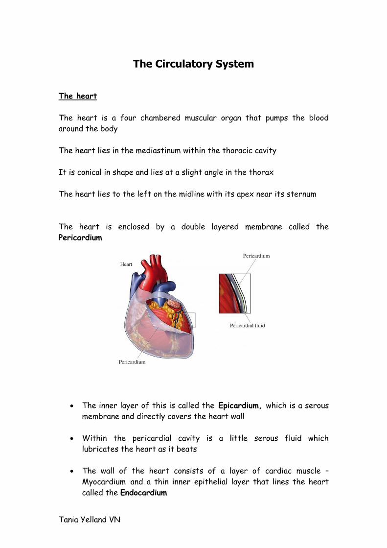
The heart is enclosed by a double layered membrane called the
Pericardium
The inner layer of this is called the Epicardium, which is a serous
membrane and directly covers the heart wall
Within the pericardial cavity is a little serous fluid which
lubricates the heart as it beats
The wall of the heart consists of a layer of cardiac muscle –
Myocardium and a thin inner epithelial layer that lines the heart
called the Endocardium
Tania Yelland VN
The heart is divided up into right and left chambers by a partition
called the Septum
Each side is then divided up into two chambers
They are:
The thin walled collecting chambers = Atrium
The thicker walled pumping chambers = Ventricles
The right side of the heart pumps blood from the heart to the lungs
and back
The left side of the heart pumps blood all around the body
Heart valves
Within the heart there are two sets of valves, the function of these
valves is to prevent backflow of blood
They are:
1. Atrioventricular valves
2. Semi lunar valves
Atrioventricular valves
These lie between the atria and ventricles
They prevent the flow of blood back into the atria when the
ventricles contract
They are attached to the papillary muscles in the heart wall by
fibres called Chordea tendinae these prevent the valves being
turned inside out by the pressure of the blood
The right atrioventricular valve lies between the right atrium and
right ventricle
Tania Yelland VN
It comprises of three fibrous cusps and is also known as tricuspid
valve
The left atrioventricular valve lies between the left atrium and
left ventricle
It comprises of two cusps and is also known as the bicuspid valve
Semilunar valve
These are situated at the base of the two major vessels leaving the heart
The Pulmonary Valve lies at the base of the pulmonary artery and
prevents backflow of blood from the pulmonary artery to the right
ventricle
The Aortic Valve lies at the base of the aorta and prevents
backflow of blood from the aorta to the left ventricle
It is the semi lunar valve closing that can be heard in the second heart
sound (dubb) if the valve is faulty it doesn’t close properly causing
turbulence. This is now heard as a whoosh instead of a lubb or dub this is
known as a Heart murmur
Tania Yelland VN
Circulation of blood through the heart
Deoxygenated blood is carried back to the heart in Veins
Two major veins collect all the blood and enter the right side of the
heart these are called:
Cranial vena cava
Caudal vena cava
The two atria (left and right) and the two ventricles (left and right)
contract in unison
Both vena cava empty into the Right Atrium which contracts when
it is full
This sends deoxygenated blood into the right ventricle via the
right atrioventricular valve
The right ventricle contracts, pumping blood into the pulmonary
artery via the pulmonary valve
The deoxygenated blood is carried in the pulmonary circulation to
the Lungs
The blood is oxygenated in the lungs and is then carried back to
the left side of the heart in the pulmonary veins which enter the
Left Atrium
When the left atrium is full of blood it contracts forcing
oxygenated blood into the left ventricle via the left
atrioventricular valve
The left ventricle contracts and pumps blood into the Aorta (by
the aortic valve) which then carries the oxygenated blood around
the body.
Oxygenated blood is delivered to the tissues; the deoxygenated blood is
collected up by the veins and transported back to the heart
Tania Yelland VN
Control of the heart beat
The heart is made of cardiac muscle which is a specialised type of muscle
that has the ability to initiate contraction from within the muscle itself
(without nervous control
The mechanism that is responsible for controlling the rate of contraction
of heart muscle is called the conduction mechanism
Within the wall of the right atrium is an area of modified cardiac muscle
called the Sino atrial node
This nodes job is to determine the rate of the heart beat and is referred
to as the Pacemaker
If the muscles have been working hard and require more oxygen the
sinoatrial node will increase the heart rate so that the oxygen can get to
those muscles
During sleep the sinoatrial node slows the heart rate down as the muscles
are relaxed
The sinoatrial node starts a wave of contraction, which passes
over the walls of the atria
The impulse passes to another specialised area called the Bundle of
his which is situated within the septum
The impulse is then conducted to the apex of the heart where it
spreads out into the ventricles in specialised nerve cells called
Purkinje Fibres
The wave of contraction in the heart muscle (myocardium) of the
ventricles starts at the apex and spreads upwards forcing blood
into the arteries that are situated at the top of the ventricles.
This is called the cardiac cycle
The period of contraction within the heart is called Systole and is
when the blood is being pumped into the ventricles or circulation
Tania Yelland VN
The period of relaxation of the heart is called Diastole and is when
the atria are filling with blood
The circulatory system is made up of a network of channels that
transport the blood from the heart to the tissues, where oxygen and
nutrients are delivered, and then transport it back again to the heart.
This network consists of arteries, capillaries and veins
Arteries
A large vessel which carries blood under pressure away from the
heart
Most arteries carry oxygenated blood except for the pulmonary
artery which carries deoxygenated blood
Arteries have thick muscular walls which enables the vessel to
dilate or constrict
Blood travels along the arteries reflecting the heartbeat which can
be felt as the pulse
As the arteries enter the tissues they branch getting smaller and
smaller these are then called Arterioles which then flow into
capillary networks
Capillaries
The capillaries form a branching network in all the tissues and link
the arteries and the veins
They are narrow with thin walled consisting of a single layer of
endothelial cells with no muscle or elastic tissue
They are permeable to gases, nutrients and waste products which
diffuse between the blood and tissues and from the tissues back
into the blood
Tania Yelland VN
Veins
A vein is a large vessel which carries blood towards the heart
The walls are thinner than arteries and they contain less muscle
and elastic tissue
Blood flows slowly under low pressure and valves may be present to
prevent pooling of the blood
Most veins carry deoxygenated blood from the tissues except for
the pulmonary veins which carry oxygenated blood from the lungs
back to the heart
Systemic and pulmonary circulation
The circulation in the mammal is referred to as a Double circulation as
blood passes through the heart twice during a complete cycle
There are two parts to the circulatory system:
1. Systemic circulation – carries oxygenated blood around the body
and returns deoxygenated blood to the heart
2. Pulmonary circulation - Carries deoxygenated blood from the
heart to the lungs where it is oxygenated and returned to the
heart
Systemic circulation
Arterial supply
Oxygenated blood leaves the left ventricle of the heart in the
major artery known as the aorta
The aorta gives off a number of arteries that then supply various
parts of the body
Tania Yelland VN
Venous return
Deoxygenated blood returns to the heart from the tissues in veins
which follow the pattern of the arteries and often have the same
name (renal artery and renal vein)
The caudal vena cave empties into the right atrium of the heart, venous
blood returns:
From the head in the Jugular vein
From the neck and forelimbs in the cephalic veins, brachial veins
and subclavian veins which drains into the cranial vena cava which
empties into the right atrium of the heart
Hepatic portal system
The liver has its own modified circulatory system within the
systemic circulation
Its function is to carry blood straight from the digestive system
to the liver so that the products of digestion can be used
immediately rather than transporting them around the body
Pulmonary system
Deoxygenated blood is pumped from the right ventricle of the
heart and is carried to the lungs in the pulmonary artery
Within the lung tissue the artery then divides into lots of
capillaries that wrap around the thin walled alveoli of the lungs
Oxygen in the inspired air diffuses into the blood and carbon
dioxide in the blood diffuses into the air in the alveoli
Tania Yelland VN
Exotic animals
Small mammals
The cardiovascular system of small mammals is similar to cats and
dogs except for the ferret which only has one vessel arising from
the aorta
Birds
The avian heart is four chambered as with mammals but is
proportionally larger with respect to its body size
The major artery (aorta) curves to the right side as it leaves the
heart rather than to the left in mammals
There are several other differences in the blood vessels in birds
Reptiles
The heart is generally considered to be three chambers in lizards,
snakes and chelonians as there is no physical division between the
right and left ventricle
They function as a four chambered heart with deoxygenated blood
being directed towards the lungs and oxygenated blood to the rest
of the body
Lizards, snakes and chelonians have paired aorta leaving the heart
one going to the right one to the left. It then fuses dorsally to
become one aorta
Fish
The heart has only one atrium and one ventricle.
Blood returns to the heart via two blood vessels the common
cardinal vein which receives blood from the head and body and the
hepatic veins
Tania Yelland VN
These enter into the a small chamber which then empties into the
ventricle
The ventricles then enter into a vessel like the aorta which divides
into brachial arteries and supplies the gills with blood
These then move on to the dorsal aorta which moves caudally to
supply the rest of the body with oxygenated blood
Tania Yelland VN
Circulatory Disorders
The primary function of the cardiovascular system is to supply
adequate blood flow to meet the body’s metabolic demands.
Cardiac output is tightly controlled which maintains blood pressure
in normal circumstances.
Cardiac output depends on the heart rate and the amount of blood
ejected from the heart with each contraction
Common Circulatory disorders
Congestive heart failure
Acute heart failure
Congenital heart diseases to include, Patent ductus arteriosis,
pulmonic stenosis, Aortic stenosis, ventricular septal defects
Acquired heart disease to include, Dilated cardiomyopathy,
hypertrophic cardiomyopathy, endocardiosis, pericardial effusion
Vascular disease – hypertension
Dysrhythmias to include; atrial fibrillation, ventricular premature
contractions, heart block
Questions to ask an a owner
Has the animal previously had signs of heart disease?
Heart disease often goes undetected for many years before
clinical signs develop
Is the animal able to exercise normally?
Heart disease will often cause exercise intolerance or collapse at
exercise
Tania Yelland VN
Is the animal eating and drinking normally?
Patients in heart failure often go off their food and are cachexic
(loss of weight, fatigue, general poor condition)
In early stages they may become polydipsic
Have there been episodes of weakness or collapse?
This could be a sign of poor circulation (reduced cardiac output)
Does the animal seem unsettled at night?
Nocturnal restlessness is common in congestive heart failure, they
find it difficult to breathe when lying down
Are there any breathing issues or is the animal coughing?
Respiratory issues may be associated with pulmonary oedema
Coughing is more commonly a sign of respiratory disease but could
be caused due to increased pressure of a large heart on the
bronchus
Tania Yelland VN
The most common type of heart failure is congestive heart failure. This
can affect the left or right sides of the heart but usually progresses to
involve both sides
Right sided heart failure results in congestion of the venous
circulation in the liver and spleen which causes ascites
Left sided CHF causes congestion of vessels in the lungs and fluid
leaking from here causes pulmonary oedema. Oedema and
congestion reduce oxygen transfer in the lungs
Causes of CHF include:
Pooling in the venous system due to:
Systolic failure (reduced efficiency if the myocardium to contract
Diastolic failure (reduced ability of the myocardium to relax and
fill with blood)
Volume overload, excess volumes of blood entering the chamber
causing myocardial stretching
Pressure overload where resistance to the outflow of blood which
increases the force of contraction needed and muscle
hyperatrophy increases pressure in the ventricle
Dysrhythmias which reduce efficiency of the pumping action
Signs of CHF include:
Exercise intolerance
Cough
Dyspnoea
Syncope/collapse
Weight loss
Pale mucous membrane (due to poor supply to the peripheral
circulation)
Ascites
Restlessness
Tania Yelland VN
Diagnostic testing:
Auscultation of the heart for the presence of murmurs
Increased heart rate and loss of sinus dysrhythmia
Compare heart and pulse rate
Pulse quality and strength
MM colour – Cyanotic with pulmonary oedema or pale if output is
reduced
Thoracic radiographs to assess size and shape of the heart and
presence of pulmonary oedema or pleural fluid
Ultrasonography for internal cardiac anatomy – valves, septa,
vessels and myocardium
ECG provides information about cardiac chamber size but also for
detection, recognition, classification and monitoring dysrhythmias
Treatment options:
Early heart disease (no signs but a murmur is detected) require the
underlying cause to be diagnosed and corrective treatment started
If corrective treatment is not possible it is likely that CHF will
progress so the patient should be checked every 3-6 months
A reduced calorie diet should be started to control weight
Exercise restrictions should be imposed, if pulmonary oedema is
present then ACE inhibitors and diuretics should be started
Supportive care such as draining pleural effusions and oxygen
therapy should be started
Tania Yelland VN
Congenital abnormalities
Patent ductus arteriosus (PDA)
The ductus arteriosus is a blood vessel that connects the two main
arteries of the body - the aorta and the pulmonary artery.
This blood vessel is normal in the foetus, but shortly after birth, it
should close.
When the ductus arteriosus remains open or patent after birth,
this abnormal communication between the aorta and pulmonary
artery passes extra volumes of blood into the lungs.
Patent ductus arteriosis (PDA) is a birth defect representing the
second most common congenital heart defect of dogs.
Approximately seven out of 1000 live birth puppies are affected.
Generally, there are no serious symptoms of PDA unless congestive
heart failure has caused fluid build-up in the lungs.
The condition is typically identified in puppies during a routine veterinary
visit for vaccinations. Continual blood flow through the PDA into the lungs
produces a continuous heart murmur.
Even when a PDA has been identified, most people believe their dog is
normal. In some cases, the dog can be smaller than littermates or play
less vigorously. However, the situation can be very misleading as
symptoms usually occur within a year of diagnosis. If untreated, about 60
percent of affected dogs die within a year of diagnosis.
When caught early, and following treatment with successful closure
of the PDA, most dogs live a normal life.
Unless there are complications from other heart defects or heart
failure has already developed, there is rarely any future need for
medication.
While special circumstances can influence the prognosis, most
cases are straightforward.
Tania Yelland VN
What to look for:
Breathing difficulties
Coughing
Exercise intolerance
Lethargy
Diagnosis
Various diagnostic tests are needed to recognize PDA, and exclude other
diseases.
Some of the necessary tests may include:
Complete medical history and physical examination including
auscultation (stethoscope examination) of the heart and lungs. The
heart murmur of PDA is characteristic and most experienced
veterinarians learn to make the diagnosis simply by listening. Since
other birth defects can also cause heart murmurs, a veterinary
cardiologist may be consulted if the diagnosis is in doubt.
A chest X-ray (radiograph) can help determine the severity of the
problem
.
An electrocardiogram (ECG) can assist with the diagnosis.
An echocardiogram with Doppler (cardiac ultrasound) is the
definitive diagnostic test. This may require referral.
Routine blood tests may be performed prior to any anaesthesia
Treatment
The conventional treatment is an operation done shortly after
diagnosis. The PDA is closed with surgical suture.
Surgery should not be delayed by waiting for symptoms to develop.
Medical treatment may be necessary before surgery if symptoms
(coughing, difficult breathing) are present.
Tania Yelland VN
In some referral centres, the PDA may be closed using special
catheterization techniques.
PDA is common in:
Miniature poodle,
collie,
Maltese
Shetland sheepdog,
German shepherd dog,
cocker spaniel,
Pomeranian, and
Labrador retriever.
Female dogs are predisposed.
Pulmonic stenosis
Cause:
A congenital narrowing of pulmonary valve or the artery leaving the
heart
Signs:
A murmur is often detected at routine examination, this can then
progress to right sided heart failure
Diagnosis
Thoracic radiographs show enlargement of the right side of the
heart
Ultrasound examination shows thickening of the right heart wall
Doppler ultrasound shows narrowing of the vessel which can
identify the severity of stenosis
Treatment
Severe cases require dilation of the artery. Symptomatic medical
treatment in non surgical cases
Tania Yelland VN
Aortic Stenosis
Cause:
A narrowing of the outflow of the left side of the heart (or
abnormal development of the aortic valve). This causes resistance
to outflow resulting in hypertrophy of the left side of the heart
Congenital aortic stenosis is probably the most common heart
defect seen in large breed dogs. Newfoundland dogs have the
highest risk for this disorder. It is also seen in the golden
retriever, Rottweiler, and boxer.
There is a mildly increased risk in:
the German shepherd,
German short-haired pointer,
Great Dane,
Samoyed
bulldog.
Signs
Depending on the severity signs may be present at a young age or
develop into adulthood
Fainting
Collapse
Sudden death
Diagnosis
Thoracic radiographs may show dilation of aorta after the point of
stenosis
Ultrasound can be used to visualise the stenotic area
Doppler to measure blood flow through the area
ECG examination shows cardiac arrythmias
Treatment
Symptomatic treatment with anti-dysrhythmic drugs and beta
blockers
Mild cases don’t require treatment
Tania Yelland VN
Venticular septal defect (hole in the heart)
A failure of the heart to develop properly which results in an
opening in the division between the left and right sides of the
heart.
Blood flows from left to right meaning that too much blood is
returned and the left side is overloaded.
VSD is a congenital disease in dogs and affected dogs should not be
used for breeding. When a dog is diagnosed with ventricular septal
defect, its parents should also not be used for breeding any longer.
As a dog embryo develops, the heart begins as a single tube. This tube
will then gradually separate into four different chambers. Abnormalities
can appear at several different steps in this intricate process and this
can lead to ventricular septal defect in a dog
Symptoms
The nature and extent of the symptoms will depend on both the
size of the abnormality and exactly where it is located. In mild
cases of ventricular septal defect, most dogs will display no
symptoms except heart murmur.
It is even possible for a small defect to close on its own as the dog
matures.
In a dog with a larger defect, the pressure in the left side of the
heart will be higher than the pressure in the right side, and there
will be a blood flow from left to right via the defect.
The left side of the dog’s heart will be forced to work harder than
normally and more blood will circulate to the lungs. This will in turn
cause a higher than normal work load for the lungs.
The symptoms can develop over the course of several months or
even years.
Common Symptoms
exercise intolerance
Increased respiratory effort
In severe cases, abnormal heart rhythm can lead to premature
death.
Tania Yelland VN
Dilated cardiomyopathy
Dilated cardiomyopathy (DCM) is a disease affecting the heart
muscle. It is the second most common heart disease in dogs (after
mitral valve disease).
In DCM the myocardium is thinned resulting in loss of
contractibility
It affects:
Mainly middle-aged large and giant breed dogs
some spaniels
Dobermans are the main breed affected.
Cats and small breeds
Male dogs are more likely to be affected than females.
What causes DCM?
Cardiomyopathy literally means disease of the heart muscle (cardio
= heart and myopathy = muscle disease).
In DCM the heart muscle becomes thin and weakened. The heart muscle
can be damaged in a number of ways including:
viral infections
Dietary deficiencies of taurine (an essential amino acid only found
in meat protein)
Dietary deficiencies in carnitine have been reported as causes of
DCM in some groups of dogs.
In most cases of DCM there is no apparent cause of the damage to the
heart and this is termed idiopathic cardiomyopathy.
The heart muscle is damaged it becomes weak and so does not
contract well. Because heart contractions are weak the heart does
not empty with each contraction and the blood supply to the body
is reduced.
Pulses are weak and the paws may feel cold. With time the heart
muscle stretches and heart becomes a flabby sac.
Tania Yelland VN
DCM eventually results in heart failure with fluid build-up in the
lungs (pulmonary oedema), the chest (pleural effusion) and abdomen
(ascites).
What are the signs of DCM:
an irregular heart beat on a routine examination
exercise intolerance
Increased respiratory effort
Anorexia
Depression
Polydipsia
Coughing
Collapse.
Cardiac cachexia.
ascites
Sudden death
How do you diagnose DCM?
Thoracic radiographs show increased cardiac size
Pulmonary oedema
Ultrasound shows thin myocardium and poor contractibility
ECG can show dysrhythmias particularly atrial fibrillation
Treatment
In almost all cases there is no treatment for the underlying muscle
disease.
Signs of heart failure can be managed according to its severity.
Taurine supplementation in some cases
Tania Yelland VN
Endocardiosis
Endocardiosis is the most common acquired disease in dogs
particularly in Cavalier King Charles Spaniels
Causes:
Degenerative condition of the atrioventricular valves (usually the
mitral valve) which causes faulty valve function and the leakage of
blood through valves when they are closed
Signs
Cardiac murmur progressing over years to CHF (usually left sided)
Diagnosis
Thoracic radiographs shows left sided heart enlargement (atrium)
Ultrasound shows distortion of valves and turbulent blood flow with
leakage
Treatment
Symptomatic management of CHF
Tania Yelland VN
Pericardial effusion
Often seen in middle sized and large breed dogs.
Fluid accumulates inside the pericardial sac which prevents the
heart from filling with blood.
Signs
Lethargy
Dyspnoea
Muffled heart sounds
Weak pulses
Pale mucous membranes
Muffled heart sounds
Ascites
Treatment
Drainage of the fluid (pericardiocentesis)
Pericardectomy
Tania Yelland VN
Drugs commonly used in the treatment of cardiac conditions
Positive inotropes e.g. digoxin
– Increase the force of contraction of the heart muscle
– Slow the heart rate
– Can cause nausea at high doses
Local anaesthetics
Given by I/V injection to treat some dysrhythmias
They reduce the sensitivity of the heart muscle so that abnormal
contractions are reduced
Only used for short term control of severe dysrhythmias
Diuretics
Promote renal excretion of fluids and reduce oedema
Inhibits the resorption of salt and water filtered by the kidneys so
that more fluid is lost in urine
Vasodilators
Reduce the workload of the heart by:
Dilating systemic veins which reduces pressure making oedema less
likely to form
Dilating arterioles making it easier for the heart to pump blood
Betablockers
Slow heart rate
Reduce the force of contraction so not used in DCM
Tania Yelland VN
Nursing the patient with congestive heart failure:
Choose a suitable kennel which can be lined with absorbent
material. This should be in a calm quiet area
Monitor and record TPR, urinary and faecal output, abnormalities
etc
A low salt/fat diet, which has a protein of a high biological value
and is highly digestible. This will reduce oedema and ascites
Water should be available at all times
Medication should be correctly administered
Exercise should be kept to a minimum