Embed Size (px)
Citation preview
The child with respiratory dysfunction
By: Murad Sawalha RN, MSN Basel Abdul-Qader RN, MSN
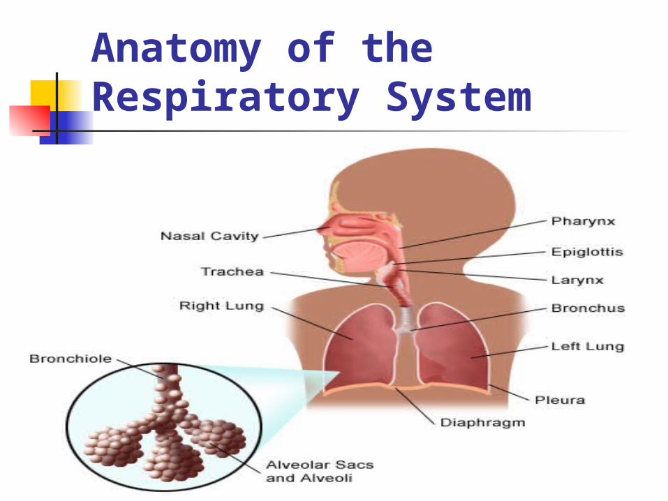
Anatomy of the Respiratory System
Anatomy of the Respiratory System (cont.)
What is respiration? Respiration is the act of breathing: inhaling (inspiration) - taking in oxygen. exhaling (expiration) - giving off carbon dioxide.
What makes up the respiratory system?
The respiratory system is made up of the organs involved in the interchanges of gases, and consists of the:
- nose - pharynx - larynx - trachea - bronchi - lungs
Anatomy of the Respiratory System (cont.)
The upper respiratory tract includes the following:
nose nasal cavity Sinuses: ethmoid, frontal, maxillary, sphenoid larynx trachea
The lower respiratory tract includes the following:
lungs airways (bronchi and bronchioles) air sacs (alveoli)
Anatomy of the Respiratory System (cont.)
What is the function of the lungs? The lungs take in oxygen, which cells need to
live and carry out their normal functions. The lungs also get rid of carbon dioxide, a waste product of the body's cells.
The lungs are enveloped in a membrane called the pleura.
The right lung has three sections, called lobes. The left lung has two lobes.
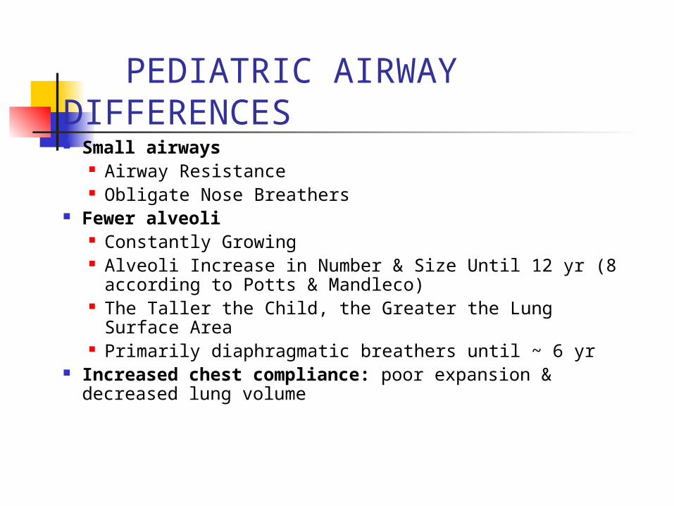
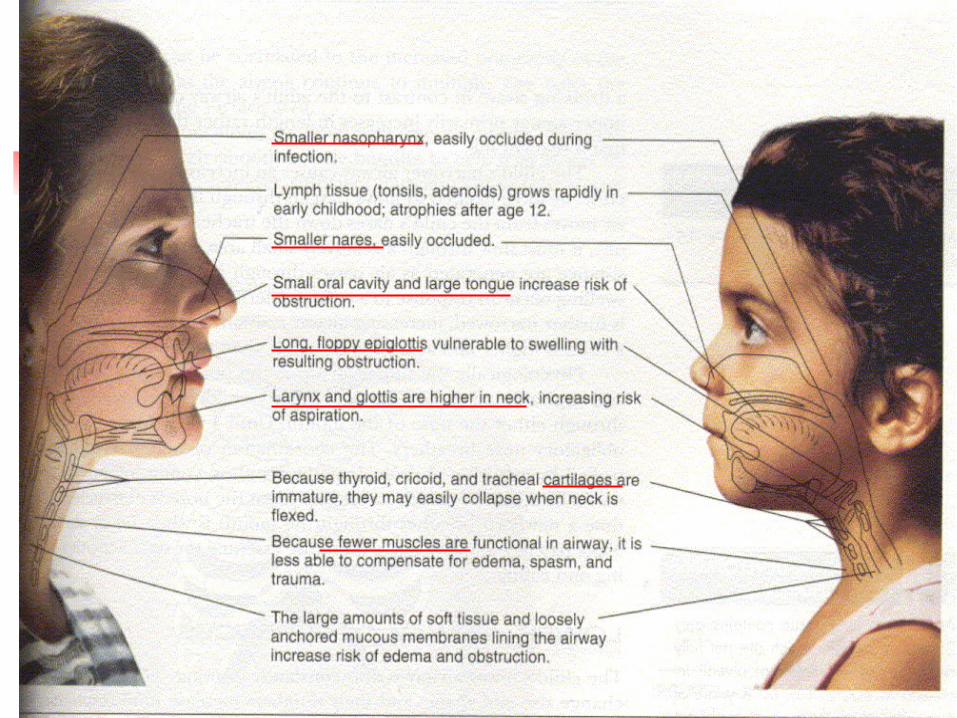
PEDIATRIC AIRWAY DIFFERENCES Small airways
Airway Resistance Obligate Nose Breathers
Fewer alveoli Constantly Growing Alveoli Increase in Number & Size Until 12 yr (8
according to Potts & Mandleco) The Taller the Child, the Greater the Lung Surface Area Primarily diaphragmatic breathers until ~ 6 yr
Increased chest compliance: poor expansion & decreased lung volume
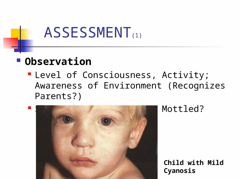
ASSESSMENT(1)
Observation Level of Consciousness, Activity;
Awareness of Environment (Recognizes Parents?)
Skin Color: Pink, Pale, Mottled?
Child with Mild Cyanosis
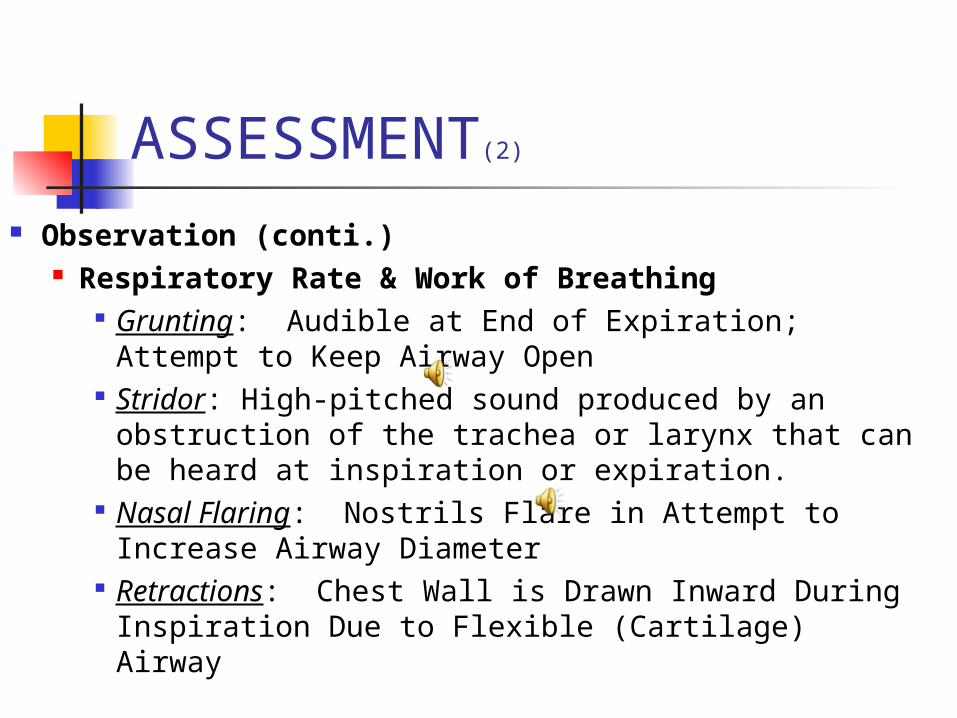
ASSESSMENT(2)
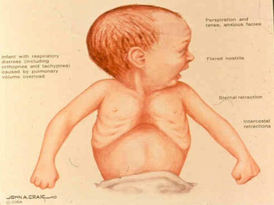
Observation (conti.) Respiratory Rate & Work of Breathing
Grunting: Audible at End of Expiration; Attempt to Keep Airway Open
Stridor: High-pitched sound produced by an obstruction of the trachea or larynx that can be heard at inspiration or expiration.
Nasal Flaring: Nostrils Flare in Attempt to Increase Airway Diameter
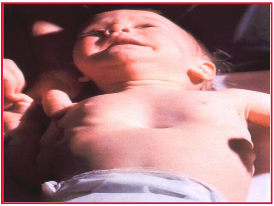
Retractions: Chest Wall is Drawn Inward During Inspiration Due to Flexible (Cartilage) Airway
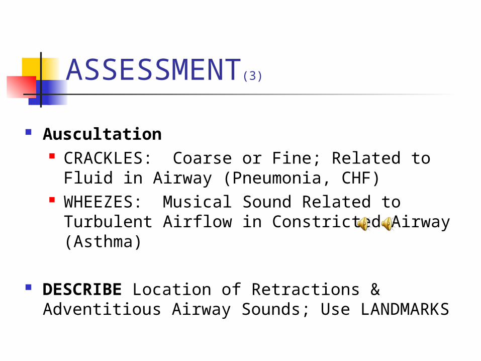
ASSESSMENT(3)
Auscultation CRACKLES: Coarse or Fine; Related to Fluid in
Airway (Pneumonia, CHF) WHEEZES: Musical Sound Related to
Turbulent Airflow in Constricted Airway (Asthma)
DESCRIBE Location of Retractions & Adventitious Airway Sounds; Use LANDMARKS
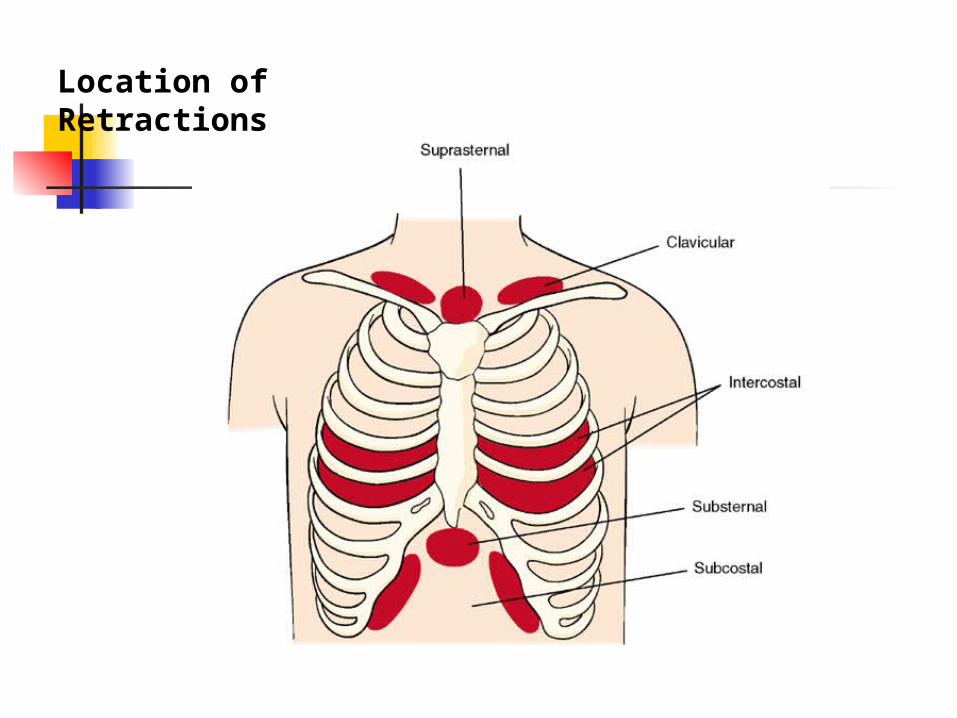
Location of Retractions
UPPER RESPIRATORY TRACT INFECTION
Nasopharyngitis Nasopharyngitis: common cold .
Causes: rhinovirus, adenovirus, influenza virus, Resp. syncytial virus (RSV), Para influenza virus.
Clinical manifestations:- Younger child :fever, irritability, restlessness,
sneezing, vomiting, diarrhea.- Older child: dryness, irritation of nose, & Throat,
cough, sneezing , chilly sensation, muscular aches.- Physical signs: edema& vasodilatation of mucosa.
Nasopharyngitis (cont.) Therapeutic management:- Mostly treated at home , no vaccine, antipyretics for
fever.- Decongestants: nose drops more effective than orally.- Cough: suppressant 22% with alcohol but not for young
child.- Antihistamine are ineffective.- Antibiotic: usually not indication. Nursing consideration: For nasal obstruction: elevate head of bed, suctioning
and vaporization, saline nasal drops. Maintain adequate fluid intake to prevent dehydration. Avoiding spread the virus.
Pharyngitis Causes : 80-90%of cases are viral cause , other is
group A and B hemolytic streptococci (GABHS).
Clinical manifestation:- May be mild so no symptoms.- Headache, fever, abdominal pain exudates on
pharynx& tonsils, 3-5 days usually symptoms are subside
Complication if not treated :- Acute glumerulonephritis syndrome in about 10 days.- Acute Rheumatic Fever (ARF) in an average 18 days
those complication if the cause is GABHS.
Pharyngitis (cont.) Diagnostic evaluation: throat culture should be
performed to rule out: GABHS
Therapeutic management: - If streptococcal sore throat infection: oral Penicillin for
10 days ,or IM Benzathine penicillin G.- Oral Erythromycin if the child has allergy to penicillin.
Nursing consideration:- Obtain throat swab for culture.- Administer penicillin & analgesic.- Cold or warm compresses to the neck may provide
relief.- Warm saline gargles.
Pharyngitis (cont.) Nursing consideration (cont.)- Soft liquid food are more acceptable than solid.
- Continue oral medication to complete the course.
- IM injection applied in deep muscle as vastus lateralis or ventrogluteal muscle, use Emla cream before IM around 2 hours.
- Nurse role to prevent the spread of disease.
- Children are considered non infectious to other 24 hours after initiation of antibiotics therapy.
Tonsillitis Tonsils are masses of lymphoid tissue, first immune
defense. Tonsillitis often occur with pharyngitis, viral or
bacterial causes. Common cause of morbidity in young children
S& S: - enlarge tonsils, difficult breathing & swallow.- Enlargement of adenoid, blocked postnasal space
&mouth breathing.
Tonsillitis Therapeutic management:- throat culture to determine the causative
agent ,viral or bacterial as GABHS.- Tonsillectomy & adenoidectomy (T&S) or (Ts &As).- Contraindicating for Ts &As: cleft palate, tonsillitis,
blood disorder.
Nursing consideration: Providing comfort & maintain minimize activities. A soft or liquid diet is prescribed. Warm salt water gargles Analgesic, antipyretic.
Tonsillitis
Post operative care: Position (place child on abdomen or side). Discourage child from coughing frequency. Some secretion are common as dried
blood. Crushed ice& ice water to relief pain. Analgesic may be rectally or IV, avoid oral
route. Avoid red or brown fluid, and citrus juice.
Tonsillitis Post operative care (cont.): Soft food, milk or ice cream not offered.
Check post operative signs of Hemorrhage: - Increase pulse more than 120b/min.- Pallor. - Frequent swallowing. - Vomiting of bright blood- Decrease blood pressure is late sign of shock.** Note: use good light to look direct on site of
operation.
Otitis Media:OM OM is inflammation of middle ear.
Episode of acute OM occur in the first 24 month, decrease at 5 years, r/to drainage through the Eustachian tube & inflammatory of Resp. system.
Etiology: - Acute (AOM): streptococcus, Haemophilus influenza,
moraxella catarrhlis, are the most common bacteria.- OM: blocked Eustachian tube from edema of URTI ,
allergic hypertrophy adenoid.- Chronic (COM): extension of AOM.
Otitis Media:OM (cont.) Diagnostic evaluation: assessment of
tympanic membrane with otoscope:- AOM: purulent discolored effusion, bulging
S&S: otalgia (earache), fever, purulent discharge, infant rolls his head from side to side, loss of appetite, crying or verbalized feeling of discomfort (older child).
COM: hearing loss, feeling of fullness, vertigo, tinnitus.
Otitis Media:OM (cont.) Therapeutic management:- Antibiotic for 10-14 days e.g. Amoxicillin.- Myringotomy: surgical incision of eardrum& grommets.- Hear test after 3 month of AOM.
Nursing consideration: Relieving pain. analgesic drug +ice bag on ear. Facilitate drainage & topical A.Biotics. Preventing complication. Instruct family to be careful when deal with child. With
temporary hearing loss. Preventing OM during infant feeding and setting after
that.
Lower respiratory tract infections
Infection of the Lower Air ways
Cartilaginous support of the air ways is not fully developed until adolescence, consequently the smooth muscle in these structures represents a major factor in the constriction of the airway.
Bronchitis Bronchitis or tracheobronchitis is inflammation of
larger air way (trachea and bronchi).
Causative agents: viruses or mycoplasma pneumonia.
Ch-ch & symptoms: dry, nonproductive cough that worsens at night then become productive in 2-3 days.
Bronchitis is a mild disease required symptomatic treatment as antipyretic, analgesic and humidity, cough suppressants may be useful at night.
Bronchiolitis & Resp. Syncytial Virus RSV
Bronchiolitis: is an acute viral infection with maximum effect at the bronchiolar level, and rare in children older of 2 years.
One of the Most Frequent Cause of Hospitalization in Infants
Virus or Bacteria Causes Inflammatory Response & Obstruction of Small Airways From Edema
RSV is responsible of 80% of cases during epidemic periods.
Bronchiolitis & Resp. Syncytial Virus RSV (cont.)
Transmission (It is easily spread): direct contact (from hand to eye, nose or other mucous membranes); virus can survive for hours on countertop, cloth, gloves; ½ hours on skin
Clinical Manifestations: URI Symptoms: Nasal Stuffiness; Cough; Fever Symptoms Increase & Cough Worsens,
Respirations More Labored, Respirations Rapid, Shallow & With Nasal Flaring & Retractions
Infant Acting Ill, Not Playing, Decreased Eating, & Spits Up Thick Mucous
Bronchiolitis & Resp. Syncytial Virus RSV (cont.)
Respiratory Syncytial Virus (RSV) Invades Mucosal Lining of Lungs, Destroys Cells,
& Causes Inflammatory Response With Increased Mucous Production Which May Cause Airway Obstruction
Airway Swelling & Mucous Obstruction Cause Air Trapping & Further Inflammation
Impaired Gas Exchange Leads to Resp. Failure
RSV antigen detection (ELISA), Viral Culture
Medical Management is Supportive
Bronchiolitis & Resp. Syncytial Virus RSV (cont.)
Medical therapy is controversial: Albuterol & Steroids?? Ribavirin, the only FDA approved antiviral agent
for RSV, Not Use Currently b/c its Side Effects Child Hospitalized if Has O2 Requirement, Cannot
Drink, or Has Apnea & Needs Continuous Monitoring
Preventive Measure for RSV Bronchiolitis Synagis: IM, monthly. During RSV season RespiGam (RSV IGIV, given IV, monthly)
Bronchiolitis & Resp. Syncytial Virus RSV (cont.)
Nursing Management: Handwashing (most important)
Patient needs separate-room (Resp. isolation) by using gloves, gown, mask, and goggles..
Maintain Respiratory Function
Support Physiologic Function
Group care
Discharge Planning & Home Care Teaching
Pneumonia Pneumonia: is inflammation of the pulmonary
parenchyma.
Common in children but more frequently occur in infancy & early childhood.
Types of pneumonia (depend on place): Lobar- Pneumonia: one-lobe or more (bilateral or
double Pneumonia). Broncho Pneumonia: begins in the terminal bronchioles
form consolidated patches in nearly lobules, also called lobular Pneumonia .
Interstitial Pneumonia: inflammatory process is confined within the alveolar walls and peribronchial and interlobular tissues.
Pneumonia (cont.) Morphology classification: viral, bacterial,
mycoplasma , aspiration of foreign body, fungal.
Viral Pneumonia: Occurs more than bacterial. Causes: RSV, parainfluenza, influenza,
adenovirus. Clinical symptoms: fever, cough, abnormal
breath sound; whitish sputum, nasal flaring, retraction, chest pain, pallor to cyanosis, irritable, restless, anorexia, vomiting, diarrhea, abdominal pain.
Pneumonia (cont.) Viral Pneumonia (cont.): Treatment:- symptomatic: O2 therapy, Comfort.- Chest physiotherapy and postural drainage.- Antipyretics, Fluid intake, & Family supports.
Primary atypical pneumonia: It is caused by mycoplasmas, the smallest free-living agents of human disease, which have the characteristics of both bacteria or viruses, but which are not classified as either, most common in children between 5-12 years, mostly in winter months, symptomatic treatments within 7-10 days.
Bacterial Pneumonia Streptococcus Pneumonia is the most common
cause in children and adult
In infant mainly followed viral infection.
Symptoms: fever, malaise, rapid& shallow respiration, cough, chest pain, abdominal pain?? Appendicitis, meningeal symptoms.
Treatment: AB therapy , bed rest, antipyretic, fluid intake, need hospitalization when pleural effusion or empyema, I.V fluid, O2 therapy.
Bacterial Pneumonia (cont.)
Complication:- Tension pneumothorax and empyema if the
causative agent is staphelococcus auoraus, - AOM,PE, lung abscess if pnumococcal pneumonia.
Prognosis: is generally good if recognize the disease early & treat early.
Prevention: pnumococcal poysaetheride vaccine for children older than 2 years who is risk.
Bacterial Pneumonia (cont.)
Nursing consideration: Administer of O2 therapy ,AB, rest,
humidity. Assess Resp. status frequently. I.V fluid intake. Antipyretic. Lying the child on affected side. Suctioning by bulb syringe for infant. Chest physiotherapy & postural drainage. Family support & reassurance.