Embed Size (px)
Citation preview
1
THE CHARACTERIZATION OF LYSINE SPECIFIC DEMETHYLASE 2 (LSD2), A NOVEL
HISTONE DEMETHYASE
By
MICHAEL S. RUTENBERG
A DISSERTATION PRESENTED TO THE GRADUATE SCHOOL
OF THE UNIVERSITY OF FLORIDA IN PARTIAL FULFILLMENT
OF THE REQUIREMENTS FOR THE DEGREE OF
DOCTOR OF PHILOSOPHY
UNIVERSITY OF FLORIDA
2007

2
Michael S. Rutenberg 2007

3
I dedicate this to God. You da man!
4
ACKNOWLEDGMENTS
I thank my mentor, Dr. Naohiro Terada, for his unending support and enthusiasm during my
research education. I also thank Dr. Yujiang Shi at Brigham and Women’s Hospital/Harvard
Medical School for allowing me the opportunity to work with him and conduct my dissertation
research in his lab. I thank the members of the Shi lab for assisting with the research contained
within this dissertation, most notably Drs. Rui Fang, Pinchou Mei, and Fumiko Shimizu, as well
as Thiago Leonor.
5
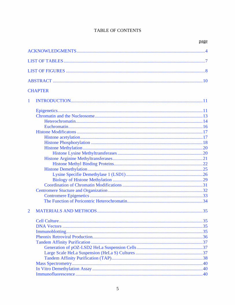
TABLE OF CONTENTS
page
ACKNOWLEDGMENTS...........................................................................................................4
LIST OF TABLES......................................................................................................................7
LIST OF FIGURES ....................................................................................................................8
ABSTRACT .............................................................................................................................10
CHAPTER
1 INTRODUCTION..............................................................................................................11
Epigenetics.........................................................................................................................11
Chromatin and the Nucleosome..........................................................................................13
Heterochromatin..........................................................................................................14
Euchromatin................................................................................................................16
Histone Modificatons .........................................................................................................17
Histone acetylation......................................................................................................17
Histone Phosphorylation .............................................................................................18
Histone Methylation....................................................................................................20
Histone Lysine Methyltransferases .......................................................................20
Histone Arginine Methyltransferases...........................................................................21
Histone Methyl Binding Proteins..........................................................................22
Histone Demethylation................................................................................................25
Lysine Specifie Demethylase 1 (LSD1) ................................................................26
Biology of Histone Methylation ...........................................................................29
Coordination of Chromatin Modifications ...................................................................31
Centromere Stucture and Organization ...............................................................................32
Contromere Epigenetics ..............................................................................................33
The Function of Pericentric Heterochromatin...............................................................34
2 MATERIALS AND METHODS........................................................................................35
Cell Culture........................................................................................................................35
DNA Vectors .....................................................................................................................35
Immunoblotting..................................................................................................................35
Pheonix Retroviral Production............................................................................................36
Tandem Affinity Purification .............................................................................................37
Generation of pOZ-LSD2 HeLa Suspension Cells .......................................................37
Large Scale HeLa Suspension (HeLa S) Cultures ........................................................37
Tandem Affinity Purification (TAP)............................................................................38
Mass Spectrometry.............................................................................................................40
In Vitro Demethylation Assay ............................................................................................40
Immunofluorescence ..........................................................................................................40
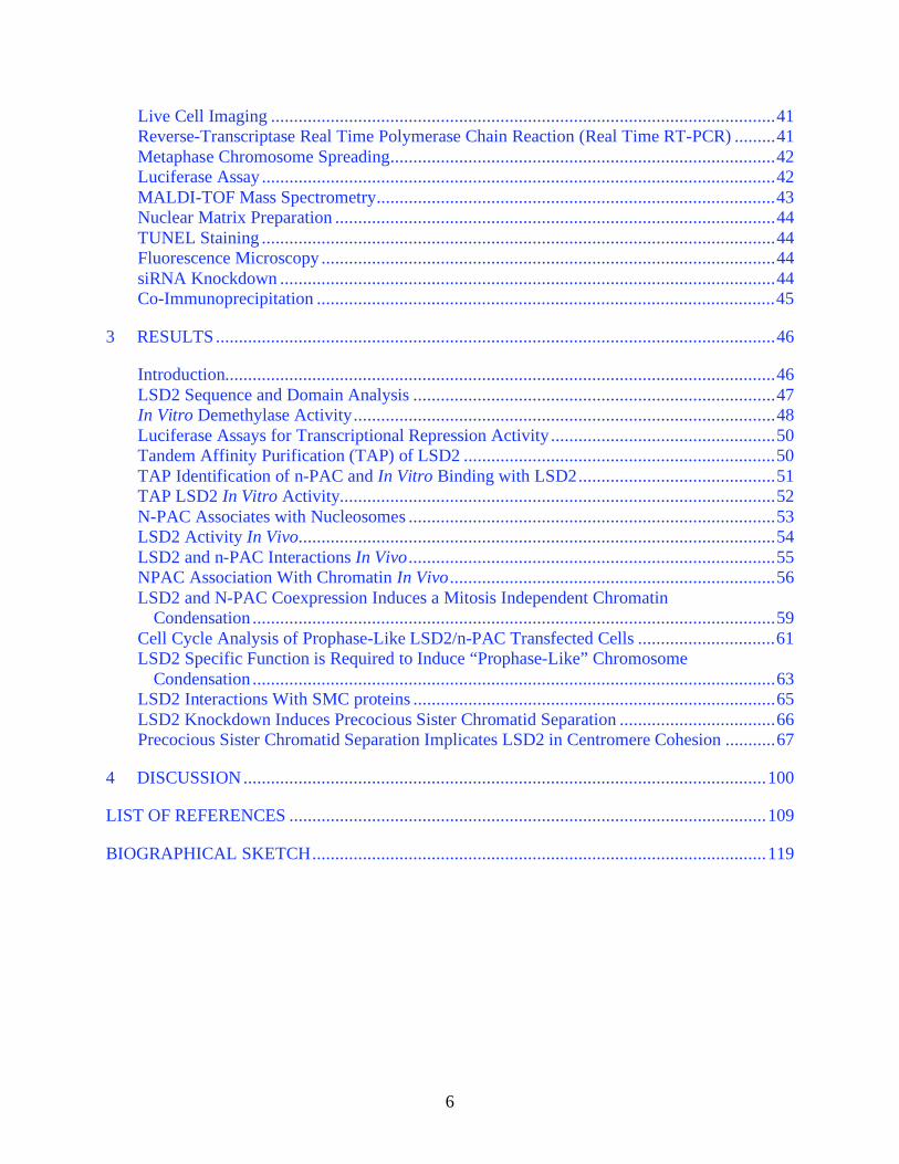
6
Live Cell Imaging ..............................................................................................................41
Reverse-Transcriptase Real Time Polymerase Chain Reaction (Real Time RT-PCR) .........41
Metaphase Chromosome Spreading....................................................................................42
Luciferase Assay ................................................................................................................42
MALDI-TOF Mass Spectrometry.......................................................................................43
Nuclear Matrix Preparation ................................................................................................44
TUNEL Staining ................................................................................................................44
Fluorescence Microscopy ...................................................................................................44
siRNA Knockdown ............................................................................................................44
Co-Immunoprecipitation ....................................................................................................45
3 RESULTS..........................................................................................................................46
Introduction........................................................................................................................46
LSD2 Sequence and Domain Analysis ...............................................................................47
In Vitro Demethylase Activity............................................................................................48
Luciferase Assays for Transcriptional Repression Activity .................................................50
Tandem Affinity Purification (TAP) of LSD2 ....................................................................50
TAP Identification of n-PAC and In Vitro Binding with LSD2...........................................51
TAP LSD2 In Vitro Activity...............................................................................................52
N-PAC Associates with Nucleosomes ................................................................................53
LSD2 Activity In Vivo........................................................................................................54
LSD2 and n-PAC Interactions In Vivo ................................................................................55
NPAC Association With Chromatin In Vivo .......................................................................56
LSD2 and N-PAC Coexpression Induces a Mitosis Independent Chromatin
Condensation ..................................................................................................................59
Cell Cycle Analysis of Prophase-Like LSD2/n-PAC Transfected Cells ..............................61
LSD2 Specific Function is Required to Induce “Prophase-Like” Chromosome
Condensation ..................................................................................................................63
LSD2 Interactions With SMC proteins ...............................................................................65
LSD2 Knockdown Induces Precocious Sister Chromatid Separation ..................................66
Precocious Sister Chromatid Separation Implicates LSD2 in Centromere Cohesion ...........67
4 DISCUSSION..................................................................................................................100
LIST OF REFERENCES ........................................................................................................109
BIOGRAPHICAL SKETCH...................................................................................................119
7
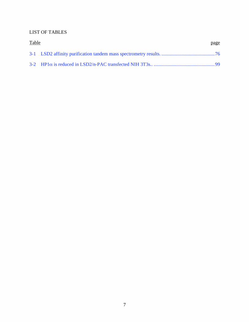
LIST OF TABLES
Table page
3-1 LSD2 affinity purification tandem mass spectrometry results. ..........................................76
3-2 HP1 is reduced in LSD2/n-PAC transfected NIH 3T3s.. ................................................99
8
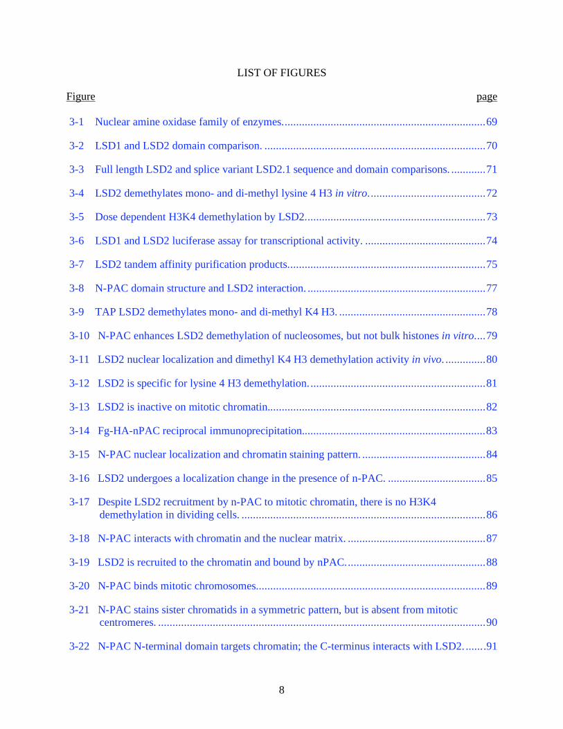
LIST OF FIGURES
Figure page
3-1 Nuclear amine oxidase family of enzymes.......................................................................69
3-2 LSD1 and LSD2 domain comparison. .............................................................................70
3-3 Full length LSD2 and splice variant LSD2.1 sequence and domain comparisons. ............71
3-4 LSD2 demethylates mono- and di-methyl lysine 4 H3 in vitro.........................................72
3-5 Dose dependent H3K4 demethylation by LSD2...............................................................73
3-6 LSD1 and LSD2 luciferase assay for transcriptional activity. ..........................................74
3-7 LSD2 tandem affinity purification products.....................................................................75
3-8 N-PAC domain structure and LSD2 interaction. ..............................................................77
3-9 TAP LSD2 demethylates mono- and di-methyl K4 H3. ...................................................78
3-10 N-PAC enhances LSD2 demethylation of nucleosomes, but not bulk histones in vitro....79
3-11 LSD2 nuclear localization and dimethyl K4 H3 demethylation activity in vivo. ..............80
3-12 LSD2 is specific for lysine 4 H3 demethylation..............................................................81
3-13 LSD2 is inactive on mitotic chromatin............................................................................82
3-14 Fg-HA-nPAC reciprocal immunoprecipitation................................................................83
3-15 N-PAC nuclear localization and chromatin staining pattern. ...........................................84
3-16 LSD2 undergoes a localization change in the presence of n-PAC. ..................................85
3-17 Despite LSD2 recruitment by n-PAC to mitotic chromatin, there is no H3K4
demethylation in dividing cells. .....................................................................................86
3-18 N-PAC interacts with chromatin and the nuclear matrix. ................................................87
3-19 LSD2 is recruited to the chromatin and bound by nPAC.................................................88
3-20 N-PAC binds mitotic chromosomes................................................................................89
3-21 N-PAC stains sister chromatids in a symmetric pattern, but is absent from mitotic
centromeres. ..................................................................................................................90
3-22 N-PAC N-terminal domain targets chromatin; the C-terminus interacts with LSD2. .......91
9
3-23 LSD2 cotransfected with n-PAC induces “prophase-like” condensed chromatin.............92
3-24 LSD2 and n-PAC cotransfected cells have “prophase-like” chromatin condensation in
the absence of mitotic markers or apoptosis. .................................................................93
3-25 LSD2 and n-PAC induce a progressive and refractory chromosome condensation. .........94
3-26 LSD2 and n-PAC cotransfected cells undergo chromatin condensation in the absence
of DNA replication. .......................................................................................................95
3-27 LSD2 knockdown induces precocious sister chromatid separation..................................96
3-28 LSD2 and n-PAC are enriched at pericentric heterochromatin in NIH3T3 cells. .............97
3-29 LSD2 and n-PAC cause a global decrease in HP1 . . .....................................................98
10
Abstract of Dissertation Presented to the Graduate School
of the University of Florida in Partial Fulfillment of the
Requirements for the Degree of Doctor of Philosophy
THE CHARACTERIZATION OF LYSINE SPECIFIC DEMETHYLASE 2 (LSD2), A NOVEL
HISTONE DEMETHYLASE
By
Michael S. Rutenberg
December 2007
Chair: Naohiro Terada
Major: Medical Sciences--Molecular Cell Biology
Covalent modification of histones plays an important role in chromatin structure and
genome function. In particular, the role of methylation of Lysine 4 on histone H3 is well
established in gene regulation. We report a novel function of the dynamic regulation of H3K4
demethylation in the maintenance of constitutive heterochromatin. We show that LSD2, a
homologue of LSD1, demethylates mono- and di-methyl K4H3 and is essential for maintaining
the stability of pericentric heterochromatin. We also identify n-PAC, an LSD2 interacting
protein, which recruits LSD2 to chromatin and enhances its demethylase activity in vitro and in
vivo. LSD2 alone associates weakly with chromatin, however, when coexpressed with n-PAC,
the two proteins associate closely with chromatin and induce a global demethylation and
chromatin condensation. The induction of chromatin condensation requires the catalytic activity
of LSD2 and its interaction with n-PAC. We also show that LSD2 knockdown results in
precocious sister chromatid separation during mitosis, a hallmark of pericentric heterochromatin
disruption. These data reveal a novel function for the dynamic regulation of H3K4 methylation
in formation/maintenance of heterochromatin. We present data showing that LSD2, while
sharing substrate specificity with its homologue, LSD1, does not share its role in gene
transcription, but rather functions as a regulator of genomic structure.
11
CHAPTER 1
INTRODUCTION
Epigenetics
The term “epigenetics” was originally used in the 1940’s to describe the unknown and
somewhat mysterious events underlying the transition from genotype to its phenotype. The
evolutionary and developmental biologist, Conrad Waddington, is credited with coining the term.
He defined epigenetics as “the interaction of genes with their environment, which bring the
phenotype into being” and described the “epigenetic landscape” as a metaphor to explain the cell
fate decisions made during cellular and tissue differentiation during development (29).
Waddington described an epigenetic landscape, in which numerous marbles (representing cells)
beginning in valleys and ridges travel via various possible trajectories from an elevated point of
undifferentiation to a stable position of differentiation. At that time, hereditary elements were
known as genes, however, the role of DNA as the heart of genetics had not been established.
Epigenetics sought to describe the mechanisms by which macromolecules, including protein,
RNA, and DNA mediated “gene” function at the cellular, tissue, and organismal level.
Since its original description, the evolution of the concept of epigenetics has been dramatic
and extensive. From H.J. Muller’s investigation of “position effect variegation” in Drosophila in
the 1930’s to Barbara McClintock’s study of transposable elements and variable mutability in
maize and Mary Lyon’s hypothesis of X-chromosome inactivation in female mammals,
epigenetics became the domain of anomolous and unpredictable hereditary phenomena.
However, with major advances in genetics, molecular biology, developmental biology, and
proteomics, many of these epigenetic mysteries began to be solved, and the concept itself, as
well as the burgeoning field of epigenetics, became more narrowly defined. The overarching
theme of contemporary epigenetics is the investigation of heritable phenotypes that can be
12
changed in the absence of changes in the underlying genotype. Holliday recently defined
epigenetics as “the sum of the alterations to the chromatin template that collectively establish and
propogate different patterns of gene expression (transcription) and silencing from the same
genome” (40). More broadly, we can describe epigenetics as the mechanisms underlying the
capacity for cells with identical genotypes to give rise to varied differentiated cells, tissues, and
organs.
As biological advances continue to be made, ever more mysterious and seemingly
impossible phenomena are discovered that fall within the realm of epigenetics. These now
include heritable phenotypes due to prions, the phenomenon of repeat induced point mutation
(RIP), and the phenomenon of wild-type gene reversion observed in the Arabidopsis HOTHEAD
gene. An important observation regarding the development of this field is the apparent
conservation of many forms of epigenetics throughout evolution (e.g. position effect variegation
and genomic imprinting/dosage compensation), suggesting an important role of these phenomena
in the evolution and survival of these organisms
Many of the underlying mechanisms for the epigenetic phenomena studied to date (e.g.
PEV, X-inactivaation, genomic imprinting) appear to lie in the covalent modification of
chromatin. This includes DNA methylation and a variety of covalent modifications added to
histone proteins. There is intense focus on the origin of these “epigenetic” marks, the means of
their faithful duplication from mother to daughter cell, and their mechanism of action in inducing
phenotypic variability in the context of a static genotype. For the remainder of this discussion, I
will largely focus on the details of histone modifications as “epigenetic marks”, with particular
emphasis on histone methylation and demethylation and their roles in genome metabolism.
13
Chromatin and the Nucleosome
The mammalian genome is massive in both size and complexity. The human genome
contains ~ 3 X 10^9 base pairs and ~25,000 genes, the length of which is about 2m in its
extended form. This presents the cell with the challenge of compacting the entire genome into
the minute volume of a nucleus (~10,000 fold compaction), while maintaining an organization
that supports the function, accessibility, and dynamics of active metabolism. Peterson and Laniel
made the analogy of “…trying to stuff about 10,000 miles of spaghetti inside a basketball. Then,
if that was not difficult enough, attempt to find a unique one inch segment of pasta from the
middle of this mess, or try to duplicate, untangle and separate individual strings to opposite
ends” .(78)The cell’s solution to this challenge lies in the assembly of chromatin; the higher
ordered organization of the genome into a structural and functional DNA/protein polymer.
Chromatin consists of DNA, histone proteins, and non-histone DNA associated proteins, which
are assembled into repeating units. These units can be further arranged into multiple levels of
higher organization.
The most basic unit of chromatin is the nucleosome, which consists of a nucleosome core
particle (NCP) comprised of 147bp of double-stranded DNA wrapped ~1.7 times around an
octamer of core histones. The core histones are a group of small, highly conserved, basic
proteins. The four basic core histones are H2A, H2B, H3, and H4. Additionally, the linker
histone, H1, is not a component of the NCP, but instead contributes to higher-order chromatin
organization and compaction.
The core histones (H2A, H2B, H3, and H4) all share structural features that enable them to
contribute structurally and functionally to the nucleosome. They each consist of a globular
domain, comprised of alpha helices, and an unstructured N-terminal “histone tail” domain. The
globular domains of the histones are responsible for polymerizing with one another and
14
interacting with the DNA duplex that wraps around the histone core. The NCP consists of a
central heterotypic tetramer of H3 and H4, flanked by two heterodimers of H2A and H2B. The
N-terminal histone tails of each histone subunit (~25 amino acids) and the carboxy-terminus of
H4 protrude beyond the globular core of the NCP as unstructured, flexible appendages extending
outside of the encircling DNA. These histone tails accessible to other nuclear proteins, yet
maintain close contact with the DNA.
Chromatin was initially identified and characterized cytologically(41). It was categorized
as either “heterochromatin”, which was condensed throughout the cell cycle and prominently
stained with basic dyes; or “euchromatin”, which was decondensed and weakly stained during
interphase (58). Heterochromatin was proposed to be condensed and metabolically inactive
(repressive for transcription and recombination) with DNA that is relatively inaccessible to cis-
acting factors. Euchromatin, on the other hand, was thought to be “relaxed”, transcriptionally
active (or at least permissive), and contained DNA that is accessible to cis-acting factors. Since
these initial descriptions, functional differences between different forms of chromatin have been
described, as well as progress made into their molecular characterization.
Heterochromatin
Mammalian heterochromatin is now divided into two categories based on structural and
functional features. “Constitutive heterochromatin” contains highly repetitive DNA sequences,
is gene poor, and is maintained independent of cell cycle and developmental stage.
Pericentromeric and telomeric heterochromatin are examples of constitutive heterochromatin that
are conserved from yeast to humans. Pericentromeric heterochromatin flanks the highly
repetitive centromere sequences of each chromosome and contributes to the formation of the
kinetechore, providing structural integrity of the mitotic chromosome and spindle apparatus.
15
Telomeric heterochromatin acts as a transcriptionally inert cap at each chrmomome end, and
ensures genomic stability during replication and nuclear division.
Much of the work characterizing constitutive heterochromatin has been done in yeast,
although the basic structure and function of these domains appear to be largely conserved.
Studies in the fission yeast, S. pombe, show that genes inserted into these genomic regions
undergo gene silencing and are subject to specific histone modifications that are characteristic of
heterochromatin(3, 32, 70, 84). These modifications include hypoacetylation of histones H3 and
H4, methylation of lysines 9 and 27 on H3 and lysine 20 on H4, and a corresponding absence of
lysine 4 methylation of histone H3(13). Additonally, non-histone proteins are associated with
the presence of these heterochromatin specific histone modifications (e.g. Swi6/HP1). Loss of
these specific histone modifications, the enzymes responsible for generating them, or the proteins
associated with them, prevents the proper formation/maintenance and function of
heterochromatin (31, 33, 72).
“Facultative heterochromatin” is the other major classification of heterochromatin found in
mammals. Similar to constitutive heterochromatin, it is compacted and transcriptionally
repressive. However, facultative heterochromatin is in gene rich regions, developmentally
regulated, and can be reversible. Metazoan facultative heterochromatin can also be more
variable from cell to cell within an organism than constitutive heterochromatin, which is
typically identical from chromosome to chromosome between different differentiated cell and
tissue types. The best-studied examples of facultative heterochromatin are genomic imprinting
and X-chromosome inactivation. These are both methods of gene dosage compensation, which
are proposed to ensure that chromosome differences between the sexes do not result in gross
differences in gene expression levels.
16
Genomic imprinting is a cis-acting epigenetic mechanism for regulating expression of
alleles based on the parental source of genetic material. There are numerous mammalian genes
that have been identified that undergo genomic imprinting, the best studied is the H19/Igf2 locus.
Imprinted genes display mono-allelic expression that is determined by epigenetic modifications
(i.e. DNA methylation) transmitted from the parental gametes to the zygote. Genomic
imprinting persists in the soma of the developing zygote, however, developing germ cells erase
their parental imprints and regenerate their own for transmission to successive generations.
X-chromosome inactivation is another example of facultative heterochromatin, which
functions to balance the gene expression between sexes with different sex chromosomes. X-
inactivation operates by silencing nearly the entirety of every X-chromosome (Xi) in excess of 1.
In every somatic cell, the silencing occurs randomly and is characterized by DNA and histone
modifications unique to the Xi. These modifications include DNA methylation, hypoacetylation
on histones H3 and H4, and histone lysine methylation patters similar to those of constitutive
heterochromatin. An additional feature of the Xi is the incorporation of the core histone variant
macroH2A.
Euchromatin
Euchromatin is cytologically less dense than heterochromatin and occupies the majority of
the 4% of coding DNA of the mammalian genome. It is less condensed than heterochromatin
and associated with transcriptional activity or permissivity. Correlative with its ability for gene
activity, recent evidence suggests characteristic patterns of euchromatic chromatin modifications.
These include DNA hypomethylation, hyperacteylation of histones H3 and H4, methylation of
lysines 4, 36, and 79 of histone H3, and a relative absence of methylation on lysines 9 and 27 of
histone H3(8, 80, 109).
17
Histone Modificatons
It has been known for decades that histones can be posttranslationally methylated and
acetylated (2). The list of histone modifications has grown substantially since then, and now
includes phosphorylation, ubiquitination, sumoylation, biotinylation and ADP-ribosylation. The
most common target of these modifications is the N-terminal tail of the core histones (especially
H3 and H4), however, recent findings show modifications of residues further into the globular
domains of histones and even on carboxy-terminal domains (61). For each of these covalent
histone modifications, there are enzymes responsible for the opposing activities that either add or
remove the particular chemical group. The identity of these enzymes, their substrate
specificities, modification specificities, and biological significance are of major interest in the
field of epigenetics.
Histone acetylation
The addition of acetyl groups to histones has been known for over forty years and for
nearly as long has been suspected to play a role in transcriptional activity (2). Studies correlated
histone lysine acetylation to active gene transcription, but it took over thirty years for a direct
mechanistic link to be made between active histone acetylation and transcriptional activation.
This came in 1996, with the identification that p55, the Tetrahymena homolog of a well-studied
yeast transcriptional activator, Gcn5p, was a nuclear histone acetyltransferase (HAT). These
findings also provided evidence that the bromodomain of HAT proteins was responsible for their
targeting to chromatin and led to the hypothesis that HATs associate with other known chromatin
associated proteins (e.g. SWI/SNF), as well as basal transcriptional machinery, to regulate gene
expression (11). Following this discovery, numerous other known transcriptional co-activators
were shown to have intrinsic HAT activity, including CBP/p300 and Tip60.
18
The role of histone lysine acetylation in transcription regulation from yeast to humans is
now well established. There is an extensive list of proteins containing nuclear HAT activity,
each with substrate specificity for a particular residue(s) along one or several histones. For
example, TAF1 acetyltransferase activity is known only to target lysine 14 on histone H3, while
p300/CBP has activity for K5 on H2A and K12 and K15 on H2B (78).
At nearly the same time as the identification of HATs as transcriptional co-activators, a
transcriptional co-repressor was shown to possess histone deacetylase activity (HDAC). Rpd3
(mammalian HDAC1) was the first known transcriptional co-repressor shown to remove acetyl
groups from histones, and its deacetylase activity was proven to be required for gene repression
and PEV (18, 104).
There are now several families described for both HATs and HDACs with varied substrate
specificities and various biological functions. While the N-terminal tails of the core histones H3
and H4 are the most decorated by lysine acetylation (6 residues on H3, 5 residues on H4), all
histones are subject to these modifications. Many of the enzymes responsible for these
modifications (both “writing” and “erasing”) are found in large protein complexes containing
other chromatin associated proteins, many of which contain additional enzymatic activities.
These large protein complexes are recruited to gene regions where they catalyze histone
acetylation or deacetylation reactions to up- or down-regulate transcription, respectively (76, 98).
Histone Phosphorylation
Phosphorylation is another well-studied post-translational histone modification.
Phosphates can be added to both serine and threonine residues in each of the core histones and
H1. There are a number of residues phosphorylated on each histone, found in both the N- and
carboxy-termini. Numerous kinases have been identified that are responsible for mediation of
this modification and their mechanisms of regulation and biological roles are of great interest.
19
One of the early reports of histone phosphorylation showed a relationship between the histone
modification and chromosome condensation during mitosis and meiosis (35). It has since been
shown that phosphorylation of serine 10 on H3 is required for the initiation of condensation in
Tetrahymena and mammalian cells (110, 114). Members of the aurora kinase family are known
to mediate this Ser 10 phosphorylation in many organisms (including humans). Threonine 3 and
11 on histone H3 are also specifically phosphorylated during chromatin condensation in mitosis,
however, their spatio-temporal patterns are somewhat distinct from Ser 10 phosphorylation (81).
DAP kinase family members are known to mediate these modifications, however, like Ser 10,
the role of threonine phosphorylation in chromatin condensation is not yet understood.
Mahadevan et al. were the first to identify a possible role for histone phosphorylation in
the induction of transcriptional activation of immediate-early genes following stimulation of cell
proliferation (57). They showed a conversion of MAP kinase pathways on the aurora B family
members MSK1 and MSK2 to induce Ser 10 phosphorylation. However, the role of histone
phosphorylation in gene activation is unclear.
Protein phophatases have been identified that reverse the phosphorylation mediated by
histone kinases. Type 1 protein phoshpatases (PP1) are responsible for removing the phosphates
from Ser 10 H3 associated with mitosis (73). There is also evidence that protein phosphatase
type 2A (PP2A) activity is responsible for dephosphorylating histones involved in transcription
regulation (74).
There are overlapping substrate specificities for many of the known histone kinases and
phosphatases. For example, in human there are at least 4 kinases capable of phosphorylating Ser
10 H3. The activities of these kinases are cell cycle dependent and appear to mediate different
biological functions attributed to this modification. The dynamic balance between the enzymes

20
responsible for adding and removing histone phosphates is likely critical to their function in
transcription regulation, chromosome condensation, and other physiological processes.
Histone Methylation
The addition of methyl groups to the -amino group of lysines on histones was first
described by Murray in 1964 (67). That same year, evidence was presented that supported
histone methylation as an active posttranslational modification (2). In addition to lysine
methylation, it has been shown that histone arginine residues are subject to methylation on their
-guanidino group. Histone methylation provides an immensely complex network of
modifications. Histones H3 and H4 are the primary targets of methylation. H3 can be
methylated on 8 residues (K4, K9, K14, K27, K36, K79, R2, R17, R26), while H4 can be
methylated on 3 residues (K20, K59, R3). Lysine methylation can also occur to varying degrees,
either mono-, di-, or tri-methylation; while arginine methylation can be either mono- or
dimethylated, with dimethylation occurring in either a symmetric or asymmetric configuration.
The variety of methylation sites and their varying degrees of methylation results in the potential
for an enourmous array of methylated histone combinations. Certain methyl-histone
combinations correlate with specific genomic activities (e.g. transcriptional regulation,
heterochromatin formation) and are likely integral to these processes.
Histone Lysine Methyltransferases
It took over thirty years after the discovery that histones are methylated for the first
enzyme with histone lysine methyltransferase activity to be identified. Suv39h1 was identified
by Thomas Jenuwein’s group in 2000 as a histone methyltransferase with specificity towards
lysine 9 on H3 (83). Suv39h1 is the human orthologue of Drosophila Su(var)3-9, a protein that
was already known to be involved in position effect variegation (107).
21
The discovery of HMTase activity by Suv39h1 led to the identification of the conserved
SET domain as the catalytic component of this group of enzymes. There are now numerous SET
domain containing histone methyltransferses that have been characterized (e.g. Ezh2, M11,
Nsd1) and nearly 50 predicted in the mammalian genome based on SET-domain analyses. Each
of the known HMTases has strict histone and residue specificity. They also have specific
degrees of processivity. Certain HMTases are capable of adding a single methyl group to a
specific unmodified residue (e.g. PR-Set7 converts H4K20 to H4K20me), while others add
multiple methyl groups to an unmodified residue (e.g. G9a converts H3K9 to H3K9dime).
Others can generate any variety of methylation states to an unmodified residue (e.g. Ezh2
converts H3K27 to mono-, di-, or tri-methyl K27). And still others only catalyze
methyltransferase reactions on residues already methylated (e.g. Suv39h1 converts H3K9me to
di- or tri-methyl H3K9) (45) Furthermore, HMTases can have specificity for free cellular
histones or nucleosomal histones.
Each of the SET containing HMTases uses S-adenosyl-L-methionine (SAM) as a cofactor,
which provides the methyl group for the methyltransferase reaction. Dot1 is the only identified
non-SET containing histone lysine methyltransferase (111). It is responsible for methylating
lysine 79 on H3. In addition to the absence of a SET domain, Dot1p is unique in targeting a
lysine residue located within the H3 globular domain (K79), as opposed to the location of the
other lysine targets of H3 and H4 in the N-terminal tails.
Histone Arginine Methyltransferases
The protein arginine methyltransferase (PRMT) family of enzymes is responsible for
methylating histone and non-histone proteins. Despite the highly conserved catalytic core of
members of the PRMT family, these proteins have a wide range of substrate specificities. Like
the lysine methyltransferases, these enzymes require SAM as a methyl group donor.
22
CARM1 was the first of these proteins reported to contain histone methyltransferase
activity. CARM1 specifically methylates H3 Arg 17, in association with nuclear hormone
receptors and p160 transcriptional co-activators involved in transcription regulation (15).
PRMT1 was later identified as another histone arginine methyltransferase that also associates
with nuclear hormone receptors as well as the p300 histone acetyltransferase (55, 112).
Generally, PRMT histone methyltransferase activity is correlated with gene activation.
Histone Methyl Binding Proteins
The biological relevance of histone methylation ultimately depends on its effect on
genomic metabolism. One mechanism for these effects is the specific recruitment of factors to
methylated histones. Effector molecules can be targeted to genomic regions based on the
methylation patterns of histones/nucleosomes. The specificity of these proteins depends on the
particular histone, amino acid residue, and the extent of methylation. Several of the known
histone methyl binding proteins contain conserved protein domains that mediate their specificity
(e.g. chromodomain,WD40 repeats) (see below). These effector proteins are responsible for
mediating the resulting changes in genome function, often through the coordination of factors
that regulate chromatin modification, transcriptional activity, DNA damage repair, and
chromosome condensation.
Heterochromatin protein 1 (HP1) is a well-studied histone methyl binding protein. It was
identified in a Drosophila screen for proteins tightly bound to DNA and was shown to
specifically associate with heterochromatin (44). It was later shown to play an essential role in
position effect variegation, suggesting a role for HP1 in heterochromatin formation (21). HP1 is
well conserved from fission yeast (Swi6) to humans. Mammalian HP1 is expressed as three
isoforms: HP1 , HP1 , and HP1 . While the functional differences between the isoforms aren’t
23
known, there are differences in nuclear localization. HP1 is primarily localized to
pericentromeric heterochromatin. HP1 localizes to pericentromeric heterochromatin, but is also
found to some extent in euchromatic regions, while HP1 more generously localizes to
euchromatic regions (64).
HP1 contains a highly conserved chromodomain (CD) in its N-terminal region, which is
found in numerous chromatin associated proteins and is responsible for its association with
methylated lysine 9 on H3 (69). HP1 binds both di- and tri-methylated lys 9 H3 via its
chromodomain, but it has a higher affinity for trimethylated K9 (5, 23, 49). The chromoshadow
domain is found in the carboxy-terminus of HP1. The chromoshadow domain is a dimerization
module, allowing interactions between HP1 molecules (both homo- and heterotypic), and
facilitating interactions with other proteins containing chromoshadow domains. These
interacting proteins include DNA methyltransferases (DNMT1, DNMT3a) and the histone
methyltransferase Su(var)3-9 (and its mammalian orthologues) (24). These interactions provide
a mechanism for heterochromatin formation (and therefore upregulation of PEV) in which HP1
is recruited to initiation sites of di- or tri-methylated K9 H3, which are then subject to further
lysine methylation by associated Su(var)3-9 to propagate a heterochromatin epigenetic signature.
The polycomb group (PcG) is a class of proteins with members that associate with specific
methylated histone motifs. Originally identified in Drosophila screens investigating homeotic
gene derepression, they are now known to maintain the spatio-temporal silencing of homeotic
genes (HOX) during development. The PcG proteins are found in complexes that are conserved
from Drosophila to mammals, and contain proteins with chromatin association domains (e.g.
chromodomain, zinc finger, and WD40) and histone modifying enzymatic domains (e.g. SET).
There are two well-characterized PcG complexes (PRC1 and PRC2) and a third that has recently
24
been identified (PhoRC). The core protein components of human PRC1 are: HPC1-3, HPH1-3,
Bmi1, and Ring1A (in Drosophila Polycomb (Pc), Polyhomeotic (Ph), Posterior sex combs (Psc),
and dRing, respectively). The PRC2 complex contains the four core components: EZH2, EED,
SUZ12, and RbAp46/48d (in Drosophila E(z) Enhancer of zeste, Esc (Extra sex combs),
Su(z)12, and Nurf55, respectively). PRC1 and 2 appear to act in concert to recognize and then
propagate the methylation of Lys27 on H3 in the repression of HOX gene expression.
The E(z) component of PRC2 contains a SET domain with methyltransferase activity
towards H3K27 (14). It has been proposed that trimethylation of H3K27 by PRC2 leads to
recruitment of PRC1 via the chromodomain of the Pc protein, which binds specifically to
methylated Lys 27 of H3 (14). This model suggests recruitment of the PRC2 complex at sites
targeted for gene repression, and subsequent binding of the PRC1 complex. While a role for
PcG protein mediated gene repression is firmly established, the precise mechanism for PRC1/2
and H3K27 methylation in mediating gene repression is unclear.
In addition to the effector pr(82)oteins that recognize methylated histones in the context of
gene repression, recent studies have provided examples of histone methyl binding proteins that
recognize gene-activation associated methylated histones. The first of these effectors identified
was ChD1, a chromodomain containing protein that is found the in yeast SAGA and SLIK
histone acetyltransferase coactivator complexes. ChD1 contains two chromodomains (CD1 and
CD2). CD2 was shown to preferentially recognize di- and tri-methylated Lys 4 H3 peptides in
vitro, and SLIK complexes containing ChD1 with a mutation in CD2 lack histone
acetyltransferase activity on methyated diMeK4H3 substrates compared to wild-type ChD1
containing complexes (17, 82).
25
The BPTF protein (bromodomain and PHD finger transcription factor) was also recently
shown to contain binding affinity for methylated Lys 4 H3 (115). BPTF is the largest subunit of
the NURF nucleosomal remodeling complex that is clearly implicated in transcription activation
(66). As indicated by the name, BPTF contains a bromodomain (which recognizes acetylated
histone tails), and a PHD finger, which is commonly found in chromatin-associated proteins (9,
19). The PHD finger was confirmed to be necessary and sufficient for preferential binding of
BPTF to trimethylated K4 H3, and suggests a mechanism for recruitment of NURF to sites of
gene activation. The PhD finger of the ING2 (inhibitor of growth) protein, a component of the
mSin3A-HDAC1 gene repression complex, was also shown to mediate preferential binding to
triMeK4 H3 in response to DNA damage induced growth suppression (92). These implicate the
PHD finger as a specialized lysine-methyl binding domain, and further advance the role of
histone methylation as a regulator of genome metabolism.
Histone Demethylation
Each of the previously discussed histone modifications (acetylation, phosphorylation,
ubiquitination, etc.) is mediated by enzymes with opposing functions (e.g. histone
acetyltransferases and histone deacetylases), enabling their reversibility and providing a dynamic
system of regulation. Histone methylation, on the other hand, was long presumed to be a stable
epigenetic mark, lacking the dynamic regulation observed with other modifications. Support for
this came largely from the thermodynamic stability of amino-methyl bonds (especially methyl-
lysine) relative to acetyl and phosphoryl groups; lack of evidence for dynamic changes in histone
methylation during the cell cycle as was observed for other modifications (e.g. phosphorylation
of Ser 10 H3 during mitosis); and an inability to identify factors responsible for the active
removal of these marks (106). It was proposed that erasure of histone methylation relied on
either passive dilution during replication, replacement of histone core subunits, or proteolytic
26
cleavage of modified histone N-terminal tails (1, 46, 106). At this time, the PAD family of
enzymes (protein arginine deiminase) had been identified. These enzymes are capable of
removing mono-methylated arginines from histones, but their activity results in the conversion of
methylated arginine to the altered amino acid citrulline. PAD proteins are limited to mono-
methylated arginines, and they don’t represent a true reversal of arginine methylation. As a
result, idea of the histone methylation as a permanent epigenetic mark persisted.
This changed in 2004, with the discovery that LSD1 (lysine specific demethylase 1), a
nuclear amine oxidase, possessed intrinsic histone demethylase activity (93). LSD1 was a
previously identified component of several chromatin associated complexes, including CoREST
transcriptional repressor complexes and nuclear hormone receptor complexes (37, 42, 94). It
was shown to remove di- and mono-methyl marks from Lys 4 of H3, however is unable to
catalyze the reaction on trimethylated Lys 4 H3.
Lysine Specifie Demethylase 1 (LSD1)
LSD1 is a nuclear homolog of the amine oxidase family of proteins and the first identified
histone demethylase. It is the 92kD protein product of the Aof2 gene with domain conservation
from yeast to human. LSD1 contains an N-terminal SWIRM domain commonly found in
chromatin-associated proteins. The catalytic activity resides in the carboxy-terminal amine
oxidase domain, which contains a binding site for FAD, a required cofactor for catalysis.
Mammalian LSD1 contains a “spacer” region contained within the amine oxidase domain,
although it not completely conserved in other species. Structural studies determined that the
spacer region contributes to a “tower” domain, which is involved in allosteric regulation of
LSD1 (97).
LSD1 catalyzes a demethylation reaction converting mono- or di-methylated Lys 4 H3 to
an unmodified lysine residue. These activities are confirmed by in vivo and in vitro studies,
27
which also confirm an inability of LSD1 to act on trimethylated Lys 4 H3. The LSD1 mediated
demethylation reaction involves a two-electron oxidation step via an aminium cation.
Hydrolysis of the aminium generates a carbinolamine, which readily breaks down to generate
formaldehyde and lysine. Two successive rounds of this reaction are required to generate
unmodified lysine from its dimethyl form. FAD is required as the electron acceptor and must be
regenerated by oxidation of FADH2 for repeated rounds of catalysis (93, 97). Structural and
binding studies indicate that the substrate binding pocket of LSD1 is incapable of discriminating
between different degrees of Lys 4 methylation (unmodified, mono-, di-, or tri-methyl), and
therefore, the chemical mechanism of the demethylation reaction provides the constraints to
mono- and di-methyl Lys 4 (97). The requirement of a free electron pair on the methylated
lysine in the two-electron oxidation reaction further supports the structural, binding, and
functional data in predicting LSD1 activity.
LSD1 is a component of several protein complexes involved in transcription regulation,
including CtBP and CoREST corepressor complexes (94, 95, 105). HDAC1/2 and other
chromatin-associated proteins are also components of these complexes. The coordination of
LSD1 mediated Lys 4 H3 demethylation and HDAC mediated H3 and H4 deacetylation helps
orchestrate their corepressor activities. CtBP/CoREST corepressor complexes are responsible
for silencing neuron specific genes in non-neural tissues. Experiments replacing wild-type LSD1
with a catalytic inactive mutant resulted in the derepression of the previously silenced genes in
non-neural cell lines (93). Subsequent studies showed that CoREST directly interacts with LSD1
as a positive regulator, facilitating its demethylase activity and preventing its proteosomal
degradation in vivo (51, 95). CoREST, via its two SANT domains, directly mediates bridging
28
between LSD1 and nucleosomal substrates, and is required for the activity of LSD1 on
nucleosomal subtrates in contrast to bulk histones (51, 95, 116).
Other LSD1 associated proteins are likely involved in its gene regulation activity. Indeed,
it has been shown that in certain contexts, the substrate specificity of LSD1 can even be changed.
Metzger et al. showed that LSD1 interacts with the androgen receptor in testes and in prostate
cell lines (62). They showed that in a cell and tissue-specific context, LSD1 is capable of
demethylating mono- and di-methyl Lys 9 H3, which mediates gene activation of the PSA
(prostate serum antigen) gene. Another report shows that LSD1 has varied roles in cell lineage
specific differentiation during pituitary organogenesis (113). This study showed LSD1
involvement in the activation of certain subsets of genes and the repression of others. They also
found that LSD1 plays a role in both gene activation and repression of the Gh (growth hormone)
gene during pituitary organogenesis. The opposing functions of LSD1 on a single gene are
determined by its association with co-activator or co-repressor complexes, which are dependant
on differentiation stage and cell lineage (113). Consistent with these data, it was shown in an
estrogen receptor (ER) positive mammary adenocarcinoma line (MCF7) that LSD1 was enriched
at the promoters of at least 22% (4212 out of 20045 total) of the genes surveyed following
stimulation by an ER ligand. It was shown that LSD1 is required for activation of many of these
genes, while it functions as a co-repressor for others (25). These data underscore the
significance of LSD1 in gene regulation, and indicate the importance of LSD1 associated
proteins in determining its role in co-activation and co-repression.
Recent studies of LSD1 homologs in lower organisms suggest additional roles for the
nuclear amine oxidase family in genome regulation. S. Pombe contains two LSD1 homologs,
spLSD1 and spLSD2, that have been shown to interact with one another (30, 50, 68). spLSD1
29
possesses Lys 9 H3 demethylase activity in vitro and in vivo. SpLSD1/spLSD2 colocalize to
promoter regions across the yeast genome. In addition to a role in gene activation, the
elimination of these proteins leads to dysregulation of constitutive heterochromatin in S. pombe.
spLSD1/2 colocalize to heterochromatin boundaries (telomeric and pericentromeric) and
inactivation of spLSD1 or reduction of expression of spLSD1/spLSD2 led to heterochromatin
spreading effects. Altogether, these data support a role for spLSD1/spLSD2 beyond that of gene
expression regulation, implicating their function in the maintenance of heterochromatin
boundaries (30, 50).
The Drosophila LSD1 ortholog is also implicated in gene regulation as well as
heterochromatin formation. However, dLSD1 appears to function mechanistically very
differently from the S. pombe demethylases. In contrast to spLSD1/2, which have K9 H3
demethylase activity, dLSD1 demethylates mono- and di-methyl K4 H3 (20, 87). dLSD1
localizes to sites of heterochromatin formation during embryonic development, and its absence
results in the spreading of euchromatin histone patterns into heterochromatin regions (contrasting
with the spreading of heterochromatin in spLSD1 null organisms). Loss of dLSD1 also
suppresses PEV, another indication of disruption in heterochromatin. It is proposed that dLSD1
demethylates K4 H3 in conjuction with Su(var)3-9 methylation of K9 H3 and HP1 to establish
characteristic epigenetic fingerprint of heterochromatin (87).
Biology of Histone Methylation
Since the initial discovery of histone methylation, a role in transcription regulation has
been suggested (2). Recent evidence indicates specific individual histone methylation patterns
determine gene activity. Generally, methylation of lysines 4, 36, and 79 of H3 correlate with
euchromatin and gene activity; while methylation of lysine 9 and 27 on H3 and lysine 20 of H4
correlate with gene repression. Histone arginine methylation is typically associated with gene
30
activation. The role of these modifications in transcription regulation is generally well conserved
(7, 53, 71, 100). But this picture is complicated by several observations: 1) the function of
specific methylated residues depends on their location within a gene region (i.e. promoter versus
intragenic); 2) the degree of methylation (mono-, di-, or tri-methylation) on a single residue is
implicated in different physiological outputs; and 3) coordination of cis and trans-histone
modifications impact each other and the physiological output.
While the role of histone methylation in gene transcription has been by far the most
extensively studied, histone methylation is implicated in several other biological pathways. It
has long been known that H3K9 methylation is important in heterochromatin formation, and
many of the methyltransferases inv(52)olved are known (e.g. yeast- Set4, drosophila- Su(var)3-
9). Importantly, a role for histone demethylation in constitutive heterochromatin
maintenance/formation has also recently been identified (see LSD1 above).
Lysine 9 methylation on histone H3 is widely correlated with gene repression (53, 71).
However, gene inactivity associated with K9H3 methylation is restricted to the promoter regions
of genes and, in contrast, H3K9 methylation in intergenic regions correlates with gene activity
(108, 109). Conversely, while trimethylated Lys 4 H3 is generally indicative of active gene
transcription, this mark is largely confined to the promoter region and transcription start site, and
isn’t maintained within intragenic regions (52, 109). The significance of the specificities of the
patterns of these epigenetics marks is largely unknown.
The biological significance of histone methylation not only depends on the residue
modified or the location within a gene region, but also depends on the degree of methylation.
Methylation of lysine 4 H3 strongly correlates to euchromatic regions of the genome (8, 80,
100). However, whether a promoter region is di- or tri-methylated determines if the gene is
31
poised for transcription (“transcriptionally competent”) or actively transcribed, respectively (88,
91). These results suggest a model in which promoters enriched for dimethyl K4H3 are poised
for gene activation (or perhaps basally expressed) and are converted to a trimethyl K4H3 upon
full activation.
Perhaps one of the more complex and least understood mechanisms of control for histone
methylation is the coordination and interdependence of many of the modifications. “Cis-
histone” regulation is defined by modifications on a histone influencing other modifications of
distinct residues on the same histone (e.g. H3K4 methylation precludes H3K9 methylation, but
often overlaps with H3K9 acetylation). Additionally, “trans-histone” regulation has been shown,
in which modifications on one core histone can regulate subsequent modifications to the other
histones within a nucleosome. Trans-histone regulation is best described in S. cerevesiae, in
which monoubiquitination of histone H2B at Lys 123 regulates the methylation of Lys 4 and 79
on H3 (10, 102).
Histone methylation is also implicated in chromosome dynamics during mitosis.
Methylation of Lys 9 H3 is required for maintenance of constitutive heterochromatin, which
comprises an essential component of the centromere for kinetochore formation and sister
chromatid cohesion during mitosis (59, 77). Without pericentromeric heterochromatin, the
cohesin complex, which is responsible for maintaining cohesion between sister chromatids,
cannot be loaded properly and cohesion is lost (6, 72).
Coordination of Chromatin Modifications
It is now becoming evident just how complex these systems of epigenetic modifications
are. Based on global patterns of chromatin modifications that are related to specific
physiological events, a “histone code” has been proposed (46, 99). The histone code proposes a
system of chromatin modifying proteins (“writers”) and a system of effector proteins (“readers”)

32
that respond to specific patterns of chromatin modifications to mediate genomic metabolism. At
the center of the histone code hypothesis is the coordination of the various chromatin
modifications. Large multi-subunit protein complexes have been identified that contain a variety
of chromatin associated proteins, including: histone modifying proteins, histone and DNA
binding proteins, nucleosome remodeling proteins, DNA methyltransferases, and transcription
factors. Such complexes have been identified that are involved in gene regulation, chromatin
condensation, sister chromatid cohesion, DNA replication, etc. (26, 36, 86, 94). These
complexes orchestrate the chromatin changes that are responsible for mediating specific
physiological events.
Centromere Stucture and Organization
The centromere is a specialized region on the eukaryotic chromosome that provides the
platform for the formation of the mitotic kinetechore, chromosome condensation, maintenance of
sister chromatid cohesion, and mediates the metaphase spindle checkpoint. Its structure and
function are essential to the faithful segregation of chromosomes during mitosis. Human
centomeres consist of -satellite repeats organized into repeating higher-order tandem arrays,
with chromosome-specific -satellites and overall lengths (from <200kb to 5Mb). Each
centromere is flanked by pericentromeric heterochromatin, consisting of clusters of -satellite
repeats interrupted by interspersed elements (SINES, LINES, and LTRs). The structure and
function of the centromeric region is dependent on the unique genetic and epigenetic
compositions. In addition to the centromere-specific arrangement of repetitive genetic elements,
the centromere incorporates a unique histone variant, CENP-A. CENP-A is a centromere
specific homolog of histone H3. Its periodic incorporation into the centromere region
contributes to the characteristic constriction observed at the centromere of each chromosome.
33
Contromere Epigenetics
The epigenetic signatures of the human centromere and pericentric heterochromatin are
unlike any other characterized regions of the genome. It is believed that the epigenetic imprint
of the region is largely responsible for its maintenance during cell divisions. The centromere
specifically contains CENP-A nucleosomes (containing tetramers of CENP-A/H4 flanked by
H2A/H2B dimers), which alternate with normal nucleosomes containing H3. Centromeric H3 is
enriched for di-methylation on Lys 4, however, unlike typical euchromatin, centromeric
nucleosomes are hypoacetylated. The centromere is depleted of methyled Lys 9 on H3. Unlike
H3, CENP-A does not appear to be modified.
The boundary region between the centromere and pericentromere is marked by an abrupt
change in epigenetic status. First, CENP-A is absent from pericentric heterochromatin.
Futhermore, pericentromeric heterochromatin is enriched for di- and tri-methylated Lys 9 H3 and
trimethylated Lys20 H4. Lysine 4 H3, on the other hand, is unmethylated in this region. The
enrichment of methylated K9 H3 recruits HP1 to pericentric heterochromatin, which is a defining
feature of the region. Flanking the distal boundaries of pericentric heterochromatin are regions
enriched for trimethylated Lys 4 H3 (13, 101). As might be predicted by its epigenetic
composition, heterochromatin is transcriptionally silent and refractory to recombination (48).
Recent work indicates a requirement for microRNAs and the RNAi pathway (Dicer, RISC, RITS
etc.) for the induction and maintenance of pericentric heterochromatin (12, 13)(HP Cam, Nat
Gen 2005; SM Buker, Nat Struct Bio, 2007). Changes in either the centromere or pericentric
domains can be induced by changes in the expression of proteins characteristic of these domains.
In Drosophila, overexpression of the K9H3 HMT, Su(var)3-9, or HP1 can induce the spreading
of heterochromatin outward from the centromere, inducing PEV into surrounding regions.
Overexpression of CENP-A induces centromere domain spreading into flanking heterochromatin
34
regions (12). While some of mediators responsible for the epigenetic profile of heterochromatin
are known (e.g. Suv39h H3K9 HMT), there is much yet to be learned about the means by which
this unique and highly specialized structure is organized and maintained.
The Function of Pericentric Heterochromatin
Pericentric heterochromatin is an essential component of the functional centromere. While
the exact details of its function aren’t completely understood, its importance is well appreciated.
One important function of pericentric heterochromatin is the maintenance of sister chromatid
cohesion during metaphase of mitosis. The cohesin complex is responsible for maintaining sister
chromatid cohesion during mitosis. Cohesin is a highly conserved multiprotein complex
consisting of four components, including two core structural maintenance of chromosome
proteins (SMC1 and SMC3) and two accessory subunits (Rad21/Scc1 and SA1/SA2) (54). The
SMC components of cohesin form a hinged clamp-like structure with an ATP-binding cassette
domain at one end. Cohesin is proposed to form a ring around newly replicated DNA and
maintain its cohesion until sister chromatid segregation during anaphase of mitosis. At
metaphase, the cohesin complex is removed from the chromosome arms, and remains only at the
centromeres (38). This centromere cohesion is required until each chromatid pair is aligned
along the metaphase plate and anaphase begins (85). HP1 association with cohesin subunits has
been clearly demonstrated in yeast, and loss of the epigenetic marks responsible for recruitment
of HP1 (di- and tri-MeK9 H3), or HP1 itself, results in the premature separation of sister
chromatids (4, 22, 77).
35
CHAPTER 2
MATERIALS AND METHODS
Cell Culture
Cells were grown in DMEM (High Glucose, w/o L-Glutamine, w/ sodium-
pyruvate)(Gibco) supplemented with 10% FBS, Penicillin (100U/ml), Streptomycin (100ug/ml),
and L-Glutamine (2mM). Transfections were done in 6-well plates on glass coverslips (12mm X
12mm). 2ug/well of DNA was transfected using Fugene (Roche) at 3:1 Fugene:DNA. Media
was changed 24 hours post transfection.
DNA Vectors
Aof1 and n-PAC cDNAs were cloned from HeLa cells and inserted into the pOZ-FH-N
tandem affinity purification vector at the XhoI and Not restriction sites. LSD2 and n-PAC GFP
fusion proteins were inserted into XhoI and HindIII sites of pCDNA3.
Immunoblotting
Adherent cells were washed 2X in cold PBS, collected using a cell scraper, and centrifuged
at 750g. The cell pellet was resuspened in 10X pellet volume of RIPA buffer [1% NP-40; 1%
sodium deoxycholate; 0.1% SDS; 0.15M NaCl; 0.01M sodium phosphate, pH 7.2; 2mM EDTA;
50mM sodium fluoride; protease inhibitors (1X Roche complete mini-EDTA free PI tablets); and
0.2mM sodium vanadate] and kept on ice for at least 30’ with vigorous vortexing every 5’.
Lysed cells were spun at 4°C, 12,000rpm, for 10 minutes. The supernatant was collected, protein
sample buffer was added, and samples were boiled for 10 minutes. Proteins were transferred
onto 0.45um nitrocellulose (0.2um was used for histone samples in in vitro demethylase assays)
with a Bio-Rad transfer apparatus (300mA/6 hours or 125mA/o/n) at 4°C. The membrane was
blocked in 3.5% non-fat milk in PBST at room temperature for 1 hour. Membranes were
incubated in primary antibody (diluted in PBST/milk) at room temperature for 1 hour (or o/n at
36
4°C), followed by two quick rinses and 3X 10’ washes in PBST/milk. Membranes were
incubated in secondary antibodies diluted 1:5000 [goat anti-mouse or goat anti-rabbit conjugated
to horseradish peroxidase (Sigma)] for 30 minutes at room temperature, followed by 2X quick
rinses, 3X 10’ mintues washes in PBST/milk, and 1X quick rinse in PBS. The membrane was
treated with Pierce ECL Western Blotting Substrate and developed using Kodak X-OMAT Blue
or X-OMAT HS.
Pheonix Retroviral Production
Pheonix A (amphotropic) cells were plated in a 10cm dish 1 day prior to tranfection to
reach ~70% confluence on the day of transfection. 5 minutes prior to DNA transfection,
chloroquine was added to the cells at a final concentration of 25uM. The retroviral DNA
construct was transfected using the following calcium phosphate precipitation method: 1) 20ug
retroviral plasmid DNA was added to a 15ml conical tube; 2) ddH20 was added (a volume
appropriate to bring the total volume of DNA, ddH2O, CaCl2 mixture to 500ul); 3) 50ul 2.5M
CaCl2 was added to the tube and mixed by flicking; 4) 500ul 2X HBS pH 7.05 (HEPES buffered
saline; 50mM HEPES, pH 7.05; 10 mM KCl; 12 mM Dextrose; 280 mM NaCl; 1.5 mM
Na2HPO4) was added dropwise to the DNA/CaCl2 mixture and bubbled vigorously for 8-10 sec.
The mixture was then added dropwise to the 10cm dish of Pheonix cells. The media was
changed 12-18 h after transfection and the cells were placed in a 32°C incubator with 5% CO2.
30-36h after the media change, the Pheonix cell supernatant was harvested and syringe filtered
(0.45um). Polybrene (final conc. 4ug/ml) was added to the viral supernatant before it was
applied to target cells for transduction. Cells in flasks were transduced overnight by incubation
in the viral supernatant. Cells in 6-well dishes were “spinoculated” by adding viral supernatant
to each transduced well, followed by centrifugation of the plate at 2000rpm at RT for 90 minutes,
37
and placed in a 32°C incubator o/n. After o/n incubation fresh 10%FBS/DMEM was added to
the transduced cells. For drug selected cells, antibiotics were added 48h post transduction.
Tandem Affinity Purification
Generation of pOZ-LSD2 HeLa Suspension Cells
Pheonix amphotropic retrovirus containing pOZ-FH-N-LSD2 was produced as described
above. 12ml of viral supernatant was applied to ~1X10^7 HeLa suspension (HeLa S) cells in a
75mm2 cell culture flasks (grown in 10%FBS/DMEM—see above). Polybrene was added to the
virus at a final concentration of 4ug/ml. HeLa cells were incubated with virus overnight. HeLa
cells were harvested and virus media was replaced with 40mL fresh 10%FBS/DMEM and
replated in 175mm2 flask.
Cells were selected 36 hours after replating the transduced HeLa S cells. 3mls medium
containing 8ul anti-IL2R was added to the HeLa cells in culture. The flask was gently swirled
every 30 minutes for 6 hours. Media was removed (unattached cells collected) and the flask was
gently washed with PBS. Cells were briefly trypsinized (~4 minutes) and divided into two T25
flasks, each filled with 50mls cells+media. Flasks were placed in a magnetic “sandwhich” and
gently rocked/rotated every 5’ for 30’. While still on magnetic plates, the T25 flasks were placed
vertically and the media was carefully removed (and replated in a new T75 flask until assurance
of selection results). The selected cells were gently washed (in T25 still sandwiched between the
magnets) with 50ml fresh media. 20ml of media was added to each T25 and the flasks were
removed from the magnets. All selected cells were collected in the fresh media and replated in
T75. Cells were grown for 2 days before selecting again.
Large Scale HeLa Suspension (HeLa S) Cultures
After selecting the HeLa S-pOZ-LSD2 (HeLa-SD2) cells twice, expression of the TAP-
tagged Aof1 construct was confirmed by immunoblot and immunofluorescence. HeLa-LSD2
38
cells were expanded to four 150mm2 tissue culture flasks. At ~70% confluence, the HeLa-LSD2
cells were transferred to 2 liters of Minimum Essential Medium Eagle-Joklik Modification
(Sigma-M0518) (12g/L plus 3g/L sodium bicarbonate, 100U/ml penicillin, 100ug/ml
Streptomycin, 2mM L-Glutamine, 10% Newborn Calf Serum) and grown in a 3L Bellco spinner
flask. The spinner flasks were grown in a non-humidified incubator at 37°C in the absence of
supplemental CO2. Prior to cell density reaching 5X10^5 cells/ml (3 days), the cells were split
(1 L each) into two 3L spinner flasks and 2L fresh media was added to each. After 1 day (prior
to cell densities reaching 5X10^5 cells/ml), the culture from each 3L flask was split into two 8L
spinner flasks (1.5L each) and 1.5L fresh Joklik media (see above) was added to each (giving a
total of 3L in each of four 8L spinner flasks). 48 hours later, 5 liters of fresh Joklik media was
added to each 8L flask. 48 hours later, cells were harvested and processed according to the
tandem affinity purification protocol.
Tandem Affinity Purification (TAP)
All of the following steps were conducted at 4°C or on ice. Cells were collected from the
8L flasks into 1 Liter polypropyline tubes and centrifuged at 3400rpm for 10’. Pelleted cells
were washed 1X with cold PBS. The volume of the cell pellet was determined, and cells were
resuspended in 5.5X the pellet volume with Hypotonic Buffer (HB-10mM Tris pH7.9, 10mM
KCL, 1.5mM MgCl2, plus fresh 200uM PMSF and 10mM B-mercaptoethanol). Cells were
incubated in HB on ice for 10’. After cells swelled in HB, they were spun down at 2500rpm for
10 minutes. The swollen pellet volume was determined and the cell pellet was resuspended in
HB (+PMSF, B-ME, and PIs) equal to the pellet volume and then applied to a dounce
homogenizer (Type A pestle). Cells were dounced until cell membrane lysis was nearly 100%
(~20-25 strokes). The cell lysate was centrifuged at 4000 rpm for 15’. After centrifugation, the
39
top lipid layer was discarded and the remaining supernatant (cytoplasmic extract) was collected
and snap-frozen in liquid nitrogen. The volume of the nuclear pellet was estimated and resuspend
in volume with Low salt buffer (LSB- 20mM Tris, pH 7.9; 20mM KCl; 1.5mM MgCl2;
0.2mM EDTA; 25% Glycerol; plus fresh 200uM PMSF and 10mM B-ME). The nuclear pellet
was stirred gently in a beaker, while an equal volume of High salt buffer (HSB- 20mM Tris, pH
7.9; 1.2M KCl; 1.5mM; 0.2mM EDTA; 25% glycerol; plus fresh 200uM PMSF and 10mmM B-
ME) was gravity dripped slowly into the stirring nuclear homogenate. The nuclear extract was
stirred for 40 minutes after the HSB was completely added. The nuclear extract was centrifuged
at 15,000rpm for 40 minutes at 4°C. Following centrifugation, the top lipid layer was discarded
and the nuclear extract was collected.
The nuclear extract was dialyzed (SpectroPor, MWCO 8,000KDa, 32mm) twice in 6L of
dialysis buffer (20mM Tris pH 7.9; 100mM KCl;. 20% glycerol; 0.2mM EDTA; plus fresh
0.2mM PMSF and 10mM B-ME), for lengths of 3.5 hours and 12 hours. The dialysate was
centrifuged at 15,000rpm, for 40’ at 4°C.
800ul of anti-FLAG antibody conjugated agarose beads (Sigma) were added to 42.5mls of
the dialyzed nuclear extract, and gently rotated at 4°C for 20 hours. Following incubation, the
mixture was centrifuged at 1200rpm for 5min at 4°C. The supernatant (“flow through”) was
collected and snap frozen. The beads were washed 3 times in 4ml wash buffer (WB- 50mM
Tris, pH7.9; 10% glycerol; 100mM KCl; 0.2mM EDTA; 5mM MgCl2; 10mM B-ME; 0.1% NP-
40) and added to a centrifuge filter tube (Bio-Rad) for elution. The FLAG purified sample was
eluted twice using 850ul of FLAG elution buffer (wash buffer+Tris pH7.9 to 200mM; 0.4mg/ml
FLAG peptide; protease inhibitors) for each. These samples constitute the FLAG E1 (FgE1) and
40
FLAG E2 (FgE2) elutions. A final elution using 0.1M Glycine pH2.7 was performed (FLAG
E3).
250ul anti-HA antibody conjugated to agarose beads (Sigma) was added to 600ul of
FLAG-E1 and rotated gently at 4°C for 6 hours. The sample was centrifuged, flow through was
collected, and the sample was washed 6 times in wash buffer. The HA samples were eluted
(HA-E1 and HA-E2) using 400ul HA elution buffer (wash buffer+Tris pH7.9 to 200mM,
0.4mg/ml HA peptide, protease inhibitors), followed by 400ul 0.1M Glycine pH2.7 elution for
HA-E3. All samples were aliquoted and snap frozen using liquid nitrogen.
Mass Spectrometry
Flag–HA double-purified material was separated by 4–20% NuPAGE (Invitrogen) gradient
SDS–polyacrylamide-gel electrophoresis (SDS–PAGE) and stained with Coomassie blue. The
various protein bands were excised and analysed by mass spectrometry at the Harvard Medical
School Taplin Biological Mass Spectrometry Facility.
In Vitro Demethylation Assay
Bulk histones, nucleosomes, or histone peptides were incubated with recombinant LSD2
protein or tandem affinity purified LSD2 in histone demethylase buffer (50mM Hepes, pH8.0,
80mM NaCl, 0.1 Units formaldehyde dehydrogenase (FDH), and 1mM NAD+) at 32°C for 2
hours to overnight. A typical reaction used 6ug of either nucleosomes (extracted from HeLa) or
bulk histones from calf thymus (Sigma), or 0.25-1ug histone peptides in 100ul total reaction
volume. The reaction mixture was analyzed by SDS-PAGE and immunoblotting using histone
modification specific antibodies, or MALDI-TOF mass spectrometry.
Immunofluorescence
Cells were plated on glass coverslips in six well dishes. Following any specific
experimental treatment/conditions, cells were washed 2X with PBS and fixed with 3%
41
paraformaldehyde/PBS for 10 minutes at room temperature (RT). Cells were rinsed 3X with
PBS and permeabilized with 0.5% Triton/PBS for 10 minutes at RT. Cells were washed in PBS
2X and blocked for 1 hour at RT in 1% BSA/PBS. Primary antibodies were diluted in
0.1%BSA/PBS and incubated for 1 hour at RT. The coverslips were washed 3X for 5’.
Secondary antibodies (1:1000) and DAPI (final concentration 500nM) was diluted in
1%BSA/PBS and incubated for 1 hour. Coverslips were washed 3X for 5’, followed by a quick
ddH20 rinse and mounted (Southern Biotechnology mounting reagent).
Live Cell Imaging
U2OS cells were plated onto glass bottom tissue culture dishes (MatTek Corp.) at ~40%
confluence. Transfections were conducted using Fugene as described above. The transfection
media was changed after 12 hours. Cells were imaged using an Olympus DSU (63X objective)
with an enclosed humidified environmental chamber perfused with 5% CO2. Cells appeared
healthy and viable under these conditions. GFP was visualized using the FITC-3540B filter set.
Images were captured approximately every hour. Differential interference contrast (DIC) images
were taken at the beginning and end of each set of images in order to visualize whole cells,
including those not transfected with GFP constructs.
Reverse-Transcriptase Real Time Polymerase Chain Reaction (Real Time RT-PCR)
Total RNA was isolated using the Trizol Reagent (Invitrogen). 2ug of RNA was used as a
template for reverse transcription reactions (RT) using Superscript Synthesis for First Strand
Synthesis Kit (Invitrogen) with random hexamer primers. Real time PCR was done in 15ul total
reactions, which included: 40-100ng RNA equivalent of first strand cDNA, 0.25nM forward
primer, 0.25nM reverse primer, 7.5ul 2X SYBR Green Supermix (Bio-Rad). Samples were run
in duplicate on 96 well plates using the Bio-Rad iQ5 Real-Time PCR Detection System. Using
the cycle number required to meet the sample threshold for each sample (Ct), quantitative
42
comparisons between samples for specific mRNAs of interest were made using the following
equation: (2^(WTg-Tg))/(2^(WTc-Tc)): where WTg= wild-type sample- gene of interest;
Tg=experimental sample- gene of interest; WTc= wild-type sample- loading control;
Tc=experimental sample- loading control.
For non-quantitative PCR, the following conditions were used: 20ng RNA equivalent of
cDNA, 10uM forward primer, 10uM reverse primer, 22ul PCR Supermix (Invitrogen).
Primers for each gene target are as follows: P1- ccaagacttatcgatgcggtatg; P2-
gtattcctcacatcatcgtccg; P3- gaggagctcagattgtcaatgg; P4- gaccacactctgctaactcc; LSD2f-
gtgctttgcaagtgcttctg; LSD2r- tgaagaaacctcgcggaaag
Metaphase Chromosome Spreading
HeLa-S cells were grown 10%FBS/DMEM media in non-treated Petri dishes to prevent
attachment. Cells were treated in 100ng/ml of Colcemid for 2-3 hours. Cells were removed
from colcemid, washed with PBS, and resuspended in hypotonic solution (0.0375 M KCl, 0.4%
sodium butyrate) for 15 min at a concentration of 5 X 10^5 cells/ml. 90ul of the cells in
hypotonic solution was spun onto glass coverslips by centrifugation at 1,900 rpm 10 min in a
cytocentrifuge (Cytopro 7620; Wescor). The coverslips were then incubated in KCM buffer
(120mM KCl; 20mM NaCl; 10mM Tris-HCl, pH7.7; 0.1% Triton X-100) for 10 min at RT and
fixed in 3% paraformaldehyde for 10 min at RT. The samples were blocked and processed for
immunofluorescence as described above.
Luciferase Assay
A Gal4-TK-Luciferase construct was used as a reporter for gene transcription activity. The
construct contains five tandem Gal4 binding sites attached to the HSV thymidine kinase (TK)
basal promoter upstream of the luciferase gene. U2OS or HEK293 cells were plated in 12-well
dishes 1 day prior to tansfection for growth to ~70% confluence at the time of transfection.
43
Gal4-Luc, wild-type or mutant LSD2 fused to Gal4-DBD, a B-galactosidase expression
construct, and pCDNA3.1 using Fugene (3:1 Fugene:DNA ratio). 36 hours after transfection,
cell extracts were harvested using Promega Reporter Lysis Buffer(plus PIs and PMSF). Cell
extracts were centrifuged at 15K rpm, for 10 min at 4°C. The supernatant was collected. 30ul of
cell extract was added to 300ul of luciferase assay buffer (25 mM Glycine-Glycine; 15 mM
MgSO4; 15 mM KPO4, pH 7.8; 4 mM EDTA; 1 mM DTT; 1mM ATP). 100ul luciferin was
added and each sample was analyzed for relative luciferase units (RLU) on a luminometer. The
-galactosidase activity assay was used for each sample to determine the relative transfection
efficiency. 30ul of each sample of cell lysate was added to -gal assay buffer (0.3μl 1 M
MgCl2; 0.9μl B-ME; 66μl ONPG (4 mg/ml ONPG in 0.1 M sodium phosphate, pH 7.5); 203μl
0.1 M sodium phosphate, pH 7.5). -gal reactions were run until a faint yellow color developed.
200μl of 1 M Na2CO3 was added to stop each reaction. Optical density was determined at 420
nm and used to determine relative transfection efficiency for each reaction.
MALDI-TOF Mass Spectrometry
Two microliters of the 100μl demethylation reaction mixture was desalted by passing
through a C18 ZipTip (Millipore). Prior to desalting, the ZipTips were activated and equilibrated
using 10μl of 50% acetonitrile/0.1% TFA (2 ), followed by 10μl of 0.1% trifluoroacetic acid
(TFA) (3 ). The reaction mixture was then loaded onto the activated ZipTips. The ZipTips were
washed with 10μl of 0.1% TFA (5 ), and the bound material was eluted from the ZipTip using 2
μl of 70% acetonitrile containing 1 mg/ml -cyano-4-hydroxycinnamic acid MALDI matrix and
0.1% TFA. The eluates were spotted onto a circle of open MALDI target areas to allow solvent
evaporation and peptide/matrix cocrystallization. The samples were analysed by a MALDI-TOF
44
MS (PerSeptive Biosystems Voyager-DE STR Bioscpectrometry Workstation) at the Dana
Farber Cancer Institute Molecular Biology Core Facilities.
Nuclear Matrix Preparation
Cells were transfected on glass coverslips as described above. Nuclear matrices were
prepared as originally described by Kantidze et al. (47) as follows: coverslips were rinsed twice
with TM buffer (50 mM Tris-HCl), pH 7.5; 3mM MgCl2), followed by a 10’ incubation on ice in
permeabilization buffer (10 mM PIPES, pH 7.8; 0.5% Triton X-100; 100 mM NaCl; 0.3 M
sucrose; 0.2 mM PMSF; 3 mM MgCl2). Cells were washed three times with TM buffer and
treated with either RNase-free DNase I (182U/ml) (Invitrogen); 1ug/ml RNase A (Qiagen); or no
nuclease treatment, for 30 min at 37°C in TM buffer. The buffer was replaced by TM buffer
supplemented with 0.5 M NaCl and the coverslips were incubated for 30 min on ice. The
coverslips were then washed twice with TM buffer, fixed with 3% pararformaldehyde for 10 min
at RT, and blocked and immunostained as described above.
TUNEL Staining
TUNEL staining was performed according to the manufacturer’s instructions for the In
Situ Cell Death Detection Kit, TMR red (Roche).
Fluorescence Microscopy
Fluorescent imaging was done using a Nikon Eclipse 90I upright microscope with a Z-
motor, and the following features: Plan Apo 63X and 100X objectives; Plan Fluor 40X and 10X
objectives; Photometrics Coolsnap ES CCD Monochrome camera; and Nikon’s NIS-Elements
Basic Research software.
siRNA Knockdown
ON-TARGETplus SMARTpool siRNAs (Dharmacon) targeting human AOF1 transcripts
were transfected into HeLa cells using Lipofectamine transfection reagent (Invitrogen). Either

45
25 picomoles (final concentration of 12.5nM) or 100 picomoles (50nM) of double-stranded RNA
was used for each transfection. Cells were transfected two times, 12 hours apart. RNA was
harvested 72 hours after the second siRNA transfection.
Co-Immunoprecipitation
Cells were washed in culture dishes 2X with cold PBS. Cells were scraped from the dishes
using PBS+0.2mM PMSF. Cells were spun at 4°C at 1500rpm for 6 minutes. The cell pellet
was resuspended in 2.5 times the pellet volume of IP Buffer II (50mM Tris, pH 7.4; 5mM
EDTA; 0.1% NP-40; 300mM NaCl; 0.2mM PMSF; protease inhibitors). Cells were incubated
on ice for 30 minutes and mixed every 5 min. IP Buffer 0 (50mM Tris, pH 7.4; 5mM EDTA;
0.1% NP-40) was added at an equal volume to Buffer II. The solution was mixed and spun at
4,200 rpm for 15 minutes at 4°C. The supernatant was collected and anti-HA agarose beads
(Sigma) was added to the solution and rotated overnight at 4°C. The bead solution was spun at
1000 rpm for 2 minutes at 4°C and the supernatant (flow through) was collected. The beads
were washed 3X at 4°C in wash buffer (50mM Tris-HCL, pH 7.9; 10% glycerol; 100mM KCl;
0.2mM EDTA; 5mM MgCl2; 10mM -mercaptoethanol; 0.1% NP-40) and SDS-PAGE sample
buffer was added directly to the beads and prepared for immunoblotting.
46
CHAPTER 3
RESULTS
Introduction
The German cell biologist Albrecht Kossol first identified histone proteins over a century
ago. For most of the period since their discovery, histones were appreciated only for their role in
packaging and organizing the eukaryotic genome. During the course of their study, it was
discovered that histones were subject to covalent modification by addition of methyl or acetyl
groups, which were suspected of playing a role in transcription regulation. However, it wasn’t
until 112 years after their discovery that a direct link was made between histone modifications
and gene regulation (11). These and other findings in epigenetic research provide the framework
for the study of epigenomics, and have led to the proposal of a “histone code”, in which the
genome is regulated by coordinated histone modifications and effector molecules that translate
the modifications into biological responses (46, 99).
A role for histone modifications in genome metabolism is now firmly established.
Processes ranging from gene transcription to DNA repair and chromosome condensation are now
characterized by specific histone modifications. At the heart of genome metabolism are the
mediators of these covalent modifications and their role in the dynamic regulation of histone
modifications. Since the original identification of acetylated and methylated histones, numerous
other histone modifications have been identified (e.g. phosphorylation, ubiquitination,
sumoylation), along with enzymes of opposing functions that mediate the “writing” and
“erasing” of these epigenetic marks (e.g. histone acetyltransferases/histone deacetylases;
kinases/phosphatases).
Despite being one of the first histone modifications identified, histone methylation was
only recently shown to undergo dynamic regulation. The recent identification of LSD1 as a bona
47
fide histone demethlyase ended the longstanding debate over the reversibility of histone
methylation (93). While histone methyltransferases (HMTs) had been well characterized, prior to
the discovery of LSD1 lysine 4 H3 demethylase acitivity, histone methylation was the only
known histone modification whose active removal hadn’t been proven. As a result, histone
methylation was often regarded as a stable and permanent epigenetic mark; an error that led to
many miscalculations of the role of histone methylation in genome physiology.
Since its histone demethylase activity was discovered, the role of LSD1 in transcription
regulation has been an area of intense research. Its participation in the neuron-specific gene
repression of a CtBP/CoREST corepressor complex in non-neuronal cells is well documented.
Other work describing a role for LSD1 demethylation in association with androgen receptor
mediated gene activation and estrogen receptor mediated gene activation and/or repression
illustrates the significance of LSD1 and histone demethylation in the dynamic regulation of gene
activity.
The nuclear amine oxidase family of proteins, of which LSD1 is a member, is well
conserved from yeast to humans. It can be divided phylogenically into two subfamilies, one
containing orthologs of LSD1, and a second containing orthologs of the human Aof1 protein
(LSD2) (Figure 3-1). The focus of the work described here includes the biochemical and
biological characterization of the human LSD1 homologue, LSD2, in an effort to advance our
understanding of nuclear amine oxidases and histone demethylation in human cellular
physiology.
LSD2 Sequence and Domain Analysis
The full length Aof1 transcript (~ 2.5 kb) coding for LSD2 was cloned from a HeLa cDNA
library. It encodes an 822 amino acid protein containing several conserved domains, including a
zinc-finger domain, a SWIRM domain, and a C-terminal amine-oxidase domain (Figure 3-2).
48
The SWIRM domain and amine oxidase domain share homology with LSD1, in which they
mediate chromatin targeting and enzyme catalysis, respectively. LSD2 noticeably lacks the
spacer region within the LSD1 amine oxidase domain, while the LSD2 N-terminal zinc finger
domain is absent in LSD1 (Figure 3-2).
Genomic database searches indicated another Aof1 transcript identified by EST screens
that encodes a 590aa protein (NP_694587), which we refer to as LSD2.1 (Figure 3-3A). LSD2
and LSD2.1 share N-terminal and C-terminal domains, however, LSD2.1 has large deletions
across two coding regions, including the SWIRM domain and the amine oxidase domain (Figure
3-3B). To confirm the transcription of both LSD2 and LSD2.1, we used the reverse-transcription
polymerase chain reaction (RT-PCR) to amplify fragments unique to either LSD2 or LSD2.1.
Using primers targeting sequences common to LSD2 and LSD2.1, but flanking the LSD2.1
deletion sites, we were able to amplify two bands in each PCR reaction. The upper band was
amplified from full length LSD2, and the lower band was amplified from LSD2.1 (Figure 3-3C).
With each primer set, the upper band (LSD2) was far more intense than the lower band
(LSD2.1), suggesting greater abundance of the full-length transcript over LSD2.1. Despite the
presence of LSD2.1 transcripts, the deletion in the SWIRM domain and the amine oxidase
domain suggest an unlikelihood that LSD2.1 possesses catalytic activity.
In Vitro Demethylase Activity
Based on its conserved SWIRM domain and the FAD-dependent amine oxidase domain,
LSD2 was predicted to catalyze a two-electron oxidation reaction similar to LSD1. Using
baculovirus generated recombinant GST-LSD2 and bacteria generated His-LSD2 and His-
LSD2.1, we tested their enzymatic activities using an in vitro demethylase assay with bulk
histone substrates. Immunoblotting the demethylase reactions with a panel of modification
specific anti-histone antibodies revealed full-length LSD2 demethylase activity for mono- and
49
di-methylated lysine 4 on histone H3, confirming similar substrate specificities to LSD1 (3-5
A&B). No demethylase activity was observed with recombinant LSD2 on dimethyl- H3K9,
H3K27, H3K36, H3K79, or H4K20 (Figure 3-4A and data not shown). Heat-treated LSD2 was
catalytically inactive, suggesting a conformational requirement for LSD2 enzymatic activity.
LSD2.1, on the other hand, had no detectable demethylase activity on H3K4 or any of the other
residues tested (Figure 3-4C and data not shown). Recombinant LSD2 demethylase activity and
substrate specificity Lys 4 H3 were further confirmed using the in vitro assay on nucleosomal
substrates (Figure 3-4C). Neither LSD2.1 nor LSD2.2 recombinant protein showed demethylase
activity on bulk histones or nuclesomes (Figure 3-4C).
Recombinant LSD2 demethylation activity in vitro was also measured using N-terminal
histone peptide fragments with specific residue modifications. The demethylase activity on the
peptide substrates was determined using MALDI-TOF mass spectrometry analysis, in which loss
of a single methyl group results in a decrease of 14 daltons from the overall mass of the peptide
substrate. As expected, LSD2 had demethylase activity only on Lys 4 methylated H3 peptides
(Figure 3-D). Interestingly, however, dimethyl K4H3 peptide was reduced only to monomethyl
K4H3, but was not reduced to the unmodified K4 H3 peptide, indicating incomplete
demethylation of the peptide substrate. This is in contrast to the complete demethylation of bulk
histone and nucleosomal dimethyl K4 to its unmodified form. The reason for the limited
demethylation of peptide substrates is unclear.
These data confirm the in vitro demethylase activity of LSD2 and reveal its specificity for
mono- and dimethyl-K4 H3 substrates. Because we were unable to detect demethylase activity
in the LSD2 variants (LSD2.1 and LSD2.2), the remainder of our focus is on the study of full
length LSD2.
50
Luciferase Assays for Transcriptional Repression Activity
Because of its identical substrate specificity to LSD1 and the known role of lysine 4 H3
methylation in transcription regulation, we suspected a role for LSD2 in gene repression. We
conducted luciferase assays to determine the effect of LSD2 on gene transcription, using a
luciferase construct driven by the herpes virus TK promoter and 5 tandem Gal4 binding sites
upstream of the promoter (Gal4-Luc). Gal4-Luc was transiently cotransfected with Gal4-DBD-
LSD1, Gal4-DBD-LSD2, or Gal4-DBD, and a B-galactosidase contruct (as a control for
transfection efficiency). The effect of each Gal4-DBD construct on transcription was determined
at different DNA transfection amounts. As previously shown, LSD1 had a strong dose-
dependent repressive effect on Gal4-Luc expression compared to Gal4-DBD alone (Figure 3-
6A). LSD2, on the other hand, had very little repression activity (Figure 3-6A). Because these
were transient transfections, we wondered if a reporter system that was fully integrated into a
nucleosomal/chromatin framework would better approximate the physiological context for LSD2
activity. We repeated the luciferase experiments using a HEK293 cell line with Gal4-Luc stably
transfected. These experiments reproduced results similar to the previous luciferase assays,
suggesting a limited role for LSD2 in transcription repression (Figure 3-6B).
Tandem Affinity Purification (TAP) of LSD2
To gain insight into the physiologic role of LSD2, we tandem affinity purified LSD2 and
identified associating proteins using tandem mass spectrometry. This was accomplished using
an N-terminal TAP (Flag:HA) tagged LSD2 expression construct (pOZ-LSD2). pOZ-LSD2
expresses very low levels of TAP tagged LSD2 from the pOZ retroviral promoter. This more
closely approximates the physiological levels (compared to conventional overexpression) of
LSD2, which helps eliminate overexpression effects on protein-protein interactions. pOZ-LSD2
was stably transduced into Hela suspension cells, which were grown to 48 liters and harvested
51
for tandem affinity purification. Following the TAP protocol for LSD2, half of the purified
sample was submitted to the Taplin Biological Mass Spectrometry Facility at Harvard Medical
School (Figure 3-7).
The results for the LSD2 tandem affinity purification have been grouped according to
known function or associations (Table 3-1). It includes proteins involved in DNA damage repair
(e.g. DNA-PKcs, Rad50, PARP1), nucleosome remodeling (BAP1, Snf2h), mitotic chromosome
structure (e.g. SMC1 (structural maintenance of chromosme 1), SMC2) and chromatin
modification (e.g. NSD3, EHMT1 EHMT2), as well as other less well-studied proteins.
Western blots using LSD2 TAP products were used to confirm the identities of many of
the proteins identified by mass spec analysis. All of the proteins tested were confirmed by
immunoblot, including: LSD2, SMC1, SMC2, Snf2h, NSD3, and NOT1 (data not shown).
TAP Identification of n-PAC and In Vitro Binding with LSD2
Of particular interest from the LSD2 TAP mass spec analysis was the identification of the
uncharacterized protein, n-PAC (NP60) (protein ID: AAQ57265). N-PAC is 553 amino acids
with a predicted molecular weight of 61 kDa. The n-PAC sequence reveals several interesting
protein domains with significance to the study of LSD2 function (Figure 3-8A). These include
two nuclear localization sequences, a PWWP (Pro-Trp-Trp-Pro) motif, an AT-hook, and an
NAD-binding dehydrogenase domain. The PWWP domain is commonly found in chromatin
associated proteins and mediates protein-protein interactions. The AT-hook is a zinc finger
domain, which functions in DNA binding. The NAD-binding dehydrogenase domain is striking
because of its likeness to the CtBP dehydrogenase domain, an LSD1 associated corepressor
whose function has yet to be determined.
Because of its interesting domain features and the high incidence of n-PAC peptides in the
LSD2 TAP mass spec analysis, we pursued the n-PAC interaction with LSD2. To confirm an
52
LSD2/n-PAC interaction we performed in vitro protein binding assays. Recombinant GST-
LSD2 was mixed with nuclear lysate from HeLa cells stably expressing Fg-HA-nPAC.
Immunoprecipitation with -GST antibodies, followed by immunoblotting for n-PAC using -
HA antibodies, show n-PAC immunoprecipitates with GST-LSD2 but not with GST alone
(Figure 3-8B). The reciprocal experiment was also done using GST-nPAC and several n-PAC
deletion mutants. GST-tagged full-length n-PAC, GST-tagged PWWP deleted n-PAC
( PWWP), or GST-tagged-PWWP were mixed with nuclear extract from Fg-HA-LSD2
expressing HeLa cells. Fg-HA-LSD2 immunoprecipitated with all of the GST-nPAC constructs,
whereas GST alone did not (Figure 3-8C). These data show an in vitro interaction between
LSD2 and n-PAC and support our mass spec data from the LSD2 TAP.
TAP LSD2 In Vitro Activity
The biological activity of chromatin/histone modifying proteins is often dependent on their
protein-protein interactions. They are usually found in association with regulatory proteins,
chromatin-binding and recruiting proteins, and other enzymes that act in concert to mediate
physiological functions. Changes in proteins associations can also provide varying contexts in
which a single protein can execute different functions. In no better place is this exemplified than
the function of LSD1. LSD1 interacts with CoREST, a positive regulator of demethylase activity
in corepressor complexes, and BHC80, a negative regulator of LSD1 acitivity (51, 95). It was
also shown that LSD1, in association with the androgen receptor, demethylates Lys 9 H3 (instead
of Lys 4 H3) and can function as a transcriptional co-activator (62).
To determine the in vitro activity of LSD2 in the context of its associated proteins, we used
TAP LSD2 products for in vitro demethylase assays using bulk histones and nucleosomes. The
assays confirmed the specificity of LSD2 demethylase activity for K4 H3 using bulk histones,
53
however, TAP LSD2 activity on nucleosomes was very weak (Figure 3-9 top). With the addition
of recombinant n-PAC, however, we were able to dramatically increase the LSD2 demethylase
activity towards mono- and di-methyl K4H3 on nucleosomes (Figure 3-9 top). Using
recombinant purified His-LSD2, we show a dose dependency on the n-PAC mediated
enhancement of dimeK4H3 demethylase activity on nucleosomes (3-10A). Interestingly, the
effect of n-PAC on LSD2 activity was exclusive to nucleosomal substrates, as TAP LSD2 alone
(without the addition of recombinant n-PAC) very efficiently demethylated diMeK4H3 on bulk
histones and the addition of n-PAC had no obvious impact on LSD2 demethylase activity (3-9
bottom and 3-10B).
N-PAC Associates with Nucleosomes
To further explore the effect of n-PAC on LSD2 activity on nucleosomes, we investigated
the n-PAC association with chromatin in vivo. We separated the soluble and insoluble nuclear
fractions from HeLa cells stably expressing Fg-HA-nPAC, and determined the amount of n-PAC
in the soluble fraction using -HA antibodies. These samples showed little detectable soluble
nuclear n-PAC. However, with the addition of increasing amounts of Micrococcal Nuclease to
digest the chromatin into polynucleosomes, and then further into oligonucleosomes, we were
able to release soluble n-PAC from the insoluble chromatin fraction. Increasing digestion of the
chromatin into smaller oligonucleosomes resulted in increasing levels of n-PAC found in the
soluble fraction (data not shown). This suggests an association of n-PAC with intact chromatin
that is disrupted by nucleosome digestion. Interestingly, LSD2 overexpressed in the absence of
n-PAC is found almost entirely in the soluble nuclear fraction, in contrast to overexpressed n-
PAC (data not shown).
Additionally, we performed pulldown assays using recombinant full-length His-nPAC or
n-PAC deletion mutants incubated with purified oligonucleosomes. Using anti-His beads to pull
54
down n-PAC, we were able to show that n-PAC pulls down histone H3 protein (data not shown).
These data combined with previous data showing in vitro LSD2 interactions with n-PAC suggest
a model in which n-PAC, via its association with chromatin, may recruit LSD2 to its
nucleosomal substrate.
LSD2 Activity In Vivo
We next turned to an in vivo system with which to study LSD2. Using a variety of LSD2
expression constructs (TAP-tagged, HA-tagged, GFP-fused LSD2), we overexpressed LSD2 in
U2OS cells. LSD2 localized exclusively to the nucleus and displayed a diffuse nuclear staining
pattern (Figure 3-11a & d). Using a variety of modification specific antibodies, we observed a
moderate to strong decrease in diMeK4 H3 staining in LSD2 transfected cells compared to
untransfected (Figure 3-11a-f). Monomethyl K4 H3 was the only other modification that had
any obvious change in LSD2 ovexpression cells (although, only in high LSD2 expressing cells)
(Figure 3-11s-u and data not shown). No change in staining was observed for triMeK4 H3,
mono- or di-methyl K9 H3, diMeK36 H3, and diMeK79 H3 (Figure 3-12). Interestingly, the
demethylation of diMeK4 by wild-type LSD2 was limited to interphase cells, whereas mitotic
cells with overexpressed LSD2 were refractory to demethylation and maintained their intense
diMeK4 staining pattern. In mitotic cells, LSD2 was excluded from the condensed chromatin
and was confined to the chromatin periphery (Figure 3-13).
In order to show a requirement for LSD2 enzymatic activity for the observed K4
demethylation in vivo, we generated several LSD2 point mutants. The LSD2-M1 mutant has a
point mutation in a conserved glutamine in the FAD binding domain (E412A), which abrogates
FAD+ binding (unpublished data) (Figure 3-2). FAD+ is a required cofactor for the amine
oxidase activity of LSD2, without which it is incapable of catalysis (unpublished data). GFP-M1
fusion constructs were transfected into U2OS cells, which showed no change in the diMeK4 H3
55
levels, confirming the requirement for the FAD+ cofactor for amine oxidase mediated
demethylation (3-12j-l). The LSD2-M2 mutant is a point mutant of a conserved lysine in the
amine oxidase domain (Figure 3-2). We mutated the lysine to alanine (K661A) and destroyed
the demethylase acitivty of LSD2, as shown in transfected U2OS cells (3-12m-o). GFP-LSD2.1
in vivo demethylase activity was similarly measured, confirming the inactivity observed in vitro
(3-12p-r). These observations were confirmed in several other cell lines, including HeLa,
MCF7, and WI-38 (not shown).
LSD2 and n-PAC Interactions In Vivo
Because of the interaction between n-PAC and LSD2, and the dramatic effect of n-PAC on
LSD2 activity on nucleosomes in vitro, we investigated their in vivo interactions. Using HeLa
cells stably transduced with pOZ-n-PAC, reciprocal immunoprecipitations were done using -
HA beads to pull down tagged n-PAC, followed by immunoblotting using a custom antibody
against LSD2. Endogenous LSD2 was efficiently pulled down with tagged n-PAC, but not in
mock transfected cells (empty pOZ vector) (Figure 3-14). This provided strong supporting
evidence for our mass spec and in vitro data showing n-PAC and LSD2 interactions.
We next used in vivo overexpression to investigate the biology of n-PAC and LSD2. Fg-
HA-nPAC transiently transfected into U2OS cells displayed an interesting pattern of n-PAC
staining. N-PAC localization was entirely nuclear and interphase cells displayed a punctate
staining pattern throughout the nucleus (Figure 3-15). The size of the n-PAC dots varied
somewhat from cell to cell, ranging from fine dots to larger foci like spots. In mitotic cells, n-
PAC staining colocalized with the chromatin, showing complete overlap with DAPI staining
throughout all stages of nuclear division (Figure 3-15). N-PAC overexpression itself had no
effect on the K4H3 methylation patterns (Figure 3-15).
56
Next, we transiently cotransfected LSD2 with n-PAC into U2OS cells. N-PAC induced a
striking change in the LSD2 nuclear staining pattern in many cotransfected cells. LSD2 no
longer diffusely stained the nucleus, but adopted the punctate nuclear staining characteristic of n-
PAC. In these cells, LSD2 and n-PAC colocalized perfectly (Figure 3-16). Cells cotransfected
with LSD2 and n-PAC also showed a more pronounced diMeK4H3 demethylation than LSD2
transfected alone, a result consistent with our in vitro data (data not shown). LSD2/n-PAC
cotransfected cells also showed a significant reduction in monoMeK4H3 staining compared to
LSD2 transfected alone, which only showed monoMeK4 demethylation in cells with high LSD2
expression. Similar to LSD2 alone, LSD2 cotransfected with n-PAC retained specificity for
mono- and di-methyl K4 H3 and showed no demethylase activity for other histone modifications
tested, including K9, K36, and K79 H3 methylation (data not shown).
In LSD2/n-PAC cotransfected cells, we observed LSD2 colocalization with n-PAC on the
condensed chromatin of mitotic cells (3-16). This was in striking contrast to the exclusion of
LSD2 from condensed mitotic chromatin, when it was transfected alone. Interestingly, despite
the localization of LSD2/n-PAC on these mitotic chromosomes, the chromatin maintained its
methylated K4H3 staining, showing no evidence of LSD2 demethylase activity (3-17). The
reason for this apparent inactivity of LSD2 demethylation on mitotic chromatin is not clear.
NPAC Association With Chromatin In Vivo
Thus far we provided solid evidence of an interaction between n-PAC and LSD2 in vitro
and in vivo, as well as shown an association between n-PAC and chromatin. While the n-PAC
staining pattern during mitosis clearly indicates an association between mitotic chromatin and n-
PAC, the interphase staining pattern is less clear. To pursue this association further, we
performed nuclear matrix preparations on transfected cells, followed by immunofuorescent
staining. U2OS cells transfected with either n-PAC alone or LSD2 and n-PAC were treated with
57
RNAse, DNAse, or no nuclease treatment, followed by 0.5M NaCl extraction. The results
indicate that RNAse treatement and salt extraction or salt extraction alone have little impact on
the n-PAC staining pattern (Figure 3-18). However, DNase treatment and salt extraction resulted
in loss of much of the n-PAC staining, leaving little residual nuclear n-PAC (Figure 3-18). It is
likely that under these experimental conditions, n-PAC’s association with chromatin was
disrupted by DNAse treatment, resulting in its solubilization and extraction. These results
confirm the n-PAC association with chromatin persists throughout the cell cycle.
Nuclear matrix preparations on cells cotransfected with n-PAC and LSD2 showed results
similar to the experiments with n-PAC alone. RNAse treatment and extraction or extraction
alone had little effect on the n-PAC staining pattern (Figure 3-19). LSD2 also colocalized
normally with n-PAC in these cells (Figure 3-19). DNAse treated and salt extracted cells, on the
other hand, lost much of their n-PAC and LSD2 staining (Figure 3-19).
With good evidence for n-PAC binding and recruitment of LSD2 to chromatin, we sought
to determine if n-PAC binds preferentially to certain chromosomal loci. HeLa-pOZ-n-PAC cells
were treated with colcemid and metaphase chromosome spreads were made.
Immunofluorescence was performed for n-PAC localization along with other chromatin markers.
The chromosome spreads revealed widespread n-PAC staining along metaphase chromosomes
(Figures 3-20 & 3-21). While we were unable to determine a pattern of binding for n-PAC
among the different chromosomes, there was striking symmetry in n-PAC binding between sister
chromatids, suggesting non-random binding of n-PAC to specific regions along the individual
chromosomes (Figures 3-20 & 3-21). Metaphase spreads stained for n-PAC and the centromere
marker, CENP-A, generally showed an absence of n-PAC staining in the centromeres of
metaphase chromosomes, although there were rare exceptions (Figure 3-21). Staining for the
58
telomere specific protein, Pin2, showed an absence of n-PAC in the telomeres of metaphase
chromosomes (data not shown). Interestingly, metaphase spreads costained for n-PAC and
dimethyl K4H3 showed a strong correlation between n-PAC staining and enriched diMeK4 H3
(3-20).
In order to further characterize n-PAC in vivo, we used Gfp-fused n-PAC deletion mutants
to determine the critical domains for its interactions with LSD2 and chromatin (Figures 3-8A &
3-22C). The full length Gfp-n-PAC protein showed the characteristic n-PAC punctate nuclear
staining (3-22A). In contrast, n-PAC with its N-terminal PWWP domain deleted (n-PAC-
PWWP) had a strikingly different pattern from wild-type n-PAC. The PWWP deletion mutant
formed large nuclear foci in chromatin deficient regions of the nucleus (Figure 3-22A).
Transfected cells immunostained for nucleolin and sp35, showed the n-PAC-del PWWP foci
were distinct from nucleolar and spliceosome regions, respectively (data not shown).
Interestingly, despite the loss of chromatin localization in the n-PAC PWWP deletion mutant, it
retained its ability to interact with LSD2, as cotransfected LSD2 was recruited to the PWWP
deletion mutant foci (Figure 3-22B). Not surprisingly, these results confirm the importance of
the PWWP domain in mediating the specific pattern of n-PAC interaction with chromatin. They
also show the PWWP domain isn’t required for n-PAC interaction with LSD2.
We also expressed a GFP fusion construct containing only the n-PAC PWWP domain.
This construct showed a diffuse nuclear staining pattern, with loss of the punctate pattern of
wild-type n-PAC (3-22A). These n-PAC deletion mutants suggest a necessity, but insufficiency,
of the PWWP domain for n-PAC localization to the chromatin, and implicate the carboxy-
terminal domains of n-PAC in LSD2 interactions.
59
LSD2 and N-PAC Coexpression Induces a Mitosis Independent Chromatin Condensation
In addition to the change in LSD2 localization and the dramatic demethylation induced by
the coexpression of LSD2 and n-PAC, we observed another striking phenotype. In U2OS cells
transiently transfected with LSD2 and n-PAC, we noticed an unusually high number of cells
displaying condensed chromatin characteristic of prophase of mitosis. Mitosis can be followed
in U2OS cells by the microscopic analysis of their condensed chromatin pattern. Each stage of
mitosis is readily visualized by a characteristic DAPI pattern (Figure 3-23A). In LSD2/n-PAC
cotransfected cells, a prophase nuclear phenotype was observed in as many as 30% of the cells,
compared to 1-2% of normally growing U2OS cell cultures. A dramatic reduction in
dimethylK4 H3 staining was observed in these cells compared to untransfected cells with a
similar DAPI pattern (3-23B). We did not observe an abnormal increase in prophase appearing
cells in U2OS cells individually transfected with LSD2 or n-PAC. Although many n-PAC
transfected cells did have observable changes in chromatin topology, it was not to the extent of
LSD2/n-PAC transfeccted cells.
We reasoned that the prophase phenotype in LSD2/n-PAC cells was likely due to one of
two possibilities: 1) forced entry into prophase induced by coexpression of LSD2/n-PAC or 2)
prophase arrest induced by LSD2/n-PAC. To begin to investigate these possibilities, we
immunostained LSD2/n-PAC cotransfected cells with various cell cycle markers.
Cyclin B1 is a well-studied marker for prophase of mitosis. Its expression is induced late
in interphase, where it localizes to the cytoplasm. The transition from late G2 of interphase to
prophase is marked by the phosphorylation of cyclin B1, resulting in its translocation to the
nucleus prior to the breakdown of the nuclear envelope. Cyclin B1 protein levels peak at
metaphase and gradually decrease during anaphase (79)(J Pines, Cell 1991). Immunofluorescent
60
analysis allows visualization of the transition from interphase to mitosis based on the expression
level and localization of cyclin B1, coupled with the DAPI staining pattern.
U2OS cells transiently cotransfected with Gfp-LSD2/n-PAC were immunostained for
cyclin B1. Using the DAPI staining pattern to identify cells with prophase-like condensed
chromatin, we analyzed cells for the presence of LSD2/n-PAC and observed their cyclin B1
staining. Unexpectedly, we found almost no cotransfected prophase cells (as determined by
DAPI) that displayed the characteristic prophase cyclin B1 staining pattern (Figure 3-24A).
Many of the cotransfected cells with prophase-like condensed chromatin had some cytoplasmic
cyclin B1, while others were cyclinB1 negative (3-24A). Very few were found that had typical
cyclin B1 staining accompanying a prophase-like DAPI pattern.
Because cyclin B1 is a hallmark for prophase, its absence in LSD2/n-PAC cotransfected
cells with prophase-like chromosome condensation was alarming. To determine the cell cycle
stage of the LSD2/n-PAC cotransfected cells, we did immunostaining for several other cell cycle
markers. Phosphorylation of Serine 10 on histone H3 (pS10 H3) is another mitosis marker (73).
Ser 10 becomes phosphorylated in late G2 of interphase, as the cell is entering prophase and
chromsome condensation begins. This histone modification is critical to proper chromosome
condensation during mitosis, and persists through anaphase before it is removed during telophase
(110, 114). Remarkably, nearly every LSD2/n-PAC cell with the prophase-like phenotype was
negative for pS10 H3 staining (Figure 3-24B).
Phosphorylated threonine 3 on histone H3 (pT3 H3) is another chromatin marker for
mitosis (81). pT3 H3 staining was also absent in the LSD2/n-PAC prophase-like cells (Figure 3-
24C). These data provide convincing evidence that while these cells display condensed
61
chromatin characteristic of prophase, they are clearly not in prophase, nor does it appear that
they are in mitosis.
Cell Cycle Analysis of Prophase-Like LSD2/n-PAC Transfected Cells
The absence of prophase/mitotic nuclear and chromatin markers in the presence of
prophase-like condensed chromatin led us to propose three probable explanations for the
phenotype. The first is that cells cotransfected with LSD2/n-PAC were undergoing apoptosis,
and we were observing the chromatin condensation associated with programmed cell death. To
explore this possibility, TUNEL staining for apoptotic cells was performed on cotransfected
U2OS cells. LSD2/n-PAC transfected cells with the condensed chromatin phenotype were
TUNEL negative, suggesting that apoptosis is not responsible for the condensation phenotype
(Figure 3-24D).
The other likely explanations for a prophase-like chromosome condensation in the absence
of other prophase markers include: 1) LSD2/n-PAC induction of premature chromosome
condensation in the absence of cellular mitotic signals (i.e. during interphase), and 2) LSD2/n-
PAC mediated impairment of chromosome decondensation at the exit of mitosis and cell
division. To address these possibilities, we turned to live cell imaging. Live imaging allowed us
to observe cotransfected cells during the cell cycle and monitor the chromatin condensation state
before, during, and after cell division. U2OS cells were cotransfected with GFP-LSD2 and Fg-
HA-nPAC and observed using time-lapse fluorescent microscopy in an enclosed environmental
chamber (humidified and perfused with 5% CO2). We visualized the nuclei of transfected cells
using the FITC channel to monitor GFP-LSD2. As early as 16 hours after transfection, many
transfected cells displayed a prophase-like phenotype. Our live imaging experiments revealed a
step-wise process of chromosome condensation in transfected cells in the absence of nuclear or
cell division. We observed cells with an interphase LSD2/n-PAC staining pattern undergo a
62
progressive condensation; from diffuse nuclear LSD2 staining, to the characteristic LSD2/n-PAC
punctate staining, to an increasingly dense prophase-like chromatin condensation (Figure 3-
26A). The prophase-like condensed state persisted for many hours in the cotransfected cells,
which contrasts with the typical prophase duration of only minutes (Figure 3-25B). In normally
cycling U2OS cells, the entirety of mitosis occurs in 1-2 hours, whereas LSD2/n-PAC
cotransfected cells maintained condensed chromatin and never underwent nuclear division. All
of the cotransfected cells observed in our study underwent apparent nuclear compaction and cell
death (as determined by DIC microscopic visualization) (Figure 3-25A). Consistent with our
negative TUNEL staining in LSD2/n-PAC cells, the condensation observed by live cell imaging
is uncharacteristic of the normally rapid apoptotic condensation. Additionally, our live imaging
results indicate the LSD2/n-PAC associated condensation occurs in the absence of cell division,
indicating an unlikelihood that the phenotype results from a dysfunction in chromosome
decondensation after mitosis. These results suggest an LSD2/n-PAC induction of chromatin
condensation independent of cell cycling. Based on the lack of mitotic markers and the absence
of a requirement for cell division for the induction of the condensed chromatin phenotype in
LSD2/n-PAC cells, we determined that the phenotype is induced during interphase of the cell
cycle.
Interphase occupies approximately 90% of the cell cycle, while mitosis occupies the
remainder of the cycle. Interphase consists of two Gap stages (G1 and G2), which border the S
phase. S phase is characterized by replication of the genome in preparation for nuclear division.
To further characterize the chromatin and cell cycle stage of LSD2/n-PAC condensed cells, we
used immunostaining to determine if the “prophase-like” cells had undergone DNA replication.
Using an antibody that stains very specifically for the centromere specific histone variant,
63
CENP-A, we were able to determine if the centromeres of condensed chromosomes had been
replicated. DNA replication that results in the generation of sister chromosomes is readily
observable, because duplication of the centromere results in CENP-A staining as doublets,
whereas unreplicated centromeres stain for CENP-A as single foci. Because centromere
replication occurs late in S phase, after the majority of the genome has been duplicated, CENP-A
staining allowed us to determine if the cell had completed S phase and, therefore, if genome
replication had been completed (58). The CENP-A staining pattern in prophase-like condensed
LSD2/n-PAC cells was strikingly different from untransfected prophase condensed cells (3-
26A). We observed very few condensed cotransfected cells displaying CENPA doublets,
whereas every untransfected cell with a prophase DAPI pattern had CENP-A doublets (Figure 3-
26 B). Our analysis showed very clearly that replication of centromeres had not occurred in
cotransfected cells despite similar chromosome condensation patterns to untransfected prophase
cells.
LSD2 Specific Function is Required to Induce “Prophase-Like” Chromosome
Condensation
We only observed the “prophase-like” condensed chromatin phenotype when LSD2 and n-
PAC were cotransfected. However, we were still uncertain of the role of each protein in the
induction of the phenotype. To address the requirement for LSD2 demethylase activity to induce
condensation, we utilized the LSD2 mutants previously described. The LSD2-M1 and LSD2-M2
demethylation inactive mutants cotransfected with n-PAC did not display the condensed
phenotype as observed with wild-type LSD2 and n-PAC. This indicated a requirement for LSD2
specific enzymatic activity for the induction of chromosome condensation, and points to a role
for K4 H3 demethylation in the process. Another interesting observation with the LSD2 mutant
cotransfections was the loss of interaction between LSD2-M1 and n-PAC. LSD2-M1 appeared
64
unable to interact with n-PAC, and failed to adopt the punctate pattern characteristic of the
LSD2/n-PAC interaction. The interaction between n-PAC and LSD2-M2 appeared unaffected.
Based on the LSD2 mutant’s inability to induce the condensation phenotype, we proposed
a role for global mono- and di-methyl K4 H3 demethylation in the condensation process. We
wondered if we could induce a similar phenotype by overexpressing n-PAC in the context of
LSD2 independent global mono- and/or di-methyl K4 H3 demethylation.
A family of Jumonji C-domain containing proteins (including SMCX, SMCY, and PLU-1)
was recently identified as lysine 4 H3 histone demethylases (43). SMCX was shown to
specifically demethylate di- and tri-methyl K4H3, but not monomethyl K4 H3 (103). PLU-1
catalyzes the demethylation of mono-, di-, and trimethyl K4H3 (43).
We transfected either human GFP-SMCX or mouse HA-tagged PLU-1 into U2OS cells to
confirm their in vivo demethylase activity and their residue and modification specificity. Indeed,
SMCX and PLU-1 displayed global demethylation towards Lys 4 H3 with the expected
specificities (data not shown). N-PAC was then cotransfected with either SMCX or PLU-1.
Cells were immunostained and analyzed for the localization patterns of the cotransfected
proteins, histone methylation status, and the chromatin condensation state of cotransfected cells.
SMCX and n-PAC had no observable interaction with one another. In cotransfected cells,
n-PAC retained its characteristic punctate nuclear staining, while SMCX displayed a diffuse
nuclear pattern (as observed in cells transfected with SMCX alone). Neither SMCX nor PLU-1
cotransfected with n-PAC induced a “prophase-like” condensation phenotype. Thus, a “non-
specific”, global demethylation of Lys 4 combined with n-PAC is insufficient to induce the
phenotype observed with LSD2 and n-PAC. There are numerous possible explanations for this
65
result, however, these data clearly implicate LSD2 specific catalytic activity and its n-PAC
interactions in the induction of chromosome condensation.
LSD2 Interactions With SMC proteins
In light of the condensation phenotype observed for LSD2/n-PAC transfected cells, we
turned to a potential role for several LSD2 interacting proteins identified in our mass spec
analysis of TAP LSD2. The SMC (structural maintenance of chromosomes) proteins are
members of a well-conserved family of chromosomal ATPases. SMC2 and SMC4 are the core
subunits of the condensin complex, while SMC1 and SMC3 form the core of the cohesin
complex, which are involved in chromosome condensation and sister chromatid cohesion,
respectively. According to our mass spec data, SMC1, 2, 3, and 4 interact with LSD2 (Table 3-
1). The mass spec results were supported by immuoblotting our LSD2 TAP samples for SMC1
and SMC2 (data not shown). We were also able to pull down SMC2 by n-PAC
immunoprecipitation, although we were unable to do so with SMC1 (Figure 3-15).
Based on these data, we proposed that the induction of chromatin condensation by
LSD2/n-PAC could be the result of the association of LSD2 and n-PAC with SMC2/SMC4 and
the condensin complex. We immunostained LSD2/n-PAC transfected cells, but were unable to
observe any changes in the amount or localization of SMC2 in either condensed or non-
condensed cotransfected cells (not shown).
We also prepared metaphase chromosome spreads of HeLa pOZ-n-PAC cells stained for n-
PAC and SMC2. SMC2 displayed a uniform staining pattern along the length of each
chromosome. Despite our Co-IP results showing an interaction between n-PAC and SMC2, we
saw no clear association between SMC2 and n-PAC in metaphase chromosomes. Furthermore,
chromosome regions specifically enriched for n-PAC showed no enrichment for SMC2.
66
From these experiments, we were unable to draw any clear association between LSD2/n-
PAC and the induction of chromosome condensation by SMC2 and the condensin complex.
LSD2 Knockdown Induces Precocious Sister Chromatid Separation
We next turned to RNA knockdown as a means to investigate the biological function of
LSD2 and to investigate a potential role in chromosome condensation. We used a pMSCV
construct containing a short hairpin sequence targeting Aof1 mRNA to knockdown LSD2. Using
this construct, we were able to effectively knockdown ~80% of Aof1 mRNA in HeLa cells, as
shown by real-time RT-PCR (Figure 3-27A). Because we suspected a role for LSD2 in
chromosome condensation, we generated metaphase condensed chromosome spreads using the
LSD2 knockdown cells. Comparisons of chromosome spreads between control HeLa and LSD2
knockdowns revealed a striking phenotype. The chromosome spreads of the LSD2 knockdown
cells showed pairs of sister chromatids already separated at their centromeres, whereas wild-type
chromosomes maintained their metaphase attachment (Figure 3-27B).
During S-phase genomic replication, duplicated sister chromosomes are held together by
the SMC1/SMC3 containing cohesin complex. The cohesin complex maintains sister
chromosome attachment along the length of the chromosomes from replication through prophase
of mitosis. During prophase, cohesin attachment along the arms of sister chromatids is lost.
However, pericentric heterochromatin cohesin persists in order to maintain sister chromatid
cohesion until anaphase initiation. At anaphase, cohesin complexes at the centromeres
disassemble and sister chromatids are segregated into daughter cells (38). The maintenance of
centromere cohesion until anaphase induction is critical for cell passage through anaphase
checkpoints and for the equal segregation of chromosomes to daughter cells (34, 63, 65, 96).
Our metaphase spreads with LSD2 knockdown cells clearly showed precocious sister chromatid
separation, indicative of a disruption in the centromere and pericentric heterochromatin cohesion.
67
Precocious Sister Chromatid Separation Implicates LSD2 in Centromere Cohesion
The striking phenotype of our LSD2 knockdown cells led us to once again revisit our
proteomic analysis of TAP LSD2. The identification of SMC1 and SMC3 in our mass spec
analysis, coupled with premature sister chromatid segregation induced by LSD2 knockdown,
pointed towards a role for LSD2 in centromere cohesion. More importantly, it led us to look at
the centromere/pericentromere as a possible unifying feature of our seemingly disparate results,
including the condensation phenotype, precocious segregation phenotype, and the proteins that
we identified in association with LSD2 and n-PAC (i.e. condensins, cohesins, histone modifying
proteins, etc). The maintenance of pericentric heterochromatin (and its characteristic epigenetic
profile) is essential to the function of the cohesin complex during mitosis and is also associated
with the initiation of chromosome condensation at the outset of mitosis (REFERNCE)
To examine a possible role for LSD2 in centromere function, we used NIH 3T3 mouse
fibroblasts. Mouse cells have clusters of pericentric heterochromatin formed from different
chromosomes, called chromocenters, which stain very prominently with DAPI and allow easy
visualization of pericentric regions within the nucleus. It has been shown that in these
chromocenters, the DAPI dense region corresponds to pericentric heterochromatin, which is
encircled by the centromeres of the contributing chromosomes (REFERENCE). We began by
tranfecting GFP-LSD2 into 3T3s and observed a diffuse nuclear localization, similar to that seen
in previously examined cell lines (Figure 3-28A). Transfected n-PAC also displayed its
characteristic punctate nuclear staining (Figure 3-28A). Neither LSD2 nor n-PAC localized to
the DAPI dense regions of pericentric heterochromatin. Surprisingly, though, when LSD2 and n-
PAC were cotransfected, there was a striking change in their localization in many of the
cotransfected 3T3s. In approximately 10-25% of the LSD2/n-PAC cotransfected cells, LSD2
and n-PAC colocalized to pericentric heterochromatin (as determined by DAPI identification of
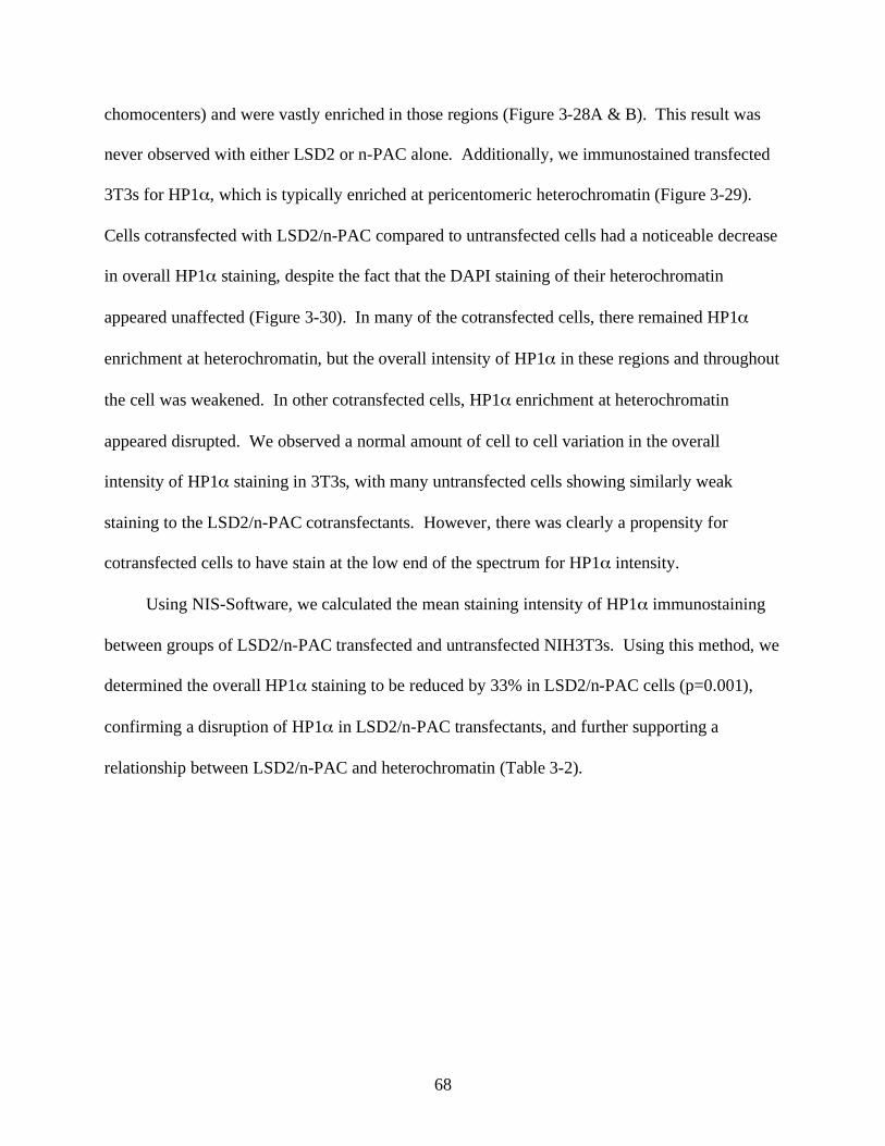
68
chomocenters) and were vastly enriched in those regions (Figure 3-28A & B). This result was
never observed with either LSD2 or n-PAC alone. Additionally, we immunostained transfected
3T3s for HP1 , which is typically enriched at pericentomeric heterochromatin (Figure 3-29).
Cells cotransfected with LSD2/n-PAC compared to untransfected cells had a noticeable decrease
in overall HP1 staining, despite the fact that the DAPI staining of their heterochromatin
appeared unaffected (Figure 3-30). In many of the cotransfected cells, there remained HP1
enrichment at heterochromatin, but the overall intensity of HP1 in these regions and throughout
the cell was weakened. In other cotransfected cells, HP1 enrichment at heterochromatin
appeared disrupted. We observed a normal amount of cell to cell variation in the overall
intensity of HP1 staining in 3T3s, with many untransfected cells showing similarly weak
staining to the LSD2/n-PAC cotransfectants. However, there was clearly a propensity for
cotransfected cells to have stain at the low end of the spectrum for HP1 intensity.
Using NIS-Software, we calculated the mean staining intensity of HP1 immunostaining
between groups of LSD2/n-PAC transfected and untransfected NIH3T3s. Using this method, we
determined the overall HP1 staining to be reduced by 33% in LSD2/n-PAC cells (p=0.001),
confirming a disruption of HP1 in LSD2/n-PAC transfectants, and further supporting a
relationship between LSD2/n-PAC and heterochromatin (Table 3-2).
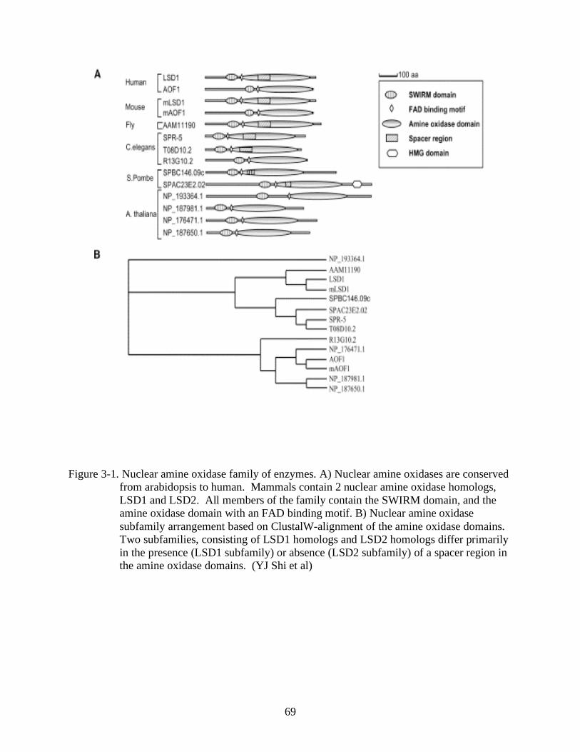
69
Figure 3-1. Nuclear amine oxidase family of enzymes. A) Nuclear amine oxidases are conserved
from arabidopsis to human. Mammals contain 2 nuclear amine oxidase homologs,
LSD1 and LSD2. All members of the family contain the SWIRM domain, and the
amine oxidase domain with an FAD binding motif. B) Nuclear amine oxidase
subfamily arrangement based on ClustalW-alignment of the amine oxidase domains.
Two subfamilies, consisting of LSD1 homologs and LSD2 homologs differ primarily
in the presence (LSD1 subfamily) or absence (LSD2 subfamily) of a spacer region in
the amine oxidase domains. (YJ Shi et al)
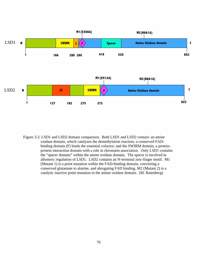
70
Figure 3-2. LSD1 and LSD2 domain comparison. Both LSD1 and LSD2 contain: an amine
oxidase domain, which catalyzes the demethylation reaction; a conserved FAD-
binding domain (F) binds the essential cofactor; and the SWIRM domain, a protein-
protein interaction domain with a role in chromatin association. Only LSD1 contains
the “spacer domain” within the amine oxidase domain. The spacer is involved in
allosteric regulation of LSD1. LSD2 contains an N-terminal zinc-finger motif. M1
(Mutant 1) is a point mutation within the FAD-binding domain, converting a
conserved glutamate to alanine, and abrogating FAD binding. M2 (Mutant 2) is a
catalytic inactive point mutation in the amine oxidase domain. (M. Rutenberg)
C LSD1 N
1 166
260 280
SWIRM
419 520
Spacer
852
Amine Oxidase domain L
M1(E308A)
F
M2(K661A)
LSD2
822
N C
1 137 192 275 373
SWIRM Amine Oxidase domain
M2(K661A) M1(E412A)
F ZF
71
Figure 3-3. Full length LSD2 and splice variant LSD2.1 sequence and domain comparisons. Full
length LSD2 was subcloned from a HeLa cDNA library and sequenced. LSD2.1 was
identified in an EST library. A) Amino acid alignment of full length LSD2 and
LSD2.1. B) Domain comparison of LSD2 and LSD2.1. LSD2.1 has deletions in the
SWIRM domain and the amine oxidase domain. PCR primers used to distinguish
between transcripts of LSD2 and LSD2.1 are denoted as P1, P2, P3, and P4. C) RT-
PCR using primers flanking the deletion sites of LSD2.1, shows the amplification of
both full length LSD2 and splice variant LSD2.1. Expected amplicon sizes are as
follows: P1/P2: LSD2- 530bp, LSD2.1- 127bp; P2/P3: LSD2- 430bp, LSD2.1-
130bp. P1/P4: only amplifies LSD2- 1,240bp. *** non-specific band. (M.
Rutenberg)
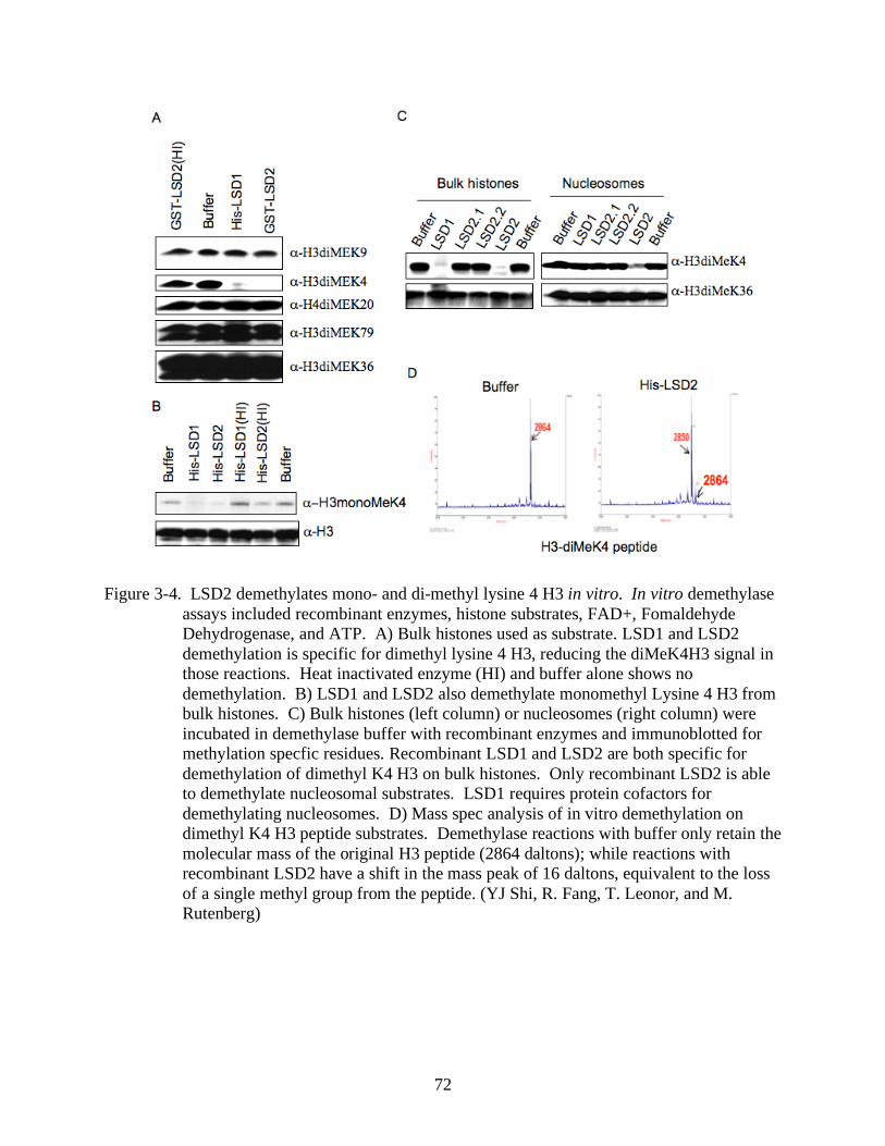
72
Figure 3-4. LSD2 demethylates mono- and di-methyl lysine 4 H3 in vitro. In vitro demethylase
assays included recombinant enzymes, histone substrates, FAD+, Fomaldehyde
Dehydrogenase, and ATP. A) Bulk histones used as substrate. LSD1 and LSD2
demethylation is specific for dimethyl lysine 4 H3, reducing the diMeK4H3 signal in
those reactions. Heat inactivated enzyme (HI) and buffer alone shows no
demethylation. B) LSD1 and LSD2 also demethylate monomethyl Lysine 4 H3 from
bulk histones. C) Bulk histones (left column) or nucleosomes (right column) were
incubated in demethylase buffer with recombinant enzymes and immunoblotted for
methylation specfic residues. Recombinant LSD1 and LSD2 are both specific for
demethylation of dimethyl K4 H3 on bulk histones. Only recombinant LSD2 is able
to demethylate nucleosomal substrates. LSD1 requires protein cofactors for
demethylating nucleosomes. D) Mass spec analysis of in vitro demethylation on
dimethyl K4 H3 peptide substrates. Demethylase reactions with buffer only retain the
molecular mass of the original H3 peptide (2864 daltons); while reactions with
recombinant LSD2 have a shift in the mass peak of 16 daltons, equivalent to the loss
of a single methyl group from the peptide. (YJ Shi, R. Fang, T. Leonor, and M.
Rutenberg)
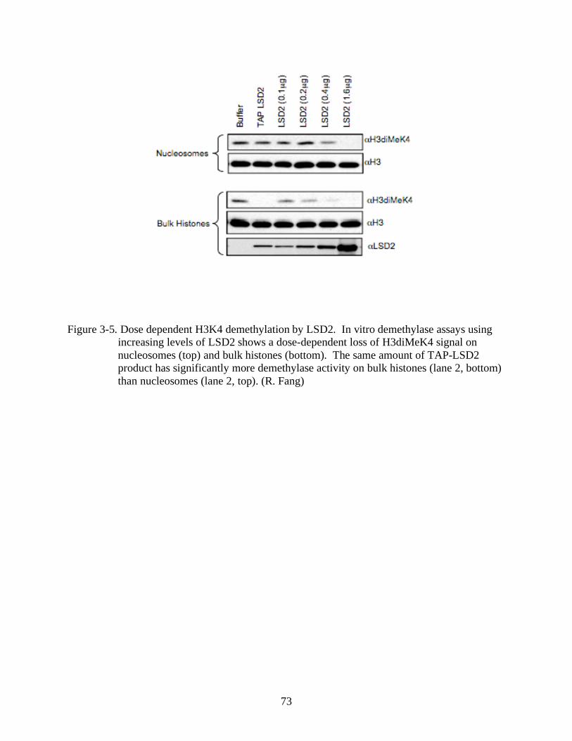
73
Figure 3-5. Dose dependent H3K4 demethylation by LSD2. In vitro demethylase assays using
increasing levels of LSD2 shows a dose-dependent loss of H3diMeK4 signal on
nucleosomes (top) and bulk histones (bottom). The same amount of TAP-LSD2
product has significantly more demethylase activity on bulk histones (lane 2, bottom)
than nucleosomes (lane 2, top). (R. Fang)
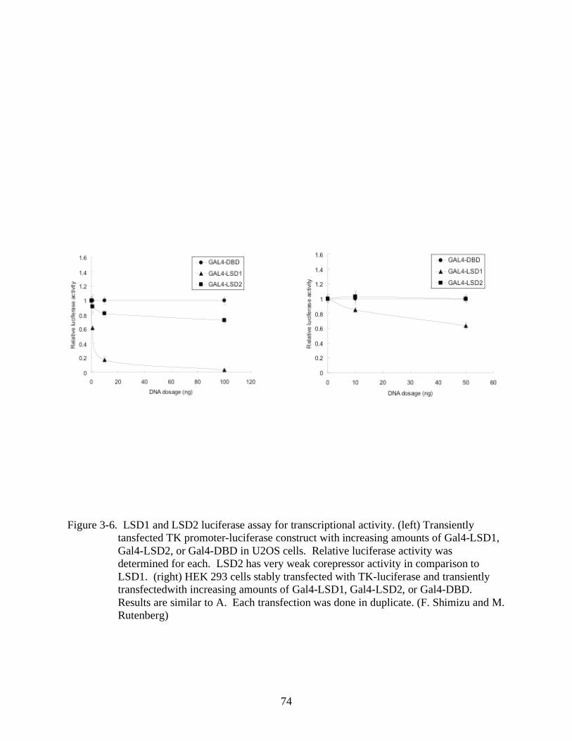
74
Figure 3-6. LSD1 and LSD2 luciferase assay for transcriptional activity. (left) Transiently
tansfected TK promoter-luciferase construct with increasing amounts of Gal4-LSD1,
Gal4-LSD2, or Gal4-DBD in U2OS cells. Relative luciferase activity was
determined for each. LSD2 has very weak corepressor activity in comparison to
LSD1. (right) HEK 293 cells stably transfected with TK-luciferase and transiently
transfectedwith increasing amounts of Gal4-LSD1, Gal4-LSD2, or Gal4-DBD.
Results are similar to A. Each transfection was done in duplicate. (F. Shimizu and M.
Rutenberg)
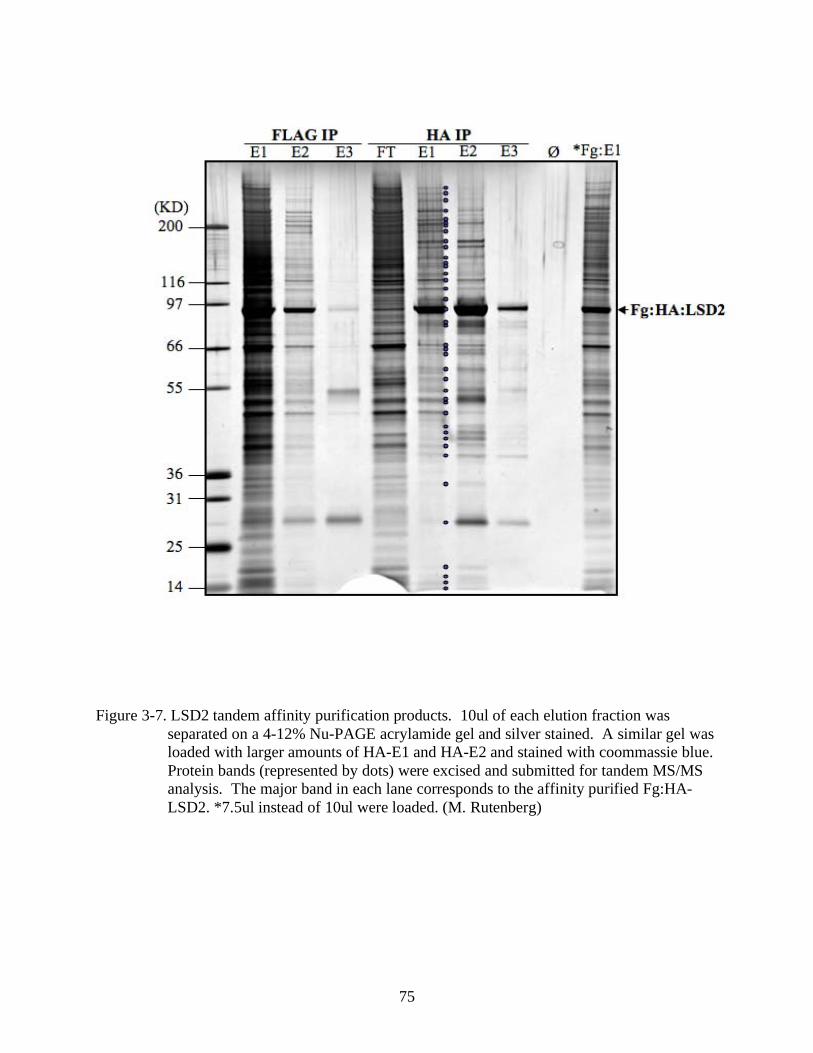
75
Figure 3-7. LSD2 tandem affinity purification products. 10ul of each elution fraction was
separated on a 4-12% Nu-PAGE acrylamide gel and silver stained. A similar gel was
loaded with larger amounts of HA-E1 and HA-E2 and stained with coommassie blue.
Protein bands (represented by dots) were excised and submitted for tandem MS/MS
analysis. The major band in each lane corresponds to the affinity purified Fg:HA-
LSD2. *7.5ul instead of 10ul were loaded. (M. Rutenberg)
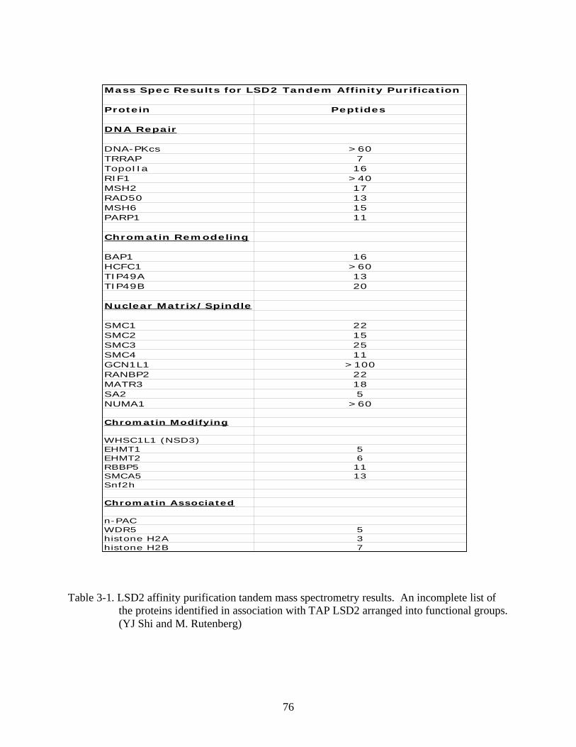
76
Table 3-1. LSD2 affinity purification tandem mass spectrometry results. An incomplete list of
the proteins identified in association with TAP LSD2 arranged into functional groups.
(YJ Shi and M. Rutenberg)
Mass Spec Results for LSD2 Tandem Affinity Purification
Protein Peptides
DNA Repair
DNA-PKcs >60
TRRAP 7
TopoIIa 16
RIF1 >40
MSH2 17
RAD50 13
MSH6 15
PARP1 11
Chromatin Remodeling
BAP1 16
HCFC1 >60
TIP49A 13
TIP49B 20
Nuclear Matrix/Spindle
SMC1 22
SMC2 15
SMC3 25
SMC4 11
GCN1L1 >100
RANBP2 22
MATR3 18
SA2 5
NUMA1 >60
Chromatin Modifying
WHSC1L1 (NSD3)
EHMT1 5
EHMT2 6
RBBP5 11
SMCA5 13
Snf2h
Chromatin Associated
n-PAC
WDR5 5
histone H2A 3
histone H2B 7
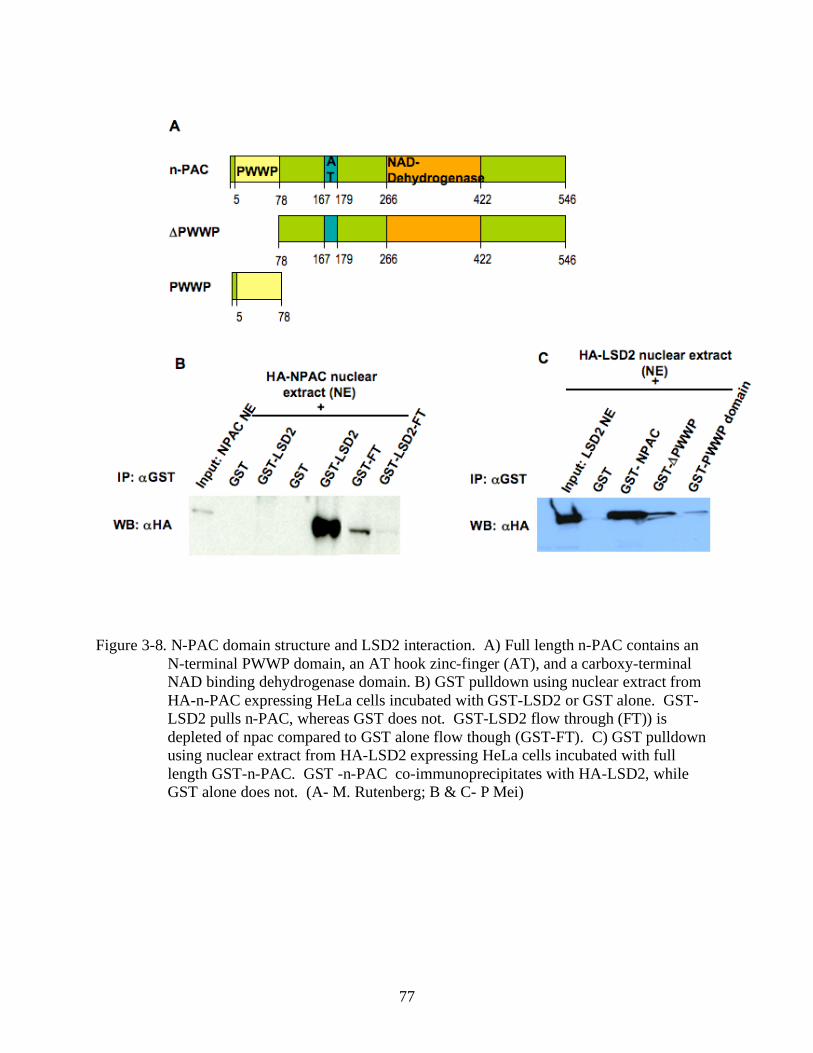
77
Figure 3-8. N-PAC domain structure and LSD2 interaction. A) Full length n-PAC contains an
N-terminal PWWP domain, an AT hook zinc-finger (AT), and a carboxy-terminal
NAD binding dehydrogenase domain. B) GST pulldown using nuclear extract from
HA-n-PAC expressing HeLa cells incubated with GST-LSD2 or GST alone. GST-
LSD2 pulls n-PAC, whereas GST does not. GST-LSD2 flow through (FT)) is
depleted of npac compared to GST alone flow though (GST-FT). C) GST pulldown
using nuclear extract from HA-LSD2 expressing HeLa cells incubated with full
length GST-n-PAC. GST -n-PAC co-immunoprecipitates with HA-LSD2, while
GST alone does not. (A- M. Rutenberg; B & C- P Mei)
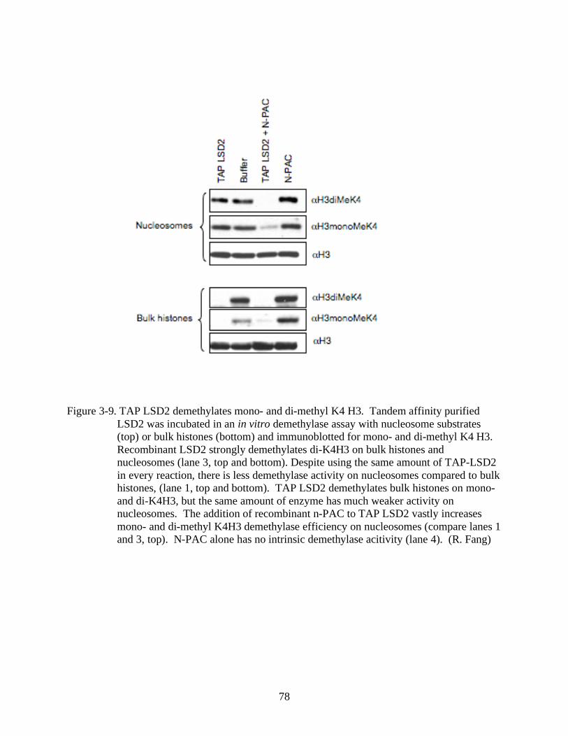
78
Figure 3-9. TAP LSD2 demethylates mono- and di-methyl K4 H3. Tandem affinity purified
LSD2 was incubated in an in vitro demethylase assay with nucleosome substrates
(top) or bulk histones (bottom) and immunoblotted for mono- and di-methyl K4 H3.
Recombinant LSD2 strongly demethylates di-K4H3 on bulk histones and
nucleosomes (lane 3, top and bottom). Despite using the same amount of TAP-LSD2
in every reaction, there is less demethylase activity on nucleosomes compared to bulk
histones, (lane 1, top and bottom). TAP LSD2 demethylates bulk histones on mono-
and di-K4H3, but the same amount of enzyme has much weaker activity on
nucleosomes. The addition of recombinant n-PAC to TAP LSD2 vastly increases
mono- and di-methyl K4H3 demethylase efficiency on nucleosomes (compare lanes 1
and 3, top). N-PAC alone has no intrinsic demethylase acitivity (lane 4). (R. Fang)
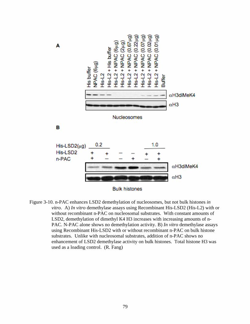
79
Figure 3-10. n-PAC enhances LSD2 demethylation of nucleosomes, but not bulk histones in
vitro. A) In vitro demethylase assays using Recombinant His-LSD2 (His-L2) with or
without recombinant n-PAC on nucleosomal substrates. With constant amounts of
LSD2, demethylation of dimethyl K4 H3 increases with increasing amounts of n-
PAC. N-PAC alone shows no demethylation activity. B) In vitro demethylase assays
using Recombinant His-LSD2 with or without recombinant n-PAC on bulk histone
substrates. Unlike with nucleosomal substrates, addition of n-PAC shows no
enhancement of LSD2 demethylase activity on bulk histones. Total histone H3 was
used as a loading control. (R. Fang)
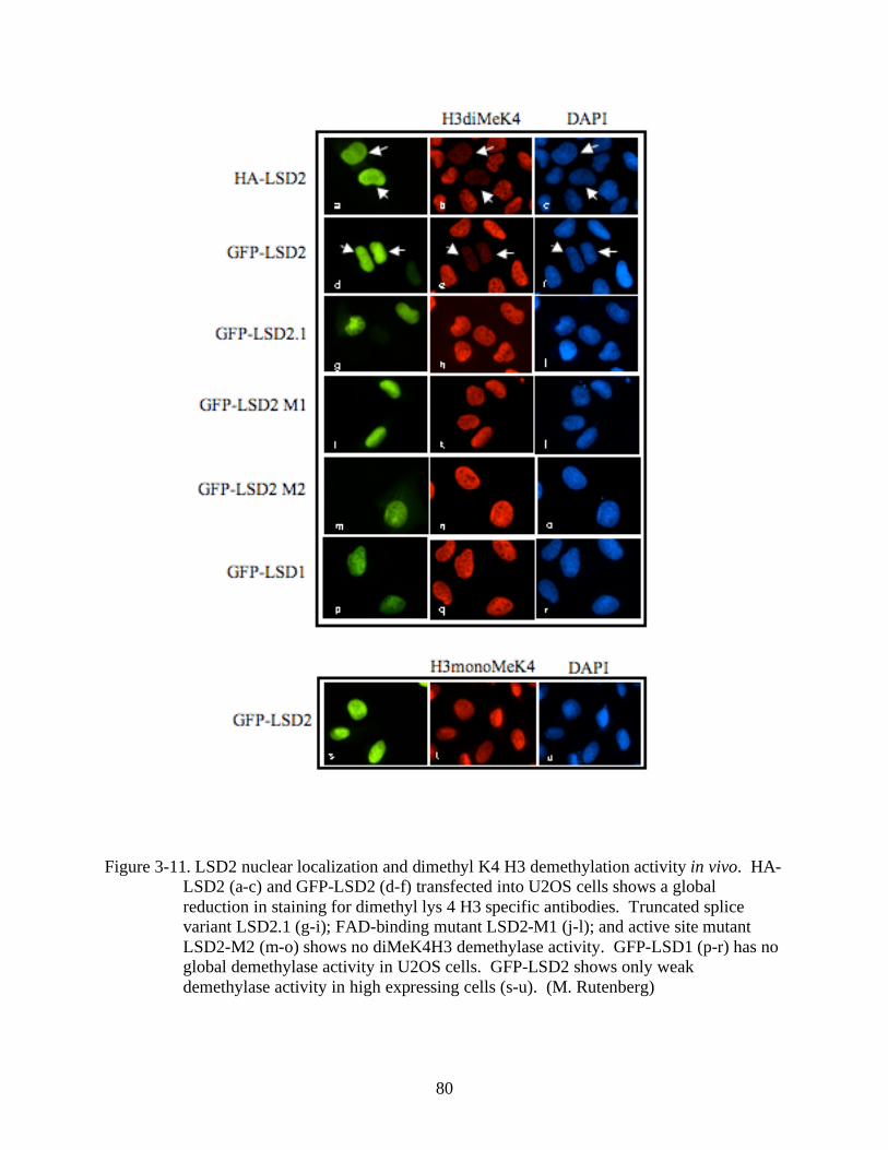
80
Figure 3-11. LSD2 nuclear localization and dimethyl K4 H3 demethylation activity in vivo. HA-
LSD2 (a-c) and GFP-LSD2 (d-f) transfected into U2OS cells shows a global
reduction in staining for dimethyl lys 4 H3 specific antibodies. Truncated splice
variant LSD2.1 (g-i); FAD-binding mutant LSD2-M1 (j-l); and active site mutant
LSD2-M2 (m-o) shows no diMeK4H3 demethylase activity. GFP-LSD1 (p-r) has no
global demethylase activity in U2OS cells. GFP-LSD2 shows only weak
demethylase activity in high expressing cells (s-u). (M. Rutenberg)
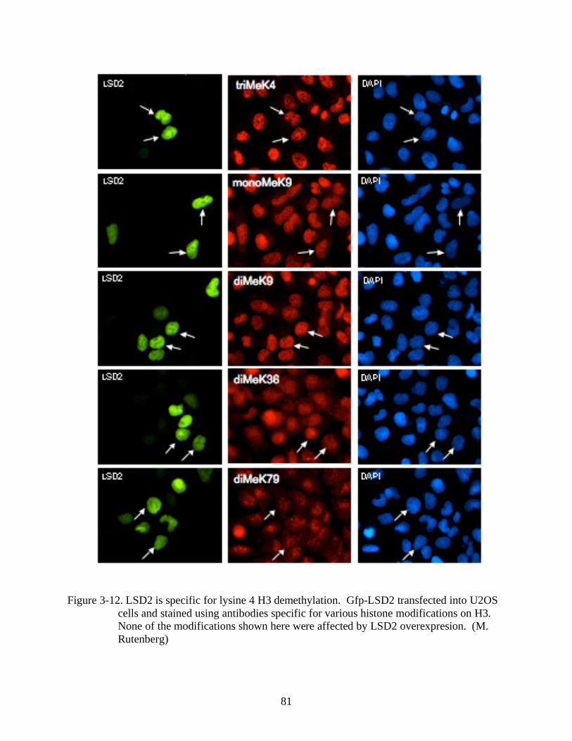
81
Figure 3-12. LSD2 is specific for lysine 4 H3 demethylation. Gfp-LSD2 transfected into U2OS
cells and stained using antibodies specific for various histone modifications on H3.
None of the modifications shown here were affected by LSD2 overexpresion. (M.
Rutenberg)
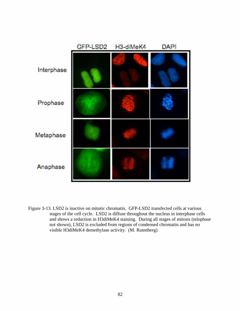
82
Figure 3-13. LSD2 is inactive on mitotic chromatin. GFP-LSD2 transfected cells at various
stages of the cell cycle. LSD2 is diffuse throughout the nucleus in interphase cells
and shows a reduction in H3diMeK4 staining. During all stages of mitosis (telophase
not shown), LSD2 is excluded from regions of condensed chromatin and has no
visible H3diMeK4 demethylase activity. (M. Rutenberg)
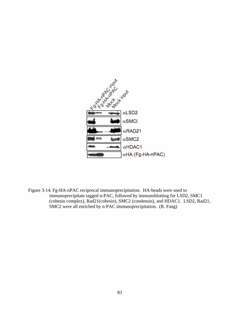
83
Figure 3-14. Fg-HA-nPAC reciprocal immunoprecipitation. HA-beads were used to
immunoprecipitate tagged n-PAC, followed by immunoblotting for LSD2, SMC1
(cohesin complex), Rad21(cohesin), SMC2 (condensin), and HDAC1. LSD2, Rad21,
SMC2 were all enriched by n-PAC immunoprecipitation. (R. Fang)
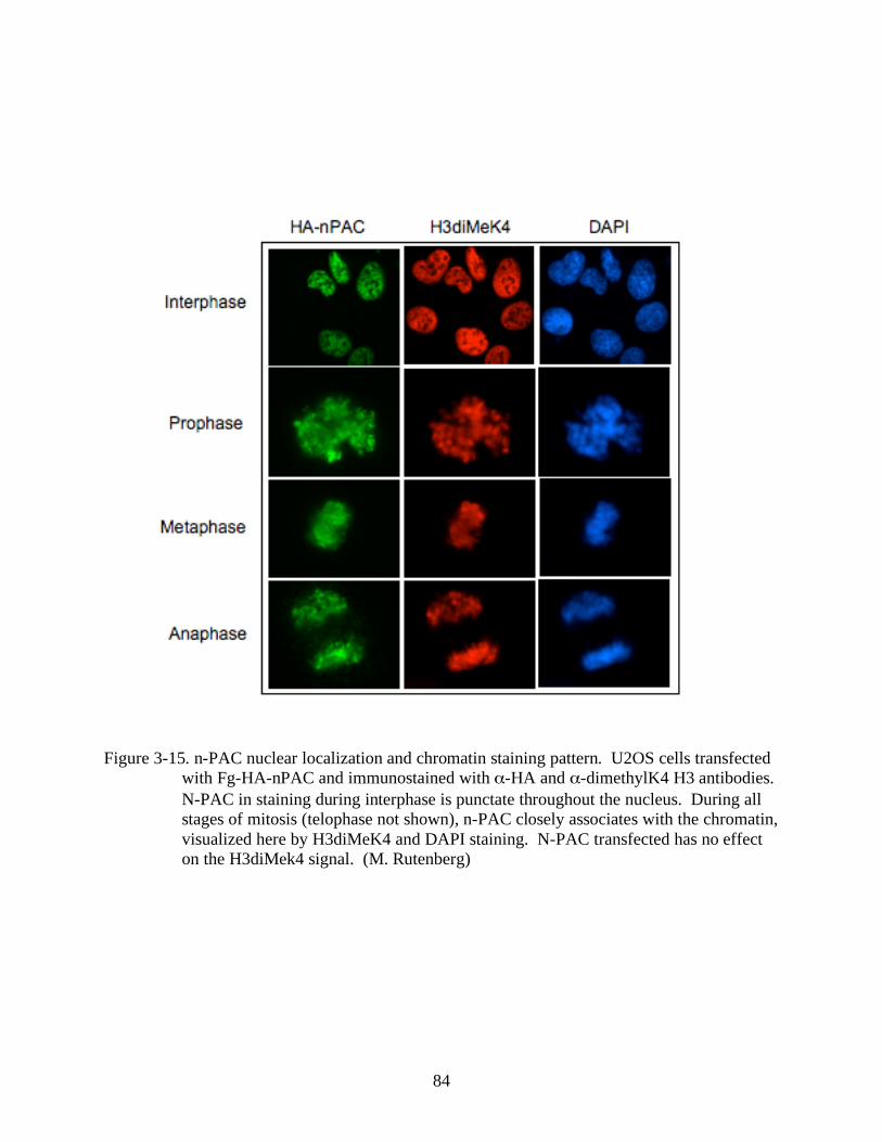
84
Figure 3-15. n-PAC nuclear localization and chromatin staining pattern. U2OS cells transfected
with Fg-HA-nPAC and immunostained with -HA and -dimethylK4 H3 antibodies.
N-PAC in staining during interphase is punctate throughout the nucleus. During all
stages of mitosis (telophase not shown), n-PAC closely associates with the chromatin,
visualized here by H3diMeK4 and DAPI staining. N-PAC transfected has no effect
on the H3diMek4 signal. (M. Rutenberg)
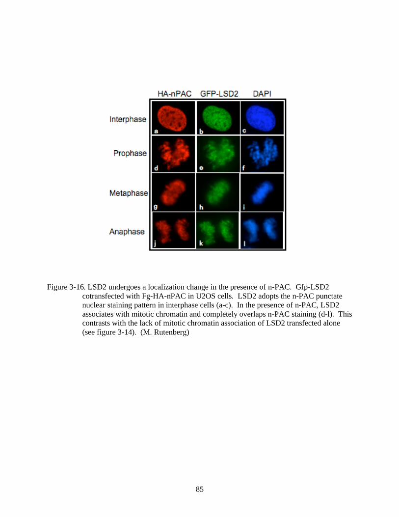
85
Figure 3-16. LSD2 undergoes a localization change in the presence of n-PAC. Gfp-LSD2
cotransfected with Fg-HA-nPAC in U2OS cells. LSD2 adopts the n-PAC punctate
nuclear staining pattern in interphase cells (a-c). In the presence of n-PAC, LSD2
associates with mitotic chromatin and completely overlaps n-PAC staining (d-l). This
contrasts with the lack of mitotic chromatin association of LSD2 transfected alone
(see figure 3-14). (M. Rutenberg)
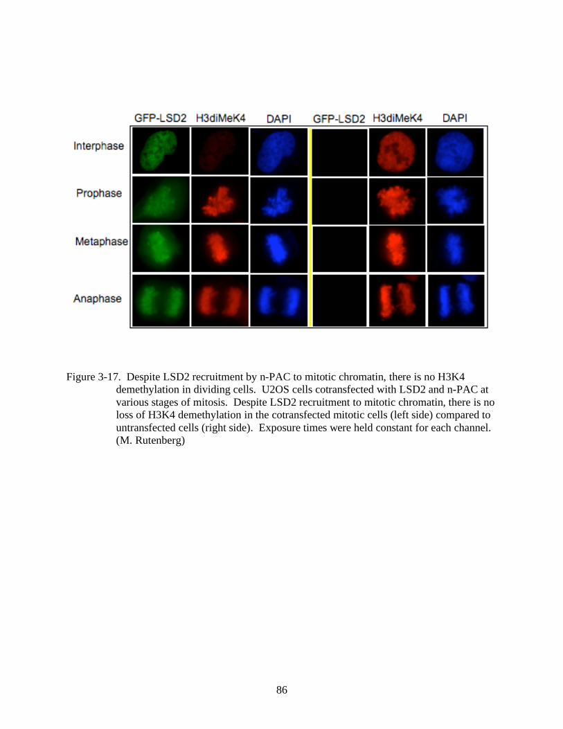
86
Figure 3-17. Despite LSD2 recruitment by n-PAC to mitotic chromatin, there is no H3K4
demethylation in dividing cells. U2OS cells cotransfected with LSD2 and n-PAC at
various stages of mitosis. Despite LSD2 recruitment to mitotic chromatin, there is no
loss of H3K4 demethylation in the cotransfected mitotic cells (left side) compared to
untransfected cells (right side). Exposure times were held constant for each channel.
(M. Rutenberg)
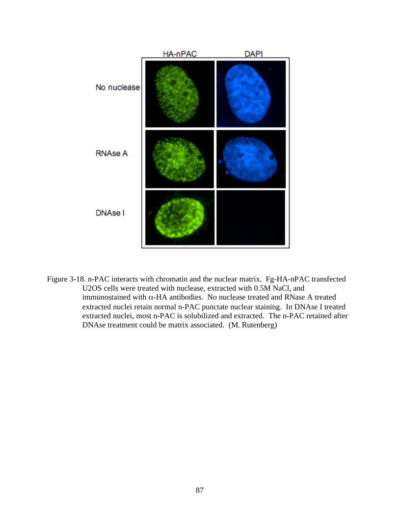
87
Figure 3-18. n-PAC interacts with chromatin and the nuclear matrix. Fg-HA-nPAC transfected
U2OS cells were treated with nuclease, extracted with 0.5M NaCl, and
immunostained with -HA antibodies. No nuclease treated and RNase A treated
extracted nuclei retain normal n-PAC punctate nuclear staining. In DNAse I treated
extracted nuclei, most n-PAC is solubilized and extracted. The n-PAC retained after
DNAse treatment could be matrix associated. (M. Rutenberg)
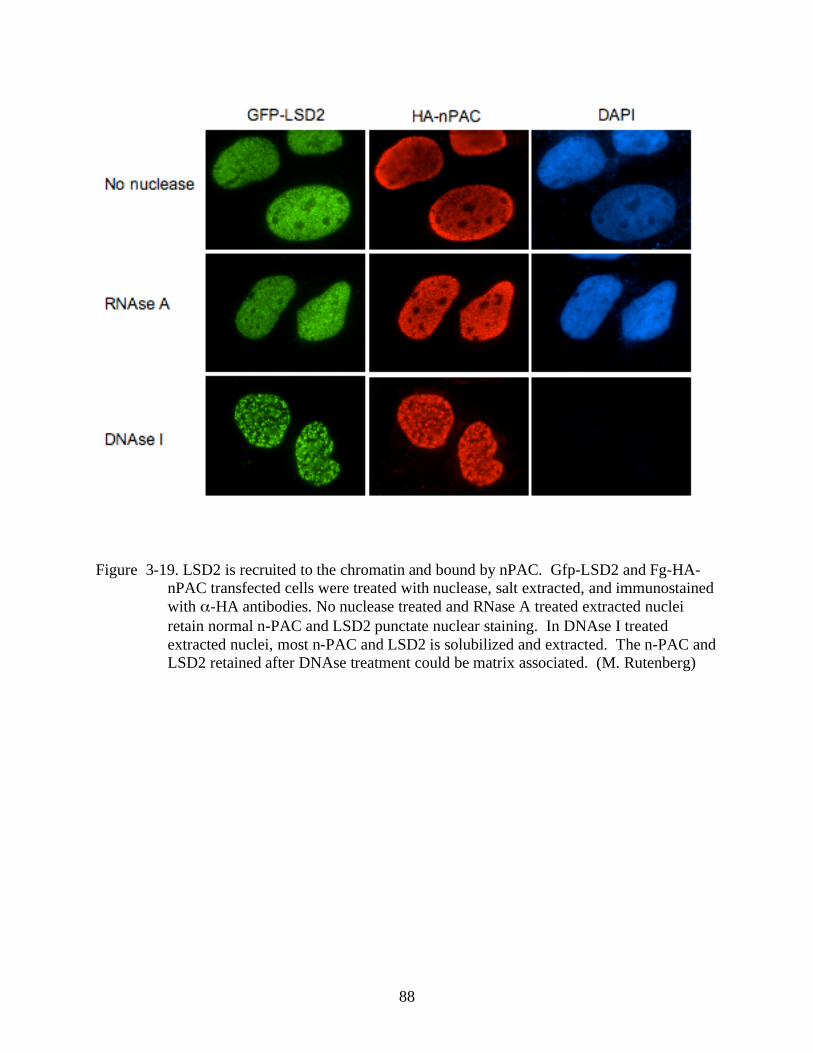
88
Figure 3-19. LSD2 is recruited to the chromatin and bound by nPAC. Gfp-LSD2 and Fg-HA-
nPAC transfected cells were treated with nuclease, salt extracted, and immunostained
with -HA antibodies. No nuclease treated and RNase A treated extracted nuclei
retain normal n-PAC and LSD2 punctate nuclear staining. In DNAse I treated
extracted nuclei, most n-PAC and LSD2 is solubilized and extracted. The n-PAC and
LSD2 retained after DNAse treatment could be matrix associated. (M. Rutenberg)
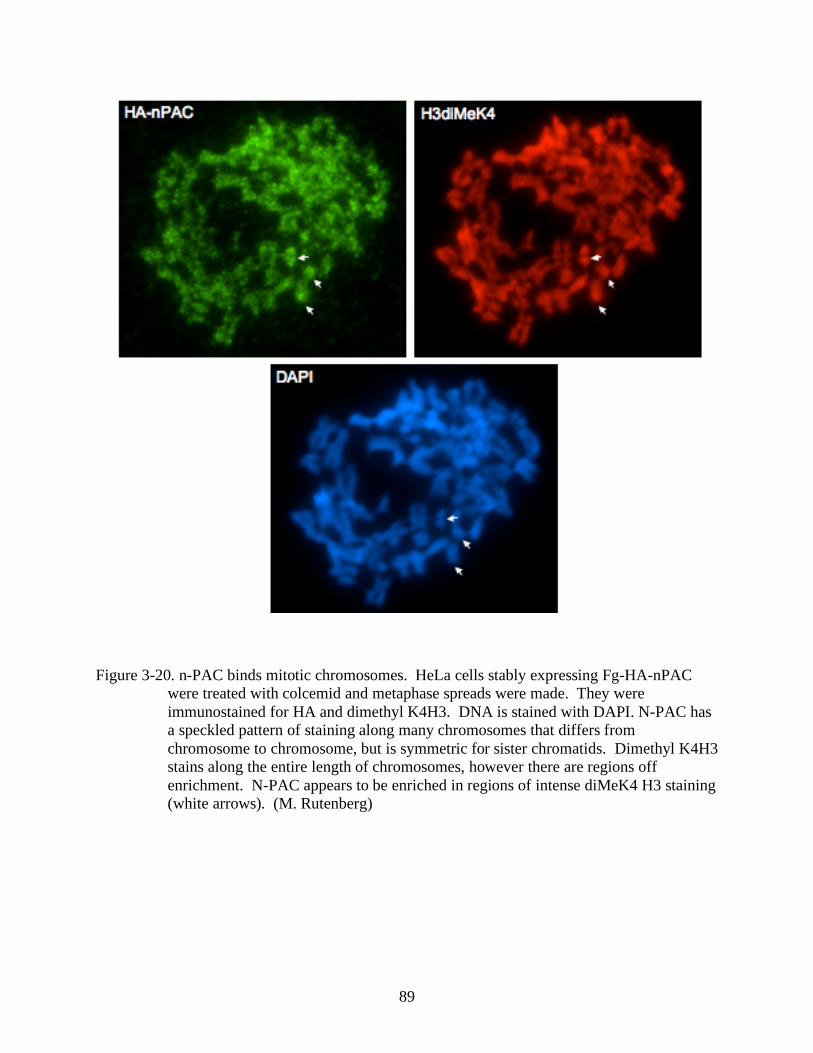
89
Figure 3-20. n-PAC binds mitotic chromosomes. HeLa cells stably expressing Fg-HA-nPAC
were treated with colcemid and metaphase spreads were made. They were
immunostained for HA and dimethyl K4H3. DNA is stained with DAPI. N-PAC has
a speckled pattern of staining along many chromosomes that differs from
chromosome to chromosome, but is symmetric for sister chromatids. Dimethyl K4H3
stains along the entire length of chromosomes, however there are regions off
enrichment. N-PAC appears to be enriched in regions of intense diMeK4 H3 staining
(white arrows). (M. Rutenberg)
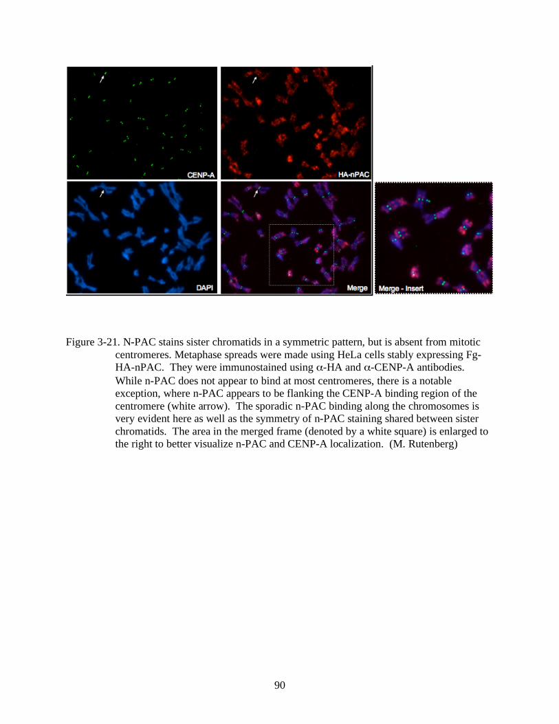
90
Figure 3-21. N-PAC stains sister chromatids in a symmetric pattern, but is absent from mitotic
centromeres. Metaphase spreads were made using HeLa cells stably expressing Fg-
HA-nPAC. They were immunostained using -HA and -CENP-A antibodies.
While n-PAC does not appear to bind at most centromeres, there is a notable
exception, where n-PAC appears to be flanking the CENP-A binding region of the
centromere (white arrow). The sporadic n-PAC binding along the chromosomes is
very evident here as well as the symmetry of n-PAC staining shared between sister
chromatids. The area in the merged frame (denoted by a white square) is enlarged to
the right to better visualize n-PAC and CENP-A localization. (M. Rutenberg)
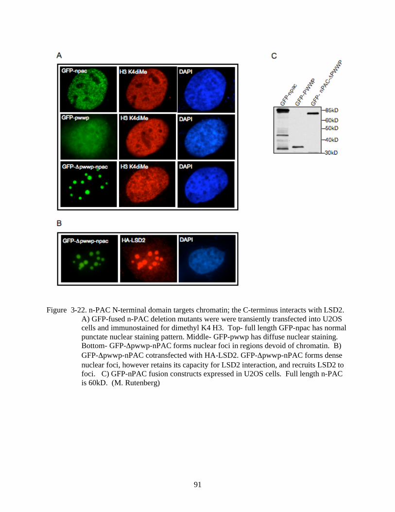
91
Figure 3-22. n-PAC N-terminal domain targets chromatin; the C-terminus interacts with LSD2.
A) GFP-fused n-PAC deletion mutants were were transiently transfected into U2OS
cells and immunostained for dimethyl K4 H3. Top- full length GFP-npac has normal
punctate nuclear staining pattern. Middle- GFP-pwwp has diffuse nuclear staining.
Bottom- GFP- pwwp-nPAC forms nuclear foci in regions devoid of chromatin. B)
GFP- pwwp-nPAC cotransfected with HA-LSD2. GFP- pwwp-nPAC forms dense
nuclear foci, however retains its capacity for LSD2 interaction, and recruits LSD2 to
foci. C) GFP-nPAC fusion constructs expressed in U2OS cells. Full length n-PAC
is 60kD. (M. Rutenberg)
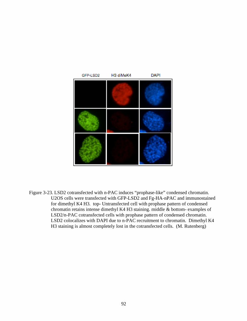
92
Figure 3-23. LSD2 cotransfected with n-PAC induces “prophase-like” condensed chromatin.
U2OS cells were transfected with GFP-LSD2 and Fg-HA-nPAC and immunostained
for dimethyl K4 H3. top- Untransfected cell with prophase pattern of condensed
chromatin retains intense dimethyl K4 H3 staining. middle & bottom- examples of
LSD2/n-PAC cotransfected cells with prophase pattern of condensed chromatin.
LSD2 colocalizes with DAPI due to n-PAC recruitment to chromatin. Dimethyl K4
H3 staining is almost completely lost in the cotransfected cells. (M. Rutenberg)

93
Figure 3-24. LSD2 and n-PAC cotransfected cells have “prophase-like” chromatin condensation
in the absence of mitotic markers or apoptosis. Reprasentative images of
untransfected or LSD2 and n-PAC cotransfected U2OS cells were immunostained for
mitotic markers or TUNEL stained for apotosis. A) Untransfected prophase cells
show intense nuclear cyclinB1 staining (top), while LSD2/n-PAC cells with similar
condensation patterns are devoid of cyclinB1 (bottom). B) Untransfected prophase
cells stain positively for the mitotic marker phosphorylated Serine 10 on H3 (H3
pS10), while LSD2/n-PAC cells with similar DAPI patterns lack H3 pS10. C)
Untransfected prophase cells are positive for phospho-Threonine 3 on H3 (H3 pThr3),
while cotransfected “prophase-like” are negative. D) TUNEL staining of LSD2/n-
PAC cells for apoptosis are negative (top & middle). Postive control cells treated
with DNAse I stain brightly for TUNEL (bottom). (M. Rutenberg)
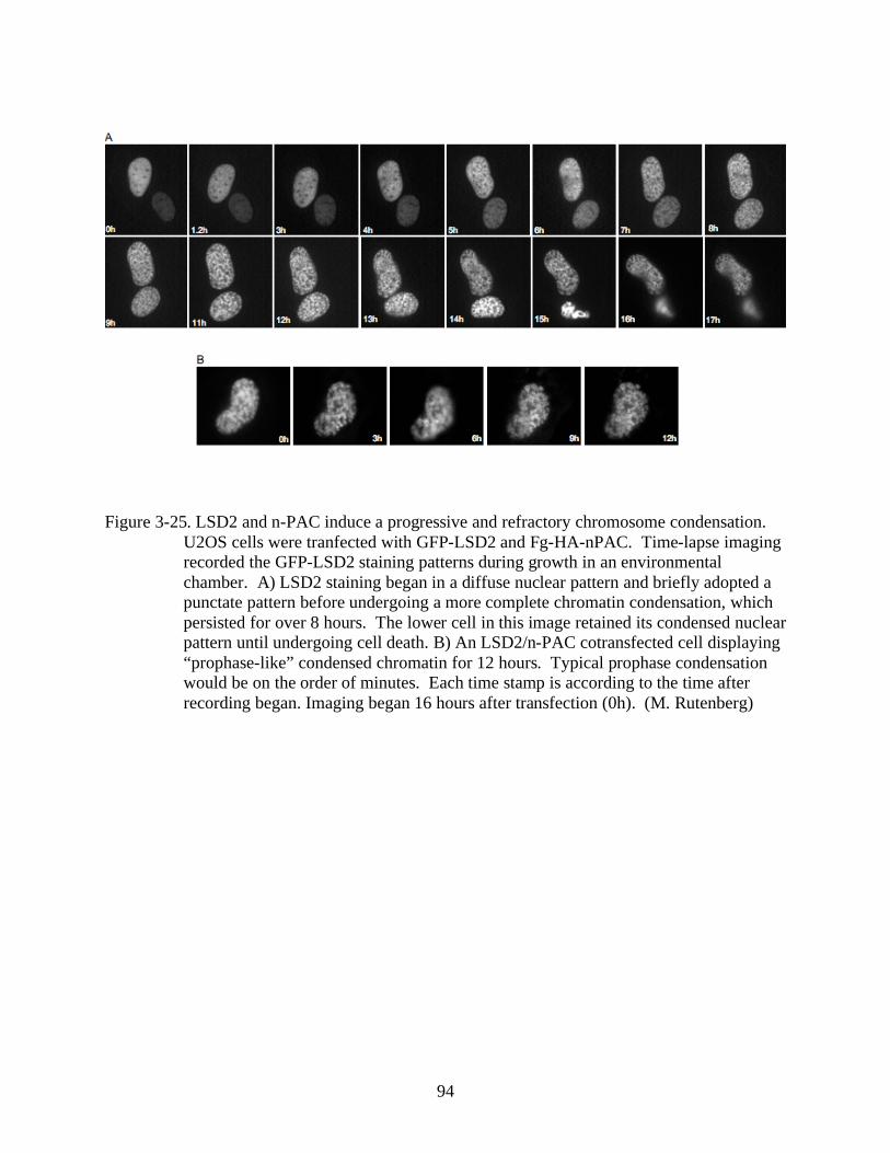
94
Figure 3-25. LSD2 and n-PAC induce a progressive and refractory chromosome condensation.
U2OS cells were tranfected with GFP-LSD2 and Fg-HA-nPAC. Time-lapse imaging
recorded the GFP-LSD2 staining patterns during growth in an environmental
chamber. A) LSD2 staining began in a diffuse nuclear pattern and briefly adopted a
punctate pattern before undergoing a more complete chromatin condensation, which
persisted for over 8 hours. The lower cell in this image retained its condensed nuclear
pattern until undergoing cell death. B) An LSD2/n-PAC cotransfected cell displaying
“prophase-like” condensed chromatin for 12 hours. Typical prophase condensation
would be on the order of minutes. Each time stamp is according to the time after
recording began. Imaging began 16 hours after transfection (0h). (M. Rutenberg)
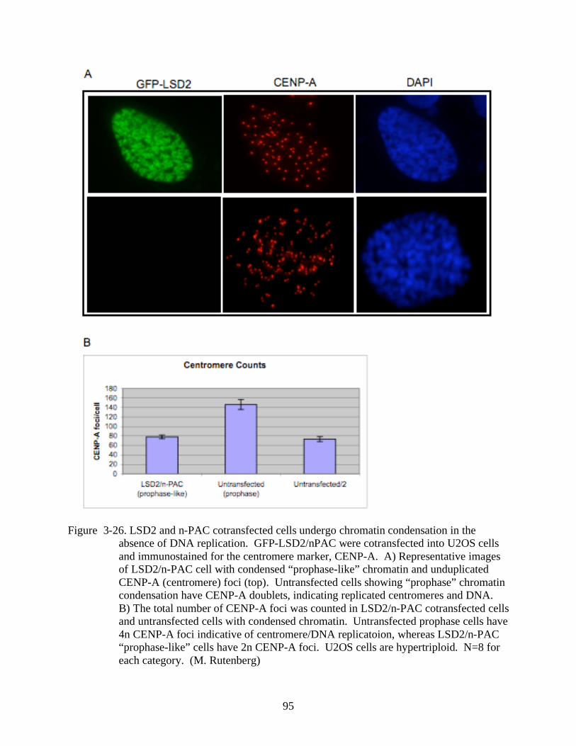
95
Figure 3-26. LSD2 and n-PAC cotransfected cells undergo chromatin condensation in the
absence of DNA replication. GFP-LSD2/nPAC were cotransfected into U2OS cells
and immunostained for the centromere marker, CENP-A. A) Representative images
of LSD2/n-PAC cell with condensed “prophase-like” chromatin and unduplicated
CENP-A (centromere) foci (top). Untransfected cells showing “prophase” chromatin
condensation have CENP-A doublets, indicating replicated centromeres and DNA.
B) The total number of CENP-A foci was counted in LSD2/n-PAC cotransfected cells
and untransfected cells with condensed chromatin. Untransfected prophase cells have
4n CENP-A foci indicative of centromere/DNA replicatoion, whereas LSD2/n-PAC
“prophase-like” cells have 2n CENP-A foci. U2OS cells are hypertriploid. N=8 for
each category. (M. Rutenberg)
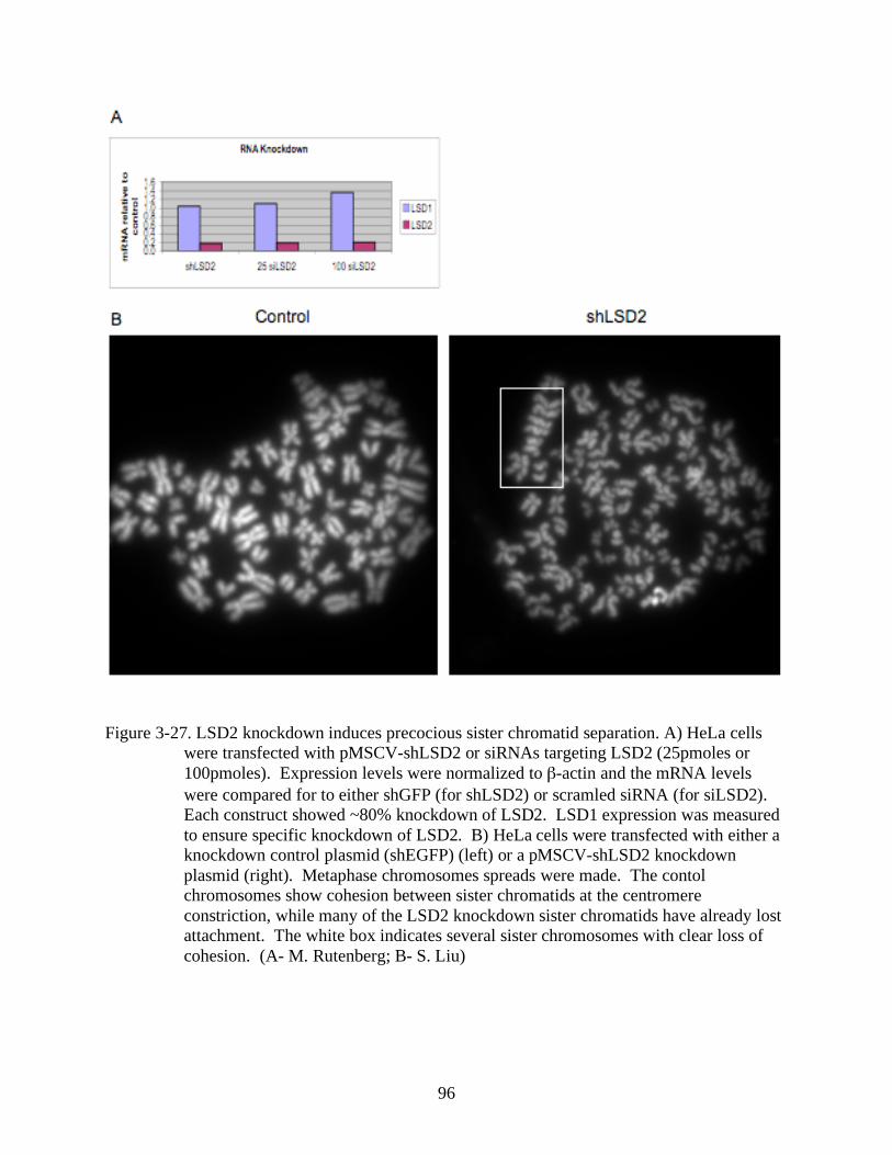
96
Figure 3-27. LSD2 knockdown induces precocious sister chromatid separation. A) HeLa cells
were transfected with pMSCV-shLSD2 or siRNAs targeting LSD2 (25pmoles or
100pmoles). Expression levels were normalized to -actin and the mRNA levels
were compared for to either shGFP (for shLSD2) or scramled siRNA (for siLSD2).
Each construct showed ~80% knockdown of LSD2. LSD1 expression was measured
to ensure specific knockdown of LSD2. B) HeLa cells were transfected with either a
knockdown control plasmid (shEGFP) (left) or a pMSCV-shLSD2 knockdown
plasmid (right). Metaphase chromosomes spreads were made. The contol
chromosomes show cohesion between sister chromatids at the centromere
constriction, while many of the LSD2 knockdown sister chromatids have already lost
attachment. The white box indicates several sister chromosomes with clear loss of
cohesion. (A- M. Rutenberg; B- S. Liu)
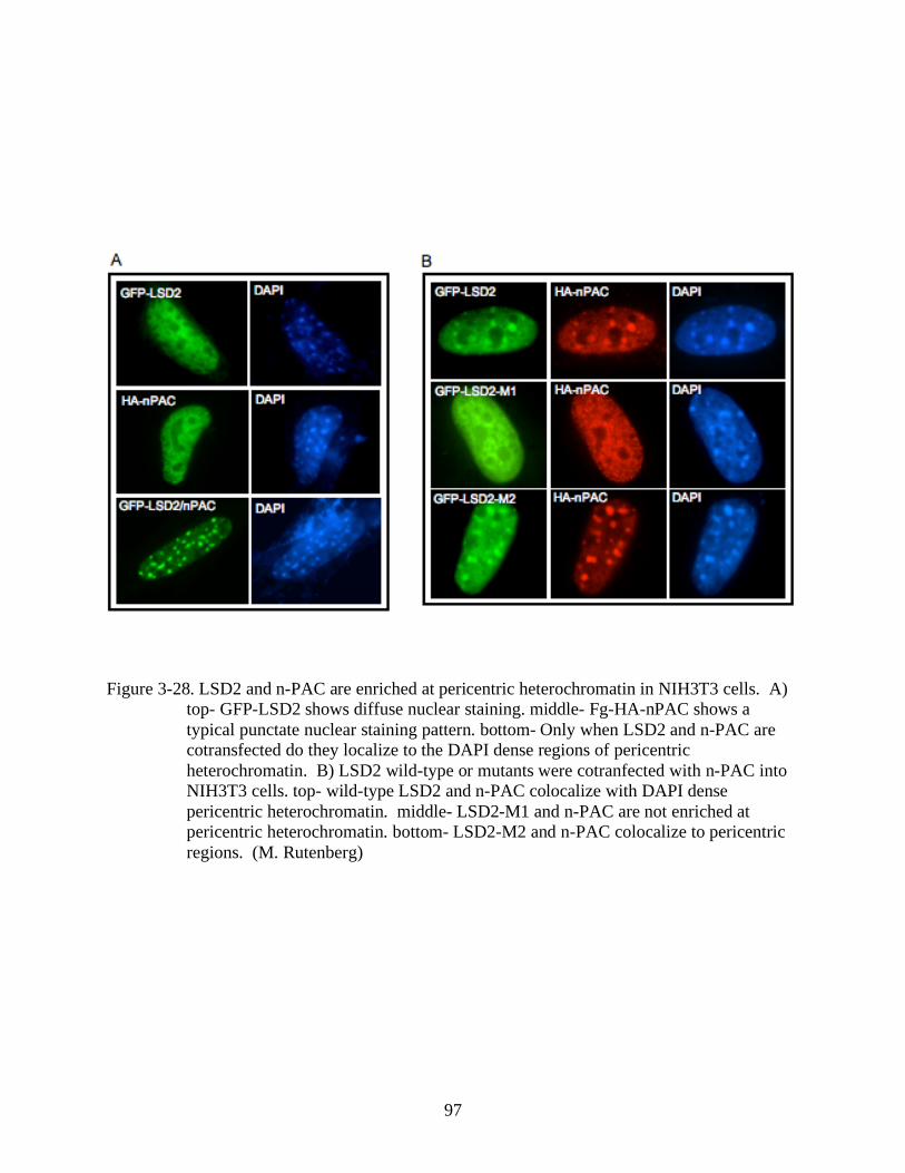
97
Figure 3-28. LSD2 and n-PAC are enriched at pericentric heterochromatin in NIH3T3 cells. A)
top- GFP-LSD2 shows diffuse nuclear staining. middle- Fg-HA-nPAC shows a
typical punctate nuclear staining pattern. bottom- Only when LSD2 and n-PAC are
cotransfected do they localize to the DAPI dense regions of pericentric
heterochromatin. B) LSD2 wild-type or mutants were cotranfected with n-PAC into
NIH3T3 cells. top- wild-type LSD2 and n-PAC colocalize with DAPI dense
pericentric heterochromatin. middle- LSD2-M1 and n-PAC are not enriched at
pericentric heterochromatin. bottom- LSD2-M2 and n-PAC colocalize to pericentric
regions. (M. Rutenberg)
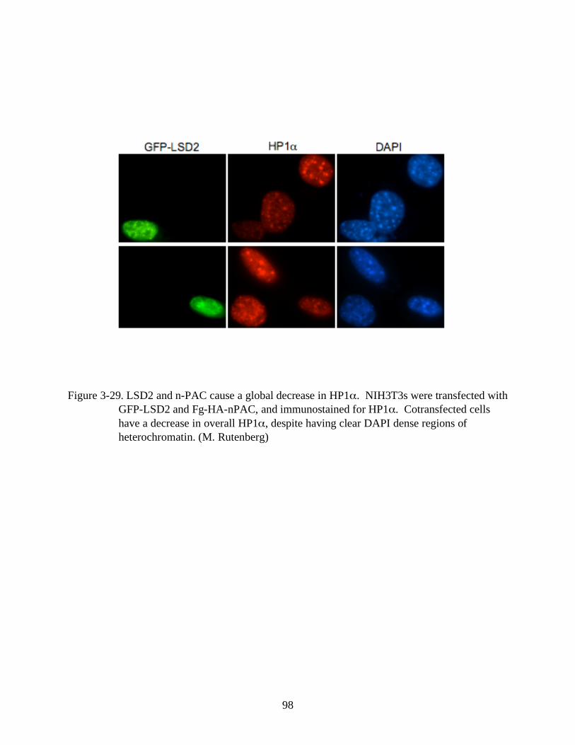
98
Figure 3-29. LSD2 and n-PAC cause a global decrease in HP1 . NIH3T3s were transfected with
GFP-LSD2 and Fg-HA-nPAC, and immunostained for HP1 . Cotransfected cells
have a decrease in overall HP1 , despite having clear DAPI dense regions of
heterochromatin. (M. Rutenberg)
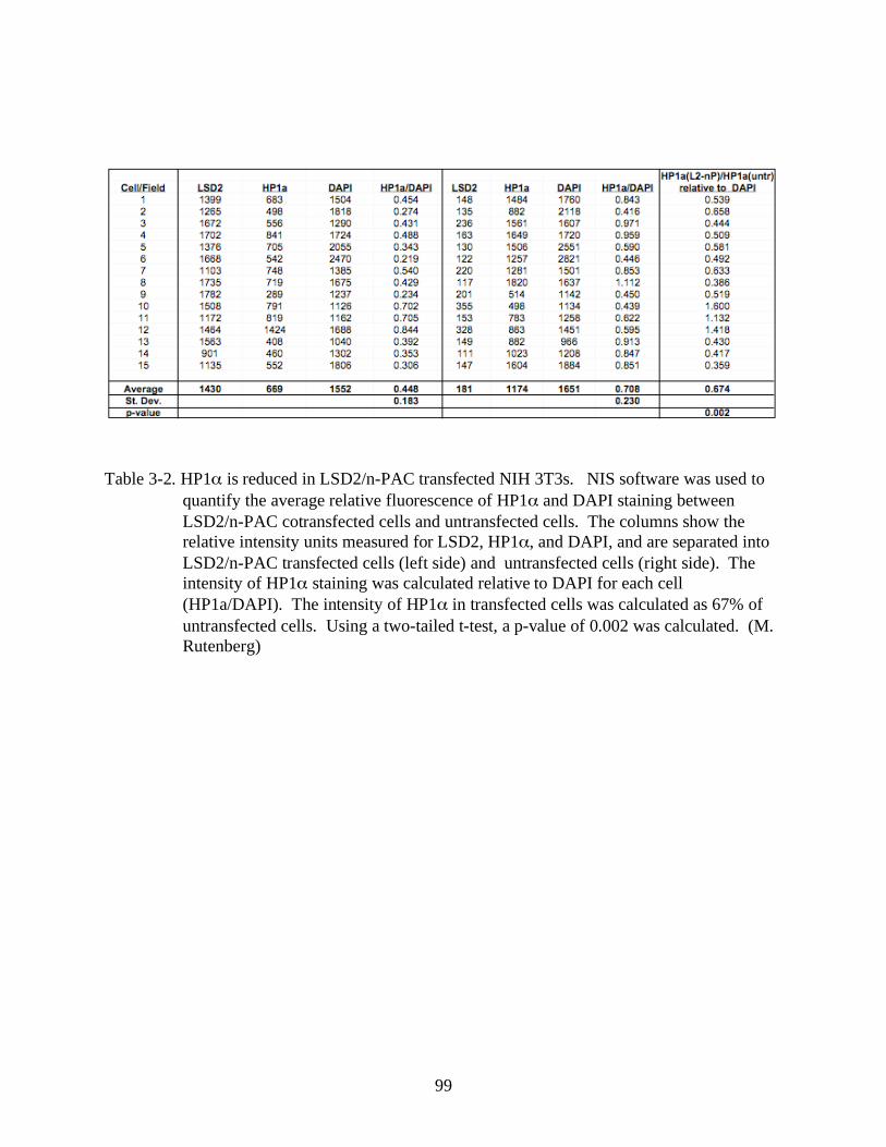
99
Table 3-2. HP1 is reduced in LSD2/n-PAC transfected NIH 3T3s. NIS software was used to
quantify the average relative fluorescence of HP1 and DAPI staining between
LSD2/n-PAC cotransfected cells and untransfected cells. The columns show the
relative intensity units measured for LSD2, HP1 , and DAPI, and are separated into
LSD2/n-PAC transfected cells (left side) and untransfected cells (right side). The
intensity of HP1 staining was calculated relative to DAPI for each cell
(HP1a/DAPI). The intensity of HP1 in transfected cells was calculated as 67% of
untransfected cells. Using a two-tailed t-test, a p-value of 0.002 was calculated. (M.
Rutenberg)
100
CHAPTER 4
DISCUSSION
The idenification of LSD1 as the first bona fide histone demethylase shook the foundation
of epigenetics. Prior to its discovery, the idea of histone methylation as a permanent epigenetic
mark was gaining favor. LSD1 demethylase activity revealed the reversibility of histone
methylation and uncovered a system by which the complex combinations of histone methylation
could be translated into a dynamic system of genome regulation.
Because much of the focus on histone methylation has been on its role in transcription
regulation, so has been the study of LSD1 and histone demethylation. Here, we have attempted
to characterize the activity and function of the LSD1 homolog, LSD2. While initial protein
domain analyses comparing LSD1 and LSD2 show conservation of their SWIRM domains and
their C-terminal, FAD-dependent amine oxidase domains, they also reveal major structural
differences between the two proteins. The tower domain of LSD1, which is noticeably absent
from LSD2, is required for LSD1 enzymatic activity and mediates allosteric regulation.
Additionally, the N-terminal zinc-finger present in LSD2 is absent in LSD1.
Despite the structural differences, we show that LSD2 shares the same substrate specificity
as LSD1. Using in vitro and in vivo demethylase assays, we confirmed LSD2 demethylase
activity on mono- and di-methyl lysine 4 H3. Using LSD2 point mutants disrupting either FAD-
binding or the amine oxidase domain, we confirmed the requirement for LSD2 catalysis for in
vivo demethylation. Based on these findings, we expected LSD2 to mediate a similar gene
repression function as LSD1. However, using a luciferase reporter system for transcriptional
activity, we found very little or no role for LSD2 in gene regulation, in contrast to strong
repression mediated by LSD1. While this does not completely rule out the possibility that LSD2
101
regulates transcription, it does suggest a simple mode of lysine 4 demethylation as a means of
repression is unlikely for LSD2.
Further differences between LSD1 and LSD2 were observed following tandem affinity
purification of LSD2. Unlike LSD1, which co-purifies with an array of transcriptional
coregulators (e.g. CtBP, CoREST, HDAC1/2), LSD2 TAP mass spectrometry sequence analysis
displayed a very different protein profile. Instead of transcription regulators, our MS/MS
analysis suggested that LSD2 associated with DNA damage repair/chromatin remodeling
complexes and mitotic chromatin associated machinery, among other broad classes of chromatin-
associated proteins. Our proteomic analysis revealed LSD2 association with several members of
the NuA4/INO80 chromatin remodeling complex, including: BAP1, TIP49A, TIP49B, and
others. We also identified the structural maintenance of chromosomes family members SMC1
and SMC3 (cohesin), and SMC2 and SMC4 (condensin). These findings indicate that LSD2
might mediate an unexpected function for histone demethylation beyond gene regulation.
In addition to a notable absence of transcriptional regulators, the LSD2 TAP MS/MS
analysis revealed a previously uncharacterized protein of considerable interest. The n-PAC
protein contains an N-terminal PWWP domain, a DNA-binding zinc-finger, and a C-terminal
NAD dependent dehydrogenase domain. The PWWP domain is a DNA/chromatin binding
domain that is also present in the DNA methyltransferases, DNMT3a and DNMT3b.
Interestingly, the PWWP in the DNMTs has been shown to specifically target them to pericentric
heterochromatin (16). We suspected the PWWP domain could mediate a similar interaction
between n-PAC associated proteins and chromatin. We were also struck by the n-PAC
dehydrogenase domain, and its relation to CtBP. CtBP is an NAD-binding dehydrogenase
domain containing corepressor that is found in LSD1 corepressor complexes. We hypothesized
102
that CtBP and n-PAC dehydrogenase activities might play similar roles in association with their
respective nuclear amine oxidases. One proposed role of their dehydrogenase domains is the
elimination of hydrogen peroxide produced as a toxic by-product of amine oxidase
demethylation. Hydrogen peroxide is produced by the reduction of molecular oxygen in the
regeneration of FAD from reduced FADH following a cycle of demethylation. While the
proposed role for CtBP and n-PAC in the elimination of hydrogen peroxide is not proven, we do
know that CtBP and its NAD binding is required for its corepressor activity (60, 117). In
addition to the mass spec data suggesting an n-PAC/LSD2 interaction, we confirmed their
interaction using an in vitro binding assay and reciprocal immunoprecipitation.
Using recombinant n-PAC, we did, in fact, show an important role for n-PAC in LSD2
enzymatic activity. In vitro, n-PAC dramatically enhanced the mono- and di-MeK4
demethylation activity of recombinant LSD2 and LSD2 TAP products on nucleosomes. This
enhancement was only observed for nucleosomal substrates, providing insight into a potential
biological role for the n-PAC/LSD2 interaction. We propose that n-PAC mediates the
recruitment and orientation of LSD2 on nucleosomes for demethylation. It is also possible,
though, that n-PAC mediates a direct enzymatic reaction on either nucleosomes or LSD2.
Further analysis of n-PAC showed that it associates closely with chromatin, and we
showed the requirement of its PWWP domain for mediating that association. We showed in vivo
that in the absence of n-PAC, LSD2 interacts weakly with chromatin. The LSD2 staining pattern
in the absence of n-PAC shows diffuse nuclear staining. In the presence of n-PAC, however,
LSD2 adopts a striking change in localization, adopting the characteristic n-PAC punctate
nuclear staining pattern. In the presence of n-PAC, LSD2 is recruited to specfic chromatin
regions, where it efficiently binds interphase and mitotic chromatin. We show that while
103
elimination of the n-PAC PWWP domain abrogates its specific chromatin localization pattern,
this domain is not required to maintain the interaction with LSD2. We also show the n-
PAC/LSD2 interaction dramatically enhances LSD2 demethylase acitivty on mono- and di-
methyl K4 H3 in vivo, consistent with our in vitro data.
While we were unable to determine specific genomic targets for n-PAC binding,
metaphase chromosome analysis showed an interesting feature of n-PAC chromosome binding.
Exogenous n-PAC has a very sporadic binding pattern along the chromosome arms and displays
very different binding patterns between chromosomes. Importantly, though, sister chromatids
showed very symmetric n-PAC staining. Because sister chromatids are genetically and
epigenetically identical, whatever the target of n-PAC binding, it would be conserved between
sisters, while binding targets could vary widely across different chromosomes. These data
suggest a targeted binding of n-PAC (and therefore LSD2) rather than a non-specific, stochastic
DNA/chromatin association. Interestingly, we also observed an enrichment of n-PAC binding on
mitotic chromosomes in regions enriched for dimethyl K4 H3 staining.
Our in vivo overexpression of LSD2 and n-PAC displayed a striking phenotype that could
give insight into a physiological role for LSD2 demethylation. As noted, coexpression of LSD2
and n-PAC induces a localization change in LSD2 and increases demethylase activity. We also
observed an unusually high number of cotransfected cells with a characteristic prophase-like
condensed chromatin phenotype, which was not observed in either LSD2 or n-PAC transfections
alone. In contrast to untransfected prophase cells, however, LSD2/n-PAC transfectants lacked
the standard markers for prophase or mitosis, including nuclear cyclin B1, phospho-serine 10 on
histone H3, and phospho-threonine 3 on H3. Cotransfected cells were TUNEL negative,
eliminating the likelihood of apoptosis as the source of the condesed chromatin phenotype.
104
In order to observe the cell cycling of LSD2/n-PAC cotransfectants, we conducted live
imaging studies. We observed the prophase condensation in LSD2/n-PAC cotransfectants results
from a progressive and protracted condensation state. The condensation can persist for many
hours (>12) before the cells eventually die. Live imaging also revealed that the LSD2/n-PAC
induced condensation does not occur as a disruption in decondensation at the exit of mitosis, as
transfected cells underwent condensation in the absence of nuclear and cell division. Analysis of
CENP-A staining in LSD2/n-PAC cells revealed the condensation occurs in the absence of
duplicated centromeres, and therefore prior to the completion of S phase. The sum of these
observations points to G1 or early S-phase as the stage for LSD2/n-PAC induced chromatin
condensation.
Using LSD2 catalytic inactive point mutants, we showed that LSD2 demethylase acitivity
is required for the induction of LSD2/n-PAC mediated chromatin condensation. This suggests
that histone demethylation is involved in chromatin condensation, however, we cannot rule out
the possibility that LSD2 catalytic activity is required to mediate demethylation reactions on
non-histone substrates. We wondered if LSD2 specific demethylase activity is required or if
non-specific lysine 4 H3 demethylation could induce a similar condensed chromatin phenotype.
In order to address this, we used the recently identified Jumonji C domain containing lysine 4 H3
histone demethylases, SMCX and PLU-1. SMCX and PLU-1 mediate demethylation on di- and
tri-methyl K4, or mono-, di-, and tri-methyl K4 H3 demethylation, respectively. Neither SMCX
nor PLU-1 coexpressed with n-PAC were able to induce the chromatin condensation as observed
with LSD2/n-PAC. Numerous conclusions can be inferred from these results, including: 1)
LSD2-specific targeted histone demethylation is required and other dem(39)ethylases don’t serve
a redundant function; 2) The required specificity of histone demethylation is not intrinsic to

105
LSD2, but is determined by its interaction with n-PAC, which is exclusive to LSD2 and absent
from SMCX and PLU-1, and 3) LSD2 demethylates a non-histone substrate that is responsible
for the condensation phenotype. Regardless, these results indicate a specific activity mediated
by the interaction of LSD2 and n-PAC that results in chromatin condensation.
Our mass spec analysis identified several proteins involved in chromatin condensation,
which are particularly interesting given our findings. The structural maintenance of chromosome
(SMC) proteins, SMC2 and SMC4 are the core subunits of the condensin I and II complexes and
were identified by proteomic analysis of TAP LSD2. SMC2 and SMC4 associate with three
other non-SMC proteins in each of the condensin complexes, and are required for proper
chromatin compaction and mitotic chromosome stability (27, 39). We suspected that LSD2/n-
PAC might induce chromosome condensation by recruitment of condensin. However, despite
confirmation of n-PAC and SMC2 interaction by co-IP, using we were unable to establish any
functional support for a role in condensin in LSD2/n-PAC induced chromatin condensation.
We then turned to RNA knockdown in order to further pursue the biological function of
LSD2. Based on our overexpression data and the assumption that LSD2 is involved in condensin
mediated chromatin compaction, we expected to see a disruption in condensation in LSD2
knockdowns. In lower organisms it has been shown that a variety of phenotypes result from
disruption of condensin, including disruption of establishment/maintenance of condensation, loss
of sister chromatid resolution, and disruption of the structure of the centromere and pericentric
heterochromatin (75, 89). Interestingly, while LSD2 knockdown metaphase chromosome
spreads had no obvious disruption in condensation, they did display a high incidence of
precocious sister chromatid segregation. This phenotype is frequently an indication of a
disruption of pericentric heterochromatin.
106
Pericentric heterochromatin is responsible for mediating many of the critical events
involved in chromosome segregation during mitosis, including kinetechore formation for
attachment of chromosomes to the mitotic spindle, sister chromatid cohesion, and providing the
initiation site of chromosomal condensation at the outset of prophase (28, 56, 90). In particular,
the pericentric heterochromatin is the initiation site for serine 10 H3 phosphorylation during
mitotic condensation, from where it spreads along the lengths of the chromosomes. Condensin is
also known to associate with these pericentric sites of serine 10 phosphorylation.
The cohesin complex (SMC1 and SMC3) is also functions at the pericentric
heterochromatin. Cohesin mediates sister chromatid attachment following replication during S
phase. It remains attached along the lengths of the sister chromatids until metaphase, at which
point cohesin is removed from the chromosome arms and only remains attached at the pericentric
heterochromatin. This heterochromatin attachment of sister chromatids is critical for
chromosome alignment and proper segregation at the metaphase plate.
The epigenetic profile of pericentric heterochromatin is essential for its specialized
functions. It is characterized by H3 and H4 hypoacetylation, hypomethylation of lysine 4 H3,
and hypermethylation of di- and tri-methyl of lysine 9 H3. The pericentric heterochromatin
flanks the centromere on both sides and is marked by a sharp transition from the epigenetic
signature of the centromere, which is enriched for lysine 4 H3 methylation, and hypomethylated
on lysine 9 H3. The centromere is also marked by the presence of the centromere specific
histone H3 variant CENP-A.
The pericentric heterochromatin is also characterized by the enrichment of HP1 , a
heterochromatin associated protein that binds to methylated H3K9 and is required for the proper
107
recruitment of the cohesin complex. Loss of lysine 9 methylation, or HP1 itself, disupts cohesin
loading and results in sister chromatid instability and precocious sister chromatid segregation.
The total of our results from these studies strongly suggest a disruption in the function of
the pericentric heterochromatin. We propose that overexpression of LSD2 and n-PAC induces a
genome-wide heterochromatinization. This could occur by n-PAC recruitment of LSD2 and
other heterochromatin associated proteins to chromatin. LSD2 demethylation of H3K4 could be
a critical step in the induction of heterochromatinization. Interestingly, our TAP purification of
LSD2 identified EHMT1 and EHMT2, two set domain containing H3K9 methyltransferases. It
is possible that these enzymes could coordinate the methylation of K9H3 with LSD2
demethylation of K4H3 to generate the epigenetic profile of heterochromatin. In this scenario,
the physiological function of LSD2 and n-PAC would be confined to pericentric
heterochromatin, our overexpression system forces a more widespread effect.
Our LSD2 knockdown data also supports the hypothesis that LSD2 and n-PAC mediate the
formation/maintenance of heterochromatin. Loss of LSD2 function could lead to the loss of
maintenance of the characteristic epigenetic profile of this region. The balance between K4H3
demethylation and K9H3 methylation is essential for heterochromatin function in sister
chromatid cohesion. LSD2/n-PAC could mediate the maintenance of K4 hypomethylation,
either throughout the cell cycle, or possibly, during DNA replication in which induction of K4H3
hypomethylation must be faithfully reproduced in newly replicated heterochromatin (Figure 4-1).
Our data showing chromatin condensation in LSD2/n-PAC overexpressing cells prior to the
completion of S-phase supports the idea that LSD2/n-PAC are active during this stage of the cell
cycle.
108
There exists a dynamic balance between the distinct domains of centromeric chromatin.
Overexpression of the K9H3 methyltransferase, Su(var)3-9, or HP1 result in the spread of
heterochromatin into adjacent regions of euchromatin and potentially into centromeric
chromatin. In contrast, overexpression of CENP-A causes spreading of the centromere
chromatin into flanking heterochromatin. LSD2 and n-PAC could serve a specific function in
maintaining the unique hypomethylated profile of this heterochromatic region, preventing
incursion by either proximal centromere chromatin or distal euchromatin. The localization of
LSD2/n-PAC to the DAPI dense regions of pericentric heterochromatin in mouse 3T3 cells
provides support for their localization to these regions.
We propose that the function of LSD2 and n-PAC represents a novel role for histone
demethylation in the formation/maintenance of heterochromatin. Because of the importance of
this region during the cell cycle, and especially for the faithful transmission of chromosomes to
daughter cells during mitosis, LSD2 activity should be tightly regulated. Perhaps n-PAC is a
major player in the regulation of LSD2 physiologically, in which case it is likely that n-PAC is
active in a cell cycle specific manner. It is possible that n-PAC is regulated by expression
changes during the cell cycle or post-translational regulation. However, there are almost
certainly other factors involved in the regulation of LSD2 and its dynamic histone demethylation.
109
LIST OF REFERENCES
1. Ahmad, K., and S. Henikoff. 2002. Epigenetic consequences of nucleosome dynamics.
Cell 111:281-4.
2. Allfrey, V. G., R. Faulkner, and A. E. Mirsky. 1964. Acetylation and Methylation of
Histones and Their Possible Role in the Regulation of Rna Synthesis. Proc Natl Acad Sci
U S A 51:786-94.
3. Allshire, R. C., J. P. Javerzat, N. J. Redhead, and G. Cranston. 1994. Position effect
variegation at fission yeast centromeres. Cell 76:157-69.
4. Allshire, R. C., E. R. Nimmo, K. Ekwall, J. P. Javerzat, and G. Cranston. 1995.
Mutations derepressing silent centromeric domains in fission yeast disrupt chromosome
segregation. Genes Dev 9:218-33.
5. Bannister, A. J., P. Zegerman, J. F. Partridge, E. A. Miska, J. O. Thomas, R. C.
Allshire, and T. Kouzarides. 2001. Selective recognition of methylated lysine 9 on
histone H3 by the HP1 chromo domain. Nature 410:120-4.
6. Bernard, P., J. F. Maure, J. F. Partridge, S. Genier, J. P. Javerzat, and R. C.
Allshire. 2001. Requirement of heterochromatin for cohesion at centromeres. Science
294:2539-42.
7. Bernstein, B. E., E. L. Humphrey, R. L. Erlich, R. Schneider, P. Bouman, J. S. Liu,
T. Kouzarides, and S. L. Schreiber. 2002. Methylation of histone H3 Lys 4 in coding
regions of active genes. Proc Natl Acad Sci U S A 99:8695-700.
8. Bernstein, B. E., M. Kamal, K. Lindblad-Toh, S. Bekiranov, D. K. Bailey, D. J.
Huebert, S. McMahon, E. K. Karlsson, E. J. Kulbokas, 3rd, T. R. Gingeras, S. L.
Schreiber, and E. S. Lander. 2005. Genomic maps and comparative analysis of histone
modifications in human and mouse. Cell 120:169-81.
9. Bienz, M. 2006. The PHD finger, a nuclear protein-interaction domain. Trends Biochem
Sci 31:35-40.
10. Briggs, S. D., T. Xiao, Z. W. Sun, J. A. Caldwell, J. Shabanowitz, D. F. Hunt, C. D.
Allis, and B. D. Strahl. 2002. Gene silencing: trans-histone regulatory pathway in
chromatin. Nature 418:498.
11. Brownell, J. E., J. Zhou, T. Ranalli, R. Kobayashi, D. G. Edmondson, S. Y. Roth,
and C. D. Allis. 1996. Tetrahymena histone acetyltransferase A: a homolog to yeast
Gcn5p linking histone acetylation to gene activation. Cell 84:843-51.
110
12. Buker, S. M., T. Iida, M. Buhler, J. Villen, S. P. Gygi, J. Nakayama, and D. Moazed.
2007. Two different Argonaute complexes are required for siRNA generation and
heterochromatin assembly in fission yeast. Nat Struct Mol Biol 14:200-7.
13. Cam, H. P., T. Sugiyama, E. S. Chen, X. Chen, P. C. FitzGerald, and S. I. Grewal.
2005. Comprehensive analysis of heterochromatin- and RNAi-mediated epigenetic
control of the fission yeast genome. Nat Genet 37:809-19.
14. Cao, R., L. Wang, H. Wang, L. Xia, H. Erdjument-Bromage, P. Tempst, R. S. Jones,
and Y. Zhang. 2002. Role of histone H3 lysine 27 methylation in Polycomb-group
silencing. Science 298:1039-43.
15. Chen, D., H. Ma, H. Hong, S. S. Koh, S. M. Huang, B. T. Schurter, D. W. Aswad,
and M. R. Stallcup. 1999. Regulation of transcription by a protein methyltransferase.
Science 284:2174-7.
16. Chen, T., N. Tsujimoto, and E. Li. 2004. The PWWP domain of Dnmt3a and Dnmt3b
is required for directing DNA methylation to the major satellite repeats at pericentric
heterochromatin. Mol Cell Biol 24:9048-58.
17. Daniel, J. A., M. G. Pray-Grant, and P. A. Grant. 2005. Effector proteins for
methylated histones: an expanding family. Cell Cycle 4:919-26.
18. De Rubertis, F., D. Kadosh, S. Henchoz, D. Pauli, G. Reuter, K. Struhl, and P.
Spierer. 1996. The histone deacetylase RPD3 counteracts genomic silencing in
Drosophila and yeast. Nature 384:589-91.
19. Dhalluin, C., J. E. Carlson, L. Zeng, C. He, A. K. Aggarwal, and M. M. Zhou. 1999.
Structure and ligand of a histone acetyltransferase bromodomain. Nature 399:491-6.
20. Di Stefano, L., J. Y. Ji, N. S. Moon, A. Herr, and N. Dyson. 2007. Mutation of
Drosophila Lsd1 disrupts H3-K4 methylation, resulting in tissue-specific defects during
development. Curr Biol 17:808-12.
21. Eissenberg, J. C., T. C. James, D. M. Foster-Hartnett, T. Hartnett, V. Ngan, and S.
C. Elgin. 1990. Mutation in a heterochromatin-specific chromosomal protein is
associated with suppression of position-effect variegation in Drosophila melanogaster.
Proc Natl Acad Sci U S A 87:9923-7.
22. Ekwall, K., E. R. Nimmo, J. P. Javerzat, B. Borgstrom, R. Egel, G. Cranston, and R.
Allshire. 1996. Mutations in the fission yeast silencing factors clr4+ and rik1+ disrupt the
localisation of the chromo domain protein Swi6p and impair centromere function. J Cell
Sci 109 ( Pt 11):2637-48.
111
23. Fischle, W., Y. Wang, S. A. Jacobs, Y. Kim, C. D. Allis, and S. Khorasanizadeh.
2003. Molecular basis for the discrimination of repressive methyl-lysine marks in histone
H3 by Polycomb and HP1 chromodomains. Genes Dev 17:1870-81.
24. Fuks, F., P. J. Hurd, R. Deplus, and T. Kouzarides. 2003. The DNA
methyltransferases associate with HP1 and the SUV39H1 histone methyltransferase.
Nucleic Acids Res 31:2305-12.
25. Garcia-Bassets, I., Y. S. Kwon, F. Telese, G. G. Prefontaine, K. R. Hutt, C. S. Cheng,
B. G. Ju, K. A. Ohgi, J. Wang, L. Escoubet-Lozach, D. W. Rose, C. K. Glass, X. D.
Fu, and M. G. Rosenfeld. 2007. Histone methylation-dependent mechanisms impose
ligand dependency for gene activation by nuclear receptors. Cell 128:505-18.
26. Geiman, T. M., U. T. Sankpal, A. K. Robertson, Y. Chen, M. Mazumdar, J. T.
Heale, J. A. Schmiesing, W. Kim, K. Yokomori, Y. Zhao, and K. D. Robertson.
2004. Isolation and characterization of a novel DNA methyltransferase complex linking
DNMT3B with components of the mitotic chromosome condensation machinery. Nucleic
Acids Res 32:2716-29.
27. Gerlich, D., T. Hirota, B. Koch, J. M. Peters, and J. Ellenberg. 2006. Condensin I
stabilizes chromosomes mechanically through a dynamic interaction in live cells. Curr
Biol 16:333-44.
28. Giet, R., and D. M. Glover. 2001. Drosophila aurora B kinase is required for histone H3
phosphorylation and condensin recruitment during chromosome condensation and to
organize the central spindle during cytokinesis. J Cell Biol 152:669-82.
29. Goldberg, A. D., C. D. Allis, and E. Bernstein. 2007. Epigenetics: a landscape takes
shape. Cell 128:635-8.
30. Gordon, M., D. G. Holt, A. Panigrahi, B. T. Wilhelm, H. Erdjument-Bromage, P.
Tempst, J. Bahler, and B. R. Cairns. 2007. Genome-wide dynamics of SAPHIRE, an
essential complex for gene activation and chromatin boundaries. Mol Cell Biol 27:4058-
69.
31. Grewal, S. I., M. J. Bonaduce, and A. J. Klar. 1998. Histone deacetylase homologs
regulate epigenetic inheritance of transcriptional silencing and chromosome segregation
in fission yeast. Genetics 150:563-76.
32. Grewal, S. I., and A. J. Klar. 1996. Chromosomal inheritance of epigenetic states in
fission yeast during mitosis and meiosis. Cell 86:95-101.
33. Grewal, S. I., and D. Moazed. 2003. Heterochromatin and epigenetic control of gene
expression. Science 301:798-802.
112
34. Guacci, V., D. Koshland, and A. Strunnikov. 1997. A direct link between sister
chromatid cohesion and chromosome condensation revealed through the analysis of
MCD1 in S. cerevisiae. Cell 91:47-57.
35. Gurley, L. R., J. A. D'Anna, S. S. Barham, L. L. Deaven, and R. A. Tobey. 1978.
Histone phosphorylation and chromatin structure during mitosis in Chinese hamster cells.
Eur J Biochem 84:1-15.
36. Hakimi, M. A., D. A. Bochar, J. A. Schmiesing, Y. Dong, O. G. Barak, D. W.
Speicher, K. Yokomori, and R. Shiekhattar. 2002. A chromatin remodelling complex
that loads cohesin onto human chromosomes. Nature 418:994-8.
37. Hakimi, M. A., Y. Dong, W. S. Lane, D. W. Speicher, and R. Shiekhattar. 2003. A
candidate X-linked mental retardation gene is a component of a new family of histone
deacetylase-containing complexes. J Biol Chem 278:7234-9.
38. Hauf, S., and Y. Watanabe. 2004. Kinetochore orientation in mitosis and meiosis. Cell
119:317-27.
39. Hirota, T., D. Gerlich, B. Koch, J. Ellenberg, and J. M. Peters. 2004. Distinct
functions of condensin I and II in mitotic chromosome assembly. J Cell Sci 117:6435-45.
40. Holliday, R. 1994. Epigenetics: an overview. Dev Genet 15:453-7.
41. Horn, P. J., and C. L. Peterson. 2006. Heterochromatin assembly: a new twist on an old
model. Chromosome Res 14:83-94.
42. Humphrey, G. W., Y. Wang, V. R. Russanova, T. Hirai, J. Qin, Y. Nakatani, and B.
H. Howard. 2001. Stable histone deacetylase complexes distinguished by the presence of
SANT domain proteins CoREST/kiaa0071 and Mta-L1. J Biol Chem 276:6817-24.
43. Iwase, S., F. Lan, P. Bayliss, L. de la Torre-Ubieta, M. Huarte, H. H. Qi, J. R.
Whetstine, A. Bonni, T. M. Roberts, and Y. Shi. 2007. The X-linked mental retardation
gene SMCX/JARID1C defines a family of histone H3 lysine 4 demethylases. Cell
128:1077-88.
44. James, T. C., and S. C. Elgin. 1986. Identification of a nonhistone chromosomal protein
associated with heterochromatin in Drosophila melanogaster and its gene. Mol Cell Biol
6:3862-72.
45. Jenuwein, T. 2006. The epigenetic magic of histone lysine methylation. Febs J
273:3121-35.
46. Jenuwein, T., and C. D. Allis. 2001. Translating the histone code. Science 293:1074-80.
113
47. Kantidze, O. L., O. V. Iarovaia, and S. V. Razin. 2006. Assembly of nuclear matrix-
bound protein complexes involved in non-homologous end joining is induced by
inhibition of DNA topoisomerase II. J Cell Physiol 207:660-7.
48. Karpen, G. H., and R. C. Allshire. 1997. The case for epigenetic effects on centromere
identity and function. Trends Genet 13:489-96.
49. Lachner, M., D. O'Carroll, S. Rea, K. Mechtler, and T. Jenuwein. 2001. Methylation
of histone H3 lysine 9 creates a binding site for HP1 proteins. Nature 410:116-20.
50. Lan, F., M. Zaratiegui, J. Villen, M. W. Vaughn, A. Verdel, M. Huarte, Y. Shi, S. P.
Gygi, D. Moazed, R. A. Martienssen, and Y. Shi. 2007. S. pombe LSD1 homologs
regulate heterochromatin propagation and euchromatic gene transcription. Mol Cell
26:89-101.
51. Lee, M. G., C. Wynder, N. Cooch, and R. Shiekhattar. 2005. An essential role for
CoREST in nucleosomal histone 3 lysine 4 demethylation. Nature 437:432-5.
52. Liang, G., J. C. Lin, V. Wei, C. Yoo, J. C. Cheng, C. T. Nguyen, D. J. Weisenberger,
G. Egger, D. Takai, F. A. Gonzales, and P. A. Jones. 2004. Distinct localization of
histone H3 acetylation and H3-K4 methylation to the transcription start sites in the
human genome. Proc Natl Acad Sci U S A 101:7357-62.
53. Litt, M. D., M. Simpson, M. Gaszner, C. D. Allis, and G. Felsenfeld. 2001.
Correlation between histone lysine methylation and developmental changes at the
chicken beta-globin locus. Science 293:2453-5.
54. Losada, A., and T. Hirano. 2005. Dynamic molecular linkers of the genome: the first
decade of SMC proteins. Genes Dev 19:1269-87.
55. Ma, H., C. T. Baumann, H. Li, B. D. Strahl, R. Rice, M. A. Jelinek, D. W. Aswad, C.
D. Allis, G. L. Hager, and M. R. Stallcup. 2001. Hormone-dependent, CARM1-
directed, arginine-specific methylation of histone H3 on a steroid-regulated promoter.
Curr Biol 11:1981-5.
56. Maddox, P. S., N. Portier, A. Desai, and K. Oegema. 2006. Molecular analysis of
mitotic chromosome condensation using a quantitative time-resolved fluorescence
microscopy assay. Proc Natl Acad Sci U S A 103:15097-102.
57. Mahadevan, L. C., A. C. Willis, and M. J. Barratt. 1991. Rapid histone H3
phosphorylation in response to growth factors, phorbol esters, okadaic acid, and protein
synthesis inhibitors. Cell 65:775-83.
58. Maison, C., and G. Almouzni. 2004. HP1 and the dynamics of heterochromatin
maintenance. Nat Rev Mol Cell Biol 5:296-304.
114
59. McManus, K. J., V. L. Biron, R. Heit, D. A. Underhill, and M. J. Hendzel. 2006.
Dynamic changes in histone H3 lysine 9 methylations: identification of a mitosis-specific
function for dynamic methylation in chromosome congression and segregation. J Biol
Chem 281:8888-97.
60. Meloni, A. R., C. H. Lai, T. P. Yao, and J. R. Nevins. 2005. A mechanism of COOH-
terminal binding protein-mediated repression. Mol Cancer Res 3:575-83.
61. Mersfelder, E. L., and M. R. Parthun. 2006. The tale beyond the tail: histone core
domain modifications and the regulation of chromatin structure. Nucleic Acids Res
34:2653-62.
62. Metzger, E., M. Wissmann, N. Yin, J. M. Muller, R. Schneider, A. H. Peters, T.
Gunther, R. Buettner, and R. Schule. 2005. LSD1 demethylates repressive histone
marks to promote androgen-receptor-dependent transcription. Nature 437:436-9.
63. Michaelis, C., R. Ciosk, and K. Nasmyth. 1997. Cohesins: chromosomal proteins that
prevent premature separation of sister chromatids. Cell 91:35-45.
64. Minc, E., J. C. Courvalin, and B. Buendia. 2000. HP1gamma associates with
euchromatin and heterochromatin in mammalian nuclei and chromosomes. Cytogenet
Cell Genet 90:279-84.
65. Mito, Y., A. Sugimoto, and M. Yamamoto. 2003. Distinct developmental function of
two Caenorhabditis elegans homologs of the cohesin subunit Scc1/Rad21. Mol Biol Cell
14:2399-409.
66. Mizuguchi, G., T. Tsukiyama, J. Wisniewski, and C. Wu. 1997. Role of nucleosome
remodeling factor NURF in transcriptional activation of chromatin. Mol Cell 1:141-50.
67. Murray, K. 1964. The Occurrence of Epsilon-N-Methyl Lysine in Histones.
Biochemistry 3:10-5.
68. Nicolas, E., M. G. Lee, M. A. Hakimi, H. P. Cam, S. I. Grewal, and R. Shiekhattar.
2006. Fission yeast homologs of human histone H3 lysine 4 demethylase regulate a
common set of genes with diverse functions. J Biol Chem 281:35983-8.
69. Nielsen, A. L., M. Oulad-Abdelghani, J. A. Ortiz, E. Remboutsika, P. Chambon, and
R. Losson. 2001. Heterochromatin formation in mammalian cells: interaction between
histones and HP1 proteins. Mol Cell 7:729-39.
70. Nimmo, E. R., G. Cranston, and R. C. Allshire. 1994. Telomere-associated
chromosome breakage in fission yeast results in variegated expression of adjacent genes.
Embo J 13:3801-11.
115
71. Noma, K., C. D. Allis, and S. I. Grewal. 2001. Transitions in distinct histone H3
methylation patterns at the heterochromatin domain boundaries. Science 293:1150-5.
72. Nonaka, N., T. Kitajima, S. Yokobayashi, G. Xiao, M. Yamamoto, S. I. Grewal, and
Y. Watanabe. 2002. Recruitment of cohesin to heterochromatic regions by Swi6/HP1 in
fission yeast. Nat Cell Biol 4:89-93.
73. Nowak, S. J., and V. G. Corces. 2004. Phosphorylation of histone H3: a balancing act
between chromosome condensation and transcriptional activation. Trends Genet 20:214-
20.
74. Nowak, S. J., C. Y. Pai, and V. G. Corces. 2003. Protein phosphatase 2A activity
affects histone H3 phosphorylation and transcription in Drosophila melanogaster. Mol
Cell Biol 23:6129-38.
75. Oliveira, R. A., P. A. Coelho, and C. E. Sunkel. 2005. The condensin I subunit
Barren/CAP-H is essential for the structural integrity of centromeric heterochromatin
during mitosis. Mol Cell Biol 25:8971-84.
76. Pazin, M. J., and J. T. Kadonaga. 1997. What's up and down with histone deacetylation
and transcription? Cell 89:325-8.
77. Peters, A. H., D. O'Carroll, H. Scherthan, K. Mechtler, S. Sauer, C. Schofer, K.
Weipoltshammer, M. Pagani, M. Lachner, A. Kohlmaier, S. Opravil, M. Doyle, M.
Sibilia, and T. Jenuwein. 2001. Loss of the Suv39h histone methyltransferases impairs
mammalian heterochromatin and genome stability. Cell 107:323-37.
78. Peterson, C. L., and M. A. Laniel. 2004. Histones and histone modifications. Curr Biol
14:R546-51.
79. Pines, J., and T. Hunter. 1991. Human cyclins A and B1 are differentially located in the
cell and undergo cell cycle-dependent nuclear transport. J Cell Biol 115:1-17.
80. Pokholok, D. K., C. T. Harbison, S. Levine, M. Cole, N. M. Hannett, T. I. Lee, G. W.
Bell, K. Walker, P. A. Rolfe, E. Herbolsheimer, J. Zeitlinger, F. Lewitter, D. K.
Gifford, and R. A. Young. 2005. Genome-wide map of nucleosome acetylation and
methylation in yeast. Cell 122:517-27.
81. Polioudaki, H., Y. Markaki, N. Kourmouli, G. Dialynas, P. A. Theodoropoulos, P. B.
Singh, and S. D. Georgatos. 2004. Mitotic phosphorylation of histone H3 at threonine 3.
FEBS Lett 560:39-44.
82. Pray-Grant, M. G., J. A. Daniel, D. Schieltz, J. R. Yates, 3rd, and P. A. Grant. 2005.
Chd1 chromodomain links histone H3 methylation with SAGA- and SLIK-dependent
acetylation. Nature 433:434-8.
116
83. Rea, S., F. Eisenhaber, D. O'Carroll, B. D. Strahl, Z. W. Sun, M. Schmid, S.
Opravil, K. Mechtler, C. P. Ponting, C. D. Allis, and T. Jenuwein. 2000. Regulation
of chromatin structure by site-specific histone H3 methyltransferases. Nature 406:593-9.
84. Renauld, H., O. M. Aparicio, P. D. Zierath, B. L. Billington, S. K. Chhablani, and D.
E. Gottschling. 1993. Silent domains are assembled continuously from the telomere and
are defined by promoter distance and strength, and by SIR3 dosage. Genes Dev 7:1133-
45.
85. Rieder, C. L., and R. Cole. 1999. Chromatid cohesion during mitosis: lessons from
meiosis. J Cell Sci 112 ( Pt 16):2607-13.
86. Rountree, M. R., K. E. Bachman, and S. B. Baylin. 2000. DNMT1 binds HDAC2 and
a new co-repressor, DMAP1, to form a complex at replication foci. Nat Genet 25:269-77.
87. Rudolph, T., M. Yonezawa, S. Lein, K. Heidrich, S. Kubicek, C. Schafer, S. Phalke,
M. Walther, A. Schmidt, T. Jenuwein, and G. Reuter. 2007. Heterochromatin
formation in Drosophila is initiated through active removal of H3K4 methylation by the
LSD1 homolog SU(VAR)3-3. Mol Cell 26:103-15.
88. Santos-Rosa, H., R. Schneider, A. J. Bannister, J. Sherriff, B. E. Bernstein, N. C.
Emre, S. L. Schreiber, J. Mellor, and T. Kouzarides. 2002. Active genes are tri-
methylated at K4 of histone H3. Nature 419:407-11.
89. Savvidou, E., N. Cobbe, S. Steffensen, S. Cotterill, and M. M. Heck. 2005. Drosophila
CAP-D2 is required for condensin complex stability and resolution of sister chromatids. J
Cell Sci 118:2529-43.
90. Schmiesing, J. A., H. C. Gregson, S. Zhou, and K. Yokomori. 2000. A human
condensin complex containing hCAP-C-hCAP-E and CNAP1, a homolog of Xenopus
XCAP-D2, colocalizes with phosphorylated histone H3 during the early stage of mitotic
chromosome condensation. Mol Cell Biol 20:6996-7006.
91. Schneider, R., A. J. Bannister, F. A. Myers, A. W. Thorne, C. Crane-Robinson, and
T. Kouzarides. 2004. Histone H3 lysine 4 methylation patterns in higher eukaryotic
genes. Nat Cell Biol 6:73-7.
92. Shi, X., T. Hong, K. L. Walter, M. Ewalt, E. Michishita, T. Hung, D. Carney, P.
Pena, F. Lan, M. R. Kaadige, N. Lacoste, C. Cayrou, F. Davrazou, A. Saha, B. R.
Cairns, D. E. Ayer, T. G. Kutateladze, Y. Shi, J. Cote, K. F. Chua, and O. Gozani.
2006. ING2 PHD domain links histone H3 lysine 4 methylation to active gene repression.
Nature 442:96-9.
93. Shi, Y., F. Lan, C. Matson, P. Mulligan, J. R. Whetstine, P. A. Cole, R. A. Casero,
and Y. Shi. 2004. Histone demethylation mediated by the nuclear amine oxidase
homolog LSD1. Cell 119:941-53.
117
94. Shi, Y., J. Sawada, G. Sui, B. Affar el, J. R. Whetstine, F. Lan, H. Ogawa, M. P.
Luke, Y. Nakatani, and Y. Shi. 2003. Coordinated histone modifications mediated by a
CtBP co-repressor complex. Nature 422:735-8.
95. Shi, Y. J., C. Matson, F. Lan, S. Iwase, T. Baba, and Y. Shi. 2005. Regulation of
LSD1 histone demethylase activity by its associated factors. Mol Cell 19:857-64.
96. Sonoda, E., T. Matsusaka, C. Morrison, P. Vagnarelli, O. Hoshi, T. Ushiki, K.
Nojima, T. Fukagawa, I. C. Waizenegger, J. M. Peters, W. C. Earnshaw, and S.
Takeda. 2001. Scc1/Rad21/Mcd1 is required for sister chromatid cohesion and
kinetochore function in vertebrate cells. Dev Cell 1:759-70.
97. Stavropoulos, P., G. Blobel, and A. Hoelz. 2006. Crystal structure and mechanism of
human lysine-specific demethylase-1. Nat Struct Mol Biol 13:626-32.
98. Sterner, D. E., and S. L. Berger. 2000. Acetylation of histones and transcription-related
factors. Microbiol Mol Biol Rev 64:435-59.
99. Strahl, B. D., and C. D. Allis. 2000. The language of covalent histone modifications.
Nature 403:41-5.
100. Strahl, B. D., R. Ohba, R. G. Cook, and C. D. Allis. 1999. Methylation of histone H3
at lysine 4 is highly conserved and correlates with transcriptionally active nuclei in
Tetrahymena. Proc Natl Acad Sci U S A 96:14967-72.
101. Sullivan, B. A., and G. H. Karpen. 2004. Centromeric chromatin exhibits a histone
modification pattern that is distinct from both euchromatin and heterochromatin. Nat
Struct Mol Biol 11:1076-83.
102. Sun, Z. W., and C. D. Allis. 2002. Ubiquitination of histone H2B regulates H3
methylation and gene silencing in yeast. Nature 418:104-8.
103. Tahiliani, M., P. Mei, R. Fang, T. Leonor, M. Rutenberg, F. Shimizu, J. Li, A. Rao,
and Y. Shi. 2007. The histone H3K4 demethylase SMCX links REST target genes to X-
linked mental retardation. Nature 447:601-5.
104. Taunton, J., C. A. Hassig, and S. L. Schreiber. 1996. A mammalian histone
deacetylase related to the yeast transcriptional regulator Rpd3p. Science 272:408-11.
105. Tong, J. K., C. A. Hassig, G. R. Schnitzler, R. E. Kingston, and S. L. Schreiber.
1998. Chromatin deacetylation by an ATP-dependent nucleosome remodelling complex.
Nature 395:917-21.
106. Trojer, P., and D. Reinberg. 2006. Histone lysine demethylases and their impact on
epigenetics. Cell 125:213-7.
118
107. Tschiersch, B., A. Hofmann, V. Krauss, R. Dorn, G. Korge, and G. Reuter. 1994.
The protein encoded by the Drosophila position-effect variegation suppressor gene
Su(var)3-9 combines domains of antagonistic regulators of homeotic gene complexes.
Embo J 13:3822-31.
108. Vakoc, C. R., S. A. Mandat, B. A. Olenchock, and G. A. Blobel. 2005. Histone H3
lysine 9 methylation and HP1gamma are associated with transcription elongation through
mammalian chromatin. Mol Cell 19:381-91.
109. Vakoc, C. R., M. M. Sachdeva, H. Wang, and G. A. Blobel. 2006. Profile of histone
lysine methylation across transcribed mammalian chromatin. Mol Cell Biol 26:9185-95.
110. Van Hooser, A., D. W. Goodrich, C. D. Allis, B. R. Brinkley, and M. A. Mancini.
1998. Histone H3 phosphorylation is required for the initiation, but not maintenance, of
mammalian chromosome condensation. J Cell Sci 111 ( Pt 23):3497-506.
111. van Leeuwen, F., P. R. Gafken, and D. E. Gottschling. 2002. Dot1p modulates
silencing in yeast by methylation of the nucleosome core. Cell 109:745-56.
112. Wang, H., Z. Q. Huang, L. Xia, Q. Feng, H. Erdjument-Bromage, B. D. Strahl, S. D.
Briggs, C. D. Allis, J. Wong, P. Tempst, and Y. Zhang. 2001. Methylation of histone
H4 at arginine 3 facilitating transcriptional activation by nuclear hormone receptor.
Science 293:853-7.
113. Wang, J., K. Scully, X. Zhu, L. Cai, J. Zhang, G. G. Prefontaine, A. Krones, K. A.
Ohgi, P. Zhu, I. Garcia-Bassets, F. Liu, H. Taylor, J. Lozach, F. L. Jayes, K. S.
Korach, C. K. Glass, X. D. Fu, and M. G. Rosenfeld. 2007. Opposing LSD1
complexes function in developmental gene activation and repression programmes. Nature
446:882-7.
114. Wei, Y., C. A. Mizzen, R. G. Cook, M. A. Gorovsky, and C. D. Allis. 1998.
Phosphorylation of histone H3 at serine 10 is correlated with chromosome condensation
during mitosis and meiosis in Tetrahymena. Proc Natl Acad Sci U S A 95:7480-4.
115. Wysocka, J., T. Swigut, H. Xiao, T. A. Milne, S. Y. Kwon, J. Landry, M. Kauer, A.
J. Tackett, B. T. Chait, P. Badenhorst, C. Wu, and C. D. Allis. 2006. A PHD finger of
NURF couples histone H3 lysine 4 trimethylation with chromatin remodelling. Nature
442:86-90.
116. Yang, M., C. B. Gocke, X. Luo, D. Borek, D. R. Tomchick, M. Machius, Z.
Otwinowski, and H. Yu. 2006. Structural basis for CoREST-dependent demethylation of
nucleosomes by the human LSD1 histone demethylase. Mol Cell 23:377-87.
117. Zhang, Q., S. Y. Wang, A. C. Nottke, J. V. Rocheleau, D. W. Piston, and R. H.
Goodman. 2006. Redox sensor CtBP mediates hypoxia-induced tumor cell migration.
Proc Natl Acad Sci U S A 103:9029-33.

119
BIOGRAPHICAL SKETCH
Michael Rutenberg was born sometime in the 1970s to the loving parents, Howard and
Judy Rutenberg, in Canton, Ohio. His older brother, Josh, has been the fortunate recipient of
Michael’s tutelage in snowboarding, mountain biking and general coolness. His younger
brother, Adam, will never be able to beat him in basketball, baseball, soccer, or John Madden’s
NFL Football (for SEGA Genesis).
Michael is the product of the rigorous academic training provided by Williams Elementary
School magnet program in Little Rock, Arkansas. His junior high school years were spent at
Forest Heights, where he generally tried to avoid having his tennis shoes stolen by larger,
stronger, meaner upperclassmen during bomb threats and fire alarms, where they were used to
play football. While attending high school at University High School in Orlando, Michael ran a
bold and courageous campaign for senior class president in an attempt unseat the evil and corrupt
incumbent, Jerry “Jer-Bear” Buchert. Despite losing the campaign, Michael is still remembered
for fighting the good fight and he is celebrated as a champion for the unheard voices of “the little
people.”
Michael graduated with a B.S. from the University of Florida in 1998.