Embed Size (px)
Citation preview

Poeciliid Research, 2017, Volume 7, Issue 1. http://www.pr.bioflux.com.ro/ 59
The cellular expression and genetics of Purple Body (Pb) in Poecilia reticulata, and its interactions with Asian Blau (Ab) and Blond (bb) under reflected and transmitted light 1Alan S. Bias, 2Richard D. Squire
1 Independent Researcher and Swordtail Guppy Breeder. Mailing address: P.O. Box 1508, Lewisburg, West Virginia 24901, USA. orcid.org/0000-0002-9093-619X;
2 Biology Department (retired), University of Puerto Rico, Mayaguez campus, Mayaguez, Puerto Rico, USA. Mailing address: P. O. Box 3227, Mayaguez, P.R., USA 00681-3227.
orcid.org/0000-0002-3916-0672. [email protected]. Corresponding author: A. S. Bias, [email protected]
Abstract. The Purple Body (Pb) gene is shown to have direct influence on chroma, brightness and hue of male ornaments. Mature Purple Body and Non-Purple Body male guppies differ from each other in several ways. Non-Purple males may have large numbers of xanthophores, erythrophores, and blue iridophores, in addition to the usual dendritic, corolla and punctate melanophores. Fewer violet iridophores are found. In contrast, homozygous Purple Body males lack collected and clustered xanthophores, although isolated single xanthophores remain. Violet iridophores and blue iridophores (violet-blue chromatophore units) abound. The dendrites of dendritic melanophores are finer and form chains with each other. Punctate and corolla melanophores in areas comprising orange ornaments are greatly reduced in number. The heterozygous Purple Body male has erythrophores similar to those of non-Purple males, but yellow pigment is reduced. The melanophores are not as greatly changed in orange ornaments. In Domestic Guppy strains, and at least in one suspected instance in wild-type, melanophore structure and populations may be further modified by one or more additional autosomal genes. Key Words: Guppy cellular microscopy, Guppy color and modifications, chromatophore, violet iridophore, blue iridophore, violet-blue iridophore, xanthophore, xantho-erythrophore, Purple Guppy, Purple Body gene, Metal Gold Iridophore, Poecilia reticulata. Poecilia wingei, Cumana’ Guppy, Campoma Guppy, Endler’s Livebearer.
Introduction. The intent of this paper is multifold; 1. To identify phenotypic and microscopic characteristics of newly described Purple Body trait (Fig 1 and 2A) as compared to non-Pb (Fig 2B). 2. Provide photographic and microscopic exhibits of Purple Body and non-Purple Body for ease in identification of chromatophores types (Fig 3A-E) and their interactions. 3. To encourage future study interest at a cellular level of populations in which Purple Body highlights near-UV (Ultra-Violet) reflective qualities. 4. To stimulate future molecular level studies of Purple Body to identify linkage groups (LGs) which correspond to haploid number of individual chromosome(s) within the Guppy genome.

Poeciliid Research, 2017, Volume 7, Issue 1. http://www.pr.bioflux.com.ro/ 60
Fig 1. Homozygous Pb/Pb male.
Fig 2. (A) Heterozygous male Pb/pb, (B) Non-Pb pb/pb male.
Fig 3. Pigment cell types and structures identified, (A) 100X, (B) 100X, (C) 100X, (D) 40X, (E) 40X. Melanophores Punctate (M1), Melanophores Corolla (M2), Melanophores Dendritic (M3); to include
visible dendritic melanophore strings and violet/blue iridophore chromatophore units. Violet Iridophores (I1), Blue Iridophores (I2); to include violet-blue iridophore collections. Erythrophores
(E). Xanthophores (X); comprised of isolated single cells, collected cells, clustered groups (dendritic structures), dense collections or Xantho-Erythrophores (X-E).

Poeciliid Research, 2017, Volume 7, Issue 1. http://www.pr.bioflux.com.ro/ 61
Materials ID Number, Pb or non-Pb, (Color / Strain), Genotype (See: Supplemental S1 for Strain Genotypes and Slide Specimen Photos). 2 non-Pb [Reticulata Female] (grey E) pb/pb. 2 Pb [Top] male (grey E x F) Pb/pb and 2 non-Pb [Lower] male (grey E x F) pb/pb. 3 non-Pb male (grey E x F) pb/pb. 3 Pb [Reticulata Female] (blond E) Pb/-. 4 non-Pb Ab male (grey E) pb/pb Ab/ab. 5 Pb Ab male (grey E) Pb/Pb Ab/ab. 5 non-Pb male (blond) pb/pb. 6 non-Pb male (grey E x F) pb/pb. 6 Pb male (grey E x F) Pb/pb. 7 non-Pb male (grey E x F) pb/pb. 7 Pb male (blond) Pb/pb. 8 Pb male (grey E) Pb/pb. 9 Pb male (grey E x F) Pb/pb. Note: dried sample photo with constricted pigments, see 2 Pb for accurate color comparison. 13 Pb male (grey E) Pb/Pb. 14 non-Pb male (grey E x F) pb/pb. 15 Pb male (grey L - Pingtung) Pb/pb. 16 non-Pb male (grey M - Kelly) pb/pb. 17 Pb (grey E, litter mate – not actual male) Pb/pb. 24 non-Pb (grey) pb/pb. 25 non-Pb (McWhite) pb/pb. 28 nonPb (blond Ginga) pb/pb. 29 Pb (blond Ginga) Pb/pb. Figure Key: ID Number, genotype, magnification, slide number. Example: 8 Pb/pb 40X (11).
Methods. All study fish were raised in 5.75, 8.75 and 10-gallon all-glass aquaria dependent upon age. They received 16 hours of light and 8 hours of darkness per day. Temperatures ranged from 78°F to 82°F. Fish were fed a blend of commercially available vegetable and algae based flake foods and Ziegler Finfish Starter (50/50 mix ratio) twice daily, and newly hatched live Artemia nauplii twice daily. A high volume feeding schedule was maintained in an attempt to produce two positive results: 1. Reduce the time to onset of initial sexual maturity and coloration, thus reduce time between breedings. 2. Increase mature size for ease of phenotypic evaluation and related microscopic study. All Digital Image processing by conventional bright and dark field equipment. AmScope M158C. Camera(s): 1. MD35, Resolution: 0.3MP. 2. MD200, Resolution: 2MP USB Digital, Sensor: Aptina (Color), Sensor Type: CMOS. Software: AmScope for Windows. An attempt was made to restrict ambient light during both daytime and nighttime imaging of specimens. Imaging was performed with reflected or transmitted practical light sources as indicated. Where delineation in results warranted, a series of three photos from each location were taken and presented in the results; reflected (top light only), transmitted (bottom light only), combined reflected + transmitted (top and bottom light). For purposes of this study low resolution photos were often preferred over higher resolution for clarity at settings of 40X, 100X or 400X. No images were stained. As identified, individual images are full body (non-dissected), or manually de-fleshed (dissected) skin samples. Samples were air dried for minimal time periods of less than one hour for aid in dissection. All samples and images from right side of body, unless otherwise noted. No cover glass was utilized, to reduce damage to chromatophore shape, structure and positioning. No preservatives were used during imaging, though rehydration was done as needed for clarity. All photos were by the senior author. Photos by the senior author were taken with a Fujifilm FinePix HS25EXR; settings Macro, AF: center, Auto Focus: continuous, varying Exposure Compensation, Image Size

Poeciliid Research, 2017, Volume 7, Issue 1. http://www.pr.bioflux.com.ro/ 62
16:9, Image Quality: Fine, ISO: 200, Film Simulation: Astia/Soft, White Balance: 0, Tone: STD, Dynamic Range: 200, Sharpness: STD, Noise Reduction: High, Intelligent Sharpness: On. Lens: Fujinon 30x Optical Zoom. Flash: External mounted EF-42 Slave Flash; settings at EV: 0.0, 35mm, PR1/1, Flash: -2/3. Photos cropped or brightness adjusted when needed with Microsoft Office 2010 Picture Manager and Adobe Photoshop CS5. All photos by the senior author. Results and Discussion I. Description and Characteristics: cellular expression of Pb vs. non-Pb. In general, while there are microscopic differences, our findings of visual distinctions between Pb and non-Pb are often more consistent, as opposed to microscopic distinctions. Much of this is likely the result of variability in both zygosity and ornament composition between individuals, both among and between populations and strains. Microscopically, structural differentiation between xantho-erythrophores appears minimal, with differences in population levels and collection or clustering of xanthophores. Heterozygous Pb exhibits partial reduction in collected xanthophores, and homozygous Pb exhibits near complete removal of collected and clustered xanthophores. Though, it is noted yellow color cell populations consisting of isolated “wild-type” single cell xanthophores remain intact. Dendritic melanophores are present in both Pb and non-Pb at various locations in the body. Observation of Pb in heterozygous and homozygous condition, for mature individuals, reveals that ectopic melanophore dendrites are often extremely extended (Fig 4). This occurs either as the result of direct modification by Pb, or indirectly through interactions as a result of xanthophore reductions or removal (Kottler et al 2013, 2014, 2015). Overall dendritic melanophore structure is of a much “finer” appearance as compared to non-Pb. Modified melanophores are more often linked together in “chain-like” strings (Fig 4), as compared to non-Pb, both within and outside of areas defined as reticulation along scale edges. While this study did not directly seek to identify an increase in melanophore populations, it was assumed higher numbers of melanophore structures would be present in Pb. While this may be the case, “darker” appearance in Pb vs. non-Pb appears to largely be the result of modification of existing melanophore structures (corolla and punctate) into extended dendrites. Thus, the number of melanophore structures does not appear to drastically increase, in any given individual, only the size of the structures themselves.
Fig 4. (A) 8 Pb/pb 40X (11) under reflected light without transmitted light (dissected). (B) 8
Pb/pb 100X (12) under reflected light without transmitted light (dissected). Pb modified dendritic melanophore strings (red arrows) and violet-blue iridophore chromatophore units (yellow circles) along scale edging producing reticulated pattern. The extreme “length” of
dendrites is a result of Pb.

Poeciliid Research, 2017, Volume 7, Issue 1. http://www.pr.bioflux.com.ro/ 63
High numbers of both iridophores and crystalline platelets were found on field edges, between circuli and annulus rings of the scales (Fig 5A and Fig 5B). The expected minimal number of xanthophores that had been found in non-Pb samples was reduced as a result of Pb modification. Erythrophores were not reliably detected in scales of near “wild-type” or feral populations in their study (Phang et al 1985). These cell types combine to produce not only background body coloration, but also increased reflectivity.
Fig 5. 10 Pb 40X (6) under two light sources. (A) Extracted scale under transmitted light without
reflected light (dissected). Lack of reflected light reveals true structure of dendrites with violet-blue iridophores within the organelle, though not reflecting (red circles). Variation in color of dendrites shows placement is at varying levels, and organelles are 3-dimentional in composition. (B) The
same field, under reflected/transmitted light (dissected). Dendritic melanophore-iridophore chromatophore units are visible in the larger structures, violet-blue iridophores laying at lower levels (non-reflecting), and crystalline platelets are all visible (red circles). Note how the use of
transmitted and reflected / transmitted images each contributed information that was unattainable using only the other image here and in subsequent figures.
The motile nature of melanosomes in ectopic melanophores may allow for changes in reflective qualities or hue of individuals. In conjunction with zygosity dependent removal of xantho-erythrophores, this may satisfy both female sexual selective preferences for conspicuous pattern of “bright orange” under specific lighting, and maintain crypsis in others (Endler 1978). While frequent evidence of dendritic and/or motile yellow color pigment (xanthophore) structures was detected in this study, none was found for dendritic and/or motile iridophores, outside of violet and blue [hereafter violet-blue] iridophore clustering associated with ectopic dendritic melanophores. Violet-blue iridophores are more visible in Pb vs. non-Pb, with variability between populations and strains, based on overall genotype. Specific to varying genotypes of individuals, there appears to be an actual increase in the ratio of violet to blue iridophores, as would be expected in an “all purple phenotype”. Whether there is an actual increase in iridophore population numbers, or simply increase visibility, due to reductions or removal of xanthophores and/or altered melanophores was not addressed in this study. Nor was the issue of the modification of the angles at which crystalline platelets reside beneath iridophore layers and basal level melanophores. Prior studies of Guppies’ scales, population and strain dependent, show they possess a compliment of chromatophores that include melanophores and xantho-erythrophores (Phang et al 1985; Ueshima et al 1998). Their presence and interactions are a component of background body coloration. While iridophores were not specifically mentioned in either study, our work indicates not only their presence in scale epidermis (Fig 5-6), but may indicate increased population numbers in conjunctions with Pb. High numbers of both iridophores and crystalline platelets (Khoo & Phang 2010; Bias, unpublished microscopic observations) were found on field edges, between circuli and annulus rings of the scales (Fig 6A and Fig 6B). Reduced, as a result of Pb modification was the expected minimal number of xanthophores that had been found in non-Pb samples. Erythrophores were not reliably detected in scales of near “wild-type” or feral

Poeciliid Research, 2017, Volume 7, Issue 1. http://www.pr.bioflux.com.ro/ 64
populations in their study (Phang et al 1985). These cell types combine to produce not only background body coloration, but also increased reflectivity.
Fig 6. 10 Pb 40X (9) under two light sources. (A) Extracted scale under transmitted light without
reflected light (dissected). Lack of reflected light reveals non-reflective violet-blue iridophores both within the dendrites and scale rings (red circle). (B) The same field, under reflected/transmitted
light (dissected). Presence of violet-blue iridophores, crystalline platelets and minimal color pigments is detected (red circle). Focal shifts failed to alter “yellow cell” coloration to either blue or
violet. Note how the use of transmitted and reflected/transmitted images each contributed information that was unattainable using only the other image here and in subsequent figures.
II. Cellular Comparison; melanophore modifications in Pb Pb/pb vs. non-Pb pb/pb under reflected and transmitted light. Dendritic melanophores in heterozygous and homozygous Pb condition, in mature individuals, reveal that dendritic arm structure is extremely extended and finer in appearance (Fig 7-8). Dendrites are linked together in “chain-like” strings intermingled with violet-blue iridophores in chromatophores units. Within the rear peduncle spot and surrounding edges a noticeable absence of corolla and punctate melanophores was often evident. This absence was abated in other regions (Fig 9-10) of the body or specific to individuals. The angle of incident lighting and spectral capabilities can alter visual perception, so too can the direction of light. Examples are here presented under both reflected and transmitted lighting, to reveal chromatophore visibility and expression for each. Pb male posterior peduncle from the caudal base to just below the dorsal base
Fig 7. (A) 8 Pb 40X (13) Pb/pb (dissected) transmitted light. (B) The same field, reflected light. Extreme dendritic melanophore modification in reticulated pattern due to heterozygous Pb (red arrows). Reflected light reveals iridophores laying beneath color pigment cells (yellow arrows).
Poeciliid Research, 2017, Volume 7, Issue 1. http://www.pr.bioflux.com.ro/ 65
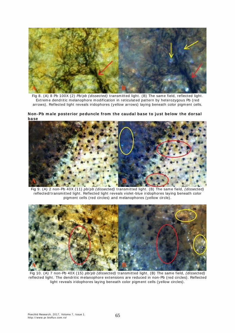
Fig 8. (A) 8 Pb 100X (2) Pb/pb (dissected) transmitted light. (B) The same field, reflected light.
Extreme dendritic melanophore modification in reticulated pattern by heterozygous Pb (red arrows). Reflected light reveals iridophores (yellow arrows) laying beneath color pigment cells.
Non-Pb male posterior peduncle from the caudal base to just below the dorsal base
Fig 9. (A) 2 non-Pb 40X (11) pb/pb (dissected) transmitted light. (B) The same field, (dissected)
reflected/transmitted light. Reflected light reveals violet-blue iridophores laying beneath color pigment cells (red circles) and melanophores (yellow circle).
Fig 10. (A) 7 non-Pb 40X (15) pb/pb (dissected) transmitted light. (B) The same field, (dissected)
reflected light. The dendritic melanophore extensions are reduced in non-Pb (red circles). Reflected light reveals iridophores laying beneath color pigment cells (yellow circles).

Poeciliid Research, 2017, Volume 7, Issue 1. http://www.pr.bioflux.com.ro/ 66
III. Cellular Comparison: early coloration in Pb Pb/pb vs. non-Pb pb/pb male Guppies (Poecilia reticulata). The following examples of early coloration in non-Pb and Pb show male expression of violet-blue iridophores macroscopically (Fig 11) and in 100x and 400x (Fig 12-14). Visual distinction is easily made between the two iridophore types. Changes in magnification, progressive focal shift, adjustment in angle of incident lighting or direction of light (reflected or transmitted) consistently failed to remove this visible distinction between violet and blue. Thus, this demonstrates two distinct iridophore populations in the blue-violet spectrum. Two distinct observations are offered based on early coloration. First, violet and blue iridophores appear “randomly collected among themselves” in similar fashion (Fig 13-14), as opposed to later mature coloration in which violet and blue iridophores are arranged together in “joined alternating color” groupings in dissimilar fashion. This shows that coloration is nearly complete, while migration to their final location is not. Second, melanophore shape is predominately corolla or punctate in early coloration (Fig 12-14), as opposed to mature coloration in which dendrites dominate. This indicates that members of the melanophore population are in place, while their final shape is not established. Side by side presentation of similar locations in Pb and non-Pb are presented.
Fig 11. (A) 6 Pb male (grey) Pb/pb. The orange peduncle ornament modified purplish-pink by Pb in red circle. (B) 3 non-Pb male (grey) pb/pb. The orange peduncle ornament unmodified in red circle.
All slide images taken from posterior orange spot (red circle).
Fig 12. Melanophore extensions in Pb vs. non-Pb in fish of similar age. (A) 6 Pb 40X (14) Pb/pb
(dissected) transmitted light. Increased numbers of dendritic melanophore extensions (red arrows) in Pb at similar age. (B) 3 non-Pb 40X (12) pb/pb (dissected) transmitted light. Decreased
numbers of dendritic melanophore extensions (red arrows) in Pb at similar age.

Poeciliid Research, 2017, Volume 7, Issue 1. http://www.pr.bioflux.com.ro/ 67
Fig 13. Melanophore extensions in Pb vs. non-Pb in fish of similar age. (A) 6 Pb 40X (8) Pb/pb (dissected) transmitted light. Increased numbers of dendritic melanophore extensions in Pb at similar age (red arrows). (B) 3 non-Pb 40X (4) pb/pb (dissected) transmitted light. More evenly
distributed blend of violet and blue irdophores in non-Pb as compared to Pb (red circles).
Fig 14. (A) 6 Pb 100X (12) Pb/pb (dissected) transmitted light. More violet iridophores than blue irdophores. (B) 3 non-Pb 100X (14) pb/pb (dissected) transmitted light. There is a more evenly
distributed blend of violet and blue irdophores in non-Pb as compared to Pb (red circles).
IV. Cellular Comparison: late coloration in Pb Pb/pb vs. non-Pb pb/pb male Guppies (Poecilia reticulata). Macroscopically (Fig 15) and microscopically (Fig 16-20) visible in heterozygous Pb are partial reductions in collected xanthophores, and in homozygous Pb near complete removal of collected and clustered xanthophores. Yellow color cell populations consisting of isolated “wild-type” single cell xanthophores remain intact. Dendritic melanophores in heterozygous and homozygous Pb condition, for mature individuals, reveal that dendrite structure is extremely extended and finer in appearance (Fig 18-19). Dendrites are linked together in “chain-like” strings intermingled with violet-blue iridophores in chromatophores units, while corolla melanophores are present in lessor numbers and punctate melanophores nearly absent (Fig 18). Darker appearance, both microscopically and phenotypically, results from modification of existing melanophore structures into extended dendrites. All major classes of chromatophores were present in the rear peduncle spot and adjoining areas in both Pb and non-Pb. Violet-blue iridophores are more visible in Pb vs. non-Pb, with variability between study specimens. An increase in the ratio of violet to blue iridophores was observed. Collected and clustered xanthophore populations, found in non-Pb members of the contemporary group, were reduced in heterozygous Pb condition and removed in homozygous Pb condition. The retention of isolated xanthophores remained intact in both heterozygous and homozygous Pb condition. Violet and blue iridophores appear arranged together in “joined alternating color” groupings in dissimilar fashion, as opposed to early mature coloration in which violet and

Poeciliid Research, 2017, Volume 7, Issue 1. http://www.pr.bioflux.com.ro/ 68
blue iridophores appear “randomly collected among themselves”. This indicates that coloration and migration are complete. Dendritic melanophores dominate shape, as opposed to early coloration in which corolla or punctate melanophores outnumber dendritic melanophores. This indicates that population numbers and shape are in place. Side by side presentation of similar locations in Pb and non-Pb are presented.
Fig 15. (A) Heterozygous 11 Pb male Pb/pb. Orange peduncle ornament modified purplish-pink by Pb in red circle. (B) 6 non-Pb male pb/pb. Orange peduncle ornament unmodified in red circle. All
slide images taken from posterior orange spot (red circle).
Fig 16. (A) 13 Pb 40X (4) Pb/Pb (dissected) reflected light. Expected higher visiblity of
erythrophores (red circle) with reduction of xanthophores by Pb modification. (B) 14 non-Pb 40X (3) pb/pb (dissected) reflected light. Expected evenly distributed xantho-erythrophores (red circle)
in non-Pb.
Fig 17. (A) 13 Pb 40X (4) Pb/Pb (dissected) reflected/transmitted light. Expected higher visiblity of erythrophores (green circle) with reduction of xanthophores by Pb modification. (B) 14 non-Pb 40X (3) pb/pb (dissected) transmitted light. Expected evenly distributed xantho-erythrophores (green
circle) in non-Pb.
Poeciliid Research, 2017, Volume 7, Issue 1. http://www.pr.bioflux.com.ro/ 69
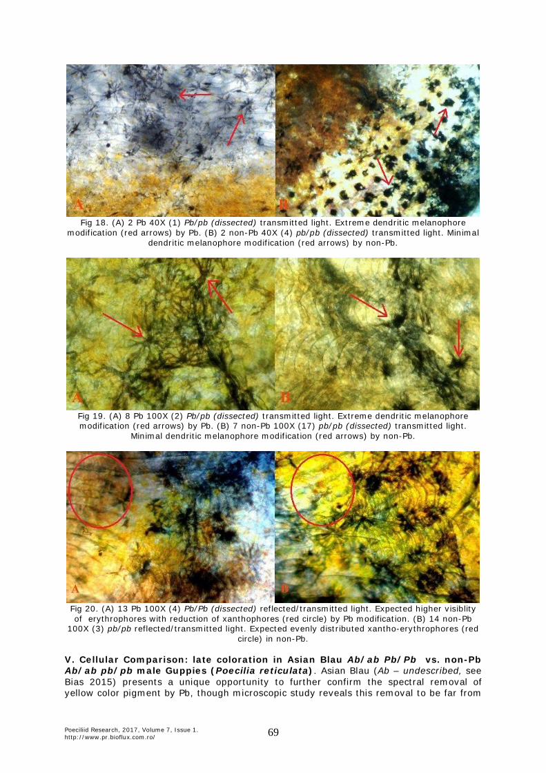
Fig 18. (A) 2 Pb 40X (1) Pb/pb (dissected) transmitted light. Extreme dendritic melanophore
modification (red arrows) by Pb. (B) 2 non-Pb 40X (4) pb/pb (dissected) transmitted light. Minimal dendritic melanophore modification (red arrows) by non-Pb.
Fig 19. (A) 8 Pb 100X (2) Pb/pb (dissected) transmitted light. Extreme dendritic melanophore modification (red arrows) by Pb. (B) 7 non-Pb 100X (17) pb/pb (dissected) transmitted light.
Minimal dendritic melanophore modification (red arrows) by non-Pb.
Fig 20. (A) 13 Pb 100X (4) Pb/Pb (dissected) reflected/transmitted light. Expected higher visiblity of erythrophores with reduction of xanthophores (red circle) by Pb modification. (B) 14 non-Pb
100X (3) pb/pb reflected/transmitted light. Expected evenly distributed xantho-erythrophores (red circle) in non-Pb.
V. Cellular Comparison: late coloration in Asian Blau Ab/ab Pb/Pb vs. non-Pb Ab/ab pb/pb male Guppies (Poecilia reticulata). Asian Blau (Ab – undescribed, see Bias 2015) presents a unique opportunity to further confirm the spectral removal of yellow color pigment by Pb, though microscopic study reveals this removal to be far from

Poeciliid Research, 2017, Volume 7, Issue 1. http://www.pr.bioflux.com.ro/ 70
complete. Autosomal incompletely dominant Ab, as opposed to autosomal recessive European Blau (r or r1, Dzwillo 1959) in heterozygous and homozygous condition removes red color pigment (Fig 21B). Collected yellow color pigment and clustered Metal Gold (Mg - undescribed, see Bias 2015) xanthophores are little affected by this erythrophore defect (Fig 21B), as shown in the pb/pb Ab/ab male. The following macroscopic photo clearly reveals near complete removal of densely packed collected yellow cells in the Pb/Pb Ab/ab (Fig 21A) male, leaving an underlying “circular ring” of violet-blue iridophores intact. As previously noted, Pb in itself has little or no effect on erythrophore populations. Albeit, Pb modification results in increased expression of violet iridophores easily seen in Ab modification (Fig 21A vs. 21B). The macroscopic presence of underlying iridophores, lacking a xantho-erythrophore (yellow-orange) overlay in Pb/Pb Ab/ab (Fig 21A) and lacking erythrophore (orange) overly in non-Pb pb/pb Ab/ab (Fig 21B), allows for the visual distinction between xantho-erythrophore populations.
Fig 21. (A) 5 Pb male (grey) Pb/Pb Ab/ab. Orange peduncle ornament modified violet-blue by
combination of Ab + Pb xantho-erythrophore removal in red circle. (B) 4 non-Pb male (grey) pb/pb Ab/ab. Orange peduncle ornament modified yellow by Ab erythrophore removal in red circle. All
slide images taken from posterior orange spot (red circle).
Though structural differences between xanthophores and erythrophores may be limited to variability in placement of underlying reflective crystalline platelets (Kottler et al 2014), microscopically the prior results are confirmed in comparison. Dendrites remain extremely extended and linked together in “chain-like” strings intermingled with violet-blue iridophores in chromatophore units. A side by side comparison of similar locations for Pb (Pb/Pb Ab/ab) and non-Pb (pb/pb Ab/ab) is presented (Fig 22-23).
Fig 22. (A) 5 Pb 40X (16) Pb/Pb Ab/ab (dissected) reflected/transmitted light. Underlying violet-
blue iridophore structure (red circle) is clearly revealed in absence of collected xantho-erythrophores. (B) 4 non-Pb 40X (8) pb/pb Ab/ab (dissected) reflected/transmitted light. Collected
xanthophores (red circle) masking violet-blue iridophores in absence of erythrophores.
Poeciliid Research, 2017, Volume 7, Issue 1. http://www.pr.bioflux.com.ro/ 71
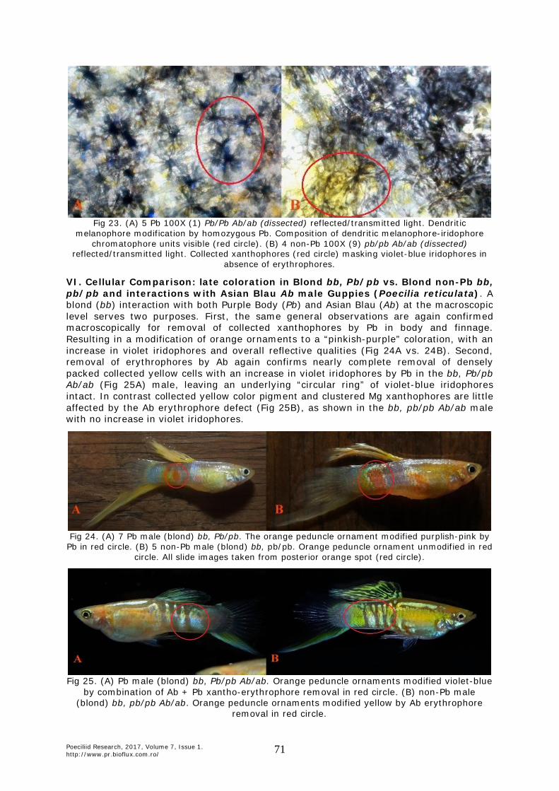
Fig 23. (A) 5 Pb 100X (1) Pb/Pb Ab/ab (dissected) reflected/transmitted light. Dendritic
melanophore modification by homozygous Pb. Composition of dendritic melanophore-iridophore chromatophore units visible (red circle). (B) 4 non-Pb 100X (9) pb/pb Ab/ab (dissected)
reflected/transmitted light. Collected xanthophores (red circle) masking violet-blue iridophores in absence of erythrophores.
VI. Cellular Comparison: late coloration in Blond bb, Pb/pb vs. Blond non-Pb bb, pb/pb and interactions with Asian Blau Ab male Guppies (Poecilia reticulata). A blond (bb) interaction with both Purple Body (Pb) and Asian Blau (Ab) at the macroscopic level serves two purposes. First, the same general observations are again confirmed macroscopically for removal of collected xanthophores by Pb in body and finnage. Resulting in a modification of orange ornaments to a “pinkish-purple” coloration, with an increase in violet iridophores and overall reflective qualities (Fig 24A vs. 24B). Second, removal of erythrophores by Ab again confirms nearly complete removal of densely packed collected yellow cells with an increase in violet iridophores by Pb in the bb, Pb/pb Ab/ab (Fig 25A) male, leaving an underlying “circular ring” of violet-blue iridophores intact. In contrast collected yellow color pigment and clustered Mg xanthophores are little affected by the Ab erythrophore defect (Fig 25B), as shown in the bb, pb/pb Ab/ab male with no increase in violet iridophores.
Fig 24. (A) 7 Pb male (blond) bb, Pb/pb. The orange peduncle ornament modified purplish-pink by
Pb in red circle. (B) 5 non-Pb male (blond) bb, pb/pb. Orange peduncle ornament unmodified in red circle. All slide images taken from posterior orange spot (red circle).
Fig 25. (A) Pb male (blond) bb, Pb/pb Ab/ab. Orange peduncle ornaments modified violet-blue
by combination of Ab + Pb xantho-erythrophore removal in red circle. (B) non-Pb male (blond) bb, pb/pb Ab/ab. Orange peduncle ornaments modified yellow by Ab erythrophore
removal in red circle.

Poeciliid Research, 2017, Volume 7, Issue 1. http://www.pr.bioflux.com.ro/ 72
Modification by Blond results in near normal population levels of melanophores, while they are reduced in size. Blond Pb corolla melanophores are similarly clumped together along scale edging or isolated groups, in the equivalent of grey Pb “chain-like” strings, and overlay violet-blue iridophore chromatophore units. Microscopically, it is noted that the presence of large Pb modified melanophore dendrites is not required for expression of modified “pinkish-purple” ornaments; rather only “brightness” is altered by their reduction. A side by side comparison of similar locations for Pb bb, Pb/Pb Ab/ab and non-Pb bb, pb/pb Ab/ab is presented (Fig 26-28). Rear Peduncle Spot (dissected)
Fig 26. (A) 7 Pb 40X (16) bb, Pb/pb (dissected) transmitted light. Minimally dendritic and corolla
melanophores have not started to form (red circle). Incomplete removal of “wild-type” xanthophores is visible (green circle). (B) 5 non-Pb 40X (3) bb, pb/pb (dissected) transmitted light. Expected evenly distributed xantho-erythrophores (green circle) present in non-Pb. Melanophores
remain punctate (red circle).
Fig 27. (A) 7 Pb 40X (6) bb, Pb/pb (dissected) transmitted light. The expected higher concentration
of erythrophores are present, while incomplete removal of “wild-type” xanthophores is visible (yellow circle). Minimally dendritic and corolla melanophores are present (red circles). (B) 5 non-Pb 40X (4) bb, pb/pb (dissected) transmitted light. Expected evenly distributed xantho-erythrophores
(yellow circle) present in non-Pb. Melanophores are corolla and punctate (red circle).

Poeciliid Research, 2017, Volume 7, Issue 1. http://www.pr.bioflux.com.ro/ 73
Fig 28. (A) 27 Pb 100X (34) bb, Pb/Pb (dissected) transmitted light revealing increased violet-blue iridophore expression (green circles). Minimally dendritic melanophores are present (green arrows).
(B) 28 non-Pb 100X (1) bb, pb/pb (dissected) transmitted light revealing balanced violet-blue iridophores (yellow circle).
VII. Cellular Comparison: late coloration in Golden gg, Pb/pb vs. Golden non-Pb gg, pb/pb male Guppies (Poecilia reticulata). Macroscopically ornamental spot coloration is modified from a highly reflective orange to a “pinkish-purple” in Golden (g, Fig 29) (Goodrich et al 1944). Orange ornament is converted to pinkish-purple in Pb by xanthophore removal, resulting in a smaller area of pigmentation and revealing increased underlying violet-blue structural color. Golden in turn reduces and aggregates melanophores, resulting in even further “constriction” of color pigments as compared to non-Golden.
Fig 29. (A) Golden Pb gg, Pb/pb expressing “constricted” modified pinkish-purple. (B) Golden non-
Pb gg, pb/pb expressing “constricted” orange ornaments. VIII. Cellular Comparison: homozygous Pb Pb/Pb, heterozygous Pb Pb/pb and non-Pb pb/pb under reflected, transmitted, reflected and transmitted lighting. Our study revealed that the presence of all major classes of chromatophores (melanophores, xanthophores, erythrophores, violet-blue iridophores) and crystalline platelets were present in Pb and non-Pb condition (Fig 30-45). Collected and clustered xanthophore populations were reduced in heterozygous Pb condition and removed in homozygous Pb condition, as seen macroscopically (Fig 30, 34, 37 and 40) and microscopically (Fig 31-33, 35-36, 38-39 and 41-45). The retention of isolated xanthophores, found in all parts of the body and fins in “wild-type”, remained intact in heterozygous and homozygous Pb condition. As previously noted, Pb in itself has little or no effect on erythrophore populations. The presence of underlying iridophores, lacking a xanthophore (yellow) overlay in Pb, with retained erythrophore (orange) overly produces a distinct “pinkish-purple” coloration in heterozygous and homozygous Pb condition, which is visible both macroscopically and microscopically. The alteration of orange spotting is complete in homozygous Pb, and limited to specific regions in heterozygous Pb (See: Bias & Squire 2017a).

Poeciliid Research, 2017, Volume 7, Issue 1. http://www.pr.bioflux.com.ro/ 74
Homozygous Pb specimens (Fig 30-33) exhibited an “overall” higher incidence of violet iridophores, a proliferation of dendritic melanophores and a dense layer of violet-blue iridophores, as compared to heterozygous Pb (Fig 34-36) and non-Pb (Fig 37-45). A “darker” appearance in Pb vs. non-Pb appears to largely be the result of modification of existing melanophore structures (corolla and punctate) into extended dendrites. An increase in melanophore population levels is not evidenced. Dendritic melanophores in heterozygous and homozygous Pb condition, for mature individuals, reveal that dendrites are extremely extended and finer in appearance. Dendrites are linked together in “chain-like” strings intermingled with violet-blue iridophores in chromatophore units forming the reticulated pattern and in isolated units. This chain-like expression is minimally present in non-Pb (Fig 38-39, 41-47), further present in heterozygous Pb (Fig 35-36), and amplified in homozygous Pb (Fig 31-33). Within the rear peduncle orange spot and surrounding edges a noticeable absence of corolla and punctate melanophores is often evident in Pb. This absence was reduced in other regions of the body or was specific to individuals.
Fig 30. 13 Pb male (grey) – Homozygous Pb/Pb. The orange peduncle ornaments are modified purplish-pink by Pb in the red circle, though dark from the high angle ambient light. All slide
images taken from posterior orange spot (red circle).
Rear Peduncle Spot (non-dissected)
Fig 31. (A) 13 Pb 40X (4) PbPb transmitted light white balance adjusted. Erythrophore positions
(green circles) are clearly indicated by altered “maroon color” in relation ship to xanthophores with transmitted light and adjusted white balance. (B) The same field, reflected/transmitted light.

Poeciliid Research, 2017, Volume 7, Issue 1. http://www.pr.bioflux.com.ro/ 75
Fig 32. (A) 13 Pb 40X (5) PbPb reflected light. Reflected light reveals that most of reflective qualities are produced by violet-blue iridophores within scale rings (red circle) and dendritic
melanophore-iridophore chromatophore units (yellow circle). (B) The same field, transmitted light white balance adjusted.
Rear Peduncle Spot (dissected)
Fig 33. (A) 13 Pb 40X (4) Pb/Pb reflected light. Violet-blue iridophores shown residing below
xantho-erythrophores, and contributing to reflective qualities (red circles). The expected presence of “chain-like” dendritic melanophores (yellow circle) present along the scale edging to form
reticulation, dendrites are “extreme” in shape compared to Pb expression (green circle). (B) The same field, transmitted light.
Fig 34. 15 Pb male (grey Pingtung feral) – Heterozygous Pb/pb. The orange peduncle ornaments modified purplish-pink by Pb in the red circle, though dark from the high angle ambient light. All
slide images taken from posterior orange spot (red circle).

Poeciliid Research, 2017, Volume 7, Issue 1. http://www.pr.bioflux.com.ro/ 76
Rear Peduncle Spot (non-dissected)
Fig 35. (A) 15 Pb 40X (6) Pb/pb reflected light. High density lower level and scale ring violet-blue iridophores (red circle) residing at the edge of spotting ornament. (B) The same field, transmitted
light white balance adjusted. (C) The same field, transmitted. Violet-blue iridophores, within ectopic dendritic melanophore-iridophore chromatophore units (green circles), while minimally
reflective remain visible under all transmitted light conditions.
Rear Peduncle Spot (dissected)
Fig 36. (A) 15 Pb 100X (4) Pb/pb reflected light. Dendritic xanthophore structures are highlighted
under reflected light (red arrows). (B) The same field, reflected/transmitted. Erythrophore expression is minimal under combined reflected/transmitted light near the edge of spotting
ornament (red circle). (B) The same field, transmitted light. Isolated erythrophore expression highlighted under transmitted light (red arrows).
Fig 37. 14 non-Pb male (grey) pb/pb. Orange peduncle ornament unmodified in red circle. All slide
images taken from posterior orange spot (red circle).

Poeciliid Research, 2017, Volume 7, Issue 1. http://www.pr.bioflux.com.ro/ 77
Rear Peduncle Spot (non-dissected)
Fig 38. (A) 14 non-Pb 40X (1) pb/pb reflected light. The location of violet-blue iridophores (yellow circles) is shown not to be limited to non-color pigmented areas. This indicates a solid structural
color “layer” regardless of presence or absence of other color pigments. (B) The same field, reflected/transmitted light. Combined reflected/transmitted light suggestive of positioning of
carotenoid orange pigment (green circles) to be slightly above that of xanthophores. (C) The same field, transmitted light. Transmitted light again allows erythrophore expression (green circle) to
“overpwer” that of xanthophores.
Rear Peduncle Spot (dissected)
Fig 39. (A) 14 non-Pb 40X (3) pb/pb reflected. The expected presence of “chain-like” dendritic
melanophores (red circle) are present along scale edging to form reticulation, though dendrites are not as “extreme” in shape compared to Pb expression. (B) The same field, transmitted light.
Higher levels of xanthophore (red circle) are expressed in non-Pb.
Fig 40. 16 non-Pb male (grey Kelly) pb/pb. The orange peduncle ornament is unmodified in red
circle. Images are taken from posterior orange spot (red circle). Reticulation expression is reduced in the absence of multiple color ornaments.

Poeciliid Research, 2017, Volume 7, Issue 1. http://www.pr.bioflux.com.ro/ 78
Rear Peduncle Spot (non-dissected)
Fig 41. (A) 16 non-Pb 40X (2) pb/pb reflected light. High concentration of violet-blue iridophores (red circles) underlying color pigments, in absence of color pigments and within scale rings (red
arrows). (B) The same field, transmitted light. Melanophores comprised of punctate (1), corolla (2) and dendritic (3) cells (green arrows).
Rear Peduncle Spot (dissected)
Fig 42. (A) 16 non-Pb 40X (2) pb/pb reflected light. (B) The same field, reflected white balance adjusted. Dendritic melanophore-iridophore chromatophore units comprise much of the dark
pattern (red circles). Erythrophores (green circles) are visible under transmitted light.
Fig 43. (A) 16 non-Pb 40X (6) pb/pb reflected light. (B) The same field, reflected/transmitted light. This mature male lacks additional spotting ornaments. As a result melanophores (yellow
circles) appear “less organized” in comparison to heavily ornamented individuals.

Poeciliid Research, 2017, Volume 7, Issue 1. http://www.pr.bioflux.com.ro/ 79
Fig 44. 16 non-Pb 100X (2) pb/pb reflected/transmitted light white balance adjusted. Violet-blue
iridophores (red arrows) visible in scale rings under reflected light contributing to reflective qualities. Their presence was also revealed in previous scale dissections. Dendrites (red circle)
under reflected/transmitted light appear much “denser” from interactions with violet-blue iridophores. Variation in dendrite shape at a single focal length indicates an ectopic orientation at
distinct layers.
Fig 45. (A) 16 non-Pb 100X 9 pb/pb transmitted light. Xantho-erythrophores (red circle) appear
“balanced” in expression lacking Pb modification. No extreme modification of dendrites (red arrows) is visible. Well defined layers between structural color and color pigment. Even with minimal
dendritic extension, some collection of violet-blue iridophores into ectopic dendritic melanophore-iridophore chromatophore units (green circles) is still visible.
IX. Subcutaneous and Spinal Chromatophores. Macroscopic qualities of non-Pb study male (Fig 46). Melanophore, xanthophore and violet-blue iridophore were found adhering to the spinal column and ribs (Fig 47). No evidence of subcutaneous erythrophores was detected below the dermis, though suspected in micro-dissected the xanthophore cluster (Fig 48); all “red coloration” identified as blood cells. This raises an issue in regard to the synthesized pteridine and dietary carotenoid pigment resource allocation in non-visible locations (Goodwin 1984; Grether et al 1999, 2001). Subcutaneous melanophore, xanthophore, violet-blue iridophore, leucophore and crystalline platelets (Fig 49) were found resident in low numbers in dissected deep tissue. Their presence at these locations is well below the dermis (Bagnara et al 1968).

Poeciliid Research, 2017, Volume 7, Issue 1. http://www.pr.bioflux.com.ro/ 80
Fig 46. 25 non-Pb (McWhite) pb/pb. The orange peduncle ornament is unmodified in red circle.
Images are taken from deep tissue and spinal extraction.
Fig 47. (A) 25 non-Pb 100X (9) (micro-dissected spinal column) reflected light. Visible dendritic melanophores (red arrows) and xanthophores (red circle) residing on the freshly extracted and cleaned spinal column, rinsed with saline solution. (B) The same fish (partially dissected spinal column) reflected/transmitted light. Visible violet-blue iridophores (red arrows) are residing just
above dendritic melanophores and xanthophores on the freshly dissected and cleaned spinal column, rinsed with saline solution.
Fig 48. (A) 25 non-Pb 40X (1) (micro-dissected tissue) reflected light. (B) The same field 100X 1
(micro-dissected tissue) reflected light. Isolated yellow xanthophore cluster (red arrows) and reflective crystalline platelets (green arrows) from subcutaneous deep tissue sample.

Poeciliid Research, 2017, Volume 7, Issue 1. http://www.pr.bioflux.com.ro/ 81
Fig 49. Proximal spine side up (inverted) views of same location. (A) 25 non-Pb 40X (1)
(dissected tissue) reflected light. Visibly radiating dendritic melanophores (red arrows) and xanthophores (green arrow) are present. Bright white areas are now inverted crystalline
platelets (yellow arrow) above scattered violet-blue iridophores. This shows that crystals are reflective both distally and proximally under any available reflected light. White leucophores
(blue arrow) are “grey” in appearance. (B) The same field 40X 1 (dissected tissue) reflected/transmitted light. Aggregated dendritic melanophores and violet-blue iridophores forming ectopic melanophore-iridophore chromatophore units (red circles) are visible with
isolated yellow xanthophore cluster (green arrow). Reflective crystalline platelets remain white (yellow arrow). White leucophores are “grey” in appearance.
Our study revealed all major classes of chromatophores (melanophores, xanthophores, erythrophores, and violet-blue iridophores) and also crystalline platelets were present in both Pb and non-Pb dermal layers. All were found present in and around the eye structure. Scales contained their own distinct populations, and all but erythrophores were found. In dermal layers, the dendrites of ectopic dendritic melanophore structures appeared to be tilted when melanosomes are fully dispersed. Collected and clustered xanthophore populations were reduced in heterozygous Pb condition and nearly removed in homozygous condition. Isolated clustered xanthophore found over all parts of the body and fins in “wild-type”, remained intact in both heterozygous and homozygous Pb condition. Melanophores, xanthophores, violet-blue iridophores, leucophores and crystalline platelets were found resident in subcutaneous deep tissue samples well below the dermis. Deep tissue crystalline platelet orientation appears to be tilted, as they are reflective both distally and proximally under reflected light. Dendritic melanophores radiate in deep tissue indicating 3-dimensional (3-D) orientation within the subcutaneous tissue. Melanophores, xanthophores, violet-blue iridophores, and also crystalline platelets were found adhering to the spinal column. An aggregation of dendritic melanophores and violet-blue iridophores forming ectopic melanophore-iridophore chromatophore units (Fig. 1 and Fig. 6c, Kottler et al 2014; Fig 17A-B, Bias & Squire 2017a), in cutaneous and subcutaneous levels is supportive of structural color cells residing at multiple layers, and being above and below the distinct iridophore layer as described in the Bagnara Dermal Chromatophore Unit (Bagnara et al 1968). The presence of underlying dermal level violet-blue iridophores and crystalline platelets, lacking a xanthophore (yellow) overlay in Pb, with a retained erythrophore (orange) layer produces a distinct “pinkish-purple” coloration in heterozygous and homozygous Pb condition, which is visible both macroscopically and microscopically. An increase in reflective qualities of violet-blue structural pigment is evidenced in Pb. Based on our study of “specific phenotypes”, Pb expresses a higher amount of violet iridophores or more balanced ratio of violet-blue iridophores. Violet-blue iridophores are shown, in both Pb and non-Pb, to form evenly distributed layers in the presence or absence of color pigments. There is evidence that xanthophores and erythrophores may reside at slightly diverse layers or angles within colored ornaments.

Poeciliid Research, 2017, Volume 7, Issue 1. http://www.pr.bioflux.com.ro/ 82
The violet-blue iridophore chromatophore unit (Fig 2A-B, Bias & Squire 2017a) and the removal of xanthophores by Pb modification are required to produce an all-purple phenotype. The Purple gene has the ability to modify existing genome-wide chromatophore populations in heterozygous and homozygous condition, with increased visibility in the UV and/or near-UV spectrum. As a result, this potentially demonstrates selection favoring “private” short wavelength signaling (Endler 1991; Millar & Hendry 2012). Visually, ornamental spot coloration is modified from a highly reflective orange to a “pinkish-purple” in Grey (g), Blond (b) and Golden (g) (Goodrich et al 1944). By further removal of erythrophores in European Blau (r) (Dzwillo 1959) and Asian Blau (Ab) (Undescribed - see Bias 2015) variants, ornaments are modified to a “violet-blue” revealing the remaining structural color. Pb modification is often most vividly noticeable in grey, blond and albino (Bias & Squire 2017d, forthcoming, PB Expression in Domestic Phenotypes). Microscopically, our results often show minimal structural differentiation between xantho-erythrophores, with differences in population levels, yellow-orange coloration, and collection or clustering of xanthophores. Yellow color cell populations consisting of isolated “wild-type” single cell xanthophores remain intact in conjunction with Pb modification. The same distinctions between heterozygous and homozygous Pb are generally confirmed in macroscopic observations. The population of melanophores does not appear to drastically increase with Pb modification, only the size and shape of the melanophores themselves are altered. When comparing the macroscopic and microscopic results, between documented autosomal genes modifying melanophores, it becomes apparent that while melanophores are modified by Pb, their modification in size or shape by Blond or Golden is not required for “Purple / Violet” or “pinkish-purple” expression by Pb. Nor is it prevented by the lack of melanophores in Albino (a) (Haskins & Haskins 1948), see examples in: (Bias & Squire 2017d, forthcoming, PB Expression in Domestic Phenotypes). Rather, melanophore presence, modification or absence only determines the actual “shade” of pinkish-purple modification in ornamental spots and overall body color when color pigment is present. Motility of melanophore structures was not addressed in this study, though constriction of melanophores is known to occur (Nayudu & Hunter 1979). Melanophore constriction was noticed during ocular study as the length of time that each slide sample was observed progressed. Frequent evidence of dendritic and/or motile yellow color pigment (xanthophore) structures was detected in this study. Both when length of time increased between preparations of euthanized specimens, and as the length of time each slide sample was observed progressed. For these reasons preparation was done immediately after euthanasia and observations kept to under an hour. No constriction was found for dendritic and/or motile iridophores, outside of violet-blue iridophore clustering associated with ectopic dendritic melanophores. It was noted on multiple occasions after extended periods of observation that what appeared to iridophores would randomly “fire” during cell death. Yet, after drying and rehydration reflective qualities of iridophores persevere. Zygosity and genotype specific, the ratio of violet to blue iridophores appears higher in Pb vs. non-Pb. Whether there is an actual increase in iridophore population numbers or simply an increase in visibility, due to reductions or removal of xanthophores and/or altered melanophores was not addressed in this study. Nor was the issue of increased reflectivity in Pb through visibility and possible modification to angles at which crystalline platelets reside beneath iridophore layers and basal level melanophores. Clearly much room exists for further and more complex cellular level research involving Purple Body modification. Taken as a whole, macroscopic and microscopic results reveal a complex interaction between all major chromatophores types and crystalline platelets is required to produce the overall purple / violet sheen and pinkish-purple modification of ornamental spotting in P. reticulata by Pb. These cell types combine to produce not only background body coloration, but also increased reflectivity in the UV and/or near-UV spectrum.

Poeciliid Research, 2017, Volume 7, Issue 1. http://www.pr.bioflux.com.ro/ 83
Poecilia reticulata exist in a recently documented (Bias & Squire 2017a) polymorphic state; Autosomal Dominant Purple Body (Pb/Pb and Pb/pb) and non-Purple Body (pb/pb). The co-existence of the two phenotypes suggests a selective advantage under predation (crypsis) and in sexual selection (conspicuous pattern) under diverse ambient lighting conditions. Pb has been identified as the first polymorphic autosomal gene to be described as existent in high frequencies in wild, feral and Domestic Guppy populations. It is capable of pleiotropic effects on all existing color and pattern elements at multiple loci. It should therefore be considered a strong candidate for further studies involving “the relationships between spectral and ultrastructure characteristics” in orange ornamentation, and extending to color and/or pattern as a whole as suggested by Kottler et al (2014). A mechanism is identified by which Pb is capable of balancing overall color and pattern polymorphisms, in turn providing fitness through heterozygosity in diverse complex habitats (Bias & Squire 2017a). We hope that Purple will be mapped to its linkage group. Conclusions. The Purple Body phenotype results from a reduction in the number of collected (but not isolated) xanthophores and an apparently increased number of violet iridophores outnumbering blue iridophores. The ectopic melanophore dendrites are often extremely extended. Many corolla and punctate melanophores are replaced by (or converted into) dendritic melanophores. These effects are much more pronounced in homozygous Pb/Pb as opposed to heterozygous Pb/pb. The presence of these iridophores is not adequately detected under conventional transmitted light, and reflected light must be used either in addition to or instead of transmitted light alone. The reduction of xanthophores and increased visibility of violet-blue iridophores converts the highly reflective orange colored spot of non-Purple Body (pb/pb) to a purplish-pink spot. Pb alters overall chroma (purity and intensity), brightness (reflection) and hue (color or shade) of male ornaments. The use of the incompletely dominant Asian Blau mutant that removes red pigment combined with the incompletely dominant Pb gene that removes yellow pigment, each with three different genotypes and phenotypes, provides us with 9 different genotypes that further dissect the contributions of erythrophores, xanthophores, and xantho-erythrophores. In doing so, supports the presence of increased violet to blue iridophores beneath all color pigments. The composition of which alters brightness of male ornaments. The added use of the recessive gene Blond allows us to further greatly reduce the physical size and structure, but not numbers, of the melanophores and thus further visually dissect the contributions of each of these cell types to the Dermal Chromatophore Unit. In result shows that while Pb is demonstrative in “wild-type” grey of an increase in dendritic arm length; this modification of melanophore structure is not needed to produce a modified purplish-pink appearance. That being darker purple in grey and lighter pink in blond. Pb melanophore modification alters both chroma and hue in male ornaments. The combination of the Purple Body and the recessive Golden gene, which reduces numbers, increases size and collects the melanophores into aggregates, provides a further “constriction” of color pigments as compared to either non-Pb Golden or non-Golden. Revealing overall melanophore populations, structure and positioning have direct influence on hue of male ornaments. In summary, macroscopic observation and microscopic study of modification by the Purple Body (Pb) gene is shown to have direct influence on chroma, brightness and hue of male ornaments. Ethics Statement. This study adhered to established ethical practices under AVMA Guidelines for the Euthanasia of Animals: 2013 Edition, S6.2.2 Physical Methods (6). All euthanized specimens were photographed immediately, or as soon as possible, after temperature reduction (rapid chilling) in water (H20) at temperatures just above freezing (0°C) to avoid potential damage to tissue and chromatophores, while preserving maximum expression of motile xantho-erythrophores in Pb and non-Pb specimens. All

Poeciliid Research, 2017, Volume 7, Issue 1. http://www.pr.bioflux.com.ro/ 84
anesthetized specimens were photographed immediately after short-term immersion in a mixture of 50% aged tank water (H20) and 50% carbonated water (H2CO3). All dried specimens photographed immediately after rehydration in cold water (H20). Prior euthanasia was by cold water (H20) immersion at temperatures just above freezing (0°C). MS-222 (Tricaine methanesulfonate) was not used to avoid the potential for reported damage and/or alterations to chromatophores, in particular melanophores, prior to slide preparation. Competing Interests and Funding. The authors declare that they have no competing interests. Senior author is a member of the Editorial Board for Poeciliid Research; International Journal of the Bioflux Society, and requested non-affiliated independent peer review volunteers. The authors received no funding for this work. Notes. This publication is number two (2) of four (4) by Bias and Squire in the study of Purple Body (Pb) in Poecilia reticulata: 1. The Cellular Expression and Genetics of an Established Polymorphism in Poecilia
reticulata; “Purple Body, (Pb)” is an Autosomal Dominant Gene, 2. The Cellular Expression and Genetics of Purple Body (Pb) in Poecilia reticulata, and its
Interactions with Asian Blau (Ab) and Blond (bb) under Reflected and Transmitted Light,
3. The Cellular Expression and Genetics of Purple Body (Pb) in the Ocular Media of the Guppy Poecilia reticulata,
4. The Phenotypic Expression of Purple Body (Pb) in Domestic Guppy Strains of Poecilia reticulata.
Acknowledgments. To my best friend and wife Deana Bias, for her support and persistence over the last several years in this four part study... To my co-author and dear friend Rick Squire for his patience as a mentor… To those Domestic Breeders who willingly provided additionally needed pedigree strains and study populations for completion of this paper… References Bagnara J. T., Taylor J. D., Hadley M. E., 1968 The dermal chromatophore unit. The
Journal of Cell Biology 38(1):67-79. Bias A. S., 2015 Working with autosomal genes for color and pattern: a domestic guppy
breeder’s best friend and often worst nightmare. Presented Sept. 5, 2015 to attendees of the 18th World Guppy Contest held in Tampa, Florida, USA. Available at: https://www.academia.edu/15488221/Working_With_Autosomal_Genes_for_Color_and_Pattern_A_Domestic_Guppy_Breeders_best_friend_and_often_worst_nightmare_. Accessed: February, 2017.
Bias A. S., Squire R. D., 2017a The cellular expression and genetics of an established polymorphism in Poecilia reticulata; “Purple Body (Pb)” is an autosomal dominant gene. Poec Res 7(1):1-32.
Bias A. S., Squire R. D., 2017d (forthcoming) The phenotypic expression of Purple Body (Pb) in domestic guppy strains of Poecilia reticulata.
Dzwillo M., 1959 Genetische Untersuchungen an domestizierten Stammen von Lebistes reticulatus Peters. Mitt Hamburg Zool Mus Inst 57:575-584.
Endler J. A., 1978 A predator’s view of animal color patterns. In: Evolutionary biology. Hecht M. K., Steere W. C., Wallace B. (eds), Springer US, pp. 319-364.
Endler J. A. 1991 Variation in the appearance of guppy color patterns to guppies and their predators under different visual conditions. Vision Research 31(3):587-608.
Goodrich H. B., Josephson N. D., Trinkaus J. P., Slate J. M., 1944 The cellular expression and genetics of two new genes in Lebistes reticulatus. Genetics 29(6):584-592.

Poeciliid Research, 2017, Volume 7, Issue 1. http://www.pr.bioflux.com.ro/ 85
Goodwin T. W., 1984 The biochemistry of the carotenoids, vol. II. Chapman and Hall, London, ISBN: 0412237709 (v.2), 224 pp.
Grether G. F., Hudon J., Millie D. F., 1999 Carotenoid limitation of sexual coloration along an environmental gradient in guppies. Proceedings of the Royal Society of London B: Biological Sciences 266(1426):1317-1322.
Grether G. F., Hudon J., Endler J. A., 2001 Carotenoid scarcity, synthetic pteridine pigments and the evolution of sexual coloration in guppies (Poecilia reticulata). Proceedings of the Royal Society of London B: Biological Sciences 268(1473):1245-1253.
Haskins C. P., Haskins E. F., 1948 Albinism, a semi-lethal autosomal mutation in Lebistes reticulatus. Heredity 2(Pt 2):251-262.
Khoo G., Phang V. P. E., 2010 Cellular basis of colour in aquarium fishes. In: FISHMAIL 18(3):9-12. Publication of the Malaysian Fisheries Society. Available at: http://www.mfs.org.my/xdoc/fishmail/Fishmail%20July-Sept%202010.pdf. Accessed: January, 2017.
Kottler V. A., Fadeev A., Weigel D., Dreyer C., 2013 Pigment pattern formation in the guppy, Poecilia reticulata, involves the Kita and Csf1ra receptor tyrosine kinases. Genetics 194(3):631-646.
Kottler V. A., Koch I., Flötenmeyer M., Hashimoto H., Weigel D., Dreyer C., 2014 Multiple pigment cell types contribute to the black, blue, and orange ornaments of male guppies (Poecilia reticulata). PloS One 9(1):e85647.
Kottler V. A., Künstner A., Koch I., Flötenmeyer M., Langenecker T., Hoffmann M., Sharma E., Weigel D., Dreyer C., 2015 Adenylate cyclase 5 is required for melanophore and male pattern development in the guppy (Poecilia reticulata). Pigment Cell and Melanoma Research 28(5):545-558.
Millar N. P., Hendry A. P., 2012 Population divergence of private and non-private signals in wild guppies. Environmental Biology of Fishes 94(3):513-525.
Nayudu P. L., Hunter C. R., 1979 Cytological aspects and differential response to melatonin of melanophore based color mutants in the guppy, Poecilia reticulata. Copeia 1979(2):232-242.
Phang V. P. E., Chow O. K., Fernando A. A., 1985 Genetic analysis of scale chromatophores of two domesticated varieties of the guppy, Poecilia reticulata. Journal of the Singapore National Academy of Science 14:1-5.
Ueshima G., Nakajima M., Fujio Y., 1998 A study on the inheritance of body color and chromatophores in the guppy Poecilia reticulata. Tohoku Journal of Agricultural Research 48(3):111-122.
Supporting Information S1 Materials; Slide Specimen Photos Received: 25 April 2017. Accepted: 21 June 2017. Published online: 24 June 2017. Authors: Alan S. Bias, Independent Researcher and Swordtail Guppy Breeder. Mailing address: P.O. Box 1508, Lewisburg, West Virginia 24901, USA. orcid.org/0000-0002-9093-619X. [email protected] Squire Richard D., Biology Department (retired), University of Puerto Rico, Mayaguez campus, Mayaguez, Puerto Rico, USA. Mailing address: P. O. Box 3227, Mayaguez, P.R., USA 00681-3227. orcid.org/0000-0002- 3916-0672. [email protected] This is an open-access article distributed under the terms of the Creative Commons Attribution License, which permits unrestricted use, distribution and reproduction in any medium, provided the original author and source are credited. How to cite this article: Bias A. S., Squire R. D., 2017 The cellular expression and genetics of Purple Body (Pb) in Poecilia reticulata, and its interactions with Asian Blau (Ab) and Blond (bb) under reflected and transmitted light. Poec Res 7(1):59-85.


![[Aviation] SNS-2 GUPPY Ultralight Plans.pdf](https://img.pdfslide.us/doc/110x75/577c81dd1a28abe054ae7355/aviation-sns-2-guppy-ultralight-planspdf.jpg)