Embed Size (px)
Citation preview

2/18/2013
1
THE CELL
History of the microscope and cell
1500’s: Europe
Merchants used magnifying glasses to determine quality of cloth
Mid 1600’s: Holland, development of the microscope and telescope
1665: Robert Hooke (English)
** first to observe DEAD CELLS
1675: Anton van Leeuwenhoek (Dutch)
** first to observe LIVING CELLS
History, cont. 1833: Robert Brown (Scottish)
Discovered nucleus
1838: Matthias Schleiden (German)
Stated “all plants are made of cells”
1839: Theodor Schwann (Dutch)
Stated “all animals are made of cells”
1855: Rudolf Verchow (German MD)
Stated “all cells arise from other cells”
Characteristics of Microscopes
• magnification: ability to make an image larger than
actual size
• resolution: power to show details clearly while enlarged
(if poor, objects seem fuzzy)
Types of Microscopes
I. compound light
- light passes through one or more lenses
- object must be sliced thinly enough to be transparent
- upper limitation is 2000X or 0.5 microns (um) in diameter
II. Electron Microscopes
- limited by physical characteristics of light
- can magnify an image up to 200,000 X, or
2 nm in diameter
- beams of electrons produces enlarged
image
2/18/2013
2
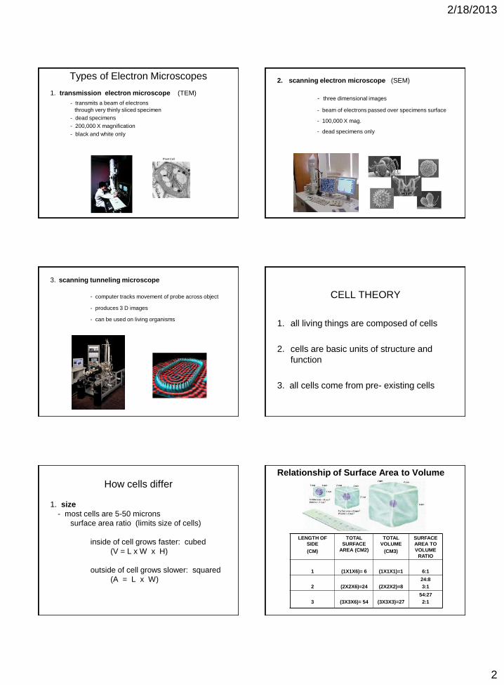
Types of Electron Microscopes
1. transmission electron microscope (TEM)
- transmits a beam of electrons
through very thinly sliced specimen
- dead specimens
- 200,000 X magnification
- black and white only
Plant Cell
2. scanning electron microscope (SEM)
- three dimensional images
- beam of electrons passed over specimens surface
- 100,000 X mag.
- dead specimens only
3. scanning tunneling microscope
- computer tracks movement of probe across object
- produces 3 D images
- can be used on living organisms
CELL THEORY
1. all living things are composed of cells
2. cells are basic units of structure and
function
3. all cells come from pre- existing cells
How cells differ
1. size
- most cells are 5-50 microns
surface area ratio (limits size of cells)
inside of cell grows faster: cubed
(V = L x W x H)
outside of cell grows slower: squared
(A = L x W)
Relationship of Surface Area to Volume
LENGTH OF
SIDE
(CM)
TOTAL
SURFACE
AREA (CM2)
TOTAL
VOLUME
(CM3)
SURFACE
AREA TO
VOLUME
RATIO
1
(1X1X6)= 6
(1X1X1)=1
6:1
2
(2X2X6)=24
(2X2X2)=8
24:8
3:1
3
(3X3X6)= 54
(3X3X3)=27
54:27
2:1
2/18/2013
3
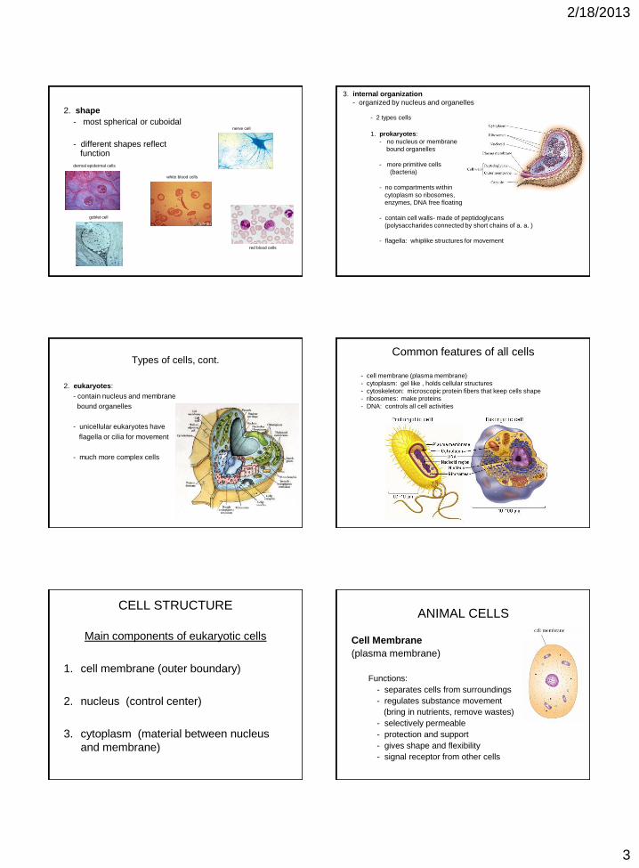
2. shape
- most spherical or cuboidal
- different shapes reflect function
dermal epidermal cells
white blood cells
goblet cell
red blood cells
nerve cell
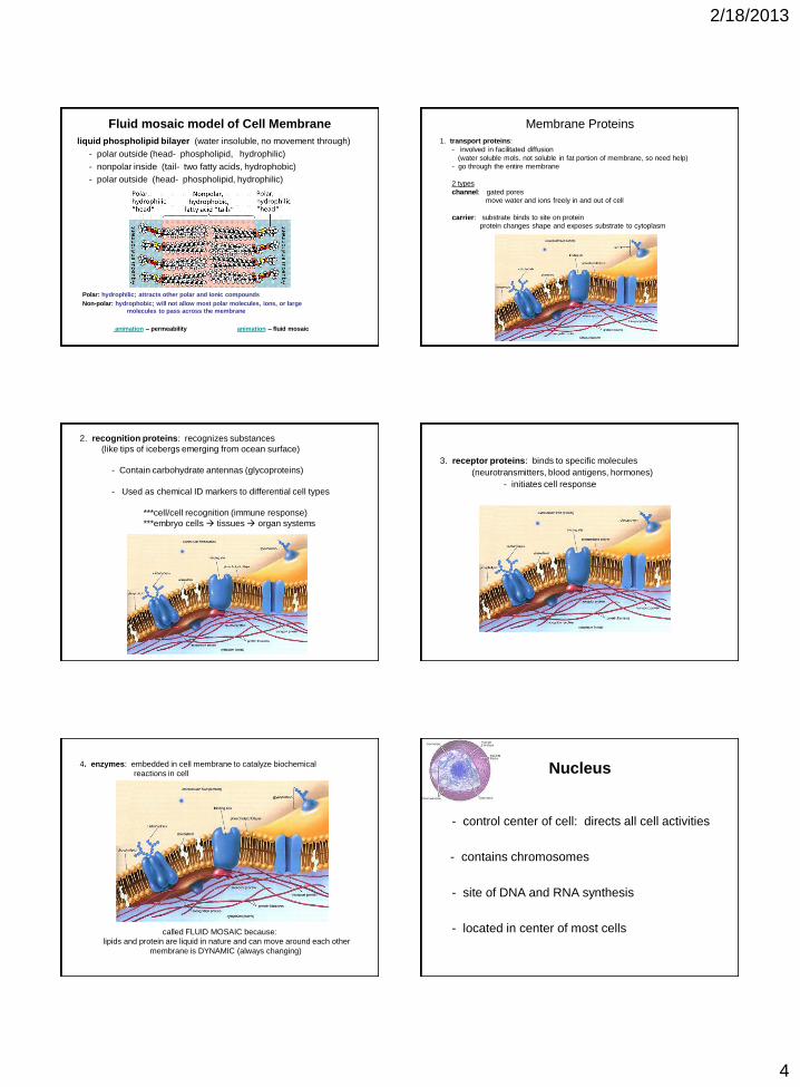
3. internal organization
- organized by nucleus and organelles
- 2 types cells
1. prokaryotes:
- no nucleus or membrane
bound organelles
- more primitive cells
(bacteria)
- no compartments within
cytoplasm so ribosomes,
enzymes, DNA free floating
- contain cell walls- made of peptidoglycans
(polysaccharides connected by short chains of a. a. )
- flagella: whiplike structures for movement
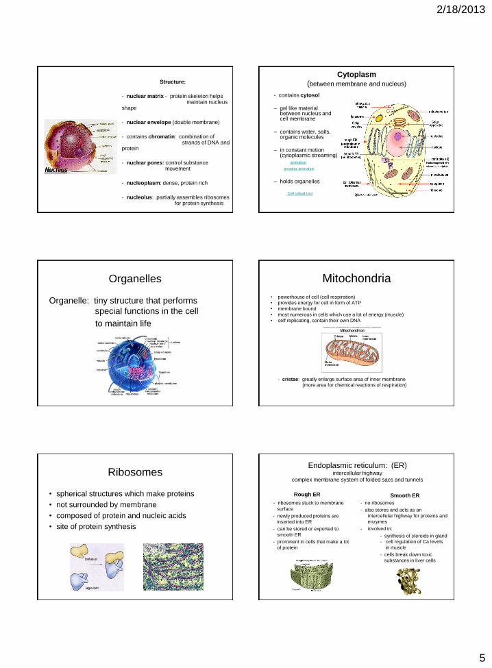
Types of cells, cont.
2. eukaryotes:
- contain nucleus and membrane
bound organelles
- unicellular eukaryotes have
flagella or cilia for movement
- much more complex cells
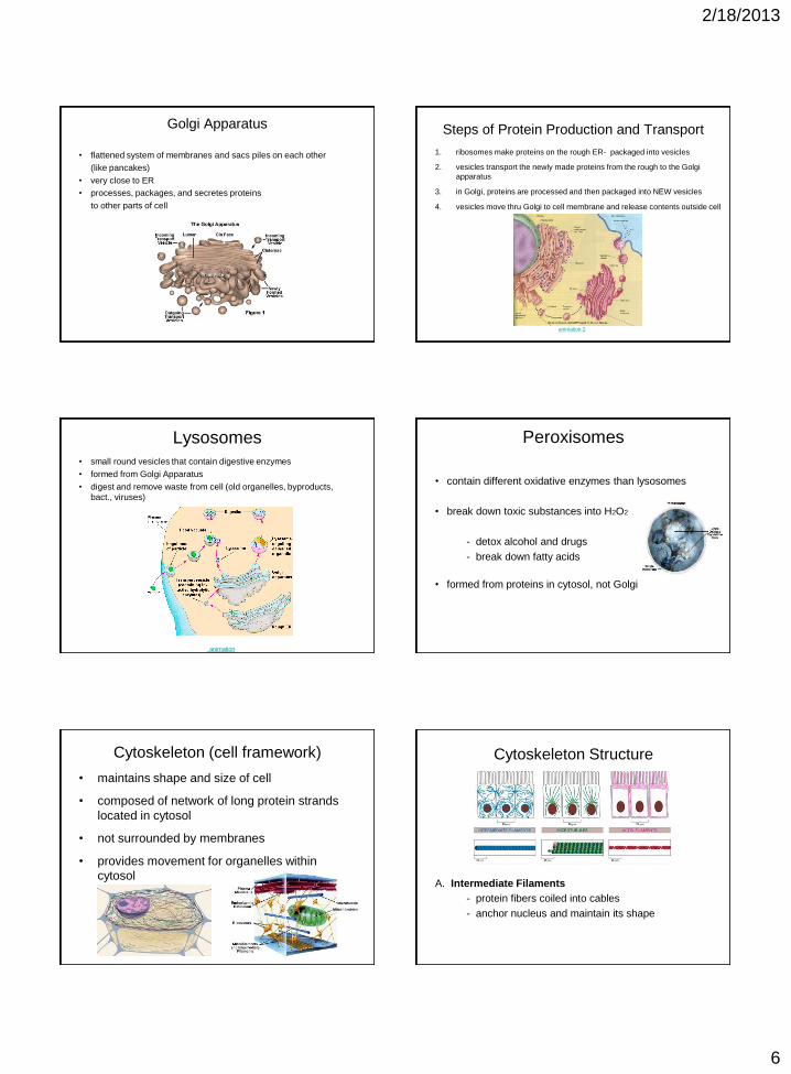
Common features of all cells
- cell membrane (plasma membrane)
- cytoplasm: gel like , holds cellular structures
- cytoskeleton: microscopic protein fibers that keep cells shape
- ribosomes: make proteins
- DNA: controls all cell activities
CELL STRUCTURE
Main components of eukaryotic cells
1. cell membrane (outer boundary)
2. nucleus (control center)
3. cytoplasm (material between nucleus
and membrane)
ANIMAL CELLS
Cell Membrane
(plasma membrane)
Functions:
- separates cells from surroundings
- regulates substance movement
(bring in nutrients, remove wastes)
- selectively permeable
- protection and support
- gives shape and flexibility
- signal receptor from other cells
2/18/2013
4
Fluid mosaic model of Cell Membrane
liquid phospholipid bilayer (water insoluble, no movement through)
- polar outside (head- phospholipid, hydrophilic)
- nonpolar inside (tail- two fatty acids, hydrophobic)
- polar outside (head- phospholipid, hydrophilic)
Polar: hydrophilic; attracts other polar and ionic compounds
Non-polar: hydrophobic; will not allow most polar molecules, ions, or large
molecules to pass across the membrane
animation – permeability animation – fluid mosaic
Membrane Proteins 1. transport proteins:
- involved in facilitated diffusion
(water soluble mols. not soluble in fat portion of membrane, so need help)
- go through the entire membrane
2 types
channel: gated pores
move water and ions freely in and out of cell
carrier: substrate binds to site on protein
protein changes shape and exposes substrate to cytoplasm
2. recognition proteins: recognizes substances
(like tips of icebergs emerging from ocean surface)
- Contain carbohydrate antennas (glycoproteins)
- Used as chemical ID markers to differential cell types
***cell/cell recognition (immune response)
***embryo cells tissues organ systems
3. receptor proteins: binds to specific molecules
(neurotransmitters, blood antigens, hormones)
- initiates cell response
4. enzymes: embedded in cell membrane to catalyze biochemical
reactions in cell
called FLUID MOSAIC because:
lipids and protein are liquid in nature and can move around each other
membrane is DYNAMIC (always changing)
Nucleus
- control center of cell: directs all cell activities
- contains chromosomes
- site of DNA and RNA synthesis
- located in center of most cells
2/18/2013
5
Structure:
- nuclear matrix - protein skeleton helps maintain nucleus shape
- nuclear envelope (double membrane)
- contains chromatin: combination of strands of DNA and protein
- nuclear pores: control substance movement
- nucleoplasm: dense, protein rich
- nucleolus: partially assembles ribosomes for protein synthesis
Cytoplasm
(between membrane and nucleus)
- contains cytosol
– gel like material between nucleus and cell membrane
– contains water, salts, organic molecules
– in constant motion (cytoplasmic streaming)
animation
amoeba animation
– holds organelles
Cell virtual tour
Organelles
Organelle: tiny structure that performs
special functions in the cell
to maintain life
Mitochondria
• powerhouse of cell (cell respiration)
• provides energy for cell in form of ATP
• membrane bound
• most numerous in cells which use a lot of energy (muscle)
• self replicating, contain their own DNA
- cristae: greatly enlarge surface area of inner membrane
(more area for chemical reactions of respiration)
Ribosomes
• spherical structures which make proteins
• not surrounded by membrane
• composed of protein and nucleic acids
• site of protein synthesis
Endoplasmic reticulum: (ER) intercellular highway
complex membrane system of folded sacs and tunnels
Rough ER - ribosomes stuck to membrane
surface
- newly produced proteins are
inserted into ER
- can be stored or exported to
smooth ER
- prominent in cells that make a lot
of protein
Smooth ER
- no ribosomes
- also stores and acts as an
intercellular highway for proteins and
enzymes
- involved in:
- synthesis of steroids in gland
- cell regulation of Ca levels
in muscle
- cells break down toxic
substances in liver cells
2/18/2013
6
Golgi Apparatus
• flattened system of membranes and sacs piles on each other
(like pancakes)
• very close to ER
• processes, packages, and secretes proteins
to other parts of cell
Steps of Protein Production and Transport
1. ribosomes make proteins on the rough ER- packaged into vesicles
2. vesicles transport the newly made proteins from the rough to the Golgi
apparatus
3. in Golgi, proteins are processed and then packaged into NEW vesicles
4. vesicles move thru Golgi to cell membrane and release contents outside cell
animation 2
Lysosomes
• small round vesicles that contain digestive enzymes
• formed from Golgi Apparatus
• digest and remove waste from cell (old organelles, byproducts,
bact., viruses)
animation
Peroxisomes
• contain different oxidative enzymes than lysosomes
• break down toxic substances into H2O2
- detox alcohol and drugs
- break down fatty acids
• formed from proteins in cytosol, not Golgi
Cytoskeleton (cell framework)
• maintains shape and size of cell
• composed of network of long protein strands
located in cytosol
• not surrounded by membranes
• provides movement for organelles within
cytosol
Cytoskeleton Structure
A. Intermediate Filaments
- protein fibers coiled into cables
- anchor nucleus and maintain its shape

2/18/2013
7
Cytoskeleton Structure
B. Microtubules
- long hollow coiled protein tubes (tubulin)
- maintain shape and support cells
- internal cell highways – move organelles thru cell
- form centrioles (cell division)
- motility (cilia and flagella)
• flagella: long whip-like structures used for movement
• cilia: short numerous hair like projections
- movement
- transport of substances across cell
ex: ear drum: transmits sound waves
respiratory tract: moves mucus etc.
Internal Organization
9 + 2 Arrangement
animation respiratory system animation
Cytoskeleton Structure
C. Microfilaments
- two strands fine protein (actin) intertwined
- used in cytoplasmic streaming, muscle
contraction
- smallest strands of cytoskeleton
cytoplasmic streaming
PLANT CELLS
Contain the same
organelles as
animal cells plus the
following:
1. cell walls
2. vacuoles
3. plastids
Cell wall
• rigid covering of plant cells, algae, and some bacteria
• composed of long chains of cellulose embedded in
hardened lignin and pectin
• very porous (O, H2O, CO2 easily
pass through)
• function: support & protection
2/18/2013
8
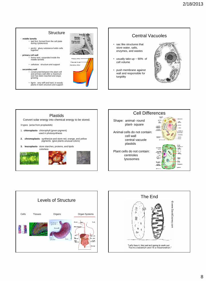
Structure - middle lamella
• laid first, formed from the cell plate during cytokenesis
• pectin: gluey substance holds cells together
- primary cell wall
• forms next, expanded inside the middle lamella
• cellulose: structure and support
- secondary wall
• constructed between the plant cell and primary wall after a maximum size has been reached and stops growing
• lignin: very stiff and hard, in woody plants in bark structure and support
Central Vacuoles
• sac like structures that
store water, salts,
enzymes, and wastes
• usually take up ~ 90% of
cell volume
• push membrane against
wall and responsible for
turgidity
Plastids Convert solar energy into chemical energy to be stored.
3 types (arise from proplastids)
1. chloroplasts- chlorophyll (green pigment)
used in photosynthesis
2. chromoplasts- synthesize and store red, orange, and yellow pigments (give plants unusual colors)
3. leucoplasts- store starches, proteins, and lipids
colorless
Cell Differences
Shape: animal- round
plant- square
Animal cells do not contain:
cell wall
central vacuole
plastids
Plant cells do not contain:
centrioles
lysosomes
Levels of Structure
Cells Tissues Organs Organ Systems
The End