Embed Size (px)
Citation preview
1
The Cardiac Cycle
Introduction
The cardiac cycle refers to a complete heartbeat from its generation to the
beginning of the next beat, and so includes the diastole (relaxation), the
systole(contraction) and the intervening pause. The frequency of the cardiac cycle
is described by the heart rate, which is typically expressed as beats per minute.
Each beat of the heart involves five major stages. The first two stages, often
considered together as the "ventricular filling" stage, involve the movement of
blood from the atria into the ventricles. The next three stages involve the
movement of blood from the ventricles to the pulmonary artery (in the case of
the right ventricle) and the aorta (in the case of the left ventricle).
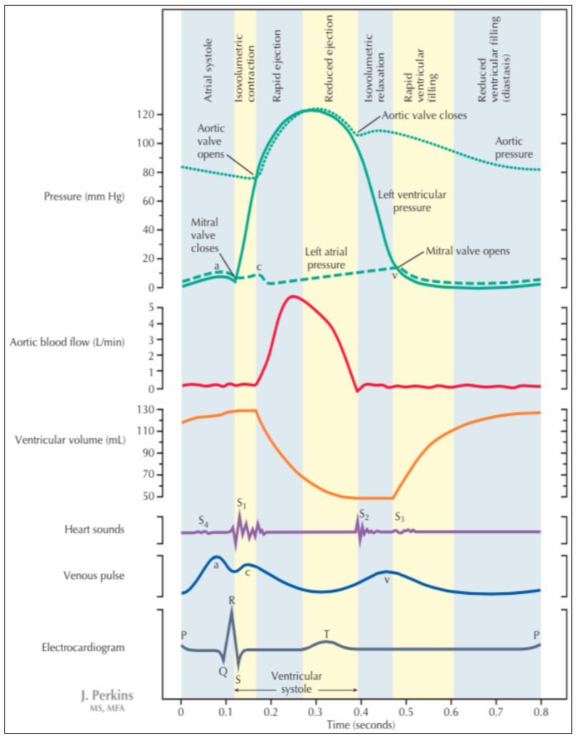
Before you continue reading this sheet, I advise you to detach the last page
{Wiggers Diagram} and always refer to it as you continue reading this sheet. It will
make things easier to visualize and understand.
Now, the cardiac cycle (if you are looking at an ECG): it is the time between
intervals: R-R Interval, P-P Interval… etc {any two consecutive points}.
During this cardiac cycle there are changes
1. Mechanical changes {Contraction (Systole) / Relaxation (Diastole)}.
2. Electrical changes {such as Action Potential or ECG}.
3. Volume changes {the volume of the ventricles concerns us more, we don’t
care about changes in the atrial volume.}.
4. Sound Changes {they are four heart sounds; we are concerned with only
two of them}.
5. Pressure changes
2
So during one cardiac cycle we have one atrial systole, one atrial diastole.
And we also have one ventricular systole, one ventricular diastole, and so on.
Remember that volume between the right and left ventricle is the same. But
what differs is the pressure between them {left ventricle > right ventricle}.
For teaching purposes, we consider the heart rate to be 75 beats/min, and
the time for one cardiac cycle is 0.8 seconds. Just to make things easier to
understand and visualize. But practically, it could be anything else from (60-100)
beats/min {in normal situations}.
What happens in the heart during One Cardiac Cycle?
1. Mechanical Changes
The atrial systole takes 0.1 seconds. And the atrial diastole takes 0.7
seconds {the total is 0.8 seconds = the cardiac cycle}.
We have AV delay; so that the atria and ventricles do not contract at the
same time. Therefore, atrial systole is followed by ventricular systole. In other
words, once the atria finish there contraction, the ventricular contraction starts.
The AV Delay takes 0.1 seconds.
The ventricle systole takes 0.3 seconds. And the ventricular diastole takes
0.5 seconds {the total is 0.8 seconds = the cardiac cycle}.
Do not be confused to think that the ventricle diastole takes 0.4 seconds.
This time (0.4 seconds) is the time where atrial and ventricular diastole overlaps
{both ventricles and atria are in diastole}. While during the other 0.1 second, the
atria are during systole (of the next cycle), and the ventricles are during diastole.
3
So the atria and ventricles work together, we consider this as mechanical changes
that occur in heart.
Remember that mechanical changes are a consequence of electrical changes. This
means that for mechanical changes to occur it should be preceded by electrical
changes.
2. Electrical Changes
ECG
Before atrial systole occurs we should record the P Wave in the ECG. And
before ventricular systole occurs we should record the QRS Complex in the ECG.
And before ventricular diastole occurs we should record the T Wave in the ECG.
Action Potential
How many action potentials we have to record? The answer should be two; one
for the atrium and one for ventricle.
The first {atrial} action potential corresponds to the P wave on the ECG
(atrial depolarization). Whereas the second {ventricular} action potential includes
QRS complex and the T wave together.
Now, by looking to the two action potentials; you should recognize that the
atrial repolarization and the ventricular depolarization occur at the same time.
Therefore, the atrial repolarization is not seen {masked}.
3. Volume & Sound Changes
As we mentioned earlier, we do not really care about the atrial volume; the
ventricular volume is the important one. The blood volume of the right ventricle is
equal to that of the left ventricle; the ventricles differ only in pressure (pressure in
the left > right).
4
Before atrial contraction takes place, ventricular volume is 100 ml, for both
the right and left ventricles.
Numbers are not to be memorized; they are only used for illustration.
Even before contraction of the atria, the AV valve is open because the atrial
pressure is more than that of the ventricle (they open passively due to the
pressure). This occurs at the beginning of ventricular diastole
How much is the atrial pressure?
The pressure in the atrium is almost zero. Accordingly, the ventricular
pressure has to be less than zero (negative).
You may wonder how can we have negative pressure?! Our reference –
when we say the pressure is zero – is the atmospheric pressure {760 mmHg}. So
when the pressure is zero in the atrium, it means that the pressure is equal to the
atmospheric pressure
The pressure in the ventricle is negative {-1 or -2}. This means that it less
than the atmospheric pressure.
Atmospheric Pressure - 2 = 760-2 = 758mmHg.
When we say someone’s blood pressure is 120/80; it means that it is
greater than atmospheric pressure by this value. Which is equivalent to
(120+760)/(80+760) = 880/840.
So to make things easier we consider zero is the pressure of the atrium. If
you don’t like dealing with negative numbers, you can consider the ventricular
pressure to be 0; so the atrial pressure is +1 or +2.
During this period, the AV valve opens and blood goes from the atrium to
the ventricle straight away. This is followed by atrial contraction, pushing a certain
extra amount {25ml} of blood to the ventricles faster. Now the blood volume
becomes 125 ml at the end of diastole of the ventricles. This is what we call the
end diastolic volume {EDV}.

5
By how much does the atrial contraction contribute to this volume? 25%
But even if the atria don’t contract {as in atrial fibrillation}; blood would
also flow to the ventricles because the AV valves are open. But it will move with
fewer amounts and less speed in this case (the volume in the ventricle would
increase by 10 ml only). This results in less contribution.
Q) Does that mean that we can live normally with only 75% efficiency? Yes
Since the atrial systole does not contribute that much to the end diastolic
volume {EDV}, then its contraction is not essential for normal function of the
heart. However, sometime we need this atrial contraction when the heart rate is
faster than normal; because there is no time for filling during ventricular diastole.
Ventricular Systole
The ventricle is guarded by two valves: the AV valve and the Semilunar
valve. The semilunar valve on the left separates the aorta from left ventricle
{aortic valve}, whereas the semilunar valve on the right separates the pulmonary
trunk from right ventricle {pulmonary valve}.
The aortic pressure during diastole is 80 mmHg, and the pulmonary pressure
during diastole is 8 mmHg.
When the ventricle starts to contract, its pressure is going to increase
above zero {that of the atrium}. Therefore, the blood tries to move from the
ventricles back to the atria: The AV Valve closes. Why? This happens because the
pressure will gradually build up in the ventricles, exceeding that of the atria. Thus,
the blood tries to move from the ventricles to the atria; the AV valves will shut.
This creates a sound. And this sound is called LUBB {the first heart sound
because of closure of AV valve). This corresponds to the QRS complex on the ECG.
During this stage, the four valves of the heart {two AV & two semilunar} are
closed. And the volume in the ventricle does not change. Therefore; we call this
phase Isovolumic Contraction.
6
Isovolumic Contraction: is a short phase in which ventricular volume is constant
and the four valves are closed. By the end of this phase the pressure increases
very rapidly, and when it reaches higher than 80 mmHg {in left ventricle} causing
the semilunar valve to open.
This leads to the first rapid ejection (about 70%), then the slow ejection (about
30%). Until the end of systole, the volume of blood that stays in the ventricle
called the end systolic volume {ESV}. It is equal to 55 ml.
The fraction of the EDV that is ejected called ejection fraction usually equal to
about 60%.
The amount of blood ejected in each beat (cardiac cycle) from either right or left
ventricle is called the stroke volume. And here it equals {125 – 55 = 70 ml/beat}.
Stroke volume = end diastolic volume {EDV} – end systolic volume {ESV}
If we want the stroke volume in one minute we multiply it by the heart rate (in
this case its 75 beats/min); this is what we call the cardiac output:
Cardiac Output (ml/minute) = Stroke Volume X Heart Rate
During systole the pressure {in the left ventricle} is very high, its value is
higher than the aortic pressure which makes the blood flow from the ventricle to
the aorta, and then to the systemic circulation.
When blood flows, its pressure will become less and less until it becomes
less than 80 mmHg in case of left ventricle (in right ventricle it gets less than 8
mmHg). So the blood tries to move from the aorta back to the left ventricle;
closing the semilunar {aortic} valve. The AV valves are still closed, and this is the
diastolic phase where the 4 valves are closed; which is called Isovolumic
Relaxation.
Usually Isovolumic Relaxation takes longer time than Isovolumic Contraction
7
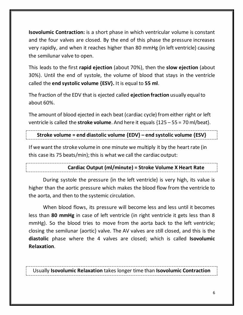
The closure of semilunar valve will cause the 2nd heart sound {DUP}
During ventricular systole, the AV valves are closed. Therefore, there is a filling of
blood inside the atria. Deoxygenated blood in the right atrium & oxygenated
blood in the left atrium.
Diastasis: is slow filling of ventricle before atria contract.1 (Refer to the figure in
the last page)
The pressure in the Isovolumic Relaxation phase keeps decreasing until it
becomes lower than the atrial pressure; causing the AV valve to open. Rapid
filling occurs, then slow filling.
Before the atrial systole, the blood volume in the ventricle rises up to 100 ml
{passively by pressure gradient}. When the atria contract, another 25 ml are
added. Total = 125 ml = end diastolic volume {EDV}.
So we have:
Rapid Filling in Ventricular Diastole & Rapid Ejection in Ventricular Systole
To sum things up:
Closure of AV valve gives us the first heart sound: LUBB {occurs at the
beginning of the ventricular systole}.
Closure of Semilunar valve gives us the second heart sound: DUPP {occurs
slightly after the end of ventricular systole}, or at beginning of its diastole.
What is the time between the 1st and 2nd heart sounds?
≈ 0.3 sec {approximately the same time of ventricle systole}
Whereas the time between the second heart sound {DUPP} and the next first
heart sound {LUBB} is about 0.5 sec {the total is 0.8 seconds = the cardiac cycle}.
1 Diastasis is the middle stage of diastole during the cycle of a heartbeat, where the initial passive fi lling of the
heart's ventricles has slowed down, but before the atria contract to complete the active fi l l ing.
8
There are sounds due to the movement of blood around a closed valve {it
sounds like murmur}. We may also hear other sounds. The third heart sound is
heard when there is rapid filling of blood from atria into the ventricles {opening of
the AV valve}. Also atrial contraction gives us the 4th heart sound. But usually, we
hear only hear the two previously mentioned heart sounds.
Pathological conditions:
If a patient has MI {Myocardial Infarction}, and there is a damage to the papillary
muscle. So the chordae tendineae cannot pull the valve down. What will happen?
Blood will go from the ventricle into the atria. It is what we call: Prolapse Valve or
Valve Incompetence.
In this case, the blood is regurgitating and when blood regurgitate it will give a
sound. So we first hear LUBB and then a sound of murmur before we finally hear
the DUPP. This is systolic {ventricle contraction} murmur; diagnosed as AV valve
incompetence.
Whereas if we hear the murmur between the DUPP and the second (next) LUBB,
it due to of Semilunar incompetence.
In children, there might be separation of the 2nd heart sounds {the aortic and
pulmonary semilunar valves don’t close at the same time} so we hear LUBB, DUPP
DUPP.
The Phonocardiograph will record the heart sounds
We already know that (in normal conditions) the stroke volume is the same
for both ventricles. However, if the stroke volume of right ventricle is 70 ml,
whereas the stroke volume of the left ventricle is 69 ml, the difference is 1 ml per
beat. In one minute: 70 ml {if we consider the heart rate to be 70 beats/min}.
9
Therefore in one min 70 ml will accumulate in the left ventricle, and after one
hour the volume will be 4200 ml (multiply by 60).
This extra volume will regurgitate back to left atrium and eventually back to the
lungs; which is abnormal and incompatible with life.
Normally, there might be difference in one beat or another but the next time it
will compensate.
4. The pressure changes
We will talk about the change of pressure in the left ventricle
Pressure in the left ventricle is different from that of the right ventricle.
If the pressure in left ventricle before atrial systole {during diastole} is zero;
the pressure in atria will be +1 or +2.
When the atria contact the pressure will increase, and it may reach up to +5
mmHg.
This is followed by ventricle contraction, the AV valve will close {1s t heart
sound) LUBB. The Isovolumetric Contraction phase starts {both AV valve
and aortic Semilunar valves are closed}.
Now there will be sharp increase in pressure until it reaches the aortic
pressure {80 mmHg}; once it exceeds it, the aortic valve will open, and it
keeps rising to be higher than the aortic pressure.
Why the ventricular pressure must be higher than the aortic pressure??
There must be a pressure gradient that maintain the flowing of blood from
the ventricles to the aorta.

10
At the end of the ventricular systole( last 1/3), the aortic pressure will be higher
than the ventricular pressure, although the blood is still moving from the
ventricle to the aorta; this happens because of the blood’s momentum.
Momentum is a force and the pressure is a force (force per unit of area)
When the force of the aortic pressure overwhelms the force of the blood’s
momentum, it closes the aortic semilunar valve, starting the Isovolumic
Relaxation phase {both valve aortic semilunar and the AV (mitral/bicuspid) valve
are closed}
The maximum pressure in the ventricle during systole is 120mmHg, while the
aortic maximum pressure will be slightly less than this, approximately 118 mmHg
when the pressure starts to decrease after the first decrease the Semilunar valves
close , and after the second decrease the AV open First SV close then AV open)
when the pressure decreases the blood tries to return back from the Aorta to the
ventricle.
When the aorta pushes blood to the closing semilunar valve, the blood is
compressed and press on the wall of the aorta and the aortic pressure slightly
rises, and makes a peak: called Dicrotic Notch or Incisura.
So the Dicrotic notch is due to the increase in pressure around a closed semilunar
valve>>> Why ? Because of the momentum of the blood.

11
Location Minimum Pressure
mmHg Maximum Pressure
mmHg
Left Ventricle 0 120
Aorta 80 120
Right Ventricle 0 25
Pulmonary Trunk 8 25
What does it mean when we say someone blood pressure is 110/70?
We mean that during the left ventricular systole the aortic pressure and
ventricular pressure reaches a maximum of 110 mmHg. Whereas during left
ventricular diastole the aortic pressure reaches a minimum of 70 mmHg.
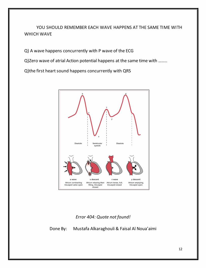
The Waves of Atria
First rise in pressure in atria occurs during atrial systole which causes a
wave called atrial pressure wave: A wave.
After atrial systole, the ventricle systole begins. And when the ventricle
contracts, blood tries to regurgitate towards the atria; causing the AV valve to
close {because of high pressure in ventricle}. This also increases the pressure
inside the atrium {because of the force of blood on its wall}. This causes another
waves called: C wave
After that the pressure inside the atria start to develop due to venous filling
{The AV valve is closed during this phase} until it reaches a maximum just before
the ventricular diastole {V wave}. This occurs just before the AV valve opens due
to the drop in ventricular pressure, and the rapid filling occurs.
12
YOU SHOULD REMEMBER EACH WAVE HAPPENS AT THE SAME TIME WITH
WHICH WAVE
Q) A wave happens concurrently with P wave of the ECG
Q)Zero wave of atrial Action potential happens at the same time with ……..
Q)the first heart sound happens concurrently with QRS
Error 404: Quote not found!
:yone oy Mustafa Alkaraghouli & Faisal Al Noua’aimi

13
14