Embed Size (px)
Citation preview

TISSUE CULTURES IN THE INVESTIGATION OF CANCER
ROBERT A. LAMBERT From the Department of Pathology, College of Physicians and Surgeons, Columbia
University, New York City
Received for publication, February 1,1916
It is recognized that progress in many departments of science is dependent in large measure, if not entirely, on the development of improved methods of investigation. In the field of cancer research it has become increasingly evident in recent years that new methods of attack must be worked out before further im- portant advances can be expected.
The discovery of the factors concerned in the unlimited law- less growth of cells constitutes, obviously, the main cancer prob lem. It would appear therefore that the recently devised method of cultivating tissues in vitro (Harrison, Burrows) by which we are enabled to study isolated somatic cells under conditions which allow the introduction of known and measurable factors into experiments, should mark an important advance in the attaok upon this fundamental question. Furthermore, since it is p o s sible to subject cancer cells, thus isolated, to various chemical and physical agencies, it may be suggested that we have also in the new method a possible means for solving another great can- cer problem, namely, the treatment of malignant growths.
In the study of human tumors, the method will probably fill a great need, since it is not possible to secure the continued prop- agation of such tumors by transplantation, by the method6 pursued in the case of tumors of certain of the lower animals.
The purpose of this paper is simply to review briefly a few of the results which have been obtained with tissue cultures in studies upon cancer and related subjects, and to suggest s o u problems in which it seems probable that the method may be
169
170 ROBERT A. LAMBERT
advantageously applied. It is possible that a review of the re- sults thus far obtained may prove disappointing. But in pal- liation it should be said that the method is young and in many respects still imperfect; and that much time and energy have been expended by a number of workers in the past five years to- ward improving the technique so as to permit the cultivation of a greater variety of tissues over longer periods of time.
Details of the history of the method of tissue cultivation in witro and its development will be found in various papers to which references are given a t the end of this article (Loeb (l), Harrison 12, 3), Burrows (4, 5) . For the oonvenience of those interested, a brief review of the technique of the method is given here.
TECHNIQUE
The simplest and most satisfactory form of culture consists of a small piece of tissue placed in a thin drop of plasma on a cover glass which is inverted and sealed over the cavity of a hollow- ground slide. To obtain the plasma in suchra way that it will remain unclotted until the tissue is added, several precautions must be observed. The blood must flow directly into the con- tainer (preferably a small test tube) without contamination with tissue juicqs, and should be kept cold in oiled or paraffined tubes both during and after centrifugation.
Blood is obtained from human beings or large laboratory ani- mals (dogs, goats), by puncturing a superficial vein with a needle of large bore, previously boiled in albolene or olive oil, and al- lowing the blood to flow directly from the needle into paraffined tubes set in large centrifuge cups which are filled with cracked ice.
Small animals such as mice, rats, and frogs may be bled from the heart in the mime way. The writer has devised another method (7), however, for bleeding small animals, which does not entail the sacrifice of the animal as does bleeding from the heart, 'a distinct advantage in certain experiments where it is important to obtain pIasma from the same animal more than once. The wrotid, jugular, or some other accessible vessel is exposed and

TISSUE CULTURES IN INVESTIGATION OF CANCER 171
clamped lightly at two points some distance apart; the vessel wall, after clean dissection, is caught by delicate forceps at a point midway between the clamps in such a way that the lumen is not occluded; the vessel is then severed beyond the forceps, the proximal clamp released and, with the end of the vessel held
l h . 1. Method of bleeding small animals for obtaining plasma. The artery is held away from the tissues and the blood allowed to spurt into paraffined tubes.
away from the tissues, the blood is allowed to spurt free into cold paraffined tubes as before (fig. 1). Even so small an animal as the mouse may be bled several times in this way.
On account of the dryness of their tissues and the proximity of the wing vessels to the skin, fowls and pigeons may be bled di-

172 ROBERT A. LAMBERT
rectly from the wing vein by making a clean incision through the skin and vessel, and allowing the blood to drip from the surface into the tubes.
Blood obtained by any of the methods mentioned is centri- fuged at high speed for several minutes, and the supernatant plasma is then put aside in a cold place, or packed in ice,
The length of time.that the plasma may be preserved unco- agulated varies greatly with different species. Fowl and pigeon plasma may be kept indefinitely if properly handled. On the other hand, rat and guinea-pig plasma will rarely remain fluid longer than two hours, often not so long. Human, rabbit, dog, and cat plasma may be kept for several hours at least, and some- times even for days.
The remainder of the technique consists in placing small pieces of tissue, 0.5 to 2.0 mm. in diameter, on a cover glass, and adding immediately a small drop of plasma. The cover glass is inverted without delay over a hollow-ground slide, sealed with vaseline, and incubated at body temperature.
It should be stated in this connection that tissue cells show no such proliferative capacity in cultures as do bacteria. Further- more, it has not been possible as yet to obtain an active growth of any of the highly differentiated body cells, though certain specialized elements, such as ganglion cells, may survive for a time.
The cultivation of human tissues has offered special difficul- ties, owing to the fact that the fibrin in the clotted human plasma, which forms the necessary scaffolding for the outgrowth of cells, is regularly digested by the tissue fragments, and the cells, not being able to wander out into the medium, soon die. The writer has overcome this difficulty by using as a culture medium a mixture of human serum (or plasma) and chick plasma, the fibrin of which resists digestion. (It was found that chick plasma alone did not form a satisfactory medium.)
That the method is in many respects surprisingly simple should be emphasized. It has been little used in cancer labora- tories, a fact which is probably to be attributed to supposed dif- ficulties in technique (danger of contamination, maintenance of
TISSUE CULTURES IN INVESTIGATION OF CANCER 173
a uniform temperature during preparation of cultures, etc.), points which have certainly received unmerited emphasis in several papers (6). As a matter of fact, practically no effort at all is required to avoid bacterial contamination; thus, the writer has used tissue from the liver of a human cadaver four hours after death (metastatic tumor nodule lying directly on the colon), and observed not a single colony of bacteria in a large number of preparations. It is possible to use even such a tissue as skin, where a certain number of bacteria are known to be present, if partial sterilization with weak alcohol be first carried out. The plasma itself, especially that of fowls, evidently possesses a defi- nite bactericidal property, by which a certain number of organ- isms are taken care of. It is not necessary to keep the tissues at or near body temperature while preparing the cultures, and within certain limits the time elapsing between removal of the tissue from the living body and the preparation of the cultures is not important. The tissue of the adult rat may be used after several days’ preservation at room temperature or in the ice- box, and the writer has obtained successful growths of human tissue kept for six days in cold storage. Embryonic tissues are capable of longer preservation-five to eighteen days, the length of time depending on the temperature.
The problems in which the method of tissue culture has been employed by the writer may be conveniently discussed under several headings: (1) Comparative study of the behavior of can- cer cells and normal cells growing in vitro; (2) Cancer immunity; (3) Stimulation of cells growing in cultures.
COMPARISON OF THE BEHAVIOR I N VITRO O F NORMAL CELLS AND
CANCER CELLS
The similarity between the growth in vitro of sarcoma and connective tissue on the one hand, and between carcinoma and normal epithelium on the other, has been emphasized in previous papers (7, 15). Sarcoma and connective tissue cells wander out singly or in chains, while epithelial cells, normal or neoplastic, tend to spread out in sheets or groups.

174 ROBERT A. LAMBERT
Certain differences which may not be without importance have been observed in the behavior of normal and malignant tissues. In the first place, a greater motility characterizes the cancer
FIQ. 2. Mouse sarcoma, ten-hour culture, showing extensive outwandering of cells which began within two hours after preparation was placed in incubator.
cell, especially the sarcoma cell, as compared with the correspond- ing normal element; a sarcoma cell may often be seen traveling through the medium at a rate almost equal to that of polymor-

TISSUE CULTURES IN INVESTIGATION OF CANCER 175
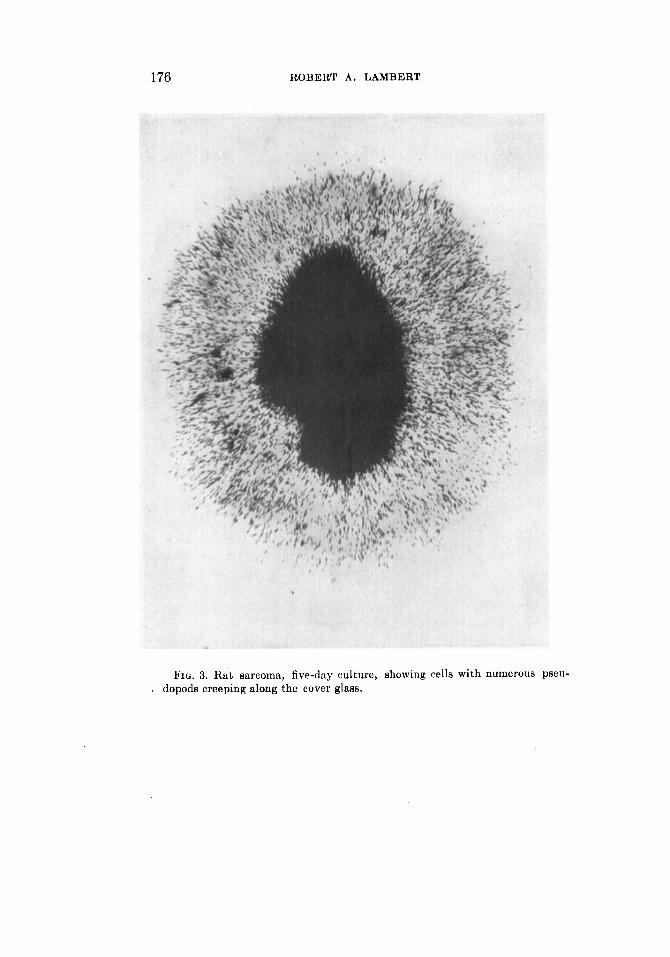
phonuclear leukocytes (figs. 2 and 3). This fact, as has been pointed out before (22), probably throws light on the mechanism of the invasive growth and spread of cancer in the body. It is not necessary to regard the formation of metastatic tumor nod- ules as always a result of the passive transportation of cells from a primary tumor by the blood or lymph stream, when the cells may easily get from place to place by their own powers of loco- motion. The writer has calculated that a tumor cell, if it sur- vived the trip, might make its way from the middle of the breast to the axilla in less than four weeks.
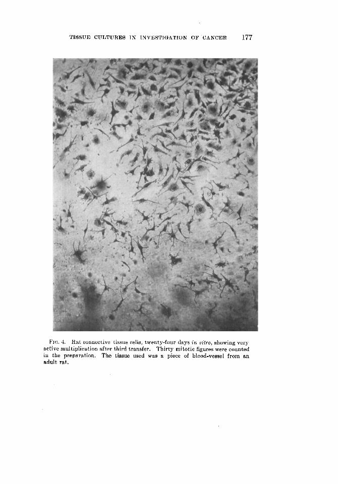
A second important, and somewhat surprising difference be- tween normal and tumor tissues lies in the fact that the contin- ued propagation of certain normal cells, especially those of con- nective tissue, is, as a rule, much easier than in the case of tumor cells. Many carcinomata and sarcomata, especially those of human beings, will not grow even in primary cultures, while others, rat and mouse tumors, for example, grow actively for a few days but tend to die, out even when transferred early to fresh plasma. On the other hand, it has been the writer’s experience that connective tissue becomes very much more active in sub- cultures, so that after several transfers a most active cell multi- plication is observed, dozens of mitotic figures being sometimes seen in a single preparation (fig. 4). The explanation for this difference in behavior is not obvious. It may be suggested either that some particular substance necessary for tumor growth is not being supplied in sufficient quantity in cultures or, stated in another way, that the cancer cell is really, in a sense, a highly differentiated cell which is particularly susceptible to changes in food supply and environment. That the cancer cell is a cell of lowered vitality is suggested by the results of some studies made by the writer on the comparative resistance of tumor cells and normal cells to heat (8). When normal connective tissue ele- ments and cells from mouse or rat sarcomata were subjected to different degrees of temperature above that of the body, for vari- able periods, it was found that the latter were definitely more susceptible to heat than were normal actively growing connec- tive tissue cells. Similar experiments with human tissues have
176 ROBERT A. LAMRERT
FIG. 3. E a t sarcoma, five-day culture, showing cells with numerous pseu- . dopods creeping along the cover glass.
T I S S U E CULTURES I N INVESTIGATION OF CANCER 177
FIG. 4. Hat connective tissue cells, twenty-four days in oitro, showing very active multiplication after third transfer. Thirty mitotic figures were counted in the preparation. The tissue used was a piece of blood-vessel from an adult rat.
178 ROBERT A. LAMBERT
recently been attempted, in the course of which the writer has found that human connective tissue cells and wandering cells are quite as resistant to high temperatures as are those of the rat; a satisfactory comparison with human tumor tissue has not been made, however, on account of the difficulty experienced in obtaining sarcoma suitable for cultivation.
That the cancer cell is a particularly hardy cell, and that ma- lignancy is due in some measure a t least to this property, is a popular notion. This idea has, indeed, received some support from studies on the resistance of certain rat and mouse tumors to freezing and to toxic chemical substances (9, 10, 11). It is to be noted, however, that these studies, with one exception, were not comparative. The writer has found that many types of nor- mal tissues isolated in tissue cultures are, like tumor cells, very resistant to low temperatures (12) and to many chemical agents. Ribbert (13) has called attention to the tendency of tumors to necrosis, maintaining that cancer cells are very susceptible to injuries of many kinds, and that this state of low vitality is referable to their poor vacularization.
While not accepting Ribbert’s explanation, the writer is in- clined, as the result of his own studies, to endorse the view that the cancer cell is a cell of relatively low resistance and that ma- lignancy is to be attributed entirely to the capacity of the cells for unlimited growth.
CANCER IMMUNITY
The results of the use of tissue cultures in the study of cancer immunity have been described fully in previous papers (14, 15, 10, 17) and need only be summarized here.
Although recent studies have shed new light on the question, the problem as to the exact nature of the resistance to trans- plantable cancer is still unsettled. Lambert and Hanes found that rat sarcoma cells will grow quite as well in the plasma of an im- mune rat (naturally or artificially immune) as in the Rlasma of a normal rat, an observation which affords further evidence that cancer immunity is not to be attributed to circulating antibodies of a cytotoxic nature.

TISSUE CULTURES IN INVESTIGATION OF CANCER 179
It was shown further that the plasma from an animal of a for- eign species (guinea-pig or rabbit) may serve also as a good medium for the cells. The fact that rat sarcoma cells can grow in such an alien plasma for at least thirty days would appear to invalidate Ehrlich’s hypothesis of athreptic immunity.
On the other hand, it was discovered that if the guinea-pig or rabbit be first immunized against rat tissues by suitable in- jections of rat blood, skin, or tumor, the plasma of the treated animal becomes no longer suitable for the growth of rat cells, but is on the contrary distinctly toxic for any rat tissue. This experiment, besides showing that antibodies for tissue cells can be readily demonstrated in tissue cultures, proves also that cyto- toxins are not specific for the cells used for immunization.
In further efforts toward demonstrating a cancer immunity reaction in vitro Dr. Edna Steinhardt and the writer attempted to cultivate rat sarcoma cells in immune rat plasma in associa- tion with normal tissues from immune animals (spleen, lymph- node, liver, and leukocyte emulsion), with the idea that the im- munity phenomenon might be the result of the combined action of immune serum and some tissue or organ. We found that the growth of the tumor cells in vitro was not influenced by such a combination of factors. This negative result may have been due, however, to faulty technique, on account of which the es- sential protecting cells may have been injured, or present in in- sufficient number. In view of the recent work of Murphy (18) who attributes immunity against cancer to the action of certain lymphoid elements, it should be possible, by a proper combina- tion of tumor cells and lymphocytes, to obtain an immunity re- action in vitro. Indeed it may even be suggested that the method of tissue cultures should serve to settle some of the questions which have been raised by this new work.
STIMULATION OF GROWTH I N VITRO
This discussion of the application of tissue cultures to cancer problems would not be complete without reference to efforts di- rected toward the stimulation of normal and tumor cells, when

180 ROBERT A. LAMBERT
freed from body restraint. Without entering into a detailed dis- cussion of physical and mechanical stimuli, the writer wishes again to emphasize the fact (19) that variations in the depth of the hanging drop and in the density of the fibrin meshwork (which can be altered by dilution) influence markedly the extent to which actively motile cells wander outward.
A single recent observation on a culture of human connective tissue exposed to a temperature of 45" for a few minutes may be of interest in this connection. Examination of the specimen just before exposure showed only two dividing cells in the entire preparation; ten minutes after heating, however, at least a dozen cells were seen in early stages of mitotic division. It may be that this single observation is without significance, but it does suggest further work on the effect of sudden elevation of tem- perature upon cell multiplication.
The action of various tissue extracts on growing cells has been studied by Dr. Carrel and his co-workers (20, 21), and very striking results have been reported on the effect of such prepa- rations, especially those of chicken sarcoma, on embryonic chick tissues. The writer has not been able to verify their findings in such of the experiments as have been repeated, and is in- clined to the opinion that the results of these authors are to be explained in other ways. It is certain that they do not possess any general application. An extract of human tumor, recently employed by the writer, appeared to inhibit rather than stimu- late the growth of normal human cells.
Results following the use of certain chemicals (Scharlach 11. and Sudan 111) which have been shown to cause an abnormal proliferation of various tissues when injected into animals, have been disappointing. Cultures of chick embryo, and of rabbit skin and cornea, were treated with these dyes through several subcultures with no apparent effect, although they were applied in different ways and in varying strengths.
In spite of these negative results, this particular field seems to offer great possibilities. Since it is known that some sort of stimulus, or possibly the loss of some restraining influence does start cells within the body on a career of reckless multiplication,

TISSUE CULTURES IN INVESTIGATION O F CANCER 181
it seems possible that patient work in this direction upon cells growing outside the organism may supply the key to this phase of the cancer problem.
REFERENCES
(1) LOEB, LEO: Ueber die Entstehung von Bindegewebe, Leucocyten, und roten Blutkorperchen aus Epithel und uber eine Methode isolierte Geweb- steile zu zuchten. Chicago, 1897, p. 41; Arch. f . Entwcklngsmechn. d. Organ., 1902, xiii, 487.
(2) HARRISON, It. G. : Observations on the living developing nerve fibre. Proc. SOC. Exper. Biol. and Med., 1906-7, iv, 140.
(3) HARRISON, R. G. : The outgrowth of nerve fibres as a mode of protoplasniic movement.
(4) BURROWS, M. T. : The growth of tissucs of the chick embryo outside animnl body with special reference to the nervous system. Jour. Exper. Zool., 1916, x, 63.
(5) BURROWS, M. T.: Method of furnishing a continuous supply of medium to tissue cultures. Anat. Rec., 1912, vi, 141.
(6) CARREL AND BURROWS: Cultivation of tissues in vitro and its technique. Jour. Exper. Med., 1911, xiii, 387.
(7) LAMBERT AND HANES : Characteristics of growth of sarcoma and carcinoma cultivated in vitro.
(8) LAMBERT, R. A.: Demonstration of the greater susceptibility to heat of sarcoma cells as compared with actively proliferating connective tis- sue cells. Jour. Amer. Med. Assn., 1912, lix, 2147.
(9) JENSEN: Experimentelle Untersuchungen uber Krebs bei Mausen. Cen- tralbl. f . Bakteriol., I Abt., Orig., 1903, xxxiv, 122.
(10) GAYLORD, H. : Resistance of embryonic epithelium, transplantable mouse cancer, and certain organisms to freezing. Jour. Infect. Dis., 1908, v, 443.
(11) SALVIN-MOORE AND BARRATT: A note upon the effect of liquid air on the graftable cancers of mice. Lancet, 1908, i, 227.
(12) LAMBERT, R. A.: The effects of cold on animal tissues. Proc. N. Y. Path. SOC., 1912, xii, 113.
(13) RIBBERT, H. : uber daa Gefasssystem und die Heilbarkeit der Geschwulste. Deutsch. Med. Wchnschr., 1904, xxx, 801.
(14) LAMBERT AND HANES: A study of cancer immunity by the method of culti- vating tissues outside the body.
(15) LAMBERT AND HANES: Cultivation of tissues in plasma of alien species. Jour. Exper. Med., 1911, xiv, 129.
(16) LAMBERT, R. A.: On the cultivation of tissues in vitru as a method for the study of cytotoxins. Jour. Exper. Med., 1911, xiv, 453.
(17) LAMBERT, R. A.: A note on the specificity of cytotoxins. Jour. Exper. Med., 1914, xix, 277.
(18) MURPHY, JAMES B.: The lymphocyte in natural and induced resistance to transplanted cancer. Jour. Exper. Med., 1915, xxii, 204.
Jour. Exper. Zool., 1910, ix, 787.
Jour. Exper. Med., 1911, xiii, 495.
Jour. Exper. Med., 1911, xiii, 505.

182 ROBERT A. LAMBERT
(19) LAMBERT, R. A.: The effect of dilution of plasma medium on the growth and fat accumulation of cells in tissue cultures. .Jour. Exper. Med., 1914, xix, 398.
(20) CARREL, A. : Artificial nctivation of the growth in t~itt-o of connective tissuc. Jour. Expcr. Med., 1913, xvii, 14.
(21) WALTON, A. J.: The effect, of various tissue extracts on the growth of adult mammalian tissues.
(22) LAMBERT AND HANES: Amijboide Bewegeungen von Krebszellen ale ein Faktor dcs invasiven und rnetastatischen Wachstums maligner Tu- moren.
Jour. Exper. Mcd., 1914, xx, 554.
Virchows Arch. f . path. Anat., 1912, ccix, 12.