Embed Size (px)
Citation preview
THE BURN MANUAL
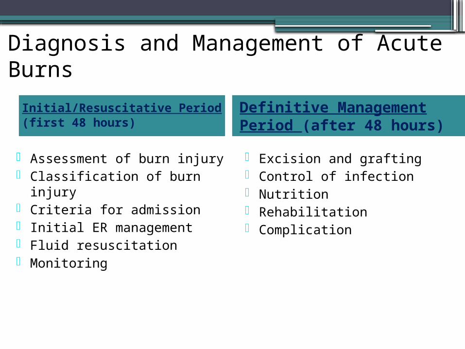
Diagnosis and Management of Acute Burns
Initial/Resuscitative Period(first 48 hours)
Definitive Management Period (after 48 hours)
Assessment of burn injury Classification of burn injury Criteria for admission Initial ER management Fluid resuscitation Monitoring
Excision and grafting Control of infection Nutrition Rehabilitation Complication
Initial Rescucitation
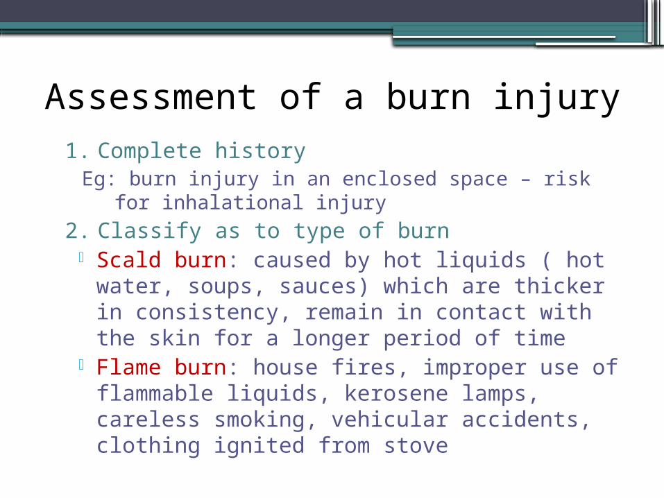
Assessment of a burn injury1. Complete history
Eg: burn injury in an enclosed space – risk for inhalational injury
2. Classify as to type of burn Scald burn: caused by hot liquids ( hot water, soups,
sauces) which are thicker in consistency, remain in contact with the skin for a longer period of time
Flame burn: house fires, improper use of flammable liquids, kerosene lamps, careless smoking, vehicular accidents, clothing ignited from stove
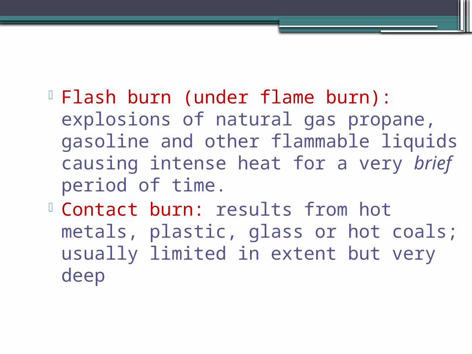
Flash burn (under flame burn): explosions of natural gas propane, gasoline and other flammable liquids causing intense heat for a very brief period of time.
Contact burn: results from hot metals, plastic, glass or hot coals; usually limited in extent but very deep
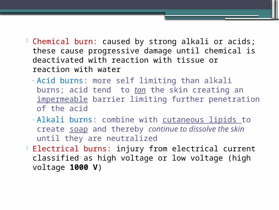
Chemical burn: caused by strong alkali or acids; these cause progressive damage until chemical is deactivated with reaction with tissue or reaction with water– Acid burns: more self limiting than alkali burns; acid
tend to tan the skin creating an impermeable barrier limiting further penetration of the acid
– Alkali burns: combine with cutaneous lipids to create soap and thereby continue to dissolve the skin until they are neutralized
Electrical burns: injury from electrical current classified as high voltage or low voltage (high voltage 1000 V)
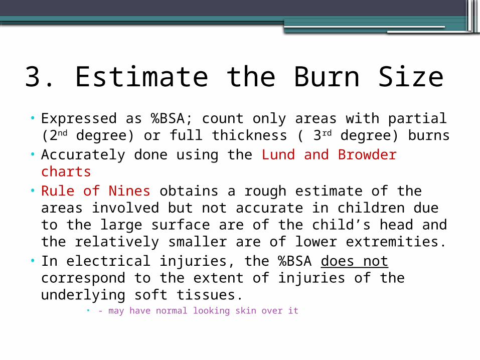
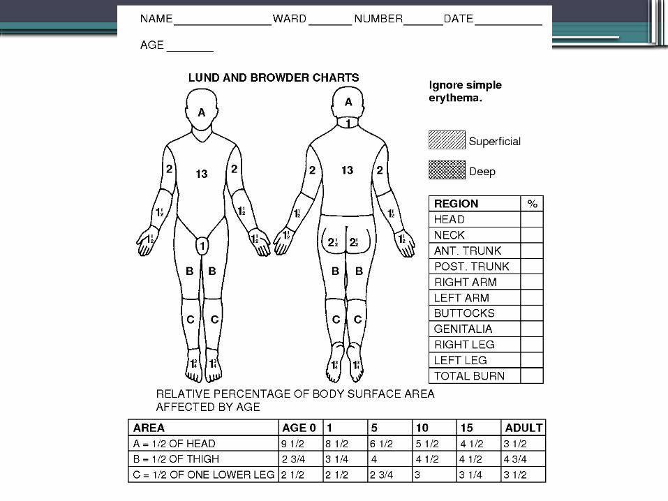
3. Estimate the Burn Size• Expressed as %BSA; count only areas with partial (2nd
degree) or full thickness ( 3rd degree) burns• Accurately done using the Lund and Browder charts• Rule of Nines obtains a rough estimate of the areas
involved but not accurate in children due to the large surface are of the child’s head and the relatively smaller are of lower extremities.
• In electrical injuries, the %BSA does not correspond to the extent of injuries of the underlying soft tissues.
• - may have normal looking skin over it
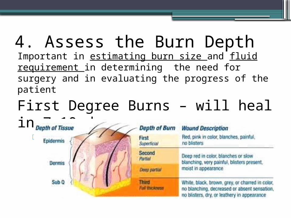
4. Assess the Burn DepthImportant in estimating burn size and fluid requirement in determining the need for surgery and in evaluating the progress of the patient
First Degree Burns – will heal in 7-10 d Ex: sunburn
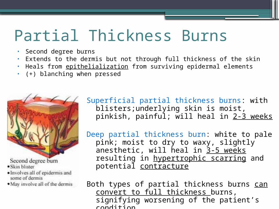
Partial Thickness Burns• Second degree burns• Extends to the dermis but not through full thickness of the skin• Heals from epithelialization from surviving epidermal elements• (+) blanching when pressed
Superficial partial thickness burns: with blisters;underlying skin is moist, pinkish, painful; will heal in 2-3 weeks
Deep partial thickness burn: white to pale pink; moist to dry to waxy, slightly anesthetic, will heal in 3-5 weeks resulting in hypertrophic scarring and potential contracture
Both types of partial thickness burns can convert to full thickness burns, signifying worsening of the patient’s condition
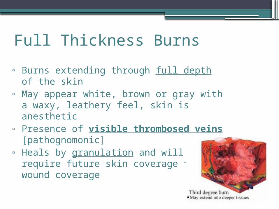
Full Thickness Burns
▫ Burns extending through full depth of the skin▫ May appear white, brown or gray with a waxy,
leathery feel, skin is anesthetic▫ Presence of visible thrombosed veins
[pathognomonic]▫ Heals by granulation and will require future skin
coverage for wound coverage
5. Check for other injuries/medical problems
These problems play a role in the origin of burn and will have to be integrated in the management of burn
Eg: seizure disorders, diabetesdisorders, fractures, blunt abdominal injuries
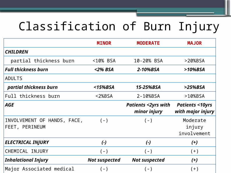
Classification of Burn InjuryMINOR MODERATE MAJOR
CHILDREN
partial thickness burn <10% BSA 10-20% BSA >20%BSA
Full thickness burn <2% BSA 2-10%BSA >10%BSA
ADULTS
partial thickness burn <15%BSA 15-25%BSA >25%BSA
Full thickness burn <2%BSA 2-10%BSA >10%BSA
AGE Patients <2yrs with minor injury
Patients <10yrs with major injury
INVOLVEMENT OF HANDS, FACE, FEET, PERINEUM
(-) (-) Moderate injury involvement
ELECTRICAL INJURY (-) (-) (+)
CHEMICAL INJURY (-) (-) (+)
Inhalational Injury Not suspected Not suspected (+)
Major Associated medical Illness (-) (-) (+)
Associated fractures, multiple trauma
(-) (-) (+)
Criteria for Admission to the Burn Unit• Acute burn patients with moderate and major injuries
• Acute burn patients <2y/o regardless of % TBSA
• Acute burn patients with injuries to the hands, face, feet and perineum
• Acute electrical burn patients
• Acute chemical burn patients
• Acute burn patients with smoke inhalation injury, other associated medical illness, or multiple trauma
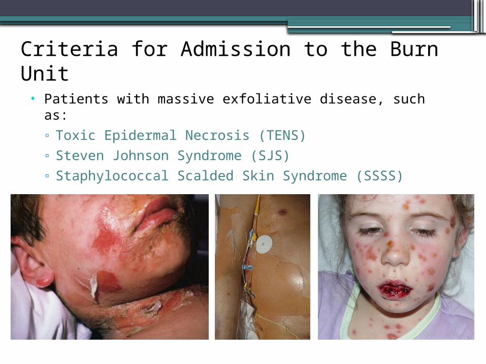
Criteria for Admission to the Burn Unit• Patients with massive exfoliative disease, such as:
▫ Toxic Epidermal Necrosis (TENS)▫ Steven Johnson Syndrome (SJS)▫ Staphylococcal Scalded Skin Syndrome (SSSS)
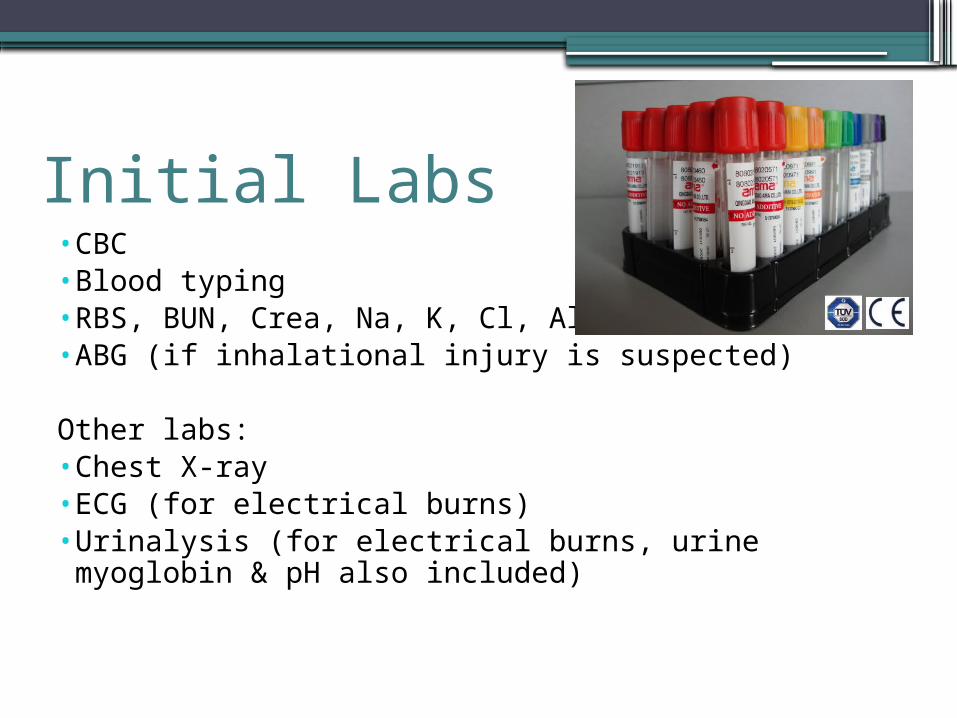
Initial Labs•CBC•Blood typing•RBS, BUN, Crea, Na, K, Cl, Albumin•ABG (if inhalational injury is suspected)
Other labs:•Chest X-ray•ECG (for electrical burns)•Urinalysis (for electrical burns, urine myoglobin & pH also
included)
1. Cool wound with tap water2. Administer tetanus prophylaxis
▫ TT booster if not received for the past 5 years▫ 0.5cc TeAna and 3000 u ATS (adults)
3. Clean wound with soap and water/betadine scrub4. Debride dead tissue
▫ Big blister unroof▫ Small blister aspirate
Initial ER Management: MINOR Burns

Initial ER Management: MINOR Burns
5. Apply bland ointment (i.e., Bacitracin, Trimycin, Vaseline) and non-stick porous gauze and wrap with gauze
6. NO systemic prophylactic antibiotics are given7. Oral/IM analgesics during wound cleaning8. Send patients home with oral analgesics and
instructions to clean the wound OD to BID and apply ointment and gauze.
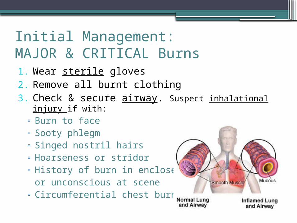
Initial Management: MAJOR & CRITICAL Burns1. Wear sterile gloves2. Remove all burnt clothing3. Check & secure airway. Suspect inhalational injury if with:
▫ Burn to face▫ Sooty phlegm▫ Singed nostril hairs▫ Hoarseness or stridor▫ History of burn in enclosed space
or unconscious at scene▫ Circumferential chest burn
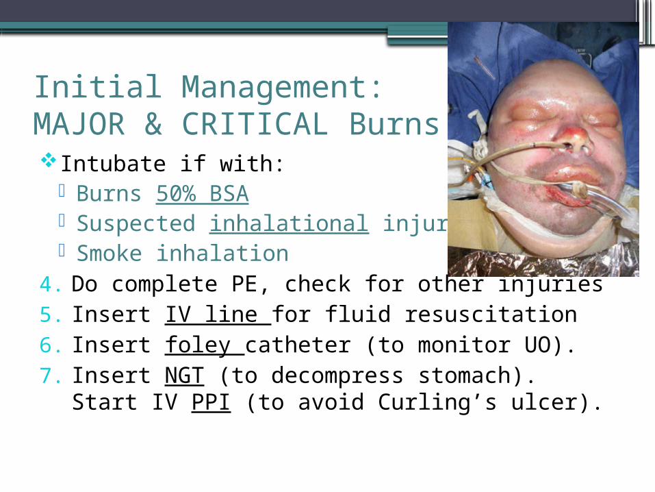
Initial Management: MAJOR & CRITICAL BurnsIntubate if with:
Burns 50% BSA Suspected inhalational injury Smoke inhalation
4. Do complete PE, check for other injuries5. Insert IV line for fluid resuscitation6. Insert foley catheter (to monitor UO).7. Insert NGT (to decompress stomach). Start IV PPI (to
avoid Curling’s ulcer).
Initial Management: MAJOR & CRITICAL Burns
8. Weigh patient and record. If not possible, estimate:▫ For children: Wt (kg) = [2 x (age in years)] + 5▫ For adults: Wt (kg) = 0.9 x [ht in cms – 100]
9. Administer ATS and TeAna10.Check pulses, assess adequacy of chest expansion
▫ Absent pulses or limited chest excursion is a surgical emergency and an indication for escharotomy
Initial Management: MAJOR & CRITICAL Burns
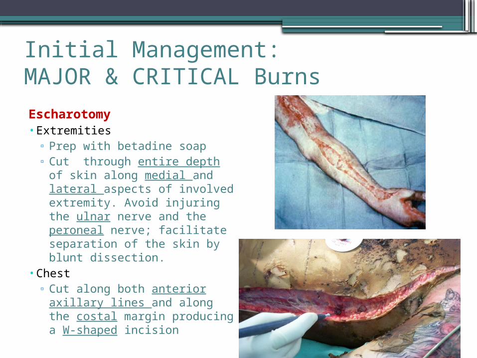
Escharotomy• Extremities
▫ Prep with betadine soap▫ Cut through entire depth of
skin along medial and lateral aspects of involved extremity. Avoid injuring the ulnar nerve and the peroneal nerve; facilitate separation of the skin by blunt dissection.
• Chest▫ Cut along both anterior
axillary lines and along the costal margin producing a W-shaped incision
Initial Management: MAJOR & CRITICAL Burns
11. Refer all pediatric patients to Pedia for co-management. Patients with other medical problems should also be referred accordingly.
12. No prophylactic antibiotics are given, unless there are concomitant medical conditions that indicate its’ early use.
Fluid Resuscitation
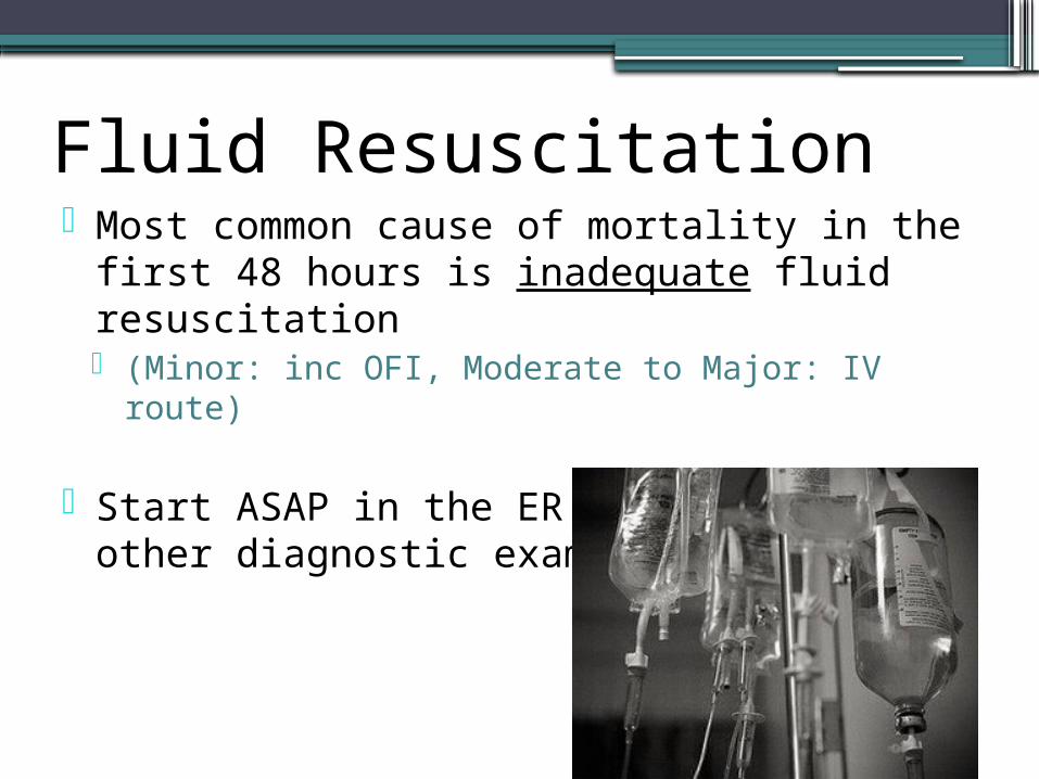
Fluid Resuscitation Most common cause of mortality in the first 48
hours is inadequate fluid resuscitation (Minor: inc OFI, Moderate to Major: IV route)
Start ASAP in the ER and even before other diagnostic exams
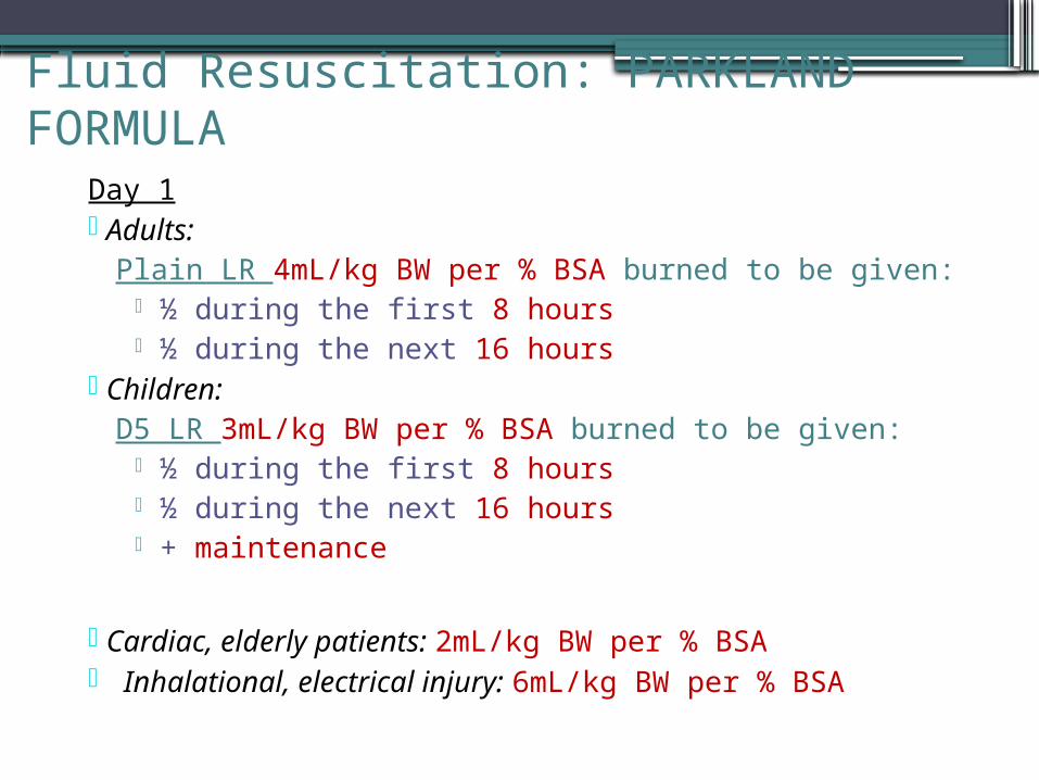
Fluid Resuscitation: PARKLAND FORMULADay 1 Adults:
Plain LR 4mL/kg BW per % BSA burned to be given: ½ during the first 8 hours ½ during the next 16 hours
Children:D5 LR 3mL/kg BW per % BSA burned to be given:
½ during the first 8 hours ½ during the next 16 hours + maintenance
Cardiac, elderly patients: 2mL/kg BW per % BSA Inhalational, electrical injury: 6mL/kg BW per % BSA
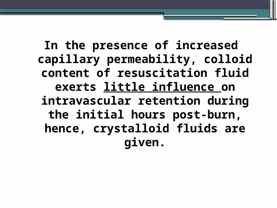
In the presence of increased capillary permeability, colloid content of
resuscitation fluid exerts little influence on intravascular retention during the
initial hours post-burn, hence, crystalloid fluids are given.
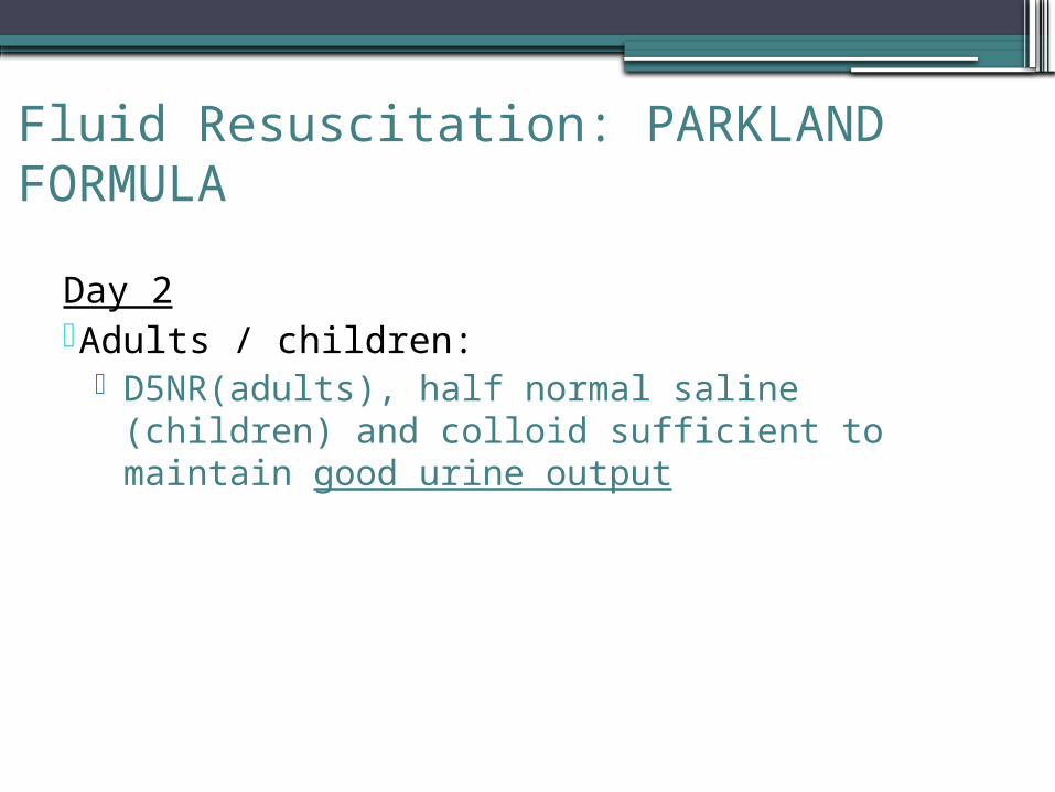
Fluid Resuscitation: PARKLAND FORMULA
Day 2Adults / children:
D5NR(adults), half normal saline (children) and colloid sufficient to maintain good urine output
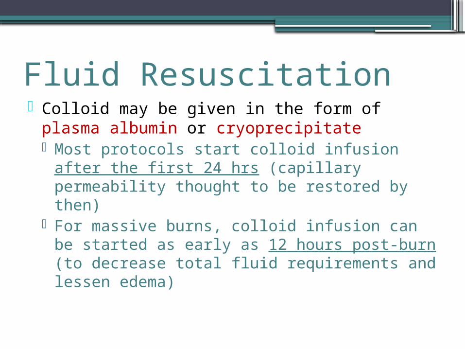
Fluid Resuscitation Colloid may be given in the form of plasma albumin or
cryoprecipitate Most protocols start colloid infusion after the first 24 hrs
(capillary permeability thought to be restored by then) For massive burns, colloid infusion can be started as
early as 12 hours post-burn (to decrease total fluid requirements and lessen edema)
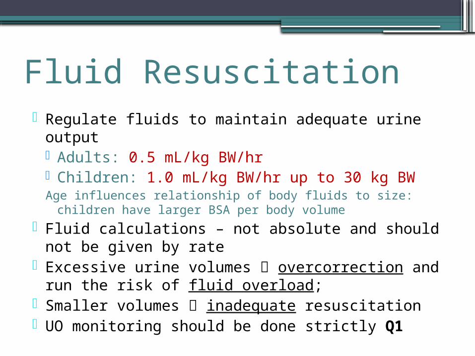
Fluid Resuscitation Regulate fluids to maintain adequate urine output
Adults: 0.5 mL/kg BW/hr Children: 1.0 mL/kg BW/hr up to 30 kg BWAge influences relationship of body fluids to size: children have
larger BSA per body volume
Fluid calculations – not absolute and should not be given by rate
Excessive urine volumes overcorrection and run the risk of fluid overload;
Smaller volumes inadequate resuscitation UO monitoring should be done strictly Q1
Fluid Resuscitation For electrical injuries:
Adjust fluid volume to maintain UO of 75-100 mL/hr (target UO: 1-2 cc/kg BW)
Mannitol 12.5-25g may be infused to promote diuresis If UO and pigment clearing do not respond to fluid
resuscitation, 12.5g osmotic diuretic mannitol may be added to each liter of resuscitation fluid
NaHCO3 can be added to maintain a slightly alkaline urine (pH>5.5) to promote solubility of heme pigments

Wound Dressing
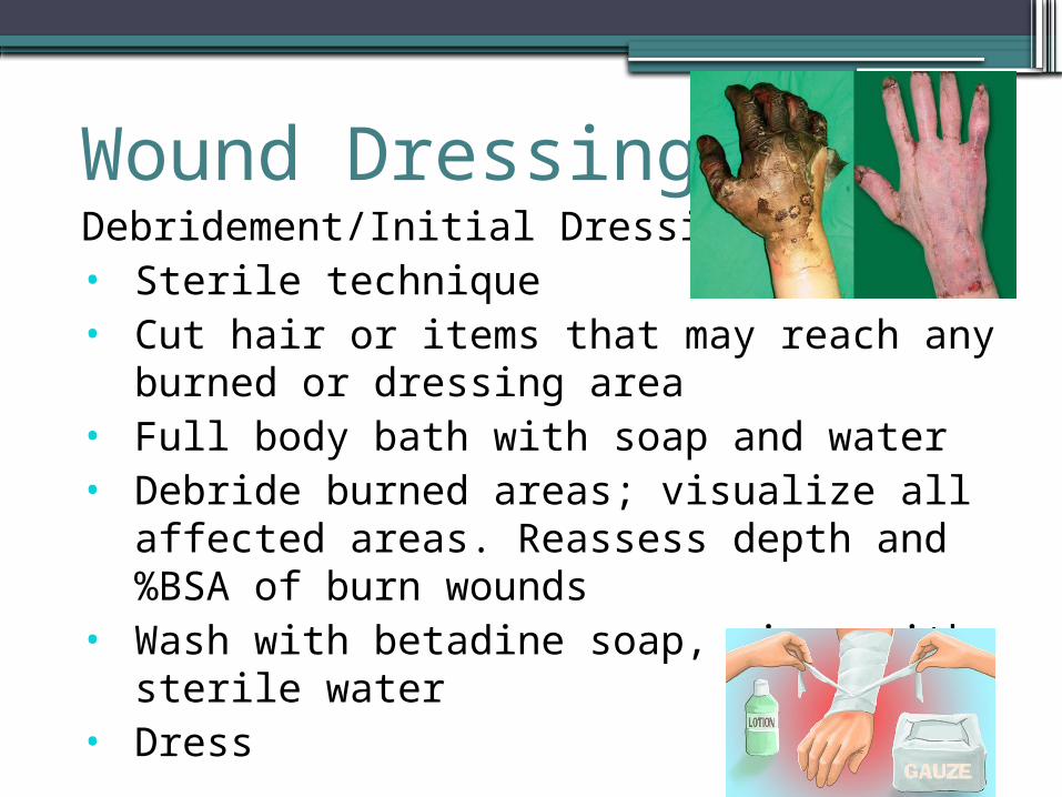
Wound DressingDebridement/Initial Dressing:• Sterile technique• Cut hair or items that may reach any burned or
dressing area• Full body bath with soap and water• Debride burned areas; visualize all affected areas.
Reassess depth and %BSA of burn wounds• Wash with betadine soap, rinse with sterile water• Dress
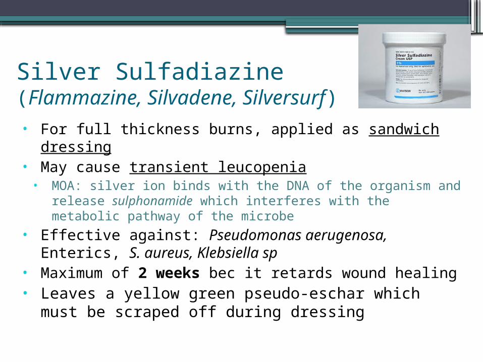
Silver Sulfadiazine (Flammazine, Silvadene, Silversurf)
• For full thickness burns, applied as sandwich dressing• May cause transient leucopenia
• MOA: silver ion binds with the DNA of the organism and release sulphonamide which interferes with the metabolic pathway of the microbe
• Effective against: Pseudomonas aerugenosa, Enterics, S. aureus, Klebsiella sp
• Maximum of 2 weeks bec it retards wound healing• Leaves a yellow green pseudo-eschar which must be
scraped off during dressing
Silver Sulfadiazine + Cerium nitrate (Flammacerium)
• Topical antimicrobial• Applied in cases wherein early excision-grafting cannot be
done (mass burn, extensive burns)• Reduces mortality by neutralizing toxin present in burned
skin• Mechanism of action:
• Cerium induces calcification of the dermal collagen remaining in the wound which produces the typical tanned, leathery crust
Silver Nitrate (not used anymore) Used as 0.5% solution Gauze dressing must be wet, solution loses effectivity
when dry Creates a brownish black discoloration with anything it
comes in contact with (will peel off with the burned skin) Bacteriostatic for S. aureus, E. Coli, P. aeruginosa Does not injure regenerating epithelium in the wound Caution with children as it tends to leach out electrolytes
(Na, Cl)
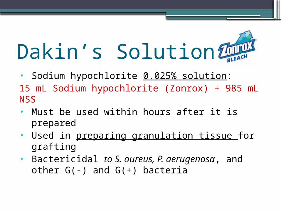
Dakin’s Solution• Sodium hypochlorite 0.025% solution:15 mL Sodium hypochlorite (Zonrox) + 985 mL NSS• Must be used within hours after it is prepared• Used in preparing granulation tissue for grafting• Bactericidal to S. aureus, P. aerugenosa, and other
G(-) and G(+) bacteria
Monitoring
•Burn injury is a dynamic process. The initial exposure to the wounding agent starts a train of physiologic events that present to the physician a patient with complex and precarious physiologic state, which has to be optimized to maximize chances of a positive outcome.
Monitoring At the ER: Check VS, UO, consciousness, pulmonary status Q1
Hgb, typing, Na, Cl, BUN, Crea, RBS CXR and ABG (for inhalational injury) ECG, urine myoglobin (for electrical burns)
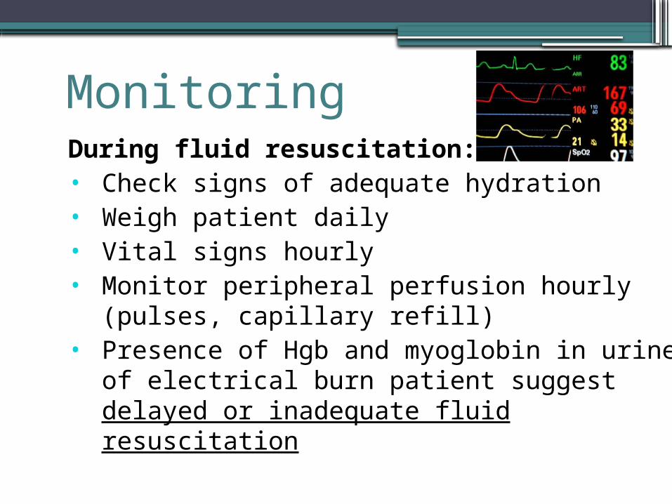
Monitoring During fluid resuscitation:• Check signs of adequate hydration• Weigh patient daily• Vital signs hourly• Monitor peripheral perfusion hourly (pulses,
capillary refill)• Presence of Hgb and myoglobin in urine of
electrical burn patient suggest delayed or inadequate fluid resuscitation
Monitoring During fluid resuscitation:• Pulmonary status every 4-5 hours• Daily determination of Hgb, Hct, WBC, Na, K,
BUN, crea• Status of wound daily during dressing change
Monitoring Post resuscitative period:• Vital signs every 4 hours• Daily determination of weight, BUN, crea, Na, K• Assess burn status daily• Burn biopsies (not swabs) twice a week• Blood CS once a week if wound is infected or
patient is septic• Weigh patient daily
Definitive ManagementPriority in the 1st 48 hours—maintain intravascular volumeOnce addressed, definitive management ensues
Classical Method:Allow eschar to spontaneously separate (3 weeks), wait until bed is ready for grafting, then place skin graft
Definitive Management
Present trend:Early excision (within 7d post burn) of burn wound, followed by skin grafting- improve survival and shorten hospital stay- adopted strategy by the PGH Burn Unit

Excision and Grafting

Excision and Grafting• To remove full thickness and deep partial burns until
clean viable bleed is encountered and a skin graft is placed immediately to cover the wound
• Early excision – done within 7 days• wound is not yet colonized by microorganisms, reducing
the chances of infection and promoting good graft take
Preparation for OR prerequisites
• Stable vital signs• Not in septic shock• Afebrile• Blood available for OR use (200-400mL/%BSA)• Normal albumin• No contraindications for surgery
Conduct for OR
• OR table covered by sterile linen• Keep OR warm• Prep patient using betadine soap and paint for the
donor site and betadine soap for the wound• Prep the donor site• Drape donor site separate from the burn wound
Tangential Excision•Principle: to excise the wound in thin layers
using a blade held at very acute angle with the skin surface
•Goal: to remove non viable tissue leaving as much dermis as possible (excellent surface for grafting)
Fascial Excision Best used when excising large flat areas When excision of the burn wounds has to be done with
minimum blood loss Less bloody than tangential excision, but with cosmetic
effect defect Limited use in extremities due to problems of edema
distal to the area of excision, presence of avascular fascia and presence of superficial nerves
Skin Graft Harvesting Preferred areas are thighs, buttocks, and abdomen The only area in which color match between donor and
recipient site is of significant concern is the face and neck. Upper chest and upper back are a good color match for face and neck.
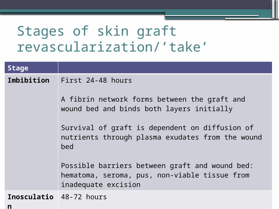
Stages of skin graft revascularization/’take’
Stage
Imbibition First 24-48 hours
A fibrin network forms between the graft and wound bed and binds both layers initially
Survival of graft is dependent on diffusion of nutrients through plasma exudates from the wound bed
Possible barriers between graft and wound bed: hematoma, seroma, pus, non-viable tissue from inadequate excision
Inosculation
48-72 hours
Old capillaries from the wound graft link with vessels on the graft, causing revascularization
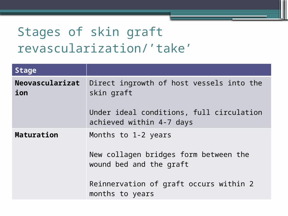
Stages of skin graft revascularization/’take’
Stage
Neovascularization
Direct ingrowth of host vessels into the skin graft
Under ideal conditions, full circulation achieved within 4-7 days
Maturation Months to 1-2 years
New collagen bridges form between the wound bed and the graft
Reinnervation of graft occurs within 2 months to years
Applying Skin Graft Best to place grafts on the wound at the time of excision Since the graft itself controls hemostasis and protects the
wound, it makes little sense to wait 24-48hrs until bleeding has stopped
This approach requires an additional procedure and there is a significant risk of the wound bed becoming desiccated or reinfected
Better to have a slight overlap of skin on the wound rather than to leave excised wound uncovered. Hypertrophic scarring will result and most evident at the edges of the graft, especially if a ridge of open wound is left to heal primarily.
Care of the Skin Graft
•First graft opening: 3-5th day post op. Open early if suspecting infection
•Remove bulky dressing slowly, not disturbing any graft using copious amounts of sterile water
•Graft uptake: Pinkish color of graft with adherence to skin bed
•Wash area gently with betadine soap and rinse with water. Dress graft with bulky wet dressing
•Staples can be removed at first dressing change
•Can be dressed everyday if not infected• If with good take, skin graft can be left open on
the 7th post op day.
Nutrition Patients with Burns have a hypermetabolic response,
which persist until burns are covered Curreri’s Formula Adult (25 x kg) + (40 x %BSA Burn) Children (60 x kg) +(35 x %BSA Burn)
Rough Guide: 2,500 cal/d in adults, proteins= 2g/kgBWAt Burn Unit, 6 egg whites/day
Carbs = 60%, fats = rest Give Vit C and Zinc Supplements
Complications• Sepsis
▫ Most common cause of death in burns▫ Suspect in the presence of: fever, hypotension, conversion from PT to
FT burns, ecthyma gangrenosum▫ Start antibiotics
• ARDS▫ Setting of electrical/Inhalational/pulmonary injury▫ Progressive hypoxemia unresponsive to inc FiO2▫ Xrays may be normal in early phase▫ Manage with intubation: 100% FiO2
• Contractures▫ Prevented with proper posture and splinting, coordinate with Rehab
Pain ControlMeperidine 50mg IV q6Nalbuphine q4Narcotics are not given IM since absorption is erratic
Criteria for Discharge• No existing complications of thermal injury such as
inhalational injury• Fluid resuscitation completed• Adequate pain tolerance• Adequate nutritional intake• No anticipated septic complications

Thank You!