Embed Size (px)
Citation preview

ORIGINAL ARTICLE
The Bronchiectasis Severity IndexAn International Derivation and Validation StudyJames D. Chalmers1, Pieter Goeminne2, Stefano Aliberti3, Melissa J. McDonnell4,5, Sara Lonni3, John Davidson4,Lucy Poppelwell1, Waleed Salih1, Alberto Pesci3, Lieven J. Dupont2, Thomas C. Fardon1, Anthony De Soyza4,5,and Adam T. Hill6
1Tayside Respiratory Research Group, University of Dundee, Dundee, United Kingdom; 2Respiratory Medicine, University HospitalGasthuisberg, Leuven, Belgium; 3Department of Health Science, University of Milan Bicocca, Clinica Pneumologica, AO San Gerardo,Monza, Italy; 4Adult Bronchiectasis Service and Sir William Leech Centre for Lung Research, Freeman Hospital, Newcastle upon TyneHospitals, Heaton, Newcastle, United Kingdom; 5Institute of Cellular Medicine, Newcastle University, Newcastle upon Tyne, UnitedKingdom; and 6Department of Respiratory Medicine Royal Infirmary of Edinburgh and the University of Edinburgh, Edinburgh, United Kingdom
Abstract
Rationale: There are no risk stratification tools for morbidity andmortality in bronchiectasis. Identifying patients at risk of exacerbations,hospital admissions, and mortality is vital for future research.
Objectives:This study describes the derivation and validation of theBronchiectasis Severity Index (BSI).
Methods:Derivationof theBSIuseddata fromaprospectivecohort study(Edinburgh, UK, 2008–2012) enrolling 608 patients. Cox proportionalhazard regressionwas used to identify independent predictors ofmortalityand hospitalization over 4-year follow-up. The score was validated inindependent cohorts from Dundee, UK (n = 218); Leuven, Belgium (n =253); Monza, Italy (n = 105); and Newcastle, UK (n = 126).
Measurements and Main Results: Independent predictors offuture hospitalization were prior hospital admissions, MedicalResearch Council dyspnea score greater than or equal to 4, FEV1 ,30% predicted, Pseudomonas aeruginosa colonization, colonizationwith other pathogenic organisms, and three or more lobes involvedon high-resolution computed tomography. Independent predictorsof mortality were older age, low FEV1, lower body mass index, priorhospitalization, and three or more exacerbations in the year beforethe study. The derived BSI predicted mortality and hospitalization:area under the receiver operator characteristic curve (AUC) 0.80(95% confidence interval, 0.74–0.86) for mortality and AUC 0.88(95% confidence interval, 0.84–0.91) for hospitalization, respectively.
Therewas a clear difference in exacerbation frequency andquality of lifeusing the St. George’s Respiratory Questionnaire between patientsclassified as low, intermediate, andhigh risk by the score (P,0.0001 forall comparisons). In the validation cohorts, the AUC for mortalityranged from 0.81 to 0.84 and for hospitalization from 0.80 to 0.88.
Conclusions: The BSI is a useful clinical predictive tool thatidentifies patients at risk of future mortality, hospitalization, andexacerbations across healthcare systems.
Keywords: bronchiectasis; mortality; Pseudomonas aeruginosa;exacerbation; prediction
At a Glance Commentary
Scientific Knowledge on the Subject: There are norecognized clinical severity criteria for non–cystic fibrosisbronchiectasis.
What This Study Adds to the Field: This study derives andvalidates a multidimensional clinical prediction tool, theBronchiectasis Severity Index (BSI), from a large internationalmulticenter study of 1,310 patients with bronchiectasis. TheBSI is a useful clinical predictive tool that identifies patients atrisk of future mortality, hospital admissions, and exacerbationsacross healthcare systems.
(Received in original form September 2, 2013; accepted in final form December 10, 2013 )
Supported by the Medical Research Council, UK; fellowship support from the Medical Research Council and the Wellcome Trust (J.D.C.); fellowship support from theEuropean Respiratory Society/European Lung Foundation (M.J.M.); and a Higher Education Funding Council for England senior lectureship and support from theNational Institute for Health Research Biomedical Research Centre (A.D.S.). L.J.D. is a senior research fellow of the Fonds Wetenschappelijk Onderzoek.
Author Contributions: All authors participated in study design, data analysis, and interpretation of the data. All authors were involved in writing and revising thearticle before submission.
Correspondence and requests for reprints should be addressed to James D. Chalmers, M.B. Ch.B., Tayside Respiratory Research Group, University of Dundee,Dundee DD1 9SY, UK. E-mail: [email protected]
This article has an online supplement, which is accessible from this issue’s table of contents at www.atsjournals.org
Am J Respir Crit Care Med Vol 189, Iss 5, pp 576–585, Mar 1, 2014
Copyright © 2014 by the American Thoracic Society
Originally Published in Press as DOI: 10.1164/rccm.201309-1575OC on December 12, 2013
Internet address: www.atsjournals.org
576 American Journal of Respiratory and Critical Care Medicine Volume 189 Number 5 | March 1 2014

Non–cystic fibrosis (CF) bronchiectasis(hereafter referred to as bronchiectasis) isa chronic respiratory disorder characterizedby recurrent cough, sputum production,and respiratory infections (1).Pathologically, patients have abnormallydilated bronchi leading to impairment ofhost defense, chronic colonization withbacteria, and airways inflammation (2, 3).
Although patients are sometimesdescribed as having mild, moderate, orsevere bronchiectasis, there is no accepteddefinition of these terms. They are oftenapplied in reference to the radiologicalappearance of disease. Radiologicalappearance is likely to be insufficient tocapture the complexity of disease impact inbronchiectasis (4).
Clinical decision making relies onaccurately identifying patients at high risk offuture mortality, hospital admissions, andexacerbations. Such a model has beensuccessful for guideline development inchronic obstructive pulmonary disease(COPD), in which different treatmentstrategies are recommended for differentGlobal Initiative for Chronic ObstructiveLung Disease stages of disease and in otherrespiratory disorders in which treatmentsare targeted to patients with a worseprognosis (5–7). There are currently noseverity scoring systems for use inbronchiectasis.
There is a need to define which patientsare most likely to benefit from newtreatments, with an increasing number ofclinical trials of inhaled and oral therapies inbronchiectasis (8–11). A severityclassification system could theoreticallyallow targeting of therapies to the patientsmost likely to benefit.
The aim of this study was develop aseverity index for bronchiectasis using fourimportant, well recognized end-points:mortality, frequency of exacerbations,hospital admissions, and health-relatedquality of life.
Methods
Derivation CohortThe clinical prediction tool was derivedusing data from a prospective cohort studyconducted at a regional specialistbronchiectasis service based at the RoyalInfirmary of Edinburgh, UK (2008–2012).The study was approved by the South EastScotland Research Ethics Committee.
Consecutive patients were enrolled on thebasis of a diagnosis of bronchiectasis madeby high-resolution computed tomography(HRCT) and a clinical history consistentwith bronchiectasis (12). The primaryobjective of the original study was toevaluate predictors of outcome inbronchiectasis, including clinical andgenetic predictors. As the goal of thispresent analysis was to derive a clinicalprediction tool using routinely availableclinical data, genetic predictors orbiomarkers that are not widely availablewere excluded from the present analysis(13, 14). Patients were excluded if they hadactive malignancy at enrollment, CF, activemycobacterial disease (including activenontuberculous mycobacteria [NTM]),HIV, or a primary diagnosis of pulmonaryfibrosis/sarcoidosis with secondary tractionbronchiectasis. Patients receiving long-termoral or inhaled antibiotic therapy atenrollment were also excluded.
Clinical AssessmentsAt the time of clinical assessment all patientswere clinically stable, with no antibiotic usein the preceding 4 weeks. All patientsunderwent spirometry (FEV1 and FVC withthe highest of three technically satisfactorymeasurements recorded). The underlyingetiology of bronchiectasis was determinedafter testing recommended by the BritishThoracic Society (BTS) guidelines (3).
Radiological SeverityRadiological severity of bronchiectasis wasassessed using a modified Reiff score, whichassesses the number of lobes involved(with the lingula considered to be a separatelobe) and the degree of dilatation (tubular =1, varicose = 2, and cystic = 3). Themaximum score is 18 and minimum scoreis 1. This score has been used previously instudies of bronchiectasis (13–16).
BacteriologyAll bacteriology was performed onspontaneous early-morning sputum samplesas previously described (2). Chroniccolonization was defined by the isolation ofpotentially pathogenic bacteria in sputumculture on two or more occasions, at least 3months apart in a 1-year period (14, 16, 17).The predominant pathogen was theorganism grown most frequently over thestudy period. Patients were asked to providesputum samples at least twice a year at clinicreviews. Patients who were unable to provide
sputum samples due to absence ofa productive cough were classified asnoncolonized for the purposes of analysis.
End-points
Mortality. At the end of the 4-year follow-up period, mortality was determined usinga computer database linked to nationaldeath records. Survival status wasconfirmed for 100% of participants. Causeof death was determined and assigned asbronchiectasis related or unrelated afterindividual case review.
Hospitalization for severe exacerbations.Severe exacerbations were defined accordingto the BTS guidelines, and unscheduledhospitalizations or emergency departmentvisits for severe bronchiectasis exacerbations orcomplications were recorded from patienthistories and verified using an administrativedatabase that records all regional hospitaladmissions (3).
Exacerbations. Exacerbations weredefined according to the BTS definitionas an acute deterioration with increasingsputum volume and purulence and/orsystemic upset (3). Frequency ofexacerbations requiring antibiotictreatment were determined frompatient histories and verified againstelectronic general practice prescriptionrecords.
Quality of life. Patients completed theSt. George’s Respiratory Questionnaire (18)as a measure of quality of life. The widelyused minimal important clinical differenceis a change of 4 units (18).
Validation CohortsThe Bronchiectasis Severity Index (BSI) wasvalidated in independent cohorts of patientswith bronchiectasis from four centers:Dundee (n = 218) and Newcastle (n = 126)in the UK (19), Leuven in Belgium (n =253) (20), and Monza in Italy (n = 105).Details of data collection in each of thesestudies are described in the onlinesupplement. Validation cohorts wereconvenience cohorts collected andanalyzed independently of the derivationstudy. Each applied definitions ofcolonization and assessments based on thederivation cohort.
Statistical Analysis and Derivation ofClinical Prediction ToolNormally distributed data are presented asmean with SD, whereas nonnormally
ORIGINAL ARTICLE
Chalmers, Goeminne, Aliberti, et al.: Predicting Morbidity and Mortality in Bronchiectasis 577
distributed data are presented as medianwith interquartile range. The chi-squaredtest and MannWhitney U test were used forcomparison of categorical and numericaldata, respectively. For comparisons of morethan two groups, one-way analysis ofvariance or the Kruskal-Wallis test wereused as appropriate. The independentrelationship of clinical variables withmortality and hospital admissions overthe study period was determined usingseparate Cox proportional hazardregression models. Variables that wereassociated with the outcome at P lessthan 0.2 on univariate analysis wereconsidered for entry into the multivariatemodels. Variables were dichotomizedusing the Youden index to identify theoptimal cut-off or using previous cut-offsidentified in the bronchiectasis literature(21). In all analyses, missing data forpredictors were assumed to be normal.Less than 0.1% of data were missing in
the five databases, and no outcome datawere missing.
To derive a prediction tool forbronchiectasis severity, the authorsidentified common variables that predictedmortality and hospital admissions. Thesevariables were then formed into a predictiontool using the rounded averagedb-coefficient to award “points” for eachvariable as previously described (7). Theperformance of the resulting model formortality and hospital admissions wasassessed using the area under the receiveroperator characteristic curve (AUC). For allanalyses, P less than 0.05 was consideredstatistically significant.
Results
The prospective derivation cohort included608 patients. The majority were classified ashaving idiopathic or postinfective
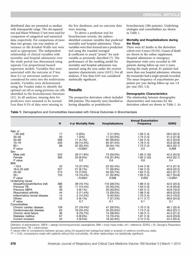
bronchiectasis (386 patients). Underlyingetiologies and comorbidities are shownin Table 1.
Mortality and Hospitalizations duringthe StudyThere were 62 deaths in the derivationcohort over 4 years (10.2%). Causes of deathare shown in the online supplement.Hospital admissions or emergencydepartment visits were recorded in 189patients during follow-up over 4 years.During the study period, 81 patients hadmore than one hospital admission, whereasthe remainder had a single episode recorded.The mean frequency of exacerbations perpatient per year during follow-up was 1.8per year (SD, 1.4).
Demographic CharacteristicsThe relationship between demographiccharacteristics and outcomes for thederivation cohort are shown in Table 1. As
Table 1: Demographics and Comorbidities Associated with Clinical Outcomes in Bronchiectasis
N 4-yr Mortality Rate HospitalizationsAnnual Exacerbation
Frequency SGRQ
Age, yr,30 (ref) 17 0 (0%) 2 (11.8%) 1.56 (1.4) 26.0 (22.2)30–49 50 1 (2%) 11 (22.0%) 1.74 (1.4) 41.4 (21.6)50–69 268 11 (4.1%) 82 (30.6%) 1.94 (1.4) 42.3 (23.9)70–79 203 29 (14.3%) 64 (31.5%) 1.78 (1.4) 45.8 (22.8)801 68 22 (32.4%) 30 (44.1%) 1.73 (1.2) 46.5 (22.1)P value ,0.0001 0.03 0.5 0.008
SexMale (ref) 243 29 (11.9%) 73 (30.0%) 1.85 (1.42) 42.2 (24.0)Female 365 33 (9.0%) 116 (31.8%) 1.82 (1.32) 44.0 (22.7)P value 0.2 0.6 0.8 0.4
BMI,18.5 42 13 (31.0%) 22 (52.4%) 2.44 (1.9) 47.5 (27.4)18.5–25 (ref) 227 21 (9.1%) 71 (30.9%) 1.84 (1.3) 40.8 (22.2)25–30 214 15 (7.0%) 55 (25.7%) 1.62 (1.2) 40.2 (21.7)301 125 13 (10.4%) 41 (32.8%) 1.93 (1.5) 50.7 (24.8)P value ,0.0001 0.008 0.003 ,0.0001
Underlying causeIdiopathic/postinfective (ref) 386 39 (10.1%) 110 (28.5%) 1.89 (1.4) 42.0 (23.3)Previous TB 82 11 (13.4%) 33 (40.2%) 1.50 (1.0) 41.8 (25.6)Previous ABPA 49 3 (6.1%) 20 (40.8%) 1.64 (1.1) 45.8 (18.4)Rheumatoid arthritis 44 5 (11.4%) 13 (29.5%) 1.98 (1.6) 46.8 (23.0)Inflammatory bowel disease 14 2 (14.3%) 6 (42.9%) 1.70 (1.4) 54.3 (19.5)Others 33 2 (6.1%) 7 (21.2%) 2.11 (1.7) 49.6 (24.0)P value 0.7 0.1 0.1 0.1
ComorbiditiesChronic cardiac disease 139 27 (19.4%)* 41 (29.5%) 1.70 (1.3) 46.1 (22.4)Cerebrovascular disease 63 16 (25.4%) 35 (55.6%)* 1.73 (1.4) 48.0 (23.1)Chronic renal failure 36 6 (16.7%) 14 (38.9%) 1.40 (1.1) 44.3 (21.2)Diabetes mellitus 67 6 (9.0%) 13 (19.4%) 1.81 (1.3) 44.9 (23.6)Current smokers 42 4 (9.5%) 18 (42.9%) 2.12 (1.5) 42.4 (20.4)
Definition of abbreviations: ABPA = allergic bronchopulmonary aspergillosis; BMI = body mass index; ref = reference; SGRQ = St. George’s RespiratoryQuestionnaire; TB = tuberculosis.P values refer to comparisons between groups using chi-squared test (categorical data) or analysis of variance (continuous data).*P , 0.05, comparisons made with patients without this comorbidity. All other relationships not statistically significant.
ORIGINAL ARTICLE
578 American Journal of Respiratory and Critical Care Medicine Volume 189 Number 5 | March 1 2014

expected, there was a strong relationshipbetween age and mortality. There was nosignificant relationship between sex andmortality. There was a strong relationshipbetween body mass index (BMI) andmortality, with a 31.0% mortality rate inpatients with a BMI less than 18.5 kg/m2.Chronic cardiac disease was associated withmortality but not with hospital admissions,exacerbation frequency, or quality of life.
Pulmonary Function TestsBased on FEV1/FVC ratio, 301 patients(49.5%) had airflow obstruction, restrictivespirometry was present in 114 patients
(18.8%), and normal spirometry waspresent in 193 patients (31.7%).
Based on the Youden index, FEV1 %predicted was most discriminatory formortality and hospital admissions and wasused for subsequent analyses of lungfunction. Lower FEV1/FVC ratio, FEV1,and FVC % predicted were all stronglyassociated with mortality. Similarly,patients with lower lung function weremore frequently hospitalized and had anincreased annual exacerbation frequencyand worse quality of life. The data for FEV1
are shown in Table 2, and the data forFVC % predicted and data for patients with
obstruction, restriction, and normalspirometry are shown in Table E1 in theonline supplement.
Hospital Admissions, Exacerbations,and Exercise CapacityAt study enrollment, 133 patients gavea history of hospitalization or emergencydepartment visits with a severe exacerbationor respiratory tract infection in thepreceding 2 years. The distribution ofpatients according to annual exacerbationfrequency is shown in Table 2. This showsthat a prior history of hospital admissionsor the annual frequency of exacerbations
Table 2: Spirometry, Previous Hospital Admissions, Exacerbations, and Baseline Medical Research Council Dyspnea Score asPredictors of Future Morbidity and Mortality
N 4-yr Mortality Hospitalizations Exacerbations SGRQ
FEV1, % predicted.80 255 15 (5.9%) 44 (17.3%) 1.60 (1.22) 34.7 (21.5)50–80 220 20 (9.1%) 71 (32.3%) 1.85 (1.41) 43.4 (20.9)30–50 110 19 (17.3%) 59 (53.6%) 2.24 (1.46) 58.5 (21.1),30 23 9 (39.1%) 15 (65.2%) 2.37 (1.36) 66.3 (21.5)P value ,0.0001 ,0.0001 ,0.0001 ,0.0001
History of hospitalization for severe exacerbationsYes 133 33 (24.8%) 123 (92.5%) 2.59 (1.5) 60.8 (22.0)No 475 29 (6.1%) 66 (13.9%) 1.62 (1.2) 38.4 (21.0)P value ,0.0001 ,0.0001 ,0.0001 ,0.0001
Frequency of outpatient exacerbations in previousyear*
0 245 19 (7.8%) 59 (24.1%) 1.09 (0.8) 36.3 (21.4)1 127 7 (5.5%) 28 (22.0%) 1.38 (0.8) 41.1 (20.8)2 97 11 (11.3%) 30 (30.9%) 2.05 (0.9) 48.2 (22.8)3 47 4 (8.5%) 18 (38.3%) 2.42 (1.0) 46.1 (25.5)4 or more 92 22 (23.9%) 54 (58.7%) 3.90 (1.4) 58.9 (21.8)P value 0.0001 ,0.0001 ,0.0001 ,0.0001
MRC dyspnea score1 228 16 (7.0%) 26 (11.4%) 1.59 (1.3) 33.2 (20.9)2 121 11 (9.1%) 29 (24.0%) 1.67 (1.2) 44.6 (21.9)3 124 10 (8.1%) 50 (40.3%) 2.01 (1.5) 46.7 (21.7)4 87 12 (13.8%) 49 (56.3%) 1.91 (1.4) 52.8 (21.2)5 48 14 (29.2%) 35 (72.9%) 2.78 (1.4) 61.9 (22.0)P value 0.0001 ,0.0001 ,0.0001 ,0.0001
Bacteriology and colonizationChronic colonization 440 52 (11.8%) 169 (38.4%) 2.04 (1.4) 45.6 (23.7)Not colonized 168 10 (6.0%) 20 (12.0%) 1.29 (0.9) 37.8 (20.2)P value 0.03 ,0.0001 ,0.0001 ,0.0001
Specific organismsHaemophilus influenzae 177 10 (5.6%) 61 (34.5%) 2.03 (1.5) 45.1 (22.0)Pseudomonas aeruginosa 70 15 (21.2%)† 62 (88.6%)† 2.85 (1.5)† 60.7 (21.7)†
Streptococcus pneumoniae 35 2 (5.7%) 11 (31.4%)† 2.13 (1.5) 49.3 (21.6)Moraxella catarrhalis 63 5 (7.9%) 26 (41.3%)† 2.08 (1.3)† 48.4 (22.1)†
Staphylococcus aureus (excluding MRSA) 43 5 (11.6%) 14 (32.6%)† 2.04 (1.7) 43.7 (21.6)†
MRSA 8 5 (62.5%)† 5 (62.5%)† 3.10 (2.4)† 50.7 (33.3)†
Gram-negative Enterobacteriaceae 40 6 (15.0%) 21 (52.5%)† 2.29 (1.5)† 55.2 (21.2)†
Definition of abbreviations: MRC = Medical Research Council; MRSA = methicillin-resistant Staphylococcus aureus; SGRQ = St. George’s RespiratoryQuestionnaire.A proportion of patients were colonized with more than one pathogen; therefore, for individual organisms the mortality rates are expressed asa percentage of all patients colonized with that pathogen and may add up to more than the total number of events in the population.*Outpatient exacerbations excludes exacerbations managed in hospital.†For microbiology, statistically significant differences are highlighted. P , 0.05 compared to the not-colonized group.
ORIGINAL ARTICLE
Chalmers, Goeminne, Aliberti, et al.: Predicting Morbidity and Mortality in Bronchiectasis 579

predicts future mortality, hospitaladmissions, exacerbations, and quality oflife. Further analysis identified a strongrelationship between baseline MedicalResearch Council dyspnea score and futuremortality, hospital admissions,exacerbations, and quality of life.
Colonization Status and BacteriologyMortality was significantly higher in patientswith chronic colonization compared withnoncolonized patients. The mortality ratevaried significantly depending on thecolonizing organism, with the highestmortality rates associated with the isolation ofPseudomonas aeruginosa and methicillin-resistant Staphylococcus aureus (Table 2).
Radiological SeverityThe analysis of radiological severity ispresented in Table E2. The data show no
significant relationship between radiologicalseverity and mortality (P = 0.3) buta significant relationship between theReiff score and hospital admissions. Thisrelationship was statistically significantabove a score of 3 or more (indicatingthree or more lobes involved or a lobewith cystic bronchiectasis). There wasa weak but statistically significantrelationship with quality of life, butthe relationship with exacerbations wasnot statistically significant (P = 0.06;Table E2).
Development of the BSI in theDerivation CohortThe Cox proportional hazard regressionmodels for hospital admission for severeexacerbations and mortality are shown inTable 3. This model identified a prior historyof hospitalization to be the strongest predictor
of future hospitalization risk. Independentof this, Medical Research Council dyspneascore, FEV1 less than 30% predicted, andcolonization with P. aeruginosa or otherorganisms were independent predictors ofhospital admissions. Mortality wassignificantly associated with priorhospitalizations, increasing age, BMI less than18.5 kg/m2, FEV1 % predicted, and three ormore exacerbations per year.
Several factors were not associated withmortality or hospital admissions afteradjustment for the other included variables,including etiology of bronchiectasis, sex,comorbidities, smoking status, and inhaledcorticosteroid use (Table 3).
Classification of Patients Accordingto the BSIPatients were classified into tertilesdesignated low (0–4 points, n = 191),
Table 3: Results of the Cox Proportional Hazard Regression Analysis for Mortality and Hospitalization
Severity MarkerHR (95% CI) for Hospital
Admissions during Follow-up HR (95% CI) for Mortality Score Points
Age, yr,50 1.0 (reference) 1.0 (reference) 050–69 1.38 (0.73–2.56) 2.21 (0.28–17.5) 270–79 1.50 (0.79–2.82) 8.57 (1.15–63.63) 4801 1.76 (0.89–3.50) 23.16 (3.09–173.7) 6
BMI,18.5 1.23 (0.73–2.08) 2.25 (1.09–4.67) 218.5–25 1.0 (reference) 1.0 (reference) 026–29 0.90 (0.62–1.30) 0.91 (0.46–1.81) 030 or more 1.14 (0.76–1.70) 1.38 (0.68–2.81) 0
FEV1 % predicted.80 1.0 (reference) 1.0 (reference) 050–80 1.17 (0.74–1.85) 1.34 (0.67–2.67) 130–49 1.40 (0.68–2.85) 1.58 (0.72–3.46) 2,30 1.52 (1.03–2.25) 4.47 (1.60–12.53) 3
Hospital admission before studyNo 1.0 (reference) 1.0 (reference) 0Yes 13.5 (9.40–19.46) 2.43 (1.30–4.53) 5
Exacerbations before the study0 1.0 (reference) 1.0 (reference) 01–2 1.67 (0.78–3.58) 1.78 (0.80–3.98) 03 or more 2.25 (0.89–5.70) 2.03 (1.02–4.03) 2
MRC dyspnea score1–3 1.0 (reference) 1.0 (reference) 04 2.42 (1.66–3.52) 1.05 (0.50–2.20) 25 2.69 (1.59–4.53) 1.15 (0.50–2.63) 3
Pseudomonas colonizationNo 1.0 (reference) 1.0 (reference) 0Yes 2.16 (1.36–3.43) 1.58 (0.75–3.34) 3
Colonization with other organismsNo 1.0 (reference) 1.0 (reference) 0Yes 1.66 (1.12–2.44) 1.10 (0.54–2.24) 1
Radiological severity: >3 lobes involvedor cystic bronchiectasis
No 1.0 (reference) 1.0 (reference) 0Yes 1.48 (1.02–2.15) 1.05 (0.57–1.94) 1
Definition of abbreviations: BMI = body mass index; CI = confidence interval; HR = hazard ratio; MRC = Medical Research Council.All factors founded to be significantly associated with either mortality or hospital admissions were included in the derivation of the severity score.
ORIGINAL ARTICLE
580 American Journal of Respiratory and Critical Care Medicine Volume 189 Number 5 | March 1 2014

intermediate (5–8 points, n = 224), andhigh BSI scores (9 or more points, n = 193).The relationship between these severityclasses and mortality and morbidity areshown in Figure 1.
The AUC for mortality was0.80 (0.74–0.86) and the AUC forhospitalization was 0.88 (0.84–0.91). Therewas a clear difference in exacerbationfrequency and quality of life betweenpatients classified as low, intermediate,and high BSI scores (P , 0.0001) for allcomparisons (Figure 1).
The above data represent predictionsover the full 4 years of follow-up. Ananalysis was performed using data fromannual follow-up visits to predict events inthe subsequent year (e.g., data from follow-up in 2009 was used to predict events from2009–2010, and so on). The AUC formortality was: 0.79 (0.66–0.91) for 2008 to2009, 0.75 (0.62–0.87) for 2009 to 2010,0.82 (0.72–0.91) for 2010 to 2011, and 0.80(0.71–0.89) for the period 2011 to 2012.This indicates that the score workedsimilarly for annual prediction as forlonger-term prediction.
For hospital admissions, the annualAUCs were: 0.87 (0.84–0.91) for 2008 to
2009, 0.86 (0.82–0.89) for 2009 to 2010,0.88 (0.82–0.94) for 2010 to 2011, and 0.87(0.78–0.96) for the period 2011 to 2012.This confirms the usefulness of the BSI inpredicting the likelihood of both short- andlong-term hospital admissions.
Validation of the BSI inIndependent CohortsThe four independent cohorts are describedin Table 4 and in the online supplement.For prediction of mortality, data are shownin Figures 2A and 2B, demonstratingprogressive increases in mortality withincreasing severity index group. The AUCsfor mortality are shown in Figure 2D andconfirmed good discrimination forpredicting mortality using the BSI. No AUCwas calculated for the Monza, Italy cohortas there were only two deaths. Similarly,each cohort with available data showeda progressive increase of hospitalizationwith BSI severity class, and the AUCs inFigure 2D confirmed a high degree ofdiscrimination (AUC, 0.80–0.88). Nohospitalization data were available in theLeuven cohort.
Three cohorts had data forexacerbation frequency during follow-up
(Figure 2C). Each of these showeda progressive increase in exacerbationfrequency with BSI severity class (P ,0.0001 for Newcastle and Dundee cohorts,P = 0.03 for Monza cohort).
In contrast to the derivation cohort, theDundee, Newcastle, and Leuven cohortsincluded patients receiving long-termantibiotic therapy. The frequency ofantibiotic therapy was 41.3% in the Dundeecohort, 40.5% in Newcastle, and 43.1% inLeuven. In these cohorts, long-termantibiotic therapy was not independentlypredictive of mortality or hospitalizations.Hazard ratio (HR) was 0.66 (95% CI,0.34–1.31) in the Leuven cohort and HRwas 1.43 (95% CI, 0.54–1.81) in theNewcastle cohort for mortality. For hospitaladmissions, the HR was 0.63 (95% CI,0.36–1.11) in the Newcastle cohort and HRwas 1.34 (95% CI, 0.81–2.23) in the Dundeecohort. The BSI score predicted mortalityand hospital admissions in current users oflong-term antibiotics similarly to theprimary analysis, suggesting this was nota major confounder. The AUC formortality in long-term antibiotic users was0.82, 0.75, and 0.80 in Dundee, Newcastle,and Leuven, respectively. For hospital
Figure 1. The performance of the Bronchiectasis Severity Index in predicting mortality, hospital admissions, exacerbations, and quality of life. All between-group comparisons were statistically significant (P, 0.0001). The exacerbation and quality-of-life data are presented as mean with SD. AUC = area underthe receiver operator characteristic curve.
ORIGINAL ARTICLE
Chalmers, Goeminne, Aliberti, et al.: Predicting Morbidity and Mortality in Bronchiectasis 581

admissions it was 0.80 and 0.76 in Dundeeand Newcastle, respectively.
Discussion
The present study is the first multicenterinternational study to describe a clinicalprediction tool for bronchiectasis. Wederived the BSI in a prospective cohort studyover 4 years and validated in severalindependent cohorts of patients withbronchiectasis. Overall, this study evaluatedthe score in 1,310 patients withbronchiectasis across five cohorts, makingthis the largest and most diverse assessmentof bronchiectasis severity so far reported.The BSI accurately stratified the risk ofmortality, hospital admissions, future risk ofexacerbations, and quality of life.
Most respiratory diseases havea disease-specific severity assessment tool,designed for guiding therapy or stratifyingrisk of complications (5–7). No such toolexists for bronchiectasis. New treatmentsare increasingly available for patients withbronchiectasis, with growing evidence forthe efficacy of long-term macrolide therapyand inhaled antibiotics (8–11). However,these therapies have attendant risks (e.g.,antimicrobial resistance and toxicity) aswell as significant healthcare costs andtreatment burdens (22). A key challenge inbronchiectasis management lies in theidentification of patients at high risk ofdeveloping bronchiectasis complicationswho may benefit from intensification oftherapy (23, 24). Whether using a risk-stratification tool such as the BSI canachieve improvements in clinical practicenow requires prospective evaluation.
This score is likely to contribute toclinical decision making for patients withbronchiectasis, for example, in identifyinghigh-risk patients who may benefit frommore intensive follow-up or aggressivetherapy, such as the administration of long-term antibiotics. The score may prove cost-efficient in reducing healthcare use andsaving valuable resources with theidentification of patients at low risk ofcomplications who may not require regularsecondary care follow-up or who can be seenless frequently (3, 24). From a researchperspective, the BSI will allow a comparisonof cohorts across different studies bydescribing them in terms of risk ofcomplications and will be useful to identifygroups of patients likely to benefit fromT
able
4:Cha
racteristic
sof
theValidationCoh
orts
Coho
rtDates
of
Enrollm
ent
N
Age(yr)
Med
ian
(IQR)
MaleSex
N(%
)
FEV1%
Predicted
Med
ian(IQ
R)
Pse
udom
onas
aeruginos
aColoniza
tion
n(%
)Durationof
Follo
w-up(m
o)End
-points
Ass
esse
dEtiology
n(%
)Outco
me
Freq
uenc
y(%
)
Dun
dee
,Sco
tland
,UK
2011
218
66(55–
77)
98(45.0)
71(51–
88)
29(13.3)
24Mortality
Idiopathic,
91(42)
Mortality,
2.3
Hos
pita
lad
mission
sABPA,21
(10)
Hos
pita
lad
mission
s,26
.6Exa
cerbation
freq
uenc
y
Pos
tinfective,
20(9)
CTD
,12
(6)
Leuv
en,Belgium
2006
–20
1225
368
(56–
78)12
7(50.2)
72(50–
91)
20(7.9)
49(m
ean)
Mortality
Idiopathic,
78(31)
Mortality,
16.6
Pos
tinfective,
50(20)
COPD,42
(17)
CTD
,25
(10)
Mon
za,Ita
ly20
11–20
1210
567
(58–
74)
45(43.0)
75(53–
98)
21(20.0)
12Mortality
Idiopathic,
35(33)
Mortality,
1.9
Hos
pita
lad
mission
sPos
tinfective,
31(30)
Hos
pita
lad
mission
s,31
.4Exa
cerbation
freq
uenc
y
COPD,23
(22)
IBD,4(4)
New
castle,
Eng
land
,UK
2009
126
61(54–
69)
51(40.5)
64(30–
84)
13(10.3)
40(m
ean)
Mortality
Idiopathic,
52(41)
Mortality,
13.5
Hos
pita
lad
mission
sPos
tinfective,
28(22)
Hos
pita
lad
mission
s,40
.4Exa
cerbation
freq
uenc
y
COPD,12
(10)
ABPA,8(6)
Definitionofabbreviatio
ns:
ABPA=allergic
bronchopulm
onary
asp
ergillosis;
COPD
=chronic
obstructivepulm
onary
disease;CTD
=connectivetissu
edisease;IBD
=inflammatory
bowel
disease;IQR=interquartile
range.
ORIGINAL ARTICLE
582 American Journal of Respiratory and Critical Care Medicine Volume 189 Number 5 | March 1 2014

novel therapies for enrollment into clinicaltrials.
Three recent trials of macrolides inbronchiectasis all showed a reduction inexacerbations with macrolide treatmentversus placebo (9–11). The Effectiveness ofMacrolides in Patients with BronchiectasisUsing Azithromycin to ControlExacerbations (EMBRACE) trial enrolledpatients with one or more exacerbations inthe previous year, the Bronchiectasis andLow-Dose Erythromycin Study (BLESS)trial required two exacerbations in theprevious year, and the Bronchiectasis andLong-Term Azithromycin Treatment(BAT) trial required three exacerbations inthe previous year and a positive sputumculture (9–11). BTS guidelines empiricallyrecommend consideration of long-termantibiotic treatment for patients with threeor more exacerbations in the previous year(3). Macrolide use is associated with asignificant increase in adverse events,with 40% of macrolide-treated patientsexperiencing gastrointestinal side effects inthe BAT trial (11). Macrolides have alsobeen linked with uncommon adverseevents, including an increased frequency of
cardiovascular events (25), andundoubtedly promote antibiotic resistance(22). Therefore, identifying patients mostlikely to benefit from antibiotic treatment,with a favorable risk:benefit ratio, is onepotential application of a severity tool. Thisrequires future prospective analysis infurther longitudinal studies.
A strength of the BSI score is that thepredictors are readily available and routinelycollected clinical parameters that do notrequire any advanced imaging or pulmonaryfunction testing, with HRCT scanningperformed as standard. The derivation andvalidation cohorts included a wide spectrumof disease severity in bronchiectasis, rangingfrom patients with infrequent exacerbationsand well-preserved lung function to patientswith radiologically defined cystic patternmultilobar bronchiectasis, frequentexacerbations, and marked airflowobstruction. The large sample size and broadinclusion criteria make this prediction toolapplicable to a wide range of patientswith bronchiectasis. The score was validatedin four cohorts—two from the UK, onefrom Italy, and one from Belgium—demonstrating its generalizability on an
international scale. Each of the validationcohorts used similar definitions andassessments to the derivation cohort, butnevertheless there were differences betweenthe cohorts, including the use of long-termantibiotic therapy. Further prospectivevalidation of the BSI in independentcohorts would be desirable. For thepurposes of analysis, we classified patientsinto similar-sized mild, moderate, andsevere groups similar to prior COPDprognostic models. Determining the optimalcut-off of the BSI score for use in clinicaldecision making will require further studies.
A recent study followed 91 patientswith bronchiectasis enrolled in a clinicalstudy for 13 years and found a mortality rateof 29.7% (26). Independent predictorsof mortality in this cohort included age,P. aeruginosa colonization, pulmonaryfunction, and the St. George’s respiratoryquestionnaire, all of which support thefindings of the present study (26). Onenand colleagues reported data from 98patients with bronchiectasis in which therewere 16 deaths and found age, BMI, andseverity of dyspnea to be the strongestpredictors of mortality (27).
Figure 2. Validation of the Bronchiectasis Severity Index (BSI) in external cohorts. (A) Mortality and hospital admissions according to mild (0–4 points),moderate (5–8 points), and severe (.8 points) risk BSI groups. (B) Kaplan-Meier survival curves (mortality) in the mild, moderate, and severe groups(P, 0.0001 by log rank test) in the Leuven cohort. (C) Exacerbation frequency in the mild, moderate, and severe groups according to the BSI (P, 0.0001for Newcastle and Dundee cohorts, P = 0.03 for Monza cohort). (D) Receiver operator characteristic curves for mortality and hospital admissionsaccording to the BSI.
ORIGINAL ARTICLE
Chalmers, Goeminne, Aliberti, et al.: Predicting Morbidity and Mortality in Bronchiectasis 583

This study has limitations: the derivedscore is relatively complex, awardingdifferent point values for each of thepredictors and including multiplepredictors. To aid calculation of the score,an online calculator is accessible at http://www.bronchiectasisseverity.com. The studyexcluded patients with active NTM disease,and therefore the validity of this tool inpatients with bronchiectasis due to activeNTM cannot be determined. The derivationcohort also excluded patients receivinglong-term antibiotic therapy, which isincreasingly being regarded as a standard ofcare for patients with severe disease. Ouranalysis in the validation cohorts whereantibiotics were widely used suggests thisdid not significantly confound the analysis.Only four patients in the derivation studywere excluded due to NTM, as this is aninfrequent underlying cause in UK centers(16, 28). In contrast, very high rates ofNTM have been reported in the UnitedStates registry (29). This tool will requirefurther international validation includingpatients with NTM. Additional variablesare likely to be associated with mortalitybeyond those included in the current BSI
score. These may include time sincediagnosis of bronchiectasis or the presenceof pulmonary hypertension, neither ofwhich was recorded routinely in this study(20). In addition, our study was primarilyof patients with idiopathic and postinfectivebronchiectasis and would not be poweredto detect anything other than very largeeffects on survival in bronchiectasis due toless common etiologies, such as allergicbronchopulmonary aspergillosis orrheumatological diseases. Investigating theeffects of less common etiologies onprognosis will require very largemulticenter registries (29). We useda simple radiological classification systemto evaluate the severity of disease onHRCT. This score has limitations, as itonly takes into account the number oflobes involved and the degree ofdilatation (15). This score has beenwidely used in studies of non-CFbronchiectasis but takes into accountfar fewer variables than scoring systemsused in CF, such as the Bhalla score (30).We are unable to address whetheradding additional radiological variableswould improve the BSI.
The predictors identified in this studyare clinically intuitive and consistent withprevious studies. The score is thereforelikely to be applicable to other secondarycare populations with bronchiectasis.Further studies determining how thisscore may impact clinical practice are nowneeded.
ConclusionsThis study has derived and validated a noveldisease-specific severity index for predictingfuture risk of mortality, hospitaladmissions, exacerbations, and quality oflife in patients with bronchiectasis. n
Author disclosures are available with the textof this article at www.atsjournals.org.
Acknowledgment: The authors thank GiuliaSuigo, M.D., and Giulia Bonaiti, M.D., from theHealth Science Department, University of MilanBicocca, and Mr Paul McAlinden, Sir WilliamLeech Centre for Lung Research, Newcastleupon Tyne Hospitals, for assistance with datacollection. They also thank the UK BronchiectasisResearch and Academic Network and theEuropean Multicentre Bronchiectasis Audit andResearchCollaboration (EMBARC) for advice andsupport in developing this study.
References
1. Chalmers JD, Hill AT. Mechanisms of immune dysfunction and bacterialpersistence in non-cystic fibrosis bronchiectasis. Mol Immunol 2013;55:27–34.
2. Chalmers JD, Smith MP, McHugh BJ, Doherty C, Govan JR, Hill AT.Short- and long-term antibiotic treatment reduces airway andsystemic inflammation in non-cystic fibrosis bronchiectasis. Am JRespir Crit Care Med 2012;186:657–665.
3. Pasteur MC, Bilton D, Hill AT; British Thoracic Society Bronchiectasisnon-CF Guideline Group. British Thoracic Society guideline for non-CF bronchiectasis. Thorax 2010;65:i1–i58.
4. McShane PJ, Naureckas ET, Tino G, Strek ME. Non-cystic fibrosisbronchiectasis. Am J Respir Crit Care Med 2013;188:647–656.
5. Lange P, Marott JL, Vestbo J, Olsen KR, Ingebrigtsen TS, Dahl M,Nordestgaard BG. Prediction of the clinical course of chronic obstructivepulmonary disease, using the new GOLD classification: a study of thegeneral population. Am J Respir Crit Care Med 2012;186:975–981.
6. Chalmers JD, Singanayagam A, Akram AR, Mandal P, Short PM,Choudhury G, Wood V, Hill AT. Severity assessment tools for predictingmortality in hospitalised patients with community-acquired pneumonia:systematic review and meta-analysis. Thorax 2010;65:878–883.
7. du Bois RM, Weycker D, Albera C, Bradford WZ, Costabel U, KartashovA, Lancaster L, Noble PW, Raghu G, Sahn SA, et al. Ascertainment ofindividual risk of mortality for patients with idiopathic pulmonaryfibrosis. Am J Respir Crit Care Med 2011;184:459–466.
8. Murray MP, Govan JRW, Doherty CJ, Simpson AJ, Wilkinson TS,Chalmers JD, Greening AP, Haslett C, Hill AT. A randomizedcontrolled trial of nebulized gentamicin in non-cystic fibrosisbronchiectasis. Am J Respir Crit Care Med 2011;183:491–499.
9. Wong C, Jayaram L, Karalus N, Eaton T, Tong C, Hockey H, Milne D,Fergusson W, Tuffery C, Sexton P, et al. Azithromycin for preventionof exacerbations in non-cystic fibrosis bronchiectasis (EMBRACE):a randomised, double-blind, placebo-controlled trial. Lancet 2012;380:660–667.
10. Serisier DJ, Martin ML, McGuckin MA, Lourie R, Chen AC, Brain B, BigaS, Schlebusch S, Dash P, Bowler SD. Effect of long-term, low-dose erythromycin on pulmonary exacerbations among patients withnon-cystic fibrosis bronchiectasis: the BLESS randomized controlledtrial. JAMA 2013;309:1260–1267.
11. Altenburg J, de Graaff CS, Stienstra Y, Sloos JH, van Haren EH,Koppers RJ, van der Werf TS, Boersma WG. Effect of azithromycinmaintenance treatment on infectious exacerbations among patientswith non-cystic fibrosis bronchiectasis: the BAT randomizedcontrolled trial. JAMA 2013;309:1251–1259.
12. Brody AS, Klein JS, Molina PL, Quan J, Bean JA, Wilmott RW. High-resolution computed tomography in young patients with cysticfibrosis: distribution of abnormalities and correlation with pulmonaryfunction tests. J Pediatr 2004;145:32–38.
13. Chalmers JD, McHugh BJ, Docherty C, Govan JR, Hill AT. Vitamin-Ddeficiency is associated with chronic bacterial colonisation anddisease severity in bronchiectasis. Thorax 2013;68:39–47.
14. Chalmers JD, McHugh BJ, Doherty C, Smith MP, Govan JR, KilpatrickDC, Hill AT. Mannose binding lectin deficiency is associated withdisease severity in non-cystic fibrosis bronchiectasis: a prospectivestudy. Lancet Respir Med 1:224–232.
15. Reiff DB, Wells AU, Carr DH, Cole PJ, Hansell DM. CT findingsin bronchiectasis: limited value in distinguishing betweenidiopathic and specific types. AJR Am J Roentgenol 1995;165:261–267.
16. Pasteur MC, Helliwell SM, Houghton SJ, Webb SC, Foweraker JE,Coulden RA, Flower CD, Bilton D, Keogan MT. An investigation intocausative factors in patients with bronchiectasis. Am J Respir CritCare Med 2000;162:1277–1284.
17. Lee TW, Brownlee KG, Conway SP, Denton M, Littlewood JM.Evaluation of a new definition for chronic Pseudomonas aeruginosainfection in cystic fibrosis patients. J Cyst Fibros 2003;2:29–34.
18. Wilson CB, Jones PW, O’Leary CJ, Cole PJ, Wilson R. Validation of theSt. George’s Respiratory Questionnaire in bronchiectasis. Am JRespir Crit Care Med 1997;156:536–541.
ORIGINAL ARTICLE
584 American Journal of Respiratory and Critical Care Medicine Volume 189 Number 5 | March 1 2014

19. Hester KL, Macfarlane JG, Tedd H, Jary H, McAlinden P, Rostron L,Small T, Newton JL, De Soyza A. Fatigue in bronchiectasis. QJM2012;105:235–240.
20. Goeminne PC, Scheers H, Decraene A, Seys S, Dupont LJ. Riskfactors for morbidity and death in non-cystic fibrosis bronchiectasis:a retrospective cross-sectional analysis of CT diagnosed bronchiectaticpatients. Respir Res 2012;13:21.
21. Youden WJ. Index for rating diagnostic tests. Cancer 1950;3:32–35.22. Serisier DJ. Risks of population antimicrobial resistance associated
with chronic macrolide use for inflammatory airway diseases. LancetRespiratory Medicine 2013;1:262–274.
23. Wilson R, Wells AU. Azithromycin in bronchiectasis: when should it beused? Lancet 2012;380:627–629.
24. De Soyza A, Brown JS, Loebinger MR; Bronchiectasis Research &Academic Network. Research priorities in bronchiectasis. Thorax2013;68:695–696.
25. Schembri S, Williamson PA, Short PM, Singanayagam A, Akram AR,Taylor JK, Singanayagam A, Hill AT, Chalmers JD. Cardiovascularevents after clarithromycin use in lower respiratory tract infections:analysis of two prospective cohort studies. BMJ 2013;346:f1235.
26. Loebinger MR, Wells AU, Hansell DM, Chinyanganya N, Devaraj A,Meister M, Wilson R. Mortality in bronchiectasis: a long-term studyassessing the factors influencing survival. Eur Respir J 2009;34:843–849.
27. Onen ZP, Gulbay BE, Sen E, Yildiz OA, Saryal S, Acican T,Karabiyikoglu G. Analysis of the factors related to mortality inpatients with bronchiectasis. Respir Med 2007;101:1390–1397.
28. Fowler SJ, French J, Screaton NJ, Foweraker J, Condliffe A, HaworthCS, Exley AR, Bilton D. Nontuberculous mycobacteria inbronchiectasis: prevalence and patient characteristics. Eur Respir J2006;28:1204–1210.
29. Aksamit TR, Carretta E, Daley CL, O’Donnell AE, Thomashow B,Dominik R, Olivier KN, Knowles MR, Griffith DE, Barker AF, et al. TheBronchiectasis Research Registry: a collaborative research cohortfor non-cystic fibrosis bronchiectasis [abstract]. Am J Respir CritCare Med 2012;185:A8654.
30. Bhalla M, Turcios N, Aponte V, Jenkins M, Leitman BS, McCauley DI,Naidich DP. Cystic fibrosis: scoring system with thin-section CT.Radiology 1991;179:783–788.
ORIGINAL ARTICLE
Chalmers, Goeminne, Aliberti, et al.: Predicting Morbidity and Mortality in Bronchiectasis 585