Embed Size (px)
Citation preview
Mauricio Castillo 1
John A. Maiko James C. Hoffman, Jr.
Received March 22, 1989; revision requested May 10, 1989; revision received June 29, 1989; accepted July 5, 1989.
'All authors: Department of Radiology, Emory University School of Medicine, Emory University Hospital, 1364 Clifton Rd., N.E., Atlanta, GA 30322. Address reprint requests to J. C. HoHman, Jr.
0195-6109{90{1101 -023 © American Society of Neuroradiology
The Bright Intervertebral Disk: An Indirect Sign of Abnormal Spinal Bone Marrow on T1-Weighted MR Images
23
Two phantoms were constructed and imaged for the purpose of reproducing and understanding the relatively increased signal intensity of intervertebral disks on T1-weighted MR images in three patients with diffuse vertebral metastases. The first phantom simulated a normal spine and showed that the disks and vertebral bodies were of similar intensity. The second phantom simulated an abnormal spine and showed that the disks were brighter than the vertebrae. Prolonged relaxation times from the vertebral bodies as well as manipulation of the window width and level are the factors responsible for the presence of bright disks on n-weighted images. Variations in the window width and level can accentuate the apparent increase in signal intensity from the disks but not reverse it.
Although the presence of bright disks may be subtle, recognition of this sign should raise the possibility of diffuse replacement of normal fatty bone marrow in the vertebrae.
AJNR 11:23-26, January/February 1990
Decreased signal intensity from the vertebral bodies on T1-weighted MR images is a well-known sign of bone marrow abnormalities such as diffuse metastatic disease or hematopoietic reconversion [1 - 3] . A more subtle and less well-known sign of abnormal vertebral body bone marrow is a relative increase in the signal intensity from the normal intervertebral disks as compared with the adjacent vertebrae. We recently encountered three patients with diffuse metastasis to the spine in whom the intervertebral disks appeared bright on the short TR/TE sequences. These findings were reproduced with the use of a phantom. We explain here the mechanisms involved in the production of these bright disks.
Materials and Methods
Three elderly male patients (63, 65, and 72 years old , respectively) with biopsy proved metastatic disease (unknown primary carcinoma, prostate carcinoma, and multiple myeloma, respectively) and positive findings on plain films and technetium bone scans underwent MR imaging of the entire spine to rule out cord compression . Two patients were studied on a 0.5-T Philips unit, and T1-weighted images (500/30) were obtained (Fig. 1 A). T2-weighted images (21 00/80) also were obtained. The third patient was studied on a 1.5-T Philips unit, and T1-weighted (600/28) and T2-weighted (2000/80) images were obtained. The spine of a healthy normal volunteer (30 years old) was imaged on the 0.5-T unit, and T1 -weighted images (500/30) were acquired (Fig . 1 B). All studies were obtained with the use of planar (40 x 1 0 em) receive-only surface coils. Only the T1 -weighted images were used for this study.
A phantom that simulated the vertebrae, intervertebral disks, and subcutaneous fat in the normal state and in patients with diffuse bone marrow metastasis was constructed (Fig. 2A).
Ratios of relative signal intensity were obtained between the normal and abnormal vertebrae and the subcutaneous fat , and between the intervertebral disks and the subcutaneous fat in both the normal and abnormal spines.
The subcutaneous fat simulator was built from an acrylic plastic cylindrical tube measuring 39 em in length and 2.5 em in diameter. This cyl inder was filled with copper sulfate solution
24
A
} .•.. ,'· ·I .·~ ...
• • '•. ·~~
B
CASTILLO ET AL.
B
c
AJNR :11 , January/February 1990
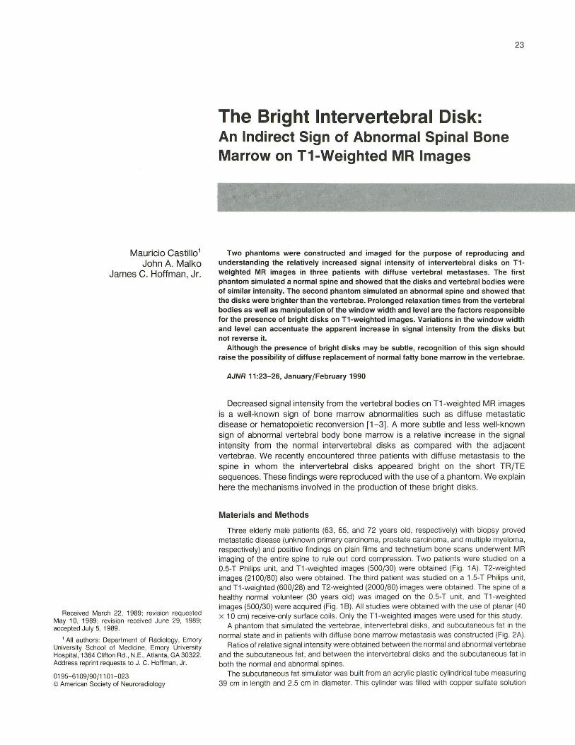
Fig. 1.-A, Sagittal MR image (500/30) in patient with diffuse metastasis to the spine from prostate carcinoma. Vertebral bodies demonstrate subtle decrease in signal intensity. Depending on window setting, it is conceivable that this study could be interpreted as normal. However, note that intervertebral disks are perceived as being of abnormally high signal intensity (an indirect sign that adjacent bone marrow is not normal).
8 , Sagittal MR image (500/30) in normal volunteer (obtained on same MR unit and surface coil as Fig. 1A). Note that intervertebral disks and marrow in vertebral bodies are of similar signal intensity.
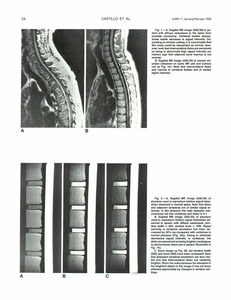
Fig. 2.-A , Sagittal MR image (500/30) of phantom used to reproduce relative signal intensities observed in normal spine. Note that disks and adjacent vertebrae are of similar signal intensity. In this phantom the ratio between subcutaneous fat and vertebrae and disks is 4: 1.
8, Sagittal MR image (500/30) of phantom used to reproduce relative signal intensities observed in spines with diffuse metastasis (window width = 464, window level = 168). Signal intensity in vertebral simulators has been decreased by 40% (as compared with vertebrae in normal phantom [Fig. 2A]). Owing to relatively decreased signal intensity in vertebrae, the disks are perceived as being brighter (analogous to phenomenon observed in patient illustrated in Fig. 1A).
C, Same image as Fig. 28, but window width (592) and level (360) have been increased. Note that diseased vertebral simulators are less visible and that intervertebral disks are relatively brighter. Since the subcutaneous fat simulator is the brightest object in the image it has not been affected appreciably by changes in window settings.
AJNR :11 , January/February 1990 MR OF INTERVERTEBRAL DISK 25
(CuSO. ) (4 mmol). The fat simulator was attached to the undersurface of an acrylic plastic board over which the vertebrae and disk simulators were placed. The normal vertebral bodies were simulated by using five plastic jars (250 ml) filled with 140 ml of CuS04 solution (1 mmol). The abnormal vertebrae were simulated by using similar containers filled with Cuso. at a different concentration (61 !'mol). The disks were simulated by using four glass vials filled with 8 ml of Cuso. solution (1 mmol) (Figs. 28 and 2C).
The Cuso. concentrations were chosen so that the relative image intensities obtained with the phantoms approximated those obtained in the patients and the normal volunteer. The normal spine phantom was imaged in the 0.5-T unit with the same surface coil and sequences (500/30) used for the patients. The abnormal vertebrae were then substituted for the normal vertebrae and images were obtained again using the same parameters.
Results
In all patients with metastatic disease, the bone scans demonstrated increased uptake throughout the spine. Plain films showed mixed sclerotic and lytic lesions in the patient with unknown primary carcinoma, multiple lytic lesions in the patient with multiple myeloma, and diffuse osteosclerosis in the patient with primary prostate carcinoma. On MR, the T1-weighted images showed the vertebral bodies to be of low signal intensity relative to the intervertebral disks (Fig. 1 A) while, in the normal volunteer, the T1-weighted images showed the intervertebral disks to be of comparable signal intensity to the marrow contained within the vertebral bodies (Fig . 1 B). In both the normal and abnormal spines the subcutaneous fat was the brightest signal in the image. The ratio between the subcutaneous fat and the normal vertebrae or disks was 4:1. For the patients with metastatic disease, the ratio between the vertebrae and the subcutaneous fat was approximately 40% lower than the ratio for the normal vertebrae, while the signal-intensity ratio between the disks and the subcutaneous fat was comparable to those found in the normal patients.
The normal spine phantom was constructed so that the vertebrae and disks would be of the same relative signal intensity shown on the T1-weighted sequences obtained in the normal volunteer (Fig . 2A). The ratio between the subcutaneous fat simulator and the normal vertebrae/disks was adjusted to 4:1. As designed, the abnormal spine phantom showed the signal intensity from the vertebrae to be approximately 40% lower relative to the fat while the signal intensity of the disks relative to the subcutaneous fat remained unchanged (Figs. 2B and 2C). The disks in the diseased phantom thus appeared brighter than those in the normal spine phantom.
Although T2-weighted images were not used in this study they were obtained in all three patients. In our patient with prostate carcinoma these images showed the diffuse vertebral metastasis to be slightly hyperintense and therefore difficult to distinguish from normal bone marrow without previous knowledge of the findings on the T1-weighted images. In the patients with either an unknown primary carcinoma or multiple myeloma, the T2-weighted images showed the metastatic foci to have a nonspecific appearance. Some metastatic deposits were brighter on the T2-weighted images
than on the T1-weighted images while others remained of low signal intensity relative to the adjacent bone marrow.
Discussion
T1-weighted images of neonates show the intervertebral disk to be of relatively high signal intensity as compared with the adjacent vertebrae [ 4] . In adults, T1-weighted images show the disks to be of similar signal intensity relative to the vertebral bodies. These findings are probably a reflection of biochemical changes that occur within the disk as it ages (5] . On long TR/TE images, normal adult disks are of increased signal intensity relative to the adjacent vertebrae. Decreased signal intensity from a disk on a T2-weighted image is abnormal and is believed to be related to degeneration, dehydration, and chemical changes (5] . Therefore, in order to study the intrinsic characteristics of disks, it is necessary to obtain T2-weighted images [6] .
MR imaging has proved to be an excellent method for assessing bone marrow (7 , 8]. With age, red marrow (active) converts into yellow marrow (inactive). Fat accounts for 80% of tl1e total volume of the yellow marrow (8] . It is because of this fatty infiltration that the adult vertebral bodies show a high signal intensity on T1-weighted images. Decreased signal intensity on T1-weighted images from the bone marrow can be seen normally in children, but in adults it correlates well with tumor infiltration of the bone marrow or with hematopoietic reconversion (8] .
In our study, T1-weighted images obtained in a normal volunteer showed that the disks and vertebrae were of similar signal intensity. As expected, in the three patients with known metastatic disease to the spine, the vertebrae were of low signal intensity relative to the adjacent disks , thus making these disks appear brighter. To our knowledge this observation has not been addressed in the literature.
The phantom was constructed to demonstrate the appearance of the normal disks relative to diseased vertebrae. The phantom that simulated the diseased spine demonstrated that the intervertebral disks were perceived as having increased intensity, although this signal was in fact unchanged relative to the signal from the subcutaneous fat.
The signal intensities of normal disks and vertebrae are similar, and the perceived contrast between them is relatively insensitive to window setting (Fig . 2A). In a diseased spine the vertebrae are generally of lower signal intensity than the disks because of their prolonged T1 relaxation [3], and this appearance of dark vertebral bodies relative to bright disks cannot be reversed by window manipulation. However, changes in the window setting can make this difference more obvious (Figs. 2B and 2C). Since the fat is the brightest tissue, changing the window does not alter its value appreciably in the gray scale and thus one is unable to perceive any change in its intensity.
Although spinal bone marrow abnormalities are usually evident by the low signal intensity from the vertebrae, relatively increased signal intensity from the disks on T1 -weighted images should raise the question of diffuse tumor infiltration or hematopoietic reconversion of the vertebral bone marrow and should lead to a review of the images on the monitor.

26 CASTILLO ET AL. AJNR:11 , January/February 1990
REFERENCES
1. Sugimura K, Yamasaki K, Kitagaki H, et al. Bone marrow diseases of the spine: differentiation with T1 and T2 relaxation times in MR imaging . Radiology 1987;165: 541 - 544
2. Rao VM , Fishman M, Mitchell DG, et al. Painful sickle cell crisis : bone marrow pattern observed with MR imaging. Radiology 1986;161 :211-215
3. Smoker WRK, Godersky JC, Knutzen RK, et al. The role of MR imaging in evaluating metastatic spinal disease. AJNR 1987;8:901-908
4. Ho PSP, Yu S, Sether LA, et al. Progressive and regressive changes in the nucleus pulposus. Part I. The neonate. Radiology 1988;169 :87-91
5. Modic MT, Pavlicek W, Weinsten M, et al. Magnetic resonance imaging of intervertebral disk disease. Radiology 1984;152: 103-111
6. Yu S, Haughton VM , Ho PSP, et al. Progressive and regressive changes in the nucleus pulposus. Part II. The adult. Radiology 1988;169 :93-97
7. Daffner RH , Lupetin AR , Dash N, et al. MRI in the detection of malignant infiltration of bone marrow. AJR 1986;146:353-358
8. Vogler JB, Murphy WA. Bone marrow imaging. Radiology 1988;168: 679- 693