Embed Size (px)
Citation preview
An Acad Bras Cienc (2021) 93(Suppl. 4): e20210297 DOI 10.1590/0001-3765202120210297Anais da Academia Brasileira de Ciências | Annals of the Brazilian Academy of SciencesPrinted ISSN 0001-3765 I Online ISSN 1678-2690www.scielo.br/aabc | www.fb.com/aabcjournal
An Acad Bras Cienc (2021) 93(Suppl. 4)
Running title: Methilprednisolone cardioprotective eff ects
Academy Section: CELLULAR
AND MOLECULAR BIOLOGY
e20210297
93 (Suppl. 4)93(Suppl. 4)
DOI10.1590/0001-3765202120210297
CELLULAR AND MOLECULAR BIOLOGY
The brief methylprednisolone administration is crucial to mitigate cardiac dysfunction after myocardial infarction
ALAN CHRISTHIAN BAHR, JULIA P. DA LUZ, RAYANE B. TEIXEIRA, PATRICK TÜRCK, ALEXSANDRA ZIMMER, ALEXANDRE L. DE CASTRO, EDUARDO E. DOS REIS, FERNANDA VISIOLI, ADRIANE BELLÓ-KLEIN, ALEX SANDER R. ARAUJO & PAULO C. SCHENKEL
Abstract: Acute myocardial infarction (AMI) is one of the major causes of heart failure and mortality. Glucocorticoids administration post-infarction has long been proposed, but it has shown confl icting results so far. This controversy may be associated with the glucocorticoid type and the period when it is administered. To elucidate these, the present aims to evaluate if the brief methylprednisolone acetate administration is determinant for heart adaptation after AMI. Male Wistar rats were divided into 3 groups: sham-operated (SHAM); infarcted (AMI); infarcted treated with methylprednisolone acetate (AMI+M). Immediately after surgery, the AMI+M group received a single dose of methylprednisolone acetate (40 mg/kg i.m.). After 56 days, the cardiac function was assessed and lungs, liver and heart were collected to determine rates of hypertrophy and congestion. Heart was used for oxidative stress and metalloproteinase activity analyses. Methylprednisolone acetate attenuated matrix metalloproteinase-2 activity, cardiac dilatation, and prevented the onset of pulmonary congestion, as well as avoided cardiac hypertrophy. Our data indicate that administration of methylprednisolone acetate shortly after AMI may be a therapeutic alternative for attenuation of detrimental ventricular remodeling.
Key words: acute myocardial infarction; heart failure; metalloproteinase; methylpred-nisolone acetate.
INTRODUCTION
Acute myocardial infarction (AMI) is one of the major health problems worldwide (Phatharajaree et al. 2007, Lei & Bin 2019). An early cardiac dysfunction due to the loss of cardiomyocytes post-AMI may lead to heart f ailure (HF) development over time (Pfeffer & Braunwald, 1990, Teixeira et al. 2017). Post-AMI HF has been classically characterized by increased left ventricular end-diastolic pressure, pulmonary congestion, and right ventricular hypertrophy (Hentschke et al. 2017, Singh et al.
1995). Several mechanisms are related to HF development, such as the increased formation of reactiv e oxygen species (ROS), the ventricular chambers dilatation, and cardiac fibrosis (Cahill & Kharbanda 2017, Mosterd et al. 2001). In this context, oxidative stress and matrix metalloproteinase 2 (MMP-2) play an important role in the HF development (Schenkel et al. 2010, Westman et al. 2016). In fact, previous studies highlighted the first week after the AMI as critical and decisive in this process, since redox-sensitive signaling pathways are modulated in this time window (Schenkel et al. 2010, 2012, Hill
ALAN CHRISTHIAN BAHR et al. METHILPREDNISOLONE CARDIOPROTECTIVE EFFECTS
An Acad Bras Cienc (2021) 93(Suppl. 4) e20210297 2 | 15
& Singal 1996). Conversely, the MMP-2 inhibition/suppression has mitigated the adverse cardiac remodeling post-AMI, whereas the elevated activity of MMP-2 has been shown as a predictor of the cardiac remodeling and worse prognosis post-AMI (Westman et al. 2016, Squire et al. 2004).
In these circumstances, hormonal therapy with glucocorticoids (GCs) seems to be promising since it can suppress both oxidative stresses triggering and MMP-2 activation (Westman et al. 2016, Xia et al. 2007). In fact, the treatment with GCs after AMI has shown a decrease in mortality rate, although this therapeutic approach still presents some contradictory results (Westman et al. 2016, Giugliano et al. 2003, Barzilai et al. 1972). Initial studies showed that GCs administration was associated with a decrease in the infarct size and mortality rate (Barzilai et al. 1972, Hafezi-Moghadam et al. 2002). Despite these benefits, GC treatment also appears to induce some detrimental effects, such as arrhythmia, cardiac cells apoptosis and increase cardiac contractile strength in rat hearts (Hammerman et al. 1983, Wester et al. 2019). Taken together, these adverse effects discouraged the GC use in patients with AMI.
Such controversial findings are probably related to the GC type used in these studies. GCs are divided according to their time and potency of action, with methylprednisolone (succinate or acetate) being classified as an intermediate action GC. Methylprednisolone succinate was the most used form in the past studies (Ko et al. 2015, Vyden et al. 1974). However, since it is administered in the form of a solution and it has a relatively short half-life, its management during treatment may compromise the outcome. On the other hand, methylprednisolone in its acetate salt form, which is a suspension, determines this drug’s gradual increase in serum levels, with a peak at
7 hours, and total clearance at day 21 (Narang et al. 1983). This pharmacokinetic profile could be more effective as a therapeutic strategy to mitigate the transition from AMI to HF. Therefore, the main objective of the present study was to determine if the methylprednisolone acetate, administered in a single dose immediately post-AMI, minimizes the cardiac remodeling by attenuate the oxidative stress and MMP-2 activity in the heart of rats.
MATERIAL AND METHODS Animals and experimental groupsMale Wistar rats (150-200 g, 45 days old) were obtained from the Center for Reproduction and Experimentation of Laboratory Animals of the Universidade Federal do Rio Grande do Sul (UFRGS). The sample size was calculated in the SigmaPlot 12.0 software using cardiac hypertrophy results showed by Schenkel et al. (2012). For this, we established a significance level of 0.05, a confidence interval of 95% and a statistical power of 80%. In this way, the sample size of 9 animals per group has been determined, reaching a total n of 27 animals. The rats were housed in polypropylene boxes (34 x 20 x 41 cm), four animals per box, under standard conditions (controlled temperature of 20-25ºC, 12h light/dark cycle, and relative humidity of 70%). Water and commercial chow (Nuvilab, Vetsul, Porto Alegre, RS, Brazil) were offered ad libitum. Animals were divided randomly into three groups: sham-operated (SHAM, n=8), infarcted (AMI, n=8) and infarcted treated with methylprednisolone acetate (AMI+M, n=7). Animals were excluded when ischemic (akinetic area) was not observed in the initial echocardiography post coronary occlusion surgery. All procedures have been made according to the International Guidelines for Use and Care of Laboratory Animals and after approved by the Ethics Committee for Animal

ALAN CHRISTHIAN BAHR et al. METHILPREDNISOLONE CARDIOPROTECTIVE EFFECTS
An Acad Bras Cienc (2021) 93(Suppl. 4) e20210297 3 | 15
Research at Universidade Federal do Rio Grande do Sul (# 30696).
Myocardial infarction surgeryTo induce myocardial infarction, rats were anesthetized (ketamine 90mg/kg and xylazine 10 mg/kg i.p.) and they submitted to ligature of the anterior descending left coronary artery as described previously (Schenkel et al. 2010, Johns & Olson 1954). Sham-operated animals were submitted to all surgical steps, except for coronary occlusion. Post-surgical analgesia was induced using tramadol (12.5 mg/kg, subcutaneously) (Brainfarma Indústria Química e Farmacêutica S.A., Anápolis, GO, Brazil), and dipyrone (200 mg/kg, orally) (Brainfarma Indústria Química e Farmacêutica S.A., Anápolis, GO, Brazil), twice a day, for 2 days.
Methylprednisolone acetate administrationImmediately after the AMI surgery, the animals from the AMI+M group received a single dose of methylprednisolone acetate (40 mg/kg i.m. - Depo-Medrol™, Pfizer Ltd., Mumbai, India). SHAM and AMI groups received a single dose of 0.9% NaCl sterile solution (Torres et al. 2014).
Echocardiographic analysisThe echocardiographic analyses (on the 2nd and 56th day after infarction) were performed in a double-blind manner by trained operators in animal echocardiography using the EnVisor Philips system (Andover, MA, USA) with a high-frequency and high-resolution transducer (12-3 MHz) (Schenkel et al. 2012). The animals were anesthetized (ketamine 90 mg/kg; xylazine 20 mg/kg, i.p.) and placed in lateral decubitus to obtain the images. LV end-systolic (ESV) and end-diastolic (EDV) volumes were measured as previously described (Schenkel et al. 2012). LV diastolic (DTA) and systolic transverse areas (STA) (cm2) were obtained by tracing the
endocardial border at three levels: basal (at the tip of the mitral valve leaflets), middle (at the papillary muscle level), and apical (distal from the papillary muscle but before the final curve cavity). Fractional area change (FAC) was used to estimate LV systolic function (Nozawa et al. 2006). On each echocardiographic transverse plane (basal, middle and apical), the arch corresponding to the segments with infarction (regions or segments of the myocardium showing one the following changes in myocardial kinetics: systolic movement akinetic and/or hypokinetic region — AHR) and the total endocardial perimeter (EP) were measured at end-diastole. Infarction size (IS) was estimated as % IS=(AHR/EP)×100 (Schenkel et al. 2010). The absolute and relative technical error and the reliability coefficient of the LV infarction area were measured as described by Perini et al. (2005) and Frainer et al. (2007).
CatheterizationAfter echocardiographic analysis, the animals had the right carotid artery cannulated with a PE50 catheter connected to a pressure-measuring transducer (Narco Biosystem pulse transducer RP-155, Houston, Texas, USA), and connected to a pressure amplifier (HP 8805C, Hewlett Packard, USA) (Schenkel et al. 2010). The catheter was inserted into the LV and the following parameters were recorded: left ventricular systolic pressure (LVSP, mmHg), left ventricular end-diastolic pressure (LVEDP, mmHg), left ventricle contractility and relaxation derivatives (dP/dtmax and dP/dtmin, mmHg/s) and cardiac contractility index((dP/dtmax)/LVEDP). The records were analyzed on a microcomputer equipped with an analog-to-digital conversion board in a double-blind manner (sampling rate of 2 kHz).
ALAN CHRISTHIAN BAHR et al. METHILPREDNISOLONE CARDIOPROTECTIVE EFFECTS
An Acad Bras Cienc (2021) 93(Suppl. 4) e20210297 4 | 15
Euthanasia and Tissue CollectionAfter LV catheterization, the animals were sacrificed by decapitation. The heart, lungs, the liver, and the tibia of the right hind paw were collected for morphometric analysis.
Morphometric AnalysisInitial and final bodyweight, as well as the body mass index (weight/length²), were evaluated. Pulmonary and hepatic congestion indexes have been obtained through the wet/dry weight (g/g) ratio for lung and liver (Pearce et al. 1965). Cardiac hypertrophy index (heart weight (g)/tibia length (mm)) was calculated (Schenkel et al. 2010). The remaining left ventricle tissue was separated from the infarction scar and immediately frozen in liquid nitrogen for further oxidative stress and molecular analyses.
Tissue Preparation Homogenization of the LV was performed for 40 seconds with Ultra-Turrax (OMNI Tissue Homogenizer, OMNI International, USA) in the presence of 1.15% KCl (5 ml/g tissue) and 100 mmol/L phenyl methyl sulfonyl fluoride. The homogenates were centrifuged for 20 minutes at 10.000 x g at 4°C, and the supernatant was collected and stored at -80°C for further oxidative stress analyses (Baldo et al. 2011, Puukila et al. 2017).
Determination of protein concentration Protein concentration in the homogenates was quantified by Lowry’s method using bovine serum albumin solution as standard (Lowry et al. 1951).
Determination of lipid peroxidationLipid peroxidation was measured by tert-butyl hydroperoxide-initiated chemiluminescence (CL), using a liquid scintillation counter in the out-of-coincidence mode (LKB Rack Beta Liquid
Scintillation Spectrometer 1215, LKB—Produkter AB, Sweden). Samples were placed in vials at a protein concentration of 0.5–1.0 mg/ml in a reaction medium consisting of 120 mol/L KCl, 30 mol/L phosphate buffer (pH = 7.4). Measurements were initiated by the addition of 3 mmol/L tert-butyl hydroperoxide and the final value (peak – basal luminescence) was expressed as counts per second per milligram of protein (cps/mg protein) (Gonzalez et al. 1991).
Determination of antioxidant enzyme activitiesThe activities of the following antioxidant enzymes were measured in the remaining LV: superoxide dismutase (SOD), catalase (CAT), and glutathione peroxidase (GPx). SOD activity, expressed as units/mg protein, was based on the inhibition of the reaction of superoxide with pyrogallol. The superoxide radical is generated by the auto-oxidation of pyrogallol in an alkaline medium. SOD activity was determined by measuring the velocity of oxidized pyrogallol formation at 420nm (Marklund 1985). The results were expressed as U SOD/mg protein. CAT activity was measured by assessing H2O2 consumption at 240nm (Aebi 1984), and it was expressed as pmol/mg protein. GPx activity was measured by NADPH oxidation at 340 nm in a reaction medium containing 0.17 mmol/L reduced glutathione, 0.2 U/mL glutathione reductase, and 0.5 mmol/L tert-butyl hydroperoxide. GPx activity was expressed as nmol of NADPH /min/mg protein (Flohé & Günzler 1984).
Determination of metalloproteinases- activityMetalloproteinases were examined by use of substrate-specific zymography. LV samples were homogenized in the presence of cacodylate buffer (10 mmol/L cacodylic acid, 150 mmol/L NaCl, 2μmol/L ZnCl2, 20 mmol/L CaCl2, 1.5 mmol/L sodium azide, 0.01% Triton X-100).
ALAN CHRISTHIAN BAHR et al. METHILPREDNISOLONE CARDIOPROTECTIVE EFFECTS
An Acad Bras Cienc (2021) 93(Suppl. 4) e20210297 5 | 15
Samples were then prepared in loading buffer and were exposed to SDS-PAGE single-phase gel electrophoresis in a batch system using 8% (W/V) running gel containing 0.15% of commercial gelatin and 4% (W/V) stacking gel. A molecular weight marker (KaleidoskopeTM, BioRad) was loaded in conjunction with the samples. Migration of the proteins occurred in running buffer (24 mmol/LTris Base, 190 mmol/L glycine, 0.1% SDS, pH 8.3) previously cooled to 4°C. The gel was then washed 3X (2.5% Triton X-100) for 15 minutes under stirring and activated in developing buffer for initially 30 minutes under stirring. Then, the gel was stained with coomassie blue solution for 1 h and it was discolored to reveal the bands indicating metalloproteinases activity, which were identified according to their molecular weight. The bands were quantified with the ImageJ 1.51 program (ImageJ, US National Institutes of Health, Bethesda, Maryland, USA) (Iyer et al. 2016).
HistologyCross-sectional samples of the heart (infarct area + left ventricle + right ventricle) were conditioned in buffered formalin and embedded in paraffin, subsequently processed and stained in hematoxylin and eosin for histological analysis. The images were captured at a magnification of 200x times using CellSens™ software (Olympus, Tokyo, Japan), in a binocular microscope model CX41RF (Olympus Latin America Inc., Miami, FL, USA) coupled to a Q-Color 5 RTV microscope camera (Olympus Latin America Inc., Miami, FL, USA). Cardiomyocytes area quantification was performed using Image-Pro Plus 6 software (Media Cybernetics Inc., Rockville, MD, USA). Ten aleatory fields were captured from each ventricle, and in these fields, 10 cells per field were analyzed, totaling 100 cells analyzed per ventricle. The right ventricle cardiomyocytes area (RVCA), and the left ventricle cardiomyocytes
area (LVCA) were analyzed in a double-blind manner and expressed as µm2 (Baldo et al. 2011).
Statistical AnalysisThe data were tested for normality by Shapiro-Wilk test. Since the data presented normal distribution, the comparison between the groups was made through one-way ANOVA, followed by the Student-Newmann-Keuls post hoc test. To analyze the size of the effect we calculated the partial Eta squared (n²) derived from the general linear model. The effects were classified as low (0.01 - 0.06), moderate (0.06 - 0.14) and high (>0.14). Pearson correlation test was used to determine the correlation between RVCA and pulmonary congestion. Results were expressed as mean ± standard deviation. The differences were considered significant when the statistical analysis showed p<0.05. GraphPad Instat 6.01 for Windows (GraphPad Software Inc, La Jolla, CA, USA) was used as a tool for data analysis.
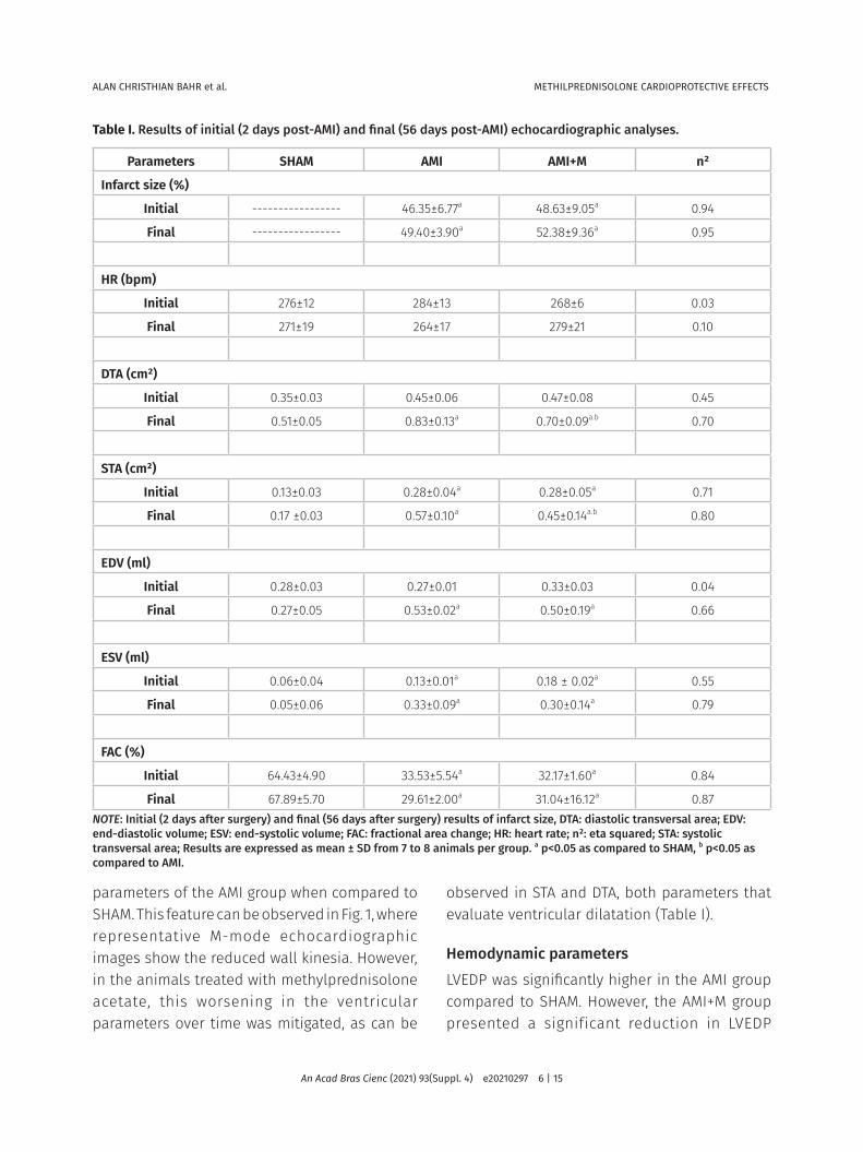
RESULTSEchocardiographic initial and final parametersAn echocardiographic analysis was performed 2 days post-AMI to confirm the effectiveness of the surgical procedure, verifying that 23 (85%) of the operated animals developed myocardial infarction. These animals present an infarcted area of 52 ± 5%, with an absolute technical error of 8.06%, a relative technical error of 16.5% and a reliability coefficient of 0.87. Two operated animals died in the first 24 hours after surgery and two were excluded because the surgery was not effective. In relation to echocardiographic morphological parameters, both infarcted groups (AMI and AMI+M) showed a significant increase in the STA, ESV and FAC when compared to the SHAM group (Table I).
Fifty-six days after myocardial infarction we observed impairment in the echocardiographic
ALAN CHRISTHIAN BAHR et al. METHILPREDNISOLONE CARDIOPROTECTIVE EFFECTS
An Acad Bras Cienc (2021) 93(Suppl. 4) e20210297 6 | 15
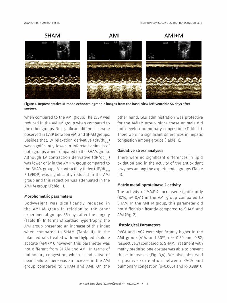
parameters of the AMI group when compared to SHAM. This feature can be observed in Fig. 1, where representative M-mode echocardiographic images show the reduced wall kinesia. However, in the animals treated with methylprednisolone acetate, this worsening in the ventricular parameters over time was mitigated, as can be
observed in STA and DTA, both parameters that evaluate ventricular dilatation (Table I).
Hemodynamic parameters LVEDP was significantly higher in the AMI group compared to SHAM. However, the AMI+M group presented a significant reduction in LVEDP
Table I. Results of initial (2 days post-AMI) and final (56 days post-AMI) echocardiographic analyses.
Parameters SHAM AMI AMI+M n²
Infarct size (%)
Initial ----------------- 46.35±6.77a 48.63±9.05a 0.94
Final ----------------- 49.40±3.90a 52.38±9.36a 0.95
HR (bpm)
Initial 276±12 284±13 268±6 0.03
Final 271±19 264±17 279±21 0.10
DTA (cm²)
Initial 0.35±0.03 0.45±0.06 0.47±0.08 0.45
Final 0.51±0.05 0.83±0.13a 0.70±0.09a.b 0.70
STA (cm²)
Initial 0.13±0.03 0.28±0.04a 0.28±0.05a 0.71
Final 0.17 ±0.03 0.57±0.10a 0.45±0.14a.b 0.80
EDV (ml)
Initial 0.28±0.03 0.27±0.01 0.33±0.03 0.04
Final 0.27±0.05 0.53±0.02a 0.50±0.19a 0.66
ESV (ml)
Initial 0.06±0.04 0.13±0.01a 0.18 ± 0.02a 0.55
Final 0.05±0.06 0.33±0.09a 0.30±0.14a 0.79
FAC (%)
Initial 64.43±4.90 33.53±5.54a 32.17±1.60a 0.84
Final 67.89±5.70 29.61±2.00a 31.04±16.12a 0.87NOTE: Initial (2 days after surgery) and final (56 days after surgery) results of infarct size, DTA: diastolic transversal area; EDV: end-diastolic volume; ESV: end-systolic volume; FAC: fractional area change; HR: heart rate; n²: eta squared; STA: systolic transversal area; Results are expressed as mean ± SD from 7 to 8 animals per group. a p<0.05 as compared to SHAM, b p<0.05 as compared to AMI.
ALAN CHRISTHIAN BAHR et al. METHILPREDNISOLONE CARDIOPROTECTIVE EFFECTS
An Acad Bras Cienc (2021) 93(Suppl. 4) e20210297 7 | 15
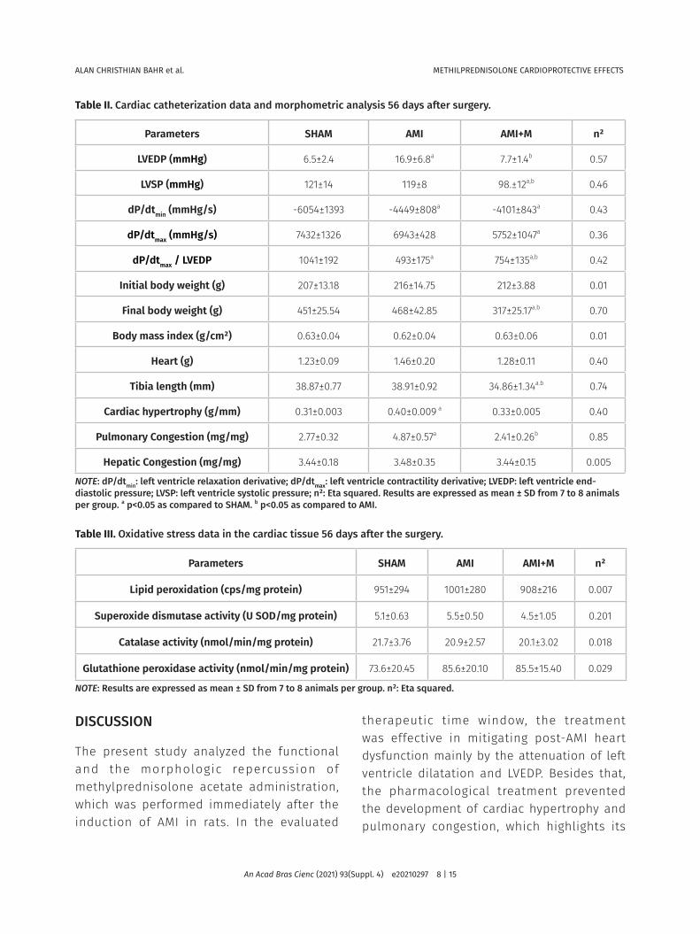
when compared to the AMI group. The LVSP was reduced in the AMI+M group when compared to the other groups. No significant differences were observed in LVSP between AMI and SHAM groups. Besides that, LV relaxation derivative (dP/dtmin) was significantly lower in infarcted animals of both groups when compared to the SHAM group. Although LV contraction derivative (dP/dtmax) was lower only in the AMI+M group compared to the SHAM group, LV contractility index (dP/dtmax / LVEDP) was significantly reduced in the AMI group and this reduction was attenuated in the AMI+M group (Table II).
Morphometric parametersBodyweight was significantly reduced in the AMI+M group in relation to the other experimental groups 56 days after the surgery (Table II). In terms of cardiac hypertrophy, the AMI group presented an increase of this index when compared to SHAM (Table II). In the infarcted rats treated with methylprednisolone acetate (AMI+M), however, this parameter was not different from SHAM and AMI. In terms of pulmonary congestion, which is indicative of heart failure, there was an increase in the AMI group compared to SHAM and AMI. On the
other hand, GCs administration was protective for the AMI+M group, since these animals did not develop pulmonary congestion (Table II). There were no significant differences in hepatic congestion among groups (Table II).
Oxidative stress analysesThere were no significant differences in lipid oxidation and in the activity of the antioxidant enzymes among the experimental groups (Table III).
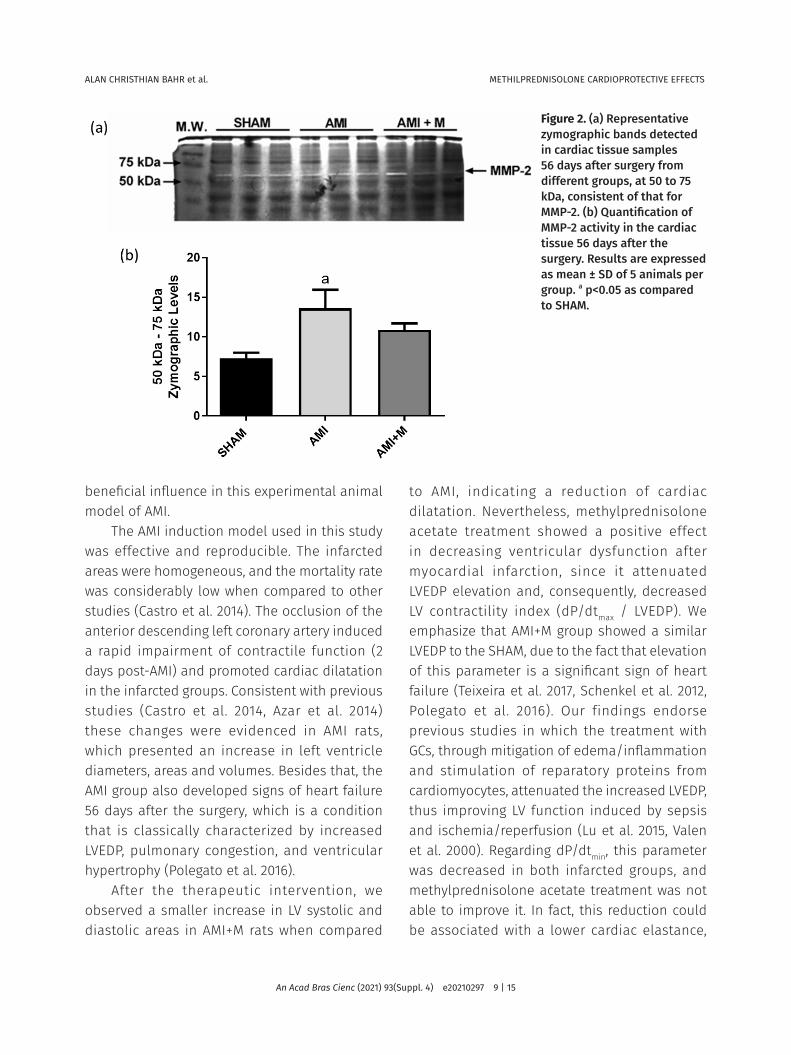
Matrix metalloproteinase 2 activityThe activity of MMP-2 increased significantly (87%, n²=0,41) in the AMI group compared to SHAM. In the AMI+M group, this parameter did not differ significantly compared to SHAM and AMI (Fig. 2).
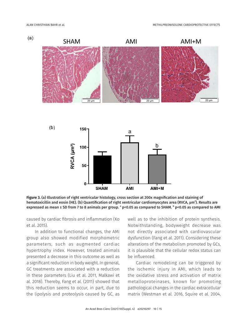
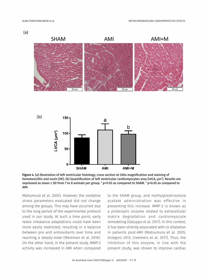
Histological ParametersRVCA and LVCA were significantly higher in the AMI group (41% and 30%, n²= 0.50 and 0.82, respectively) compared to SHAM. Treatment with methylprednisolone acetate was able to prevent these increases (Fig. 3,4). We also observed a positive correlation between RVCA and pulmonary congestion (p=0,0001 and R=0,8891).
Figure 1. Representative M-mode echocardiographic images from the basal view left ventricle 56 days after surgery.
ALAN CHRISTHIAN BAHR et al. METHILPREDNISOLONE CARDIOPROTECTIVE EFFECTS
An Acad Bras Cienc (2021) 93(Suppl. 4) e20210297 8 | 15
DISCUSSION
The present study analyzed the functional and the morphologic repercussion of methylprednisolone acetate administration, which was performed immediately after the induction of AMI in rats. In the evaluated
therapeutic time window, the treatment was effective in mitigating post-AMI heart dysfunction mainly by the attenuation of left ventricle dilatation and LVEDP. Besides that, the pharmacological treatment prevented the development of cardiac hypertrophy and pulmonary congestion, which highlights its
Table II. Cardiac catheterization data and morphometric analysis 56 days after surgery.
Parameters SHAM AMI AMI+M n²
LVEDP (mmHg) 6.5±2.4 16.9±6.8a 7.7±1.4b 0.57
LVSP (mmHg) 121±14 119±8 98.±12a,b 0.46
dP/dtmin (mmHg/s) -6054±1393 -4449±808a -4101±843a 0.43
dP/dtmax (mmHg/s) 7432±1326 6943±428 5752±1047a 0.36
dP/dtmax / LVEDP 1041±192 493±175a 754±135a,b 0.42
Initial body weight (g) 207±13.18 216±14.75 212±3.88 0.01
Final body weight (g) 451±25.54 468±42.85 317±25.17a.b 0.70
Body mass index (g/cm²) 0.63±0.04 0.62±0.04 0.63±0.06 0.01
Heart (g) 1.23±0.09 1.46±0.20 1.28±0.11 0.40
Tibia length (mm) 38.87±0.77 38.91±0.92 34.86±1.34a.b 0.74
Cardiac hypertrophy (g/mm) 0.31±0.003 0.40±0.009 a 0.33±0.005 0.40
Pulmonary Congestion (mg/mg) 2.77±0.32 4.87±0.57a 2.41±0.26b 0.85
Hepatic Congestion (mg/mg) 3.44±0.18 3.48±0.35 3.44±0.15 0.005
NOTE: dP/dtmin: left ventricle relaxation derivative; dP/dtmax: left ventricle contractility derivative; LVEDP: left ventricle end-diastolic pressure; LVSP: left ventricle systolic pressure; n²: Eta squared. Results are expressed as mean ± SD from 7 to 8 animals per group. a p<0.05 as compared to SHAM. b p<0.05 as compared to AMI.
Table III. Oxidative stress data in the cardiac tissue 56 days after the surgery.
Parameters SHAM AMI AMI+M n²
Lipid peroxidation (cps/mg protein) 951±294 1001±280 908±216 0.007
Superoxide dismutase activity (U SOD/mg protein) 5.1±0.63 5.5±0.50 4.5±1.05 0.201
Catalase activity (nmol/min/mg protein) 21.7±3.76 20.9±2.57 20.1±3.02 0.018
Glutathione peroxidase activity (nmol/min/mg protein) 73.6±20.45 85.6±20.10 85.5±15.40 0.029
NOTE: Results are expressed as mean ± SD from 7 to 8 animals per group. n²: Eta squared.
ALAN CHRISTHIAN BAHR et al. METHILPREDNISOLONE CARDIOPROTECTIVE EFFECTS
An Acad Bras Cienc (2021) 93(Suppl. 4) e20210297 9 | 15
beneficial influence in this experimental animal model of AMI.
The AMI induction model used in this study was effective and reproducible. The infarcted areas were homogeneous, and the mortality rate was considerably low when compared to other studies (Castro et al. 2014). The occlusion of the anterior descending left coronary artery induced a rapid impairment of contractile function (2 days post-AMI) and promoted cardiac dilatation in the infarcted groups. Consistent with previous studies (Castro et al. 2014, Azar et al. 2014) these changes were evidenced in AMI rats, which presented an increase in left ventricle diameters, areas and volumes. Besides that, the AMI group also developed signs of heart failure 56 days after the surgery, which is a condition that is classically characterized by increased LVEDP, pulmonary congestion, and ventricular hypertrophy (Polegato et al. 2016).
After the therapeutic intervention, we observed a smaller increase in LV systolic and diastolic areas in AMI+M rats when compared
to AMI, indicating a reduction of cardiac dilatation. Nevertheless, methylprednisolone acetate treatment showed a positive effect in decreasing ventricular dysfunction after myocardial infarction, since it attenuated LVEDP elevation and, consequently, decreased LV contractility index (dP/dtmax / LVEDP). We emphasize that AMI+M group showed a similar LVEDP to the SHAM, due to the fact that elevation of this parameter is a significant sign of heart failure (Teixeira et al. 2017, Schenkel et al. 2012, Polegato et al. 2016). Our findings endorse previous studies in which the treatment with GCs, through mitigation of edema/inflammation and stimulation of reparatory proteins from cardiomyocytes, attenuated the increased LVEDP, thus improving LV function induced by sepsis and ischemia/reperfusion (Lu et al. 2015, Valen et al. 2000). Regarding dP/dtmin, this parameter was decreased in both infarcted groups, and methylprednisolone acetate treatment was not able to improve it. In fact, this reduction could be associated with a lower cardiac elastance,
Figure 2. (a) Representative zymographic bands detected in cardiac tissue samples 56 days after surgery from different groups, at 50 to 75 kDa, consistent of that for MMP-2. (b) Quantification of MMP-2 activity in the cardiac tissue 56 days after the surgery. Results are expressed as mean ± SD of 5 animals per group. a p<0.05 as compared to SHAM.
ALAN CHRISTHIAN BAHR et al. METHILPREDNISOLONE CARDIOPROTECTIVE EFFECTS
An Acad Bras Cienc (2021) 93(Suppl. 4) e20210297 10 | 15
caused by cardiac fibrosis and inflammation (Ko et al. 2015).
In addition to functional changes, the AMI group also showed modified morphometric parameters, such as augmented cardiac hypertrophy index. However, treated animals presented a decrease in this outcome as well as a significant reduction in body weight. In general, GC treatments are associated with a reduction in these parameters (Liu et al. 2011, Malkawi et al. 2018). Thereby, Fang et al. (2011) showed that this reduction seems to occur, in part, due to the lipolysis and proteolysis caused by GC, as
well as to the inhibition of protein synthesis. Notwithstanding, bodyweight decrease was not directly associated with cardiovascular dysfunction (Fang et al. 2011). Considering these alterations of the metabolism promoted by GCs, it is plausible that the cellular redox status can be influenced.
Cardiac remodeling can be triggered by the ischemic injury in AMI, which leads to the oxidative stress and activation of matrix metalloproteinases, known for promoting pathological changes in the cardiac extracellular matrix (Westman et al. 2016, Squire et al. 2004,
Figure 3. (a) Illustration of right ventricular histology, cross section at 200x magnification and staining of hematoxicillin and eosin (HE). (b) Quantification of right ventricular cardiomyocytes area (RVCA, µm2). Results are expressed as mean ± SD from 7 to 8 animals per group. a p<0.05 as compared to SHAM. b p<0.05 as compared to AMI
ALAN CHRISTHIAN BAHR et al. METHILPREDNISOLONE CARDIOPROTECTIVE EFFECTS
An Acad Bras Cienc (2021) 93(Suppl. 4) e20210297 11 | 15
Matsumura et al. 2005). However, the oxidative stress parameters evaluated did not change among the groups. This may have occurred due to the long period of the experimental protocol used in our study. At such a time point, early redox imbalance adaptations could have been more easily stabilized, resulting in a balance between pro and antioxidants over time and reaching a steady-state (Westman et al. 2016). On the other hand, in the present study, MMP-2 activity was increased in AMI when compared
to the SHAM group, and methylprednisolone acetate administration was effective in preventing this increase. MMP-2 is known as a proteolytic enzyme related to extracellular matrix degradation and cardiomyocyte remodeling (Galuppo et al. 2017). In this context, it has been directly associated with LV dilatation in patients post-AMI (Matsumura et al. 2005, Grzegorz 2013, Creemers et al. 2011). Thus, the inhibition of this enzyme, in line with the present study, was shown to improve cardiac
Figure 4. (a) Illustration of left ventricular histology, cross section at 200x magnification and staining of hematoxicillin and eosin (HE). (b) Quantification of left ventricular cardiomyocytes area (LVCA, µm2). Results are expressed as mean ± SD from 7 to 8 animals per group. a p<0.05 as compared to SHAM. b p<0.05 as compared to AMI.
ALAN CHRISTHIAN BAHR et al. METHILPREDNISOLONE CARDIOPROTECTIVE EFFECTS
An Acad Bras Cienc (2021) 93(Suppl. 4) e20210297 12 | 15
remodeling, reducing the extent of myocardial necrosis, cardiac dilatation and ventricular dysfunction after myocardial injury (Grzegorz 2013, Creemers et al. 2011). Additionally, the mortality post-AMI observed in the present study (15%) corroborates with that showed previously for Galuppo et al. (2017). These authors showed that monocytes/macrophages GC receptors are critical to attenuate mortality from 56% to 12% seven days post-AMI. Additionally, it was also shown for them the importance of these receptors to mitigate cardiac remodeling. In this scenario, our results could be related to GC direct effect in inhibiting MMP-2 gene expression or by stimulating the tissue inhibitors of metalloproteinases (Matsumura et al. 2005, Podesser et al. 2001).
Histological evaluation of the cardiomyocytes area showed that AMI+M animals did not present an increase in RVCA and LVCA. Since dilatation and hypertrophy are features of the post-infarction cardiac maladaptive remodeling (Schenkel et al. 2010), methylprednisolone acetate administration seems to mitigate these pathological processes. Furthermore, we evidenced a positive correlation between RVCA and pulmonary congestion. Since this pulmonary disturb is compatible with heart failure, methylprednisolone acetate administration was able to control the development of heart failure, at least in part, in infarcted treated animals. Regarding lung congestion, methylprednisolone treatment in rats with acute lung injury and respiratory distress syndrome was effective in reducing the levels of pulmonary congestion by lowering pro-inflammatory macrophages and enhancing tissue repair macrophages (Tu et al. 2017).
In conclusion, the results of the present study indicate that administration of methylprednisolone acetate shortly after AMI attenuates MMP-2 activity and, thus, may
be a therapeutic alternative for attenuation of ventricular remodeling. However, the evaluation of methylprednisolone acetate effects in a long-term period after infarction and the determination of the optimum dose are questions that remain to be answered, to bring these results from animal testing to clinical application.
Acknowledgments This work was supported by the following Brazilian Research Agencies: Coordenação de Aperfeiçoamento de Pessoal de Nível Superior (CAPES) and Conselho Nacional de Desenvolvimento Científico e Tecnológico (CNPq).
REFERENCES
AEBI H. 1984. Catalase in vitro. Meth Enzy 105: 121-126.
AZAR A, TAVAKOLI F, MOLADOUST H, ZARE A & SADEGHPOUR A. 2014. Echocardiographic evaluation of cardiac function in ischemic rats: Value of m-mode echocardiography. Res Cardio Med 3(4): e22941.
BALDO G ET AL. 2011. Bone Marrow Cells Reduce Collagen Deposition in the Rat Model of Common Bile Duct Ligation. J Cell Sci Ther 2: 112.
BARZILAI D, PLAVNICK J, HAZANI A, EINATH R, KLEINHAUS N & KANTER Y. 1972. Use of hydrocortisone in the treatment of acute myocardial infarction. Chest 61(5): 488-491.
CAHILL TJ & KHARBANDA RK. 2017. Heart failure after myocardial infarction in the era of primary percutaneous coronary intervention: Mechanisms, incidence and identification of patients at risk. World J Cardiol 9(5): 407.
CASTRO AL ET AL. 2014. Cardioprotective effects of thyroid hormones in a rat model of myocardial infarction are associated with oxidative stress reduction. Mol Cell Endocrinol 391(1-2): 22-29.
CREEMERS EEJM, CLEUTJENS JPM, SMITS JFM & DAEMEN MJAP. 2001. Matrix metalloproteinase inhibition after myocardial infarction: a new approach to prevent heart failure? Circulat Res 89(3): 201-210.
FANG J, DUBOIS DC, HE Y, ALMON RR & JUSKO WJ. 2011. Dynamic modeling of methylprednisolone effects on body weight and glucose regulation in rats. J Pharmacokinet Pharmacodyn 38: 293-316.
ALAN CHRISTHIAN BAHR et al. METHILPREDNISOLONE CARDIOPROTECTIVE EFFECTS
An Acad Bras Cienc (2021) 93(Suppl. 4) e20210297 13 | 15
FLOHÉ L & GÜNZLER WA. 1984. Assays of glutathione peroxidase. Methods Enzymol 105: 114-121.
FRAINER DES, ADAMI F, VASCONSELOS FAG, ASSIS MAA, CALVO MCM & KERPE R. 2007. Padronização e confiabilidade das medidas antropométricas para pesquisa populacional. ALAN 57(4): 335-342.
GALUPPO P ET AL. 2017. The glucocorticoid receptor in monocyte-derived macrophages is critical for cardiac infarct repair and remodeling. FASEB J 31(11): 5122-5132.
GIUGLIANO G, GIUGLIANO R, GIBSON C & KUNTZ R. 2003. Meta-analysis of corticosteroid treatment in acute myocardial infarction. Am J Cardiol 91(9): 1055-1059.
GONZALEZ FB , LLESUY S & BOVERIS A . 1991 . Hydroperoxideinitiated chemiluminescence: an assay for oxidative stress in biopsies of heart, liver, and muscle. Free Radic Biol Med 10: 93-100.
GRZEGORZ S. 2013. Intracellular Regulation of Matrix Metalloproteinase-2 Activity: New Strategies in Treatment and Protection of Heart Subjected to Oxidative Stress. Scientifica 130451.
HAFEZI-MOGHADAM A ET AL. 2002. Acute cardiovascular protective effects of corticosteroids are mediated by non-transcriptional activation of endothelial nitric oxide synthase. Nature Medicine 8(5): 473-479.
HAMMERMAN H, KLONER RA, HALE S, SCHOEN FJ & BRAUNWALD E. 1983. Dose-dependent effects of short-term methylprednisolone on myocardial infarct extent, scar formation, and ventricular function. Circulation 68(2): 446-452.
HENTSCHKE V ET AL. 2017. Functional capacity in a rat model of heart failure: impact of myocardial infarct size. Exp Physiol 102(11): 1448-1458.
HILL MF & SINGAL PK. 1996. Antioxidant and oxidative stress changes during heart failure subsequent to myocardial infarction in rats. Am J Pathol 148: 291-300.
IYER RP, JUNG M & LINDSEY ML. 2016. MMP-9 signaling in the left ventricle following myocardial infarction. Am J Physiol Heart Circ Physiol 311(1): H190-198.
JOHNS T & OLSON BJ. 1954. Experimental myocardial infarction. A method of coronary occlusion in small animals. Ann Surg 140: 675-682.
KO YH ET AL. 2015. Methylprednisolone protects cardiac pumping mechanics from deteriorating in lipopolysaccharide-treated rats. Front Physiol 6: 348.
LEI L & BIN Z. 2019. Risk Factor Differences in Acute Myocardial Infarction between Young and Older People:
A Systematic Review and Meta-Analysis. Int J Cardiovasc Sci 32(2): 163-176.
LIU X ET AL. 2011. Glucocorticoids decrease body weight and food intake and inhibit appetite regulatory peptide expression in the hypothalamus of rats. Exp Therap Med 2(5): 977-984.
LOWRY OH, ROSEBROUGH NJ, FARR AL & RANDALL RJ. 1951. Protein measurement with the folin phenol reagent. J Biol Chem 193: 265-275.
LU Z, LU J & DENG Y. 2015. Glucocorticoids offer protection against myocardial injury in a murine model of sepsis. Int J Clin Exp Med 8(8): 12211-12218.
MALKAWI A ET AL. 2018. Metabolomics Based Profiling of Dexamethasone Side Effects in Rats. Frontiers in Pharmacology.
MARKLUND S. 1985. Handbook of methods for oxygen radical research. Boca Rat. CRC Press, p. 243-247.
MATSUMURA S, IWANAGA S, MOCHIZUKI S, OKAMOTO H, OGAWA S & OKADA Y. 2005. Targeted deletion or pharmacological inhibition of MMP-2 prevents cardiac rupture after myocardial infarction in mice. J Clin Invest 115: 599-609.
MOSTERD A, COST B, HOES AW, DE BRUIJNE MC, DECKERS JW, HOFMAN A ET AL. 2001. The prognosis of heart failure in the general population: The Rotterdam Study. Eur Heart J 22, 1318-1327.
NARANG PK, WILDER R, CHATTERJI DC, YEAGER RL & GALLELLI JF. 1983. Systemic bioavailability and pharmacokinetics of methylprednisolone in patients with rheumatoid arthritis following ‘high-dose’ pulse administration. Biopharm Drug Dispos 4(3): 233-248.
NOZAWA E ET AL. 2006. Performance of twodimensional Doppler echocardiography for the assessment of infarct size and left ventricular function in rats. Braz J Med Biol Res 39: 687-695.
PEARCE ML, YAMASHITA J & BEAZELL J. 1965. Measurement of pulmonary edema. Circ Res 16: 482-488.
PERINI TA, OLIVEIRA GL, ORNELLAS JS & OLIVEIRA FP. 2005. Cálculo do erro técnico de medição em antropometria. Rev Bras Med Esporte, vol. 11.
PFEFFER M & BRAUNWALD E. 1990. Ventricular remodeling after myocardial infarction. Experimental observations and clinical implications. Circulation 81: 1161-1172.
PHATHARAJAREE W, PHROMMINTIKUL A & CHATTIPAKORNAL N. 2007. Matrix metalloproteinases and myocardial infarction. Canad J Cardiol 23(9): 727-733.

ALAN CHRISTHIAN BAHR et al. METHILPREDNISOLONE CARDIOPROTECTIVE EFFECTS
An Acad Bras Cienc (2021) 93(Suppl. 4) e20210297 14 | 15
PODESSER BK ET AL. 2001. ET(A)-receptor blockade prevents matrix metalloproteinase activation late post-myocardial infarction in the rat. Am J Physiol Heart Circ Physiol 280(3): H984-991.
POLEGATO B ET AL. 2016. Association between Functional Variables and Heart Failure after Myocardial Infarction in Rats. Arq Bras Cardiol 106(2): 105-112.
PUUKILA S ET AL. 2017. Secoisolariciresinol diglucoside attenuates cardiac hypertrophy and oxidative stress in monocrotaline-induced right heart dysfunction. Mol Cell Biochem 432(1-2): 33-39.
SCHENKEL PC ET AL. 2010. Redox-sensitive prosurvival and proapoptotic protein expression in the myocardial remodeling post-infarction in rats. Mol Cell Biochem 341(1-2): 1-8.
SCHENKEL PC ET AL. 2012. Time course of hydrogen peroxide-thioredoxin balance and its influence on the intracellular signalling in myocardial infarction. Exp Physiol 97: 741-749.
SINGH N ET AL. 1995. Oxidative stress and heart failure. Mol Cell Biochem 147: 77-81.
SQUIRE IB, EVANS J, NG LL, LOFTUS IM & THOMPSON MM. 2004. Plasma MMP-9 and MMP-2 following acute myocardial infarction in man: correlation with echocardiographic and neurohumoral parameters of left ventricular dysfunction. J Card Fail 10(4): 328-333.
TEIXEIRA RB ET AL. 2017. Long-term T3 and T4 treatment as an alternative to aerobic exercise training in improving cardiac function post-myocardial infarction. Biomed Pharmacother 95: 965-973.
TORRES RL, TORRES IL, LASTE G, FERREIRA MB, CARDOSO PF & BELLÓ-KLEIN A. 2014. Effects of acute and chronic administration of methylprednisolone on oxidative stress in rat lungs. J Bras Pneumol 40: 238-243.
TU GW ET AL. 2017. Glucocorticoid attenuates acute lung injury through induction of type 2 macrophage. J Transl Med 15(1): 181.
VALEN G, KAWAKAMI T, TÄHEPÔLD P, DUMITRESCU A, LÖWBEER C & VAAGE J. 2000. Glucocorticoid pretreatment protects cardiac function and induces cardiac heat shock protein 72. Am J Physiol-Heart Circ Physiol 279(2): H836-H843.
VYDEN JK ET AL. 1974. Effects of methylprednisolone administration in acute myocardial infarction. J Cardiol 34: 677-686.
WESTER M, HELLER A, GRUBER M, MAIER L, SCHACH C & WAGNER S. 2019. Glucocorticoid stimulation increases cardiac
contractility by SGK1-dependent SOCE-activation in rat cardiac myocytes. PLOS ONE.14(9): e0222341.
WESTMAN P ET AL. 2016. Inflammation as a Driver of Adverse Left Ventricular Remodeling After Acute Myocardial Infarction. J Am Coll Cardiol 67(17): 2050-2060.
XIA QG, NA T, GUO YM, BI YT, ZHANG HY & DAI DZ. 2007. Improvement of chronic heart failure by dexamethasone is not associated with downregulation of leptin in rats. Acta Pharmacol Sin 28(2): 202-210.
How to citeBAHR AC ET AL. 2021. The brief methylprednisolone administration is crucial to mitigate cardiac dysfunction after myocardial infarction. An Acad Bras Cienc 93: e20210297. DOI 10.1590/0001-3765202120210297.
Manuscript received on March 2, 2021;accepted for publication on July 1, 2021
ALAN CHRISTHIAN BAHR1
https://orcid.org/0000-0001-6938-4928
JULIA PAIM DA LUZ1
https://orcid.org/0000-0003-0024-9183
RAYANE BRINCK TEIXEIRA1
https://orcid.org/0000-0001-9749-6580
PATRICK TÜRCK1
https://orcid.org/0000-0001-5994-5218
ALEXSANDRA ZIMMER1
https://orcid.org/0000-0001-9322-8511
ALEXANDRE LUZ DE CASTRO1
https://orcid.org/0000-0002-4212-8374
EDUARDO ECHER DOS REIS1
https://orcid.org/0000-0003-0925-806X
FERNANDA VISIOLI2
https://orcid.org/0000-0002-4033-8431
ADRIANE BELLÓ-KLEIN1
https://orcid.org/0000-0002-8340-7759
ALEX SANDER DA ROSA ARAUJO1
https://orcid.org/0000-0002-9482-7388
PAULO CAVALHEIRO SCHENKEL1,3
https://orcid.org/0000-0002-7386-231X
ALAN CHRISTHIAN BAHR et al. METHILPREDNISOLONE CARDIOPROTECTIVE EFFECTS
An Acad Bras Cienc (2021) 93(Suppl. 4) e20210297 15 | 15
1Universidade Federal do Rio Grande do Sul, Departamento de Fisiologia, Instituto de Ciências Básicas da Saúde, R. Sarmento Leite, 500, 90050-170 Porto Alegre, RS, Brazil2Universidade Federal do Rio Grande do Sul, Departamento de Patologia Oral, R. Sarmento Leite, 500, 90050-170 Porto Alegre, RS, Brazil3Universidade Federal de Pelotas, Departamento de Fisiologia e Farmacologia, R. Gomes Carneiro, 1, 96010-610 Pelotas, RS, Brazil
Correspondence to: Paulo Cavalheiro SchenkelE-mail: [email protected]
Author contributions All authors contributed to the study conception and design. Bahr, Luz, Teixeira, Türck, Zimmer, Reis, and Visioli performed experiments. The first draft of the manuscript has been written by Bahr and Schenkel and all authors commented on previous versions of the manuscript. Castro, Belló-Klein and Araujo revised the manuscript, and all authors approved the final manuscript.