Embed Size (px)
Citation preview
The biochemistry and genetics of immune-related disease
Meeting times: Wednesdays 4-6 pm BPB 201
486-4350
MCB 5255 intro meeting spring 2012

Today’s plans
•Course organization, grading, etc•Course topic
•Overview•my own research interests•goals for the class
•Course organization, grading, etc
•Look over schedule (next slide)•Grading:
•3 P’s•Participation•Presentation•(grant) Proposal
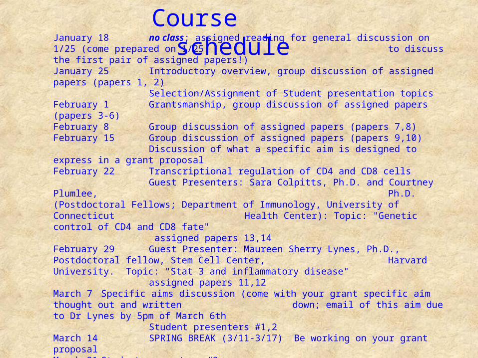
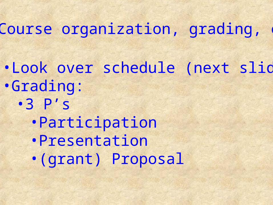
Course scheduleJanuary 18 no class; assigned reading for general discussion on 1/25 (come prepared on 1/25 to discuss the first pair of assigned papers!)January 25 Introductory overview, group discussion of assigned papers (papers 1, 2)
Selection/Assignment of Student presentation topicsFebruary 1 Grantsmanship, group discussion of assigned papers (papers 3-6)February 8 Group discussion of assigned papers (papers 7,8)February 15 Group discussion of assigned papers (papers 9,10)
Discussion of what a specific aim is designed to express in a grant proposalFebruary 22 Transcriptional regulation of CD4 and CD8 cells
Guest Presenters: Sara Colpitts, Ph.D. and Courtney Plumlee, Ph.D. (Postdoctoral Fellows; Department of Immunology, University of Connecticut Health Center): Topic: "Genetic control of CD4 and CD8 fate"
assigned papers 13,14February 29 Guest Presenter: Maureen Sherry Lynes, Ph.D., Postdoctoral fellow, Stem Cell Center, Harvard University. Topic: "Stat 3 and inflammatory disease"
assigned papers 11,12March 7 Specific aims discussion (come with your grant specific aim thought out and written down; email of this aim due to Dr Lynes by 5pm of March 6th
Student presenters #1,2March 14 SPRING BREAK (3/11-3/17) Be working on your grant proposalMarch 21 Student presenters #3March 28 Student presenters #4April 4 Student presenters #5April 11 Student presenters #6April 18 Student presenters #7
Student Grant Proposals are due today; reviewer assignments will be distributedApril 25 Grant review panel meetingMay 2-7 Final exam period

• •Broad topic, so we will refine by selection of topics (to be done next in class)•Schedule for presentations will be emailed before next week, schedule will be based on topics selected•ONE WEEK before you are to present, you must forward either a link to the papers you will present, or pdfs of the papers (which will be assigned to all of the class)•March 7: you must have specific aim for your grant proposal defined and approved
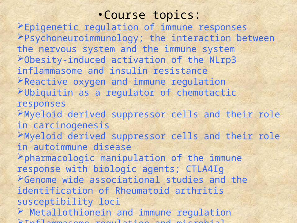
•Course topics:Epigenetic regulation of immune responsesPsychoneuroimmunology; the interaction between the nervous system and the immune systemObesity-induced activation of the NLrp3 inflammasome and insulin resistanceReactive oxygen and immune regulationUbiquitin as a regulator of chemotactic responsesMyeloid derived suppressor cells and their role in carcinogenesisMyeloid derived suppressor cells and their role in autoimmune diseasepharmacologic manipulation of the immune response with biologic agents; CTLA4IgGenome wide associational studies and the identification of Rheumatoid arthritis susceptibility loci Metallothionein and immune regulationInflammasome regulation and microbial infectionsmTOR as a central regulator of immunitycatastrophic dysregulation of the immune response; the cytokine storm hemophagocytic syndrome and multi-organ failure in sepsis

Presentation Format
OverviewHow do the two papers you have selected to present to the class tie together? What is the general theme of your presentation?
IntroductionAnswer the question:“What does the audience need to know to appreciate the importance of this topic?”what was known before the work described in the two papers was donewhat was the experimental approach/explain novel techniquesmake sure to be familiar with the biological materials (e.g. reagents, cell lines, animals)
Identify the most important experiments and discuss those with the controls that help to understand the data
Summarize the conclusion of each experiment (what did the experiment tell us and what did it open up as the next unknown)
Conclusion of paper: what is the big picture of the paper, what do we know now that is most important and what is left unknownwhat would be the next interesting experiments

Practical aspects of presentation
One week before your presentation you must have emailed me links or pdf files of the two papers that you will present and lead the discussion.(papers should be related to each other and to the topic you have chosen, and should have been published in the past 1-2 years.
Your presentation will be either 55 or 75 minutes (if presenting alone or with another student). Earlier presentations will be doubled up and shorter since there is less time to prepare
Plan on leading the discussion of the papers (remembering that each of you will be graded on the quality and frequency of your participation in class)
•My own research interests•The role of metallothionein in immune modulation
•I’ll tell you a bit about the protein and its biochemistry•Examples of immunomodulation•We have parallel interests in biotechnology development
•Mab production (UC1MT)•ECIS/taxis•Grating coupled surface plasmon resonance
metallothionein
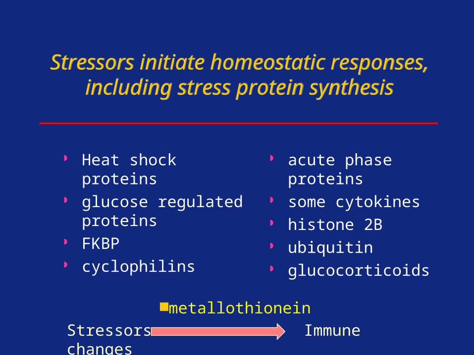
Stressors initiate homeostatic responses, including stress protein synthesis
Stressors initiate homeostatic responses, including stress protein synthesis
Heat shock proteins glucose regulated
proteins FKBP cyclophilins
acute phase proteins some cytokines histone 2B ubiquitin glucocorticoids
Stressors Immune changes
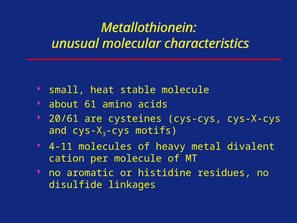
Metallothionein: unusual molecular characteristics
Metallothionein: unusual molecular characteristics
small, heat stable molecule about 61 amino acids 20/61 are cysteines (cys-cys, cys-X-cys and cys-X3-
cys motifs) 4-11 molecules of heavy metal divalent cation per
molecule of MT no aromatic or histidine residues, no disulfide
linkages
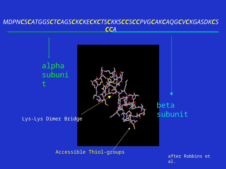
MDPNCSCATGGSCTCAGSCKCKECKCTSCKKSCCSCCPVGCAKCAQGCVCKGASDKCSCCA
alpha subunit
beta subunit
Lys-Lys Dimer Bridge
Accessible Thiol-groupsafter Robbins et al.
ISRE GRE BLE MRE TRE GC MRE TATA+1
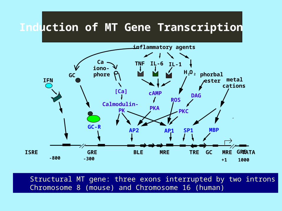
Induction of MT Gene Transcription
IFN
Ca iono-phore
TNF IL-6 IL-1
phorbalester metal
cations
GC
GC-R
DAG
PKC
cAMP
PKA
[Ca]
Calmodulin-PK
MBPAP2 SP1AP1
-300-800
H 2O 2
ROS
1000
GRE
inflammatory agents
Structural MT gene: three exons interrupted by two intronsChromosome 8 (mouse) and Chromosome 16 (human)
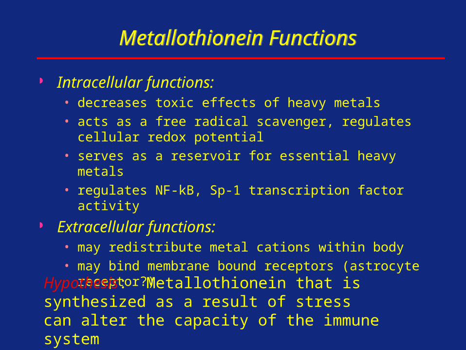
Metallothionein FunctionsMetallothionein Functions
Intracellular functions:• decreases toxic effects of heavy metals• acts as a free radical scavenger, regulates cellular redox
potential• serves as a reservoir for essential heavy metals• regulates NF-kB, Sp-1 transcription factor activity
Extracellular functions:• may redistribute metal cations within body• may bind membrane bound receptors (astrocyte receptor?)
Hypothesis: Metallothionein that is synthesized as a result of stress can alter the capacity of the immune system
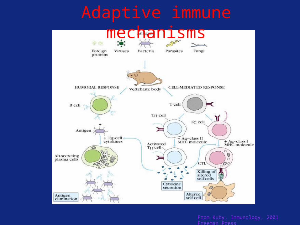
From Kuby, Immunology, 2001 Freeman Press
Adaptive immune mechanisms
0
10
20
30
40
50
Cyto-toxicity(%)
30:1 10:1 1:1
MT
PBS
Effector:Target
CTL inductionphase
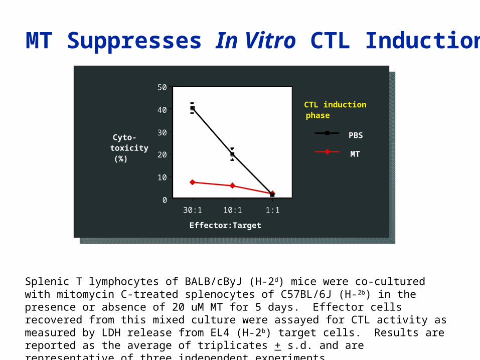
MT Suppresses In Vitro CTL Induction
Splenic T lymphocytes of BALB/cByJ (H-2d) mice were co-cultured with mitomycin C-treated splenocytes of C57BL/6J (H-2b) in the presence or absence of 20 uM MT for 5 days. Effector cells recovered from this mixed culture were assayed for CTL activity as measured by LDH release from EL4 (H-2b) target cells. Results are reported as the average of triplicates + s.d. and are representative of three independent experiments.
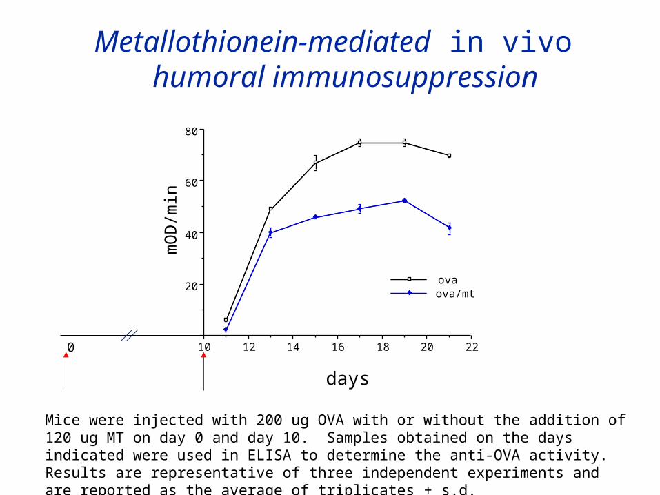
Metallothionein-mediated in vivo
humoral immunosuppression
Metallothionein-mediated in vivo
humoral immunosuppression
22201816141210
20
40
60
80
ovaova/mt
days
mOD/min
Mice were injected with 200 ug OVA with or without the addition of 120 ug MT on day 0 and day 10. Samples obtained on the days indicated were used in ELISA to determine the anti-OVA activity. Results are representative of three independent experiments and are reported as the average of triplicates + s.d.
0
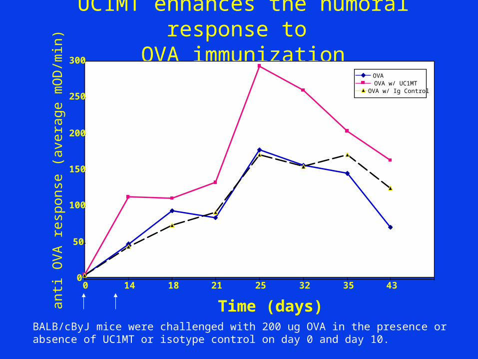
UC1MT enhances the humoral response to
OVA immunization
0
50
100
150
200
250
300
0 14 18 21 25 32 35 43
Time (days)anti OVA response (average mOD/min)
OVA OVA w/ UC1MT
OVA w/ Ig Control
BALB/cByJ mice were challenged with 200 ug OVA in the presence or absence of UC1MT or isotype control on day 0 and day 10.
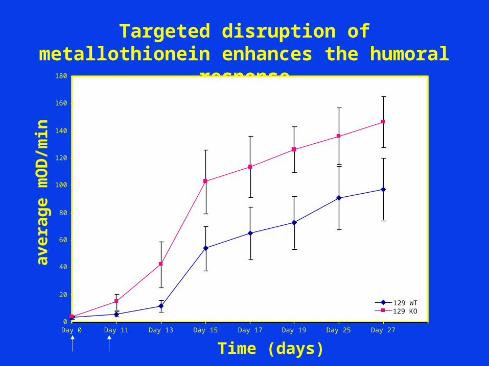
Targeted disruption of metallothionein enhances the humoral
response
0
20
40
60
80
100
120
140
160
180
Day 0 Day 11 Day 13 Day 15 Day 17 Day 19 Day 25 Day 27
Time (days)
average mOD/min
129 WT129 KO
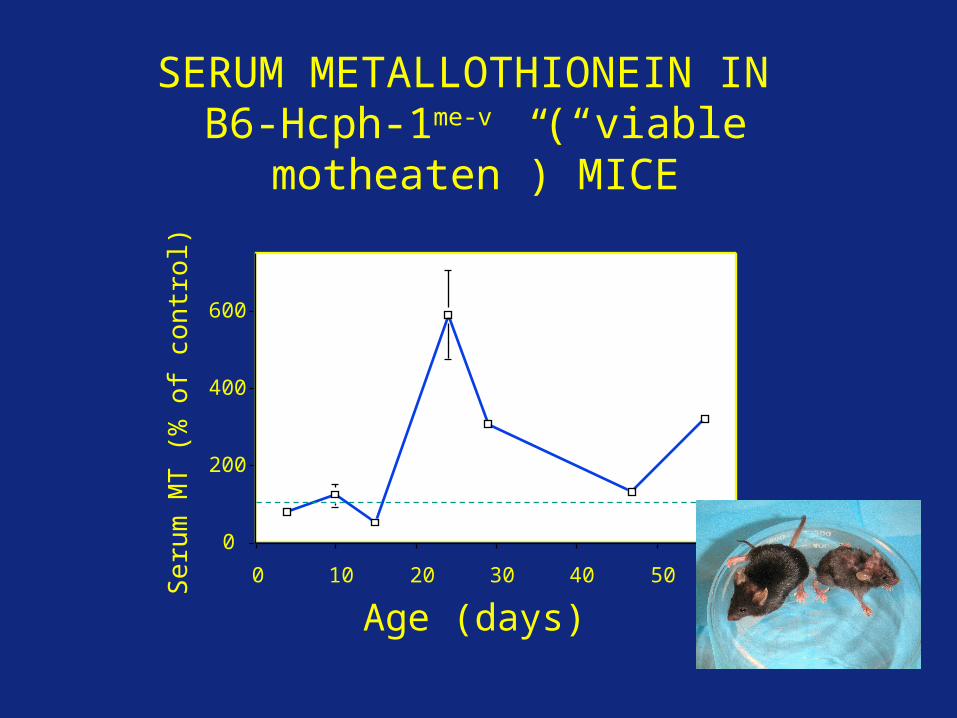
0
200
400
600
Serum MT (% of control)
0 10 20 30 40 50 60
Age (days)
SERUM METALLOTHIONEIN IN B6-Hcph-1me-v (“viable
motheaten”) MICE
UC1MT-FITC binding to splenocytes from B6-Hcph-1mev/mev and B6-Hcph-1+/mev mice
Metallothionein is also found on the surface of leukocytes from mice with a congenital chronic inflammatory disease

post immunization
MT injection
ZnCl2 injection
23 24 25 26 27 28 29 30 31 32 33
Art
hriti
c in
dex
0
1
2
3
4
5
6
vehicle groupMT groupZnCl2 group
MT suppresses the severity of collagen-induced arthritis
To induce CIA, DBA/1J mice were initially immunized by injection with CII/CFA into the base of
the tail. 2 weeks later, mice received a booster injection with CII/IFA in the footpad. After
7 days, each group of mice were injected i.p. with MT (), ZnCl2 () or PBS(vehicle control) and
were assessed for indications of arthritis. The clinical severity of CIA, determined by
arthritis index, was significantly suppressed in MT and ZnCl2 treatment groups.
-Youn et al. 2002
Metallothionein also influences the onset and severity of inflammatory bowel disease (collaboration with University of Gent, Belgium); with inflammation associated with optic wound healing (collaboration with University of Tasmania, Australia), and with experimental epidermolysis bullosa acquisita (Universitätsklinikum Schleswig-Holstein , Germany)
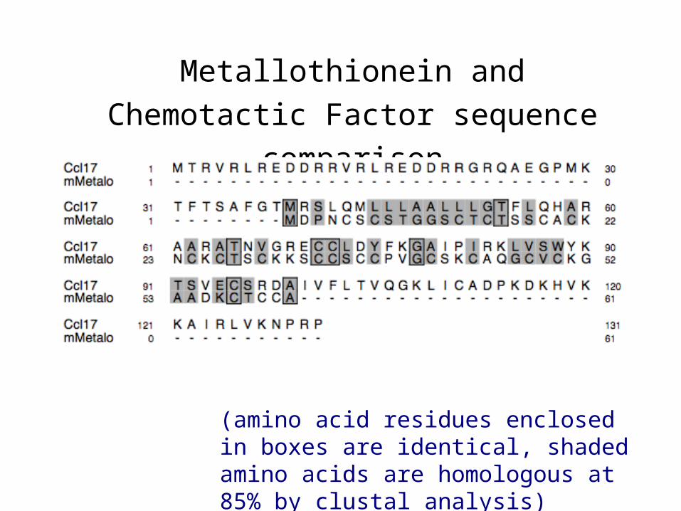
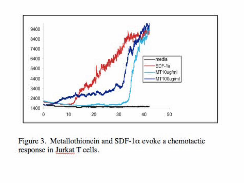
Metallothionein and Chemotactic
Factor sequence comparison
(amino acid residues enclosed in boxes are identical, shaded amino acids are homologous at 85% by clustal analysis)
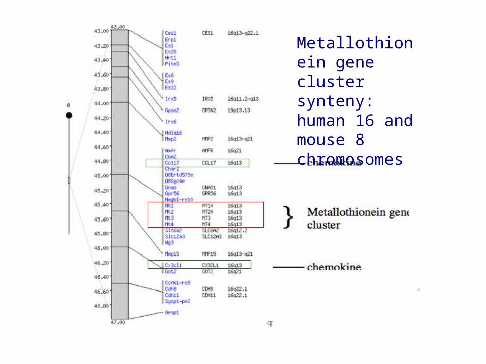
Metallothionein gene cluster synteny: human 16 and mouse 8 chromosomes
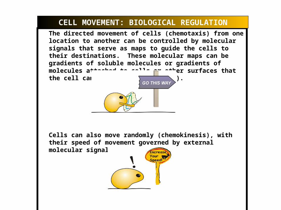
The directed movement of cells (chemotaxis) from one location to another can be controlled by molecular signals that serve as maps to guide the cells to their destinations. These molecular maps can be gradients of soluble molecules or gradients of molecules attached to cells or other surfaces that the cell can encounter (haptotaxis).
Cells can also move randomly (chemokinesis), with their speed of movement governed by external molecular signals.
CELL MOVEMENT: BIOLOGICAL REGULATION
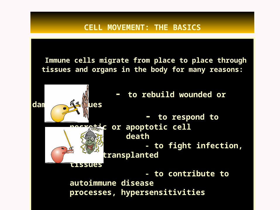
Immune cells migrate from place to place through tissues and organs in the body for many reasons:
- to rebuild wounded or damaged tissues
- to respond to necrotic or apoptotic cell
death - to fight infection,
attack transplanted tissues
- to contribute to autoimmune disease processes, hypersensitivities
CELL MOVEMENT: THE BASICS
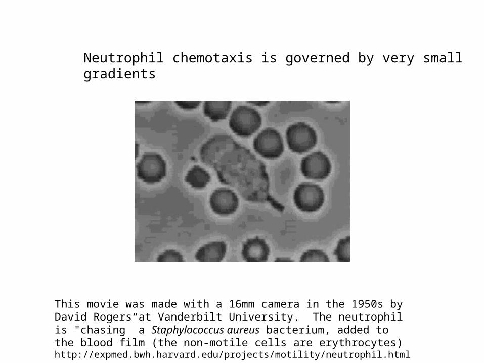
This movie was made with a 16mm camera in the 1950s by David Rogers at Vanderbilt University. The neutrophil is "chasing” a Staphylococcus aureus bacterium, added to the blood film (the non-motile cells are erythrocytes) http://expmed.bwh.harvard.edu/projects/motility/neutrophil.html.
Neutrophil chemotaxis is governed by very small gradients

8 Well ECIS Chamber
Courtesy of Applied Biophysics Inc.
Collaboration with David Knecht’s
laboratory and with Applied Biophysics,
Inc.
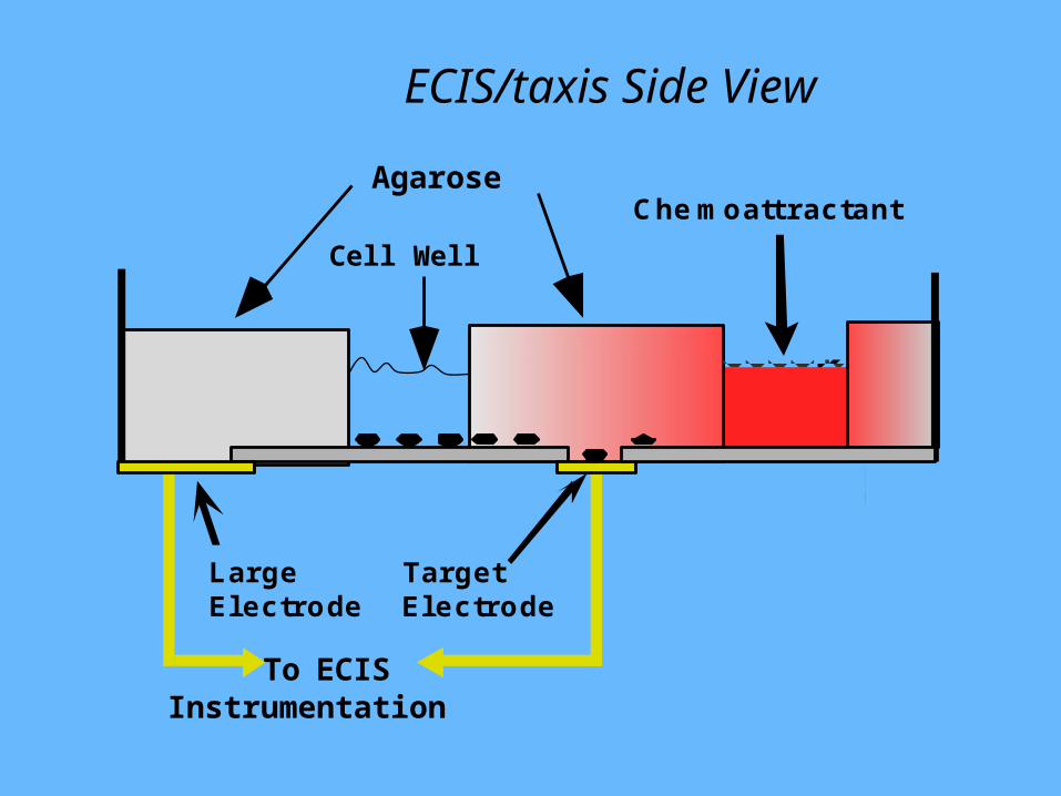
ECIS/taxis Side View
Cell Well
LargeElectrode
TargetElectrode
C he m oattractantAgarose
To ECISInstrumentation
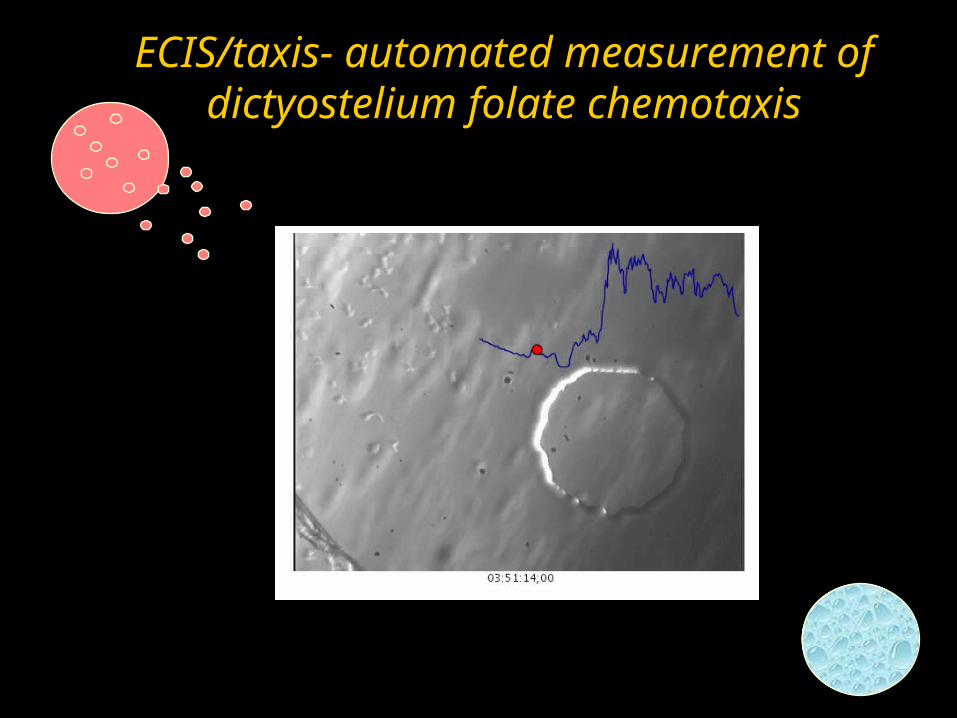
ECIS/taxis- automated measurement of dictyostelium folate chemotaxis

Surface Plasmon Resonance; the basicsSurface Plasmon Resonance; the basics
Based on the physical phenomenon of energy transfer at a metal-dielectric interface (e.g., a gold and water interface ).
Under specific optical conditions (e.g. wave vector, incident angle and frequency of the incident light), the energy of the light excites electron movement (the plasmon) within the metal. This energy transfer reduces the intensity of the reflected light.
(e-)
SPR technologies can use different ways to match the wave vector of the illuminating beam of light with the plasmon wave vector
Using a prism (Kretschman configuration) Using a diffraction grating (Grating-coupled surface plasmon resonance ; GCSPR) -- the first order diffracted light from the grating matches the plasmon requirements There is a critical angle of illumination
at which this vector matching is best, and at which the energy transfer into the metal is largest.
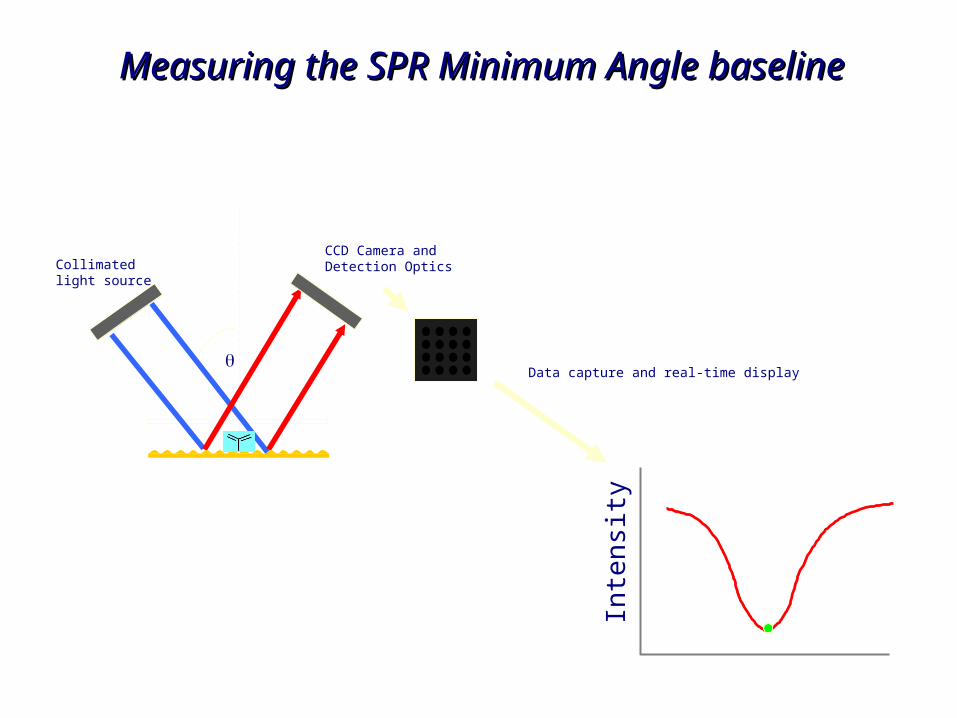
The angle at which maximum energy transfer occurs is the “SPR angle ” or “resonance angle”. Energy transfer is sensitive to the refractive index of the dielectric/metal interfaceThe addition of proteins to the sensor chip surface (which will then have a higher index of refraction than water) will increase the angle at which maximum coupling occurs.
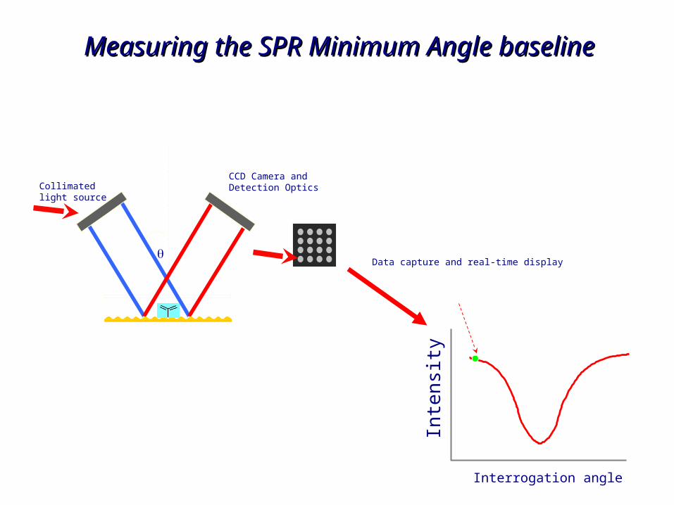
Collimatedlight source
CCD Camera andDetection Optics
Inte
nsi
ty
Data capture and real-time display
Interrogation angle
Measuring the SPR Minimum Angle Measuring the SPR Minimum Angle baselinebaseline
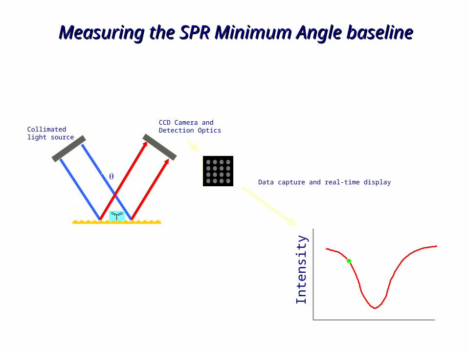
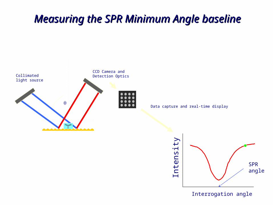
Collimatedlight source
CCD Camera andDetection Optics
Inte
nsi
ty
Data capture and real-time display
Measuring the SPR Minimum Angle Measuring the SPR Minimum Angle baselinebaseline
Collimatedlight source
CCD Camera andDetection Optics
Inte
nsi
ty
Data capture and real-time display
Measuring the SPR Minimum Angle Measuring the SPR Minimum Angle baselinebaseline
Collimatedlight source
CCD Camera andDetection Optics
Inte
nsi
ty
Data capture and real-time display
Interrogation angle
SPR angle
Measuring the SPR Minimum Angle Measuring the SPR Minimum Angle baselinebaseline
Collimatedlight source
CCD Camera andDetection Optics
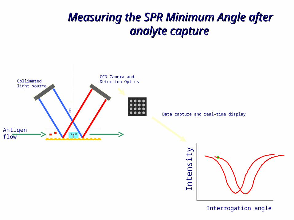
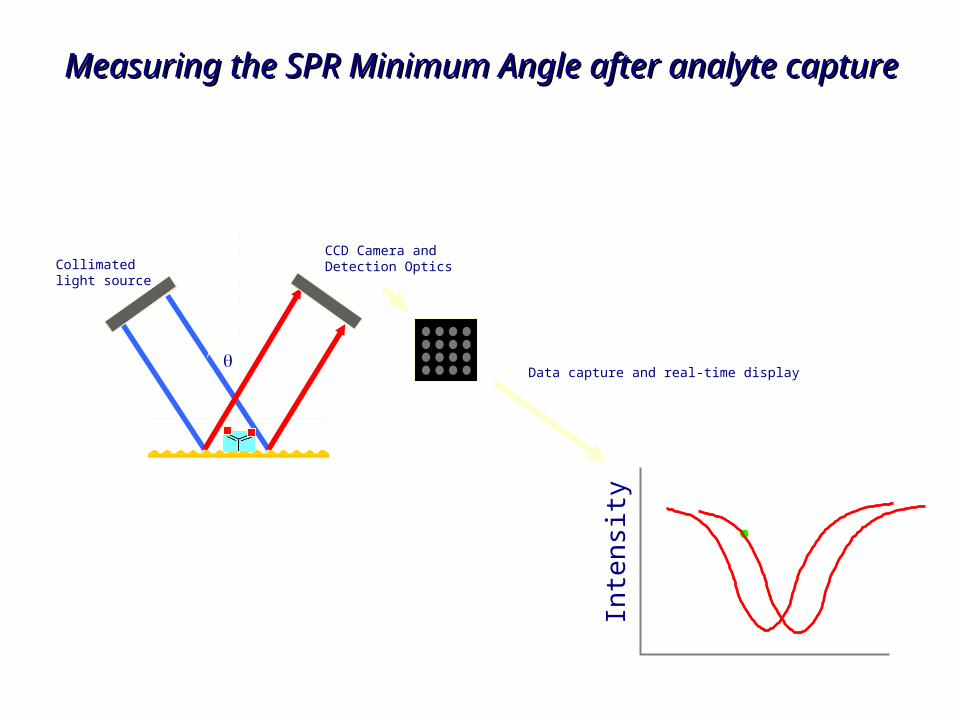
Measuring the SPR Minimum Angle Measuring the SPR Minimum Angle after analyte captureafter analyte capture
Inte
nsi
ty
Data capture and real-time display
Interrogation angle
Antigen flow
Collimatedlight source
CCD Camera andDetection Optics
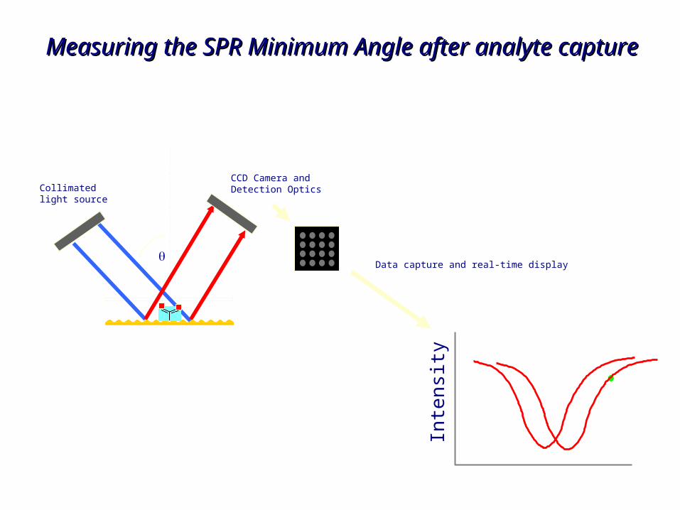
Measuring the SPR Minimum Angle Measuring the SPR Minimum Angle after analyte captureafter analyte capture
Inte
nsi
ty
Data capture and real-time display
Interrogation angle
Antigen flow
Collimatedlight source
CCD Camera andDetection Optics
Inte
nsi
ty
Data capture and real-time display
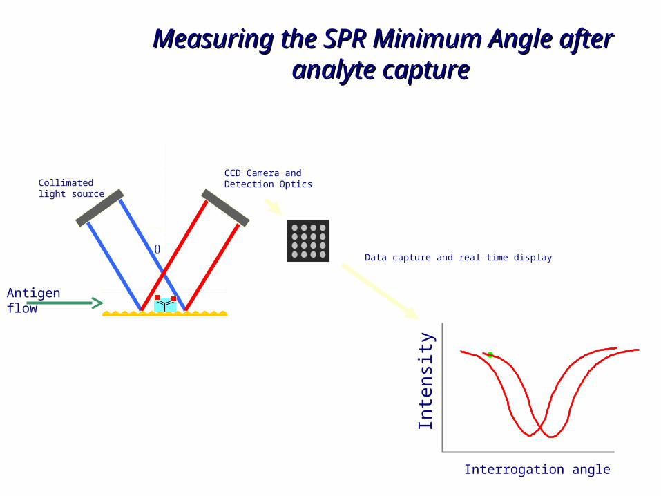
Measuring the SPR Minimum Angle after Measuring the SPR Minimum Angle after analyte captureanalyte capture

Collimatedlight source
CCD Camera andDetection Optics
Inte
nsi
ty
Data capture and real-time display
Measuring the SPR Minimum Angle after Measuring the SPR Minimum Angle after analyte captureanalyte capture
Collimatedlight source
CCD Camera andDetection Optics
Inte
nsi
ty
Data capture and real-time display
Measuring the SPR Minimum Angle after Measuring the SPR Minimum Angle after analyte captureanalyte capture
Collimatedlight source
CCD Camera andDetection Optics
Inte
nsi
ty
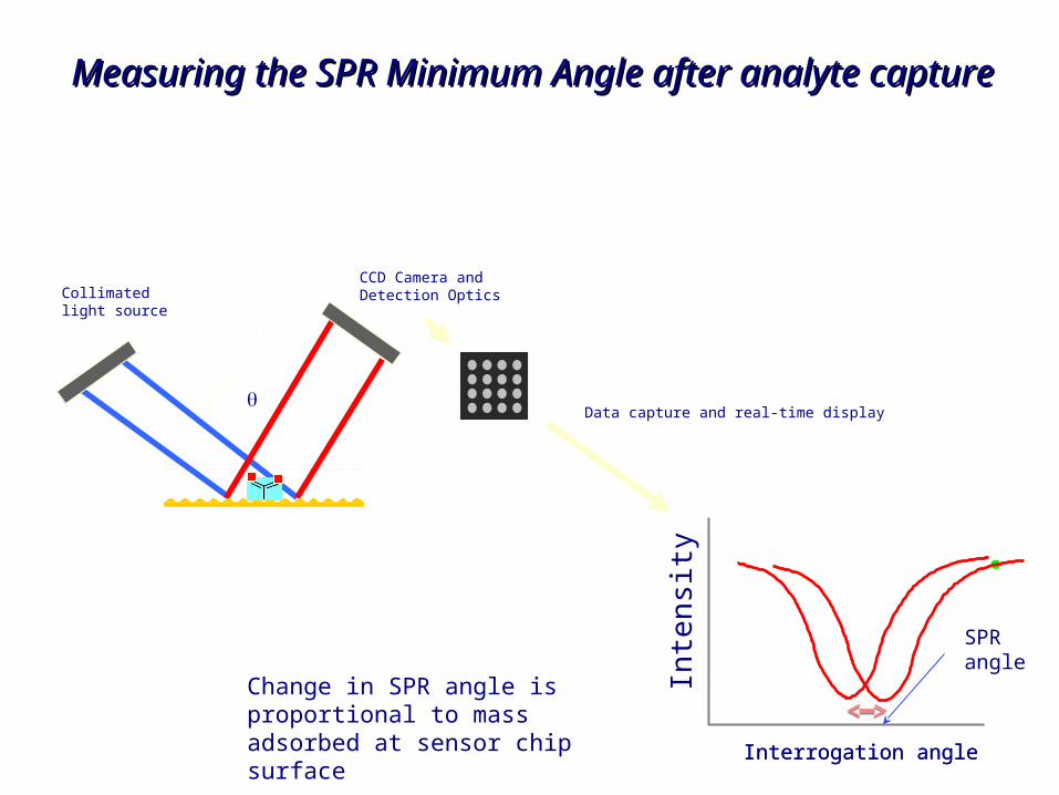
Data capture and real-time display
Interrogation angleInterrogation angle
SPR angle
Change in SPR angle is proportional to mass adsorbed at sensor chip surface
Measuring the SPR Minimum Angle after Measuring the SPR Minimum Angle after analyte captureanalyte capture
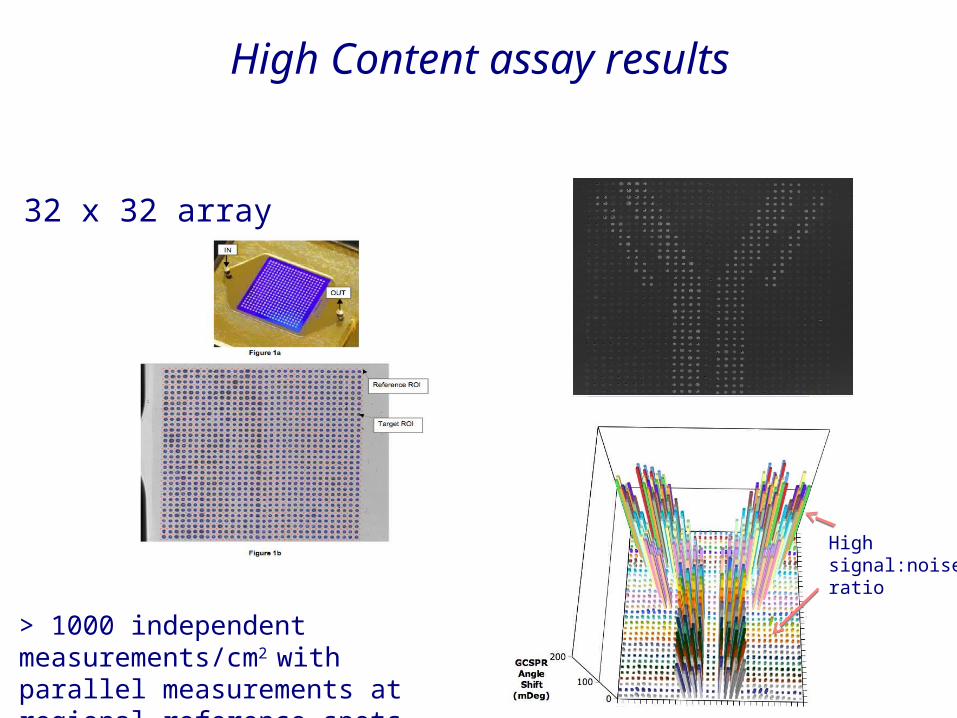
High Content assay results
> 1000 independent measurements/cm2 with parallel measurements at regional reference spots
32 x 32 array
High signal:noise ratio
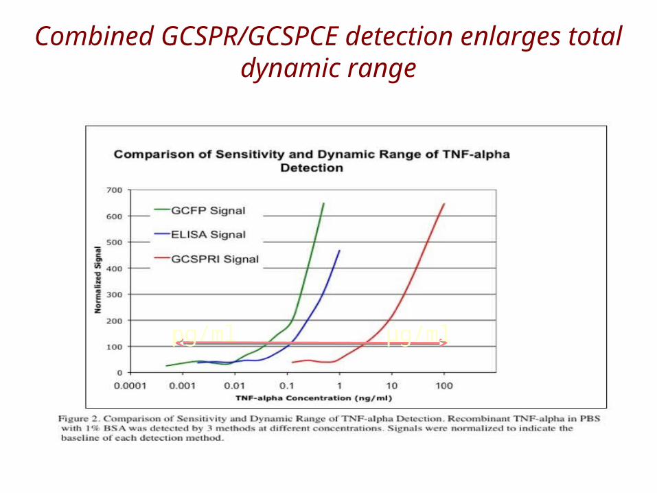
Combined GCSPR/GCSPCE detection enlarges total dynamic range
pg/ml µg/ml
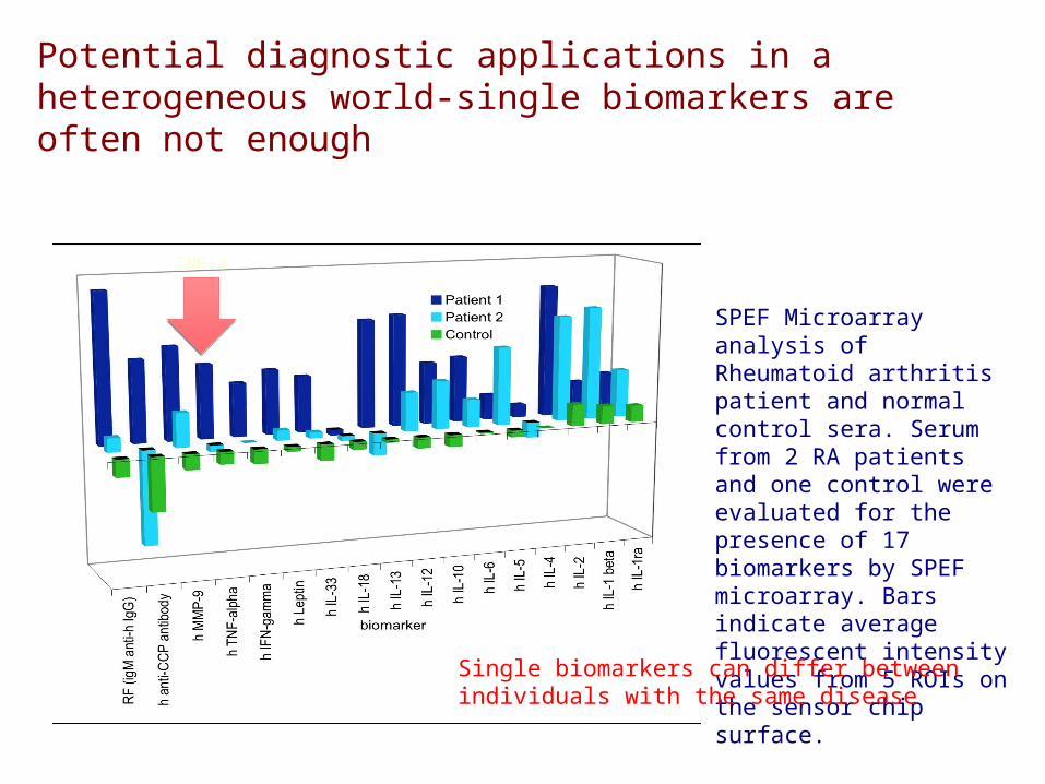
SPEF Microarray analysis of Rheumatoid arthritis patient and normal control sera. Serum from 2 RA patients and one control were evaluated for the presence of 17 biomarkers by SPEF microarray. Bars indicate average fluorescent intensity values from 5 ROIs on the sensor chip surface.
Potential diagnostic applications in a heterogeneous world-single biomarkers are often not enough
Single biomarkers can differ between individuals with the same disease
TNF-a
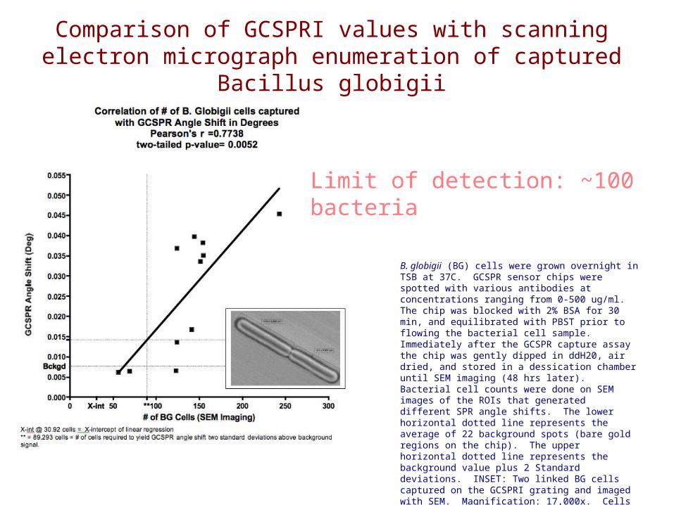
B. globigii (BG) cells were grown overnight in TSB at 37C. GCSPR sensor chips were spotted with various antibodies at concentrations ranging from 0-500 ug/ml. The chip was blocked with 2% BSA for 30 min, and equilibrated with PBST prior to flowing the bacterial cell sample. Immediately after the GCSPR capture assay the chip was gently dipped in ddH20, air dried, and stored in a dessication chamber until SEM imaging (48 hrs later). Bacterial cell counts were done on SEM images of the ROIs that generated different SPR angle shifts. The lower horizontal dotted line represents the average of 22 background spots (bare gold regions on the chip). The upper horizontal dotted line represents the background value plus 2 Standard deviations. INSET: Two linked BG cells captured on the GCSPRI grating and imaged with SEM. Magnification: 17,000x. Cells measure approximately 3.3 x 0.86 M.
Comparison of GCSPRI values with scanning electron micrograph enumeration of captured
Bacillus globigii
Limit of detection: ~100 bacteria
Influenza Peptide Reactive CD4+ T-Cell Capture and Detection on a GCSPRI
0
50
100
150
200
250
300
100 50 10 1 0.1 0.01
Percent HA Reactive T cells
∆ SPR Angle (mdeg)
MHC II / HA peptide
MHC II / Tet peptide
anti-CD3 antibody
MHC II / HA peptide
MHC II / Tet peptide (negative control)
anti-CD3 Antibody
(positive control)
A
B
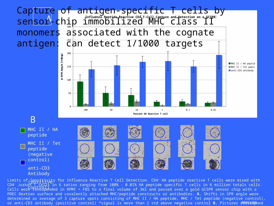
Limits of Sensitivity for Influenza Reactive T Cell Detection. CD4+ HA peptide reactive T cells were mixed with CD4+ Jurkat T cells in 6 ratios ranging from 100% - 0.01% HA peptide specific T cells in 6 million totals cells. Cells were resuspended in RPMI + FBS to a final volume of 3ml and passed over a gold GCSPR sensor chip with a PDEC Dextran surface and covalently attached MHC/peptide constructs or antibodies. A. Shifts in SPR angle were determined as average of 3 capture spots consisting of MHC II / HA peptide, MHC / Tet peptide (negative control), or anti-CD3 antibody (positive control) *signal is more than 2 std above negative control B. Pictures correspond to one representative cell capture spot on sensor surface.
*
* *
* *
JMR021208
Capture of antigen-specific T cells by sensor-chip immobilized MHC class II monomers associated with the cognate antigen: can detect 1/1000 targets
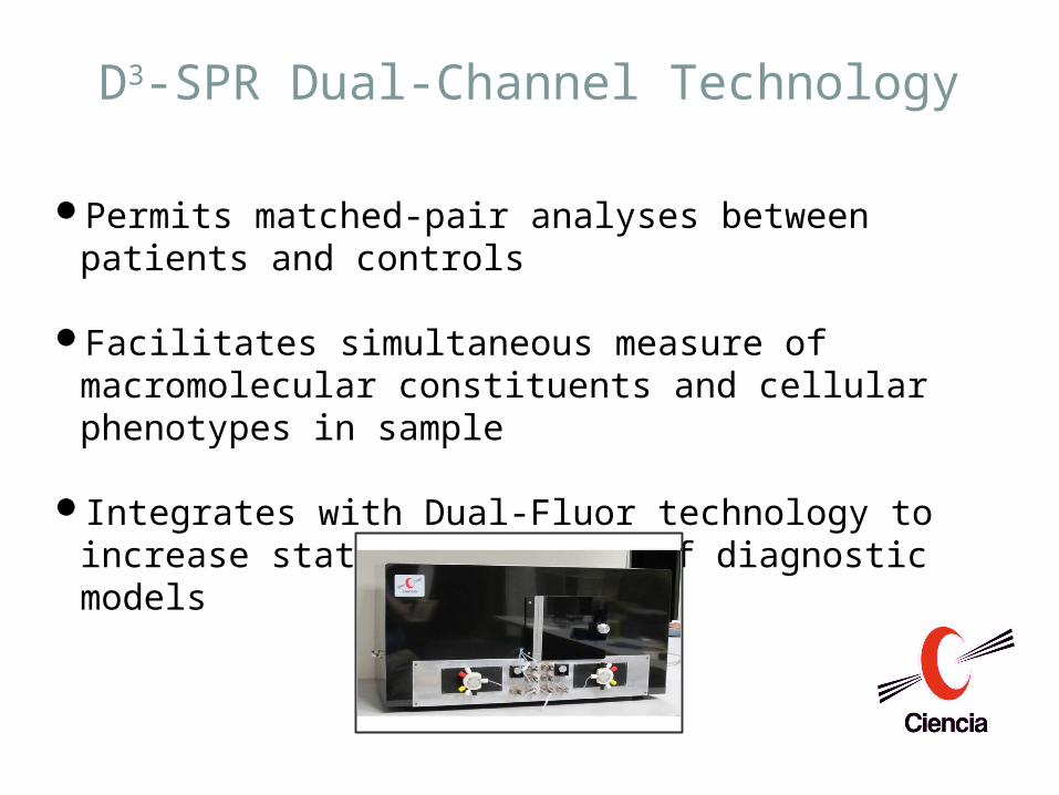
D3-SPR Dual-Channel Technology
Permits matched-pair analyses between patients and controls
Facilitates simultaneous measure of macromolecular constituents and cellular phenotypes in sample
Integrates with Dual-Fluor technology to increase statistical power of diagnostic models
Conclusions
Metallothionein synthetic capacity represents a risk factor for toxicant-induced immunomodulation
Induction of metallothionein synthesis may have therapeutic effects in some forms of autoimmunity
Metallothionein analogs may represent novel immunotherapeutics
The propensity to synthesize metallothionein may influence disease susceptibility
Monoclonal antibody-mediated manipulation of metallothionein or other metallothionein antagonists in vivo can enhance vaccination efficacy
Technologies on our horizon Grating-coupled surface plasmon resonance imaging as diagnostic for
molecular signatures of agricultural diseases Grating coupled fluorescence plasmonics for the identification of CD4+ and
CD8+ T cells that are diabetogenic• Capture of cells with immobilized MHC Class I and Class II monomers folded
with diabetogenic peptides• Functional analysis of captured cells
Design and validate chips for specific diagnostic/therapeutic/monitoring needs
• Stress responses• Protein synthesis and secretion• Cellular differentiation• Vaccine responses (simultaneous assessments of vaccine efficacy, immune
capacity, and pathogen presence using the same unfractionated blood sample).
• Immune assessment (infection, autoimmune disease and cancer)• Environmental assessment
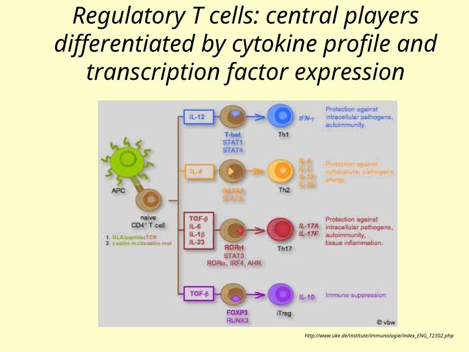
Regulatory T cells: central players differentiated by cytokine profile and
transcription factor expression
http://www.uke.de/institute/immunologie/index_ENG_72302.php
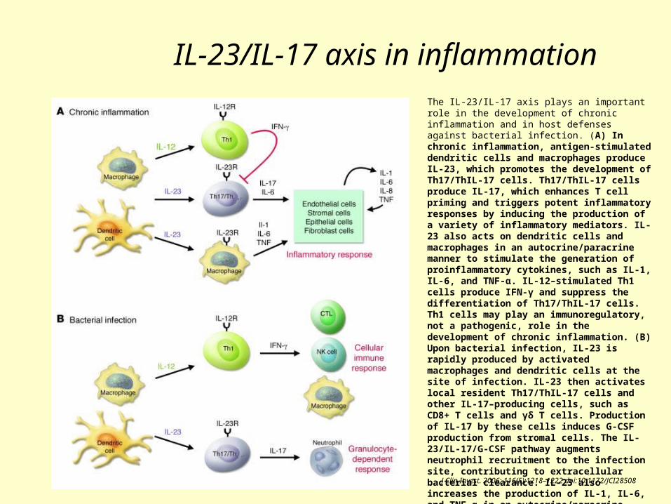
IL-23/IL-17 axis in inflammationThe IL-23/IL-17 axis plays an important role in the development of chronic inflammation and in host defenses against bacterial infection. (A) In chronic inflammation, antigen-stimulated dendritic cells and macrophages produce IL-23, which promotes the development of Th17/ThIL-17 cells. Th17/ThIL-17 cells produce IL-17, which enhances T cell priming and triggers potent inflammatory responses by inducing the production of a variety of inflammatory mediators. IL-23 also acts on dendritic cells and macrophages in an autocrine/paracrine manner to stimulate the generation of proinflammatory cytokines, such as IL-1, IL-6, and TNF-α. IL-12–stimulated Th1 cells produce IFN-γ and suppress the differentiation of Th17/ThIL-17 cells. Th1 cells may play an immunoregulatory, not a pathogenic, role in the development of chronic inflammation. (B) Upon bacterial infection, IL-23 is rapidly produced by activated macrophages and dendritic cells at the site of infection. IL-23 then activates local resident Th17/ThIL-17 cells and other IL-17–producing cells, such as CD8+ T cells and γδ T cells. Production of IL-17 by these cells induces G-CSF production from stromal cells. The IL-23/IL-17/G-CSF pathway augments neutrophil recruitment to the infection site, contributing to extracellular bacterial clearance. IL-23 also increases the production of IL-1, IL-6, and TNF-α in an autocrine/paracrine manner. In contrast, Th1 cells produce IFN-γ and stimulate CD8+ cytotoxic T lympocytes, NK cells, and macrophages. IFN-γ enhances antigen presentation by inducing expression of MHC molecules and activates cells to produce cytolytic molecules, including perforin and granzyme, which promote the elimination of intracellular bacteria.
J Clin Invest. 2006; 116(5):1218–1222 doi:10.1172/JCI28508