Embed Size (px)
Citation preview

~ 1 ~
The Australian Cretaceous ichthyosaur Platypterygius australis: understanding its taxonomy, morphology, and palaeobiology
MARIA ZAMMIT
Environmental Biology School of Earth and Environmental Sciences
The University of Adelaide South Australia
A thesis submitted for the degree of Doctor of Philosophy at the University of Adelaide
10 January 2011
~ 2 ~
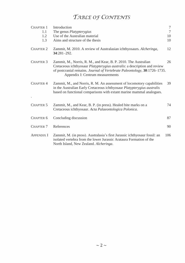
TABLE OF CONTENTS CHAPTER 1 Introduction 7 1.1 The genus Platypterygius 7 1.2 Use of the Australian material 10 1.3 Aims and structure of the thesis 10 CHAPTER 2 Zammit, M. 2010. A review of Australasian ichthyosaurs. Alcheringa, 12 34:281–292. CHAPTER 3 Zammit, M., Norris, R. M., and Kear, B. P. 2010. The Australian 26 Cretaceous ichthyosaur Platypterygius australis: a description and review of postcranial remains. Journal of Vertebrate Paleontology, 30:1726–1735. Appendix I: Centrum measurements CHAPTER 4 Zammit, M., and Norris, R. M. An assessment of locomotory capabilities 39 in the Australian Early Cretaceous ichthyosaur Platypterygius australis based on functional comparisons with extant marine mammal analogues. . CHAPTER 5 Zammit, M., and Kear, B. P. (in press). Healed bite marks on a 74 Cretaceous ichthyosaur. Acta Palaeontologica Polonica. CHAPTER 6 Concluding discussion 87 CHAPTER 7 References 90 APPENDIX I Zammit, M. (in press). Australasia’s first Jurassic ichthyosaur fossil: an 106 isolated vertebra from the lower Jurassic Arataura Formation of the North Island, New Zealand. Alcheringa.
~ 3 ~
ABSTRACT
The Cretaceous ichthyosaur Platypterygius was one of the last representatives of the
Ichthyosauria, an extinct, secondarily aquatic group of reptiles. Remains of this genus occur
worldwide, but the Australian material is among the best preserved and most complete. As a
result, the Australian ichthyosaur fossil finds were used to investigate the taxonomy, anatomy,
and possible locomotory methods and behaviours of this extinct taxon.
Understanding the importance of the Australian Platypterygius species has been
complicated by the use of two specific names, P. australis and P. longmani, and confused
further by the loss of holotype material. Examination of Australian material has demonstrated
that both species belong to the same taxon. P. australis was shown as the valid taxon name,
relegating P. longmani to a junior synonym, and thus resolving the taxonomic uncertainty of
the only Australasian ichthyosaur that can be identified to species-level.
Examination of P. australis postcranial anatomy revealed four postcranial characters
that, used in conjunction with previously identified cranial and postcranial diagnostic features,
distinguish the Australian taxon from other species of Platypterygius. The morphology of the
postcranial elements (including bones that had not previously been described for the genus)
was then used to hypothesise the locomotory mode in this ichthyosaur based on osteological
comparisons with extant marine mammals. Results indicated that a decoupled locomotor
system was most plausible for P. australis, where the caudal fin was used for long distance
swimming and the broad forelimbs for manoeuvring. In addition, the broad forelimbs, for
which the genus is named, are thought to increase acceleration when either stationary or
whilst moving.
In addition to the functional studies, palaeobehaviour in this ichthyosaur could also be
inferred from bite traces. Palaeopathologies in the form of bite marks on a partial ichthyosaur
skull were examined. The bite marks were attributed to another ichthyosaur (most likely of

~ 4 ~
the same species), thus indicating that P. australis individuals engaged in aggressive
behaviour.
This thesis examined the known Australian ichthyosaur material to address taxonomic,
anatomical, and behavioural aspects of Platypterygius, and demonstrated the utility of the
Australian Cretaceous record for this purpose. Collection of additional specimens, particularly
from localities in Western Australia and South Australia where diagnostic remains are yet to
be found, would significantly add to our knowledge of this extinct taxon.
~ 5 ~
DECLARATION
This thesis contains no material which has been accepted for the award of any other
degree or diploma in any university of other tertiary institution to Maria Zammit and, to the
best of my knowledge and belief, contains no material previously published or written by
another person, except where due reference is made in the text.
The author consents to this copy of the thesis, once deposited in the University of
Adelaide Library, being made available for photocopying and loan, subject to the provisions
of the Copyright Act 1968.
The author acknowledges that copyright of published works contained within this
thesis (as listed below) resides with the copyright holders of those works.
Zammit, M. 2010. A review of Australasian ichthyosaurs. Alcheringa, 34:281–292.
Zammit, M., Norris, R., and Kear, B.P. 2010. The Australian Cretaceous ichthyosaur
Platypterygius australis: a description and review of postcranial remains. Journal of
Vertebrate Paleontology, 30:1726–1735.
Zammit, M., and Kear, B.P. (in review). Healed bite marks on a Cretaceous ichthyosaur. Acta
Palaeontologica Polonica.
Zammit, M. (in press). Australasia’s first Jurassic ichthyosaur: an isolated vertebra from the
lower Jurassic Arataura Formation of the North Island, New Zealand. Alcheringa.
Further, the author also gives permission for the digital version of this thesis to be
made available on the web, via the University’s digital research repository, the Library
catalogue, the Australasian Digital Theses Program (ADTP), and also through web search
engines, unless permission has been granted by the University to restrict access for a period of
time.
………………………………………………. Maria Zammit
10 January 2011

~ 6 ~
ACKNOWLEDGEMENTS First and foremost, I thank the School of Earth and Environmental Sciences for my
Divisional Scholarship and John Jennings for agreeing to act as my principal supervisor,
facilitating my candidature for a Doctor of Philosophy at the University of Adelaide. My co-
supervisor, Rachel Norris, and external supervisor, Benjamin Kear, have my profound thanks
for their encouragement, support, and advice throughout my candidature. Without their
continuous efforts, this work may not have eventuated at all.
I am extremely grateful to the following institutions and their curators and collection
managers for granting access to specimens, or facilitating loans: AM, Australian Museum,
Sydney, New South Wales: Robert Jones; AU, University of Auckland, Auckland, New
Zealand: Neville Hudson; KKM, Kronosaurus Korner Museum, Richmond, Queensland: Paul
Stumkat; MV, Museum Victoria, Melbourne, Victoria: David Pickering; NMMUK, Natural
History Museum United Kingdom, London, UK: Sandra Chapman; QM, Queensland
Museum, Brisbane, Queensland: Scott Hocknull, Kristen Spring, Heather Janetzki, and
Patrick Couper; SAM, South Australian Museum, Adelaide, South Australia: Ben McHenry,
Mary-Anne Binnie, Natalie Schroeder, David Stemmer, Catherine Kemper, Philippa Horton,
and Carolyn Kovach. Travel to these institutions was supported by the University of Adelaide
and funding from the Sir Mark Mitchell Foundation.
I also wish to thank my fellow PhD candidates for making my time at the University
of Adelaide more enjoyable, especially Christina Adler, Jessica Wadley, and Keridwen
Barber. The sound palaeontological advice provided by Trevor Worthy was always much
appreciated.
Last but not least, I’d like to thank my husband Nicolas Rawlence for being an
understanding partner and wonderful friend, and providing invaluable support.
~ 7 ~
CHAPTER 1
INTRODUCTION
Ichthyosaurs are a group of extinct, fish-shaped marine reptiles that were first
described from Jurassic deposits of Europe in the early 19th century (Home 1814, 1816). Their
remains are now known from the Lower Triassic (Olenekian: Callaway and Massare 1989)
through to the Upper Cretaceous (Cenomanian: Bardet 1992), and can be found all over the
world. The origins of the ichthyosaurs have proved problematic, and almost all major
vertebrate groups have been hypothesised as the possible sister group – such groups include
fish, crocodiles, turtles, squamates, and monotremes (McGowan and Motani 2003). Studies
on more basal Triassic forms suggested that the Ichthyosauria are diapsid reptiles (Massare
and Callaway 1990; Motani et al. 1998), though some ichthyosaur researchers consider the
evidence for this hypothesis to be inconclusive (Maisch and Hungerbühler 2001). Post-
Triassic ichthyosaurs are still considered the ‘typical’ ichthyosaurian form, and the majority
of previous studies have focused on these genera, particularly the Jurassic representatives. For
example, phylogenetic, ontogenetic, functional, and palaeobiological studies have been
undertaken mainly on Jurassic ichthyosaurs (Johnson 1977; Buchholtz 2001), but have rarely
been extended to their Cretaceous counterparts.
1.1 The genus Platypterygius
The Cretaceous genus Platypterygius is one of the last representatives of the
Ichthyosauria (Neocomian–Cenomanian: McGowan and Motani 2003), and is found on
almost every continent (Maisch and Matzke 2000). Until recently, most Cretaceous
ichthyosaurs were referred to this genus (McGowan 1991), resulting in Platypterygius
becoming almost the default taxon for any Cretaceous ichthyosaur material. However, several
Jurassic genera are now known to extend into the Cretaceous (Brachypterygius, McGowan
~ 8 ~
and Motani 2003; Aegirosaurus, Fischer 2009; Fischer et al. in press; and Ophthalmosaurus,
McGowan and Motani 2003; Fischer 2009), and two new genera have been erected for
Cretaceous material (Maiaspondylus, Maxwell and Caldwell 2006a; and Athabascasaurus,
Druckenmiller and Maxwell 2010). Thus, a reassessment of the genus Platypterygius is
required to aid in the taxonomic identification of new Cretaceous specimens.
The most extensive recent review of ichthyosaurs to date (McGowan and Motani
2003) recognised five valid species of Platypterygius: P. americanus (Nace 1939, 1941) from
the Albian–Cenomanian of North America (Maxwell and Kear 2010); P. australis (McCoy
1867a) from the Albian of Australia (Kear 2003); P. campylodon (Carter 1846; Kiprijanoff
1881) from the Albian–Cenomanian of England, France, and Russia (McGowan and Motani
2003); P. hauthali (von Huene 1927) from the Barremian of Argentina (Fernández and
Aguirre-Urreta 2005); and P. platydactylus (Broili 1907) from the Aptian of Germany
(McGowan and Motani 2003). McGowan and Motani (2003) consider their review to be
taxonomically inflated, and suggested that fewer species would probably be maintained if
more data were available for adequate comparisons. However, recently the resurrection of P.
hercynicus (from P. platydactylus) from the Aptian of Germany has been proposed (Kolb and
Sander 2009), and two additional species, P. sachicarum (Páramo 1997) and P. ochevi
(Arkhangelsky et al. 2008) have been established, from the Barremian–Aptian of Colombia
and the Albian–Cenomanian of Russia, respectively. Despite this, species diversity within
Platypterygius remains unclear (Maxwell and Kear 2010), with McGowan (1991) raising the
possibility that the genus may comprise only a single species if more individuals were known.
Recent studies have reviewed the cranial (Kear 2005a) and postcranial (Fernández and
Aguirre-Urreta 2005; Kolb and Sander 2009; Maxwell and Kear 2010) anatomy of well-
known species of Platypterygius to provide greater discrimination between species, a
necessary step in establishing species diversity within the genus. The reviews of postcranial
material also showed its usefulness for taxonomic identification. Postcranial remains of P.
australis and P. platydactylus are yet to be discriminated from other Platypterygius species –

~ 9 ~
this is difficult as the holotypes of these two species have been lost (Wade 1990) or destroyed
(McGowan and Motani 2003), respectively. Additionally, the collection of further material is
required to distinguish the two South American species, P. hauthali and P. sachicarum, as
there are no comparable elements between the type specimens.
Most research undertaken on Platypterygius has focussed on the description of
specimens and taxonomy of the genus. However, some studies have inferred feeding habits
and method of locomotion. Stomach contents (Kear et al. 2003) and dentition (Kear 2002;
Lingham-Soliar 2003) of P. australis indicated a diet predominantly of fish and belemnites
with opportunistic predation upon larger vertebrates. Shake feeding was also hypothesised in
this ichthyosaur species based on the presence of dental caries in a juvenile specimen (Kear
2002b). The osteological morphology of Platypterygius has been used to infer two different
locomotory methods. Wade (1984, 1990) proposed that P. australis was capable of using both
caudal and forelimb propulsion at different speeds, while von Huene (1923), McGowan
(1972a), and Riess (1982) suggested that Platypterygius used forelimb propulsion only. Both
of these hypotheses are contrary to the caudal propulsion inferred for other post-Triassic
ichthyosaurs (Lingham-Soliar 2003). Of the studies mentioned, only the research of Riess
(1982) used functional morphology to support the hypothesis – Wade (1984, 1990) did not
present evidence, and the functional significance of von Huene’s (1927) and McGowan’s
(1972) observations were not tested. While Riess (1982) utilised functional morphology, the
swimming style inferred for one of the extant taxa examined casts doubt on the swimming
style proposed for post-Triassic ichthyosaurs. Therefore, the propulsive method of
Platypterygius is unclear despite the importance of swimming style in diet, habitat
preferences, and geographical distribution.
~ 10 ~
1.2 Australian material
Platypterygius material from the Australian Cretaceous is among the most complete
and best preserved (Maxwell and Kear 2010). All specimens are currently referred to a single
species (Kear 2006), although there is some debate regarding the correct specific name for
this taxon. Both P. australis (McCoy 1867a) and P. longmani (Wade 1990) persist as species
names for this material in the literature, and a reassessment of the original P. australis
remains is required to resolve the taxonomic status of the Australian species (Kear 2005a).
Most skeletal elements are represented (not, however, in a single specimen), providing
the material necessary for a description of elements previously unknown for the genus and a
functional interpretation of the postcranial remains. Foetal material has also been preserved
within the body cavity of an adult skeleton (Kear et al. 2003), providing the only example
currently known of viviparity in the genus Platypterygius. Further, pathological specimens are
preserved in the Australian fossil record, as highlighted by Kear (2002), and include a partial
jaw exhibiting tooth marks, which has been figured (Kear 2006) but not described. Bite marks
are rarely recorded on ichthyosaurian elements, but have been well documented among other
Mesozoic vertebrates (e.g. Clarke and Etches 1991; Everhart 2004; Shimada and Hooks
2004). Thus, a detailed examination of ichthyosaur specimens from the Australian fossil
record has the potential to greatly enhance what is currently known regarding Cretaceous
ichthyosaurs.
1.3 Aims and structure of the thesis
The overall aim of this thesis is to understand the taxonomy, morphology, and
palaeobiology of the ichthyosaur Platypterygius through examining material referred to the
Australian exemplar of this genus. The research is divided into four complementary aims: (1)
to resolve the taxonomy of the Australian Cretaceous ichthyosaur material; (2) to describe the
postcranial anatomy of the endemic Australian species of Platypterygius; (3) to develop an
hypothesis of swimming style in this extinct animal based on osteological comparisons to
~ 11 ~
extant marine taxa; and (4) to infer behaviour in this ichthyosaur based on available
palaeopathological evidence.
This thesis consists of six chapters (including this introductory chapter) prepared in
manuscript format to allow submission for publication with minimal alteration. The
preparation of each chapter in manuscript format results in some repetition, particularly in the
introduction of each chapter, and in indirect cross-referencing of chapters.
A single appendix is included within this thesis. It is a paper that has been accepted for
publication in Alcheringa, and describes the only Jurassic ichthyosaur fossil known from the
Australasian region. While the remains discussed are indeterminate, they show that
ichthyosaurs were present in New Zealand during the Early Jurassic.

~ 12 ~
CHAPTER 2
A review of Australasian ichthyosaurs
Maria Zammit
School of Earth and Environmental Sciences, University of Adelaide, Adelaide, SA, 5005, Australia
Alcheringa (2010), 34(3):281–292

~ 13 ~
STATEMENT OF AUTHORSHIP A review of Australasian ichthyosaurs Alcheringa (2010), 34(3):281–292 Maria Zammit (Candidate) Designed research, examined all fossils, wrote the manuscript, produced all figures, was responsible for its submission, and acted as corresponding author. I hereby certify that the statement of contribution is accurate. Signed…………………………………………………………………….Date……………

Zammit, M. (2010) A review of Australasian ichthyosaurs. Alcheringa, v. 34 (3), pp. 281-292, September 2010
NOTE: This publication is included in the print copy of the thesis
held in the University of Adelaide Library.
It is also available online to authorised users at:
http://dx.doi.org/10.1080/03115511003663939

~ 26 ~
CHAPTER 3
The Australian Cretaceous ichthyosaur Platypterygius australis: a description and review of
postcranial remains
Maria Zammit1, Rachel M. Norris2, and Benjamin P. Kear3
1 School of Earth and Environmental Sciences, North Terrace Campus, University of Adelaide, Adelaide, South Australia, Australia 5005
2 School of Animal and Veterinary Sciences, Roseworthy Campus, University of Adelaide, Adelaide, South Australia, Australis, 5371
3 Palaeobiology Programme, Department of Earth Sciences, Uppsala University, Villavägen 16, SE-752 36 Uppsala, Sweden
Journal of Vertebrate Paleontology (2010), 30(6):1726–1735

~ 27 ~

~ 28 ~

Zammit, M., Norris, R.M. and Kear, B.P. (2010) The Australian Cretaceous ichthyosaur Platypterygius australis: a description and review of postcranial remains Journal of Vertebrate Palaeontology, v. 30 (6), pp. 1726-1735, 2010
NOTE: This publication is included in the print copy of the thesis
held in the University of Adelaide Library.
It is also available online to authorised users at:
http://dx.doi.org/10.1080/02724634.2010.521930
~ 39 ~
CHAPTER 4
An assessment of locomotory capabilities in the Australian Early Cretaceous ichthyosaur Platypterygius australis based on functional comparisons with extant marine mammal
analogues
Maria Zammit1 and Rachel M. Norris2
1 School of Earth and Environmental Sciences, North Terrace Campus, University of Adelaide, Adelaide, South Australia, Australia 5005
2 School of Animal and Veterinary Sciences, Roseworthy Campus, University of Adelaide, Adelaide, South Australia, Australis, 5371
Text in manuscript Prepared for submission Acta Palaeontologica Polonica
~ 40 ~

~ 41 ~
An assessment of locomotory capabilities in the Australian Early Cretaceous ichthyosaur
Platypterygius australis based on functional comparisons with extant marine mammal
analogues
MARIA ZAMMIT1* and RACHEL M. NORRIS2
1 School of Earth and Environmental Sciences, North Terrace Campus, University of
Adelaide, Adelaide, South Australia, Australia 5005
2 School of Animal and Veterinary Sciences, Roseworthy Campus, University of Adelaide,
Adelaide, South Australia, Australis, 5371
* Corresponding author:
Maria Zammit
School of Earth and Environmental Sciences
Darling Building DP 418
North Terrace Campus
University of Adelaide
Adelaide, SA
Australia 5005
Ph:+61 (0)8 83035148
~ 42 ~
ABSTRACT
Swimming style in modern marine tetrapods has critical implications for diet, habitat
preferences, and geographical distribution, and is therefore also important for inferring the
palaeobiology of comparable extinct lineages. Ichthyosaurs are one group that has long been
the subject of investigation in this regard, because, alongside cetaceans, they can be
characterised by a highly specialised ‘fish-like’ body plan. The dominant locomotory mode
for the majority of derived, post-Triassic ichthyosaurs is thought to have been caudal fin-
driven propulsion. However, forelimb-based swimming has been suggested for the
widespread Cretaceous genus Platypterygius because of its robust humeral morphology. This
uncertainty has led us to assess the locomotory capabilities of Platypterygius through
functional comparisons with extant marine tetrapods because different forms of propulsion
are reflected in different skeletal frameworks. Detailed anatomical examination of the
postcranial skeleton of one of the best known species, Platypterygius australis from the Early
Cretaceous of Australia, showed that its pelvic girdle and limbs most closely resembled those
of cetaceans in the following features: shape of the propodials, hyperphalangy, absence of
functional elbow/knee joints, tightly interlocking carpals, and extreme reduction of the pelvic
girdle. Since cetaceans swim via caudal fin propulsion with the forelimbs aiding in
stabilisation and steering, it is reasonable to extrapolate a similar locomotory mode for P.
australis. The relatively wide forelimb and apparently complex musculature also suggest a
role in manoeuvrability and increased acceleration performance.
INTRODUCTION
Ichthyosaurs were a group of extinct marine amniotes whose fossil record extends
from the Lower Triassic (Olenekian) through to the Upper Cretaceous (Cenomanian:
McGowan and Motani 2003). Their characteristic fish-like body shape, convergent on that of
dolphins and sharks (e.g. Hildebrand 1974; Webb 1984), implies efficient cruising and
~ 43 ~
sprinting (i.e. long distance swimming) over acceleration and manoeuvring (Webb 1984),
giving evidence for a pursuit predator lifestyle (e.g. Massare 1988; Buchholtz 2001a). The
hypotheses of swimming styles for ichthyosaurs have traditionally been based on body outline
(Alexander 1975), but more recently hydrodynamic (McGowan 1992; Massare 1994) and
mathematical models (Motani 2002) have been employed. Comparative vertebral morphology
has also identified cetaceans as the most viable functional analogues for post-Triassic
ichthyosaur taxa (Buchholtz 2001a; Massare and Sharkey 2003) in contrast to sharks
(Massare and Faulkner 1997). However, to date no study has compared ichthyosaur
postcranial anatomy to a wide range of marine swimming analogues to eliminate other forms
of propulsion as implausible.
In an alternative thesis, Riess (1986) proposed that the broader osteology of the
postcranium, particularly the limb girdles and limbs, could be used to classify ichthyosaurs
into several locomotory ‘types’, with post-Triassic ichthyosaurs conforming to an Inia-type
(Amazon River dolphin) forelimb-driven method of propulsion. However, this finding
contradicts those of Klima et al. (1980) who previously showed that swimming in the
Amazon River dolphin relied predominantly on the caudal fin, with the limbs being used to
manoeuvre around its complex habitat.
Swimming style in the latest surviving, and arguably the most derived of the post-
Triassic ichthyosaurs, Platypterygius (Motani 1999a; Maisch and Matzke 2000), is still very
unclear. Forelimb propulsion has been suggested by von Huene (1923) and McGowan (1972)
based on humeral morphology and the presumed size of the caudal fin, but the functional
significance of these observations has yet to be investigated. In contrast, Wade (1984)
proposed that the most completely known species, Platypterygius australis from the Lower
Cretaceous (Albian) of Australia (see Zammit 2010 for taxonomic review), used both the
caudal fin and forelimbs for propulsion but at different speeds (i.e. caudal fin for fast and
forelimb for slower propulsion); this is termed ‘decoupled locomotion’ (Blake 2004), and has
been reported in the Amazon River dolphin (Klima et al. 1980) and various species of fish

~ 44 ~
(Webb and Keyes 1981). As with the observations of von Huene (1923) and McGowan
(1972), Wade’s (1984) proposal was not supported by comparisons with extant swimming
tetrapods. It is important to note that decoupled locomotion is not a defined swimming style –
rather, it involves the use of varying locomotory behaviours.
In this paper, we aim to assess the locomotory capabilities of P. australis through
detailed osteological comparisons with a number of marine tetrapod analogues: (1) the
bottlenose dolphin (Tursiops aduncus), a fast caudal propulsor; (2) the dugong (Dugong
dugon), a slow caudal propulsor; (3) the Australia sea lion (Neophoca cinerea), a forelimb
propulsor; and (4) two genera of phocid seal (Lobodon carcinophagus and Hydrurga
leptonyx), hind limb propulsors.
Institutional abbreviations: AM, Australian Museum, Sydney; KKM, Kronosaurus
Korner Museum, Richmond; QM, Queensland Museum, Brisbane; SAM, South Australian
Museum, Adelaide.
METHODS
Thirty-nine bottlenose dolphins, Tursiops aduncus (SAM), six dugongs, Dugong
dugon (SAM, QM), 23 Australian sea lions, Neophoca cinerea (SAM), and three phocids, one
Lobodon carcinophagus and two Hydrurga leptonyx (SAM), were examined. Dolphins were
used for the cetacean model of propulsion rather than their larger cetacean counterparts
because: (a) swimming in cetaceans does not appear to vary across different groups
(Buchholtz 2001b); (b) their swimming style has been widely investigated (e.g. Buchholtz and
Schur 2004; Buchholtz et al. 2005); and (c) dolphin specimens were more widely available
for study. Other non-mammalian marine analogues, such as penguins and marine turtles were
eliminated from this study because of their highly modified osteological anatomy due to
specialised adaptations towards flight and the formation of a carapace, respectively. The
ichthyosaur specimens used in this study were: QM F2453, 83 vertebral centra (cranial-most
~ 45 ~
20 associated with partial neural arches),
coracoids, scapulae, humeri, and partial
forelimbs; QM F2473, a humerus and partial
forelimb; QM F3389, a scapula, humerus,
and partial forelimb; QM F10686, two
incomplete forelimbs, numerous vertebral
centra, and pectoral girdle material; QM
F18906, partial hind limbs; QM F40821,
fused ischiopubis and unidentifiable
elements; QM F40822, complete right and
incomplete left coracoid; and SAM P44323,
unprepared specimen including vertebrae,
pectoral girdle elements, and forelimb
elements. Features that have previously been
correlated with swimming style (e.g. height
of the vertebral neural spines: Buchholtz and
Schur 2004; areas for forelimb muscle
attachment on the humerus: Klima et al.
1980) were identified in the ichthyosaur
specimens.
SWIMMING IN EXTANT MAMMALIAN
ANALOGUES
Caudal propulsors
Caudal propulsion is used by many
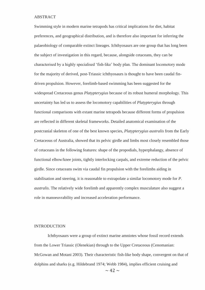
Figure 1: Cervical vertebrae of (A) bottlenose dolphin SAM M21243, (B) dugong SAM M847, (C) Australia fur seal SAM M15964, (D) leopard seal SAM M16638, and (E) Platypterygius australis QM F2453. Mammalian taxa are shown in lateral (left) and anterior (right) views. P. australis is shown in lateral view only. The fifth vertebra (C5) is shown for the extant mammalian taxa, while vertebrae 3–8 are shown for P. australis. The traditional vertebral regions (e.g. cervical thoracic) are difficult to define in the P. australis vertebral column, and are conventionally numbered according to their position in the vertebral column rather than their position in a given vertebral region. However, the vertebrae of extant mammalian taxa are conventionally numbered according to their position in a vertebral region. Thus, for the mammalian specimens used in this study, vertebral number in this and all proceeding figures is given as both the position within the vertebral column (to compare with P. australis) and in the more conventional method of position within a region of the vertebral column. Scale bar = 10 cm.

~ 46 ~
Figure 2: Thoracic vertebrate of (A) bottlenose dolphin SAM M21243, (B) dugong SAM M847, (C) Australian fur seal SAM M15964, (D) leopard seal SAM M16638, and (E) Platypterygius australis QM F2453. Mammalian taxa are shown in lateral (left) and anterior (right) views. P. australis is shown in lateral view only. The vertebrae shown are as follows (left to right): tenth (T3) and 15th (T8) for the dolphin; tenth (T3), 15th (T8), 20th (T13), and 25th (T18) for the dugong; tenth (T3), 15th (T8), and 20th (T13) for the otariid and phocid seal; and vertebrate 14–23 for P. australis. Scale bar = 10 cm.
~ 47 ~
marine taxa, including the secondarily aquatic cetaceans, dugongs, and manatees. Of these
three mammalian taxa, cetaceans are fast swimmers, while dugongs generally swim at slower
speeds.
Axial skeleton. Short cervical centra are common to all caudal propulsors (manatees,
Buchholtz et al. 2007; dolphins, Figure 1A and Buchholtz and Schur 2004; dugongs, Figure
1B), but is only obvious when compared to the height of the centrum (i.e. cervical centra are
disc-like in shape for caudal propulsors, but cylindrical in pectoral propulsors). This is
considered an adaptation to streamline the anterior torso (Hildebrand 1974) and reduce drag
(Alexander 1975). In addition, fusion of the atlas and axis, or the entire cervical region can
occur, limiting movement at the atlanto-axial joint (Osburn 1903). However, in extant taxa
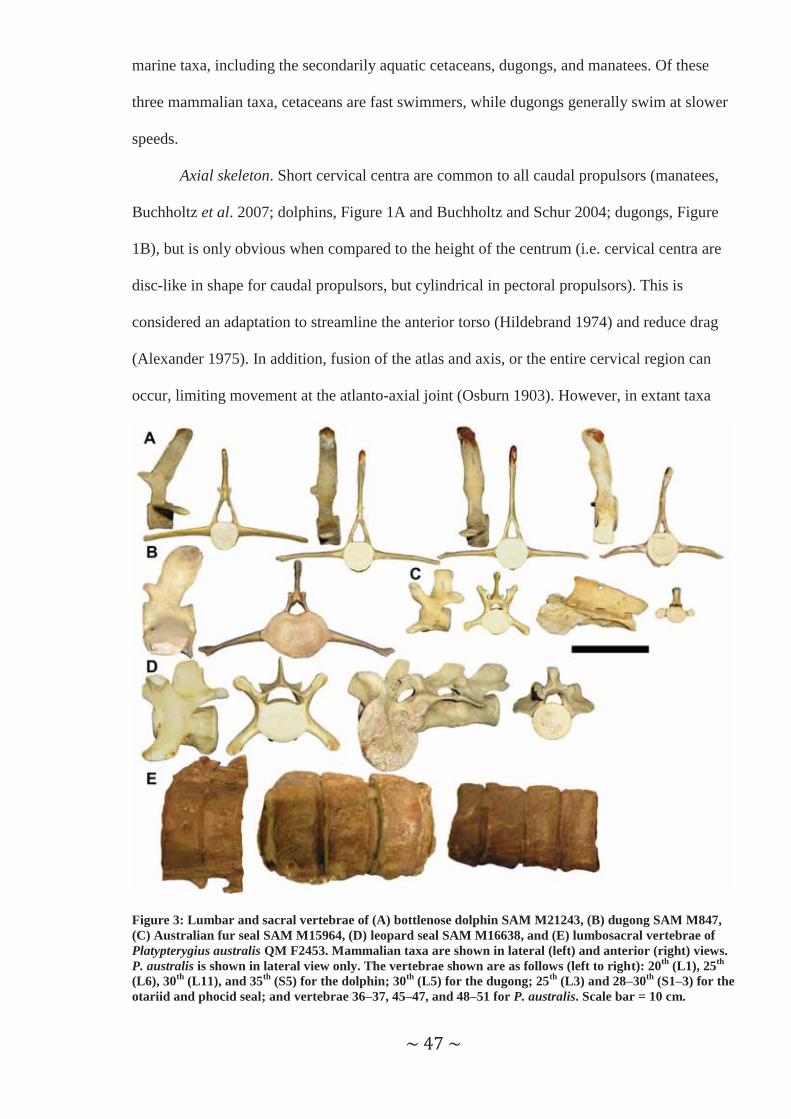
Figure 3: Lumbar and sacral vertebrae of (A) bottlenose dolphin SAM M21243, (B) dugong SAM M847, (C) Australian fur seal SAM M15964, (D) leopard seal SAM M16638, and (E) lumbosacral vertebrae of Platypterygius australis QM F2453. Mammalian taxa are shown in lateral (left) and anterior (right) views. P. australis is shown in lateral view only. The vertebrae shown are as follows (left to right): 20th (L1), 25th (L6), 30th (L11), and 35th (S5) for the dolphin; 30th (L5) for the dugong; 25th (L3) and 28–30th (S1–3) for the otariid and phocid seal; and vertebrae 36–37, 45–47, and 48–51 for P. australis. Scale bar = 10 cm.

~ 48 ~
this is restricted to fast caudal propulsors, as the manatee (Buchholtz et al. 2007) and dugong
do not have fused cervicals.
The thoracic vertebrae in caudally propulsive tetrapods also have restricted movement.
This is evidenced by the increasing height of the neural spine and the presence of rib facets
(Figure 2A-B), both correlated with decreasing flexibility through limiting rotation between
adjacent vertebrae (Buchholtz and Schur 2004). Mobility within the vertebral column is
generally greater posteriorly, and is often associated with dorsoventrally short, caudally-
inclined neural spines (Buchholtz and Schur 2004) (Figure 3A-B). The precise point at which
Figure 4: Caudal vertebrae of (A) bottlenose dolphin SAM M21243, (B) dugong SAM M847, (C) Australian fur seal SAM M15964, (D) leopard seal SAM M16638, and (E) lumbosacral vertebrae of Platypterygius australis KKM R519. Mammalian taxa are shown in lateral (left) and anterior (right) views. P. australis is shown in lateral view only. The vertebrae shown are as follows (left to right): 40th (Ca4), 45th (Ca9), 50th (Ca14), 55th (Ca19), and 60th (Ca24) for the dolphin; 35th (Ca3), 40th (Ca8), 45th (Ca13), and 50th (Ca18) for the dugong; and 35th (Ca5) and 40th (Ca10) for the otariid and phocid seal. The position of the vertebrae for P. australis is unknown as the anterior section of the skeleton is unpreserved. Scale bar = 10 cm.

~ 49 ~
flexibility increases is species-specific in the Cetacea and depends upon the length of
vertebral column used in propulsion – in Tursiops, this occurs at the beginning of the caudal
region (approximately at vertebra 40 in SAM M21243). The lack of discrete morphological
boundaries between the classically defined lumbar, sacral, and caudal vertebrae are also
typical in caudal propulsors. The centra in the caudal region correspond to the most mobile
part of the spinal column, with the neural spines either greatly reduced or absent, and the
centra dorsoventrally compressed (Figure 4A-B) – the latter feature allows for increased
dorsoventral versus lateral mobility (Hildebrand 1974), and is related to the dorsoventral axial
movement of the mammalian vertebral column.
Pectoral girdle and forelimb. Both caudal propulsors had ‘symmetrical’ scapulae –
except for this feature, scapular shape and morphology differed between the two caudal
propulsors examined (Figure 5). In the fast caudal propulsor (i.e. the dolphin, Tursiops
aduncus), the scapula was generally broader than long (Table 1) with concave and convex
surfaces, but did not bear a spinous process on its external surface. The scapula of the slow
caudal propulsor (i.e. the dugong, Dugong dugon), however, was longer than broad (Table 1)
with a spinous process on its external surface. A more complex surface texture (i.e. not
completely smooth) has been correlated with greater differentiation of musculature in the
Amazon River dolphin, Inia geoffrensis, and associated with the greater manoeuvrability
achieved by the extensive movements of the forelimb (Klima et al. 1980). The forelimbs of
Table 1: Length:width ratio of appendicular elements for each taxon. Length/width ratio of appendicular elements for each taxon Element Platypterygius Fast caudal
propulsor Slow caudal propulsor
Pectoral propulsor
Pelvic propulsor
Scapula Unknown 0.6-1.1 1.3-1.6 0.7-1.0 0.7-0.8 Humerus 1.3 1.7-2.1 1.9-2.8 2.6-2.9 1.8 Radius 0.7-1.2 2.5-3.5 5.9-6.9 4.9-5.8 4.8 Ulna 0.7-1.5 1.9-2.7 3.6-4.8 2.3-3.3 2.6-2.9 Metacarpals 1.1 1.2-1.8 3.5-4.2 2.7-3.2 3.7-4.0 Forelimb phalanges 0.6-0.9 0.5-1.7 1.7-2.4 3.2-3.8 3.7-4.7 Femur 1.9 Absent Absent 2.1-2.3 1.9 Tibia 1.0 Absent Absent 4.8-6.0 4.2 Fibula 1.3 Absent Absent 8.3-14.6 10.6 Metatarsals 0.8 Absent Absent 2.9-3.3 3.3-3.4 Hind limb phalanges 0.8 Absent Absent 3.9-4.7 4.0-4.5
~ 50 ~
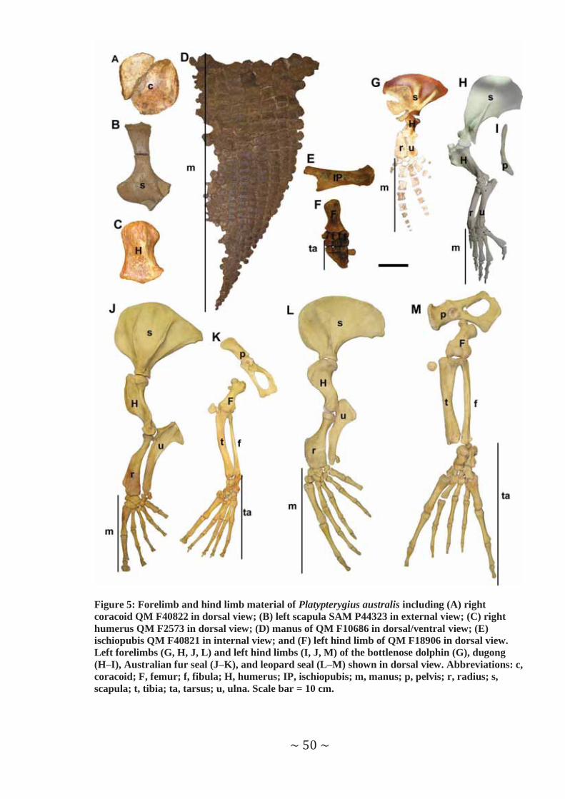
Figure 5: Forelimb and hind limb material of Platypterygius australis including (A) right coracoid QM F40822 in dorsal view; (B) left scapula SAM P44323 in external view; (C) right humerus QM F2573 in dorsal view; (D) manus of QM F10686 in dorsal/ventral view; (E) ischiopubis QM F40821 in internal view; and (F) left hind limb of QM F18906 in dorsal view. Left forelimbs (G, H, J, L) and left hind limbs (I, J, M) of the bottlenose dolphin (G), dugong (H–I), Australian fur seal (J–K), and leopard seal (L–M) shown in dorsal view. Abbreviations: c, coracoid; F, femur; f, fibula; H, humerus; IP, ischiopubis; m, manus; p, pelvis; r, radius; s, scapula; t, tibia; ta, tarsus; u, ulna. Scale bar = 10 cm.
~ 51 ~
the slow caudal propulsor may be capable of a wider range of motion than the fast caudal
propulsor, and this is supported by the range of functions served by the limbs in the dugong
(Brown 1878; Harrison and King 1975; Berta and Sumich 1999). Further, the scapula
appeared to have a smaller surface area in the slow caudal propulsor, despite the size ranges
of the two species only differing by approximately 20 cm (sensu van Dyck and Strahan 2008),
indicating that the fast caudal propulsor had a greater scapular area for muscle attachment.
The humerus also differed considerably between the two caudal propulsors (Figure 5).
Humeral morphology in the fast caudal propulsor reflected the flipper formed by the distal
section of the forelimb (Figure 5G). The proximal head of the dolphin humerus had a single
tubercle for muscle attachment and formed a ball-and-socket articulation with the scapula,
while the distal end was antero-posteriorly expanded into the plane of the flipper. Between the
two ends, the humeral shaft narrowed to exhibit a ‘waisted’ appearance. In contrast, the
humerus of the slow caudal propulsor was expanded proximally and tapered towards its distal
end (in lateral view, Figure 5H). Additional tuberosities were present on the proximal end of
the dugong humerus, a feature that has been correlated with increased muscle attachment to
improve slow manoeuvring in the Amazon River dolphin (Klima et al. 1980). Further, the
humerus of the slow caudal propulsor was longer (both in absolute length and relative to body
size) than the fast caudal propulsor, and more elongate. Shortening of the humerus has been
correlated with increased streamlining of the body (Hildebrand 1974), indicating that the fast
caudal propulsor was the more streamlined of the two taxa considered here.
The most obvious difference in the distal portion of the forelimb for these two taxa is
the presence of an elbow joint in the slow caudal propulsor (Hill 1945) while the joints distal
to the shoulder exhibit little or no movement in the fast caudal propulsor (Cooper et al.
2007b). This limb stiffness may be related to the use of the flipper as a hydrofoil in the
dolphin (Cooper and Dawson 2009), while the use of the forelimb in the dugong is more
varied, and includes feeding (Harrison and King 1975) and manoeuvring (Berta and Sumich
1999). However, movement of the radius and ulna is somewhat restricted in the dugong by

~ 52 ~
the fusion of the two elements at both the proximal and distal ends. The carpals are also fused
in the slow caudal propulsor, limiting movement in the wrist. In contrast, fusion in the distal
forelimb of the fast caudal propulsor is more varied (i.e. sometimes radius and ulna are fused,
sometimes the two elements fused with the humerus, and sometimes carpal elements fuse).
For both taxa, the radius and ulna are longer than broad, and carpal elements are broader than
long.
In the manus, digit morphology varies between the two taxa examined. All phalanges
are longer than broad, although the distal phalanges of the fast caudal propulsor are almost
circular. The fourth digit is the longest in the slow caudal propulsor, while the second is the
longest in the fast caudal propulsor – this might be associated with the formation of the flipper
in the latter taxon. Individual phalanges are longer in the slow caudal propulsor; however, the
fast caudal propulsor exhibits hyperphalangy (i.e. where the number of phalanges exceeds the
ancestral condition), and has the longer manus of the two taxa. Hyperphalangy has been
correlated with a steering and stabilising function in fast-swimming caudally propulsive
marine tetrapods (Cooper et al. 2007a; Cooper and Dawson 2009). Cetaceans have also been
known to exhibit hyperdactyly in rare cases as a digital anomaly (Cooper and Dawson 2009),
and a single modern cetacean, Phocoena sinus, is known to show non-anomalous
hyperdactyly within its populations (Ortega-Ortiz et al. 2000). However, the significance of
hyperdactyly for function is currently unclear.
Pelvic girdle and hind limb. Loss or reduction of the hind limb and corresponding
girdle in cetaceans is thought to have occurred after the evolution of caudal propulsion
(Thewissen et al. 2006). All extant mammalian caudal propulsors have no external hind limbs
and a greatly reduced pelvis. Indeed, one species of manatee, Trichechus inunguis, has lost all
elements of the pelvic girdle (Husar 1977). The ilia also lack any connection to the vertebral
column, which is reflected in the loss of an identifiable sacrum in these animals (Fig. 3A–B).
Summary. In summary, the important skeletal features correlated with caudal
propulsion include: (1) shortened cervical region; (2) dorsoventral compression for increased
~ 53 ~
dorsoventral flexibility in the most mobile vertebrae; (3) difference in neural spine height and
inclination posteriorly, associated with increased flexibility; (4) lack of discrete
morphological boundaries between regions of the vertebral column; (5) absence of a fused
sacrum; (6) reduction or loss of the pelvic girdle; and (7) loss of the hind limb. Several
skeletal features also appear to distinguish fast and slow caudal propulsors. Fast caudal
propulsors have a fused cervical region, which would limit movement at the atlanto-axial
joint. The propodials were also shorter in the fast caudal propulsor, perhaps to increase
streamlining, while the shape of the humerus was associated with the formation of the flipper.
In addition, the forelimb of the fast caudal propulsor had very little movement distal to the
shoulder joint – this is perhaps related to the flipper functioning as a single unit, and its use a
hydrofoil. In contrast, the slow caudal propulsor had an unfused cervical region and more
elongate propodials, possibly as a result of streamlining being less important at lower speeds.
The greater mobility in the forelimbs of the slow caudal propulsor reflects its multiple
functions.
Pectoral propulsors
Pectoral propulsion, where the forelimbs are used as the main swimming apparatus, is
present in a wide range of secondarily aquatic tetrapods. The most highly adapted extant
mammalian forms include otariid seals (fur seals and sea lions).
Axial skeleton. Cervical centra (Figure 1C) in the pectoral propulsor are cylindrical
(i.e. long relative to their height and width), and the vertebrae in this region are unfused. Both
of these characteristics indicate a flexible cervical region (Buchholtz and Schur 2004), and
likely relate to the use of the neck during terrestrial locomotion (English 1976b in Berta and
Sumich 1999) and in changing direction during swimming (Ray 1963). The neural spines
increase in height posteriorly through the cervical vertebrae (in contrast to the pelvic
propulsor), providing a greater area for attachment of the neck musculature (Berta and
Sumich 1999), and, again, likely relates to its use in terrestrial locomotion.
~ 54 ~
High neural spines are also present in the thoracic region – as the main muscular
power is concentrated at the anterior end (Harrison and King 1965) – specifically for
attachment of the multifidus lumborum and longissimus thoracics (Berta and Sumich 1999).
The length of the neural spines also increases posteriorly (Howell 1929), possibly decreasing
the flexibility present between adjacent vertebrae. Howell (1929) also noted that the
flexibility in the posterior region of the thorax in one pectoral propulsor was reduced by tight
interlocking of the zygapophyses. Further, the presence of ribs decreases movement between
adjacent vertebrae as Buchholtz and Schur (2004) found in caudal propulsors. Thus, the
thoracic region of the spinal column is less flexible that the cervical vertebrae.
Unlike in the caudal propulsors, the lumbar, sacral, and caudal regions are readily
distinguishable in the pectoral propulsor examined. The lumbar vertebrae share a similar
morphology to the posterior-most thoracic vertebrae, except for the presence of looser
articulations (Howell 1929), and the absence of rib facets. The centra in the lumbar region
have a cylindrical shape (length > width > height), a feature also present in both the sacral and
caudal regions. A fused sacrum consisting of three vertebrae is present (though the first
caudal was fused to the sacrum in one specimen examined), and the three centra show a
regular decrease in width (Howell 1929). The neural spine decreases in height throughout the
caudal region, and is virtually absent in the posterior-most caudal vertebrae. This morphology
indicates that the immobile, fused sacrum separates two relatively flexible regions – the
lumbar region anteriorly, and the caudal region posteriorly.
Pectoral girdle and forelimb. The scapula in the pectoral propulsor examined was
broader than long (Table 1), and was longer than the equivalent element in the caudal
propulsors (Figure 5). This provides a much greater surface area for muscle attachment
(Howell 1929) compared with non-pectoral propulsive tetrapods. In addition, the fan-shape of
the scapula (Figure 5J) is also associated with muscle attachments (English 1977). The
scapula also bears two prominent ridges which, in addition to the enlargement of the
supraspinous fossa, are correlated to the strong development of the supraspinatus muscle
~ 55 ~
(Berta and Sumich 1999). Further support for the strong development of the supraspinatus
includes the greater area for the attachment of this muscle, giving the scapula an asymmetric
appearance. This is perhaps unsurprising, as the supraspinatus assists in movement of the
humerus (Howell 1929).
In comparison to the caudal propulsors, the humerus of the pectoral propulsor is both
longer and more elongate (Figure 5, Table 1) – however, the element is described as short and
massive (Howell 1929; English 1977), thus exhibiting characteristics Hildebrand (1974)
associated with increased streamlining. Additionally, the otariid humerus is more robust with
more surfaces for muscle attachment than the fast caudal propulsor. This is demonstrated by
the two enlarged tubercles present adjacent to the proximal head, while the well-developed
deltoid crest on the humeral shaft also provides a greater surface area for muscle attachment.
The former feature is also associated with increasing the moment arm of the rotator cuff
musculature (Berta and Sumich 1999), thus increasing the power and manoeuvrability in the
otariid seal. Howell (1929) also interpreted the extension of the humerus as being more
powerful in pectoral propulsors relative to pelvic propulsors among the Pinnipedia. The
proximal articular surface of the humerus forms a ball-and-socket articulation at the shoulder
joint, and this articular surface is quite extensive, providing a large surface area for increased
ranges of movement (English 1977).
Like the humerus, the radius and ulna of the pectoral propulsor have been shortened,
and thus exhibit adaptations to streamlining (English 1977) – however, both elements are also
longer and more elongate than the comparable bones in the fast caudal propulsor (Figure 5,
Table 1). The epipodial elements do not exhibit any fusion, in contrast to the caudal
propulsors, perhaps reflecting the employment of a rowing swim stroke (Feldkamp 1987) or
the use of the forelimb in both aquatic propulsion and terrestrial locomotion (English 1976b).
English (1977) described the elbow joint as functioning as a modified hinge joint – this is in
stark contrast to the absence of a functional elbow joint in the fast caudal propulsor. However,
most movement seems to occur at the glenohumeral joint with the elbow possibly being used

~ 56 ~
for control (English 1977). The radius and ulna, along with the more distal forelimb elements,
exhibit flattening associated with the formation of the flipper (English 1977); in fact, Howell
(1929) considered the broadness of the bones distally and proximally, respectively, to be
unique to the Pinnipedia.
The remainder of the pectoral propulsor forelimb has more in common with the pelvic
propulsor than the caudal propulsors (Figure 5). None of the wrist bones exhibit fusion (in
contrast to the caudal propulsors), and the differences in morphology between the carpal
elements of the two types of limb propulsors has been attributed to the position of the pectoral
propulsor’s forelimb during terrestrial locomotion (Howell 1929). Like many of the
mammalian taxa examined in this study, the otariid seal exhibits flattening and elongation of
the digits (Figures 5J–K, Table 1), perhaps related to the formation of the flipper and/or to
increase the surface area of the distal forelimb – however, in the pectoral propulsor, the first
digit is the longest, with the digits progressively decreasing in size anterior to posterior.
Pelvic girdle and hind limb. In contrast to caudal propulsors, pectoral propulsors do
not show any reduction in the pelvic girdle, perhaps resulting from the use of the hind limbs
in terrestrial locomotion. All modern pectoral propulsors return to land, and therefore none
are purely aquatic. An obligate aquatic animal that uses only its forelimbs for propulsion and
does not require its hind limbs for terrestrial locomotion might exhibit reduction of the pelvis
– however, this hypothesis cannot be examined using modern taxa as no obligate aquatic,
pectoral propulsor is currently known. The pectoral propulsor exhibits a lengthening of the
ischium and pubis, and migration of muscle attachments distally to increase leverage – the
importance of the former feature is unclear (Howell 1929).
The femur in the pectoral propulsor is a flattened bone with expanded proximal and
distal ends (Figure 5K). As with the cetacean humerus, the femoral shaft has a ‘waisted’
appearance. Two trochanters are present (the greater adjacent to the ball-and-socket
articulation to the pelvic girdle, and the lesser on the lateral surface on the femoral shaft),
allowing muscles to insert separately on the femur (Howell 1929). Separate insertions may
~ 57 ~
allow for more manoeuvrability in the hind limb of the pectoral propulsor, as reported in the
forelimb for the Amazon River dolphin (Klima et al. 1980).
Both the tibia and fibula are elongate elements in the pectoral propulsor (Figure 5K,
Table 1). Fusion of these two bones occurs at the proximal end of these elements, and the
fused head of these elements is sharply angled to allow greater flexibility of the knee (Howell
1929). This may reflect a steering or manoeuvring function for the hind limb in aquatic
locomotion, and/or its use in terrestrial movement. The morphology of the astragalus,
however, indicates restricted movement relative to the surrounding elements when compared
to that of the pelvic propulsor (Howell 1929) – the reason for this remains unclear. Like the
forelimb, the metatarsals and phalanges are elongate and flattened, and probably for similar
reasons – the formation of the flipper and to increase the surface area of the hind limb.
However, while the first digit is clearly the most robust and well-developed, and the first
metatarsal is the longest of the five, none of the digits appear to be longer than the others.
Summary. Several important features appear to correlate with pectoral propulsion.
These include: (1) a longer cervical region than found in caudal propulsors; (2) large surface
areas for muscle attachment on the pectoral girdle and forelimb elements; (3) elongate
epipodial elements; and (4) flattened and elongate digits. Further, the rowing style employed
by the pectoral propulsor examined may require flexibility at the elbow joints and in the wrist
– however, the forelimb is also used extensively in terrestrial locomotion, and the flexibility
observed in these joints may also reflect this function of the limb.
Pelvic propulsors
Only a single group of extant, secondarily aquatic tetrapods rely upon their pelvic
limbs for propulsion – phocid seals (true seals). Usually, pelvic propulsors generate thrust
through alternating strokes of the left and right hind limbs (Alexander 1975) assisted by
lateral movements of the lumbar and caudal vertebrae (King 1989). Additionally, the hind
limbs can be moved as a single unit (Ray 1963) via lateral sweeps of the vertebral column

~ 58 ~
(Alexander 1975). In contrast to the pectoral propulsors, the forelimbs have a reduced role in
both aquatic and terrestrial locomotion – in aquatic locomotion the pectoral limbs are
generally folded against the body, but may be used for an initial acceleration (Ray 1963) or
for stabilisation and manoeuvring (Feldkamp 1987).
Axial skeleton. The cervical vertebrae in the pelvic propulsor exhibit an anatomy
intermediate between the pectoral and caudal propulsors. Like the pectoral propulsors, the
vertebrae of this region are unfused and cylindrical in shape, suggesting that this region was
capable of greater flexibility than in the caudal propulsors. This is perhaps to be expected as
the neck is used to change direction, and assumes an S-shape to achieve a more streamlined
body (Ray 1963). However, the cervical centra are shorter in the pelvic propulsor compared to
the pectoral propulsor (Howell 1929), and are wider than they are long or high – in the caudal
propulsors, shorter centra are associated with a more streamlined, torpedo-shaped body, and
this may be the reason for shortened centra in the pelvic propulsor. Additionally, there is
virtually no neural spine present in the vertebrae in the cervical region (Howell 1929) – in
contrast to the otariids, but similar to the caudal propulsors (Figure 1) – this is likely a result
of the neck not being used in terrestrial locomotion as in the otariid seals. Thus, the cervical
region of the pelvic propulsor exhibits several features associated with a flexible neck, while
others are correlated with the requirement to improve streamlining, as observed in the pectoral
and caudal propulsors respectively.
Like the majority of the cervical centra, the thoracic centra (all but the most posterior)
are wider than they are long or high, indicating a decreased flexibility compared with the
lumbar region. In addition, the presence of ribs (as with caudal propulsors: Buchholtz and
Schur 2004) decrease the mobility in this region relative to the cervical vertebrae. The
reduced neural spines (Figure 2D) have been noted by several authors (e.g. Howell 1929) –
this likely results from the reduced musculature, relative to the pectoral propulsor, required
anteriorly (Harrison and King 1965). Conversely, the elongate transverse processes of the
lumbar region are for the attachment of the vertebral muscle blocks involved in the lateral
~ 59 ~
movements of the hind body, reflecting the importance the location of the main muscular
power in the posterior part of the body and thus the mode of swimming (Harrison and King
1965; King 1989; Berta and Sumich 1999). In addition, the lumbar vertebrae are also
characterised by the loose articulations that allow great flexibility (Harrison and King 1965),
and the zygapophyseal shape in the last lumbar and first sacral that allow the sacrum to be
raised above the vertebral axis (Howell 1929) – the latter feature was not observed by Howell
(1929) in the pectoral propulsor. Otherwise, the morphology of the lumbar region are similar
to that of the posterior thoracic vertebrae (Howell 1929), including a centrum length that is
greater than both width and height (as observed in the last thoracic centra).
As in the pectoral propulsor, but unlike the caudal propulsors, a fused sacrum and
clearly defined vertebral regions are present in the pelvic propulsor (Figure 3). However,
unlike the pectoral propulsor, the width of the first sacral is considerably greater than the
remaining two centra (Howell 1929), demonstrated by the width being the greatest dimension
in the first sacral centrum (unlike in the pectoral propulsor) while length was the largest
measurement in the remaining centra (as observed in the pectoral propulsor). Similarly, the
caudal vertebrae are cylindrical with low neural spines, indicating a greater flexibility than the
preceding sacral region, though the centra appear to be more elongate than the corresponding
centra in the pectoral propulsor (Figure 4).
Pectoral girdle and forelimb. The scapula in the pelvic propulsor examined was
almost oval in shape (Figure 5L), in contrast to the fan- or sickle-shape Howell (1929)
observed in an otariid and phocid, respectively. As with the pectoral propulsor, but unlike the
caudal propulsors, the scapula is asymmetric in appearance – however, in the phocid
propulsor, this results from a greater area for muscle attachment caudad to the spinous
process, and thus a less developed supraspinatus musculature. Only a single spine can be
observed on the external surface (like the slow caudal propulsor), compared with two in the
pectoral propulsor and none in the fast caudal propulsor, and the scapula is otherwise
relatively smooth. This indicates a smaller area for muscle attachment compared to the former
~ 60 ~
(King 1989), but a greater area of muscle attachment relative to the latter. There is also a
tendency for this spine to be reduced in the phocids relative to their otariid counterparts
(Harrison and King 1965). As with the pectoral and fast caudal propulsors, but unlike the
slow caudal propulsor, the scapula is broader than it is long (Table 1) – however, the scapula
is reduced relative to the total size of the animal compared to the pectoral propulsors.
As suggested by Harrison and King (1965), the general humeral morphology (as well
as the rest of the forelimb) of the pelvic propulsor is similar to that of the pectoral propulsor.
The humerus has a deltoid crest (though this is less robust than in the otariid seal) and two
tubercles, which, as discussed for the pectoral propulsor, are associated with greater areas for
muscle attachment and increased power and manoeuvrability (Berta and Sumich 1999). A
ball-and-socket articulation is present at the glenohumeral joint, indicating a similar flexibility
was present at the shoulder as in the pectoral propulsor. This element is also longer than it is
wide and described as short (Howell 1929; King 1989) with the latter feature associated with
increased streamlining – however, the humerus is not as elongate as that of the pectoral
propulsor, exhibiting a length:width ratio more comparable with the caudal propulsors (Table
1). An additional difference to the otariid seal includes the position of the greater tuberosity
no higher than the humeral head, related to the different movement requirements of the
forelimb compared to pectoral propulsors (Howell 1929). Thus, the musculature of the
forelimb does not appear to be as developed as in the pectoral propulsor, but the amount of
flexibility is similar – perhaps the flexibility is required to fold the forelimbs alongside the
body during aquatic locomotion, while the musculature is less important as the primary
propulsive power is at the posterior part of the body.
The radius and ulna (Figure 5L) of the pelvic propulsor are also similar in overall
morphology to the pectoral propulsor (Harrison and King 1965). Both elements are shortened,
flattened distally (radius) or proximally (ulna), and longer than broad (Osburn 1903; Howell
1929; Harrison and King 1965; King 1989) (Table 1). A functional elbow joint is present, and
fusion is absent between these elements – the forelimb of the pelvic propulsor has not been as

~ 61 ~
extensively studied as the pectoral propulsor, but it may also have functioned as a modified
hinge joint as the morphology does not differ considerably between the two propulsors.
However, the radial styloid process of English (1977) appears to extend further proximally
and the proximal end of the ulna does not appear to be as broad in the pelvic propulsor. The
reasons for this are unclear, but the latter perhaps relates to the greater muscle attachments
required in the pectoral propulsor.
As outlined above, the pelvic and pectoral propulsors showed the greatest similarity in
the morphology of the manus. These similarities included the absence of fusion in the carpal
elements, lengthening (Table 1) and flattening of the digits, and the first digit being the
longest with the remaining digits decreasing in size anterior to posterior. The decrease in size
of the digits, however, appears to be more pronounced in the pectoral propulsor (Howell
1929). One difference in manus morphology includes the crowding of the carpal elements on
the ulnar side of the limb (Harrison and King 1965), attributed to the position in which the
phocid seal holds its forelimb (Harrison and King 1965).
Pelvic girdle and hind limb. Unsurprisingly, the pelvic propulsors show no reduction
of the pelvic girdle – instead, the innominate bone in this type of propulsor shows several
adaptations that highlight the importance of the pelvic girdle in locomotion. The ilium shows
the lateral eversion characteristic of the phocines (Harrison and King 1965; King 1983 in
Berta and Sumich 1999). This morphology provides a much greater attachment area for the
iliocostalis lumboram muscle, which flexes the lateral part of the body during swimming, and
to increase the area for the attachment of muscles that insert on the femur (m. gluteus medius,
minimis, and pyriformis), which serve as the lateral abductors of the hip (Berta and Sumich
1999). The iliac wing of the innominate bone is deflected outwards, providing attachment
points for the iliocostalis muscles (Smith and Savage 1956). Both the ischium and pubis are
long relative to the ischium to increase the lever arm of the muscles used in the adductional
movements while swimming (Howell 1929). As a result of the morphology of the last lumbar
~ 62 ~
and first sacral vertebrae, the ischium can be elevated to the level of the backbone (Howell
1929), lengthening the ischiopubic muscles (Smith and Savage 1956).
Overall, the femur of the pelvic propulsor is very similar to that of the pectoral
propulsor – it is a shortened, flat element (Harrison and King 1965) with a ‘waisted’ shaft,
and bears a greater trochanter lateral to the femoral head. Howell (1929) described this
trochanter to be wedge-shaped, with a deep fossa resulting from complete fusion of the
tendons inserting at this point. In addition, the lesser trochanter is absent in the pelvic
propulsor, correlated with a decrease in the attachment area of muscles that rotate the femur
posteriorly (e.g. mm. iliacus and psoas major) – instead, these muscle insert either onto the
iliac wing or distal to the medial femur, increasing the flexibility of the leg and lateral
undulation of the lumbosacral region of the vertebral column (Berta and Sumich 1999).
Further, the femur of the pelvic propulsor is ‘stockier’ and less elongate than that of the
pectoral propulsor (Table 1), particularly at this distal end. This could be related to the greater
development of the gluteus maximus, gastrocneumius medialis, and plantaris Howell (1929)
noted for the phocid seals.
The remaining elements of the pelvic limb show many similarities to the pectoral
propulsor. Both the tibia and fibula of the pelvic propulsors are elongate elements (Table 1),
and are fused into a single head at the proximal end. However, the fused head of the two
elements is approximately level, compared with the sharp angle observed in the pectoral
propulsor, indicating that less flexibility was present in the knee joint compared to the otariid
seal (Howell 1929). As stated previously, the morphology of the astragalus indicates that a
greater range of movement relative to the surrounding elements was present in the pelvic
propulsor (Howell 1929). The metatarsals and phalanges are elongate and flattened (Table 1),
associated with the formation of the flipper and to increase the surface area of the hind limb
(Hildebrand 1974) – however, while the first metatarsal is the more robust, the first and fifth
digits are both longer than the middle three. Howell (1929) described the metatarsals of the

~ 63 ~
phocid seal he examined as being crowded and thus having restricted movement – this may
also be true of the pelvic propulsors examined in this study.
Summary. Pelvic propulsion appears to correlate with several morphological features.
This includes: (1) a cervical region intermediate in length between caudal and pectoral
propulsors; (2) large surface areas for muscle attachment on the pelvic girdle; and (3)
flattened and elongate digits. In addition, the lumbar region of the pelvic propulsor appears to
be quite flexible, but this may result from the lumbar part of the body assisting with
propulsion rather than pelvic propulsion per se.
DISCUSSION
Comparisons with Platypterygius australis
The vertebral centra of Platypterygius australis are deeply amphicoelous, a feature
typical of caudally propulsive ichthyosaurs (Buchholtz 2001a). Fusion of the atlas and axis
occurs in both P. australis and caudally propulsive ichthyosaurs (McGowan and Motani
2003?), and was only observed in the fast caudal propulsor examined in this study. Disc-like
centra are present throughout the vertebral column of this ichthyosaur, and are more similar in
appearance to the shortened cervical centra of the caudal propulsors rather than the cylindrical
centra of the pectoral and pelvic propulsors. The latter two features have been correlated with
shortening of the cervical region, which, in turn, is an adaptation to increase streamlining
(Hildebrand 1974). There is a pronounced lack of sharply defined regions along the vertebral
column (a feature shared with both the fast and slow caudal propulsors but not the pectoral or
pelvic propulsor, pers. obs.) – this may be due to the absence of a fused sacrum and
corresponding disconnection of the pelvis from the vertebral column. The occurrence of
definitive tail fluke centra demonstrates the presence of a caudal fin, and the lateral
compression of these elements facilitates lateral movement of the vertebral column (similar to
caudal propulsors). However, the length of the tail region cannot be estimated as only a few

~ 64 ~
centra are preserved (contrary to McGowan’ s1972 interpretation of a small caudal fin). The
neural arches are also incompletely preserved (associated only with the anterior-most centra),
preventing examination of changes in neural spine morphology throughout the vertebral
column. Dorsal facets for articulation with an unpreserved neural arch were observed for all
centra examined in the vertebral column.
Scapular morphology varied widely among the different extant taxa (Figure 5), and
more investigations of the functional significance is required before conclusions can be drawn
on how these differences relate to propulsive method. However, what can be observed is that
animals using their forelimbs as a hydrofoil have a less complex (i.e., smoother) surface
texture (as well as fewer areas for insertion on their humerus). Further, relative areas for
muscle attachment appear to vary between the caudal, pectoral, and pelvic propulsors, but
mycological studies are required for Tursiops and Dugong to confirm these observations.
How these observations relate to ichthyosaurs is also unclear, for several reasons: (1) the
ichthyosaur shoulder girdle is composed of four bones rather than the single element observed
in all extant mammalian taxa examined; (2) a complex terminology for reptilian limb
musculature exists, with several names being used for the same muscle (Romer 1956); and (3)
the difficulties in inferring muscle origins and insertions (and thus function) in extinct species.
Thus, the functional significance of the shoulder girdle morphology in P. australis remains
unresolved.
Reduction of the pelvis in the caudal propulsors is considered an adaptation to
increase streamlining (Hildebrand 1974). Only the ischium and pubis are fused in P. australis,
and reflect a decreasing role of the hind limb in locomotion (vis-à-vis caudal propulsors). The
absence of the obturator foramen implies that the obturator nerve (Romer 1956) and blood
vessels passed around rather than through the pubis. Further, the muscles typically associated
with this foramen, such as the puboischiofemoralis (Romer 1956) and rotators of the hind
limb, must have been similarly reduced or absent.
~ 65 ~
The humerus and femur of P. australis have a very similar morphology (Zammit et al.
2010), and will therefore be discussed together – this is in contrast to the pectoral and pelvic
propulsors, where the humeral and femoral morphology differ, and the caudal propulsors,
where the femur and hind limb are absent. Both propodials are short and broad, showing a
general morphology more similar to the stout cetacean humerus than the elongate morphology
of the dugong, otariid, and phocid humeri. The proximal head of the humerus and femur is
characterised by prominent dorsal and ventral trochanters (termed dorsal trochanter and
deltopectoral crest respectively in the humerus, Motani 1999b). This is indicative of strong
attachments for muscles originating on the limb girdles (e.g. m. latissimus dorsi,
subscapulocoracoideus, scapulohumeralis anterior, deltoid, pectoralis and supacoracoideus;
and m. pubioischiofemoralis externus and adductor femoris respectively), and were probably
used to achieve joint stability as well as movement for the limb (Romer 1956). McGowan
(1972) correlated this with some degree of forelimb propulsion, perhaps as a compensator for
the hypothesised low aspect ratio small caudal fin (McGowan 1972). However, increased
attachment surfaces for musculature do not necessarily impart better propulsive ability, and
indeed robust humeral morphology might have aided in manoeuvring, especially at low
speeds (see Klima et al. 1980 for observations of the Amazon River dolphin). It can also be
linked to increased stability (Moore et al. 2010).
Both propodials of P. australis have a ‘waisted’ shaft that expands into distal ends that
follow the same plane as the remainder of the limb (Figure 5C and F). Narrowing of the shaft
distal to the proximal head (in dorso-ventral view) is not observed in the humeri of the slow
caudal, pectoral, and pelvic propulsors – this is perhaps related to the formation of the deltaic
crest in these extant taxa, a feature which has no equivalent in P. australis or the fast caudal
propulsor. The humerus of the fast caudal propulsor (Figure 5G), and the femora of the
pectoral and pelvic propulsors (Figure 5K and M, respectively) all exhibit a ‘waisted’ shaft.
However, the femora of the pectoral and pelvic propulsors exhibit a slightly different
morphology to that of P. australis – the femur of the former has trochanter on its femoral

~ 66 ~
shaft to increase the area for muscle attachment, while the femur of both taxa do not show the
same gradual distal expansion observed in both P. australis and the fast caudal propulsor. The
distal expansion of the propodial occurs in all taxa examined (but perhaps to a lesser extent in
the slow caudal, pectoral, and pelvic propulsors), and is associated with the formation of the
paddle. Of all extant taxa examined, the cetacean humerus shows the greatest similarity to the
morphology observed in P. australis.
The fore- and hind limb of P. australis exhibit the following features: tightly
interlocking elements that do not have functional elbow or knee joints; pronounced
hyperphalangy; and hyperdactyly that is amongst the most extreme of all ichthyosaurs.
Comparable immobile joints distal to the humerus have been noted in cetaceans (Cooper et al.
2007b), where they serve to stiffen the limb as it moves as a unit (Hildebrand 1974), but the
remaining extant taxa examined in this study did not exhibit this morphology. However, some
pectoral propulsors (e.g. penguins) also exhibit relatively stiff forelimbs, indicating that
immobile limbs are not correlated with a particular method of propulsion. Instead, it may
affect the potential of the limb to function as a lift-generating structure (Fish 2004; Cooper et
al. 2007b).
Hyperphalangy has been linked with increasing the rigidity in the distal parts of the
limbs (Hildebrand 1975). Cetaceans are the only modern mammals to exhibit hyperphalangy,
where it has been related to steering and balance (Richardson and Chipman 2003). The fast
caudal propulsor was the only extant taxa examined in this study to show hyperphalangy – the
slow caudal, pectoral, and pelvic propulsors did not have additional phalanges beyond the
ancestral number. The presence of hyperphalangy in plesiosaurs, a group of extinct marine
reptiles inferred to use pectoral and/or pelvic propulsion, suggests that this morphology may
not correlate directly with method of propulsion, and supports the proposal of Cooper et al.
(2007b) that it may instead be indicative of the use of the limb as a hydrofoil to generate lift.
The hyperdactyly observed in Platypterygius is the most extreme of all ichthyosaurs,
having at least seven digits (McGowan and Motani 2003). Hyperdactyly was not observed in

~ 67 ~
any of the extant taxa examined in this study – however, individuals of some cetacean species
have been reported as exhibiting this feature (Watson et al. 2008; Cooper and Dawson 2009),
and hyperdactyly is consistently expressed in a single porpoise species (e.g. Phocoena sinus:
Ortega-Ortiz et al. 2000). Thus, hyperdactyly cannot be considered evidence of a particular
method of locomotion. Instead, it may be correlated with increasing the size, particularly the
width of the limb (Hildebrand 1974), and as such, it could have improved acceleration
performance (Webb 1977) and/or function as a hydroplane.
The postcranial anatomy of P. australis shows several features that can be used to
elucidate their propulsive method. Three morphological characteristics were common to P.
australis and both caudal propulsors – shortened neck, compression of the caudal centra, and
complete fusion of the ischiopubis. An additional feature, fusion of the atlas-axis complex,
was observed only in P. australis and the fast caudal propulsor. Further, the limbs exhibited
features correlated with a lift-generating function – propodial morphology, hyperphalangy,
hyperdactyly, and tightly interlocking phalanges. In contrast, very few features were shared
between P. australis and the pectoral or pelvic propulsors, and some characters, such as the
short, broad phalanges, were in direct contrast to the morphology observed in these extant
taxa.
CONCLUSIONS
P. australis has many postcranial features in common with caudal propulsors. Further,
some of these features, such as the shortened neck, are in direct contrast to the morphology
observed in pectoral or pelvic propulsors. Thus, P. australis is inferred to use caudal
propulsion as the predominant method of locomotion. The convergence in limb anatomy
between P. australis and the cetaceans was particularly striking, specifically in the stiffness of
the limbs and hyperphalangy, and indicates a similar function – that of a hydrofoil used for
steering and balance. However, the additional areas for muscle attachment, which are also

~ 68 ~
observed in Inia in comparison to marine dolphins (Klima et al. 1980), indicate that use of the
fore (and perhaps hind) limbs for low speed propulsion and manoeuvring is feasible (Wade
1984). P. australis might have had a decoupled locomotor system – where the caudal fin was
used for long-distance cruising and the limbs for acceleration and movement – as some fish
(Webb and Keyes 1981; Blake 2004) and aquatic mammals (e.g. Inia, Klima et al. 1980) do
today. The increased surface area of the limbs, particularly the distal forelimb, would have
improved acceleration (sensu Hildebrand 1974).
ACKNOWLEDGEMENTS
Many thanks to the staff at various museum for arranging access to specimens: Robert Jones
(AM); Paul Stumkat (KKM); Scott Hocknull, Patrick Couper, and Heather Janetzki (QM);
and Ben McHenry, Mary-Anne Binnie, David Stemmer, Catherine Kemper, and Phillippa
Horton (SAM).
REFERENCES
Alexander, R. M. 1975. The Chordates. Cambridge University Press, London, UK, 480pp.
Berta, A., and Sumich, J. L. 1999. Marine Mammals: Evolutionary Biology. Academic Press,
San Diego, California.
Blake, R. W. 2004. Fish functional design and swimming performance. Journal of Fish
Biology, 65:1193–1222.
Buchholtz, E. A. 2001a. Swimming styles in Jurassic ichthyosaurs. Journal of Vertebrate
Paleontology, 21:61–73.
Buchholtz, E. A. 2001b. Vertebral osteology and swimming style in living and fossil whales
(Order: Cetacea). Journal of Zoology, London, 253:175–190.
~ 69 ~
Buchholtz, E. A., and Schur, S. A. 2004. Vertebral osteology in Delphinidae (Cetacea).
Zoological Journal of the Linnean Society, 140:383–401.
Buchholtz, E. A., and Stepien, C. C. 2009. Anatomical transformation in mammals:
developmental origin of aberrant cervical anatomy in tree sloth. Evolution and
Development, 11:69–79.
Buchholtz, E. A., Booth, A. C., and Webbink, K. E. 2007. Vertebral anatomy in the Florida
manatee, Trichechus manatus latirostris: a developmental and evolutionary analysis.
The Anatomical Record, 290:624–637.
Buchholtz, E. A., Wolkovich, E. M., and Cleary, R. J. 2005. Vertebral osteology and
complexity in Lagenorhynchus acutus (Delphinidae) with comparison to other
delphinoid genera. Marine Mammal Science, 21:411–428.
Cooper, L. N., and Dawson, S. D. 2009. The trouble with flippers: a report on the prevalence
of digital anomalies in Cetacea. Zoological Journal of the Linnean Society, 155:722–
735.
Cooper, L. N., Berta, A., Dawson, S. D., and Reidenberg, J. S., 2007a. Evolution of
hyperphalangy and digit reduction in the cetacean manus. The Anatomical Record,
290:654–672.
Cooper, L. N., Dawson, S. D., Reidenberg, J. S., and Berta, A. 2007b. Neuromuscular
anatomy and evolution of the cetacean forelimb. The Anatomical Record, 290:1121–
1137.
English, A. W. M. 1976a. Functional anatomy of the hands of fur seals and sea lions.
American Journal of Anatomy, 147:1–18.
English, A. W. M. 1976b. Limb movements and locomotor function in the California sea lion
(Zalophus californianus). Journal of the Zoological Society, London, 178:341–364.
English, A. W. M. 1977. Structural correlates of forelimb function in fur seals and sea lions.
Journal of Morphology, 151:325–352.

~ 70 ~
Feldkamp, S. D. 1987. Foreflipper propulsion in the California sea lion, Zalophus
californianus. Journal of Zoology, London, 212:43–57.
Fish, F. E. 2004. Structure and mechanics of nonpiscine control surfaces. IEEE Journal of
Oceanic Engineering, 29:605–621.
Harrison, R. J., and King, J. E. 1965. Marine Mammals. Hutchinson and Co. Limited,
London. 192 pp.
Hildebrand, M. 1974. Analysis of Vertebrate Structure. John Wiley & Sons, New York. 710
pp.
Howell, A. B. 1929. Contribution to the comparative anatomy of the eared and earless seals
(genera Zalophus and Phoca). Proceedings of the United States National Museum,
73:1–142.
von Huene, F. 1923. Lines of phyletic and biological development of the Ichthyopterygia.
Bulletin of the Geological Society of America, 34:463–468.
Husar, S. L. 1977. Trichechus inunguis. Mammalian Species, 72:1–4.
King, J. E. 1983. Seals of the World. Oxford University Press, London.
King, J. E. 1989. Otariidae and Phocidae. Fauna of Australia (eds D.W. Walton and B.J.
Richardson). Australian Government Publishing. 35 pp.
Klima, M., Oelschläger, H. A., and Wünsch, D. 1980. Morphology of the pectoral girdle in
the Amazon dolphin Inia geoffrensis with special reference to the shoulder joint and
movements of the flippers. Zeitschrift fur Säugetierkunde, 45:288–309.
Maisch, M. W., and Matzke, A. T. 2000. The Ichthyosauria. Stuttgarter Beiträge zur
Naturkunde Serie B (Geologie und Paläontologie), 298:1–159.
Massare, J. A. 1988. Swimming capabilities of Mesozoic marine reptiles: implications for
method of predation. Paleobiology, 14:187–205.
Massare, J. A. 1994. Swimming capabilities of Mesozoic marine reptiles: a review. Pp. 133–
149 in Mechanics and Physiology of Animal Swimming (eds Maddock, L., Bone, Q.,
and Rayner, J.) Cambridge University Press, New York.

~ 71 ~
Massare, J. A., and Faulkner, J. 1997. Sharks as morphological analogs for ichthyosaurs.
Paludicola, 1:117–125.
Massare, J. A., and Sharkey, S. J. 2003. Centrum shape in sharks: not a good analog for
ichthyosaurs. Paludicola, 4:27–36.
Maxwell, E. E., and Kear, B. P. 2010. Postcranial anatomy of Platypterygius americanus
(Reptilia: Ichthyosauria) from the Cretaceous of Wyoming. Journal of Vertebrate
Paleontology, 30:1059–1068.
McGowan, C. 1972. Evolutionary trends in longipinnate ichthyosaurs with particular
reference to the skull and fore fin. Life Sciences Contributions, Royal Ontario Museum,
83:1–38.
McGowan, C. 1992. The ichthyosaurian tail: sharks do not provide an appropriate analogue.
Palaeontology, 35:555–570.
McGowan, C., and R. Motani. 2003. Ichthyopterygia; in H.-D. Sues (ed.), Handbook of
Paleoherpetology, Volume 8. Verlag Dr. Friedrich Pfeil, München, 175 pp.
Moore, K. L., Dalley, A. F., and Agur, A. M. R. 2010. Clinically Oriented Anatomy.
Lippincott Williams and Wilkins, Philadelphia, USA. 1134 pp.
Motani, R. 1999a. Phylogeny of the Ichthyopterygia. Journal of Vertebrate Paleontology,
19:473–496.
Motani, R. 1999b. On the evolution and homologies of ichthyopterygian forefins. Journal of
Vertebrate Paleontology, 19:28–41.
Motani, R. 2002. Swimming speed estimation of extinct marine reptiles: energetic approach
revisited. Paleobiology, 28:251–262.
Ortega-Ortiz, J. G., Villa-Ramírez, B., and Gersenowies, J. R. 2000. Polydactyly and other
features of the manus of the vaquita, Phocoena sinus. Marine Mammal Science,
16:277–286.
Osburn, R. C. 1903. Adaptation to aquatic, arboreal, fossorial and cursorial habits in
mammals. I. Aquatic adaptations. American Naturalist, 37:651–665.
~ 72 ~
Ray, C. 1963. Locomotion in pinnipeds. Natural History, 72:10–21.
Renjun, L., Gewalt, W., Neurohr, B., and Winkler, A. 1994. Comparative studies on the
behaviour of Inia geoffrensis and Lipotes vexillifer in artificial environments. Aquatic
Mammals, 20:39–45.
Richardson, M. K., and Chipman, A. D. 2003. Developmental constraints in a comparative
framework: a test case using variations in phalanx number during amniote evolution.
Journal of Experimental Zoology. Part B, Molecular and Developmental Evolution,
296B:8–22.
Riess, J. 1986. Fortbewegunsweise, schwimmbiophysik und phylogenie der ichthyosaurier.
Palaeontographica Abteilung A, 192:93–155.
Romer, A. S. 1956. Osteology of the Reptiles. University of Chicago Press, Chicago. 772 pp.
Thewissen, J. G. M., Cohn, M. J., Stevens, L. S., Bajpai, S., Heyning, J., and Horton Jr., W.
E. 2006. Developmental basis for hind-limb loss in dolphins and origin of the cetacean
bodyplan. Proceedings of the National Academy of Sciences of the United States of
America, 103:8414–8418.
Wade, M. 1984. Platypterygius australis, an Australian Cretaceous ichthyosaur. Lethaia,
17:93–113.
Wade, M. 1990. A review of the Australian Cretaceous longipinnate ichthyosaur
Platypterygius, (Ichthyosauria, Ichthyopterygia). Memoirs of the Queensland Museum,
28:115–137.
Watson, A., Kuo, T.-F., Yang, W.-C., Yao, C.-J., and Chou, L.-S. 2008. Distinctive osteology
of distal flipper bones of tropical bottlenose whales, Indopacetus pacificus, from
Taiwan: mother and calf, calf with polydactyly. Marine Mammal Science, 24:398–410.
Webb, P. W. 1977. Effects of median-fin amputation on fast-stat performance of rainbow
trout (Salmo gairdneri). Journal of Experimental Biology, 68:123–135.
Webb, P. W. 1984. Form and function in fish swimming. Scientific American, 25:58–70.

~ 73 ~
Webb, P. W. and Keyes, R. S. 1981. Division of labor between median fins in swimming
dolphin (Pisces: Coryphenidae). Copeia, 1981:901–904.
Zammit, M. 2010. A review of Australasian ichthyosaurs. Alcheringa, 34:281–292.
Zammit, M., Norris, R., and Kear, B.P. 2010. The Australian Cretaceous ichthyosaur
Platypterygius australis: a description and review of postcranial remains. Journal of
Vertebrate Paleontology, 30:1726–1735.

~ 74 ~
CHAPTER 5
Healed bite marks on a Cretaceous ichthyosaur
Maria Zammit1 and Benjamin P. Kear2
1 School of Earth and Environmental Sciences, North Terrace Campus, University of Adelaide, Adelaide, South Australia, Australia 5005
2 Palaeobiology Programme, Department of Earth Sciences, Uppsala University, Villavägen 16, SE-752 36 Uppsala, Sweden
Acta Palaeontological Polonica (2010), in review
~ 75 ~

Zammit, M. and Kear, B.P. (2011) Healed bite marks on a Cretaceous ichthyosaur. Acta Palaeontological Polonica, v. 56 (4), pp. 859-863, December 2011
NOTE: This publication is included in the print copy of the thesis
held in the University of Adelaide Library.
It is also available online to authorised users at:
http://dx.doi.org/10.4202/app.2010.0117

~ 87 ~
CHAPTER 6
CONCLUDING DISCUSSION
Platypterygius was a geographically widespread ichthyosaur genus (Maisch and
Matzke 2000), and one of the last representatives of the Ichthyosauria (McGowan and Motani
2003). The genus itself requires extensive revision, having become a ‘waste-basket’ taxon for
Cretaceous ichthyosaur material, and very few ontogenetic, functional, or palaeobiological
studies have been undertaken (both for Platypterygius and Cretaceous ichthyosaurs in
general) despite the availability of material. Thus, Australian occurrences of Platypterygius
were used to address some of these gaps in knowledge by meeting the following objectives:
(1) resolving the taxonomy of the Australian Cretaceous ichthyosaur material; (2) describing
the postcranial anatomy of the endemic Australia species of Platypterygius; (3) developing an
hypothesis of swimming style in this extinct animal; and (4) inferring behaviour in this
ichthyosaur based on palaeopathological evidence.
Examination of the neotype of P. australis (designated by Zammit 2010) and holotype
of P. longmani confirmed the supposition of Kear (2003) that both species belong to the same
taxon – thus, P. australis is considered as the valid taxon and P. longmani its junior synonym.
The taxonomic stabilisation of the Australian ichthyosaur material resolves the two-decade
debate surrounding the nomenclature of P. australis, allowing future work to concentrate on
species diversity of Platypterygius. In Australia, such research should concentrate on
collecting additional fossil material from South Australian and Western Australian deposits
that are stratigraphically older and younger, respectively, than Queensland occurrences.
Determining the ichthyosaur species present in these regions will demonstrate if P. australis
remains span this entire range in Australia (Berriasian–Cenomanian: Kear 2003), or if other
forms of the genus were present as suggested by Choo (1999). In addition, further collecting
in Western Australia (which includes the youngest deposits containing ichthyosaurs in the
~ 88 ~
country) may indicate that ichthyosaurs persisted in Australia until their extinction in the
Cenomanian.
The redescription of P. australis postcranial material identified diagnostic characters
that, when combined with previously identified cranial and postcranial features, distinguish it
from other species of Platypterygius. This study provides a valuable resource for other
researchers as the description and illustrations can be compared with other ichthyosaurs.
Further, re-examination of P. australis remains decreases the number of Platypterygius
species requiring redescription and comparison to other members of the genus. Future work
could follow several directions. Firstly, this study can be used in conjunction with other
papers (e.g. Kolb and Sander 2009; Maxwell and Kear 2010) to identify phylogenetically
significant, postcranial characters within Platypterygius. These characters can then be
included in previously developed character matrices (e.g. Motani 1999b; Maisch and Matzke
2000) to investigate phylogenetic relationships within the genus. Secondly, foetal material can
be examined to determine which diagnostic characters alter between life stages. In Australia,
foetal remains are associated with gravid females (Kear et al. 2003), and can therefore be
confidently assigned to species. This would allow morphological comparisons between the
two life stages (e.g. foetal versus adult) that can shed light on how diagnostic characters vary
during ontogeny.
Osteological comparisons between P. australis and extant mammalian analogues
suggested that the most plausible swimming style in this extinct ichthyosaur was a decoupled
system using the caudal fin and forelimbs for different types of propulsion. In this marine
reptile, it is hypothesised that the caudal fin was used for long distance cruising, and the
forelimbs for manoeuvring and possibly improving fast start acceleration (i.e. the type of
acceleration employed to escape predators). Applying a similar methodology to other
Australian Cretaceous marine reptiles could allow their dispersal abilities and hunting styles
to be explored, and may result in understanding the niche specialisation of the extinct marine
reptile fauna present during this period.
~ 89 ~
Bite marks on a partial ichthyosaur snout were hypothesised to result from aggressive
interactions with another ichthyosaur. This is the first record of such behaviour in
ichthyosaurs, despite being recorded in many extinct reptilian taxa (e.g. mosasaurs: Everhart
2008; crocodiles: Avilla et al. 2004; Mackness and Sutton 2000; dinosaurs, Tanke and Currie
1998). However, injuries have been figured (Martill 1996) or noted (Lingham-Soliar 2003) on
ichthyosaur remains, but are yet to be described – further studies could examine such
pathologies in detail, leading to a greater understanding of the interactions between Mesozoic
vertebrates.
The research presented in this thesis adds to our understanding of the Australian
Cretaceous ichthyosaur Platypterygius australis. It has resolved the taxonomy of this taxon,
examined the postcranial remains of this ichthyosaur and developed functional hypotheses for
several characteristic features of the genus, and inferred palaeobehaviours from pathological
remains. However, the Australian Cretaceous record has much more to contribute regarding
the species diversity and ontogeny of this ichthyosaur, and the collection of additional fossils
will allow many more discoveries in the future.
~ 90 ~
CHAPTER 7
BIBLIOGRAPHY
Abel, O. 1922. Die Schnauzenverletzungen der Parasuchier und ihre biologische Bedeutung.
Paläontologische Zeitschrift, 5:26–57.
Aitchison, J. C., Clarke, G. L., Meffre, S., and Cluzel, D. 1995. Eocene arc-continent collision
in New Caledonia and implications for regional southwest Pacific tectonic evolution.
Geology, 23:161–164.
Alexander, R. M. 1975. The Chordates. Cambridge University Press, London. 480 pp.
Alley, N. F., and Pledge, N. S. 2000. The plants, animals, and environments of the last 280
(290) million years. Lake Eyre Monograph Series, 5:35–82.
Anonymous 1924. The Australian Museum Magazine, 2:36.
Arkhangelsky, M. S. 1998. On the ichthyosaurian genus Platypterygius. Paleontological
Journal, 32:611–615.
Arkhangelsky, M. S., Averianov, A. O., Pervushov, E. M., Ratnikov, V. Y., and Zozyrev, N.
Y. 2008. On ichthyosaur remains from the Cretaceous of the Voronezh Region.
Paleontological Journal, 42:287–291.
Audley-Charles, M. G. 1968. The geology of Portuguese Timor. Memoirs of the Geological
Society of London, 4:1–76.
Audley-Charles, M. G. 1978. The Indonesian and Philippine archipelagos. In The
Phanerozoic Geology of the World II. The Mesozoic, A, M. Moullade and A.E.M.
Nairn eds, Elsevier Scientific Publishing Company, Amsterdam, Netherlands, 165-
207.
Aufderheide, A. C., and Rodriguez-Martin, C. 1988. The Cambridge Encyclopedia of Human
Palaeopathology. Cambridge University Press, New York, 496 pp.
Avilla, L. S. A., Fernandes, R., and Ramos, D. F. B. 2004. Bite marks on a crocodylomorph
from the Upper Cretaceous of Brazil: evidence of social behaviour? Journal of
Vertebrate Paleontology, 24:971–973.
Bardet, N. 1992. Stratigraphic evidence for the extinction of the ichthyosaurs. Terra Nova,
4:649–656.
Bardet, N. 1994. Extinction events among Mesozoic marine reptiles. Historic Biology, 7:313–
324.
Barnes, K. M., and Hillier, N. 2010. The taphonomic attributes of a Late Cretaceous
plesiosaur skeleton from New Zealand. Alcheringa, 34:333–344.
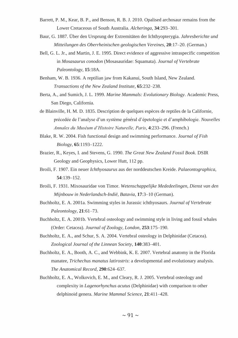
~ 91 ~
Barrett, P. M., Kear, B. P., and Benson, R. B. J. 2010. Opalised archosaur remains from the
Lower Cretaceous of South Australia. Alcheringa, 34:293–301.
Baur, G. 1887. Über den Ursprung der Extremitäten der Ichthyopterygia. Jahresberichte und
Mitteilungen des Oberrheinischen geologischen Vereines, 20:17–20. (German.)
Bell, G. L. Jr., and Martin, J. E. 1995. Direct evidence of aggressive intraspecific competition
in Mosasaurus conodon (Mosasauridae: Squamata). Journal of Vertebrate
Paleontology, 15:18A.
Benham, W. B. 1936. A reptilian jaw from Kakanui, South Island, New Zealand.
Transactions of the New Zealand Institute, 65:232–238.
Berta, A., and Sumich, J. L. 1999. Marine Mammals: Evolutionary Biology. Academic Press,
San Diego, California.
de Blainville, H. M. D. 1835. Description de quelques espèces de reptiles de la Californie,
précedée de l’analyse d’un système général d’épetologie et d’amphibologie. Nouvelles
Annales du Muséum d’Histoire Naturelle, Paris, 4:233–296. (French.)
Blake, R. W. 2004. Fish functional design and swimming performance. Journal of Fish
Biology, 65:1193–1222.
Brazier, R., Keyes, I. and Stevens, G. 1990. The Great New Zealand Fossil Book. DSIR
Geology and Geophysics, Lower Hutt, 112 pp.
Broili, F. 1907. Ein neuer Ichthyosaurus aus der norddeutschen Kreide. Palaeontographica,
54:139–152.
Broili, F. 1931. Mixosauridae von Timor. Wetenschappelijke Mededeelingen, Dienst van den
Mijnbouw in Nederlandsch-Indië, Batavia, 17:3–10 (German).
Buchholtz, E. A. 2001a. Swimming styles in Jurassic ichthyosaurs. Journal of Vertebrate
Paleontology, 21:61–73.
Buchholtz, E. A. 2001b. Vertebral osteology and swimming style in living and fossil whales
(Order: Cetacea). Journal of Zoology, London, 253:175–190.
Buchholtz, E. A., and Schur, S. A. 2004. Vertebral osteology in Delphinidae (Cetacea).
Zoological Journal of the Linnean Society, 140:383–401.
Buchholtz, E. A., Booth, A. C., and Webbink, K. E. 2007. Vertebral anatomy in the Florida
manatee, Trichechus manatus latirostris: a developmental and evolutionary analysis.
The Anatomical Record, 290:624–637.
Buchholtz, E. A., Wolkovich, E. M., and Cleary, R. J. 2005. Vertebral osteology and
complexity in Lagenorhynchus acutus (Delphinidae) with comparison to other
delphinoid genera. Marine Mammal Science, 21:411–428.

~ 92 ~
Buffetaut, E. 1983. Wounds on the jaw of an Eocene mesosuchian crocodilian as possible
evidence for the antiquity of crocodilian intraspecific fighting behaviour.
Paläontologische Zeitschrift, 57:143–145.
Callaway, J. M., and Massare, J. A. 1989. Geographic and stratigraphic distribution of the
Triassic Ichthyosauria (Reptilia; Diapsida). Neues Jahrbuch für Geologie und
Paläontologie, Abhandlungen, 178:37–58.
Camp, C. L. 1942. Ichthyosaur rostra from central California. Journal of Paleontology,
16:362–371.
Campbell, H. J. 1979. Permian-Mesozoic stratigraphy of the Moindou-Terémba area, west
coast, New Caledonia. MSc Thesis, University of Auckland, 273 pp. (unpublished).
Campbell, H. J. 1984. New records and taxa of Permian and Triassic fossils from New
Caledonia and New Zealand. Alcheringa, 8:151–167.
Campbell, J. D. 1965. New Zealand Triassic saurians. New Zealand Journal of Geology and
Geophysics, 8:505–509.
Campbell, J. D. and Warren, G. 1965. Fossil localities in the Torlesse Group in the South
Island. Transactions of the Royal Society of New Zealand, 3:99–137.
Carter, J. 1846. On the occurrence of a new species of Ichthyosaurus in the Chalk in the
neighbourhood of Cambridge. London Geological Journal, 1:7–9.
Chapman, F. 1914. Australasian Fossils: A Student’s Manual of Palaeontology. George
Robertson and Company, Melbourne, Australia, 341pp.
Choo, B. 1999. Cretaceous ichthyosaurs from Western Australia. Records of the Western
Australian Museum, Supplement, 57:207–218.
Clarke, J., and Etches, S. 1991. Predation amongst Jurassic marine reptiles. Proceedings of
the Dorset Natural History and Archaeological Society, 113:202–205.
Cooper, L. N., and Dawson, S. D. 2009. The trouble with flippers: a report on the prevalence
of digital anomalies in Cetacea. Zoological Journal of the Linnean Society, 155:722–
735.
Cooper, L. N., Berta, A., Dawson, S. D., and Reidenberg, J. S., 2007a. Evolution of
hyperphalangy and digit reduction in the cetacean manus. The Anatomical Record,
290:654–672.
Cooper, L. N., Dawson, S. D., Reidenberg. J. S., and Berta, A. 2007b. Neuromuscular
anatomy and evolution of the cetacean forelimb. The Anatomical Record, 290:1121–
1137.

~ 93 ~
Cosgriff, J. W. and Garbutt, N. K. 1972. Erythrobatrachus noonkanbahensis, a trematosaurid
species from the Blina Shale. Journal of the Royal Society of Western Australia, 55:5–
18.
Cox, G. 1991. Prehistoric Animals of New Zealand. HarperCollins Publishers (New Zealand)
Limited, Auckland, 40 pp.
Day, R. W. 1969. The Lower Cretaceous of the Great Artesian Basin. In Stratigraphy and
Palaeontology. Essays in Honour of Dorothy Hill, K.S.W. Campbell ed, Australian
National University Press, Canberra, 140–173.
Druckenmiller, P. S., and Maxwell, E. E. 2010. A new Lower Cretaceous (lower Albian)
ichthyosaur genus from the Clearwater Formation, Alberta, Canada. Canadian
Journal of Earth Sciences, 47:1037–1053.
Einarsson, E., Lindgren, J., Kear, B. P., and Siverson, M. 2010. Mosasaur bite marks on a
plesiosaur propodials from the Campanian (Late Cretaceous) of southern Sweden.
FGG, 132:123–128.
Embleton, B. J. J. 1984. Australia’s global setting: past global settings. In Phanerozoic Earth
History of Australia, J. J. Veevers (ed), Clarendon Press, Oxford, United Kingdom,
11–16.
English, A. W. M. 1976. Functional anatomy of the hands of fur seals and sea lions. American
Journal of Anatomy, 147:1–18.
English, A. W. M. 1977. Structural correlates of forelimb function in fur seals and sea lions.
Journal of Morphology, 151:325–352.
Etheridge Jnr, R. 1888. On additional evidence of the genus Ichthyosaurus in the Mesozoic
rocks (“Rolling Downs Formation”) of north-eastern Australia. Proceedings of the
Linnean Society of New South Wales, 2:405–409.
Etheridge Jnr, R. 1904. A second sauropterygian converted to opal, from the Upper
Cretaceous of White Cliffs, New South Wales. Records of the Australian Museum,
5:306–316.
Everhart, M. J. 2004. Late Cretaceous interaction between predators and prey. Evidence of
feeding by two species of shark on a mosasaur. PalArch, Vertebrate Palaeontology
Series, 1:1–7.
Everhart, M. J. 2005. Oceans of Kansas. A Natural History of the Western Interior Sea.
Indiana University Press, Bloomington, 322 pp.
Everhart, M. J. 2008. A bitten skull of Tylosaurus kansasensis (Squamata: Mosasauridae) and
a review of mosasaur-on-mosasaur pathology in the fossil record. Transactions of the
Kansas Academy of Science, 111:251–262.

~ 94 ~
Feldkamp, S. D. 1987. Foreflipper propulsion in the California sea lion, Zalophus
californianus. Journal of Zoology, London, 212:43–57.
Fernández, M. 1997. A new ichthyosaur from the Tithonian (Late Jurassic) of the Neuquén
Basin, northwestern Patagonia, Argentina. Journal of Paleontology, 71:479–484.
Fernández, M. 2007. Redescription and phylogenetic position of Caypullisaurus
(Ichthyosauria: Ophthalmosauridae). Journal of Paleontology, 81:368–375.
Fernández, M., and Aguirre-Urreta, M. B. 2005. Revision of Platypterygius hauthali von
Huene, 1927 (Ichthyosauria: Ophthalmosauridae) from the Early Cretaceous of
Patagonia, Argentina. Journal of Vertebrate Paleontology, 25:583–587.
Fish, F. E. 2004. Structure and mechanics of nonpiscine control surfaces. IEEE Journal of
Oceanic Engineering, 29:605–621.
Fischer, V. 2009. Les ichthyosaures du sud-est de la France. MSc Thesis. Universite de Liege.
180 pp. (unpublished) (in French)
Fischer, V., Clément, A., Guiomar, M., and Godefroit, P. in press. The first definite record of
a Valanginian ichthyosaur and its implications on the evolution of post-Liassic
Ichthyosauria. Cretaceous Research.
Fleming, C. A. 1979. The Geological History of New Zealand and its Life. Auckland
University Press, Auckland, 141 pp.
Fleming, C. A., Gregg, D. R. and Welles, S. P. 1971. New Zealand ichthyosaurs – a summary,
including new records from the Cretaceous. New Zealand Journal of Geology and
Geophysics, 14:734–741.
Fordyce, R. E. 1982. The fossil vertebrate record of New Zealand. In The Fossil Vertebrate
Record of Australasia, P. Rich and E. Thompson eds, Monash University Offset,
Clayton, Victoria, 629–698.
Fordyce, R. E. 1991. A new look at the fossil vertebrate record of New Zealand. In Vertebrate
Palaeontology of Australasia, P. Vickers-Rich, J. M. Monaghan, R. F. Baird and T. H.
Rich eds, Pioneer Design Studio Pty Ltd, Lilydale, Victoria, 1191–1316.
Fordyce, R. E. 2003. Fossils and the history of life. In The Natural History of Southern New
Zealand, J. Darby, R. E. Fordyce, A. Mark, K. Probert and C. Townsend (eds),
University of Otago Press, Dunedin, 37–64.
Frakes, L. A., Alley, N. F., and Deynoux, M. 1995. Early Cretaceous ice rafting and climate
zonation in Australia. International Geology Review, 37:567–583.
Frakes, L. A., Burger, D., Apthorpe, M., Wiseman, J., Dettmann, M., Alley, N., Flint, R.,
Gravestock, D., Ludbrook, N., Backhouse, J., Skwarko, S., Scheibnerova, V.,
McMinn, A., Moore, P. S., Bolton, B. R., Douglass, J. G., Christ, R., Wade, M.,
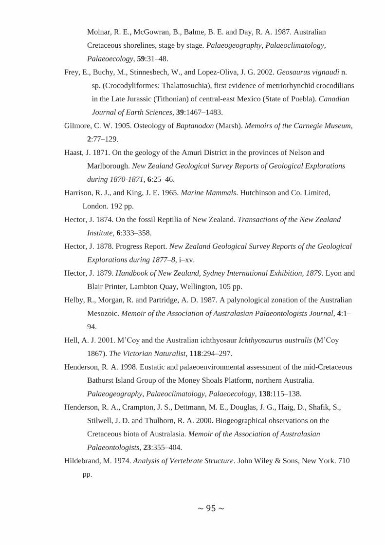
~ 95 ~
Molnar, R. E., McGowran, B., Balme, B. E. and Day, R. A. 1987. Australian
Cretaceous shorelines, stage by stage. Palaeogeography, Palaeoclimatology,
Palaeoecology, 59:31–48.
Frey, E., Buchy, M., Stinnesbech, W., and Lopez-Oliva, J. G. 2002. Geosaurus vignaudi n.
sp. (Crocodyliformes: Thalattosuchia), first evidence of metriorhynchid crocodilians
in the Late Jurassic (Tithonian) of central-east Mexico (State of Puebla). Canadian
Journal of Earth Sciences, 39:1467–1483.
Gilmore, C. W. 1905. Osteology of Baptanodon (Marsh). Memoirs of the Carnegie Museum,
2:77–129.
Haast, J. 1871. On the geology of the Amuri District in the provinces of Nelson and
Marlborough. New Zealand Geological Survey Reports of Geological Explorations
during 1870-1871, 6:25–46.
Harrison, R. J., and King, J. E. 1965. Marine Mammals. Hutchinson and Co. Limited,
London. 192 pp.
Hector, J. 1874. On the fossil Reptilia of New Zealand. Transactions of the New Zealand
Institute, 6:333–358.
Hector, J. 1878. Progress Report. New Zealand Geological Survey Reports of the Geological
Explorations during 1877–8, i–xv.
Hector, J. 1879. Handbook of New Zealand, Sydney International Exhibition, 1879. Lyon and
Blair Printer, Lambton Quay, Wellington, 105 pp.
Helby, R., Morgan, R. and Partridge, A. D. 1987. A palynological zonation of the Australian
Mesozoic. Memoir of the Association of Australasian Palaeontologists Journal, 4:1–
94.
Hell, A. J. 2001. M’Coy and the Australian ichthyosaur Ichthyosaurus australis (M’Coy
1867). The Victorian Naturalist, 118:294–297.
Henderson, R. A. 1998. Eustatic and palaeoenvironmental assessment of the mid-Cretaceous
Bathurst Island Group of the Money Shoals Platform, northern Australia.
Palaeogeography, Palaeoclimatology, Palaeoecology, 138:115–138.
Henderson, R. A., Crampton, J. S., Dettmann, M. E., Douglas, J. G., Haig, D., Shafik, S.,
Stilwell, J. D. and Thulborn, R. A. 2000. Biogeographical observations on the
Cretaceous biota of Australasia. Memoir of the Association of Australasian
Palaeontologists, 23:355–404.
Hildebrand, M. 1974. Analysis of Vertebrate Structure. John Wiley & Sons, New York. 710
pp.
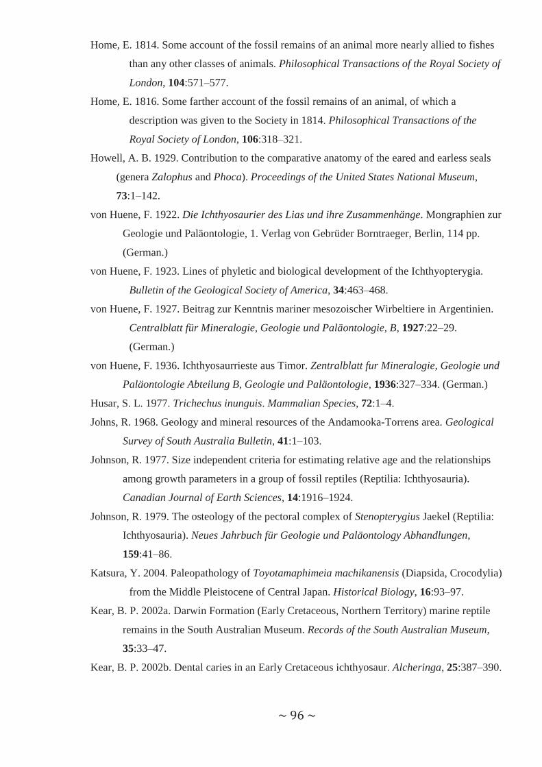
~ 96 ~
Home, E. 1814. Some account of the fossil remains of an animal more nearly allied to fishes
than any other classes of animals. Philosophical Transactions of the Royal Society of
London, 104:571–577.
Home, E. 1816. Some farther account of the fossil remains of an animal, of which a
description was given to the Society in 1814. Philosophical Transactions of the
Royal Society of London, 106:318–321.
Howell, A. B. 1929. Contribution to the comparative anatomy of the eared and earless seals
(genera Zalophus and Phoca). Proceedings of the United States National Museum,
73:1–142.
von Huene, F. 1922. Die Ichthyosaurier des Lias und ihre Zusammenhänge. Mongraphien zur
Geologie und Paläontologie, 1. Verlag von Gebrüder Borntraeger, Berlin, 114 pp.
(German.)
von Huene, F. 1923. Lines of phyletic and biological development of the Ichthyopterygia.
Bulletin of the Geological Society of America, 34:463–468.
von Huene, F. 1927. Beitrag zur Kenntnis mariner mesozoischer Wirbeltiere in Argentinien.
Centralblatt für Mineralogie, Geologie und Paläontologie, B, 1927:22–29.
(German.)
von Huene, F. 1936. Ichthyosaurrieste aus Timor. Zentralblatt fur Mineralogie, Geologie und
Paläontologie Abteilung B, Geologie und Paläontologie, 1936:327–334. (German.)
Husar, S. L. 1977. Trichechus inunguis. Mammalian Species, 72:1–4.
Johns, R. 1968. Geology and mineral resources of the Andamooka-Torrens area. Geological
Survey of South Australia Bulletin, 41:1–103.
Johnson, R. 1977. Size independent criteria for estimating relative age and the relationships
among growth parameters in a group of fossil reptiles (Reptilia: Ichthyosauria).
Canadian Journal of Earth Sciences, 14:1916–1924.
Johnson, R. 1979. The osteology of the pectoral complex of Stenopterygius Jaekel (Reptilia:
Ichthyosauria). Neues Jahrbuch für Geologie und Paläontology Abhandlungen,
159:41–86.
Katsura, Y. 2004. Paleopathology of Toyotamaphimeia machikanensis (Diapsida, Crocodylia)
from the Middle Pleistocene of Central Japan. Historical Biology, 16:93–97.
Kear, B. P. 2002a. Darwin Formation (Early Cretaceous, Northern Territory) marine reptile
remains in the South Australian Museum. Records of the South Australian Museum,
35:33–47.
Kear, B. P. 2002b. Dental caries in an Early Cretaceous ichthyosaur. Alcheringa, 25:387–390.

~ 97 ~
Kear, B. P. 2003. Cretaceous marine reptiles of Australia: a review of taxonomy and
distribution. Cretaceous Research, 24:277–303.
Kear, B. P. 2004. Biogeographic and biostratigraphic implications of Australian Mesozoic
marine reptiles. Australian Biologist, 17:4–22.
Kear, B. P. 2005a. Cranial morphology of Platypterygius longmani Wade, 1990 (Reptilia:
Ichthyosauria) from the Lower Cretaceous of Australia. Zoological Journal of the
Linnean Society, 145:583–622.
Kear, B. P. 2005b. Marine reptiles from the Lower Cretaceous (Aptian) deposits of White
Cliffs, southeastern Australia: implications of a high latitude, cold water assemblage.
Cretaceous Research, 26:769–782.
Kear, B. P. 2006. Marine reptiles from the Lower Cretaceous of South Australia: elements of
a high-latitude cold-water assemblage. Palaeontology, 49:837–856.
Kear, B. P., Boles, W. E., and Smith, E. T. 2003. Unusual gut contents in a Cretaceous
ichthyosaur. Proceedings of the Royal Society of London, Series B, 270:S206–S208.
Kear, B. P., Schroeder, N. I., and Lee, M. S. Y. 2006. An archaic crested plesiosaur in opal
from the Lower Cretaceous high latitude deposits of Australia. Biology Letters,
2:615–619.
King, J. E. 1989. Otariidae and Phocidae. Fauna of Australia (eds D.W. Walton and B.J.
Richardson). Australian Government Publishing. 35 pp.
Kiprijanoff, W. 1881. Studien über die fossilen Reptilien Russlands. Theil 1. Gattung
Ichthyosaurus König aus dem Sewerischen Sandstein oder Osteolith der Kreide-
Gruppe. Mémoires de l’Académie Impériale des Sciences de Saint-Pétersbourg, 7e
Série, 28:1–103.
Kirton, A. J. 1983. A review of British Upper Jurassic ichthyosaurs. PhD dissertation,
University of Newcastle-Upon-Tyne, United Kingdom, 239 pp.
Klima, M., Oelschaläger, H. A., and Wünsch, D. 1980. Morphology of the pectoral girdle in
the Amazon dolphin Inia geoffrensis with special reference to the shoulder joint and
the movements of the flippers. Zeitschrift fur Säugetierkunde, 45:288–309.
Kolb, C., and Sander, P. M. 2009. Redescription of the ichthyosaur Platypterygius hercynicus
(Kuhn 1946) from the Lower Cretaceous of Salzgitter (Lower Saxony, Germany).
Palaeontographica Abteilung A, 288:151–192.
Krieg, G. W. and Rodgers, P. A. 1995. Stratigraphy – marine succession. In The Geology of
South Australia. Volume 2, The Phanerozoic, J.F. Drexel and W.V. Preiss eds,
Geological Survey of South Australian Bulletin 54, 112–123.

~ 98 ~
Kuhn, O. 1946. Ein Skelett von Ichthyosaurus (Platypterygius) hercynicus n. sp. aus dem
Aptium von Gitter. Bericht der Naturforschenden Gesellschaft Bamberg, 29:69–82.
Laird, M. G. and Bradshaw, J. D. 2004. The break-up of a long-term relationship: the
Cretaceous separation of New Zealand from Gondwana. Gondwana Research, 7:273–
286.
Landis, C. A., Campbell, H. J., Aslund, T., Cawood, P. A., Douglas, A., Kimbrough, D. L.,
Pillai, D. D. L., Raine, J. I. and Willsman, A. 1999. Permian-Jurassic strata at
Productus Creek, Southland, New Zealand: implications for terrane dynamics of the
eastern Gondwanaland margin. New Zealand Journal of Geology and Geophysics,
42:255–278.
Lingham-Soliar, T. 1995. Anatomy and functional morphology of the largest marine reptile
known, Mosasaurus hoffmanni (Mosasauridae, Reptilia) from the Upper Cretaceous,
Upper Maastrichtian of The Netherlands. Philosophical Transactions of the Royal
Society of London B, 347:155–180.
Lingham-Soliar, T. 1998. Unusual death of a Cretaceous giant. Lethaia, 31:308–310.
Lingham-Soliar, T. 2003. Extinction of ichthyosaurs: a catastrophic or evolutionary
paradigm? Neues Jahrbuch für Geologie und Paläontologie, Abhandlungen,
228:421–452.
Lingham-Soliar, T. 2004. Palaeopathology and injury in the extinct mosasaurs
(Lepidosauromorpha, Squamata) and implications for modern reptiles. Lethaia,
37:255–262.
Longman, H. A. 1922. An ichthyosaurian skull from Queensland. Memoirs of the Queensland
Museum, 7:246–256.
Longman, H. A. 1924. Some Queensland fossil vertebrates. Memoirs of the Queensland
Museum, 10:16–28.
Longman, H. A. 1943. Further notes on Australian ichthyosaurs. Memoirs of the Queensland
Museum, 12:101–104.
Ludbrook, N. H. 1966. Cretaceous biostratigraphy of the Great Artesian Basin in South
Australia. Geological Survey of South Australia Bulletin, 40:1–223.
Lydekker, R. 1889. Catalogue of the Fossil Reptilia and Amphibia in the British Museum
(Natural History). Part II. Containing the Orders Ichthyopterygia and Sauropterygia.
London, Printed by Order of the Trustees of the British Museum, London, 307 pp.
Mackness, B., and Sutton, R. 2000. Possible evidence for intraspecific aggression in a
Pliocene crocodile form northern Australia. Alcheringa, 24:55–62.

~ 99 ~
Maisch, M. W., and Hungerbühler, A. 2001. New evidence for a discrete supratemporal bone
in the Jurassic ichthyosaur Temnodontosaurus. Historical Biology, 15:335–345.
Maisch, M. W., and Matzke, A. T. 2000. The Ichthyosauria. Stuttgarter Beiträge zur
Naturkunde Serie B (Geologie und Paläontologie), 298:1–159.
Marsh, O. C. 1880. The limbs of Sauranodon, with notice of a new species. American Journal
of Science, 19:168–171.
Martill, D. M. 1996. Fossils explained: ichthyosaurs. Geology Today, 12:194–196.
Massare, J. A. 1988. Swimming capabilities of Mesozoic marine reptiles: implications for
method of predation. Paleobiology, 14:187–205.
Massare, J. A. 1994. Swimming capabilities of Mesozoic marine reptiles: a review. pp. 13–
149 in Maddock, L., Bone, Q., and Rayner, J. M. V. (eds) Mechanics and Physiology
of Animal Swimming. Cambridge University Press, New York.
Massare, J. A., and Callaway, J. M. 1990. The affinities and ecology of Triassic ichthyosaurs.
Geological Society of America Bulletin, 102:409–416.
Massare, J. A., and Faulkner, J. 1997. Sharks as morphological analogs for ichthyosaurs.
Paludicola, 1:117–125.
Massare, J. A., and Sharkey, S. J. 2003. Centrum shape in sharks: not a good analog for
ichthyosaurs. Paludicola, 4:27–36.
Massare, J. A., Buchholtz, E. A., Kenney, J. M. and Chomat, A.-M. 2006. Vertebral
morphology of Ophthalmosaurus natans (Reptilia: Ichthyosauria) from the Jurassic
Sundance Formation of Wyoming. Paludicola, 5:242–254.
Maxwell, E. E. and Caldwell, M. W. 2006. A new genus of ichthyosaur from the Lower
Cretaceous of western Canada. Palaeontology, 49:1043–1052.
Maxwell, E. E., and Kear, B. P. 2010. Postcranial anatomy of Platypterygius americanus
(Reptilia: Ichthyosauria) from the Cretaceous of Wyoming. Journal of Vertebrate
Palaeontology, 30:1059–1068.
Mazin, J.-M. 1983. Repartition stratigraphique et géographique des Mixosauria
(Ichthyopterygia) provincialité marine au Trias moyen. In Actes du Symposium
Päléontologie Georges Cuvier, E. Buffetaut, J.M. Mazin and E. Salmon eds,
Montbéliard, 375–387.
Mazin, J.-M. 1985. Les Ichthyopterygia du trias superieur de Nouvelle Caledonia.
Implications paleobiogeographiques. Revue de Paléobiologie, 4:177–182.
Mazin, J.-M. 1986. Paleobiogeography of Triassic ichthyopterygian reptiles; some working
hypotheses. Neues Jahrbuch für Geologie und Paläontologie, Abhandlungen,
173:117–129.

~ 100 ~
McCoy, F. 1867a. On the occurrence of Ichthyosaurus and Plesiosaurus in Australia. Annals
and Magazine of Natural History, Third Series, 19:355–356.
McCoy, F. 1867b. On the discovery of the Enaliosauria and other Cretaceous fossils in
Australia. Transactions and Proceedings of the Royal Society of Victoria, 8:41–42.
McCoy, F. 1869. On the fossil eye and teeth of the Ichthyosaurus australis (M’Coy), from the
Cretaceous formations of the source of the Flinder’s River; and on the palate of
Diprotodon, from the Tertiary limestone of Limeburner’s Point, near Geelong.
Transactions and Proceedings of the Royal Society of Victoria, 2:77–78.
McGowan, C. 1972a. Evolutionary trends in longipinnate ichthyosaurs with particular
reference to the skull and fore fin. Life Sciences Contribution, Royal Ontario
Museum, 83:1–38.
McGowan, C. 1972b. The systematics of Cretaceous ichthyosaurs with particular reference to
the material from North America. Contributions to Geology, University of Wyoming,
11:9–29.
McGowan, C. 1974. A revision of the longipinnate ichthyosaurs of the Lower Jurassic of
England, with descriptions of two new species (Reptilia: Ichthyosauria). Royal
Ontario Museum Life Sciences Contributions, 97:1–37.
McGowan, C. 1991. Dinosaurs Spitfires and Seadragons. Harvard University Press,
Cambridge, Massachusetts. 365 pp.
McGowan, C. 1992. The ichthyosaurian tail: sharks do not provide an appropriate analogue.
Palaeontology, 35:555–570.
McGowan, C., and Motani, R. 2003. Ichthyopterygia. In Sues, H.-D. (ed.), Handbook of
Paleoherpetology, Volume 8. Verlag Dr. Friedrich Pfeil, München. 175 pp.
McLoughlin, S., Haig, D. W., Backhouse, J., Holmes, M. A., Ellis, G., Long, J. A. and
McNamara, K. J. 1995. Oldest Cretaceous sequence, Giralia Anticline, Carnarvon
Basin, Western Australia: late Hauterivian-Barremian. AGSO Journal of Australian
Geology and Geophysics, 15:445–468.
McMinn, A. and Burger, D. 1986. Palynology and palaeoenvironments of the Toolebuc
Formation (sensu lato) in the Eromanga Basin. Geological Society of Australia Special
Publication, 12:139–154.
Molnar, R. E. 1982. Australian Mesozoic reptiles. In The Fossil Vertebrate Record of
Australasia, P. Rich and E. Thompson eds, Monash University Offset, Clayton,
Victoria, 169–225.
Moore, K. L., Dalley, A. F., and Agur, A. M. R. 2010. Clinically Oriented Anatomy.
Lippincott Williams and Wilkins, Philadelphia, USA. 1134 pp.
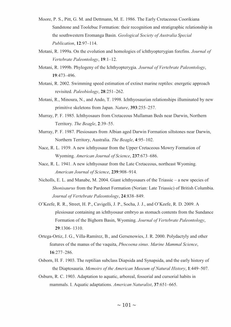
~ 101 ~
Moore, P. S., Pitt, G. M. and Dettmann, M. E. 1986. The Early Cretaceous Coorikiana
Sandstone and Toolebuc Formation: their recognition and stratigraphic relationship in
the southwestern Eromanga Basin. Geological Society of Australia Special
Publication, 12:97–114.
Motani, R. 1999a. On the evolution and homologies of ichthyopterygian forefins. Journal of
Vertebrate Paleontology, 19:1–12.
Motani, R. 1999b. Phylogeny of the Ichthyopterygia. Journal of Vertebrate Paleontology,
19:473–496.
Motani, R. 2002. Swimming speed estimation of extinct marine reptiles: energetic approach
revisited. Paleobiology, 28:251–262.
Motani, R., Minoura, N., and Ando, T. 1998. Ichthyosaurian relationships illuminated by new
primitive skeletons from Japan. Nature, 393:255–257.
Murray, P. F. 1985. Ichthyosaurs from Cretaceous Mullaman Beds near Darwin, Northern
Territory. The Beagle, 2:39–55.
Murray, P. F. 1987. Plesiosaurs from Albian aged Darwin Formation siltstones near Darwin,
Northern Territory, Australia. The Beagle, 4:95–102.
Nace, R. L. 1939. A new ichthyosaur from the Upper Cretaceous Mowry Formation of
Wyoming. American Journal of Science, 237:673–686.
Nace, R. L. 1941. A new ichthyosaur from the Late Cretaceous, northeast Wyoming.
American Journal of Science, 239:908–914.
Nicholls, E. L. and Manabe, M. 2004. Giant ichthyosaurs of the Triassic – a new species of
Shonisaurus from the Pardonet Formation (Norian: Late Triassic) of British Columbia.
Journal of Vertebrate Paleontology, 24:838–849.
O’Keefe, R. R., Street, H. P., Cavigelli, J. P., Socha, J. J., and O’Keefe, R. D. 2009. A
plesiosaur containing an ichthyosaur embryo as stomach contents from the Sundance
Formation of the Bighorn Basin, Wyoming. Journal of Vertebrate Paleontology,
29:1306–1310.
Ortega-Ortiz, J. G., Villa-Ramírez, B., and Gersenowies, J. R. 2000. Polydactyly and other
features of the manus of the vaquita, Phocoena sinus. Marine Mammal Science,
16:277–286.
Osborn, H. F. 1903. The reptilian subclass Diapsida and Synapsida, and the early history of
the Diaptosauria. Memoirs of the American Museum of Natural History, 1:449–507.
Osburn, R. C. 1903. Adaptation to aquatic, arboreal, fossorial and cursorial habits in
mammals. I. Aquatic adaptations. American Naturalist, 37:651–665.

~ 102 ~
Owen, R. 1840. Report on British fossil reptiles. Reports of the British Association for the
Advancement of Science, 9:43–126.
Páramo, M. E. 1997. Platypterygius sachicarum (Reptilia, Ichthyosauria) neuva especie del
Cretacico de Colombia. Revista Ingeominas, 6:1–12.
Pledge, N. S. 1980. Vertebrate Fossils of South Australia. Government Printer, South
Australia, Adelaide, 11 pp.
Pledge, N. S., and Tedford, R. H. 1990. Vertebrate Fossils; pp. 199–209 in M. J. Tyler, C. R.
Twidale, M. Davies and C. B. Wells (eds.), Natural History of the North East
Deserts. Royal Society of South Australia, Inc, Adelaide, Australia.
Pollard, J. E. 1968. The gastric contents of an ichthyosaur from the Lower Lias of Lyme
Regis, Dorset. Palaeontology, 11:376–388.
Ray, C. 1963. Locomotion in pinnipeds. Natural History, 72:10–21.
Renjun, L., Gewalt, W., Neurohr, B., and Winkler, A. 1994. Comparative studies on the
behaviour of Inia geoffrensis and Lipotes vexillifer in artificial environments. Aquatic
Mammals, 20:39–45.
Revell, P. A. 1986. Pathology of Bone. Springer-Verlag, Berlin, 451 pp.
Richardson, M. K., and Chipman, A. D. 2003. Developmental constraints in a comparative
framework: a test case using variations in phalanx number during amniote evolution.
Journal of Experimental Zoology. Part B, Molecular and Developmental Evolution,
296B:8–22.
Riess, J. 1986. Fortbewegunsweise, Schwimmbiophysik und Phylogenie der Ichthyosaurier.
Palaeontographica Abteilung A, 192:93–155.
Romer, A. S. 1956. Osteology of the Reptiles. University of Chicago Press, Chicago, 772 pp.
Romer, A. S. 1968. An ichthyosaur skull from the Cretaceous of Wyoming. Contributions to
Geology, University of Wyoming, 7:27–41.
Rothschild, B. M., and Martin, L. D. 1993. Paleopathology. Disease in the Fossil Record.
CRC Press, Boca Raton, 386 pp.
Rothschild, B. M., Martin, L. D., and Schulp, A. S. 2005. Sharks eating mosasaurs, dead or
alive? Netherlands Journal of Geosciences, 84:335–340.
Sachs, S. and Grant-Mackie, J. A. 2003. An ichthyosaur fragment from the Cretaceous of
Northland, New Zealand. Journal of the Royal Society of New Zealand, 33:307–314.
Sander, P. M. 1992. Cymbospondylus (Shastasauridae: Ichthyosauria) from the Middle
Triassic of Spitsbergen: filling a paleobiogeographic gap. Journal of Paleontology,
66:332–337.

~ 103 ~
Sander, P. M. 2000. Ichthyosauria: their diversity, distribution, and phylogeny.
Paläontologishche Zeitschrift, 74:1–35.
Schellart, W. P., Lister, G. S. and Toy, V. G. 2006. A Late Cretaceous and Cenozoic
reconstruction of the southwest Pacific region: tectonics controlled by subduction and
slab rollback processes. Earth-Science Reviews, 76:191–233.
Schwimmer, D. R. 2002. King of the Crocodylians. The Paleobiology of Deinosuchus.
Indiana University Press, Bloomington, 220 pp.
Schwimmer, D. R., Steward, J. D., and Williams, G. D. 1997. Scavenging by sharks of the
genus Squalicorax in the Late Cretaceous of North America. Palaios, 12:71–73.
Shimada, K. 1997. Paleoecological relationships of the Late Cretaceous lamniform shark,
Cretoxyrhina mantelli (Agassiz). Journal of Paleontology, 71:926–933.
Shimada, K., and Everhart, M. J. 2004. Shark-bitten Xiphactinus audax (Teleostei:
Ichthyodectiformes) from the Niobrara Chalk (Upper Cretaceous) of Kansas. The
Mosasaur, 7:35–39.
Shimada, K., and Hooks III, G. E. 2004. Shark-bitten protostegid turtles form the Upper
Cretaceous Mooreville Chalk, Alabama. Journal of Paleontology, 78:205–210.
Shimada, K., Tsuihiji, T., Sato, T., and Hasegawa. Y. 2010. A remarkable case of a shark-
bitten elasmosaurid plesiosaur. Journal of Vertebrate Paleontology, 30:592–597.
Siverson, M. 1999. A new large lamniform shark from the uppermost Gearle Siltstone
(Cenomanian, Late Cretaceous) of Western Australia. Transactions of the Royal
Society of Edinburgh: Earth Sciences, 90:49–66.
Storrs, G. W., Arkhangelsky, M. S., and Efimov, V. E. 2000. Mesozoic marine reptiles of
Russia and other former Soviet republics; pp 187-210 in M. J. Benton, M. A. Shiskin,
D. M. Unwin, and E. N. Kurochkin (eds.), The Age of Dinosaurs in Russia and
Mongolia. Cambridge University Press, Cambridge, U.K.
Tanke, D. H., and Currie, P. J. 1998. Head-biting behaviour in theropod dinosaurs:
paleopathological evidence. Gaia, 15:167–184.
Tanke, D. H., and Rothschild, B. M. 2002. Dinosores: An annotated bibliography of dinosaur
paleopathology and related topics – 1838–2001. New Mexico Museum of Natural
History and Science Bulletin, 20:1–96.
Teichert, C. and Matheson, R.S. 1944. Upper Cretaceous ichthyosaurian and plesiosaurian
remains from Western Australia. Australian Journal of Science, 6:167–178.
Thewissen, J. G. M., Cohn, M. J., Stevens, L. S., Bajpai, S., Heyning, J., and Horton Jr., W.
E. 2006. Developmental basis for hind-limb loss in dolphins and origin of the cetacean

~ 104 ~
bodyplan. Proceedings of the National Academy of Sciences of the United States of
America, 103:8414–8418.
Thulborn, T., and Turner, S. 1993. An elasmosaur bitten by a pliosaur. Modern Geology,
18:489–501.
Turner, S., and Rozefelds, A. 1992. Tip of the pyramid? Cretaceous megasharks from
Australia. The Beagle. Records of the Northern Territory Museum and Art Gallery,
9:262–263.
Turner, S., and Thulborn, T. 2003. Second course for Kronosaurus? Further evidence of
pliosaur predation in the Cretaceous of Queensland; pp. 29–30 in Abstracts, 9th
Conference on Australasian Vertebrate Evolution, Palaeontology & Systematics –
Heber A. Longman Symposium, Brisbane.
Veevers, J. J., Powell, C. McA., and Roots, S. R. 1991. Review of seafloor spreading around
Australia. I. Synthesis of the patterns of spreading. Australian Journal of Earth
Sciences, 38:373–389.
Wade, M. 1984. Platypterygius australis, an Australian Cretaceous ichthyosaur. Lethaia,
17:99–113.
Wade, M. 1985. Platypterygius australis (McCoy) 1867: a Cretaceous marine reptile. In
Kadimakara: Extinct Vertebrates of Australia, P.V. Rich and G.F. van Tets eds, 137–
142.
Wade, M. 1990. A review of the Australian Cretaceous longipinnate ichthyosaur
Platypterygius (Ichthyosauria, Ichthyopterygia). Memoirs of the Queensland
Museum, 28:115–137.
Watson, A., Kuo, T.-F., Yang, W.-C., Yao, C.-J., and Chou, L.-S. 2008. Distinctive osteology
of distal flipper bones of tropical bottlenose whales, Indopacetus pacificus, from
Taiwan: mother and calf, calf with polydactyly. Marine Mammal Science, 24:398–410.
Webb, P. W. 1977. Effects of median-fin amputation on fast-stat performance of rainbow
trout (Salmo gairdneri). Journal of Experimental Biology, 68:123–135.
Webb, P. W. 1984. Form and function in fish swimming. Scientific American, 25:58–70.
Webb, P. W., and Keyes, R. S. 1981. Division of labor between median fins in swimming
dolphin (Pisces: Corphenidae). Copeia, 1981:901–904.
White, T. E. 1935. On the skull of Kronosaurus queenslandicus Longman. Occasional Papers
of the Boston Society of Natural History, 8:219–228.
Worley, W. F. 1894. Geology of Nelson. Transactions of the New Zealand Institute, 26:414–
421.
Zammit, M. 2010. A review of Australasian ichthyosaurs. Alcheringa, 34:281–292.
~ 105 ~
Zammit, M., Norris, R. M., and Kear, B. P. 2010. The Australian Cretaceous ichthyosaur
Platypterygius australis: a description and review of postcranial remains. Journal of
Vertebrate Paleontology, 30:1726–1735.
~ 106 ~
APPENDIX I
Zammit, M. 2010
Australasia’s first Jurassic ichthyosaur fossil: an isolated vertebra from the lower Liassic Arataura Formation of the North Island, New Zealand.
Alcheringa (2010), in press.
Zammit, M. (2011) Australasia’s first Jurassic ichthyosaur fossil: an isolated vertebra from the lower Liassic Arataura Formation of the North Island, New Zealand. Alcheringa, v. 35 (3), pp. 341-343, September 2011
NOTE: This publication is included in the print copy of the thesis
held in the University of Adelaide Library.
It is also available online to authorised users at:
http://dx.doi.org/10.1080/03115518.2010.497056