Embed Size (px)
Citation preview
The Athletic Knee
Shannon M. Wolfe


The Problem
• Young active patients with articular cartilage defects!– Which defects progress to OA ?– Which defects are symptomatic ?– How do we most effectively treat these
defects?
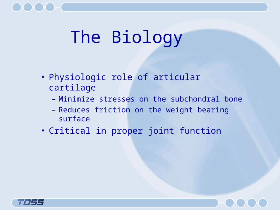
The Biology
• Physiologic role of articular cartilage– Minimize stresses on the subchondral bone– Reduces friction on the weight bearing
surface
• Critical in proper joint function

Goals of Treatment
• Restore integrity of load bearing surface
• Obtain full range of motion• Obtain pain free motion• Inhibit further degeneration


Treatment Considerations
• Age of the patient
• Defect size
• Knee stability
• Knee alignment
• Level of activity
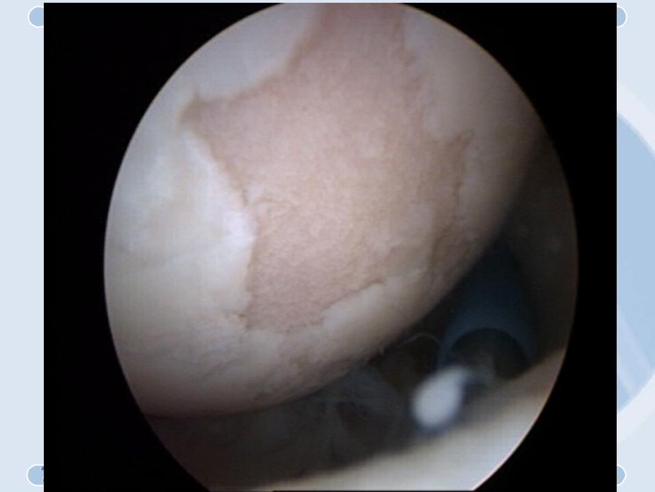
Partial Thickness Defects
• Articular cartilage lacks the capacity to repair structural damage
• Progresses when exposed to mechanical wear
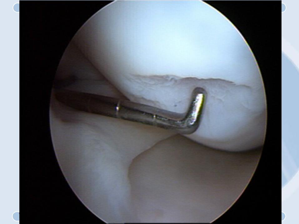
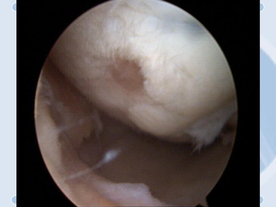
Full Thickness Defects
• Do not heal with hyaline cartilage• Healing by subchondral stimulation leads to
the formation of fibrocartilage– Lacks physiological role of hyaline cartilage– Poor wear characteristics
• Progress to osteoarthritis

Non-Surgical Options
• Activity modification (decrease load)• Muscle strengthening (load
absorption)• Bracing (selective joint unloading)• Aspiration (decrease painful joint
distention)
Non-Surgical Options• Pharmacological
– Oral• Non-steroidal anti-inflammatory medication• Chondrotin sulfate• Glucosamine
– Injectable• Corticosteroids - decrease the inflammatory response but
have no mechanical benefit• Synvisc - may improve the status of the articular surface by
improving chondrocyte “health”
Surgical Options• Arthroscopic lavage - remove debris• Arthroscopic shaving - smooth surface• Drilling or microfracture - create fibrocartilage
scar• Osteotomy - realignment to unload diseased
compartment• Osteochondral autograft - replace a damaged
surface• Autologous chondrocyte transplant - replace
injured cartilage• Allograft osteochondral transplantation

Arthroscopic Lavage
• Remove debris and inflammation mediators• Temporary relief• Not a definitive procedure - not curative• Not normally sufficient for athletic or active
patients
Arthroscopic Debridement
• Lavage and chondroplasty• No sub-chondral stimulation• May lead to improvement for up to 5 yrs.• 10-20% may become worse• Debridement does nothing to promote
repair• Malaligned or unstable knees do poorly
Thermal Chondroplasty
• New procedure• Requires bi-polar or ultrasonic device• “Seal” the articular surface with heat• Keplan L,M.D. reported no injury to the
chondrocytes of the involved or peripheral cartilage. “Radio-frequency energy appears to be safe for use on articular surface.” Arthroscopy, Jan-Feb. 2000, pp 2-5.

Abrasion Arthroplasty
• Debridement and stimulation of subchondral bone
• 1 - 1.5mm deep results in fibrocartilage repair• intracortical rather than cancellous
Results : Abrasion Arthroplasty
• Johnson 399 patients• 66% with continued pain• 99% with activity
restriction
Results : Abrasion Arthroplasty
• Unpredictable• May not be better than debridement alone• Rand noted 50% of patients who had an
abrasion underwent TKR within 3 yrs.

Drilling or Microfracture• Debride lose cartilage
• Subchondral bone penetration drill or pick, 3/cm squared
• Results in fibrocartilage repair
• Lacks durability
• Lacks the mechanical properties of hyaline cartilage


Drilling Results
Joseph Tippet,M.D.• 62 month follow
up• 71% Excellent• 15% Good• 14% Fair / Poor

Results :• Richard Steadman, M.D. reported
improvement in 364 of 485 patients (75%) at 7 years post-op– 90 - 100% of the defects were healed at
4 wks. with 30% hyaline cartilage– 12 mos. 42% hyaline cartilage
• Myron Spector, M.D. demonstrated complete filling of the lesions at 3 months in an animal model

Microfracture Results :
• Unpublished– 75% improvement– 50% returned to
sports
• Steadman / Hawkins

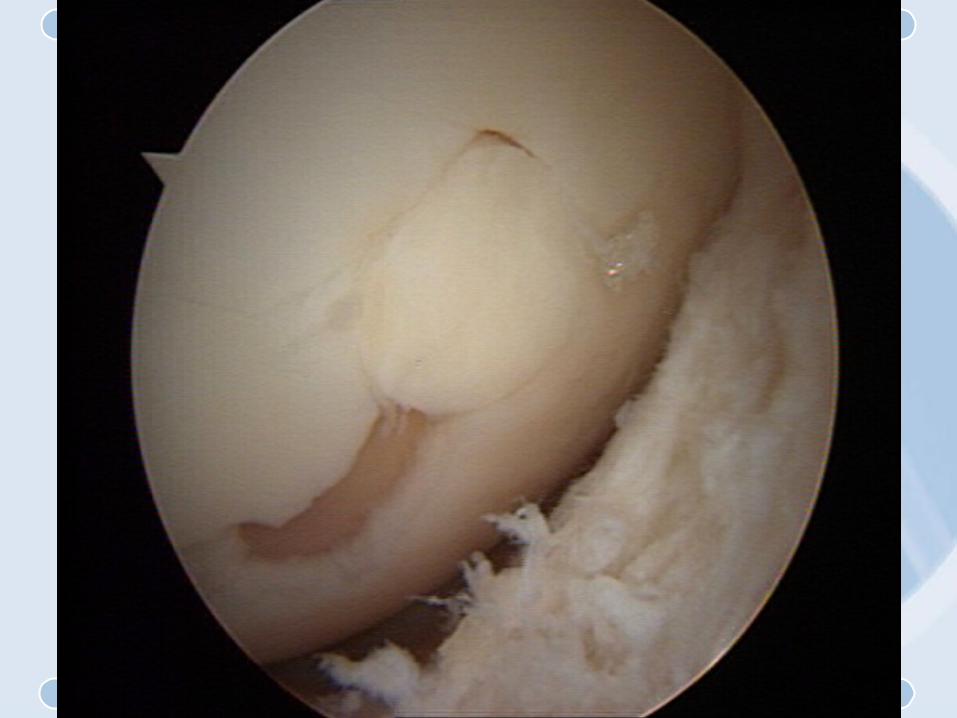
Osteochondral Grafting
• Autologous plugs of bone with hyaline cartilage cap
• Best done for small lesions (< 2cm.)
• New technique
• Limited data at follow-up
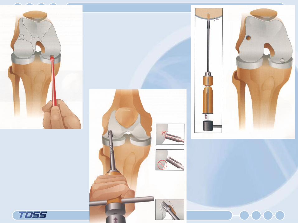

Osteochondral Autografting
• Indications– Full thickness (grade IV) lesions in the
weight bearing surface of the femoral condyles
– Well circumscribed lesion - sharp transition zone
– <2 cm diameter lesion– Young patient (< 45 yrs.)– Normal alignment and stability
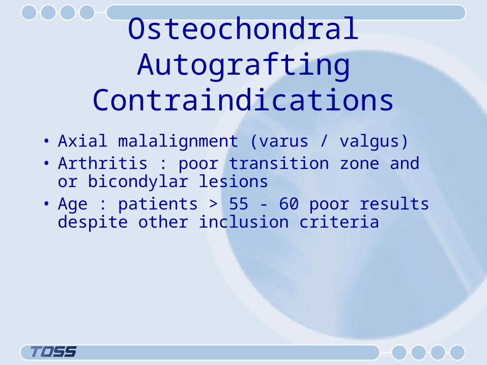
Osteochondral Autografting Contraindications
• Axial malalignment (varus / valgus)• Arthritis : poor transition zone and or bicondylar
lesions • Age : patients > 55 - 60 poor results despite other
inclusion criteria

Osteochondral Autografting Contraindications
• Lesions > 2cm. (rare)• Osteochondritis dessicans• Large OCD usually exceed donor area limitations
& large bony defects w/ no subchondral reference points


Osteochondral Autografting
• Advantages– Potential for physiologic hyaline
cartilage– Single stage procedure– Can be done all arthroscopically

Osteochondral Autografting
• Disadvantages / Concerns– Damage to the subchondral plate– Creates bleeding and fibrocartilage– Donor site morbidity– Incongruence of the plugs / articular
surface

Donor Site Morbidity : Osteochondral Autografts
• Morgan, Carter & Bobic 104 cases - no donor morbidity
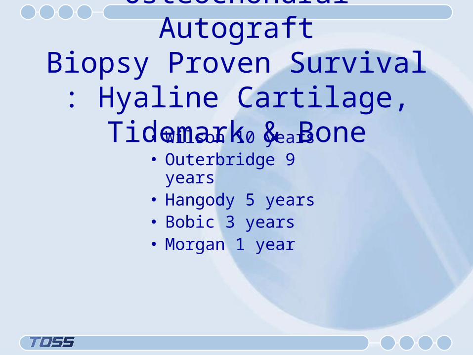
Osteochondral AutograftBiopsy Proven Survival :
Hyaline Cartilage, Tidemark & Bone
• Wilson 10 years• Outerbridge 9
years• Hangody 5 years• Bobic 3 years• Morgan 1 year

Osteochondral Autografting : Results
• Bobic– 12 Cases– Lesion 1 - 2.2cm.– 10/12 excellent results at 2
yrs.

Osteochondral Autografting : Results
• Morgan & Carter– 52 Cases– IKDC evaluation– Pain
• 65% improved 2 grades
• 31% improved 1 grade• 4% no change (failure)

LIMITATIONS OF OATS
• Potential for DJD at donor site is real
• No clinical support for repair of single or multiple plugs
–Prophylactic surgery• Difficult to justify the procedure


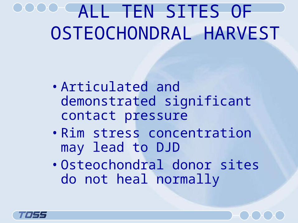
ALL TEN SITES OF OSTEOCHONDRAL HARVEST
• Articulated and demonstrated significant contact pressure
• Rim stress concentration may lead to DJD
• Osteochondral donor sites do not heal normally

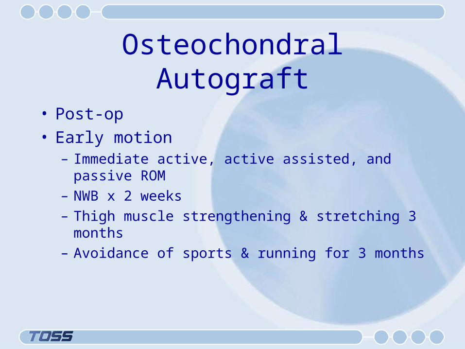
Osteochondral Autograft
• Post-op• Early motion
– Immediate active, active assisted, and passive ROM
– NWB x 2 weeks– Thigh muscle strengthening & stretching 3 months– Avoidance of sports & running for 3 months

RECOVERY FROM OATS
• Allow 6 weeks for plug to heal
• Desk job RTW 1-2 weeks
• Laborer RTW 3-4 months

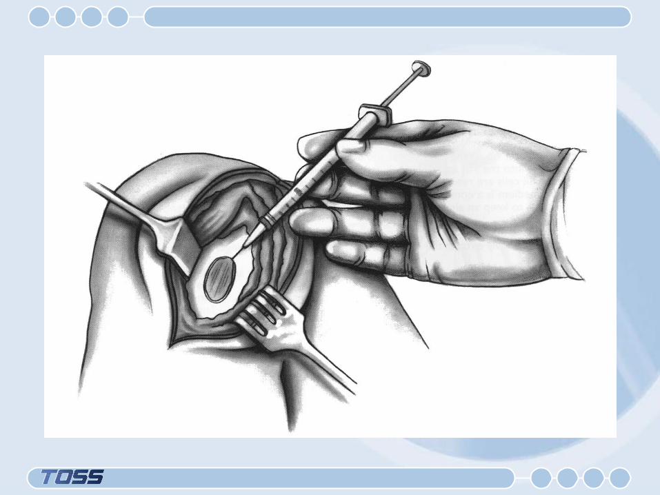
Autologous Chondrocyte Implantation
• First procedure : biopsy– Arthroscopic chondrocyte harvest from
upper medial femoral condyle
• Cultivation of cells 14-21 days• Second procedure : implantation
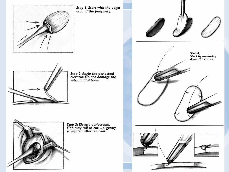
– Arthrotomy & debridement of lesion– Defect covered with periosteal flap– Cultured chondrocytes injected into defect
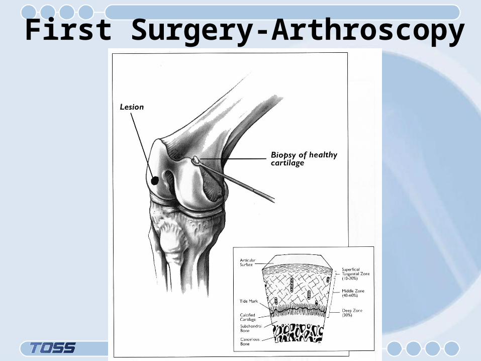
First Surgery-Arthroscopy

Second Surgery-Arthrotomy


Inject $10,000 worth of cells!

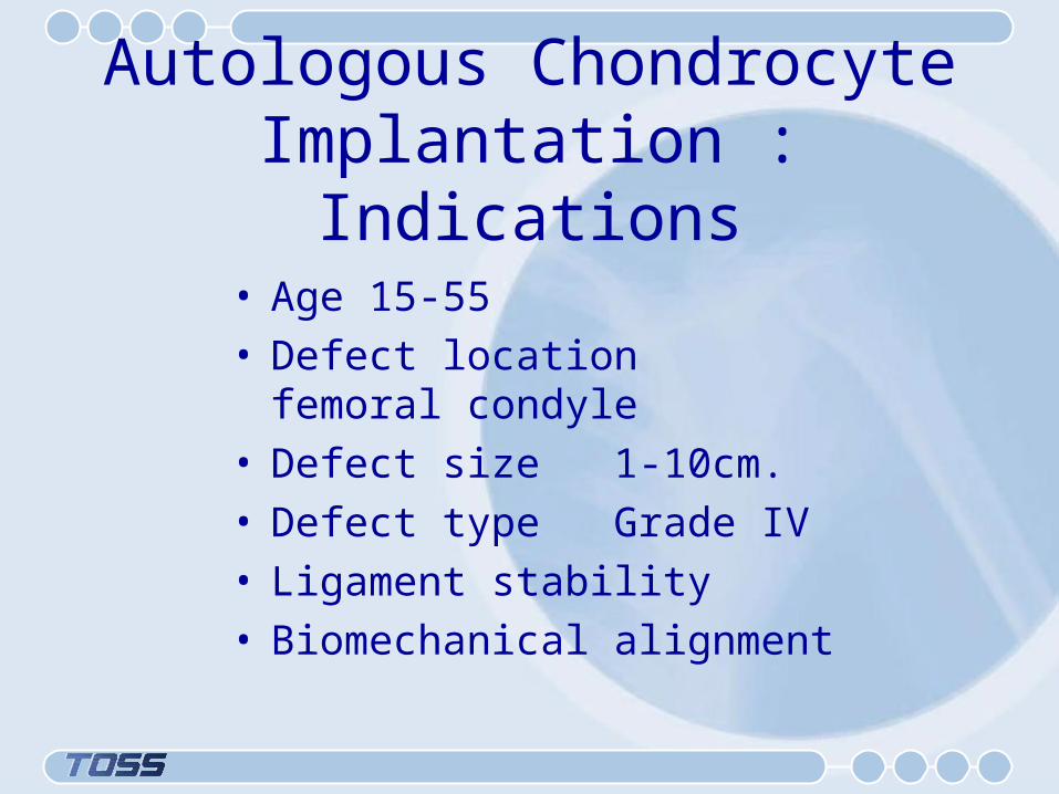
Autologous Chondrocyte Implantation : Indications
• Age 15-55• Defect location femoral
condyle• Defect size 1-10cm.• Defect type Grade IV• Ligament stability• Biomechanical alignment

Autologous Chondrocyte Implantation
• Contraindications– Kissing lesions– Inflammitory arthritis– Total meniscectomy– Over 50 (psychologic)– Unstable knee– Generalized degenerative disease– Unhealed lesion through subchondral bone

Dedifferentiation / Redifferentiation

Method of Restoration

Autologous Chondrocyte Implantation: Advantages
• Less donor site morbidity• Larger and multiple defects can be
addressed• Good results with longer follow-up• No violation of host’s subchondral plate• FDA approved

Autologous Chondrocyte Implantation : Disadvantages
• Requires 2 procedures• Not arthroscopic• Expensive• No long term results

Autologous Chondrocyte Implantation
• Post-op– CPM– Active ROM– Toe touch weight bearing for 6
weeks– week 7-12 closed chair exercises– Jogging at 6 months– Sports at 1 year

Autologous Chondrocyte Implantation
US Clinical Experience• 121 patients 6 month follow-up• 42 patients 12 month follow-up• 85% improved overall condition• 80% improved pain scores at 12
months
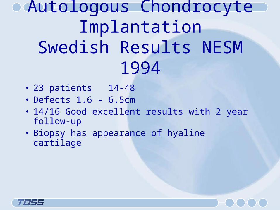
Autologous Chondrocyte Implantation
Swedish Results NESM 1994
• 23 patients 14-48• Defects 1.6 - 6.5cm• 14/16 Good excellent results with 2 year
follow-up• Biopsy has appearance of hyaline cartilage

Autologous Chondrocyte Implantation
Swedish Results 1997
• 100 patients 2-9 year follow-up– 90% improvement with femoral condyle lesions– 74% with femoral condyle and ACL reconstruction– 58% for trochlear lesions– 75% for multiple defects

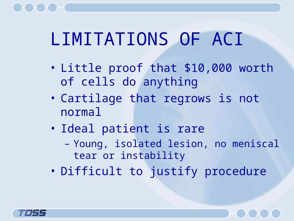
LIMITATIONS OF ACI
• Little proof that $10,000 worth of cells do anything
• Cartilage that regrows is not normal • Ideal patient is rare
– Young, isolated lesion, no meniscal tear or instability
• Difficult to justify procedure

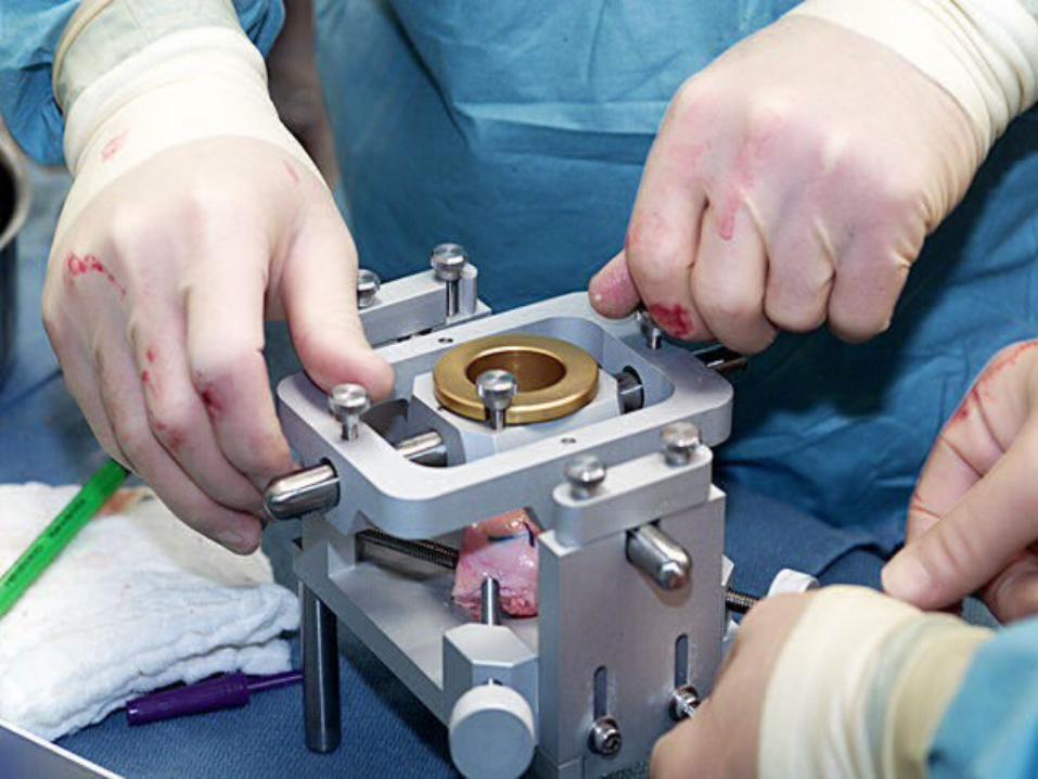
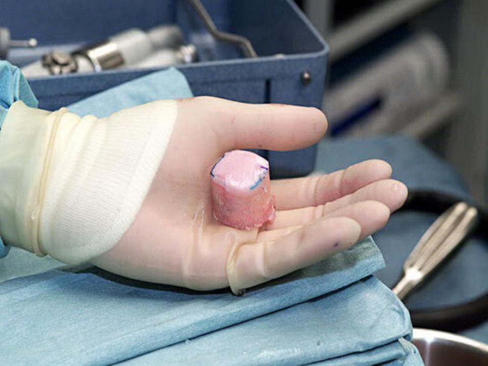
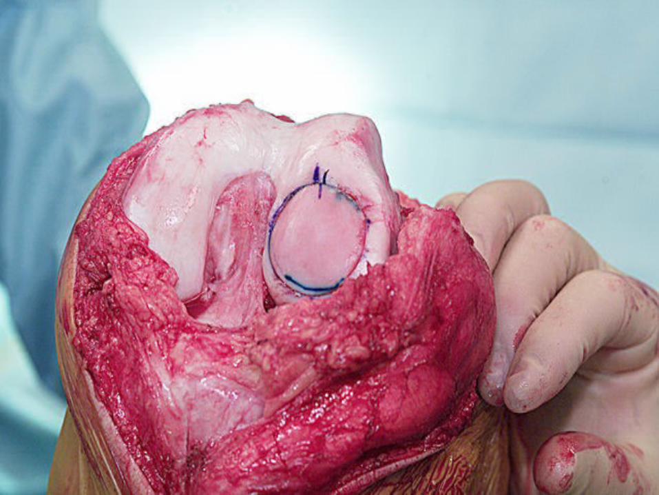
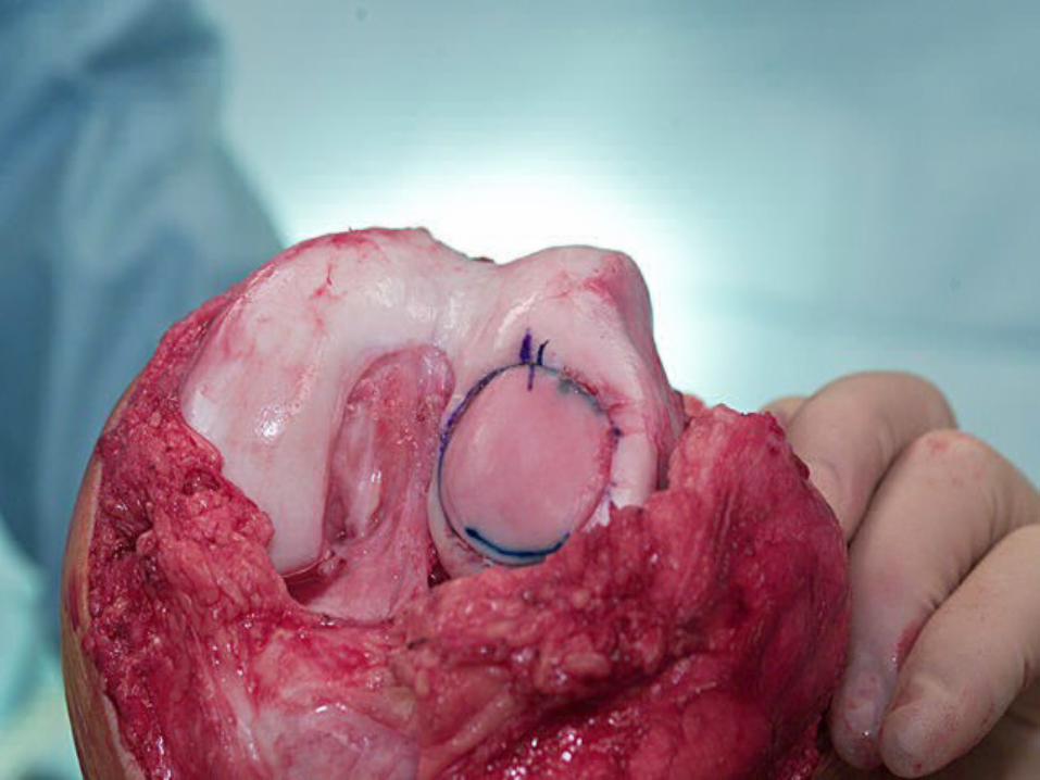
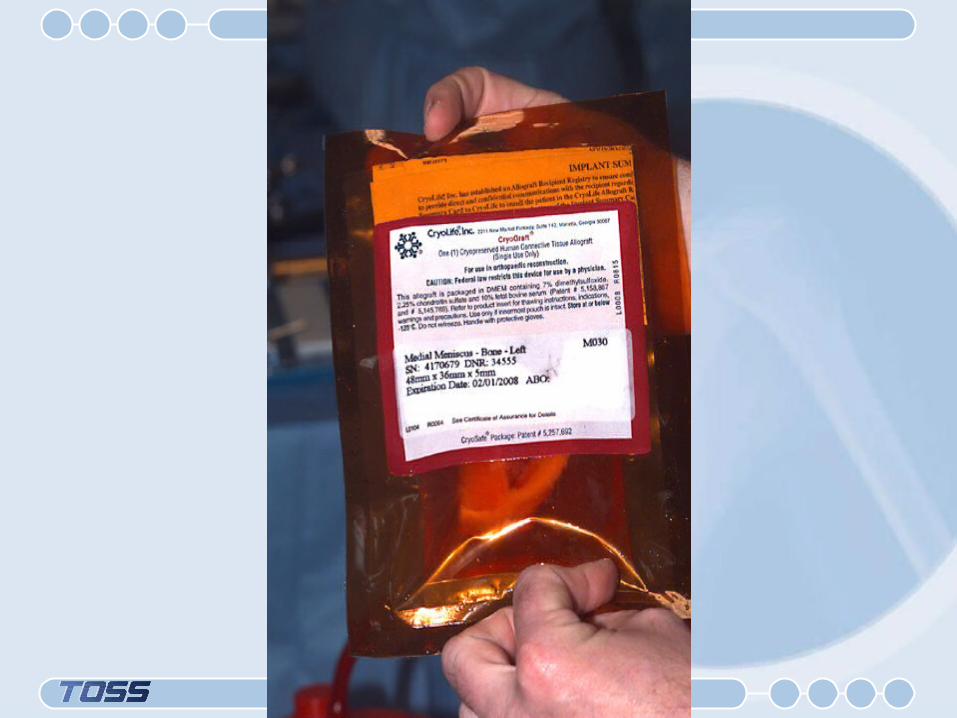
Osteochondral Allograft Transplantation
• Joint resurfacing with fresh or fresh frozen cadeveric tissue
Allograft Procedure
• Open procedure
• Expose the degenerative lesion
• Remove the defective articular cartilage and a “thin” bony base
• Utilize allograft tissue to replace and restore the articular surface

Allograft Advantages
• Replaces articular hyaline cartilage with hyaline cartilage
• Single procedure
Allograft Disadvantages
• Cost
• Risk of disease transmission from fresh allograft tissue

Allograft Results


What to do??

Treatment Recommendations
• Low demand patients
• Small focal lesion (<2cm)
• Arthroscopic chondroplasty– 50% relief up to 5 years
• Autograft Osteochondral or chondrocyte if failed chondroplasty

Treatment Recommendations
• High demand patient• Small focal lesion (<2cm)• Debridement plus drilling / fx
– 75% success with all– 50% success with sports
• Osteochondral grafting or chondrocyte transplant if failure

Treatment Recommendations
• Low demand patient
• Large lesion (>2cm)
• Debridement or microfracture with chondrocyte harvest
• If persistent pain - osteochondral or chondrocyte transplant

Treatment Recommendations
• High demand patients
• Large lesion (>2cm.)
• Chondrocyte transplant 1st line treatment yields 90% success

Long HistoryNo Acute SymptomsVarus KneeMarked DJD
Arthroscopic ResultsUnpredictable
Little Improvement

Conclusions
• Articular cartilage does not repair itself• Numerous treatments with varying results• Most treatments fail in the long term due to
articular cartilage’s inability to produce hyaline cartilage

Conclusions
• Osteochondral auto grafts and chondrocyte transplants show promising results
• Osteochondral auto grafts allow transplantation of bone capped with hyaline cartilage
• Autologous chondrocyte implantation allows near normal hyaline cartilage growth into defects

Meniscal Allograft Indications
• Patient age - young - 20-40
• Previous meniscectomy• Painful compartment• Minimal Arthritic Changes• Correct alignment• Stable knee

Sterilization
• Viral contamination risk 1:1.6 million to 1:1.2 billion
• Radiation– > 2.5 mrads destroys collagen– <2.5 mrads does not kill viruses
• Sterile harvest and storage with donor screening


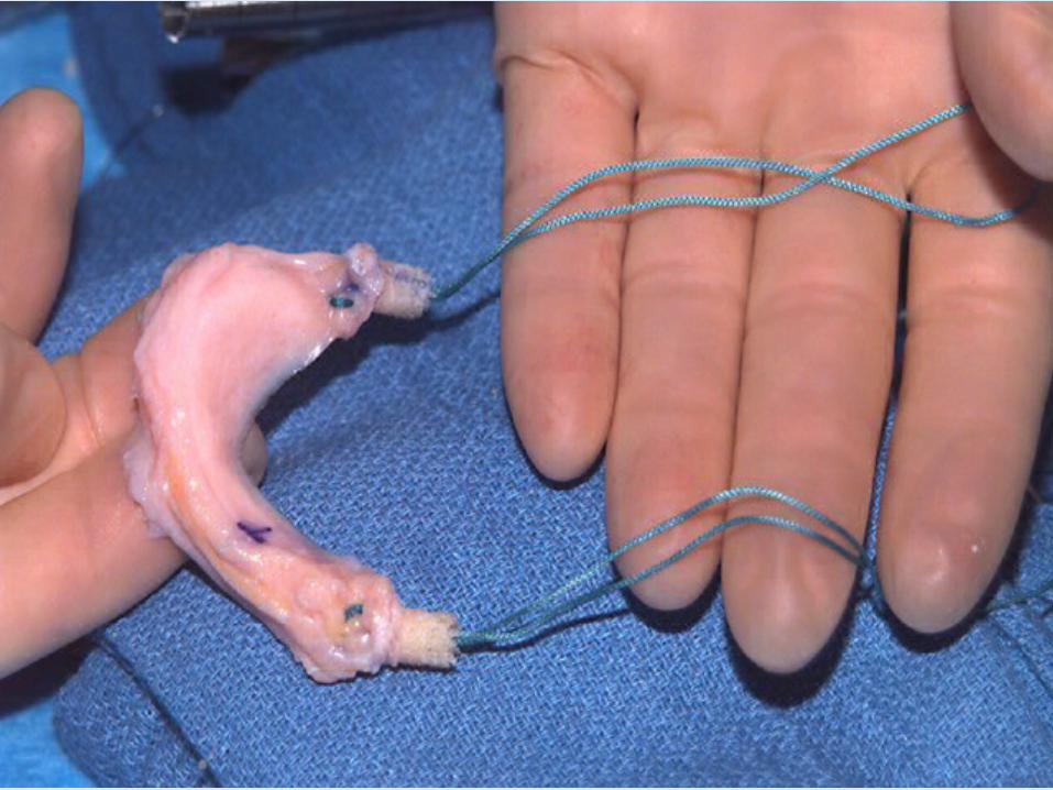
Meniscal Allograft Technique
• Bone anchors for anterior and posterior horns– Plugs for medial meniscus– Slot for lateral meniscus
• Increases the difficulty


Meniscal Allograft Technique
• Open– Easier
• Arthroscopic– Less morbidity– More technically demanding
• Collateral ligament release if necessary– Increases exposure & facilitates graft
passage under condyles

Allograft Meniscal Transplant
• Postoperative protocol– Not completely elucidated– Reflect meniscal repair protocols– Most incorporate early full ROM– Restricted weight bearing (6
weeks)– CPM early in post operative
course
Allograft Meniscal Transplant: Results
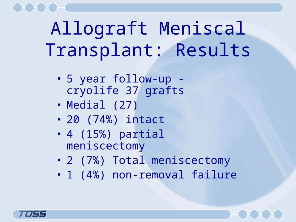
• 5 year follow-up - cryolife 37 grafts
• Medial (27)• 20 (74%) intact• 4 (15%) partial meniscectomy• 2 (7%) Total meniscectomy• 1 (4%) non-removal failure

Allograft Meniscal Transplant : Results
• Goble - 69 allografts
• 40 patients > 2 yr. follow-up
• 11 (16%) failures
• 70% of patients had subjective improvements with pain

Cryo-Life 5 Year Results
• Lateral (10)• 5 (5%) intact• 4 (40%) partial
meniscectomy• 1 (10%) total meniscectomy