Embed Size (px)
Citation preview
Word count: 9,073
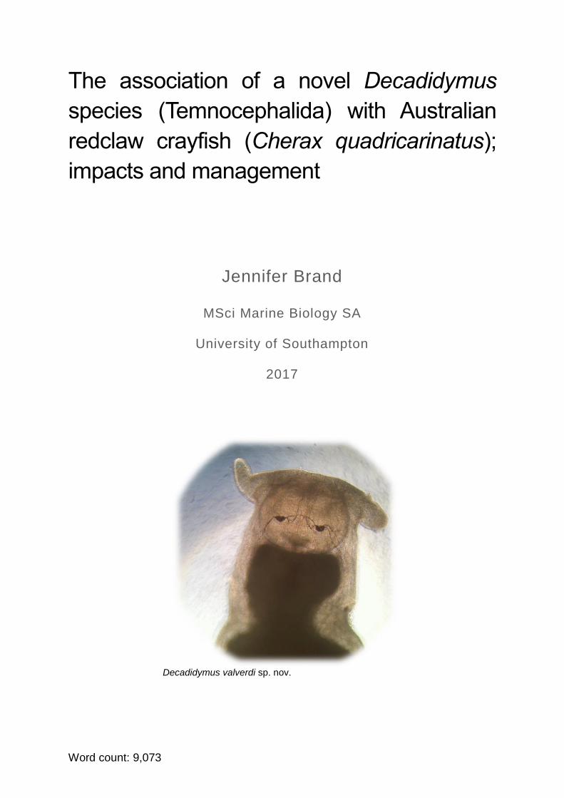
The association of a novel Decadidymus
species (Temnocephalida) with Australian
redclaw crayfish (Cherax quadricarinatus);
impacts and management
Jennifer Brand
MSci Marine Biology SA
University of Southampton
2017
Decadidymus valverdi sp. nov.
2
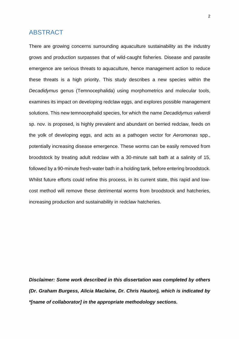
ABSTRACT
There are growing concerns surrounding aquaculture sustainability as the industry
grows and production surpasses that of wild-caught fisheries. Disease and parasite
emergence are serious threats to aquaculture, hence management action to reduce
these threats is a high priority. This study describes a new species within the
Decadidymus genus (Temnocephalida) using morphometrics and molecular tools,
examines its impact on developing redclaw eggs, and explores possible management
solutions. This new temnocephalid species, for which the name Decadidymus valverdi
sp. nov. is proposed, is highly prevalent and abundant on berried redclaw, feeds on
the yolk of developing eggs, and acts as a pathogen vector for Aeromonas spp.,
potentially increasing disease emergence. These worms can be easily removed from
broodstock by treating adult redclaw with a 30-minute salt bath at a salinity of 15,
followed by a 90-minute fresh-water bath in a holding tank, before entering broodstock.
Whilst future efforts could refine this process, in its current state, this rapid and low-
cost method will remove these detrimental worms from broodstock and hatcheries,
increasing production and sustainability in redclaw hatcheries.
Disclaimer: Some work described in this dissertation was completed by others
(Dr. Graham Burgess, Alicia Maclaine, Dr. Chris Hauton), which is indicated by
*[name of collaborator] in the appropriate methodology sections.
3
Table of Contents
ABSTRACT ................................................................................................................ 2
Acknowledgements .................................................................................................... 3
Introduction ................................................................................................................ 4
Chapter 1: Species Characterisation .......................................................................... 8
1.1 Introduction ....................................................................................................... 8
1.2 Sampling, materials and methods ................................................................... 10
1.3 Results ............................................................................................................ 14
1.4 Discussion ....................................................................................................... 19
Chapter 2: Impacts ................................................................................................... 21
2.1 Introduction ..................................................................................................... 21
2.2 Materials and methods .................................................................................... 23
2.3 Results ............................................................................................................ 28
2.4 Discussion ....................................................................................................... 33
Chapter 3: Management ........................................................................................... 37
3.1 Introduction ..................................................................................................... 37
3.2 Materials and methods .................................................................................... 39
3.3 Results ............................................................................................................ 41
3.4. Discussion ...................................................................................................... 45
Conclusions .............................................................................................................. 48
References ............................................................................................................... 50
Appendices .............................................................................................................. 54
Acknowledgements I would like to thank Dr. Ellen Ariel, Dr. Lisa Elliott and Dr. Chris Hauton for their
guidance and support throughout this project. Enormous thanks to Colin Valverde for
providing the study site (AquaVerde) and crayfish for this research. Thanks to James
Cook University and the University of Southampton for providing required facilities.
Thanks also to Sue and Laurie Riley, Dr. Jen Whan, Dr. Graham Burgess and Alicia
Maclaine for their advice and contributions. Lastly, huge thanks to my family, friends,
and the University of Southampton, who have all supported me throughout my studies.
4
Introduction
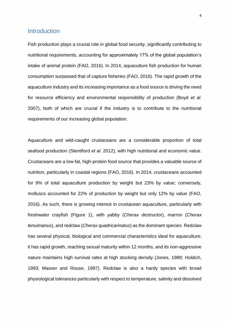
Fish production plays a crucial role in global food security, significantly contributing to
nutritional requirements, accounting for approximately 17% of the global population’s
intake of animal protein (FAO, 2016). In 2014, aquaculture fish production for human
consumption surpassed that of capture fisheries (FAO, 2016). The rapid growth of the
aquaculture industry and its increasing importance as a food source is driving the need
for resource efficiency and environmental responsibility of production (Boyd et al.
2007), both of which are crucial if the industry is to contribute to the nutritional
requirements of our increasing global population.
Aquaculture and wild-caught crustaceans are a considerable proportion of total
seafood production (Stentiford et al. 2012), with high nutritional and economic value.
Crustaceans are a low-fat, high-protein food source that provides a valuable source of
nutrition, particularly in coastal regions (FAO, 2016). In 2014, crustaceans accounted
for 9% of total aquaculture production by weight but 23% by value; conversely,
molluscs accounted for 22% of production by weight but only 12% by value (FAO,
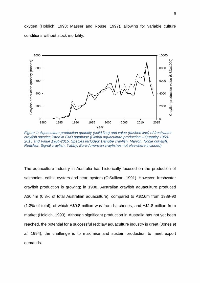
2016). As such, there is growing interest in crustacean aquaculture, particularly with
freshwater crayfish (Figure 1), with yabby (Cherax destructor), marron (Cherax
tenuimanus), and redclaw (Cherax quadricarinatus) as the dominant species. Redclaw
has several physical, biological and commercial characteristics ideal for aquaculture;
it has rapid growth, reaching sexual maturity within 12 months, and its non-aggressive
nature maintains high survival rates at high stocking density (Jones, 1989; Holdich,
1993; Masser and Rouse, 1997). Redclaw is also a hardy species with broad
physiological tolerances particularly with respect to temperature, salinity and dissolved
5
oxygen (Holdich, 1993; Masser and Rouse, 1997), allowing for variable culture
conditions without stock mortality.
Figure 1: Aquaculture production quantity (solid line) and value (dashed line) of freshwater crayfish species listed in FAO database (Global aquaculture production – Quantity 1950-2015 and Value 1984-2015. Species included: Danube crayfish, Marron, Noble crayfish, Redclaw, Signal crayfish, Yabby, Euro-American crayfishes not elsewhere included)
The aquaculture industry in Australia has historically focused on the production of
salmonids, edible oysters and pearl oysters (O'Sullivan, 1991). However, freshwater
crayfish production is growing; in 1988, Australian crayfish aquaculture produced
A$0.4m (0.3% of total Australian aquaculture), compared to A$2.6m from 1989-90
(1.3% of total), of which A$0.8 million was from hatcheries, and A$1.8 million from
market (Holdich, 1993). Although significant production in Australia has not yet been
reached, the potential for a successful redclaw aquaculture industry is great (Jones et
al. 1994); the challenge is to maximise and sustain production to meet export
demands.
0
2000
4000
6000
8000
10000
0
200
400
600
800
1000
1980 1985 1990 1995 2000 2005 2010 2015
Cra
yfis
h p
roduction v
alu
e (
US
Dx1000)
Cra
yfis
h p
roduction q
uantity
(to
nnes)
Year
6
Hatchery development
Hatchery and nursery technology has been developed for most farmed aquatic
species (FAO, 2014), including crustaceans such as shrimp, lobster, freshwater
prawn, crab and some freshwater crayfish (Nelson and Dendy, 1979; Malecha, 1983;
Charmantier-Daures and Charmantier, 1991; Jackson et al. 1992; Jones,
1995; Kittaka, 1997). Implementing hatcheries in crayfish aquaculture addresses the
key limiting factors on production; seed stock availability (Villarreal and Pelaez, 2000)
and the intensity and inconsistency of pond rearing, hence the interest in the
development of economical hatchery and nursery protocols is growing.
However, constraints on the success of hatchery and nursery facilities exist, most
significantly of which is frequent and seemingly sporadic crop failures, thought to be
caused by disease. Disease has caused substantial animal and economic losses in
hatcheries and the industry (Walker and Winton, 2010), and the occurrence of
outbreaks is expected to increase as the industry grows (Edgerton et al. 2002).
Therefore, to improve global food security in the crustacean aquaculture industry and
reduce production losses, a greater understanding of crayfish diseases and parasites
is required, coupled with management efforts towards their eradication (Murray and
Peeler, 2005; Ghanawi and Saoud, 2012; Stentiford et al. 2012).
Crustacean disease and parasites in aquaculture
The most significant threat to the continued expansion of aquaculture is disease and
parasite emergence (Meyer, 1991; Bondad-Reantaso et al. 2005), exacerbated by
aquaculture providing a conducive environment for disease emergence, establishment
and transmission (Murray and Peeler, 2005). Decapod crustaceans at all life stages
are susceptible to a variety of pathogens and parasites, including viral, bacterial,
7
fungal and metazoan (Longshaw, 2011; Stentiford et al. 2012). Whilst a variety of
metazoan parasites are found on crustaceans (such as trematodes, cestodes,
turbellaria and small parasitic crustaceans), the most severe losses to production are
attributable to viruses and bacteria (Bower et al. 1994; Stentiford et al. 2012).
Several reviews have examined diseases specific to crayfish (Edgerton, 1999;
Edgerton et al. 2002; Longshaw, 2011; Saoud et al. 2013), although much greater
understanding of redclaw diseases is needed (Ghanawi and Saoud, 2012). Disease
and parasite reviews often include fouling organisms such as Branchiobdellida and
Temnocephalida, although they receive little attention in comparison to disease. This
is due to their classification as ectocommensals or ectosymbiotes, rather than
parasitoids or predators, which would cause harm to hosts and reduce production.
This study aims to investigate the ecological relationship between a Decadidymus
temnocephalid and redclaw crayfish (Cherax quadricarinatus). The identity of this
species is discussed, with regards to morphological characteristics and phylogeny.
The epidemiology of this species is examined, in terms of its prevalence, abundance
and impact on hatchery productivity, as a potential egg predator and pathogen vector.
Lastly, this research explores possible management solutions for the control of this
species.
8
Chapter 1: Species Characterisation
1.1 Introduction
Temnocephalids (phylum Platyhelminthes) are small, active flatworms found on fresh
water crustaceans such as redclaw (Volonterio, 2009). They are characterised by the
presence of eyespots, a posterior sucker and anterior processes used for attachment
and movement around the host (Sewell and Whittington, 1995; Edgerton et al. 2002).
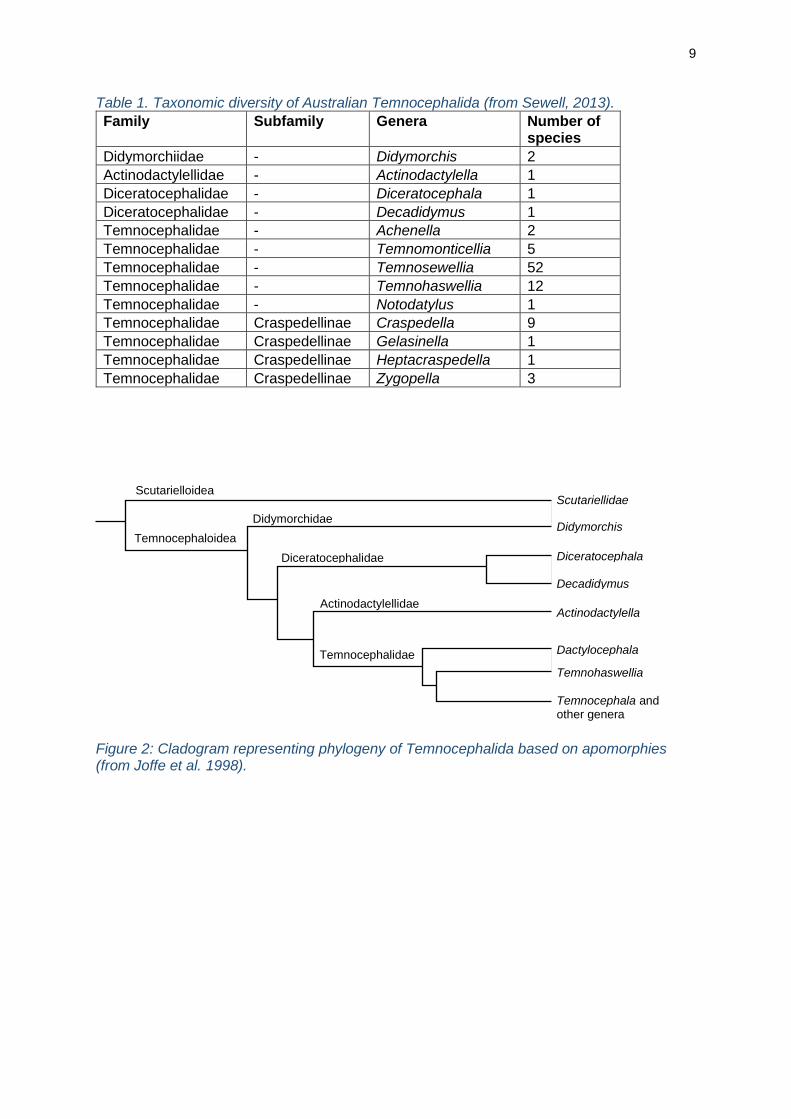
Australia is considered the global centre of temnocephalan diversity, with 91 named
temnocephalid species within 13 genera (Table 1, Sewell, 2013). Some
temnocephalids have been examined in detail, such as Diceratocephala boschmai,
Temnocephala minor and Craspedella spenceri, however most remain understudied.
One somewhat understudied group within the Temnocephalida is the genus
Decadidymus (Family Diceratocephalidae). The only species currently listed within
this genus is Decadidymus gulosus (Cannon, 1991), with a description of its gross
morphology and key anatomical features such as reproductive systems, mouth,
pharynx and gut. Since this description, the apomorphies of the Diceratocephalidae
have been identified (Joffe et al. 1998). However, there is no published data on the
molecular characterisation of Decadidymus species; suggested phylogenetic
placement of Decadidymus within the Diceratocephalidae is based on morphological
data (Figure 2, Joffe et al. 1998).
This investigation aims to identify a temnocephalid that occurs in redclaw crayfish
broodstock. For the first time, a genetic sequence of a Decadidymus species was
provided. Combining molecular tools with morphological characteristics of the species
in question, the possibility of a novel species is discussed.
9
Table 1. Taxonomic diversity of Australian Temnocephalida (from Sewell, 2013).
Family Subfamily Genera Number of species
Didymorchiidae - Didymorchis 2
Actinodactylellidae - Actinodactylella 1
Diceratocephalidae - Diceratocephala 1
Diceratocephalidae - Decadidymus 1
Temnocephalidae - Achenella 2
Temnocephalidae - Temnomonticellia 5
Temnocephalidae - Temnosewellia 52
Temnocephalidae - Temnohaswellia 12
Temnocephalidae - Notodatylus 1
Temnocephalidae Craspedellinae Craspedella 9
Temnocephalidae Craspedellinae Gelasinella 1
Temnocephalidae Craspedellinae Heptacraspedella 1
Temnocephalidae Craspedellinae Zygopella 3
Figure 2: Cladogram representing phylogeny of Temnocephalida based on apomorphies (from Joffe et al. 1998).
Temnohaswellia
Temnocephala and other genera
Dactylocephala
Actinodactylella
Diceratocephala
Decadidymus
Didymorchis
Scutariellidae
Temnocephalidae
Actinodactylellidae
Diceratocephalidae
Didymorchidae
Temnocephaloidea
Scutarielloidea
10
1.2 Sampling, materials and methods
Redclaw were obtained from indoor, climate controlled broodstock tanks from a
hatchery in Atherton, North Queensland during the wet season (Jan-Mar 2016).
Decadidymus specimens were separated from adult crayfish and their eggs following
egg-stripping of berried females. The abdomen and cephalothorax of the crayfish were
examined and any remaining worms were removed with fine forceps.
1.2.1 Morphology
Decadidymus specimens were held alive in 40ml sterile physiologically-buffered
saline (0.9% NaCl solution) (PBS) for no longer than an hour before subsequent
treatments. Worms required anaesthetising before exposure to fixative to prevent
body distortion. Anaesthetisation trials were conducted to determine the most
effective method (Appendix A); the selected method was dropwise addition of room-
temperature 95% ethanol to a specimen jar containing worms and 20ml of sterile
water, until activity ceased and worms became fully relaxed. All morphological
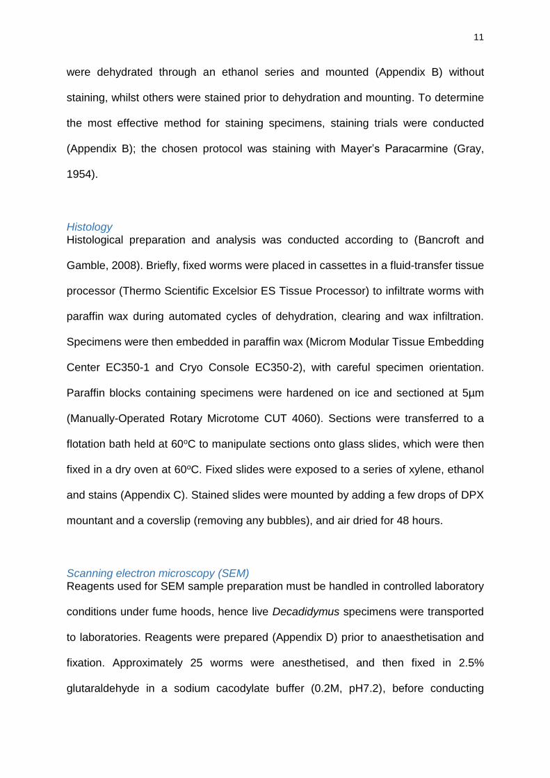
characteristics identified were compared to Temnocephalida morphology (Table 2)
and dorsal facies (Figure 3) to identify the species to genus level.
Whole mounts and staining Following anaesthetisation, worms were washed in PBS three times. Smaller worms
were fixed in a specimen jar containing 40ml 10% neutral-buffered formalin (NBF).
Larger worms were positioned between two glass slides separated by Vaseline™ so
as not to destroy the specimens, and placed inside a screw-cap Coplin Jar containing
10% NBF, producing slightly flattened fixed specimens for whole mounting of larger
specimens. Specimens were transported to laboratories in 10% NBF, for staining and
mounting. Worms were washed three times in PBS to remove traces of NBF. Some
11
were dehydrated through an ethanol series and mounted (Appendix B) without
staining, whilst others were stained prior to dehydration and mounting. To determine
the most effective method for staining specimens, staining trials were conducted
(Appendix B); the chosen protocol was staining with Mayer’s Paracarmine (Gray,
1954).
Histology Histological preparation and analysis was conducted according to (Bancroft and
Gamble, 2008). Briefly, fixed worms were placed in cassettes in a fluid-transfer tissue
processor (Thermo Scientific Excelsior ES Tissue Processor) to infiltrate worms with
paraffin wax during automated cycles of dehydration, clearing and wax infiltration.
Specimens were then embedded in paraffin wax (Microm Modular Tissue Embedding
Center EC350-1 and Cryo Console EC350-2), with careful specimen orientation.
Paraffin blocks containing specimens were hardened on ice and sectioned at 5µm
(Manually-Operated Rotary Microtome CUT 4060). Sections were transferred to a
flotation bath held at 60oC to manipulate sections onto glass slides, which were then
fixed in a dry oven at 60oC. Fixed slides were exposed to a series of xylene, ethanol
and stains (Appendix C). Stained slides were mounted by adding a few drops of DPX
mountant and a coverslip (removing any bubbles), and air dried for 48 hours.
Scanning electron microscopy (SEM) Reagents used for SEM sample preparation must be handled in controlled laboratory
conditions under fume hoods, hence live Decadidymus specimens were transported
to laboratories. Reagents were prepared (Appendix D) prior to anaesthetisation and
fixation. Approximately 25 worms were anesthetised, and then fixed in 2.5%
glutaraldehyde in a sodium cacodylate buffer (0.2M, pH7.2), before conducting
12
standard SEM sample preparation (Appendix D). A JEOL Scanning electron
microscope (JSM-5410LV) was used to capture images, adjusting the position and
orientation of the specimen within the vacuum by moving, tilting and rotating the
specimen stage, and adjusting the quality of the image using resolution, magnification,
and contrast and brightness settings.
Table 2. Morphological characteristics to identify Temnocephalida to genus (Sewell, 2013).
Genus Locomo-tory cilia
Number of tentacles
Medial tentacle bulb-shaped
Dorsal scales
Number of dorsal papillate ridges
Ciliated papillae in rows on tentacles
Number of pairs of testes
Didymorchis Y 0 - N 0 N 1
Diceratocephala Y 2 - N 0 N 1
Decadidymus N 2 - N 0 N 10
Actinodactylella N 12 - N 0 Y 2
Temnohaswellia N 6 N N 0 N 2
Temnomonticellia N 5 Y N 0 N 2
Temnosewellia N 5 N N 0 N 2
Achenella N 5 N N 0 N 1
Notodactylus N 5 N Y 0 N 2
Zygopella N 5 N N 1 Y 2
Gellasinella N 5 N N 2 Y 2
Craspedella N 5 N N 3 Y 2
Heptacraspedella N 5 N N 7 Y 2
Figure 3: Dorsal facies of Australian Temnocephalida. Scale bar = ~1 mm (Sewell, 2013).
13
1.2.2 Molecular characterisation *[Dr. Graham Burgess and Alicia Maclaine]
Once separated from crayfish eggs, specimens were immediately washed in sterile
PBS five times and preserved in 80% ethanol (no anaesthetisation required). DNA
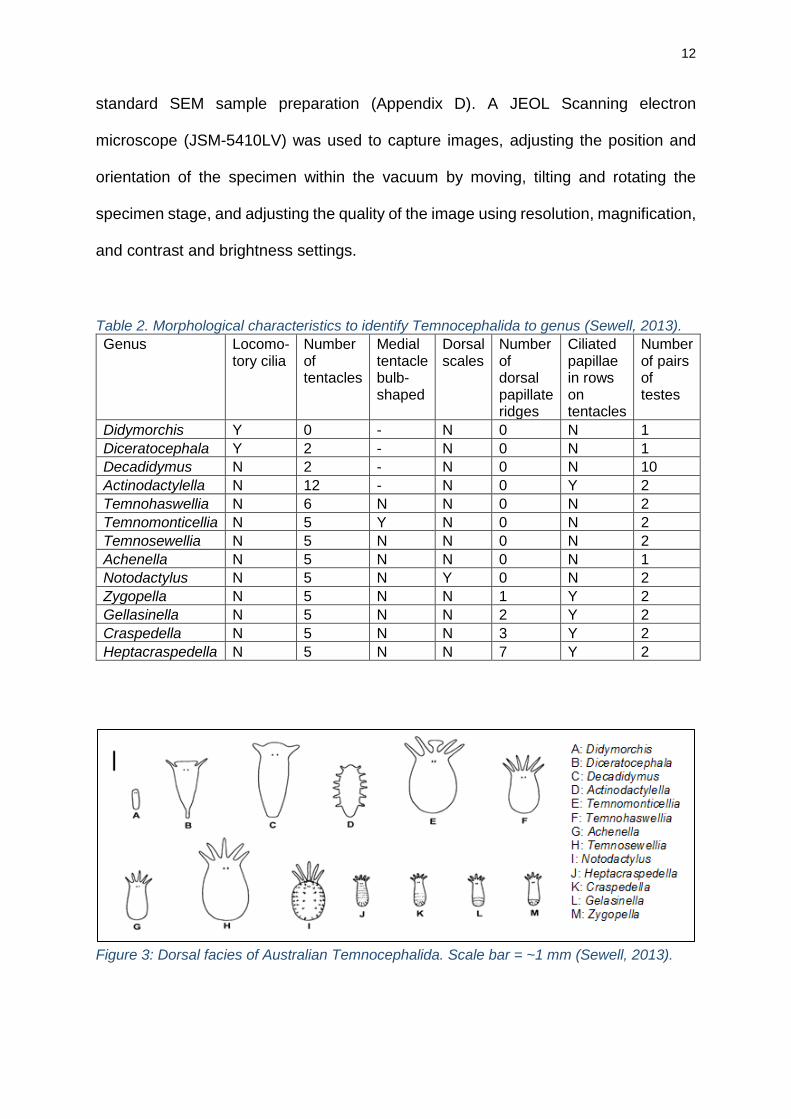
was extracted (Bioline ISOLATE II Genomic DNA Kit), and primers chosen (Table 3)
produced amplicons of 28S and 18S ribosomal RNA sequences. PCR products were
sequenced (Macrogen Inc. South Korea), and sequences were assembled using
Geneious 9.1 (Biomatters Ltd New Zealand). A search on NCBI BLAST returned the
sequences with greatest homology to the sequence of this study species.
Table 3: Primers used to sequence Decadidymus valverdi sp. nov.
Primer name Primer sequence Sequence amplified
Expected size (bp)
Purpose Reference
U178 GCACCCGCTGAAYTTAAG 28s 1525 PCR
Lockyer et al. 2003
L1642 CCAGCGCCATCCATTTTCA
U1148 GACCCGAAAGATGGTGAA 28s 1391 PCR
Lockyer et al. 2003
L2450 GCTTTGTTTTAATTAGACAGTCGGA
FW-28s-1322-F AGCAGGTCTCCAAGGTTA 28s 1332 PCR This study
FW-28s-1322-R ACTTAGAGGCGTTCAGTCT
FW-28s-401-F AGTAACGCAGGTGTCCAA 28s 401 Sequencing This study
FW-28s-401-R CTCTCGTACTGAGCAGGATTA
FW-18s-1786 F GTCTCAAAGATTAAGCCATGC 18s 1786 PCR This study
FW-18s-1786 R CGGAAACCTTGTTACGACTT
FW-28s-570-F AGAACTGGCACGGACAAG 18s 570 Sequencing This study
FW-28s-570-R GCTCACCTTTGGACACCT

14
1.3 Results
1.3.1 Morphology
This study species belongs to the Decadidymus genus; specimens possess the three
apomorphies of the Diceratocephalidae family (Joffe et al. 1998), as well as the
morphological characteristics of Decadidymus (Table 2 and Figure 3). Features of
these study specimens consistent with the Decadidymus gulosus description
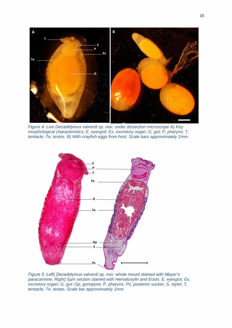
(Cannon, 1991) include; heavy-bodied worms, 3-4mm in length, a posterior, muscular
adhesive disk, and excretory pores on each side on the anterolateral margin (Figures
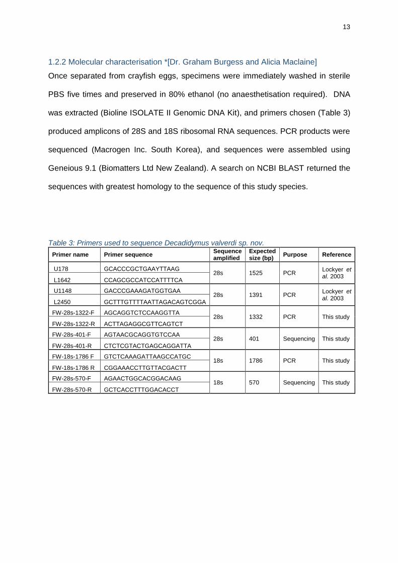
4 and 5). The presence of two prominent anterior eyespots, sensory papillae covering
the body (particularly on the tentacles and posterior sucker), 10 pairs of testes and a
stylet (Figures 4 and 5) are also features consistent with Cannon’s description of D.
gulosus.
However, there are notable differences between Cannon’s description of D. gulosus
(1991) and these study specimens; the location and size of the mouth, the relative size
of the pharynx compared to total body length, the size and location of the gonopore,
the location of the excretory pores and the size of the stylet (Figures 4, 5 and 6, Table
4). These features are consistent across all worms that were examined
morphologically (approximately 80 worms across wholemount, histology and SEM
examinations). Furthermore, SEM images of D. gulosus (Cannon, 1991) showing
overall body plan and position of the mouth appear vastly different to the SEM images
produced in this study (Figure 6). As such, the name Decadidymus valverdi sp. nov.
is proposed.
15
Figure 4: Live Decadidymus valverdi sp. nov. under dissection microscope A) Key morphological characteristics; E, eyespot; Ex, excretory organ; G, gut; P, pharynx; T, tentacle; Te, testes. B) With crayfish eggs from host. Scale bars approximately 1mm.
Figure 5: Left) Decadidymus valverdi sp. nov. whole mount stained with Mayer’s paracarmine. Right) 5µm section stained with Hematoxylin and Eosin. E, eyespot; Ex, excretory organ; G, gut; Gp, gonopore; P, pharynx; Ps, posterior sucker; S, stylet; T, tentacle; Te, testes. Scale bar approximately 1mm.
A B
T
P
Te
G
E
Ex
Te
E
S Gp
P T
Ps
G
Ex
16
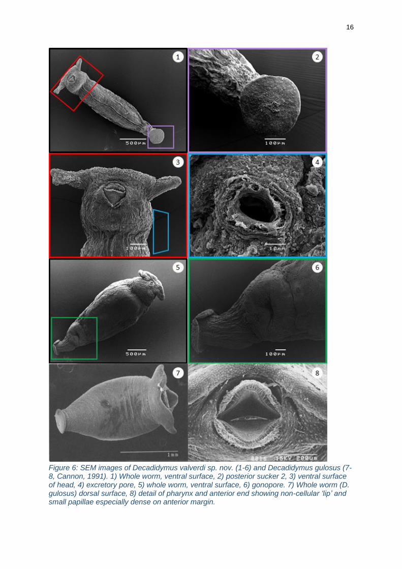
Figure 6: SEM images of Decadidymus valverdi sp. nov. (1-6) and Decadidymus gulosus (7-8, Cannon, 1991). 1) Whole worm, ventral surface, 2) posterior sucker 2, 3) ventral surface of head, 4) excretory pore, 5) whole worm, ventral surface, 6) gonopore. 7) Whole worm (D. gulosus) dorsal surface, 8) detail of pharynx and anterior end showing non-cellular ‘lip’ and small papillae especially dense on anterior margin.
17
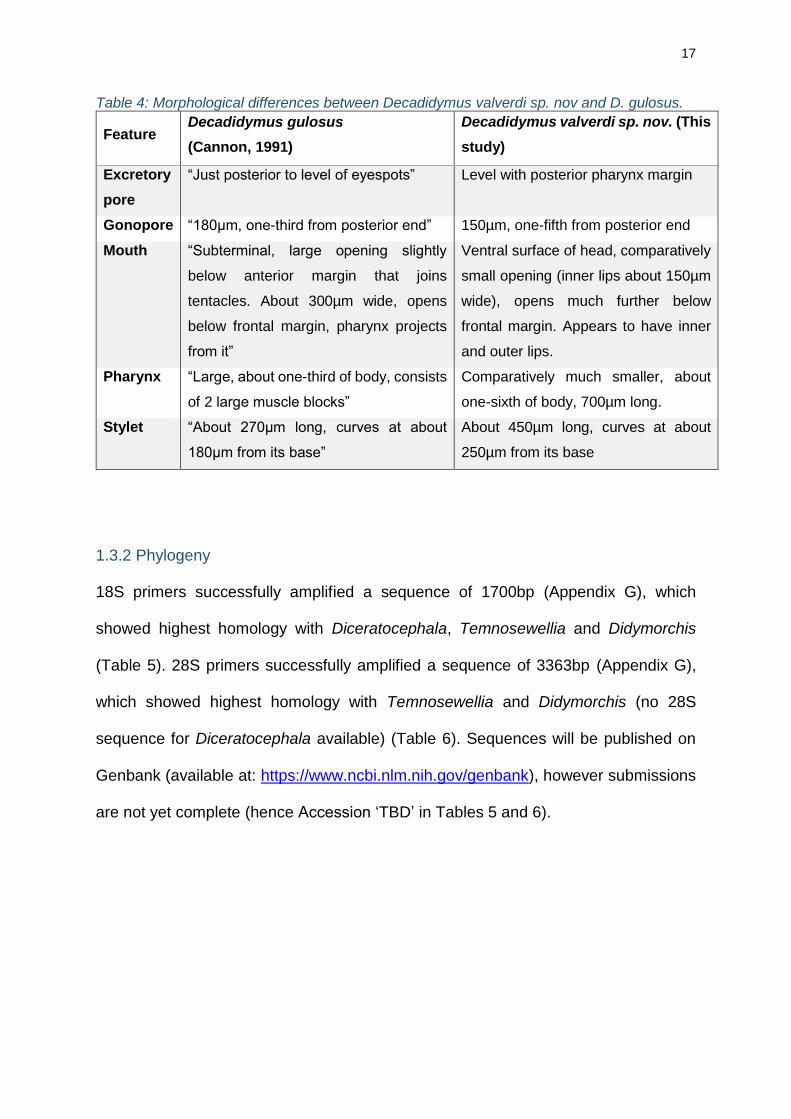
Table 4: Morphological differences between Decadidymus valverdi sp. nov and D. gulosus.
Feature Decadidymus gulosus
(Cannon, 1991)
Decadidymus valverdi sp. nov. (This
study)
Excretory
pore
“Just posterior to level of eyespots” Level with posterior pharynx margin
Gonopore “180µm, one-third from posterior end” 150µm, one-fifth from posterior end
Mouth “Subterminal, large opening slightly
below anterior margin that joins
tentacles. About 300µm wide, opens
below frontal margin, pharynx projects
from it”
Ventral surface of head, comparatively
small opening (inner lips about 150µm
wide), opens much further below
frontal margin. Appears to have inner
and outer lips.
Pharynx “Large, about one-third of body, consists
of 2 large muscle blocks”
Comparatively much smaller, about
one-sixth of body, 700µm long.
Stylet “About 270µm long, curves at about
180µm from its base”
About 450µm long, curves at about
250µm from its base
1.3.2 Phylogeny
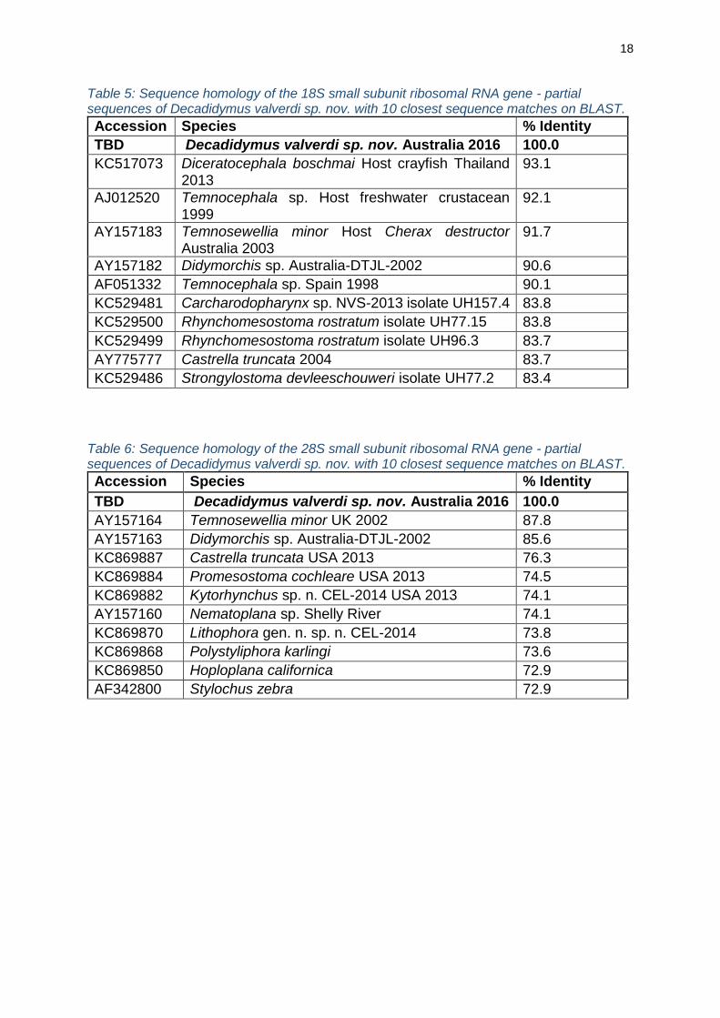
18S primers successfully amplified a sequence of 1700bp (Appendix G), which
showed highest homology with Diceratocephala, Temnosewellia and Didymorchis
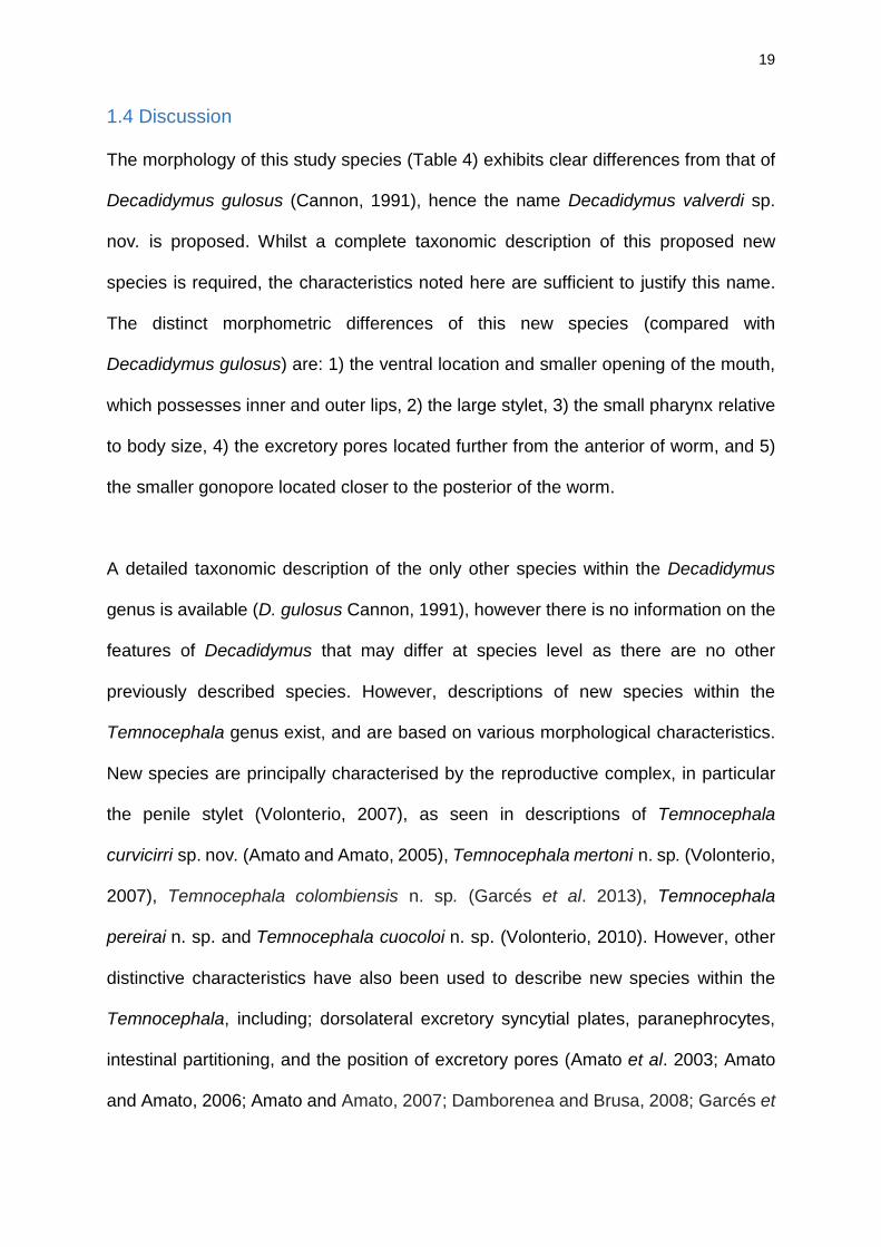
(Table 5). 28S primers successfully amplified a sequence of 3363bp (Appendix G),
which showed highest homology with Temnosewellia and Didymorchis (no 28S
sequence for Diceratocephala available) (Table 6). Sequences will be published on
Genbank (available at: https://www.ncbi.nlm.nih.gov/genbank), however submissions
are not yet complete (hence Accession ‘TBD’ in Tables 5 and 6).
18
Table 5: Sequence homology of the 18S small subunit ribosomal RNA gene - partial sequences of Decadidymus valverdi sp. nov. with 10 closest sequence matches on BLAST.
Accession Species % Identity
TBD Decadidymus valverdi sp. nov. Australia 2016 100.0
KC517073 Diceratocephala boschmai Host crayfish Thailand 2013
93.1
AJ012520 Temnocephala sp. Host freshwater crustacean 1999
92.1
AY157183 Temnosewellia minor Host Cherax destructor Australia 2003
91.7
AY157182 Didymorchis sp. Australia-DTJL-2002 90.6
AF051332 Temnocephala sp. Spain 1998 90.1
KC529481 Carcharodopharynx sp. NVS-2013 isolate UH157.4 83.8
KC529500 Rhynchomesostoma rostratum isolate UH77.15 83.8
KC529499 Rhynchomesostoma rostratum isolate UH96.3 83.7
AY775777 Castrella truncata 2004 83.7
KC529486 Strongylostoma devleeschouweri isolate UH77.2 83.4
Table 6: Sequence homology of the 28S small subunit ribosomal RNA gene - partial sequences of Decadidymus valverdi sp. nov. with 10 closest sequence matches on BLAST.
Accession Species % Identity
TBD Decadidymus valverdi sp. nov. Australia 2016 100.0
AY157164 Temnosewellia minor UK 2002 87.8
AY157163 Didymorchis sp. Australia-DTJL-2002 85.6
KC869887 Castrella truncata USA 2013 76.3
KC869884 Promesostoma cochleare USA 2013 74.5
KC869882 Kytorhynchus sp. n. CEL-2014 USA 2013 74.1
AY157160 Nematoplana sp. Shelly River 74.1
KC869870 Lithophora gen. n. sp. n. CEL-2014 73.8
KC869868 Polystyliphora karlingi 73.6
KC869850 Hoploplana californica 72.9
AF342800 Stylochus zebra 72.9
19
1.4 Discussion
The morphology of this study species (Table 4) exhibits clear differences from that of
Decadidymus gulosus (Cannon, 1991), hence the name Decadidymus valverdi sp.
nov. is proposed. Whilst a complete taxonomic description of this proposed new
species is required, the characteristics noted here are sufficient to justify this name.
The distinct morphometric differences of this new species (compared with
Decadidymus gulosus) are: 1) the ventral location and smaller opening of the mouth,
which possesses inner and outer lips, 2) the large stylet, 3) the small pharynx relative
to body size, 4) the excretory pores located further from the anterior of worm, and 5)
the smaller gonopore located closer to the posterior of the worm.
A detailed taxonomic description of the only other species within the Decadidymus
genus is available (D. gulosus Cannon, 1991), however there is no information on the
features of Decadidymus that may differ at species level as there are no other
previously described species. However, descriptions of new species within the
Temnocephala genus exist, and are based on various morphological characteristics.
New species are principally characterised by the reproductive complex, in particular
the penile stylet (Volonterio, 2007), as seen in descriptions of Temnocephala
curvicirri sp. nov. (Amato and Amato, 2005), Temnocephala mertoni n. sp. (Volonterio,
2007), Temnocephala colombiensis n. sp. (Garcés et al. 2013), Temnocephala
pereirai n. sp. and Temnocephala cuocoloi n. sp. (Volonterio, 2010). However, other
distinctive characteristics have also been used to describe new species within the
Temnocephala, including; dorsolateral excretory syncytial plates, paranephrocytes,
intestinal partitioning, and the position of excretory pores (Amato et al. 2003; Amato
and Amato, 2006; Amato and Amato, 2007; Damborenea and Brusa, 2008; Garcés et
20
al. 2013). These other characteristics have significant taxonomic weight and will aid
species identification (Volonterio, 2007), demonstrating that a variety of morphological
characteristics are important in species descriptions within the Temnocephalida.
Therefore, the features of this Decadidymus study species that have been described
are considered sufficient to propose the novel species Decadidymus valverdi sp. nov.,
and to justify a future taxonomic description of this species.
The genetic sequence obtained from this study species shows greatest homology with
other temnocephalids and confirms its position within the Temnocephalida (Tables 5
and 6). Sequence homology comparisons cannot be made between D. gulosus and
D. valverdi sp. nov., as there are no published sequences for D. gulosus; originally
described by Cannon (1991), type species are held at the Museum of Queensland
only as wholemounts and serial sections (none preserved in alcohol), hence
sequencing cannot be undertaken without prohibited destructive sampling.
Nonetheless, a new species, Decadidymus valverdi sp. nov., is proposed based on
the significant morphological characteristics, and a genetic sequence is provided,
aiding future research in resolving the molecular phylogeny within the
Temnocephalida.
21
Chapter 2: Impacts
2.1 Introduction
Temnocephalids, universally described as ectocommensals, occupy specific sites on
host crayfish, such as the carapace or gill chamber (Edgerton et al. 2002). Most
commonly found feeding on fouling organisms including other temnocephalids
(Longshaw, 2011), there has been no evidence of parasitism within the
Temnocephalida (Jennings, 1971). The only exception to this is Scutariella didactyla,
which feeds on host body fluids (Jennings, 1971). Whilst another temnocephalid
species (Diceratocephala boschmai) has been observed feeding on damaged crayfish
eggs, it was not considered to play a significant role in crayfish egg mortality (Jones
and Lester, 1993).
A few studies have examined impacts of temnocephalids on crayfish, including egg
predation by D. boschmai (Jones and Lester, 1993), host asphyxiation when inhabiting
the gill cavity (Sammy, 1988; Edgerton et al. 2002), and the impact on crayfish
aesthetics and marketability (Herbert 1987). The presence of temnocephalids may
also provide a niche for bacteria and other epibiota (Jennings, 1971), which can affect
ventilation of crayfish eggs, as well as increase the emergence of disease in the
system. However, stock mortalities from temnocephalid infestations have never been
reported, despite heavy infestations (Herbert 1987; Edgerton et al. 2002). This
suggests that either these temnocephalid impacts are not significantly reducing
production, or the causative agent of crop failure has been previously misidentified.
These rare studies on temnocephalid impacts typically focus on individual species,
leading to the potential to overlook significant impacts of the lesser-known
22
Temnocephalida. The impacts of Decadidymus spp. on crayfish health and
aquaculture production remain unstudied. Since its description, few publications have
investigated this species, with one study on its ultrastructure and spermiogenesis
(Watson et al. 1995) and one on the phylogeny within the Temnocephalida including
Decadidymus (Joffe et al. 1998). It is essential to understand the impacts of
Decadidymus vavlerdi sp. nov. in hatcheries, due to their suggested egg predation
and pathogen transmission. To assess its potential impact on redclaw juvenile survival
and hatchery productivity, this study examined its prevalence, abundance and
infestation intensity, its feeding behaviour, and its bacterial load hence its potential to
act as a pathogen vector between facilities and individuals.
23
2.2 Materials and methods
2.2.1 Prevalence, abundance and intensity
Most worms were attached to crayfish eggs by their posterior sucker and were
therefore removed from the females as the eggs were stripped. Worms and crayfish
eggs were placed in a labelled container for counting. The crayfish abdomen and
cephalothorax were subsequently examined for any remaining worms, which were
removed with fine forceps and placed into the container. When collecting
Decadidymus valverdi sp. nov. specimens, their location on host crayfish was noted.
The colour of the worm gut was also photographed and described in comparison to
crayfish egg yolk. The sex and weight of each crayfish, the number of worms and the
number of crayfish eggs per brood were recorded. The prevalence, abundance and
intensity of worm infections was analysed relative to these factors.
Mean worm burden (zeros included) and mean worm intensity (zeros excluded) were
calculated from abundance data for berried and unberried crayfish. All other statistical
analysis was performed using transformed worm abundance data (SQRT+1), which
produced a normally-distributed data set (Anderson-Darling test, AD=7.976, P < 0.005,
α=0.05). To compare transformed worm abundance data against crayfish (berried
female, unberried female or male), Kruskal-Wallis One Way Analysis of Variance on
Ranks was required (data failed test for equal variance; F-test P < 0.05, α=0.05), and
pairwise comparisons used Dunn’s Method. Transformed abundance data was
compared against the weight of crayfish and number of eggs per crayfish using
Pearson’s Correlation (normally-distributed data).
24
2.2.2 Feeding behaviour
In vitro observations A feed trial was conducted with 12 live worms to monitor their activity, interaction with
and predation on crayfish eggs. Each specimen jar contained one worm and three
redclaw eggs in approximately 30ml ozone-treated freshwater held at 26oC. Four
worms were incubated with live eggs, four with live eggs that were punctured just
before the trial began, and four with intact dead eggs; eggs were determined as live
or dead by observing movement under a dissection microscope. Worms and eggs
were monitored and photographed every 24 hours under a dissection microscope to
determine if eggs were damaged or lost volume, and if any changes to worm gut (size
or colour) were visible. A full water exchange was completed for each specimen jar
after observations.
Gut content analysis To determine if these worms feed on bacteria amongst the egg mass, sections of
paraffin blocks from histological morphology studies were stained for the presence of
bacteria (Gram-Twort); dark blue or pink stained gut contents would indicate the
presence of bacteria. Sections were cut to 5µm, heat fixed onto glass slides,
dehydrated, cleared, stained, infiltrated with xylene and mounted (Appendix C). An
unstained slide, with tissue containing Gram positive and Gram negative bacteria was
taken through the above protocol, providing a positive control for the staining
procedure. In this protocol, Gram-positive bacteria are stained dark blue, Gram
negative are stained pink, nuclei are stained red and cytoplasm is stained light green.
To determine if these worms feed on crayfish egg yolk, cryosections were stained for
the presence of lipids (Herxheimer’s). Approximately 20 worms and 20 crayfish eggs
were transported back to laboratories alive. Specimens were anaesthetised and
25
transferred to a cryostat (Leica CM1850 Cryostat). Worms were embedded in Jung
Tissue freezing medium, sections were cut to 5µm and transferred to glass slides, all
within the cryostat. Sections were fixed with FAA (formaldehyde–acetic acid–ethanol)
for two minutes before staining (Appendix C) and mounting in aqueous mounting
media (Grey and Weisse). Lipids are stained bright red, nuclei are stained blue. This
process was repeated for crayfish eggs, acting as a positive control by confirming the
presence of lipids within the egg.
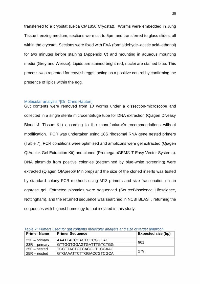
Molecular analysis *[Dr. Chris Hauton] Gut contents were removed from 10 worms under a dissection-microscope and
collected in a single sterile microcentrifuge tube for DNA extraction (Qiagen DNeasy
Blood & Tissue Kit) according to the manufacturer’s recommendations without
modification. PCR was undertaken using 18S ribosomal RNA gene nested primers
(Table 7). PCR conditions were optimised and amplicons were gel extracted (Qiagen
QIAquick Gel Extraction Kit) and cloned (Promega pGEM®-T Easy Vector Systems).
DNA plasmids from positive colonies (determined by blue-white screening) were
extracted (Qiagen QIAprep® Miniprep) and the size of the cloned inserts was tested
by standard colony PCR methods using M13 primers and size fractionation on an
agarose gel. Extracted plasmids were sequenced (SourceBioscience Lifescience,
Nottingham), and the returned sequence was searched in NCBI BLAST, returning the
sequences with highest homology to that isolated in this study.
Table 7: Primers used for gut contents molecular analysis and size of target amplicon.
Primer Name Primer Sequence Expected size (bp)
23F – primary AAATTACCCACTCCCGGCAC 901
23R – primary GTTGGTGGAGTGATTTGTCTGG
25F – nested TGCTTACTGTCACGCTCCGAAC 279
25R – nested GTGAAATTCTTGGACCGTCGCA
26
2.2.3 Bacteriology
It is important to understand the bacterial load of these worms with respect to its role
in pathogen transmission between broodstock and hatcheries. Bacterial isolates from
these worms were identified to species level (where possible), to determine if these
worms could act as a pathogen vector for strains thought to cause frequent and
sporadic batch failures in hatcheries. If these worms do possess such pathogens, their
removal from aquaculture facilities is required to reduce production losses.
Ten worms were removed from each of two crayfish (20 worms total), and were
washed three times with sterile physiological saline (PBS) to remove most bacteria on
the epidermis originating from the environment or crayfish host. Primary plates were
prepared on site; the ten worms from each crayfish were homogenized in a sterile
environment, swabbed and plated onto a tryptone soy agar (TSA) plate. Inoculated
primary plates were incubated at 30oC for 24 hours, and bacterial growth on primary
plates was quantified according to Drew (1997); 3+ characterised by heavy bacterial
growth on primary and secondary streaks; 2+ by heavy bacterial growth only on
primary streak; 1+ has 10 to 60 colonies on the entire plate; 1 has less than 10 colonies
on the entire plate; and 0 has no bacterial growth on any area of plate. After 24 hours
of incubation at 30oC, primary plates were incubated at 4oC and transported to
laboratories within 24 hours for subculturing.
Primary plates were examined and subcultures of all distinct colony types were
prepared under sterile conditions. A single colony of each type was removed from the
primary plate and streaked onto separate sterile TSA plates which were incubated for
24 hours at 30oC, producing monocultures that could be identified.
27
All monocultures were identified to species level where possible. Initial
characterisation of cultures was achieved by Gram staining (Gram, 1884) (Appendix
E), identifying cultures as Gram positive or negative, and characterising the shape of
the bacteria. Oxidase and catalase tests were also conducted, before biochemical
tests could be used. Biochemical tests (bioMérieux API 20E and 20NE) were used to
identify Gram negative bacterial isolates down to species level; Gram negative,
oxidase positive isolates required API 20NE tests, and Gram negative, oxidase
negative isolates required API 20E tests (Appendix E). Results were entered into an
online database for identification, and species identification was accepted if the
confidence level was above 95%. Gram positive isolates cannot be identified using the
above biochemical tests, so were identified by molecular techniques *[Dr. Graham
Burgess and Alicia Maclaine]. Once identified, bacterial isolates were preserved
immediately by freezing each of the strains on polypropylene beads at -80oC
(Appendix E).
28
2.3 Results
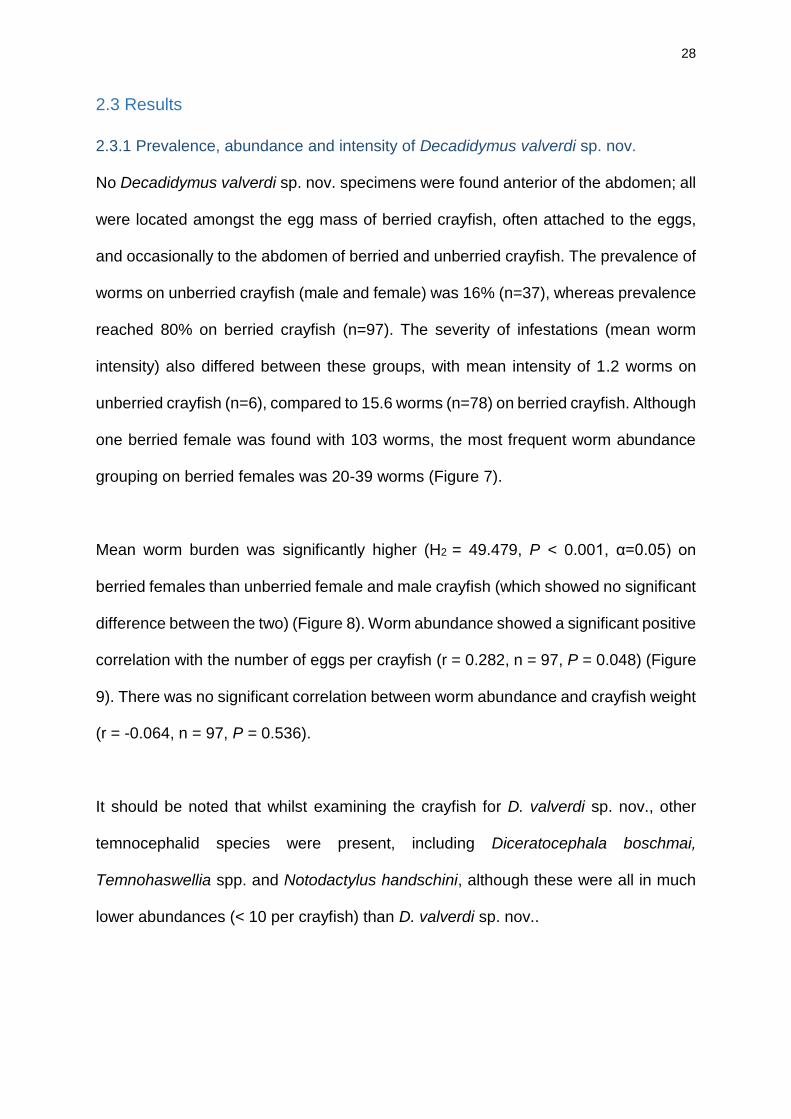
2.3.1 Prevalence, abundance and intensity of Decadidymus valverdi sp. nov.
No Decadidymus valverdi sp. nov. specimens were found anterior of the abdomen; all
were located amongst the egg mass of berried crayfish, often attached to the eggs,
and occasionally to the abdomen of berried and unberried crayfish. The prevalence of
worms on unberried crayfish (male and female) was 16% (n=37), whereas prevalence
reached 80% on berried crayfish (n=97). The severity of infestations (mean worm
intensity) also differed between these groups, with mean intensity of 1.2 worms on
unberried crayfish (n=6), compared to 15.6 worms (n=78) on berried crayfish. Although
one berried female was found with 103 worms, the most frequent worm abundance
grouping on berried females was 20-39 worms (Figure 7).
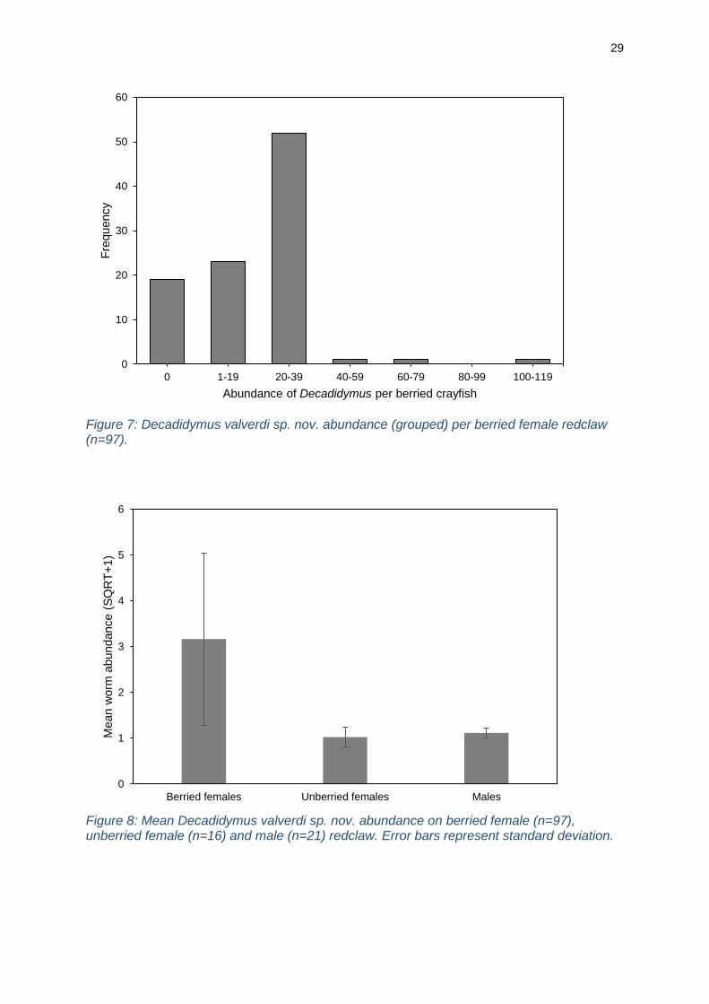
Mean worm burden was significantly higher (H2 = 49.479, P < 0.001, α=0.05) on
berried females than unberried female and male crayfish (which showed no significant
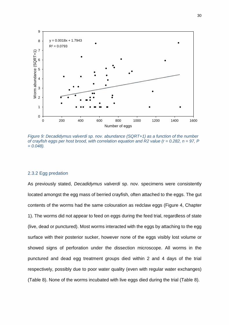
difference between the two) (Figure 8). Worm abundance showed a significant positive
correlation with the number of eggs per crayfish (r = 0.282, n = 97, P = 0.048) (Figure
9). There was no significant correlation between worm abundance and crayfish weight
(r = -0.064, n = 97, P = 0.536).
It should be noted that whilst examining the crayfish for D. valverdi sp. nov., other
temnocephalid species were present, including Diceratocephala boschmai,
Temnohaswellia spp. and Notodactylus handschini, although these were all in much
lower abundances (< 10 per crayfish) than D. valverdi sp. nov..
29
Figure 7: Decadidymus valverdi sp. nov. abundance (grouped) per berried female redclaw (n=97).
Figure 8: Mean Decadidymus valverdi sp. nov. abundance on berried female (n=97), unberried female (n=16) and male (n=21) redclaw. Error bars represent standard deviation.
0
10
20
30
40
50
60
0 1-19 20-39 40-59 60-79 80-99 100-119
Fre
quency
Abundance of Decadidymus per berried crayfish
0
1
2
3
4
5
6
Berried females Unberried females Males
Mean w
orm
abundance (
SQ
RT
+1)
30
Figure 9: Decadidymus valverdi sp. nov. abundance (SQRT+1) as a function of the number of crayfish eggs per host brood, with correlation equation and R2 value (r = 0.282, n = 97, P = 0.048).
2.3.2 Egg predation
As previously stated, Decadidymus valverdi sp. nov. specimens were consistently
located amongst the egg mass of berried crayfish, often attached to the eggs. The gut
contents of the worms had the same colouration as redclaw eggs (Figure 4, Chapter
1). The worms did not appear to feed on eggs during the feed trial, regardless of state
(live, dead or punctured). Most worms interacted with the eggs by attaching to the egg
surface with their posterior sucker, however none of the eggs visibly lost volume or
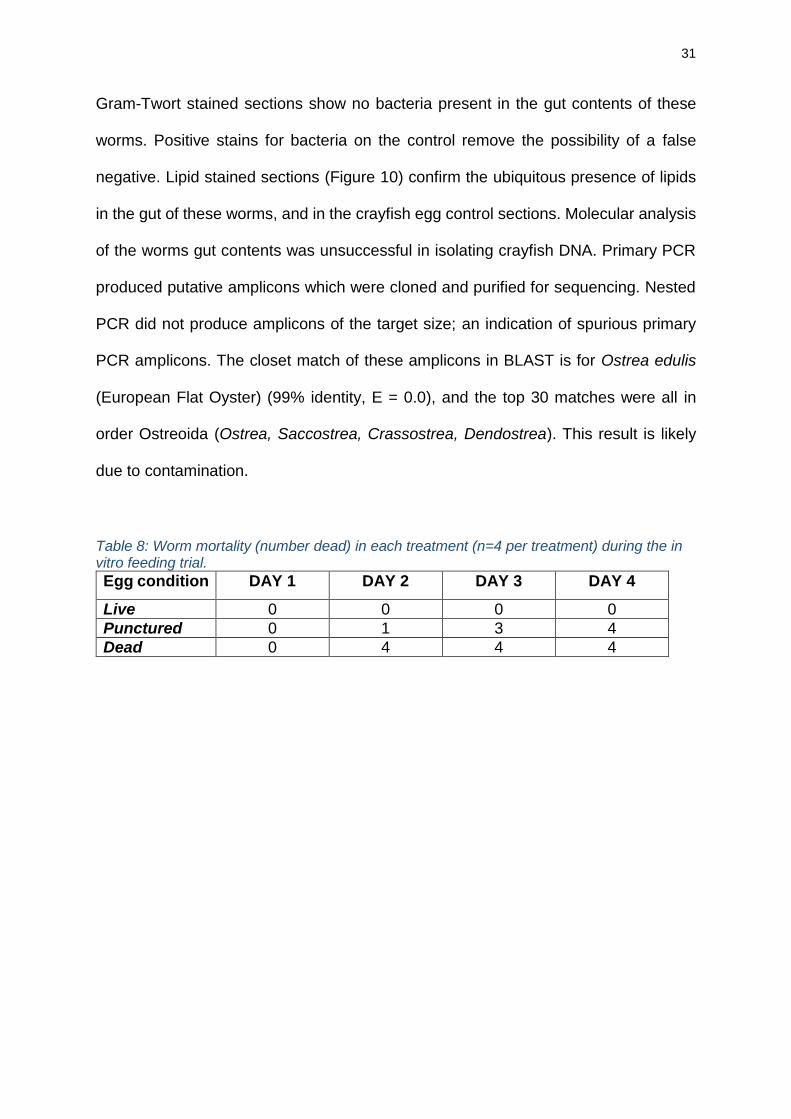
showed signs of perforation under the dissection microscope. All worms in the
punctured and dead egg treatment groups died within 2 and 4 days of the trial
respectively, possibly due to poor water quality (even with regular water exchanges)
(Table 8). None of the worms incubated with live eggs died during the trial (Table 8).
y = 0.0018x + 1.7943
R² = 0.0793
0
1
2
3
4
5
6
7
8
9
0 200 400 600 800 1000 1200 1400 1600
Worm
abundance (
SQ
RT
+1)
Number of eggs
31
Gram-Twort stained sections show no bacteria present in the gut contents of these
worms. Positive stains for bacteria on the control remove the possibility of a false
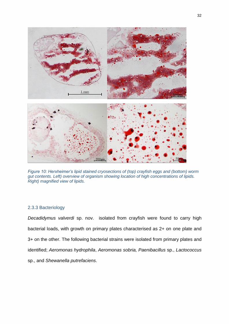
negative. Lipid stained sections (Figure 10) confirm the ubiquitous presence of lipids
in the gut of these worms, and in the crayfish egg control sections. Molecular analysis
of the worms gut contents was unsuccessful in isolating crayfish DNA. Primary PCR
produced putative amplicons which were cloned and purified for sequencing. Nested
PCR did not produce amplicons of the target size; an indication of spurious primary
PCR amplicons. The closet match of these amplicons in BLAST is for Ostrea edulis
(European Flat Oyster) (99% identity, E = 0.0), and the top 30 matches were all in
order Ostreoida (Ostrea, Saccostrea, Crassostrea, Dendostrea). This result is likely
due to contamination.
Table 8: Worm mortality (number dead) in each treatment (n=4 per treatment) during the in vitro feeding trial.
Egg condition DAY 1 DAY 2 DAY 3 DAY 4
Live 0 0 0 0
Punctured 0 1 3 4
Dead 0 4 4 4
32
Figure 10: Herxheimer’s lipid stained cryosections of (top) crayfish eggs and (bottom) worm gut contents. Left) overview of organism showing location of high concentrations of lipids. Right) magnified view of lipids.
2.3.3 Bacteriology
Decadidymus valverdi sp. nov. isolated from crayfish were found to carry high
bacterial loads, with growth on primary plates characterised as 2+ on one plate and
3+ on the other. The following bacterial strains were isolated from primary plates and
identified; Aeromonas hydrophila, Aeromonas sobria, Paenibacillus sp., Lactococcus
sp., and Shewanella putrefaciens.
33
2.4 Discussion
2.4.1 Prevalence, abundance and intensity
A substantial ecological relationship between Decadidymus valverdi sp. nov. and
redclaw eggs was found; identified by the worms’ consistent location amongst the egg
mass, the high prevalence of these worms on berried females (80% of broodstock
were infested), and a significantly larger number of worms on berried redclaw than on
unberried redclaw, male or female (Figure 8). The correlation between the infestation
intensity and the number of eggs per crayfish brood (Figure 9) further supports this
association between these worms and redclaw eggs. Additionally, worm infestation
intensity is unaffected by the host crayfish weight, indicating that these worms are not
directly dependent on adult crayfish resources, concurrent with their previous
description as ectocommensals with regards to the adult host. It has been previously
recorded that host grooming behaviours and molting can affect temnocephalid
(Diceratocephala boschmai) populations on the host (Jones and Lester, 1996). This
may account for the large variation of infestation intensities on berried females (Figure
8), but does not nullify the evident association between the presence and number of
crayfish eggs and the infestation intensity of D. valverdi sp. nov.. This association is
unlikely to harm the adult crayfish directly; whilst some temnocephalids may damage
the host, such as by asphyxiation (Sammy, 1988; Edgerton et al. 2002), the consistent
location of this species amongst the egg mass suggests they are unable to harm the
adult host, consistent with findings of similar temnocephalid species (D. boschmai,
Herbert, 1987).
Whilst the most common infestation intensity per crayfish was 20-39 (Figure 7),
intensity surpassed 100 on one individual. Although host grooming behaviour reduces
34
the number of temnocephalids on the crayfish (Saoud et al. 2013), the efficacy of such
behaviours could decline as infestation intensity increases. Moreover, infestations
may harm developing crayfish eggs, as ventilation of the egg mass will reduce, and a
niche for opportunistic pathogens is provided (Jennings, 1971). Furthermore, other
impacts of this worm (as discussed below) will be exacerbated at high infestation
intensities.
2.4.2 Feeding behaviour
Clear evidence for the consumption of redclaw egg yolk by D. valverdi sp. nov. is
demonstrated in the lipid-stained cryosections of the worm gut and crayfish egg
contents, confirming high lipid concentrations in both (Figure 10). Crustacean egg yolk
is known to contain mostly lipids, proteins and carbohydrate (Adiyodi, 1985),
supporting these findings. Whilst most temnocephalids feed on other fouling
organisms (Longshaw, 2011), the high concentration of lipids found in the guts of D.
valverdi sp. nov. indicate that this is not true of this species, as the gut contents would
not be almost exclusively lipids if they were feeding on whole bodies of other
ectocommensals. The high lipid concentration of the yolk likely resulted in molecular
methods failing to isolate redclaw sequences from the gut contents of the worm; the
scarcity of redclaw 18S target amplicon in the yolk consumed by these worms led to
the production of spurious amplicons later identified as Ostrea edulis, a known
contaminant in the facilities used. Future research should explore lipid profiling
techniques to confirm the origin of the lipids present in the gut of these worms.
35
Additionally, the aforementioned association between these worms and redclaw eggs,
the location of the worms amongst the egg mass (rather than the dorsal carapace
which most temnocephalids inhabit), and the remarkable similarity between the gut
colour of these worms and the crayfish egg yolk further suggests that these worms
rely on redclaw eggs as a food source. Herbert (1987) noted a similar resemblance
between host crayfish eggs and the gut colour of D. boschmai, which was later found
feeding on damaged crayfish eggs (Jones and Lester, 1993), substantiating the link
between gut colour and yolk ingestion.
During the in vitro feed trial, D. valverdi sp. nov. interacted with redclaw eggs, but did
not feed on the yolk. Reasons for this (when other evidence points towards yolk
ingestion) may include; the worms were not starved for long enough prior to trial
commencement, the artificial environment deterred usual feeding behaviours, or the
rapid mortality in punctured and dead egg treatments due to poor water quality led to
early mortality before the need to feed arose. Despite the failure of the feed trial to
witness the consumption of egg yolk by these worms, the above evidence is sufficient
to determine that these worms feed on redclaw egg yolk. If future research can monitor
these worms feeding on crayfish eggs in situ, the extent to which this affects redclaw
juvenile survival and hatchery productivity should be examined, to quantify the
magnitude of the impact this Decadidymus species has on redclaw crayfish
hatcheries. Nonetheless, the evidence provided highlights the importance of
investigating management techniques to eradicate this species from broodstock and
hatchery facilities.
36
2.4.3 Bacteriology
Quantification of bacterial growth on primary plates exhibits the high bacterial load of
these worms. Most bacteria isolated from these worms are commonly found in soil
and water and pose no significant threats to hatchery production (Paenibacillus sp.,
Lactococcus sp., and Shewanella putrefaciens). It is likely that these worms carry a
variety of environmental bacteria, of which this study has isolated a small sample. This
was not examined further as the focus of this study was to determine if these worms
possess pathogens and aid their transmission between broodstock and hatchery
environments.
The isolation of Aeromonas hydrophila and Aeromonas sobria confirms that these
worms contain pathogens and will transmit these between broodstock and hatcheries
if transferred to the hatchery amongst redclaw eggs. Aeromonas spp. are opportunistic
pathogens that are especially damaging and of high concern in culture conditions,
since stressed animals are predisposed to disease (Sung et al. 2000; Nielsen et al.
2001; Quaglio et al. 2006). Stress is often caused by high stocking density, heavy
parasite infestations, and previous infections or disease, all of which frequently occur
in crayfish aquaculture. A. hydrophila is a highly virulent pathogen in crayfish
aquaculture, causing rapid and severe stock mortality after infection (Jiravanichpaisal
et al. 2009); A. sobria may have similar effects, although this is untested. Nonetheless,
these worms do possess Aeromonas spp. and will act as a vector for these pathogens.
Therefore, to prevent disease transmission from broodstock to hatchery, either the
pathogens inside the worms must be eradicated, or the worms must be removed from
crayfish eggs prior to transferral to the hatchery.
37
Chapter 3: Management
3.1 Introduction
These worms pose two key threats to the production of redclaw hatcheries; the
transferral of pathogens into hatcheries, and the predation on crayfish eggs, both of
which are exacerbated with high prevalence and infestation intensity.
To minimise pathogen transmission to the hatchery, crayfish eggs (once stripped from
females) are routinely treated with antifungal and antimicrobial chemicals before being
transferred into the hatchery. Formaldehyde (formalin) treatments are effective and
the most common (Celada et al. 2004; Melendre et al. 2006; Sáez-Royuela et al. 2009;
Kouba et al. 2010), although others such as peracetic acid, copper hydroxide and
sodium chloride have also proven successful (van West, 2006; Carral et al 2009;
Jussila et al. 2011; Kouba et al. 2012). It is time consuming to remove worms by hand
from stripped redclaw eggs as the worms are small, abundant, and similar in
appearance to redclaw eggs. Chemical treatments will kill temnocephalids present in
the egg mass (O’Donoghue et al. 1990), but still result in worm bodies and any viable
pathogens inside being transferred to the hatchery, potentially causing crop failure.
Therefore, for chemical treatment to sufficiently control the impacts of these worms, it
must kill the worms and all pathogens they contain. However, whilst chemical
treatments address the key concern of pathogen transmission, they do not address
the potentially severe egg predation which is likely to occur both prior to egg-stripping
and in the hatchery.
Chemical treatments of diseases such as the crayfish plague (Aphanomyces astaci)
and Saprolegnia infections are relatively well-understood, however treatments for
38
ectoparasites and ectocommensals remain vastly understudied, due to their perceived
lower pathogenicity. However, with the findings that Decadidymus valverdi sp. nov. is
likely to impact hatchery productivity, it is more important to consider treatments that
will remove these worms from crayfish broodstock. Small doses of chemical
treatments such as peracetic acid have been suggested to remove ectocommensals
from broodstock (Jusilla et al. 2011), however exposing adult crayfish to chemical
treatments may have significant adverse effects. Salt solutions have been previously
suggested as a method to control ectocommensals, with evidence supporting its ability
to remove temnocephalids from crayfish hosts (Jones et al. 1994; Soleng et al. 1998).
This research aims to test the efficacy of two suggested control methods for this
temnocephalid species; formalin baths to remove pathogens carried by D. valverdi sp.
nov., and exploiting salinity tolerances of redclaw and D. valverdi sp. nov. to remove
these worms from infested adult crayfish. In doing so, this study pinpoints the
production stage where control is required; with worms on berried crayfish or amongst
the stripped egg mass.
39
3.2 Materials and methods
3.2.1 Formalin treatment – removing pathogens inside worms
To examine the effect of formalin treatments on the bacterial load of Decadidymus
valverdi sp. nov., 60 randomly selected worms were exposed to each of the following
treatments in 70ml specimen jars; 1) five washes with sterile PBS (control), 2)
incubation with 25ml 1000mg/L formalin for 15 minutes, and 3) incubation with 25ml
1500mg/L formalin for 15 minutes. 20 worms were exposed to each treatment, with
ten worms per treatment specimen jar (2 replicates). After treatment, worms were
washed twice in PBS to remove any traces of formalin left on the outside of the worms
which would hinder bacterial growth when incubated. Bacteriology for each treatment
group of ten worms was conducted (Chapter 2 and Appendix E) to determine if worm
bacteriology was affected by formalin treatments.
3.2.2 Exploiting salinity tolerances – removing worms from adult crayfish
To determine the salinity tolerance of D. valverdi sp. nov., worms were exposed to
salinities between 0 and 30 in specimen jars for a total of 150 minutes, and mortality
was recorded every 15 minutes for one hour, with a final assessment after 150
minutes. Specimens were classed as dead when no movement was observed when
pushed with forceps and the posterior sucker was detached from the jar. Ten worms
were placed in each 70ml specimen jar containing 40ml of sterile water at the
appropriate salinity. Due to a limited number of specimens available, for the control
group (0 salinity) only one jar (ten worms) was used. For salinities 2-30, three jars (30
worms) were used and mean mortality per treatment (n=3) was calculated. Results
were normally-distributed (AD = 7.976, P < 0.005, α=0.05) and had equal variance (P
= 0.656, α=0.05), so the effect of salinity and treatment exposure time on mean
40
accumulated mortality was analysed using a two-way repeated measures ANOVA with
pairwise comparisons (Holm-Sidak).
Salinity treatments were then tested on worms still attached to crayfish, to examine
the viability of this treatment in removing worms from adult host crayfish. 20 berried
crayfish in individual containers were exposed to salinity treatments (five crayfish per
treatment), with enough water to submerse crayfish (approximately 2 litres). Salinities
tested were 0 (control), 10, 15 and 20, all followed by a 90-minute fresh water bath.
The number of worms that had fallen off the crayfish was recorded after the 30-minute
salt water treatment, and subsequent 90-minute fresh water bath. Crayfish were then
stripped and the number of worms remaining on each female and amongst the egg
mass was recorded. The percentage of total worms per crayfish that had fallen off was
calculated (hereafter referred to as ‘worm drop-off’) for each salinity treatment and
subsequent fresh water bath, and arcsine transformed. After transformation, data was
tested for normality (AD = 1.198, P < 0.005, α=0.05) and equal variance (P = 0.635,
α=0.05). A two-way repeated-measures ANOVA with pairwise comparisons (Holm-
Sidak) was undertaken to compare results across different treatments.
Behaviour of crayfish curling of tail underneath abdomen) was monitored throughout
the trial. Mortality of crayfish used in this trial was observed for seven days following
the trial, to determine if salinity treatments increased stress of broodstock and
increased mortality.
41
3.3 Results
3.3.1 Formalin treatment – bacterial control
Bacterial growth on primary plates decreased as the concentration of formalin in
treatments increased. In the control group, both plates were characterised as 2+
(heavy bacterial growth on primary streak only), and six isolates were identified;
Aeromonas hydrophila, A. sobria, Paenibacillus sp., Lactococcus sp., Shewanella
putrefaciens and Citrobacter youngae. When treated with 1000mg/L formalin, one
plate was characterised as 1+ (10 - 60 colonies on entire plate), the other as 2+, and
six isolates were identified; A. hydrophila, A. sobria, Chromobacterium violaceum, S.
putrefaciens, Kocuria sp. and Lactococcus sp. When treated with 15000mg/L formalin,
both plates had bacterial growth characterised as 1 (< 10 colonies on entire plate),
and 2 isolates were identified; A. sobria and Microbacterium sp..
As with the initial bacteriology study (Section 2.4.3), most isolates identified are of
environmental origin and considered harmless, and A. hydrophila and A. sobria were
isolated again, confirming that these worms contain pathogens. A formalin
concentration of 1000mg/L is not enough to reduce the bacterial load carried by these
worms, whereas 1500mg/L will greatly reduce bacterial load. However, even at the
highest formalin concentration tested (higher than that commonly used in crayfish
aquaculture industry), Aeromonas sobria was still present and viable.
3.3.2 Salinity treatment – worm removal
Initial salinity trials showed that increasing salinity and exposure time increased
accumulated worm mortality (Figure 11). As salinity increased, the time required for
42
mean accumulated mortality to reach 100% (LT100) decreased; 15 minutes in salinity
of 30, 30 minutes in salinities of 20 and 25, and 150 minutes for salinities of 10 and 15
(Figure 11). LT100 was not reached during the 150 minutes of this trial for salinities
between 0 and 8. This highlights the significant interaction between salinity and
exposure time on the mean accumulated mortality of these worms (F36,139=5.913, P
<0.001). Due to this interaction, main effects of salinity and exposure could not be
independently examined, hence pairwise comparisons were required. After 15
minutes of exposure, there was a significant increase in the mean accumulated
mortalities between treatment salinities of 6, 20, 25 and 30 from the control, whereas
salinities of 2, 4, 8, 10 and 15 showed no significant difference from the control (Figure
11). At 30, 45 and 60 minutes of exposure, salinities of 15 and above showed
significant differences from the control, whereas salinities of 2-10 showed no
significant difference. After 150 minutes, salinities of 10 and above showed significant
differences from the control, whereas salinities of 2-8 showed no significant difference
from the control (Figure 11).
Salinity trials undertaken with worms on host crayfish found a significant interaction
between treatment salinity and the treatments used (30-minute salt water and
subsequent 90-minute fresh water) on worm drop-off (F3,16=17.41, P < 0.001) (Figure
12). There are no significant differences in worm drop-off between the 30-minute salt
bath and subsequent 90-minute fresh water bath in salinities of 0 (P = 1.000) and 10
(P = 0.134), but there are significant differences in this when treated with initial
salinities of 15 (P < 0.01) and 20 (P < 0.01) (Figure 12). After the 30-minute salt-bath,
mean worm drop-off was 0% in the control group, 38% at a salinity of 10, 87% at a
salinity of 15, and 88% at a salinity of 20. Pairwise comparisons showed there were
43
significant differences in the worm drop-off at all salinities tested (P < 0.01), except
between 15 and 20 (P = 0.834) (Figure 12; 0 < 10 < 15, 20). Variation within the
treatment group at a salinity of 10 was much greater than within groups at salinities of
15 and 20 (Figure 12).
After the 90-minute fresh water bath following salinity exposure, the same trend was
observed amongst the treatment groups; there were significant differences in the worm
drop-off at all salinities tested (P < 0.01), except between 15 and 20 (P = 0.393) (Figure
12; 0 < 10 < 15, 20). Mean worm drop-off remained at 0% in the control group, but
rose to 41% in subjects initially treated with a salinity of 10, 94% in subjects initially
treated with a salinity of 15, and 97% in subjects initially treated with a salinity of 20.
Variation within the treatment group at a salinity of 10 remained much greater than
within groups at salinities of 15 and 20 after the fresh water treatment (Figure 12).
In the salt-bath treatment at salinity of 20, all crayfish curled their tails underneath their
body, which was not seen in any of the other treatment groups. Mortality was
monitored for seven days following the trial; one individual died from the treatment
salinity of 10 (on day 1), and one individual died from the treatment salinity of 15 (on
day 2). No individuals from salinities of 0 or 20 died. Other temnocephalid species
inhabiting the redclaw were also removed at high salinity treatments, although this was
not quantified.
44
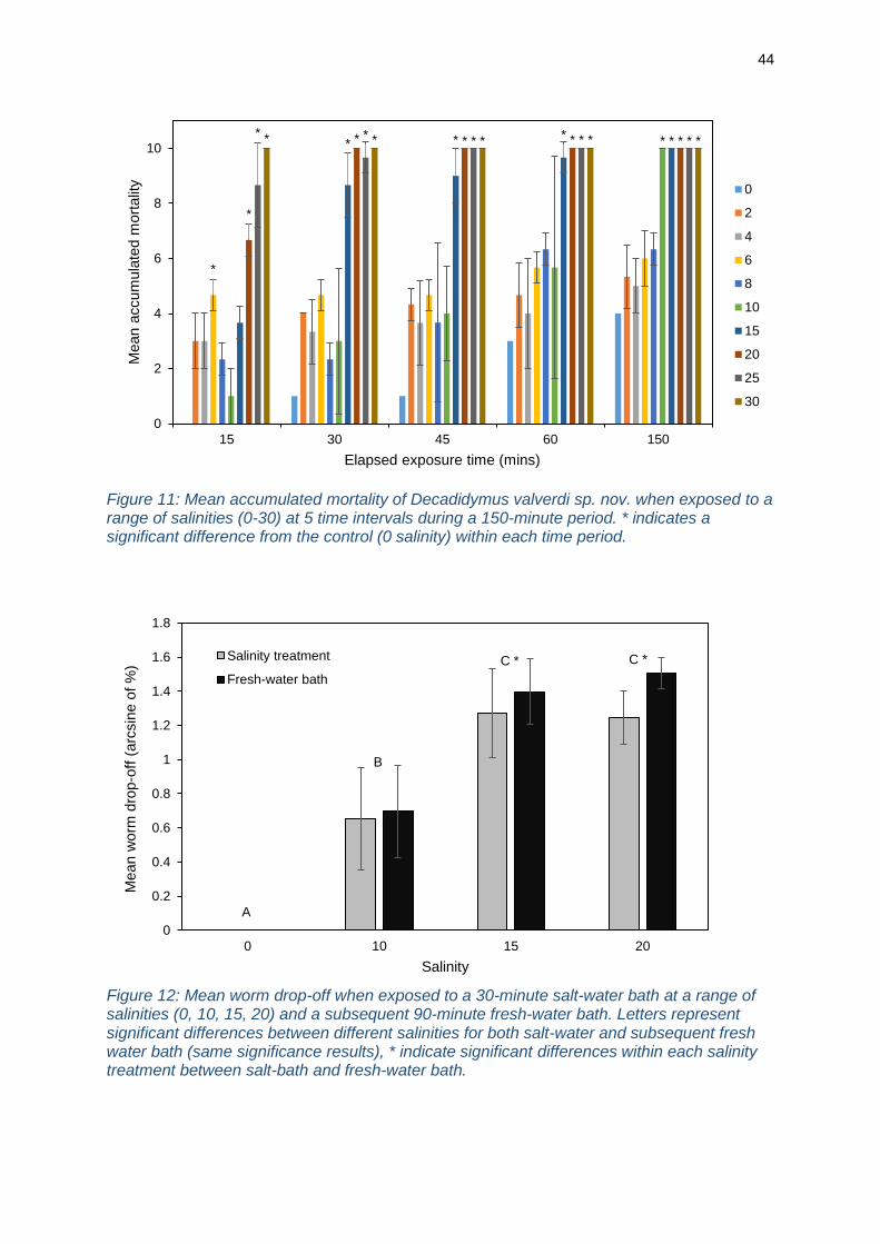
Figure 11: Mean accumulated mortality of Decadidymus valverdi sp. nov. when exposed to a range of salinities (0-30) at 5 time intervals during a 150-minute period. * indicates a significant difference from the control (0 salinity) within each time period.
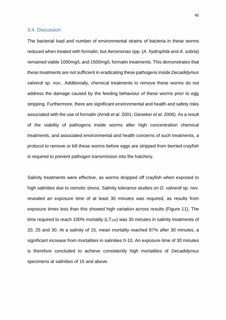
Figure 12: Mean worm drop-off when exposed to a 30-minute salt-water bath at a range of salinities (0, 10, 15, 20) and a subsequent 90-minute fresh-water bath. Letters represent significant differences between different salinities for both salt-water and subsequent fresh water bath (same significance results), * indicate significant differences within each salinity treatment between salt-bath and fresh-water bath.
*
** * * *
*
* * * ** * * * ** * * * *
0
2
4
6
8
10
15 30 45 60 150
Mean a
ccum
ula
ted m
ort
alit
y
Elapsed exposure time (mins)
0
2
4
6
8
10
15
20
25
30
A
B
C * C *
0
0.2
0.4
0.6
0.8
1
1.2
1.4
1.6
1.8
0 10 15 20
Mean w
orm
dro
p-o
ff (
arc
sin
e o
f %
)
Salinity
Salinity treatment
Fresh-water bath
45
3.4. Discussion
The bacterial load and number of environmental strains of bacteria in these worms
reduced when treated with formalin, but Aeromonas spp. (A. hydrophila and A. sobria)
remained viable 1000mg/L and 1500mg/L formalin treatments. This demonstrates that
these treatments are not sufficient in eradicating these pathogens inside Decadidymus
valverdi sp. nov.. Additionally, chemical treatments to remove these worms do not
address the damage caused by the feeding behaviour of these worms prior to egg
stripping. Furthermore, there are significant environmental and health and safety risks
associated with the use of formalin (Arndt et al. 2001; Gieseker et al. 2006). As a result
of the viability of pathogens inside worms after high concentration chemical
treatments, and associated environmental and health concerns of such treatments, a
protocol to remove or kill these worms before eggs are stripped from berried crayfish
is required to prevent pathogen transmission into the hatchery.
Salinity treatments were effective, as worms dropped off crayfish when exposed to
high salinities due to osmotic stress. Salinity tolerance studies on D. valverdi sp. nov.
revealed an exposure time of at least 30 minutes was required, as results from
exposure times less than this showed high variation across results (Figure 11). The
time required to reach 100% mortality (LT100) was 30 minutes in salinity treatments of
20, 25 and 30. At a salinity of 15, mean mortality reached 87% after 30 minutes, a
significant increase from mortalities in salinities 0-10. An exposure time of 30 minutes
is therefore concluded to achieve consistently high mortalities of Decadidymus
specimens at salinities of 15 and above.
46
Results from trials with worms on crayfish hosts found that whilst there is a significant
increase in worm drop-off at a salinity of 10 in comparison to the control, over half of
the worms remained on the crayfish (Figure 12). Furthermore, the results were
variable between individuals treated with a salinity of 10, hence salinities above 10 are
required if enough worms are to be consistently removed from individuals in
broodstock. There was no significant difference in worm drop-off between salinity
treatments of 15 and 20 (Figure 12), thus a 30-minute bath in a salinity of 15 is
considered the best practise method for worm removal, as lower salinities are less
stressful for redclaw adults and juveniles.
The crayfish mortality observed in these trials was likely due to handling stress rather
than salinity exposure, as no mortality occurred in redclaw exposed to the highest
salinity treatments. Furthermore, redclaw are known to tolerate salinities and exposure
times used in this study; Prymaczok et al. (2008) found growth performance and adult
redclaw survival were unaffected by long exposure (3 weeks) to salinities up to 15g/L.
Similarly, although increased salinity decreases juvenile hatching rate (Anson and
Rouse 1994), there is no significant difference in juvenile growth when reared in
salinities between 0 and 14g/L for 12 weeks (Austin 1995). This indicates that salinity
treatments recommended in this study will have no significant detrimental effect on the
growth and survival of both adult and juvenile redclaw.
Treatment groups with salinities of 15 and 20 showed a significant increase in worm
drop-off after the additional 90-minute fresh water bath than after the initial salt bath
(Figure 12). This is likely due to salinity exposure and resultant behaviour of crayfish;
individuals in the salinity of 20 treatment tucked their tail underneath their abdomen
47
and around the egg mass, trapping some worms that would have otherwise fallen off
due to osmotic stress. When these crayfish are transferred to the fresh water bath, a
less stressful environment, their tails uncurled allowing for the worms that had died
from salinity exposure to drop out of the egg mass. This resulted in a significantly
higher worm removal when the salt water treatment is combined with a subsequent
fresh water bath than in the salt water bath alone.
Salinity treatments will effectively remove D. valverdi sp. nov. from redclaw, with the
potential to also act as a disease and antifungal treatment (Marking et al. 1994;
Schreier et al. 1996; Mifsud and Rowland, 2008; Kozák et al. 2009; Policar et al. 2011).
Therefore, a 30-minute salt bath with a salinity of 15, followed by a fresh water bath
for 90 minutes in a holding tank is considered the best practice method for eradicating
D. valverdi sp. nov. from broodstock and preventing reinfestation. This will remove
worms from the broodstock before eggs are stripped and transferred to the hatchery,
preventing egg mortality from predation and the transferral of pathogens between the
environments. This recommended protocol is effective, rapid, low-cost and simple
(with regards to facility requirements and training), hence has already been adopted
by the aquaculture facility used for this research. The industry would benefit from
future research refining this protocol, with particular focus on the time required for the
subsequent fresh water bath, as only one time was tested in this study. Furthermore,
it would be beneficial to examine any potential long term effects of this treatment, both
in terms of how the eradication of these worms affects juvenile survival and hatchery
production, and any potential negative effects of this recommended treatment on
juvenile growth, survival and hatching rate.
48
Conclusions
The name Decadidymus valverdi sp. nov. is proposed for this species. Identifying
morphological features of this species (compared to Decadidymus gulosus) are; 1) the
ventral location and smaller opening of the mouth, which possesses inner and outer
lips, 2) the large stylet, 3) the small pharynx, relative to overall body size, 4) the
excretory pores located further from the anterior of worm, and 5) the smaller gonopore
located closer to the posterior of the worm. These features are sufficient to justify the
proposal of a new species, hence genetic sequencing was undertaken to further
describe this new species. Sequence homology confirms the presence of this new
species within the Temnocephalida, however cannot be compared to D. gulosus as
no sequence for this species was obtained during its description. The phylogeny within
the Temnocephalida requires more attention in future studies, as does a detailed
taxonomic description of this proposed new species.
This new species is highly prevalent and abundant on berried redclaw and exhibits a
strong association with host egg masses. Whilst adult redclaw are likely to be
unaffected by infestations, the presence and intensity of infestation of this
temnocephalid species will cause significant harm to developing juveniles. These
worms feed on the yolk of developing eggs, which is predicted to severely hinder
juvenile development and survival. D. valverdi sp. nov. is also capable of transmitting
pathogens into the hatchery, potentially leading to crop failure and therefore a
reduction in hatchery productivity and sustainability. Future research should quantify
the extent to which D. valverdi sp. nov. affects juvenile survival and hatchery output.
Nonetheless, the strong association with redclaw eggs, feeding behaviour and
pathogens present in this worm signify a severe detrimental impact to production,
49
highlighting the need for the management and eradication of this Decadidymus
species in aquaculture facilities.
Salinity treatments are commonly used in redclaw aquaculture as a rapid, low-cost
solution to ectoparasites and disease, due to their high salinity tolerance. Salt baths
are effective for removing these worms from adult crayfish; a 30-minute salt bath with
a salinity of 15 is recommended, followed by a 90-minute fresh water bath in a holding
tank before entering broodstock. This is the more effective management protocol to
reduce the impacts of this Decadidymus species, as formalin treatments will not
remove pathogens inside these worms and do not address the issue of yolk
consumption by these worms prior to egg stripping.
This recommended protocol has already been implemented at the study site facility,
although long term benefits to production are yet to be quantified. Future assessments
of this technique should test the efficacy of shorter fresh water baths after salinity
treatment, as the full 90 minutes used in this study may not be required. Another
aspect to consider in future development of this protocol is the possibility of long-term
effects of salinity exposure on juvenile growth, survival and hatching rate; the
advantages of increased survival and production by removing these worms with
salinity treatments, against the disadvantages of potentially reduced growth, survival
and hatching rates of juveniles should be compared.
50
References
Adiyodi, R. G. (1985). Reproduction and its control. The biology of Crustacea, 9, 147-215.
Amato, J. F. R., Amato, S. B., & Daudt, L. C. C. (2003). New species of Temnocephala Blanchard (Platyhelminthes, Temnocephalida) ectosymbiont on Aegla serrana Buckup & Rossi (Crustacea, Anomura) from southern Brazil. Revista Brasileira de Zoologia, 20(3), 493-500.
Amato, J. F., & Amato, S. B. (2005). New species of Temnocephala Blanchard (Platyhelminthes, Temnocephalida) ectosymbiont on giant water bugs, Belostoma spp. (Hemiptera, Belostomatidae) from southern Brazil. Revista Brasileira de Zoologia, 22(1), 107-118.
Amato, J. F., Amato, S. B., & Seixas, S. A. (2006). A new species of Temnocephala Blanchard (Platyhelminthes, Temnocephalida) ectosymbiont on Trichodactylus fluviatilis Latreille (Crustacea, Decapoda, Trichodactylidae) from southern Brazil. Revista Brasileira de Zoologia, 23(3), 796-806.
Amato, J. F., Seixas, S. A., & Amato, S. B. (2007). A new species of Temnocephala Blanchard (Platyhelminthes, Temnocephalida) ectosymbiont on creeping water bugs, Cryphocricos granulosus De Carlo (Hemiptera, Naucoridae) from southern Brazil. Revista Brasileira de Zoologia, 24(4), 1043-1051.
Andersen, W. C., Turnipseed, S. B., & Roybal, J. E. (2006). Quantitative and confirmatory analyses of malachite green and leucomalachite green residues in fish and shrimp. Journal of agricultural and food chemistry, 54(13), 4517-4523.
Anson, K. J., & Rouse, D. B. (1994). Effects of Salinity on Hatching and Post‐Hatch Survival of the Australian Red Claw Crayfish Cherax quadricarinatus. Journal of the World Aquaculture Society, 25(2), 277-280.
Arndt, R. E., Wagner, E. J., & Routledge, M. D. (2001). Reducing or withholding hydrogen peroxide treatment during a critical stage of rainbow trout development: effects on eyed eggs, hatch, deformities, and fungal control. North American Journal of Aquaculture, 63(2), 161-166.
Austin, C. M. (1995). Effect of temperature and salinity on the survival and growth of juvenile redclaw (Cherax quadricarinatus). Freshwater Crayfish, 10, 419-426.
Bancroft, J. D., & Gamble, M. (2008). Theory and practice of histological techniques. (6th edition) Churchill Livingstone, Elsevier, Philadelphia, USA.
Bondad-Reantaso, M. G., Subasinghe, R. P., Arthur, J. R., Ogawa, K., Chinabut, S., Adlard, R., Tan, Z. & Shariff, M. (2005). Disease and health management in Asian aquaculture. Veterinary parasitology, 132(3), 249-272.
Bower, S. M., McGladdery, S. E., & Price, I. M. (1994). Synopsis of infectious diseases and parasites of commercially exploited shellfish. Annual Review of Fish Diseases, 4, 1-199.
Boyd, C. E., Tucker, C., McNevin, A., Bostick, K., & Clay, J. (2007). Indicators of resource use efficiency and environmental performance in fish and crustacean aquaculture. Reviews in Fisheries science, 15(4), 327-360.
Cannon, L. R. (1991). Temnocephalan symbionts of the freshwater crayfish Cherax quadricarinatus from northern Australia. Hydrobiologia, 227(1), 341-347.
Carral, J. M., González, A., Celada, J. D., Sáez-Royuela, M., Melendre, P. M., González, R., & García, V. (2009). Antifungal treatments in artificial incubation of crayfish eggs (Pacifastacus leniusculus, Astacidae): searching for alternatives to formaldehyde. Knowledge and Management of Aquatic Ecosystems, (394-395), 16.
Celada, J. D., Carral, J. M., Sáez-Royuela, M., Melendre, P. M., & Aguilera, A. (2004). Effects of different antifungal treatments on artificial incubation of the astacid crayfish (Pacifastacus leniusculus Dana) eggs. Aquaculture, 239(1), 249-259.
Charmantier-Daures, M., & Charmantier, G. (1991). Mass-culture of Cancer irroratus larvae (Crustacea, Decapoda): adaptation of a flow-through sea-water system. Aquaculture, 97(1), 25-39.
Damborenea, C., & Brusa, F. (2008). A new species of Temnocephala (Platyhelminthes, Temnocephalida) commensal of Pomella megastoma (Mollusca, Ampullariidae) from Misiones, Argentina. Revista mexicana de biodiversidad, 79, 1-7.
51
Drew, W. L. (1977). Value of sputum culture in diagnosis of pneumococcal pneumonia. Journal of clinical microbiology, 6(1), 62-65.
Edgerton, B. F. (1999). A review of freshwater crayfish viruses. Freshwater Crayfish, 12, 261–278.
Edgerton, B. F., Evans, L. H., Stephens, F. J., & Overstreet, R. M. (2002). Synopsis of freshwater crayfish diseases and commensal organisms. Aquaculture, 206(1), 57-135.
FAO (Food and Agriculture Organisation) (2014). The State of World Fisheries and Aquaculture 2014. Opportunities and Challenges. Rome. 223 pp.
FAO (Food and Agriculture Organisation) (2016). The State of World Fisheries and Aquaculture 2016. Contributing to food security and nutrition for all. Rome. 200 pp.
Garcés, A. C., Puerta, L., Tabares, Y., Lenis, C., & Velásquez, L. E. (2013). Temnocephala colombiensis n. sp. (Platyhelminthes: Temnocephalidae) from Antioquia, Colombia. Revista mexicana de biodiversidad, 84(4), 1090-1099.
Ghanawi, J., & Saoud, I. P. (2012). Molting, reproductive biology, and hatchery management of redclaw crayfish Cherax quadricarinatus (von Martens 1868). Aquaculture, 358, 183-195.
Gieseker, C. M., Serfling, S. G., & Reimschuessel, R. (2006). Formalin treatment to reduce mortality associated with Saprolegnia parasitica in rainbow trout, Oncorhynchus mykiss. Aquaculture, 253(1), 120-129.
Gram, C. (1884). The differential staining of Schizomycetes in tissue sections and in dried preparations. Fortschritte der Medizin, 2, 185-9.
Herbert, B. (1987). Notes on diseases and epibionts of Cherax quadricarinatus and C. tenuimanus (Decapoda: Parastacidae). Aquaculture, 64(3), 165-173.
Holdich, D. M. (1993). A review of astaciculture: freshwater crayfish farming. Aquatic Living Resources, 6(4), 307-317.
Jackson, C. J., Pendrey, R. C., & Rothlisberg, P. C. (1992). The Larvatron: a computer-controlled apparatus for rearing planktonic animals under experimental conditions. Marine Ecology Progress Series, 86(2), 189-199.
Jennings, J. B. (1971). Parasitism and commensalism in the Turbellaria. Advances in parasitology, 9, 1-32.
Jiravanichpaisal, P., Roos, S., Edsman, L., Liu, H., & Söderhäll, K. (2009). A highly virulent pathogen, Aeromonas hydrophila, from the freshwater crayfish Pacifastacus leniusculus. Journal of invertebrate pathology, 101(1), 56-66.
Joffe, B. I., Cannon, L. R., & Schockaert, E. R. (1998). On the phylogeny of families and genera within the Temnocephalida. Hydrobiologia, 383(1), 263-268.
Jones, C. M. (1995). Production of juvenile redclaw crayfish, Cherax quadricarinatus (von Martens) (Decapoda, Parastacidae) I. Development of hatchery and nursery procedures. Aquaculture, 138(1), 221-238.
Jones, C. M., Medley, P. B., & Avault Jr, J. W. (1994). Redclaw crayfish, Cherax quadricarinatus: production, economics and marketing. World Aquaculture, 25, 4.
Jones, T. C., & Lester, R. G. J. (1993). Aspects of the biology and pathogenicity of Diceratocephala boschmai (Platyhelminthes: Temnocephalida), an ectosymbiont on the redclaw crayfish, Cherax quadricarinatus. Marine and Freshwater Research, 44(6), 927-933.
Jones, T. C., & Lester, R. J. (1996). Factors influencing populations of the ectosymbiont Diceratocephala boschmai (Platyhelminthes; Temnocephalida), on the redclaw crayfish Cherax quadricarinatus maintained under laboratory conditions. Aquaculture, 143(3-4), 233-243.
Jussila, J., Makkonen, J., & Kokko, H. (2011). Peracetic acid (PAA) treatment is an effective disinfectant against crayfish plague (Aphanomyces astaci) spores in aquaculture. Aquaculture, 320(1), 37-42.
Kittaka, J. (1998). Culture of larval spiny lobsters: a review of work done in northern Japan. Marine and Freshwater Research, 48(8), 923-930.
52
Kouba, A., Carral, J. M., Buřič, M., Mráz, J., Policar, T., & Kozák, P. (2010). Artificial incubation of noble crayfish (Astacus astacus) eggs in a partially recirculating system using formaldehyde as an antifungal treatment. Aquaculture Research, 41(10), e618-e623.
Kouba, A., Kuklina, I., Niksirat, H., Máchová, J., & Kozák, P. (2012). Tolerance of signal crayfish (Pacifastacus leniusculus) to Persteril 36 supports use of peracetic acid in astaciculture. Aquaculture, 350, 71-74.
Kozák, P., Policar, T., Fedotov, V. P., Kuznetsova, T. V., Buřič, M., & Kholodkevich, S. V. (2009). Effect of chloride content in water on heart rate in narrow-clawed crayfish (Astacus leptodactylus). Knowledge and Management of Aquatic Ecosystems, (394-395), 08.
Lockyer, A. E., Olson, P. D., & Littlewood, D. T. J. (2003). Utility of complete large and small subunit rRNA genes in resolving the phylogeny of the Neodermata (Platyhelminthes): implications and a review of the cercomer theory. Biological Journal of the Linnean Society, 78(2), 155-171.
Longshaw, M. (2011). Diseases of crayfish: a review. Journal of Invertebrate Pathology, 106(1), 54-70.
Marking, L. L., Rach, J. J., & Schreier, T. M. (1994). American fisheries society evaluation of antifungal agents for fish culture. The Progressive Fish-Culturist, 56(4), 225-231.
Masser, M. P., & Rouse, D. (1997). Australian red claw crayfish. Southern Regional Aquaculture Center, 224.
Melecha, S. (1983). Commercial seed production of the freshwater prawn, Macrobrachium rosenbergii in Hawaii. CRS Handbook of Mariculture, 1, 203-205.
Melendre, P. M., Celada, J. D., Carral, J. M., Sáez-Royuela, M., & Aguilera, A. (2006). Effectiveness of antifungal treatments during artificial incubation of the signal crayfish eggs (Pacifastacus leniusculus Dana. Astacidae). Aquaculture, 257(1), 257-265.
Meyer, F. P. (1991). Aquaculture disease and health management. Journal of animal science, 69(10), 4201-4208.
Mifsud, C., & Rowland, S. J. (2008). Use of salt to control ichthyophthiriosis and prevent saprolegniosis in silver perch, Bidyanus bidyanus. Aquaculture Research, 39(11), 1175-1180.
Murray, A. G., & Peeler, E. J. (2005). A framework for understanding the potential for emerging diseases in aquaculture. Preventive veterinary medicine, 67(2), 223-235.
Nelson, R. G., & Dendy, J. S. (1979). Conditions for holding and propagating crawfish brood stock (Procambarus clarkii). In Proceedings of the World Mariculture Society, 10(1‐4), 503-509. Blackwell Publishing Ltd.
Nielsen, M. E., Høi, L., Schmidt, A. S., Qian, D., Shimada, T., Shen, J. Y., & Larsen, J. L. (2001). Is Aeromonas hydrophila the dominant motile Aeromonas species that causes disease outbreaks in aquaculture production in the Zhejiang Province of China?. Diseases of aquatic organisms, 46(1), 23-29.
O'Donoghue, P., Beveridge, I., & Phillips, P. (1990). Parasites and ectocommensals of yabbies and marron in South Australia. Central Veterinary Laboratories (VETLAB), South Australian Department of Agriculture, Adelaide.
O'Sullivan, D. (1991). Status of Australian aquaculture 1989-90. Austasia Aquaculture, 2-13.
Policar, T., Smyth, J., Flanigan, M., Kouba, A., & Kozák, P. (2011). Sodium chloride as effective antifungal treatment for artificial egg incubation in Austropotamobius pallipes. Knowledge and Management of Aquatic Ecosystems, (401), 13.
Prymaczok, N. C., Medesani, D. A., & Rodríguez, E. M. (2008). Levels of ions and organic metabolites in the adult freshwater crayfish, Cherax quadricarinatus, exposed to different salinities. Marine and Freshwater Behaviour and Physiology, 41(2), 121-130.
Quaglio, F., Morolli, C., Galuppi, R., Bonoli, C., Marcer, F., Nobile, L., De Luise, G. & Tampieri, M. P. (2006). Preliminary investigations of disease-causing organisms in the white-clawed crayfish Austropotamobius pallipes complex from streams of northern Italy. Bulletin Francais de la Peche et de la Pisciculture, (380-381), 1271-1290.
53
Sáez-Royuela, M., Melendre, P. M., Celada, J. D., Carral, J. M., González, A., González, R., & García, V. (2009). Possibilities of artificial incubation of signal crayfish (Pacifastacus leniusculus Dana) eggs at high densities and reduced flow rate using formaldehyde as antifungal treatment. Aquaculture, 288(1), 65-68.
Sammy, N. (1988). Breeding biology of Cherax quadricarinatus in the Northern Territory. In Proceedings of the First Australian Shellfish Aquaculture Conference, Perth. (Eds L. H. Evans and D. O'Sullivan.) pp. 79-88.
Saoud, I. P., Ghanawi, J., Thompson, K. R., & Webster, C. D. (2013). A review of the culture and diseases of redclaw crayfish Cherax quadricarinatus (von Martens 1868). Journal of the World Aquaculture Society, 44(1), 1-29.
Sapkota, A., Sapkota, A. R., Kucharski, M., Burke, J., McKenzie, S., Walker, P., & Lawrence, R. (2008). Aquaculture practices and potential human health risks: current knowledge and future priorities. Environment international, 34(8), 1215-1226.
Schreier, T. M., Rach, J. J., & Howe, G. E. (1996). Efficacy of formalin, hydrogen peroxide, and sodium chloride on fungal-infected rainbow trout eggs. Aquaculture, 140(4), 323-331.
Sewell, K. B. (2013). Key to the genera and checklist of species of Australian temnocephalans (Temnocephalida). Museum Victoria Science Reports, 17, 1-13.
Sewell, K. B., & Whittington, I. D. (1995). A light microscope study of the attachment organs and their role in locomotion of Craspedella sp. (Platyhelminthes: Rhabdocoela: Temnocephalidae), an ectosymbiont from the branchial chamber of Cherax quadricarinatus (Crustacea: Parastacidae) in Queensland, Australia. Journal of natural history, 29(5), 1121-1141.
Soleng, A., Bakke, T. A., & Hansen, L. P. (1998). Potential for dispersal of Gyrodactylus salaris (Platyhelminthes, Monogenea) by sea-running stages of the Atlantic salmon (Salmo salar): field and laboratory studies. Canadian Journal of Fisheries and Aquatic Sciences, 55(2), 507-514.
Stentiford, G. D., Neil, D. M., Peeler, E. J., Shields, J. D., Small, H. J., Flegel, T. W., Vlak, J. M., Jones, B., Morado, F., Moss, S., Lotz, J., Bartholomay, L., Behringer, D. C., Hauton, C. & Lightner, D. V. (2012). Disease will limit future food supply from the global crustacean fishery and aquaculture sectors. Journal of invertebrate pathology, 110(2), 141-157.
Sung, H. H., Hwang, S. F., & Tasi, F. M. (2000). Responses of giant freshwater prawn (Macrobrachium rosenbergii) to challenge by two strains of Aeromonas spp. Journal of invertebrate pathology, 76(4), 278-284.
van West, P. (2006). Saprolegnia parasitica, an oomycete pathogen with a fishy appetite: new challenges for an old problem. Mycologist, 20(3), 99-104.
Villarreal, H., & Pelaez, A. (2000). Commercial semi intensive production in clay ponds of the freshwater red claw crayfish Cherax quadricarinatus. In Proceedings of the World Aquaculture America 2000 symposium, New Orleans, USA (p. 271).
Volonterio, O. (2007). A new species of Temnocephala (Platyhelminthes, Temnocephalida) and a description of T. axenos from Uruguay. Journal of Natural History, 41(21-24), 1245-1257.
Volonterio, O. (2009). First report of the introduction of an Australian temnocephalidan into the New World. Journal of Parasitology, 95(1), 120-123.
Volonterio, O. (2010). Two new species of Temnocephala (Platyhelminthes, Temnocephalida) from the South American snake-necked turtle Hydromedusa tectifera (Testudines, Chelidae). Zoological science, 27(12), 965-970.
Walker, P. J., & Winton, J. R. (2010). Emerging viral diseases of fish and shrimp. Veterinary research, 41(6), 51-75.
Watson, N. A., Rohde, K., & Sewell, K. B. (1995). Ultrastructure of spermiogenesis and spermatozoa of Decadidymus gulosus, Temnocephala dendyi, T. minor, Craspedella sp., C. spenceri and Diceratocephala boschmai (Platyhelminthes, Temnocephalida, Temnocephalidae), with emphasis on the intercentriolar body and zone of differentiation. Invertebrate reproduction & development, 27(2), 131-143.
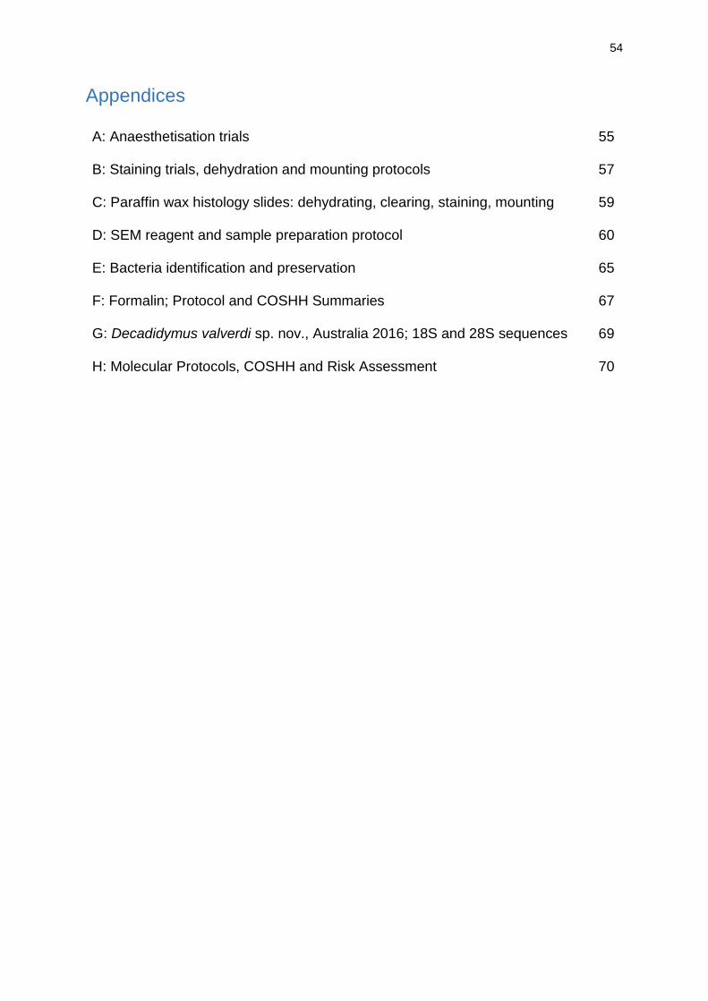
54
Appendices
A: Anaesthetisation trials
B: Staining trials, dehydration and mounting protocols
C: Paraffin wax histology slides: dehydrating, clearing, staining, mounting
D: SEM reagent and sample preparation protocol
E: Bacteria identification and preservation
F: Formalin; Protocol and COSHH Summaries
G: Decadidymus valverdi sp. nov., Australia 2016; 18S and 28S sequences
H: Molecular Protocols, COSHH and Risk Assessment
55
57
59
60
65
67
69
70
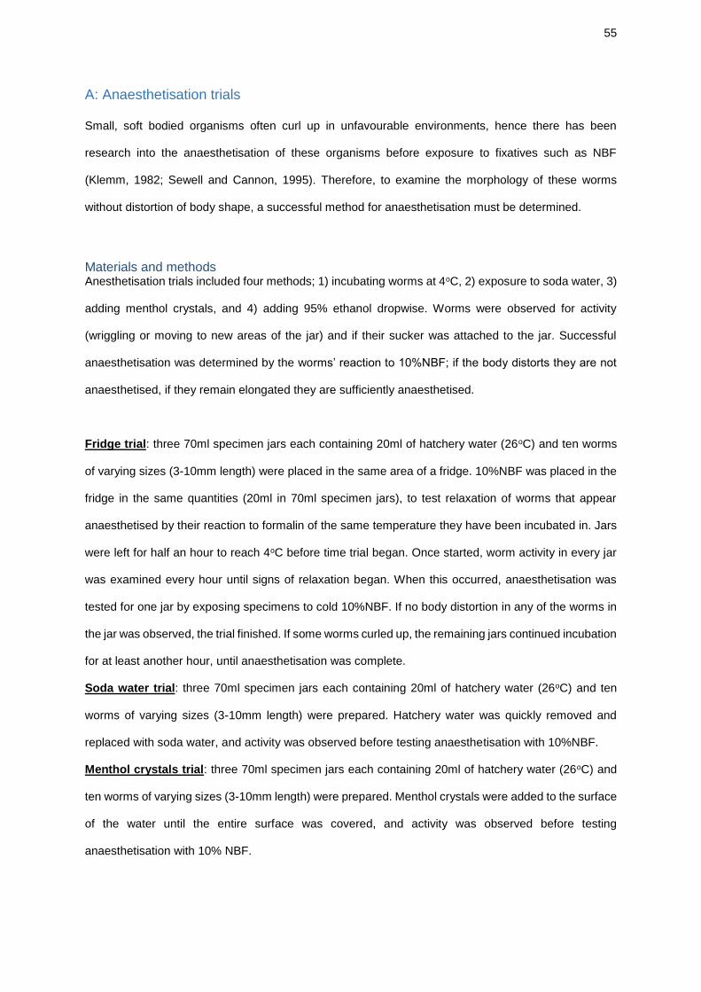
55
A: Anaesthetisation trials
Small, soft bodied organisms often curl up in unfavourable environments, hence there has been
research into the anaesthetisation of these organisms before exposure to fixatives such as NBF
(Klemm, 1982; Sewell and Cannon, 1995). Therefore, to examine the morphology of these worms
without distortion of body shape, a successful method for anaesthetisation must be determined.
Materials and methods Anesthetisation trials included four methods; 1) incubating worms at 4oC, 2) exposure to soda water, 3)
adding menthol crystals, and 4) adding 95% ethanol dropwise. Worms were observed for activity
(wriggling or moving to new areas of the jar) and if their sucker was attached to the jar. Successful
anaesthetisation was determined by the worms’ reaction to 10%NBF; if the body distorts they are not
anaesthetised, if they remain elongated they are sufficiently anaesthetised.
Fridge trial: three 70ml specimen jars each containing 20ml of hatchery water (26oC) and ten worms
of varying sizes (3-10mm length) were placed in the same area of a fridge. 10%NBF was placed in the
fridge in the same quantities (20ml in 70ml specimen jars), to test relaxation of worms that appear
anaesthetised by their reaction to formalin of the same temperature they have been incubated in. Jars
were left for half an hour to reach 4oC before time trial began. Once started, worm activity in every jar
was examined every hour until signs of relaxation began. When this occurred, anaesthetisation was
tested for one jar by exposing specimens to cold 10%NBF. If no body distortion in any of the worms in
the jar was observed, the trial finished. If some worms curled up, the remaining jars continued incubation
for at least another hour, until anaesthetisation was complete.
Soda water trial: three 70ml specimen jars each containing 20ml of hatchery water (26oC) and ten
worms of varying sizes (3-10mm length) were prepared. Hatchery water was quickly removed and
replaced with soda water, and activity was observed before testing anaesthetisation with 10%NBF.
Menthol crystals trial: three 70ml specimen jars each containing 20ml of hatchery water (26oC) and
ten worms of varying sizes (3-10mm length) were prepared. Menthol crystals were added to the surface
of the water until the entire surface was covered, and activity was observed before testing
anaesthetisation with 10% NBF.
56
Dropwise 95% ethanol trial: three 70ml specimen jars each containing 20ml of hatchery water (26oC)
and ten worms of varying sizes (3-10mm length) were prepared. Room-temperature 95% ethanol was
added dropwise using a pipette and the jar was swirled to ensure the ethanol diffused evenly throughout
the water. This continued until activity reduced and anaesthetisation was tested with 10% NBF.
Results Fridge: In all jars, no narcotisation was observed until after 6 hours. Effective, but slow.
Soda: almost instantaneous anaesthetisation of larger specimens, but smaller specimens showed some
body distortion upon exposure to soda water (before NBF).
Menthol crystals: effective but slow, lots of crystals required.
Dropwise ethanol: Initial activity of worms increased after first few drops added, but as more ethanol
was added activity decreased. Smaller worms were narcotised with a lower concentration of ethanol
than larger worms. This method worked quickly on all worm sizes once enough ethanol was added
(approximately 5-10ml).
Chosen method of anaesthetisation was dropwise ethanol until observed anaesthetisation, as provided
most rapid and effective treatment.
57
B: Staining trials, dehydration and mounting protocols
Five stains were selected for trials; Mayer’s paracarmine, Harris’ hematoxylin, Solphenyl blue, Toluidine blue and Kirkpatrick’s carmine.
Stain preparation
Protocols Mayer’s paracarmine and Harris’ hematoxylin: cover specimens in cavity block with stain solution for 24
hours. Destain with acidic alcohol (1%HCl-70% ethanol) until differentiation of tissues is visible.
Neutralise with 1%NH4-70% ethanol.
Solphenyl blue: cover specimens in cavity block with solphenyl blue (approximately 3 drops), leave for
1 minute. Fill cavity block with water, check stain for differentiation (empty water and refill if
differentiation is unclear). Remove water when differentiation visible and mount immediately without
dehydrating (see below).
Toluidine blue: half fill cavity block with distilled water and specimens for staining. Add five drops of
toluidine blue and leave for 2 minutes. Wash briefly with water, remove with pipette. Add differentiator
(FAA – formaldehyde, acetic acid, ethanol) and watch for differentiation of tissues. Remove FAA with
pipette when differentiation is visible.
Mayer’s parcarmine: Stain Aluminium chloride 0.5g 70% ethanol (heated) 100ml Calcium chloride 4.0g Carmine 1.0g Once cooled, mixture is filtered Differentiator 70% ethanol 99ml Glacial acetic acid 1ml
Kirkpatrick’s carmine: Stain Carmine 2.5g Glacial acetic acid 2.5ml Potassium aluminium sulphate 2.5g Distilled water 100ml Differentiator Distilled water
Solphenyl blue: Stain; JCU stock solution Differentiator; Distilled water
Toluidine blue: Stain Toluidine blue 1.0g Borax 1.0g Boric acid 1.0g Distilled water 100ml Differentiator Formaldehyde 5ml Acetic acid 5ml 80% ethanol 90ml
Harris’ Hematoxylin: Stain Stock Harris Hematoxylin (JCU) 100ml Glacial acetic acid 4ml Differentiator 70% ethanol 99ml Glacial acetic acid 1ml
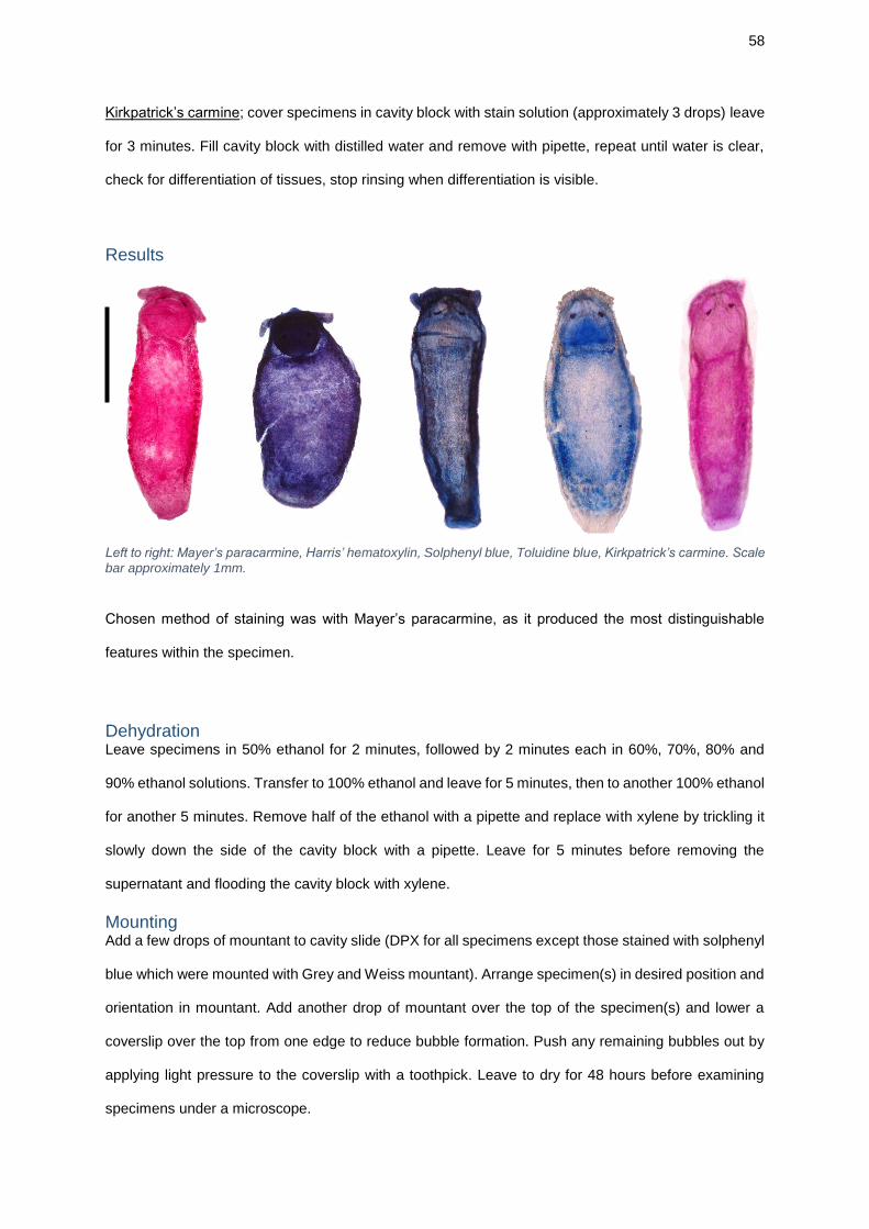
58
Kirkpatrick’s carmine; cover specimens in cavity block with stain solution (approximately 3 drops) leave
for 3 minutes. Fill cavity block with distilled water and remove with pipette, repeat until water is clear,
check for differentiation of tissues, stop rinsing when differentiation is visible.
Results
Left to right: Mayer’s paracarmine, Harris’ hematoxylin, Solphenyl blue, Toluidine blue, Kirkpatrick’s carmine. Scale
bar approximately 1mm.
Chosen method of staining was with Mayer’s paracarmine, as it produced the most distinguishable
features within the specimen.
Dehydration Leave specimens in 50% ethanol for 2 minutes, followed by 2 minutes each in 60%, 70%, 80% and
90% ethanol solutions. Transfer to 100% ethanol and leave for 5 minutes, then to another 100% ethanol
for another 5 minutes. Remove half of the ethanol with a pipette and replace with xylene by trickling it
slowly down the side of the cavity block with a pipette. Leave for 5 minutes before removing the
supernatant and flooding the cavity block with xylene.
Mounting Add a few drops of mountant to cavity slide (DPX for all specimens except those stained with solphenyl
blue which were mounted with Grey and Weiss mountant). Arrange specimen(s) in desired position and
orientation in mountant. Add another drop of mountant over the top of the specimen(s) and lower a
coverslip over the top from one edge to reduce bubble formation. Push any remaining bubbles out by
applying light pressure to the coverslip with a toothpick. Leave to dry for 48 hours before examining
specimens under a microscope.
59
C: Paraffin wax histology slides: dehydrating, clearing, staining, mounting
Dehydrating and clearing of fixed glass slides: • Xylene 2 minutes
• Xylene 2 minutes
• Ethanol 1 minute
• Ethanol 1 minute
• Ethanol 1 minute
• Water wash 1 minute
Stains:
Xylene infiltration: • Ethanol 1 minute
• Ethanol 1 minute
• Ethanol 2 minutes
• Xylene 2 minutes
• Xylene 2 minutes
• Xylene until mounting
Mounting: Remove stained slides from xylene bath and stand up to allow xylene to drip off (without drying out
slides). Add a few drops of DPX mountant (or Grey and Weisse if aqueous mountant is required) to
slide and lower a coverslip over the top from one edge to reduce bubble formation. Push any remaining
bubbles out by applying light pressure to the coverslip with a toothpick. Leave to dry for 48 hours before
examining specimens under a microscope.
Haematoxylin and Eosin:
• Mayer’s haematoxylin 8 minutes
• Water Wash 30 seconds
• Scott’s tap water substitute 30 seconds
• Water wash 2 minutes
• Young’s Eosin 4 minutes
Differentiate in water wash ~20 seconds
Gram-Twort:
• 2% crystal violet 2 minutes
• Water wash 5 seconds
• Lugol’s iodine 3 minutes
• Rinse with water 5 seconds
• Decolourise with acetone 5 seconds
• Twort’s solution (1:3 water) 10 minutes
Rinse with water 5 seconds
Herxheimer’s:
• Herxheimer’s 8 minutes
• Rinse with 70% ethanol 5 seconds
• Rinse with water 5 seconds
• Mayer’s Haematoxylin 2 minutes
• Rinse with water 5 seconds
• Scott’s tap water substitute 30 seconds
Rinse with water 5 seconds
60
D: SEM reagent and sample preparation protocol
Note: using SEM also required completing JCU AAC risk assessments
Hazardous chemicals used in this procedure:
1. Glutaraldehyde solution. Toxic if swallowed or inhaled, causes severe skin burns and eye damage, may cause an allergic skin reaction, may cause allergy or asthma symptoms or breathing difficulties if inhaled, may cause respiratory irritation, very toxic to aquatic life with long lasting effects, corrosive to the respiratory tract.
2. Sodium cacodylate trihydrate. Toxic if swallowed or inhaled, suspected of causing cancer, very toxic to aquatic life with long lasting effects.
3. HMDS (Hexamethyldisilazane). Highly flammable liquid and vapour, harmful if swallowed or if inhaled, toxic in contact with skin, harmful to aquatic life with long lasting effects.
Protocol:
• Dissolve sodium cacodylate in distilled water, correct pH with HCl to obtain a sodium cacodylate buffer of 0.2M and pH7.2
• Mix with glutaraldehyde to produce 2.5% glutaraldehyde in sodium cacodylate buffer
• Incubate anaesthetised worms in this fixative buffer solution for 24 hours
• Transfer sample into fresh buffer solution (no fixative) using forceps
• Mix well and all of buffer removed, leaving sample behind
• Add fresh buffer, repeated twice
• Removed as much buffer as possible from sample, replace with 50% ethanol in
deionized water solution, leave for 15 minutes
• Repeat using increasing ethanol concentrations (60%, 70%, 80%, 90% and 100%)
• Repeat 100% ethanol step twice
• Replace 100% ethanol with a 1:1 solution of 100% ethanol and HMDS, leave for 15
minutes
• Replace ethanol/HMDS solution with 100% HMDS, leave for 15 minutes, repeat twice
• At third 100% HMDS stage, leave sample immersed in solution to evaporate off in fume
hood, loosely covered by lid (overnight)
• Carefully orientated samples onto SEM stub (with dissecting scope if needed), mount
using carbon based, electrically conductive, double sided adhesive discs (Leit tabs)
• Gold-coat stubs (conducted by JCU Advanced Analytical Centre staff)
• Nitrile rubber gloves, safety glasses and a lab coat worn
• Risk assessments and COSHH completed and MSDS consulted (JCU) before carrying out this protocol
• Completed under a fume hood
61
COSHH Summary: Glutaraldehyde solution, Sigma-Aldrich 340855
MSDS supplier and revision date: Sigma-Aldrich, 24.02.17
Task description: Glutaraldehyde mixed with sodium cacodylate buffer of 0.2M and pH7.2, to produce 2.5% glutaraldehyde in sodium cacodylate buffer; used to fix specimens for SEM.
Hazard statements: Precautionary statements: H301 + H331 H314 H317 H334 H335 H410 EUH071
Toxic if swallowed or inhaled Causes severe skin burns and eye damage May cause an allergic skin reaction May cause allergy or asthma symptoms or breathing difficulties if inhaled May cause respiratory irritation Very toxic to aquatic life with long lasting effects Corrosive to the respiratory tract
P260 P280 P301 + P330 + P331 + P310 P304 + P340 + P310 P305 + P351 + P338 P403 + P233
Do not breathe dust/ fume/ gas/ mist/ vapours/ spray Wear protective gloves/ protective clothing/ eye protection/ face protection IF SWALLOWED: Rinse mouth. Do NOT induce vomiting. Immediately call a POISON CENTER/doctor IF INHALED: Remove person to fresh air and keep comfortable for breathing. Immediately call a POISON CENTER/doctor IF IN EYES: Rinse cautiously with water for several minutes. Remove contact lenses, if present and easy to do. Continue rinsing Store in a well-ventilated place. Keep container tightly closed
Could a less hazardous substance be used instead? No Control measures Practice: Handle in accordance with good laboratory hygiene and safety practice. Wash hands before leaving laboratory. Avoid contact with skin and eyes. Avoid inhalation of vapour or mist, handle in a fume hood. Ensure adequate ventilation. Personal protective equipment: Safety glasses and face shield, nitrile rubber gloves, lab coat. Storage: Store in cool place. Keep container tightly closed in a dry and well-ventilated place, keep upright to prevent leakage. Emergency procedure General advice:
Consult a physician. Show MSDS (attached) to doctor in attendance.
Inhalation: Move person into fresh air. If not breathing, give artificial respiration. Consult a physician.
Skin contact: Take off contaminated clothing and shoes immediately. Wash off with soap and plenty of water. Take victim immediately to hospital. Consult a physician.
Eye contact: Rinse thoroughly with plenty of water for at least 15 minutes and consult a physician.
Ingestion Do NOT induce vomiting. Never give anything by mouth to an unconscious person. Rinse mouth with water. Consult a physician.
Spill procedure:
Prevent further leakage or spillage if safe to do so. Do not let product enter drains, discharge into the environment must be avoided. Soak up with inert absorbent material, keep in closed container and dispose of as hazardous waste. Follow hazardous waste disposal protocol for laboratory.
62
Fire-fighting measures:
Use water spray, alcohol resistant foam, dry chemical or carbon dioxide. Fire-fighters to wear self-contained breathing apparatus where possible
Health surveillance not required. Specific training not required. Hazards and risks are suitably controlled using above measures.
COSHH Summary: Sodium cacodylate trihydrate, Sigma-Aldrich C0250
MSDS supplier and revision date: Sigma-Aldrich, 28.12.15
Task description: Dissolved in sterile water to produce buffer of 0.2M and pH7.2, then mixed with glutaraldehyde to produce 2.5% glutaraldehyde in sodium cacodylate buffer; used to fix specimens for SEM.
Hazard statements: Precautionary statements: H301 + H331
Toxic if swallowed or inhaled P261 P273
Avoid breathing dust Avoid release to the environment
H351 Suspected of causing cancer H410 Very toxic to aquatic life with long
lasting effects P281 Use personal protective equipment as
required.
P301 + P310
IF SWALLOWED: Immediately call a POISON CENTER or doctor/ physician.
P311 Call a POISON CENTER or doctor/ physician
P501 Dispose of contents/ container to an approved waste disposal plant.
Could a less hazardous substance be used instead? No Control measures Practice: Handle in accordance with good laboratory hygiene and safety practice. Wash hands before leaving laboratory. Avoid formation of dust and aerosols. Avoid eye and skin contact, and inhalation of dust or vapours. Handle in a fume hood. Ensure adequate ventilation. Personal protective equipment: Safety glasses, nitrile rubber gloves, lab coat. Storage: Store in cool place. Keep container tightly closed in a dry and well-ventilated place, keep upright. Emergency procedure General advice:
Consult a physician. Show MSDS (attached) to doctor in attendance.
Inhalation: Move person into fresh air. If not breathing, give artificial respiration. Consult a physician.
Skin contact: Wash off with soap and plenty of water. Take victim immediately to hospital. Consult a physician.
Eye contact: Flush eyes with water as a precaution.
Ingestion Never give anything by mouth to an unconscious person. Rinse mouth with water. Consult a physician.
Spill procedure:
Prevent further leakage or spillage if safe to do so. Do not let product enter drains, discharge into the environment must be avoided. Pick up and arrange
63
disposal without creating dust. Sweep up and shovel. Keep in suitable, closed containers for disposal. Follow hazardous waste disposal protocol for laboratory.
Fire-fighting measures:
Use water spray, alcohol resistant foam, dry chemical or carbon dioxide. Fire-fighters to wear self-contained breathing apparatus if necessary
Health surveillance not required. Specific training not required. Hazards and risks are suitably controlled using above measures.
COSHH Summary: Hexamethyldisilazane (HMDS), Sigma-Aldrich H4875
MSDS supplier and revision date: Sigma-Aldrich, 28.07.15
Task description: Samples incubated in 1:1 HMDS and ethanol, and 100% HMDS. 100% HMDS left to evaporate under fume hood
Hazard statements: Precautionary statements: H225 H302 + H332
Highly flammable liquid and vapour. Harmful if swallowed or if inhaled
P210 P261
Keep away from heat, hot surfaces, sparks, open flames and other ignition sources. No smoking Avoid breathing dust
H311 Toxic in contact with skin P273 Avoid release to the environment H412 Harmful to aquatic life with
long lasting effects P280 Wear protective gloves/ protective clothing/
eye protection/ face protection
P302 + P352 + P312
IF ON SKIN: Wash with plenty of water. Call a POISON CENTER or doctor/ physician if you feel unwell
P304 + P340 + P312
IF INHALED: Remove person to fresh air and keep comfortable for breathing. Call a POISON CENTER or doctor/ physician if you feel unwell
Could a less hazardous substance be used instead? No Control measures Practice: Handle in accordance with good laboratory hygiene and safety practice. Wash hands before leaving laboratory. Avoid contact with skin and eyes. Avoid breathing vapours, mist or gas. Ensure adequate ventilation. Keep away from sources of ignition - no smoking. Take measures to prevent the build-up of electrostatic charge. Handle in a fume hood. Personal protective equipment: Safety glasses and face shield, nitrile rubber gloves, lab coat. Storage: Store in cool place. Keep container tightly closed in a dry and well-ventilated place. Containers which are opened must be carefully resealed and kept upright to prevent leakage. Emergency procedure General advice:
Consult a physician. Show MSDS (attached) to doctor in attendance.
Inhalation: Move person into fresh air. If not breathing, give artificial respiration. Consult a physician.
Skin contact: Wash off with soap and plenty of water. Take victim immediately to hospital. Consult a physician.
64
Eye contact: Flush eyes with water as a precaution.
Ingestion Do NOT induce vomiting. Never give anything by mouth to an unconscious person. Rinse mouth with water. Consult a physician.
Spill procedure:
Prevent further leakage or spillage if safe to do so. Do not let product enter drains, discharge into the environment must be avoided. Contain spillage, and then collect with an electrically protected vacuum cleaner or by wet-brushing and place in suitable, closed containers for disposal. Follow hazardous waste disposal protocol for laboratory.
Fire-fighting measures:
Use water spray, alcohol resistant foam, dry chemical or carbon dioxide. Fire-fighters to wear self-contained breathing apparatus if necessary
Health surveillance not required. Specific training not required. Hazards and risks suitably controlled using above measures.
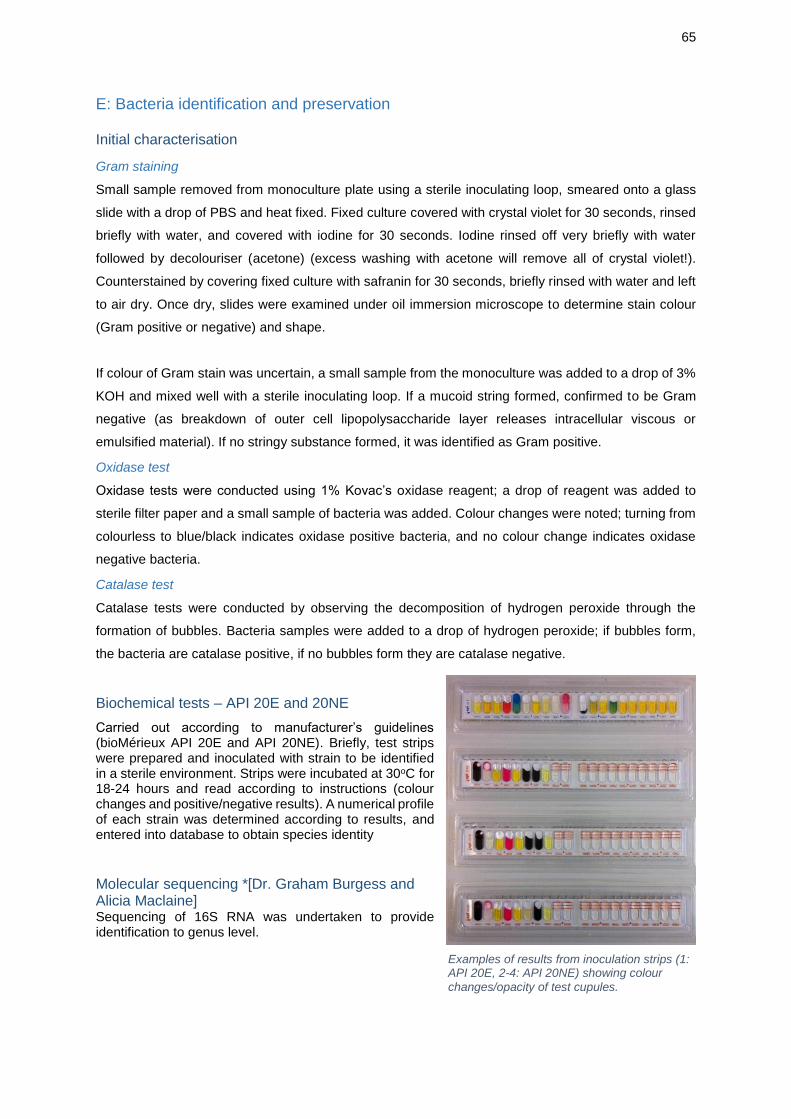
65
E: Bacteria identification and preservation
Initial characterisation
Gram staining
Small sample removed from monoculture plate using a sterile inoculating loop, smeared onto a glass
slide with a drop of PBS and heat fixed. Fixed culture covered with crystal violet for 30 seconds, rinsed
briefly with water, and covered with iodine for 30 seconds. Iodine rinsed off very briefly with water
followed by decolouriser (acetone) (excess washing with acetone will remove all of crystal violet!).
Counterstained by covering fixed culture with safranin for 30 seconds, briefly rinsed with water and left
to air dry. Once dry, slides were examined under oil immersion microscope to determine stain colour
(Gram positive or negative) and shape.
If colour of Gram stain was uncertain, a small sample from the monoculture was added to a drop of 3%
KOH and mixed well with a sterile inoculating loop. If a mucoid string formed, confirmed to be Gram
negative (as breakdown of outer cell lipopolysaccharide layer releases intracellular viscous or
emulsified material). If no stringy substance formed, it was identified as Gram positive.
Oxidase test
Oxidase tests were conducted using 1% Kovac’s oxidase reagent; a drop of reagent was added to
sterile filter paper and a small sample of bacteria was added. Colour changes were noted; turning from
colourless to blue/black indicates oxidase positive bacteria, and no colour change indicates oxidase
negative bacteria.
Catalase test
Catalase tests were conducted by observing the decomposition of hydrogen peroxide through the
formation of bubbles. Bacteria samples were added to a drop of hydrogen peroxide; if bubbles form,
the bacteria are catalase positive, if no bubbles form they are catalase negative.
Biochemical tests – API 20E and 20NE
Carried out according to manufacturer’s guidelines (bioMérieux API 20E and API 20NE). Briefly, test strips were prepared and inoculated with strain to be identified in a sterile environment. Strips were incubated at 30oC for 18-24 hours and read according to instructions (colour changes and positive/negative results). A numerical profile of each strain was determined according to results, and entered into database to obtain species identity
Molecular sequencing *[Dr. Graham Burgess and Alicia Maclaine] Sequencing of 16S RNA was undertaken to provide identification to genus level.
Examples of results from inoculation strips (1: API 20E, 2-4: API 20NE) showing colour
changes/opacity of test cupules.
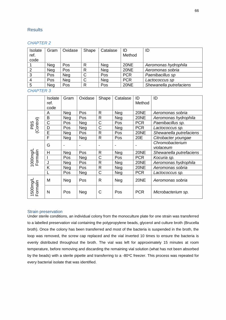
66
Results
CHAPTER 2
Isolate ref. code
Gram Oxidase Shape Catalase ID Method
ID
1 Neg Pos R Neg 20NE Aeromonas hydrophila
2 Neg Pos R Neg 20NE Aeromonas sobria
3 Pos Neg C Pos PCR Paenibacillus sp
4 Pos Neg C Neg PCR Lactococcus sp
5 Neg Pos R Pos 20NE Shewanella putrefaciens
CHAPTER 3
Isolate ref. code
Gram Oxidase Shape Catalase ID Method
ID
PB
S
(Co
ntr
ol)
A Neg Pos R Neg 20NE Aeromonas sobria
B Neg Pos R Neg 20NE Aeromonas hydrophila
C Pos Neg C Pos PCR Paenibacillus sp.
D Pos Neg C Neg PCR Lactococcus sp.
E Neg Pos R Pos 20NE Shewanella putrefaciens
F Neg Neg R Pos 20E Citrobacter youngae
100
0m
g/L
Form
alin
G - - - - - Chromobacterium violaceum
H Neg Pos R Neg 20NE Shewanella putrefaciens
I Pos Neg C Pos PCR Kocuria sp.
J Neg Pos R Neg 20NE Aeromonas hydrophila
K Neg Pos R Neg 20NE Aeromonas sobria
L Pos Neg C Neg PCR Lactococcus sp.
150
0m
g/L
Form
alin
M Neg Pos R Neg 20NE Aeromonas sobria
N Pos Neg C Pos PCR Microbacterium sp.
Strain preservation Under sterile conditions, an individual colony from the monoculture plate for one strain was transferred
to a labelled preservation vial containing the polypropylene beads, glycerol and culture broth (Brucella
broth). Once the colony has been transferred and most of the bacteria is suspended in the broth, the
loop was removed, the screw cap replaced and the vial inverted 10 times to ensure the bacteria is
evenly distributed throughout the broth. The vial was left for approximately 15 minutes at room
temperature, before removing and discarding the remaining vial solution (what has not been absorbed
by the beads) with a sterile pipette and transferring to a -80oC freezer. This process was repeated for
every bacterial isolate that was identified.
67
F: Use of formalin; Protocol and COSHH Summaries
Hazardous chemicals used in these procedures:
1. Formalin. Harmful if swallowed, may cause an allergic skin reaction, suspected of causing genetic defects, may cause cancer
Protocol summaries:
1. Fixation of specimens for morphological studies
• Specimens held in 70ml screw cap specimen jar with no more than 40ml of formalin (10% NBF) per jar, specimens added to jar in a well-ventilated area
• Jars transported from study site to laboratories in secondary container to avoid spillage. No waste disposal of formalin on site
• In laboratories, all work with formalin completed under fume hood. Specimens washed prior to subsequent treatments. Formalin waste disposed of according to laboratory procedures
2. Chemical treatment (disinfectant)
• Dilutions required (100mg/L and 1500mg/L) prepared in laboratories under fume hood prior to use in field
• Specimens held in 70ml screw cap specimen jar for 15 minutes, with no more than 40ml of formalin per jar, completed in a well-ventilated area
• Specimens washed before completing bacteriology studies. Formalin waste retained and transported to laboratories and disposed of according to standard procedures
3. Anaesthetisation trials
• After anaesthetisation method completed, tested by adding specimens to formalin (10% NBF), in 70ml screw cap specimen jar with no more than 40ml of formalin per jar, specimens added to jar in a well-ventilated area
• Worms assessed for activity and body distortion. Specimens washed prior to subsequent treatments. Formalin waste retained and transported to laboratories and disposed of according to standard procedures
COSHH Summary: Formalin solution, neutral buffered, 10%, Sigma-Aldrich HT501128
MSDS supplier and revision date: Sigma-Aldrich, 26.03.15
Task description: Specimens incubated in formalin in screw-cap specimen jars for fixation or for disinfectant trials (specimens incubated in formalin up to 1500mg/L).
• Nitrile rubber gloves, safety glasses and a lab coat worn
• Risk assessments and COSHH completed and MSDS consulted (JCU) before carrying out this protocol
• Completed under fume hood (laboratories) or in well-ventilated area (study site)
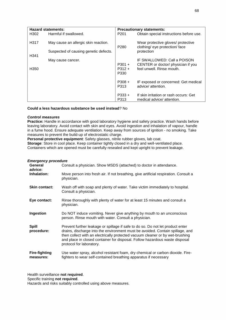
68
Hazard statements: Precautionary statements: H302 H317 H341 H350
Harmful if swallowed. May cause an allergic skin reaction. Suspected of causing genetic defects. May cause cancer.
P201 P280 P301 + P312 + P330
Obtain special instructions before use. Wear protective gloves/ protective clothing/ eye protection/ face protection IF SWALLOWED: Call a POISON CENTER or doctor/ physician if you feel unwell. Rinse mouth.
P308 +
P313
IF exposed or concerned: Get medical advice/ attention.
P333 + P313
If skin irritation or rash occurs: Get medical advice/ attention.
Could a less hazardous substance be used instead? No Control measures Practice: Handle in accordance with good laboratory hygiene and safety practice. Wash hands before leaving laboratory. Avoid contact with skin and eyes. Avoid ingestion and inhalation of vapour, handle in a fume hood. Ensure adequate ventilation. Keep away from sources of ignition - no smoking. Take measures to prevent the build-up of electrostatic charge. Personal protective equipment: Safety glasses, nitrile rubber gloves, lab coat. Storage: Store in cool place. Keep container tightly closed in a dry and well-ventilated place. Containers which are opened must be carefully resealed and kept upright to prevent leakage. Emergency procedure General advice:
Consult a physician. Show MSDS (attached) to doctor in attendance.
Inhalation: Move person into fresh air. If not breathing, give artificial respiration. Consult a physician.
Skin contact: Wash off with soap and plenty of water. Take victim immediately to hospital. Consult a physician.
Eye contact: Rinse thoroughly with plenty of water for at least 15 minutes and consult a physician.
Ingestion Do NOT induce vomiting. Never give anything by mouth to an unconscious person. Rinse mouth with water. Consult a physician.
Spill procedure:
Prevent further leakage or spillage if safe to do so. Do not let product enter drains, discharge into the environment must be avoided. Contain spillage, and then collect with an electrically protected vacuum cleaner or by wet-brushing and place in closed container for disposal. Follow hazardous waste disposal protocol for laboratory.
Fire-fighting measures:
Use water spray, alcohol resistant foam, dry chemical or carbon dioxide. Fire-fighters to wear self-contained breathing apparatus if necessary
Health surveillance not required. Specific training not required. Hazards and risks suitably controlled using above measures.
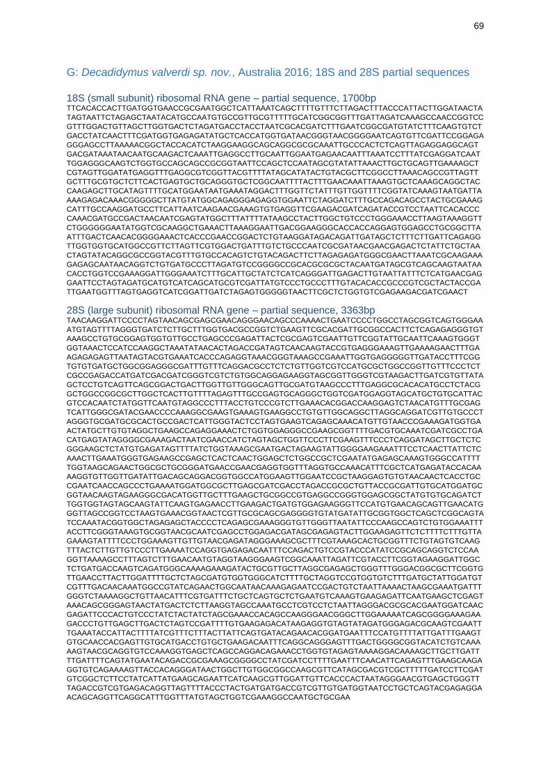
69
G: Decadidymus valverdi sp. nov., Australia 2016; 18S and 28S partial sequences
18S (small subunit) ribosomal RNA gene – partial sequence, 1700bp TTCACACCACTTGATGGTGAACCGCGAATGGCTCATTAAATCAGCTTTTGTTTCTTAGACTTTACCCATTACTTGGATAACTA
TAGTAATTCTAGAGCTAATACATGCCAATGTGCCGTTGCGTTTTTGCATCGGCGGTTTGATTAGATCAAAGCCAACCGGTCC
GTTTGGACTGTTAGCTTGGTGACTCTAGATGACCTACCTAATCGCACGATCTTTGAATCGGCGATGTATCTTTCAAGTGTCT
GACCTATCAACTTTCGATGGTGAGAGATATGCTCACCATGGTGATAACGGGTAACGGGGAATCAGTGTTCGATTCCGGAGA
GGGAGCCTTAAAAACGGCTACCACATCTAAGGAAGGCAGCAGGCGCGCAAATTGCCCACTCTCAGTTAGAGGAGGCAGT
GACGATAAATAACAATGCAAGACTCAAATTGAGGCCTTGCAATTGGAATGAGAACAATTTAAATCCTTTATCGAGGATCAAT
TGGAGGGCAAGTCTGGTGCCAGCAGCCGCGGTAATTCCAGCTCCAATAGCGTATATTAAACTTGCTGCAGTTGAAAAGCT
CGTAGTTGGATATGAGGTTTGAGGCGTCGGTTACGTTTTATAGCATATACTGTACGCTTCGGCCTTAAACAGCCGTTAGTT
GCTTTGCGTGCTCTTCACTGAGTGCTGCAGGGTGCTCGGCAATTTTACTTTGAACAAATTAAAGTGCTCAAAGCAGGCTAC
CAAGAGCTTGCATAGTTTTGCATGGAATAATGAAATAGGACTTTGGTTCTATTTGTTGGTTTTCGGTATCAAAGTAATGATTA
AAAGAGACAAACGGGGGCTTATGTATGGCAGAGGGAGAGGTGGAATTCTAGGATCTTTGCCAGACAGCCTACTGCGAAAG
CATTTGCCAAGGATGCCTTCATTAATCAAGAACGAAAGTGTGAGGTTCGAAGACGATCAGATACCGTCCTAATTCACACCC
CAAACGATGCCGACTAACAATCGAGTATGGCTTTATTTTATAAGCCTACTTGGCTGTCCCTGGGAAACCTTAAGTAAAGGTT
CTGGGGGGAATATGGTCGCAAGGCTGAAACTTAAAGGAATTGACGGAAGGGCACCACCAGGAGTGGAGCCTGCGGCTTA
ATTTGACTCAACACGGGGAAACTCACCCGAACCGGACTCTGTAAGGATAGACAGATTGATAGCTCTTTCTTGATTCAGAGG
TTGGTGGTGCATGGCCGTTCTTAGTTCGTGGACTGATTTGTCTGCCCAATCGCGATAACGAACGAGACTCTATTCTGCTAA
CTAGTATACAGGCGCCGGTACGTTTGTGCCACAGTCTGTACAGACTTCTTAGAGAGATGGGCGAACTTAAATCGCAAGAAA
GAGAGCAATAACAGGTCTGTGATGCCCTTAGATGTCCGGGGCCGCACGCGCGCTACAATGATAGCGTCAGCAAGTAATAA
CACCTGGTCCGAAAGGATTGGGAAATCTTTGCATTGCTATCTCATCAGGGATTGAGACTTGTAATTATTTCTCATGAACGAG
GAATTCCTAGTAGATGCATGTCATCAGCATGCGTCGATTATGTCCCTGCCCTTTGTACACACCGCCCGTCGCTACTACCGA
TTGAATGGTTTAGTGAGGTCATCGGATTGATCTAGAGTGGGGGTAACTTCGCTCTGGTGTCGAGAAGACGATCGAACT
28S (large subunit) ribosomal RNA gene – partial sequence, 3363bp TAACAAGGATTCCCCTAGTAACAGCGAGCGAACAGGGAACAGCCCAAAACTGAATCCCCTGGCCTAGCGGTCAGTGGGAA
ATGTAGTTTTAGGGTGATCTCTTGCTTTGGTGACGCCGGTCTGAAGTTCGCACGATTGCGGCCACTTCTCAGAGAGGGTGT
AAAGCCTGTGCGGAGTGGTGTTGCCTGAGCCCGAGATTACTCGCGAGTCGAATTGTTCGGTATTGCAATTCAAAGTGGGT
GGTAAACTCCATCCAAGGCTAAATATAACACTAGACCGATAGTCAACAAGTACCGTGAGGGAAAGTTGAAAAGAACTTTGA
AGAGAGAGTTAATAGTACGTGAAATCACCCAGAGGTAAACGGGTAAAGCCGAAATTGGTGAGGGGGTTGATACCTTTCGG
TGTGTGATGCTGGCGGAGGGCGATTTGTTTCAGGACGCCTCTCTGTTGGTCGTCCATGCGCTGGCCGGTTGTTTCCCTCT
CGCCGAGACCATGATCGACGATCGGGTCGTCTGTGGCAGGAGAAGGTAGCGGTTGGGTCGTAAGACTTGATCGTGTTATA
GCTCCTGTCAGTTCAGCGGACTGACTTGGTTGTTGGGCAGTTGCGATGTAAGCCCTTTGAGGCGCACACATGCCTCTACG
GCTGGCCGGCGCTTGGCTCACTTGTTTTAGAGTTTGCCGAGTGCAGGGCTGGTCGATGGAGGTAGCATGCTGTGCATTAC
GTCCACAATCTATGGTTCAATGTAGGCCCTTTACCTGTCCCGTCTTGAAACACGGACCAAGGAGTCTAACATGTTTGCGAG
TCATTGGGCGATACGAACCCCAAAGGCGAAGTGAAAGTGAAGGCCTGTGTTGGCAGGCTTAGGCAGGATCGTTGTGCCCT
AGGGTGCGATGCGCACTGCCGACTCATTGGGTACTCCTAGTGAAGTCAGAGCAAACATGTTGTAACCCGAAAGATGGTGA
ACTATGCTTGTGTAGGCTGAAGCCAGAGGAAACTCTGGTGGAGGGCCGAAGCGGTTTTGACGTGCAAATCGATCGCCTGA
CATGAGTATAGGGGCGAAAGACTAATCGAACCATCTAGTAGCTGGTTCCCTTCGAAGTTTCCCTCAGGATAGCTTGCTCTC
GGGAAGCTCTATGTGAGATAGTTTTATCTGGTAAAGCGAATGACTAGAAGTATTGGGGAAGAAATTTCCTCAACTTATTCTC
AAACTTGAAATGGGTGAGAAGCCGAGCTCACTCAACTGGAGCTCTGGCCGCTCGAATATGAGAGCAAAGTGGGCCATTTT
TGGTAAGCAGAACTGGCGCTGCGGGATGAACCGAACGAGGTGGTTTAGGTGCCAAACATTTCGCTCATGAGATACCACAA
AAGGTGTTGGTTGATATTGACAGCAGGACGGTGGCCATGGAAGTTGGAATCCGCTAAGGAGTGTGTAACAACTCACCTGC
CGAATCAACCAGCCCTGAAAATGGATGGCGCTTGAGCGATCGACCTAGACCGCGCTGTTACCGCGATTGTGCATGGATGC
GGTAACAAGTAGAAGGGCGACATGGTTGCTTTGAAGCTGCGGCCGTGAGGCCGGGTGGAGCGGCTATGTGTGCAGATCT
TGGTGGTAGTAGCAAGTATTCAAGTGAGAACCTTGAAGACTGATGTGGAGAAGGGTTCCATGTGAACAGCAGTTGAACATG
GGTTAGCCGGTCCTAAGTGAAACGGTAACTCGTTGCGCAGCGAGGGGTGTATGATATTGCGGTGGCTCAGCTCGGCAGTA
TCCAAATACGGTGGCTAGAGAGCTACCCCTCAGAGCGAAAGGGTGTTGGGTTAATATTCCCAAGCCAGTCTGTGGAAATTT
ACCTTCGGGTAAAGTGCGGTAACGCAATCGAGCCTGGAGACGATAGCGAGAGTACTTGGAAGAGTTCTCTTTTCTTTGTTA
GAAAGTATTTTCCCTGGAAAGTTGTTGTAACGAGATAGGGAAAGCGCTTTCGTAAAGCACTGCGGTTTCTGTAGTGTCAAG
TTTACTCTTGTTGTCCCTTGAAAATCCAGGTGAGAGACAATTTCCAGACTGTCCGTACCCATATCCGCAGCAGGTCTCCAA
GGTTAAAAGCCTTTAGTCTTTGAACAATGTAGGTAAGGGAAGTCGGCAAATTAGATTCGTACCTTCGGTAGAAGGATTGGC
TCTGATGACCAAGTCAGATGGGCAAAAGAAAGATACTGCGTTGCTTAGGCGAGAGCTGGGTTTGGGACGGCGCTTCGGTG
TTGAACCTTACTTGGATTTTGCTCTAGCGATGTGGTGGGCATCTTTTGCTAGGTCCGTGGTGTCTTTGATGCTATTGGATGT
CGTTTGACAACAAATGGCCGTATCAGAACTGGCAATAACAAAGAGAATCCGACTGTCTAATTAAAACTAAGCGAAATGATTT
GGGTCTAAAAGGCTGTTAACATTTCGTGATTTCTGCTCAGTGCTCTGAATGTCAAAGTGAAGAGATTCAATGAAGCTCGAGT
AAACAGCGGGAGTAACTATGACTCTCTTAAGGTAGCCAAATGCCTCGTCCTCTAATTAGGGACGCGCACGAATGGATCAAC
GAGATTCCCACTGTCCCTATCTACTATCTAGCGAAACCACAGCCAAGGGAACGGGCTTGGAAAAATCAGCGGGGAAAGAA
GACCCTGTTGAGCTTGACTCTAGTCCGATTTTGTGAAGAGACATAAGAGGTGTAGTATAGATGGGAGACGCAAGTCGAATT
TGAAATACCATTACTTTTATCGTTTCTTTACTTATTCAGTGATACAGAACACGGATGAATTTCCATGTTTTATTGATTTGAAGT
GTGCAACCACGAGTTGTGCATGACCTGTGCTGAAGACAATTTCAGGCAGGGAGTTTGACTGGGGCGGTACATCTGTCAAA
AAGTAACGCAGGTGTCCAAAGGTGAGCTCAGCCAGGACAGAAACCTGGTGTAGAGTAAAAGGACAAAAGCTTGCTTGATT
TTGATTTTCAGTATGAATACAGACCGCGAAAGCGGGGCCTATCGATCCTTTTGAATTTCAACATTCAGAGTTTGAAGCAAGA
GGTGTCAGAAAAGTTACCACAGGGATAACTGGCTTGTGGCGGCCAAGCGTTCATAGCGACGTCGCTTTTTGATCCTTCGAT
GTCGGCTCTTCCTATCATTATGAAGCAGAATTCATCAAGCGTTGGATTGTTCACCCACTAATAGGGAACGTGAGCTGGGTT
TAGACCGTCGTGAGACAGGTTAGTTTTACCCTACTGATGATGACCGTCGTTGTGATGGTAATCCTGCTCAGTACGAGAGGA
ACAGCAGGTTCAGGCATTTGGTTTATGTAGCTGGTCGAAAGGCCAATGCTGCGAA
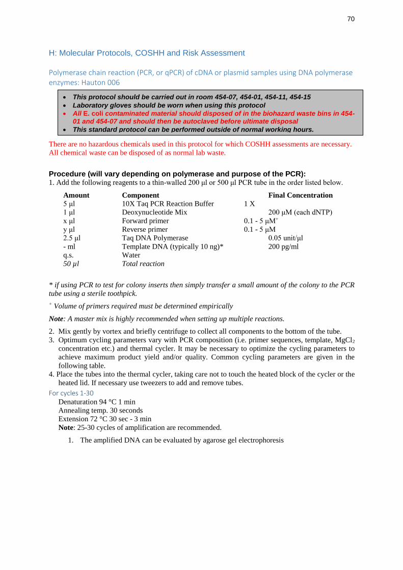
70
H: Molecular Protocols, COSHH and Risk Assessment
Polymerase chain reaction (PCR, or qPCR) of cDNA or plasmid samples using DNA polymerase enzymes: Hauton 006
There are no hazardous chemicals used in this protocol for which COSHH assessments are necessary.
All chemical waste can be disposed of as normal lab waste.
Procedure (will vary depending on polymerase and purpose of the PCR): 1. Add the following reagents to a thin-walled 200 μl or 500 μl PCR tube in the order listed below.
Amount Component Final Concentration
5 μl 10X Taq PCR Reaction Buffer 1 X
1 μl Deoxynucleotide Mix 200 μM (each dNTP)
x μl Forward primer 0.1 - 5 μM+
y μl Reverse primer 0.1 - 5 μM
2.5 μl Taq DNA Polymerase 0.05 unit/μl
- ml Template DNA (typically 10 ng)* 200 pg/ml
q.s. Water
50 µl Total reaction
* if using PCR to test for colony inserts then simply transfer a small amount of the colony to the PCR
tube using a sterile toothpick.
+ Volume of primers required must be determined empirically
Note: A master mix is highly recommended when setting up multiple reactions.
2. Mix gently by vortex and briefly centrifuge to collect all components to the bottom of the tube.
3. Optimum cycling parameters vary with PCR composition (i.e. primer sequences, template, MgCl2
concentration etc.) and thermal cycler. It may be necessary to optimize the cycling parameters to
achieve maximum product yield and/or quality. Common cycling parameters are given in the
following table.
4. Place the tubes into the thermal cycler, taking care not to touch the heated block of the cycler or the
heated lid. If necessary use tweezers to add and remove tubes.
For cycles 1-30 Denaturation 94 °C 1 min
Annealing temp. 30 seconds
Extension 72 °C 30 sec - 3 min
Note: 25-30 cycles of amplification are recommended.
1. The amplified DNA can be evaluated by agarose gel electrophoresis
• This protocol should be carried out in room 454-07, 454-01, 454-11, 454-15
• Laboratory gloves should be worn when using this protocol
• All E. coli contaminated material should disposed of in the biohazard waste bins in 454-01 and 454-07 and should then be autoclaved before ultimate disposal
• This standard protocol can be performed outside of normal working hours.
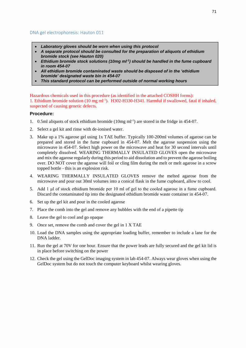
71
DNA gel electrophoresis: Hauton 011
Hazardous chemicals used in this procedure (as identified in the attached COSHH forms):
1. Ethidium bromide solution (10 mg ml-1). H302-H330-H341. Harmful if swallowed, fatal if inhaled,
suspected of causing genetic defects.
Procedure:
1. 0.5ml aliquots of stock ethidium bromide (10mg ml-1) are stored in the fridge in 454-07.
2. Select a gel kit and rinse with de-ionised water.
3. Make up a 1% agarose gel using 1x TAE buffer. Typically 100-200ml volumes of agarose can be
prepared and stored in the fume cupboard in 454-07. Melt the agarose suspension using the
microwave in 454-07. Select high power on the microwave and heat for 30 second intervals until
completely dissolved. WEARING THERMALLY INSULATED GLOVES open the microwave
and mix the agarose regularly during this period to aid dissolution and to prevent the agarose boiling
over. DO NOT cover the agarose will foil or cling film during the melt or melt agarose in a screw
topped bottle - this is an explosion risk.
4. WEARING THERMALLY INSULATED GLOVES remove the melted agarose from the
microwave and pour out 30ml volumes into a conical flask in the fume cupboard, allow to cool.
5. Add 1 μl of stock ethidium bromide per 10 ml of gel to the cooled agarose in a fume cupboard.
Discard the contaminated tip into the designated ethidium bromide waste container in 454-07.
6. Set up the gel kit and pour in the cooled agarose
7. Place the comb into the gel and remove any bubbles with the end of a pipette tip
8. Leave the gel to cool and go opaque
9. Once set, remove the comb and cover the gel in 1 X TAE
10. Load the DNA samples using the appropriate loading buffer, remember to include a lane for the
DNA ladder.
11. Run the gel at 70V for one hour. Ensure that the power leads are fully secured and the gel kit lid is
in place before switching on the power
12. Check the gel using the GelDoc imaging system in lab 454-07. Always wear gloves when using the
GelDoc system but do not touch the computer keyboard whilst wearing gloves.
• Laboratory gloves should be worn when using this protocol
• A separate protocol should be consulted for the preparation of aliquots of ethidium bromide stock (see Hauton 020)
• Ethidium bromide stock solutions (10mg ml-1) should be handled in the fume cupboard in room 454-07
• All ethidium bromide contaminated waste should be disposed of in the ‘ethidium bromide’ designated waste bin in 454-07
• This standard protocol can be performed outside of normal working hours
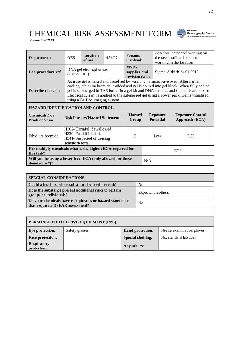
72
CHEMICAL RISK ASSESSMENT FORM Version Sept 2013
Department: OES Location
of use: 454/07
Persons
involved:
Assessor, personnel working on
the task, staff and students
working in the location
Lab procedure ref: DNA gel electrophoresis
(Hauton 011)
MSDS
supplier and
revision date:
Sigma-Aldrich 24.04.2012
Describe the task:
Agarose gel is mixed and dissolved by warming in microwave oven. After partial
cooling, ethidium bromide is added and gel is poured into gel block. When fully cooled,
gel is submerged in TAE buffer in a gel kit and DNA samples and standards are loaded.
Electrical current is applied to the submerged gel using a power pack. Gel is visualised
using a GelDoc imaging system.
HAZARD IDENTIFICATION AND CONTROL
Chemical(s) or
Product Name
Risk Phrases/Hazard Statements
Hazard
Group
Exposure
Potential
Exposure Control
Approach (ECA)
Ethidium bromide
H302- Harmful if swallowed.
H330- Fatal if inhaled.
H341- Suspected of causing
genetic defects.
E Low EC3
For multiply chemicals what is the highest ECA required for
this task? EC3
Will you be using a lower level ECA (only allowed for those
denoted by*)? N/A
SPECIAL CONSIDERATIONS
Could a less hazardous substance be used instead? No
Does the substance present additional risks to certain
groups or individuals? Expectant mothers.
Do your chemicals have risk phrases or hazard statements
that require a DSEAR assessment? No
PERSONAL PROTECTIVE EQUIPMENT (PPE)
Eye protection: Safety glasses Hand protection: Nitrile examination gloves
Face protection: Special clothing: No, standard lab coat
Respiratory
protection: Any others:
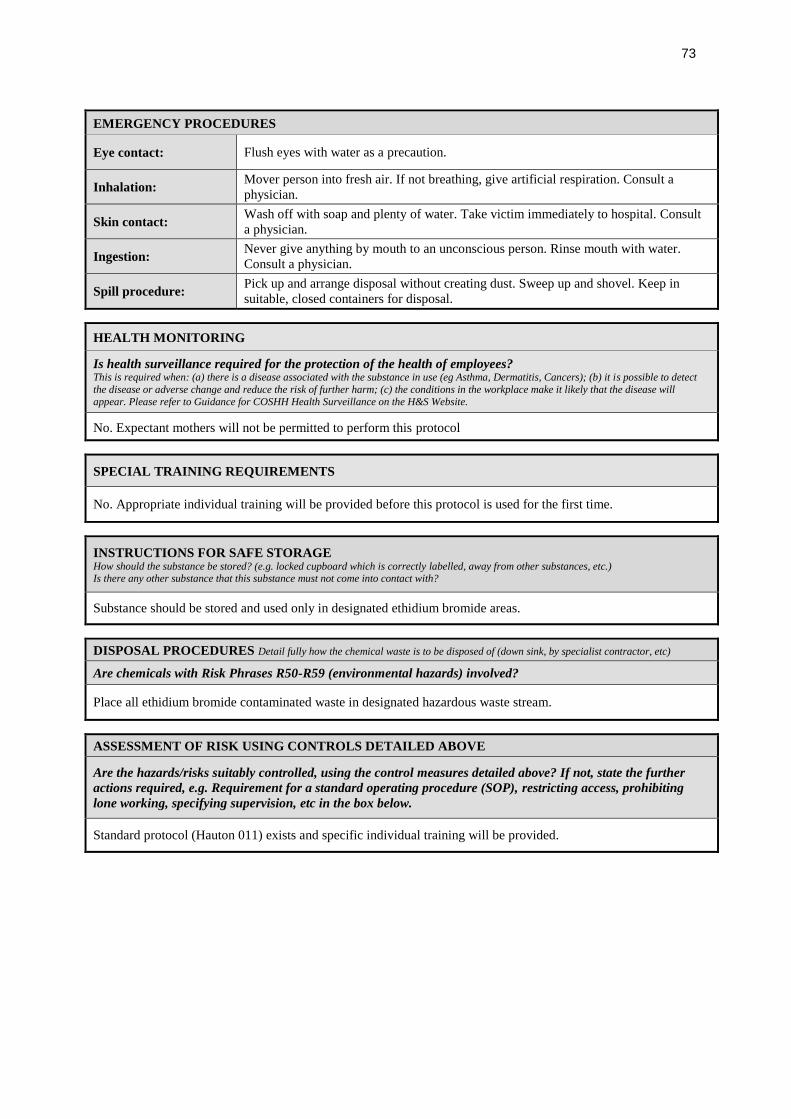
73
EMERGENCY PROCEDURES
Eye contact: Flush eyes with water as a precaution.
Inhalation: Mover person into fresh air. If not breathing, give artificial respiration. Consult a
physician.
Skin contact: Wash off with soap and plenty of water. Take victim immediately to hospital. Consult
a physician.
Ingestion: Never give anything by mouth to an unconscious person. Rinse mouth with water.
Consult a physician.
Spill procedure: Pick up and arrange disposal without creating dust. Sweep up and shovel. Keep in
suitable, closed containers for disposal.
HEALTH MONITORING
Is health surveillance required for the protection of the health of employees? This is required when: (a) there is a disease associated with the substance in use (eg Asthma, Dermatitis, Cancers); (b) it is possible to detect
the disease or adverse change and reduce the risk of further harm; (c) the conditions in the workplace make it likely that the disease will
appear. Please refer to Guidance for COSHH Health Surveillance on the H&S Website.
No. Expectant mothers will not be permitted to perform this protocol
SPECIAL TRAINING REQUIREMENTS
No. Appropriate individual training will be provided before this protocol is used for the first time.
INSTRUCTIONS FOR SAFE STORAGE How should the substance be stored? (e.g. locked cupboard which is correctly labelled, away from other substances, etc.) Is there any other substance that this substance must not come into contact with?
Substance should be stored and used only in designated ethidium bromide areas.
DISPOSAL PROCEDURES Detail fully how the chemical waste is to be disposed of (down sink, by specialist contractor, etc)
Are chemicals with Risk Phrases R50-R59 (environmental hazards) involved?
Place all ethidium bromide contaminated waste in designated hazardous waste stream.
ASSESSMENT OF RISK USING CONTROLS DETAILED ABOVE
Are the hazards/risks suitably controlled, using the control measures detailed above? If not, state the further
actions required, e.g. Requirement for a standard operating procedure (SOP), restricting access, prohibiting
lone working, specifying supervision, etc in the box below.
Standard protocol (Hauton 011) exists and specific individual training will be provided.
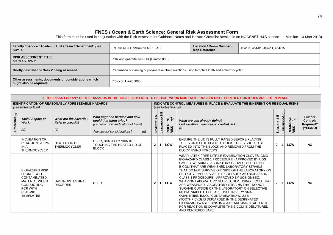
74
FNES / Ocean & Earth Science: General Risk Assessment Form This form must be used in conjunction with the Risk Assessment Guidance Notes and Hazard Checklist *available on NOCSNET H&S section Version 1.3 (Jan 2013)
Faculty / Service / Academic Unit / Team / Department: (see Note 1)
FNES/ERE/OES/Hauton MIPI-LAB Location / Room Number / Map Reference:
454/07, 454/01, 454-11, 454-15
RISK ASSESSMENT TITLE MAIN ACTIVITY
PCR and quantitative PCR (Hauton 006)
Briefly describe the ‘tasks’ being assessed: Preparation of running of polymerase chain reactions using template DNA and a thermocycler
Other assessments, documents or considerations which might also be required:
Protocol: Hauton006.
IF THE RISKS FOR ANY OF THE HAZARDS IN THE TABLE IS DEEMED TO BE HIGH, WORK MUST NOT PROCEED UNTIL FURTHER CONTROLS ARE PUT IN PLACE.
IDENTIFICATION OF REASONABLY FORESEEABLE HAZARDS
(see Notes 2i & 2ii) INDICATE CONTROL MEASURES IN PLACE & EVALUATE THE INHERENT OR RESIDUAL RISKS (see Notes 3i & 3ii)
Refe
ren
ce (
a)
Task / Aspect of Work. (b)
What are the hazards? Refer to checklist (c)
Who might be harmed and how could that harm arise? (i.e. Who, how and nature of harm) Any special considerations? (d) S
EV
ER
ITY
1-5
LIK
EL
IHO
OD
1-5
INH
ER
EN
T
RIS
K (
e)
What are you already doing? List existing measures to control risk. (f)
SE
VE
RIT
Y 1
-5
LIK
EL
IHO
OD
1-5
RE
SID
UA
L
RIS
K (
g) Further
Controls Required? (YES/NO)
INCUBATION OF REACTION STEPS IN A THERMOCYCLER
HEATED LID OF THERMOCYCLER
USER, BURNS TO SKIN IF TOUCHING THE HEATED LID OR BLOCK
2 1 LOW
ENSURE THE LID IS FULLY RAISED BEFORE PLACING TUBES ONTO THE HEATED BLOCK. TUBES SHOULD BE PLACED INTO THE BLOCK AND REMOVED FROM THE BLOCK USING FORCEPS
2 1 LOW NO
BIOHAZARD RISK FROM E.COLI CONTAMINATED MATERIAL WHEN CONDUCTING PCR WITH PLASMID TEMPLATES
GASTROINTESTINAL DISORDER
USER 2 1 LOW
WEAR LATEX-FREE NITRILE EXAMINATION GLOVES. GMO BIOHAZARD CLASS 1 PROCEDURE - APPROVED BY UOS GMBSC. WEARING LABORATORY GLOVES, GLP. USING E.COLI THAT ARE WEAKENED LABORATORY STRAINS THAT DO NOT SURVIVE OUTSIDE OF THE LABORATORY ON SELECTIVE MEDIA. VIABLE E.COLI ARE GMO BIOHAZARD CLASS 1 PROCEDURE - APPROVED BY UOS GMBSC. WEARING LABORATORY GLOVES, GLP. USING E.COLI THAT ARE WEAKENED LABORATORY STRAINS THAT DO NOT SURVIVE OUTSIDE OF THE LABORATORY ON SELECTIVE MEDIA. VIABLE E.COLI ARE USED IN VERY SMALL QUANTITIES. E.COLI CONTAMINATED WASTE (TOOTHPICKS) IS DISCARDED IN THE DESIGNATED BIOHAZARD WASTE BINS IN 454-01 AND 454-07. AFTER THE PCR REACTION IS COMPLETE THE E.COLI IS DENATURED AND RENDERED SAFE
2 1 LOW NO
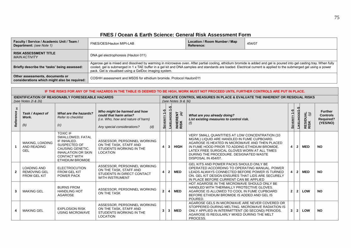
75
FNES / Ocean & Earth Science: General Risk Assessment Form
Faculty / Service / Academic Unit / Team / Department: (see Note 1)
FNES/OES/Hauton MIPI-LAB Location / Room Number / Map Reference:
454/07
RISK ASSESSMENT TITLE MAIN ACTIVITY
DNA gel electrophoresis (Hauton 011)
Briefly describe the ‘tasks’ being assessed: Agarose gel is mixed and dissolved by warming in microwave oven. After partial cooling, ethidium bromide is added and gel is poured into gel casting tray. When fully cooled, gel is submerged in 1 x TAE buffer in a gel kit and DNA samples and standards are loaded. Electrical current is applied to the submerged gel using a power pack. Gel is visualised using a GelDoc imaging system.
Other assessments, documents or considerations which might also be required:
COSHH assessment and MSDS for ethidium bromide. Protocol Hauton011
IF THE RISKS FOR ANY OF THE HAZARDS IN THE TABLE IS DEEMED TO BE HIGH, WORK MUST NOT PROCEED UNTIL FURTHER CONTROLS ARE PUT IN PLACE.
IDENTIFICATION OF REASONABLY FORESEEABLE HAZARDS
(see Notes 2i & 2ii) INDICATE CONTROL MEASURES IN PLACE & EVALUATE THE INHERENT OR RESIDUAL RISKS (see Notes 3i & 3ii)
Refe
ren
ce (
a)
Task / Aspect of Work. (b)
What are the hazards? Refer to checklist (c)
Who might be harmed and how could that harm arise? (i.e. Who, how and nature of harm) Any special considerations? (d) S
EV
ER
ITY
1-5
LIK
EL
IHO
OD
1-5
INH
ER
EN
T
RIS
K (
e)
What are you already doing? List existing measures to control risk. (f)
SE
VE
RIT
Y 1
-5
LIK
EL
IHO
OD
1-5
RE
SID
UA
L
RIS
K (
g) Further
Controls Required? (YES/NO)
1 MAKING, LOADING AND READING GEL
TOXIC IF SWALLOWED, FATAL IF INHALED. SUSPECTED OF CAUSING GENETIC. INHALATION OR SKIN CONTACT WITH ETHIDIUM BROMIDE
ASSESSOR, PERSONNEL WORKING ON THE TASK, STAFF AND STUDENTS WORKING IN THE LOCATION
4 3 HIGH
VERY SMALL QUANTITIES AT LOW CONCENTRATION (10 MG/ML) LIQUID ARE HANDLED IN FUME CUPBOARD. AGAROSE IS HEATED IN MICROWAVE AND THEN PLACED IN FUME HOOD PRIOR TO ADDING ETHIDIUM BROMIDE. LATEX FREE SURGICAL GLOVES WORN AT ALL TIMES DURING THE PROCEDURE. DESIGNATED WASTE DISPOSAL IN 454/07.
4 2 MED NO
2 LOADING AND REMOVING GEL FROM GEL KIT
ELECTROCUTION FROM GEL KIT POWER PACK
ASSESSOR, PERSONNEL WORKING ON THE TASK, STAFF AND STUDENTS IN DIRECT CONTACT WITH INSTRUMENT
4 2 MED
GEL KITS AND POWER PACKS SHOULD ONLY BE OPERATED ACCORDING TO OPERATING MANUAL. POWER LEADS ALWAYS CONNECTED BEFORE POWER IS TURNED ON. GEL KIT DESIGN ENSURES THAT LIDS ARE SECURELY IN PLACE BEFORE CURRENT CAN BE APPLIED
4 2 MED NO
3 MAKING GEL BURNS FROM HANDLING HOT AGAROSE
ASSESSOR, PERSONNEL WORKING ON THE TASK
2 4 MED
HOT AGAROSE IN THE MICROWAVE SHOULD ONLY BE HANDLED WITH THERMALLY PROTECTIVE GLOVES. AGAROSE IS ALLOWED TO COOL IN FUME CUPBOARD BEFORE ETHIDIUM BROMIDE IS ADDED AND GEL IS POURED.
2 2 LOW NO
4 MAKING GEL EXPLOSION RISK USING MICROWAVE
ASSESSOR, PERSONNEL WORKING ON THE TASK, STAFF AND STUDENTS WORKING IN THE LOCATION
3 3 MED
AGAROSE GELS IN MICROWAVE ARE NEVER COVERED OR STOPPERED DURING MELTING. MICROWAVE RADIATION IS ONLY APPLIED IN INTERMITTENT (30 SECOND) PERIODS. AGAROSE IS REGULARLY MIXED DURING THE MELT PROCESS.
3 2 LOW NO

76


![Comparative Ultrastructure and Carbohydrate Composition of ... et...It has been suggested for two freshwater crayfish species, Procambarus clarkii and Cherax quadricarinatus [8,9],](https://img.pdfslide.us/doc/110x75/60e359f5e1e7bd32a1046092/comparative-ultrastructure-and-carbohydrate-composition-of-et-it-has-been.jpg)