Embed Size (px)
Citation preview
1
The SkeletalSystem:
AppendicularSkeleton
The Big Idea• The Appendicular Skeleton & Homeostasis
– The bones of the appendicular skeletoncontribute to homeostasis by providingattachment points and leverage for muscles,which aids body movements; by providingsupport and protection of internal organs, suchas the reproductive organs; and by storing andreleasing calcium
• The 126 bones of theappendicular skeleton areprimarily concerned withmovement
• As “appendages” to thecentral skeleton, thesebones include those of theupper and lower limbs(including the girdles thatattach them to the axialskeleton)
• Based on the position of its major joints andcomponent bones, the upper limb is divided intothe shoulder, arm, forearm, and hand
• The shoulder is the area of upper limbattachment to the trunk
• The arm is the part of the upper limb betweenthe shoulder and the elbow joint
• The forearm is between the elbow and thewrist
• The hand is distal to the wrist
8.1 Pectoral (Shoulder) Girdle• Objectives
• Identify the bones of the pectoral (shoulder)girdle, their functions, and their principalmarkings
• The bones of theshoulder (pectoral)girdle include thescapula and the clavicle
• The shoulder jointalso incorporates theupper part of thehumerus
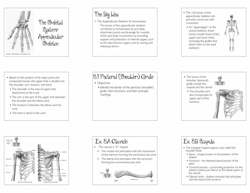
Ex. 8A Clavicle• The clavicle is “S” shaped
• The medial end articulates with the manubriumof the sternum forming the sternoclavicular joint
• The lateral end articulates with the acromionforming the acromioclavicular joint
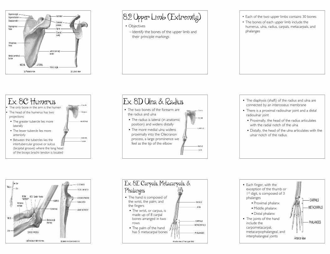
Ex. 8B Scapula• The triangular shaped scapula is also called the
shoulder blade• Spine - a large process on the posterior of the
scapula• Acromion - the flattened lateral portion of the
spine• Coracoid process - a protruding projection on the
anterior surface just inferior to the lateral aspect ofthe clavicle
• Glenoid cavity - shallow concavity that articulateswith the head of the humerus
2
8.2 Upper Limb (Extremity)• Objectives
– Identify the bones of the upper limb andtheir principle markings
• Each of the two upper limbs contains 30 bones
• The bones of each upper limb include thehumerus, ulna, radius, carpals, metacarpals, andphalanges
Ex. 8C Humerus• The only bone in the arm is the humerus
• The head of the humerus has twoprojections
• The greater tubercle lies morelaterally
• The lesser tubercle lies moreanteriorly
• Between the tubercles lies theintertubercular groove or sulcus(bicipital groove) where the long headof the biceps brachii tendon is located
Ex. 8D Ulna & Radius• The two bones of the forearm are
the radius and ulna
• The radius is lateral (in anatomicposition) and widens distally
• The more medial ulna widensproximally into the Olecranonprocess, a large prominence wefeel as the tip of the elbow
• The diaphysis (shaft) of the radius and ulna areconnected by an interosseus membrane
• There is a proximal radioulnar joint and a distalradioulnar joint
• Proximally, the head of the radius articulateswith the radial notch of the ulna
• Distally, the head of the ulna articulates with theulnar notch of the radius
Ex. 8E Carpals, Metacarpals, &Phalanges• The hand is composed of
the wrist, the palm, andthe fingers• The wrist, or carpus, is
made up of 8 carpalbones arranged in tworows
• The palm of the handhas 5 metacarpal bones
• Each finger, with theexception of the thumb or1st digit, is composed of 3phalanges• Proximal phalanx• Middle phalanx• Distal phalanx
• The joints of the handinclude thecarpometacarpal,metacarpophalangeal, andinterphalangeal joints
3
8.3 Pelvic (Hip) Girdle• Objectives
• Identify the bones of the pelvic girdle andtheir principle markings
• The lower limb is directly anchored to the axialskeleton by a sacroiliac joint which links the pelvicbone to the sacrum
• Based on the position of its major joints andcomponent bones, the lower limb is divided intothe gluteal region (the major bones forming thehip girdle), thigh, leg, and foot• The gluteal region is between the iliac crest
and hip joint• The thigh is between the hip and the knee
joint• The leg is between the knee and the ankle• The foot is distal to the ankle
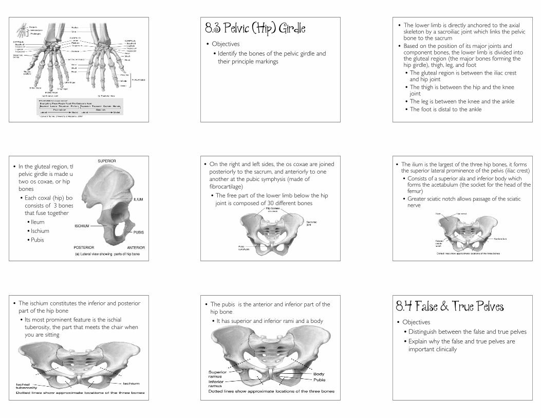
• In the gluteal region, thepelvic girdle is made up oftwo os coxae, or hipbones
• Each coxal (hip) boneconsists of 3 bonesthat fuse together
• Ileum
• Ischium
• Pubis
• On the right and left sides, the os coxae are joinedposteriorly to the sacrum, and anteriorly to oneanother at the pubic symphysis (made offibrocartilage)
• The free part of the lower limb below the hipjoint is composed of 30 different bones
• The ilium is the largest of the three hip bones, it formsthe superior lateral prominence of the pelvis (iliac crest)• Consists of a superior ala and inferior body which
forms the acetabulum (the socket for the head of thefemur)
• Greater sciatic notch allows passage of the sciaticnerve
• The ischium constitutes the inferior and posteriorpart of the hip bone
• Its most prominent feature is the ischialtuberosity, the part that meets the chair whenyou are sitting
• The pubis is the anterior and inferior part of thehip bone
• It has superior and inferior rami and a body
8.4 False & True Pelves• Objectives
• Distinguish between the false and true pelves
• Explain why the false and true pelves areimportant clinically
4
• The false pelvis is separated from the true pelvis by thepelvic brim• The pelvic brim is a line from the sacral promontory to
the upper part of the pubic symphysis• The false pelvis lies above this line• It contains no pelvic organs except the urinary
bladder (when full), the lower intestines, and theuterus, uterine tubes, and ovaries
• The true pelvis is the bony pelvis inferior to the pelvicbrim• It has an inlet, an outlet and a cavity• Surrounds the pelvic cavity and houses the rectum and
urinary bladder, the vagina and cervix in females, andthe prostate in males
• The pelvic axis is the path of childbirth during the firstand second stages of labor
8.5 Comparison of Female &Male Pelves• Objectives
• Compare the principal differences betweenfemale and male pelves
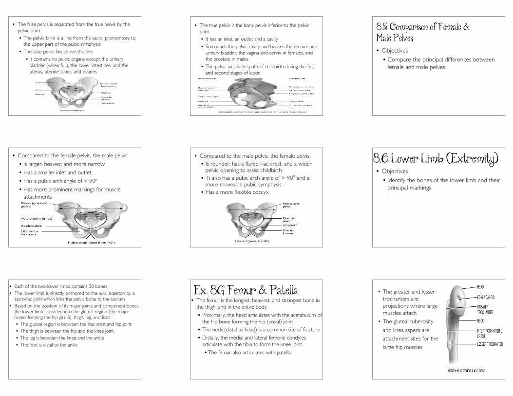
• Compared to the female pelvis, the male pelvis
• Is larger, heavier, and more narrow
• Has a smaller inlet and outlet
• Has a pubic arch angle of < 90o
• Has more prominent markings for muscleattachments
• Compared to the male pelvis, the female pelvis• Is rounder, has a flared iliac crest, and a wider
pelvic opening to assist childbirth• It also has a pubic arch angle of > 90o and a
more moveable pubic symphysis• Has a more flexible coccyx
8.6 Lower Limb (Extremity)• Objectives
• Identify the bones of the lower limb and theirprincipal markings
• Each of the two lower limbs contains 30 bones• The lower limb is directly anchored to the axial skeleton by a
sacroiliac joint which links the pelvic bone to the sacrum• Based on the position of its major joints and component bones,
the lower limb is divided into the gluteal region (the majorbones forming the hip girdle), thigh, leg, and foot• The gluteal region is between the iliac crest and hip joint• The thigh is between the hip and the knee joint• The leg is between the knee and the ankle• The foot is distal to the ankle
Ex. 8G Femur & Patella• The femur is the longest, heaviest, and strongest bone in
the thigh, and in the entire body• Proximally, the head articulates with the acetabulum of
the hip bone forming the hip (coxal) joint• The neck (distal to head) is a common site of fracture• Distally, the medial and lateral femoral condyles
articulate with the tibia to form the knee joint• The femur also articulates with patella
• The greater and lessertrochanters areprojections where largemuscles attach
• The gluteal tuberosity
and linea aspera are
attachment sites for the
large hip muscles
5
• The femur has sitesfor attachment ofthe knee muscles atthe medial andlateral epicondyles(above the femoralcondyles)
• The patella (knee cap) is the largestand only named sesamoid bone in thebody
• A thick articular cartilage lines theposterior surface
• At the distal femur, thepatella forms thepatellofemoral jointwhere it functions toincrease the leverage ofthe quadriceps muscles
• Runner’s knee(patellofemoral stresssyndrome) is acommon sports injury
Ex. 8H Tibia & Fibula• Of the two bones in the leg, the
tibia (always medial) is the largestand bears all the weight
• The lateral and medial condylesat the proximal end articulatewith the femur
• It articulates distally with thetalus of the ankle and the fibula
• The fibula is the smaller, laterallyplaced bone of the leg
• It is non-weight bearing
• The head forms the proximaltibiofibular joint
• At the distal end, the lateralmalleolus articulates with thetibia and the talus at the ankle
• Much like the forearm bones,the tibia and fibula are joined byan interosseous membrane
• The distal end of the leg bonesform the medial and lateralmalleoli of the ankle
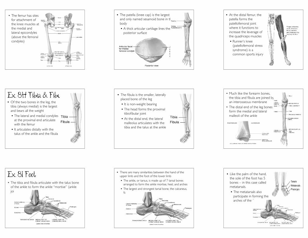
Ex. 8I Foot• The tibia and fibula articulate with the talus bone
of the ankle to form the ankle “mortise” (anklejoint)
• There are many similarities between the hand of theupper limb and the foot of the lower limb• The ankle, or tarsus, is made up of 7 tarsal bones
arranged to form the ankle mortise, heel, and arches• The largest and strongest tarsal bone, the calcaneus,
forms the heel
• Like the palm of the hand,the sole of the foot has 5bones – in this case calledmetatarsals.
• The metatarsals alsoparticipate in forming thearches of the foot.
6
• Each toe with the exceptionof the hallux (big toe) iscomposed of 3 phalanges• Proximal phalanx• Middle phalanx• Distal phalanx
• The joints of the foot includethe tarsometatarsal,metatarsophalangeal, andinterphalangeal joints
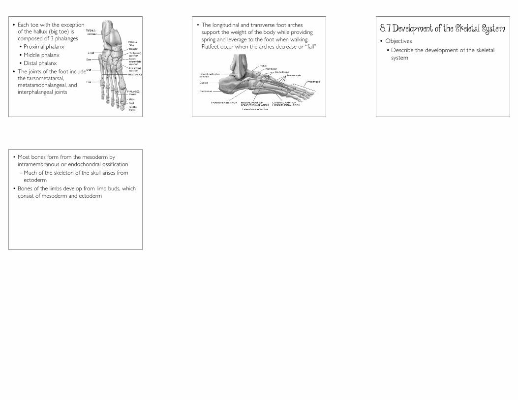
• The longitudinal and transverse foot archessupport the weight of the body while providingspring and leverage to the foot when walking.Flatfeet occur when the arches decrease or “fall”
8.7 Development of the Skeletal System• Objectives
• Describe the development of the skeletalsystem
• Most bones form from the mesoderm byintramembranous or endochondral ossification
– Much of the skeleton of the skull arises fromectoderm
• Bones of the limbs develop from limb buds, whichconsist of mesoderm and ectoderm