Embed Size (px)
Citation preview

Submitted 7 March 2018Accepted 5 September 2018Published 11 October 2018
Corresponding authorNorhaniza Aminudin,[email protected]
Academic editorElena González-Burgos
Additional Information andDeclarations can be found onpage 15
DOI 10.7717/peerj.5694
Copyright2018 Abrahim et al.
Distributed underCreative Commons CC-BY 4.0
OPEN ACCESS
The antioxidant activities, cytotoxicproperties, and identification of water-soluble compounds of Ficus deltoidealeavesNoor Nazirahanie Abrahim1, Puteri Shafinaz Abdul-Rahman1,2 andNorhaniza Aminudin2,3
1Department of Molecular Medicine, Faculty of Medicine, University of Malaya, Kuala Lumpur, Malaysia2University of Malaya Centre for Proteomics Research (UMCPR), Faculty of Medicine, University of Malaya,Kuala Lumpur, Malaysia
3 Institute of Biological Sciences, Faculty of Science, University of Malaya, Kuala Lumpur, Malaysia
ABSTRACTLeaves from three varieties of Ficus deltoidea, colloquially termed small- (FDS),medium- (FDM), and big-type leaf (FDB), were subjected to water extraction. Thecrude extracts were fractionated usingwater (WF) and ethyl acetate (EAF). The phenolicand flavonoid content, antioxidant activity, and cytotoxicity of the fractions wereinvestigated. The EAF had the highest phenolic and flavonoid content compared to theother FDS fractions. Conversely, the FDM crude extract had the highest phenolic andflavonoid content compared to the other FDMsamples. Antioxidant activitywas highestin the FDB crude extract. Ultra-high–performance liquid chromatography showed thattwo compounds, vitexin and coumaric acid, were present in the FDB crude extract.Additionally, the F. deltoidea leaves caused no signs of toxicity in a normal liver cellline. Our findings show that F. deltoidea varieties have excellent antioxidant activitywith no cytotoxic effects on normal liver cells.
Subjects Biochemistry, Cell Biology, Food Science and Technology, Plant Science, TranslationalMedicineKeywords Ficus deltoidea, Antioxidant, Flavonoids, Phenolic acid
INTRODUCTIONIt has been widely reported that various plant phytochemicals have biological activityand have the potential to affect disease risk through complementary mechanisms. Alarge number of these phytochemicals, particularly polyphenols, have been identifiedand have a wide range of biological activities, including neuroprotective effects (Zhuet al., 2007); inflammatory and immune response regulation (Rotelli et al., 2003); andanti-fungal (Raj et al., 2001), anti-cancer, hypoglycemic (Sharma, Chandrajeet & Partha,2008), anti-hyperlipidemic (Ghule et al., 2006), and anti-atherosclerotic effects (Kim etal., 2005a). Despite such phytochemical activity, one of the main research interests isinvestigating their antioxidant potential for preventing or delaying autoxidation by freeradicals. Free radical autoxidation leads tomacromolecule deterioration, especially of lipids,proteins, carbohydrates, and DNA (Carocho & Ferreira, 2013). The oxidation of these
How to cite this article Abrahim et al. (2018), The antioxidant activities, cytotoxic properties, and identification of water-soluble com-pounds of Ficus deltoidea leaves. PeerJ 6:e5694; DOI 10.7717/peerj.5694
macromolecules, particularly in humans, leads to various illnesses, such as Alzheimer’sdisease, cancer, and cardiovascular diseases.
Due to its wide medicinal uses, we selected Ficus deltoidea for the present study. Alsoknown as mistletoe fig or Mas cotek, F. deltoidea is a shrub that is native to SoutheastAsia. It is a well-known shrub, especially among the Malays, and is used to treat diabetes,headache, sore throat, and cold. Its leaves have been studied for their hypoglycemic,antinociceptive (Sulaiman et al., 2008), anti-inflammatory (Abdullah et al., 2009), andantioxidant (Hakiman & Maziah, 2009) activity. However, its antioxidant activity has notbeen fully elucidated due to the presence of various bioactive compounds that mightcontribute to different antioxidant capacities. These complex mixtures, especially in plantextracts, can interact synergistically, additively, or antagonistically in different assays (Wanget al., 2011; Colon & Nerin, 2016). According to Misbah, Abdul Aziz & Aminudin (2013), acombination of assays incorporating various mechanisms of action would be very helpfulfor providing complete information on the antioxidant capacity of a specific plant. Thus,the aim of the present study is to determine the antioxidant capacity of F. deltoidea leavesin different in vitro systems as well as to determine their cytotoxic effect on a normal livercell line.
METHODSSample preparationThe leaves of three varieties of F. deltoidea were obtained from a plantation in Rembau,Negeri Sembilan. The F. deltoidea varieties (small, FDS; medium, FDM; big, FDB) weredeposited in the Herbarium, Rimba Ilmu, University of Malaya, Kuala Lumpur, andassigned individual voucher specimen numbers (KLU046467, KLU046469, KLU046471,respectively). The leaves were rinsed and air-dried at room temperature until they reacheda constant weight and then ground into powder using a commercial blender. The powderwas kept at −20 ◦C for further analysis.
Extraction and liquid–liquid fractionationThe dried leaf powder underwent extraction according toMisbah, Abdul Aziz & Aminudin(2013) to yield the crude extract. Then, the crude extracts were fractionated using partialliquid–liquid separation for finer separation of the plant constituents into fractions ofdifferent polarity. The process involved the use of two immiscible solvents of differentpolarities, i.e., water and ethyl acetate using the method established byMisbah, Abdul Aziz& Aminudin (2013) to yield the water and ethyl acetate fractions. Subsequent experimentswere conducted using the FDS, FDM, and FDB crude extracts along with their respectivewater and ethyl acetate fractions.
Ultraviolet–visible (UV-Vis) spectroscopyUV-Vis spectroscopy was used to distinguish the presence of phenolic components inthe samples. The UV-Vis absorption pattern of phytoconstituents can be measured invery dilute solution against a solvent blank using a UV-Vis spectrophotometer. Samplesolutions prepared in water were used for this analysis, and the spectra were recordedagainst a control (water). The wavelength maxima (λmax) of each samples was recorded.
Abrahim et al. (2018), PeerJ, DOI 10.7717/peerj.5694 2/20

Extract analysis assaysFolin-Ciocalteu assayThe total polyphenolic content (TPC) of the samples was determined using the Folin-Ciocalteu assay. Folin-Ciocalteu reagent (0.1 ml) was added to 1 µl sample and incubatedfor 5 min. Sodium carbonate (0.07 ml) was added to the mixture and left in the dark for2 h. The absorbance of the mixture was measured at 765 nm using a microplate reader(BioTek, USA). Gallic acid (0–200 µg/ml) was used as the standard and was processedunder similar conditions as above. The TPC in the samples was expressed as mg gallic acidequivalents (GAE)/g dry weight. All experiments were carried out in triplicate.
Aluminum chloride assayQuercetin (0–100 µg/ml) was prepared to generate the standard curve. Sample (500 µl) orquercetin (1 mg/ml) was combined with 95% ethanol (1.5 ml), 10% aluminum chloride(0.1 ml), 1 M potassium acetate (0.1 ml), and distilled water (2.8 ml). The absorbanceof the mixture was determined at 415 nm after 30-min incubation. The total flavonoidcontent (TFC) was expressed as mg quercetin equivalents (QE)/g dry weight. All analyseswere performed in triplicate.
Cupric ion (Cu2+) reducing antioxidant capacity (CUPRAC) assayThe CUPRAC assay is the most commonly used assay for in vitro determination of theantioxidant activity of food elements, biological fluids, and also plant extracts. It uses copper(II)-neocuproine [Cu(II)-Nc] as the oxidizing agent to measure antioxidant activity closeto physiological pH conditions. For this assay, 10 mM copper solution (1 ml) was mixedwith 7.5 mMneocuproine (1 ml), 1M ammonium acetate buffer (1 ml), and sample (1 ml),incubated for 30 min, and the absorbance of the mixture was determined at 450 nm. Thesamples and the positive control quercetin (0–1,000 µg/ml) were tested. All experimentswere performed in triplicate.
2,2-Diphenyl-1-picryl-hydrazyl (DPPH) assayWe determined the radical scavenging activity of antioxidants in the samples and quercetin(positive control, 0–1,000 µg/ml) using DPPH. Samples (100 µl) were added to 600 µlDPPH reagent and mixed vigorously. The mixture was incubated in the dark for 30 minat room temperature, following which the decrease in absorbance was detected at 517 nm.The same procedure was repeated with the positive control. The absorbance of the radicalwithout antioxidants was used as the negative control. The experiment was carried out intriplicate, and the percentage of inhibition (%) was calculated using the following formula:[absorbance(blank) –absorbance(sample)/absorbance(blank)] ×100.
Non-enzymatic lipid peroxidation (thiobarbituric acid–reactive substances,TBARS) assayFowl egg yolk homogenate was used as the lipid-rich medium and underwent non-enzymatic peroxidation when incubated with ferrous sulphate (FeSO4), which acts as amediator for the initiation of lipid peroxidation. The yolk was separated from the albuminand the yolk membrane was removed. We used 0, 0.1, 0.5, 1, and 2 mg/ml sample andpositive control were used. Samples (100µl) weremixed with 500µl buffered egg yolk (1%)
Abrahim et al. (2018), PeerJ, DOI 10.7717/peerj.5694 3/20

and 100 µl FeSO4 (1 M) in a test tube. The mixture was incubated at 37 ◦C for 1 h, and then250 µl trichloroacetic acid (TCA,15%) and 500 µl TBA (1%) were added to the mixture.Subsequently, the mixture was heated for 10 min at 100 ◦C and left to cool, centrifuged at3,500 rpm for 10 min, and its absorbance was detected at 532 nm. Each experiment wascarried out independently in triplicate. The percentage of inhibition (%) was calculatedusing the following formula: [absorbance(blank)—absorbance(sample)/absorbance(blank)]×100.
Ferrous ion chelating (FIC, ferrozine) activity assayThe FIC activity assay was used to investigate the FIC capacity of the samples (Singh& Rajini, 2004). Briefly, 2 mM FeSO4 (0.005 ml) was mixed with 0–400 µg/ml sample(0.1 ml), followed by the addition of 5 mM ferrozine (0.02 ml). The absorbance of thereaction mixture was detected at 562 nm after 10-min incubation. A higher absorbanceat 562 nm indicated the weaker FIC strength of the chelator. Ethylenediaminetetraaceticacid disodium salt (EDTA-Na2) was used as the reference standard (Diaz et al., 2012).A blank, containing water, was also incorporated under the same conditions. The FICcapacity of the extracts (%) was estimated using the following equation: [(absorbance[blank]–absorbance[test])/absorbance(blank)] ×100, where absorbancetest is the absorbance ofthe reaction mixture containing the extract or ascorbic acid and absorbanceblank is theabsorbance of the blank. All determinations were carried out in triplicate.
Ferricyanide (Prussian blue) assayThe reducing power of the crude extracts and fractions were measured using the methodof Bursal & Koksal (2011) with a slight adjustment. The formation of the Prussian bluecomplex is due to the reduction of ferric ions (Fe3+) to ferrous ions (Fe2+), the absorbanceof which can be detected at 700 nm. Sample (0, 0.1, 0.5, 1 mg/ml) was mixed with sodiumphosphate buffer (0.2 M, pH 6.6) and potassium ferricyanide (1%). The mixture wasincubated at 50 ◦C for 20 min using a dry bath, followed by the addition of TCA (10%) andferric trichloride (FeCl3, 0.1%) to the reactionmixture.Distilledwaterwas used as the blank.The absorbance of the mixture was detected at 700 nm using a UV spectrophotometer.
Toxicity studyThe inhibition of cell growth was measured using the 3-(4,5-dimethylthiazol-2-yl)-2,5-diphenyltetrazolium bromide (MTT) assay. MTT is a tetrazolium salt that is cleaved intoformazan crystals by succinate dehydrogenase and is only active in viable cells. A higheramount of formazan dye produced indicates a higher number of viable cells. WRL68normal liver cells were seeded in 96-well culture plates (5× 10³ cells/well) and left to attachovernight at 37 ◦C in a 5% CO2 atmosphere. F. deltoidea leaf crude extracts and fractionswere added to each well to yield final concentrations of 50, 100, 200, and 500 µg/ml. Thecells were also treated with the same amount of vehicle (water) present in the plant extracts,and this was used as the negative control. Cytotoxicity was measured after 24-, 48-, and72-h treatment by adding 10 µl MTT reagent (5 mg/ml) to the cells and incubating themfor an additional 4 h. Finally, the medium and MTT reagent were discarded and replaced
Abrahim et al. (2018), PeerJ, DOI 10.7717/peerj.5694 4/20
with 100 µl isopropanol. The absorbance was detected at 595 nm and the percentage ofinhibition (%) was calculated as follows: [(total cells – viable cells)/total cells] ×100.
The median inhibitory concentrations (IC50) were determined. All experiments werecarried out in three separate batches, each in triplicate.
Phytochemical analysis and identificationThe sample with the most activity was subjected to ultra-high–performance liquidchromatography (UHPLC) identification. The sample was prepared using two hydrolysismethods: acidic and alkaline. The acidic hydrolysis was prepared by mixing 10 mg samplewith 1.2 M hydrochloric acid (HCl) in 50% methanol, while alkaline hydrolysis wasperformed by mixing 10 mg sample with 0.5 M sodium hydroxide (NaOH) in 50%methanol. Both mixtures were heated for 2 h at 90 ◦C using a dry water bath (Labnet,USA), left to cool, and centrifuged at 5000 rpm for 20 min. The supernatant was filteredand stored at −20 ◦C until used.
The hydrolyzed samples were analyzed using a UHPLC system (Agilent, Santa Clara, CA,USA) comprising a dual wavelength absorbance detector, quaternary pumps, auto-injectorwith a 6-µl sample loop, and a column oven. Reverse-phase separations were carried outat 30 ◦C using a ZORBAX C18 column (Agilent, Santa Clara, CA, USA) (3. 9× 50 mm).Trifluoroacetic acid (TFA) in water at pH 2.6 (solvent A) and acetonitrile (solvent B) wereused as the mobile phase. The flow rate was maintained at 0.3 ml/min for a total run of 9min, and the gradient program consisted of 5% to 15% solvent B for 3 min, 15% to 50%solvent B for 3 min, and 50% to 100% solvent B for 2 min, and it then was reduced toinitial conditions for another 1 min. The eluted peaks were detected at 280 nm and 335nm. The samples were diluted in solvent A to yield 5% methanol, and 3 µl sample wasinjected into the UHPLC system. All samples were prepared and analyzed in triplicate.The standards used in the UHPLC analysis were coumaric acid, catechin, gallic acid, andvitexin. Standard stock solutions of each compound were prepared inmethanol at 1 mg/ml.A mixture of the standard solutions was prepared at various concentrations (100–500 ng,and 3 µl was injected into the UHPLC and run using the conditions described above.Peak identification was carried out by spiking the samples with the standards of possiblecompounds and by spectral analysis. The UV spectra of individual peaks were recordedin the 200–400-nm range. Data acquisition and processing were performed using a LabAdvisor chromatography manager (Agilent, Santa Clara, CA, USA).
Statistical analysisThe data were analyzed using the Excel statistical package forWindows software (Microsoft,USA); all analyses were done in triplicate. The results are expressed as the mean± standarddeviation (SD). The correlation coefficient was used to detect the relationship betweenthe extracts’ phenolic content and the antioxidant activity. Statistical significance wascalculated using Student’s t -test. Differences between means at the 95% confidence level(p< 0.05) were considered statistically significant as compared to the control.
Abrahim et al. (2018), PeerJ, DOI 10.7717/peerj.5694 5/20
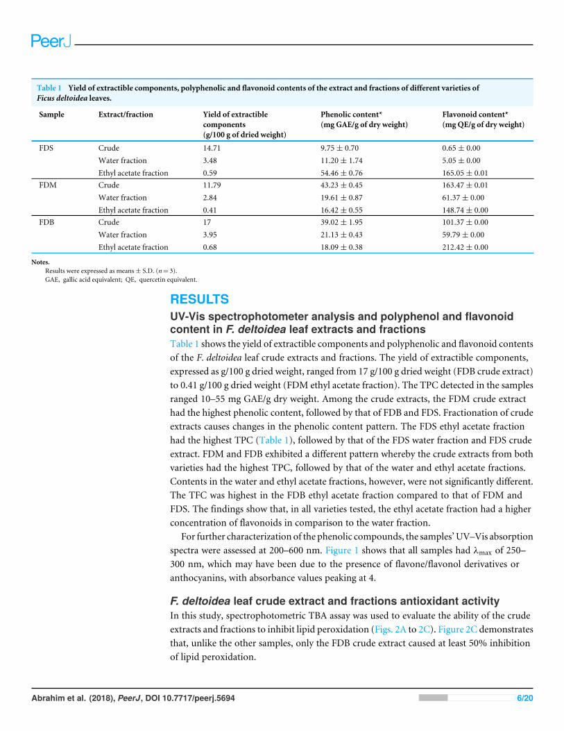
Table 1 Yield of extractible components, polyphenolic and flavonoid contents of the extract and fractions of different varieties ofFicus deltoidea leaves.
Sample Extract/fraction Yield of extractiblecomponents(g/100 g of dried weight)
Phenolic content*(mg GAE/g of dry weight)
Flavonoid content*(mg QE/g of dry weight)
FDS Crude 14.71 9.75± 0.70 0.65± 0.00Water fraction 3.48 11.20± 1.74 5.05± 0.00Ethyl acetate fraction 0.59 54.46± 0.76 165.05± 0.01
FDM Crude 11.79 43.23± 0.45 163.47± 0.01Water fraction 2.84 19.61± 0.87 61.37± 0.00Ethyl acetate fraction 0.41 16.42± 0.55 148.74± 0.00
FDB Crude 17 39.02± 1.95 101.37± 0.00Water fraction 3.95 21.13± 0.43 59.79± 0.00Ethyl acetate fraction 0.68 18.09± 0.38 212.42± 0.00
Notes.Results were expressed as means± S.D. (n= 3).GAE, gallic acid equivalent; QE, quercetin equivalent.
RESULTSUV-Vis spectrophotometer analysis and polyphenol and flavonoidcontent in F. deltoidea leaf extracts and fractionsTable 1 shows the yield of extractible components and polyphenolic and flavonoid contentsof the F. deltoidea leaf crude extracts and fractions. The yield of extractible components,expressed as g/100 g dried weight, ranged from 17 g/100 g dried weight (FDB crude extract)to 0.41 g/100 g dried weight (FDM ethyl acetate fraction). The TPC detected in the samplesranged 10–55 mg GAE/g dry weight. Among the crude extracts, the FDM crude extracthad the highest phenolic content, followed by that of FDB and FDS. Fractionation of crudeextracts causes changes in the phenolic content pattern. The FDS ethyl acetate fractionhad the highest TPC (Table 1), followed by that of the FDS water fraction and FDS crudeextract. FDM and FDB exhibited a different pattern whereby the crude extracts from bothvarieties had the highest TPC, followed by that of the water and ethyl acetate fractions.Contents in the water and ethyl acetate fractions, however, were not significantly different.The TFC was highest in the FDB ethyl acetate fraction compared to that of FDM andFDS. The findings show that, in all varieties tested, the ethyl acetate fraction had a higherconcentration of flavonoids in comparison to the water fraction.
For further characterization of the phenolic compounds, the samples’UV–Vis absorptionspectra were assessed at 200–600 nm. Figure 1 shows that all samples had λmax of 250–300 nm, which may have been due to the presence of flavone/flavonol derivatives oranthocyanins, with absorbance values peaking at 4.
F. deltoidea leaf crude extract and fractions antioxidant activityIn this study, spectrophotometric TBA assay was used to evaluate the ability of the crudeextracts and fractions to inhibit lipid peroxidation (Figs. 2A to 2C). Figure 2C demonstratesthat, unlike the other samples, only the FDB crude extract caused at least 50% inhibitionof lipid peroxidation.
Abrahim et al. (2018), PeerJ, DOI 10.7717/peerj.5694 6/20
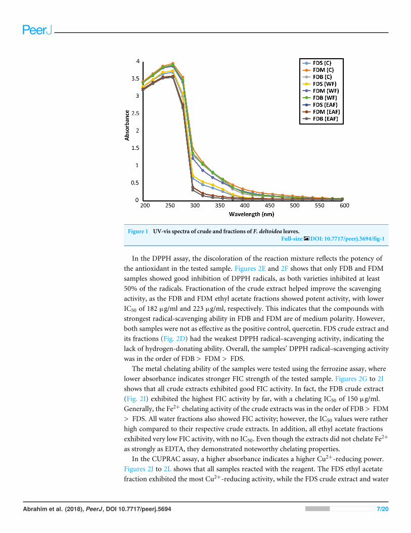
Figure 1 UV-vis spectra of crude and fractions of F. deltoidea leaves.Full-size DOI: 10.7717/peerj.5694/fig-1
In the DPPH assay, the discoloration of the reaction mixture reflects the potency ofthe antioxidant in the tested sample. Figures 2E and 2F shows that only FDB and FDMsamples showed good inhibition of DPPH radicals, as both varieties inhibited at least50% of the radicals. Fractionation of the crude extract helped improve the scavengingactivity, as the FDB and FDM ethyl acetate fractions showed potent activity, with lowerIC50 of 182 µg/ml and 223 µg/ml, respectively. This indicates that the compounds withstrongest radical-scavenging ability in FDB and FDM are of medium polarity. However,both samples were not as effective as the positive control, quercetin. FDS crude extract andits fractions (Fig. 2D) had the weakest DPPH radical–scavenging activity, indicating thelack of hydrogen-donating ability. Overall, the samples’ DPPH radical–scavenging activitywas in the order of FDB > FDM > FDS.
The metal chelating ability of the samples were tested using the ferrozine assay, wherelower absorbance indicates stronger FIC strength of the tested sample. Figures 2G to 2Ishows that all crude extracts exhibited good FIC activity. In fact, the FDB crude extract(Fig. 2I) exhibited the highest FIC activity by far, with a chelating IC50 of 150 µg/ml.Generally, the Fe2+ chelating activity of the crude extracts was in the order of FDB > FDM> FDS. All water fractions also showed FIC activity; however, the IC50 values were ratherhigh compared to their respective crude extracts. In addition, all ethyl acetate fractionsexhibited very low FIC activity, with no IC50. Even though the extracts did not chelate Fe2+
as strongly as EDTA, they demonstrated noteworthy chelating properties.In the CUPRAC assay, a higher absorbance indicates a higher Cu2+-reducing power.
Figures 2J to 2L shows that all samples reacted with the reagent. The FDS ethyl acetatefraction exhibited the most Cu2+-reducing activity, while the FDS crude extract and water
Abrahim et al. (2018), PeerJ, DOI 10.7717/peerj.5694 7/20

Figure 2 FDS, FDM and FDB were tested for various antioxidant activities which are lipid peroxida-tion (A–C), DPPH radical scavenging (D–F), ferrozine (G–I), CUPRAC (J–L) and ferricyanide (M–O)assays. Crude extract (grey bar) Water fraction (white bar) Ethyl acetate fraction (black bar).
Full-size DOI: 10.7717/peerj.5694/fig-2
Abrahim et al. (2018), PeerJ, DOI 10.7717/peerj.5694 8/20

Table 2 Correlation analyses between phenolic (PC) and flavonoid content (FC) and antioxidant ac-tivities of the crude extracts and fractions of F deltoidea leaves.
DPPHassay
Cupracassay
Ferrozineassay
Ferricyanideassay
Lipid peroxidationassay
R2 R2 R2 R2 R2
PC 0.1697* 0.9161** −0.0213 0.3255** 0.6658FC 0.7765* 0.5867** −0.6321* 0.3358** 0.5225*
Notes.*Data with p value < 0.05 were considered significant.**Data with p value < 0.01 were considered significant.
fraction had the lowest activity (Fig. 2J). A similar pattern of activity was observed for FDMand FDB (Figs. 2K and 2L) in the order of crude extract > water fraction > ethyl acetatefraction. The reducing activity of both the FDM and FDB crude extracts might have beendue to the presence of various phytochemicals that interacted synergistically. On the otherhand, fractionation causes the loss of reducing activity, which could be observed in boththe FDM and FDB fractions.
The ferricyanide assay is a reducing power assay based on the ability of test samples toreduce yellow Fe3+ to blue Fe2+. The resulting blue color is considered linearly connectedto the total reducing capacity of electron-donating antioxidants. Figures 2M to 2O showsthat the reducing activity could be divided into three types: high, moderate, and low. Theextracts that exhibited the highest reducing activity were the FDM crude extract (0.482),followed by the FDB crude extract (0.432). The samples that showed moderate activitywere the FDM water fraction (0.378), FDB ethyl acetate fraction (0.370), and FDB waterfraction (0.330). The samples that exhibited low reducing abilities were in the order ofFDS crude extract > FDM ethyl acetate fraction > FDS ethyl acetate fraction > FDS waterfraction.
Correlation coefficient between antioxidant assaysTable 2 shows the relationship between the TPC and TFC and the antioxidant activitiesof each sample. Strong interaction was observed between the TPC and Cu2+-reducingactivity (R2
= 0.9161, p< 0.01) and between the TFC and DPPH radical–scavengingactivity (R2
= 0.7765, p< 0.05). There were moderate correlations between the TFC andCu2+-reducing activity (R2
= 0.5867, p< 0.01) and between the TFC and lipid peroxidationactivity (R2
= 0.5225, p< 0.05). In contrast, the TPC was poorly correlated with DPPHradical–scavenging and metal-chelating activity (R2
= 0.1697, p< 0.05; R2=−0.0213).
Effects of F. deltoidea leaf extracts on WRL68 cell growthNormal liver cell, WRL68 was used for toxicity evaluation. Table 3 summarizes the resultsof the cytotoxic activity of the F. deltoidea leaf extracts. The data are expressed as theIC50 for all incubation times. No IC50 was detected for FDS and FDM even up to 72-hincubation. On the other hand, the FDB crude extract inhibited 50% of the cells after 72-hincubation, with an IC50 of 340 µg/ml. The WRL68 cells were also more sensitive to theFDB water fraction, which had IC50 of 375 µg/ml, 300 µg/ml, and 227 µg/ml after 24-, 48-,and 72-h incubation, respectively.
Abrahim et al. (2018), PeerJ, DOI 10.7717/peerj.5694 9/20

Table 3 Cytotoxicity of the extracts of F. deltoidea leaves againstWRL68 cells.
Sample Extract/fraction Incubation
24 hr(IC50 -µg/ml)
48 hr(IC50 -µg/ml)
72 hr(IC50 -µg/ml)
FDS Crude N/D N/D N/DWater fraction N/D N/D N/DEthyl acetate fraction N/D N/D N/D
FDM Crude N/D N/D N/DWater fraction N/D N/D N/DEthyl acetate fraction N/D N/D N/D
FDB Crude N/D N/D 347.67± 2.52Water fraction 378.3± 5.51 306.7± 7.37 224± 21.17Ethyl acetate fraction N/D N/D N/D
Notes.The experiment was conducted in a 96-well plate, each in triplicate. Cells were allowed to attach for 24 h after seeding. WRL68cells were treated with various concentrations of the extracts of F.deltoidea crude extracts and fractions for 24, 48 and 72 h.Results were expressed as means± S.D. (n= 3).IC50= concentration of plant extracts (µ g/ml) that inhibited 50% of the cells.N/D, no inhibition detected
The morphological changes of the WRL68 cells following treatment with the F. deltoidealeaf extracts were observed using a phase contrast microscope after 72-h incubation.Figure 3 shows that there was an obvious difference between the untreated (Fig. 3A) andtreated (Figs. 3B–3D) cells. The distinct changes observed in the cells treated with the FDBcrude extract (Fig. 3D) included shrinkage, rounding, and detachment from the surface ofthe wells. These alterations became increasingly noticeable as the dose increased, but werenot observed in the control cells. In contrast, the FDS and FDM extracts (Figs. 3B and 3C)showed no indications of cytotoxicity, as no morphological changes were observed.
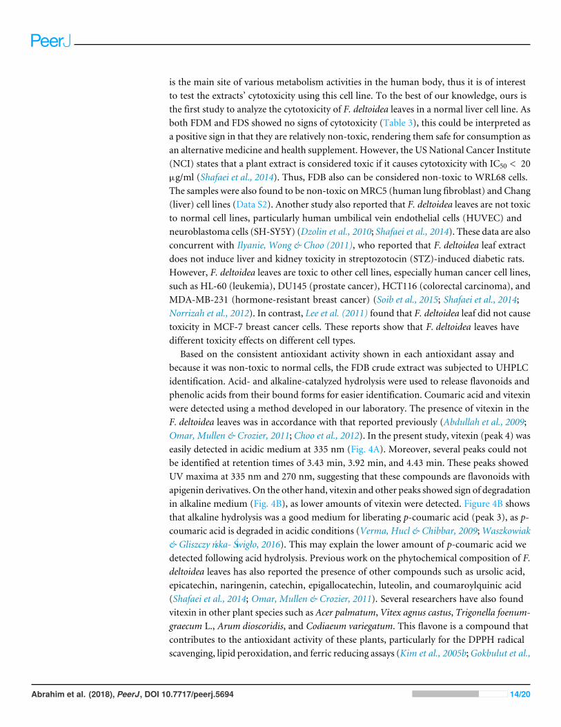
UHPLC phytochemical analysis of F. deltoidea leavesThe sample with good activity in most antioxidant assays was subsequently subjected toUHPLC for phytochemical identification. Figure 4C depicts the separation of a standardmixture of four flavonoids and phenolic acids. Good dissolution was obtained in a shortseparation time of 9 min. Gallic acid, catechin, and p-coumaric acid were detected at 280nm. The absorbance at 335 nmwas used to detect the presence of coumaric acid and vitexin.
Figure 4A and 4B show the chromatogram of the FDB crude extract under acidic andalkaline hydrolysis conditions, respectively. Two peaks were positively identified based ontheir retention time, UV spectra, and commercial standards spiking test. Coumaric acidwas present in a higher concentration under alkaline conditions as compared to acidicconditions. In contrast, there was a higher amount of vitexin following acidic hydrolysisrather than alkaline hydrolysis. Figure 4D shows that the UV spectrum of p-coumaric acidwas characterized by the presence of two maxima at 226 nm and 310 nm, while the vitexinspectrum consisted of a prominent band at 214 nm and 268 nm, with a shoulder at the338 nm region.
Abrahim et al. (2018), PeerJ, DOI 10.7717/peerj.5694 10/20

Figure 3 Representative images for morphological changes ofWRL68 cells after 72 h of incubationwithout (A) and with treatment of FDS (B), FDM (C) and FDB (D) at the highest concentration (origi-nal magnification:10×).
Full-size DOI: 10.7717/peerj.5694/fig-3
DISCUSSIONIn the present study, water was used as amedium for extracting the hydrophilic antioxidantspresent in F. deltoidea leaves. This is of interest, as typically in the preparation of food andnutraceuticals, aqueous plant extracts are nutritionally more useful and have apparentbenefit in relation to safety. On top of that, this is also a similar method as how theplant extract was prepared and consumed traditionally. Many researchers have foundthat the physiological functions of natural foods can be associated with the presence ofphenolic components. Furthermore, flavonoids have various biological properties, suchas anti-bacterial, anti-inflammatory, anti-viral, and anti-thrombotic effects. Therefore,it is reasonable to determine the TPC and TFC contents of the F. deltoidea leaf extracts.Three most commonly found varieties of F. deltoidea were evaluated in this study and
Abrahim et al. (2018), PeerJ, DOI 10.7717/peerj.5694 11/20
Figure 4 HPLC chromatograms of crude water extract of FDB in acid (A), alkaline (B) hydrolysis andstandard solutionmixture (C). (D) UV-spectra of peak 3 and 4 corresponding to p-coumaric acid andvitexin. These compounds were detected at a total run of 9 min by using two different wavelength at 280and 335 nm. Standard solution mixture consists of gallic acid (1), catechin (2), p-coumaric acid (3) andvitexin (4).
Full-size DOI: 10.7717/peerj.5694/fig-4
the results show that some of our findings differ from that of others. Pushpanathan &Nithyanandam (2015) reported that the F. deltoidea leaf TPC was 96.225 mg GAE/100 gdried weight (water extract), 225.917 mg GAE/100 g dried weight (80% methanol), and264.765 mg/100 g dried weight (80% ethanol), which was lower than that detected in oursample. By contrast,Mun et al. (2017) detected higher F. deltoidea leaf TPC compared thatin the present study, i.e., 368.42 ± 6.37 mg GAE/g (aqueous extract), 295.03 ± 16.65mg GAE/g (methanol extract), and 263.45 ± 5.28 mg/g (ethanol extract). By contrast,we detected higher TFC as compared to others (Hakiman & Maziah, 2009; Dzolin et al.,2010; Soib et al., 2015). Thus, it is difficult to compare the results obtained from differentinvestigations, as differences may arise for various reasons. Differing extraction methodsare a factor in the varied TPC and TFC determinations between studies. Various methodscan be used to extract plant compounds. Parameters such as the nature and volume of thesolvent, temperature, and time can affect compound extraction (Soong & Barlow, 2004;Maisuthisakul & Pongsawatmanit, 2004). Moreover, the presence of interfering substancesin plant extracts, such as lipophilic compounds, sugar, ascorbic acid, and aromatic amines
Abrahim et al. (2018), PeerJ, DOI 10.7717/peerj.5694 12/20

may contribute to the variations in TPC estimation between studies (Ikram et al., 2009;Khan, Bakht & Shafi, 2016). It is also important to note that the TPC and TFC evaluationmethods are based on the general structure of phenolics and flavonoids; and hence,complexity of the compounds and structural modifications that could have occurredduring growth process may lead to variations. The choice of using different varieties alsowill results in differences in term of compound extracted, their types and quantity. Each ofthis Ficus variety differs in term of morphology and growth behavior and requirements, allof which that may contribute to compound variations. Eight varieties of F. deltoidea canbe found in Malaysia; thus, genetic and geographical origins may also affect the chemicalcomposition between plants (Zimisuhara et al., 2015; Chen, Wang & Chen, 2014).
Many methods can be used to demonstrate the antioxidant activity of plant extracts.However, no single assay can establish the complete antioxidant potential of suchcompounds, as multiple reactions and mechanisms are involved in the antioxidativeprocesses (González-Centeno et al., 2013). Hence, we tested the antioxidant activity of F.deltoidea leaves using several assays involving different mechanisms. Fractionation of thecrude extracts led to the loss of antioxidant activity, especially for FDM and FDB. Thefinding suggests that the bioactive components in the crude extracts may act synergisticallyto produce the antioxidant effects, and fractionation might have eliminated some ofthe compounds (Zahin, Aqil & Ahmad, 2010). In fact, the crude extracts’ TPC and TFC(Table 1) correlated well with the antioxidant activity (Fig. 2), but not that of the fractions.Furthermore, the potential antioxidants in both FDM and FDB were mainly high-polaritycompounds. Antioxidant activity was also observed for the FDS ethyl acetate fraction, whichwas also due to the presence of high TPC and TFC. This suggests that medium-polaritycompounds contribute more to that particular activity. Based on these observations, wecould see differences in antioxidants capability showed by the varieties evaluated; FDB andFDM demonstrated a more similar pattern contrasting with FDS. This may also suggeststhat antioxidant effects of F.deltoidea could be contributed by variety of compounds otherthan the commonly known phenolics. Nevertheless, Table 2 shows the lack of correlationbetween the TPC and TFC and the antioxidant activity. Thus, highlighting that the phenolicand flavonoid compounds were not major contributors to the antioxidant activity of F.deltoidea leaves. Our findings are in agreement with the studies of Norra (2011) and Munet al. (2017). The differing antioxidant activity of the samples was most probably due to thediffering phenolic content and phenolic composition. In some instances, sampleswith lowerphenolic and flavonoid content had higher antioxidant activity. In plant extracts, the typesand amounts of phenolics and flavonoids present does not necessarily affect the antioxidantactivity; in fact, it also depends on the degree of polymerization, concentration, and thesynergistic interaction between the diverse chemical structures of the antioxidants and theantioxidant assays (Stratil, Klejdus & Kubaan, 2006; Sulaiman et al., 2011). Furthermore,antioxidants can exert their protective effects at different stages of oxidation and throughdifferent mechanisms (Parejo et al., 2002;Wang et al., 2011; Chanudom et al., 2014).
Despite the good antioxidant activity in different assay systems, determining the extracts’toxicity, especially in a normal cell line, was essential. Here, we tested the cytotoxicity of theF. deltoidea leaf crude extracts and fractions on the WRL68 normal liver cell line. The liver
Abrahim et al. (2018), PeerJ, DOI 10.7717/peerj.5694 13/20
is the main site of various metabolism activities in the human body, thus it is of interestto test the extracts’ cytotoxicity using this cell line. To the best of our knowledge, ours isthe first study to analyze the cytotoxicity of F. deltoidea leaves in a normal liver cell line. Asboth FDM and FDS showed no signs of cytotoxicity (Table 3), this could be interpreted asa positive sign in that they are relatively non-toxic, rendering them safe for consumption asan alternative medicine and health supplement. However, the US National Cancer Institute(NCI) states that a plant extract is considered toxic if it causes cytotoxicity with IC50 < 20µg/ml (Shafaei et al., 2014). Thus, FDB also can be considered non-toxic to WRL68 cells.The samples were also found to be non-toxic onMRC5 (human lung fibroblast) and Chang(liver) cell lines (Data S2). Another study also reported that F. deltoidea leaves are not toxicto normal cell lines, particularly human umbilical vein endothelial cells (HUVEC) andneuroblastoma cells (SH-SY5Y) (Dzolin et al., 2010; Shafaei et al., 2014). These data are alsoconcurrent with Ilyanie, Wong & Choo (2011), who reported that F. deltoidea leaf extractdoes not induce liver and kidney toxicity in streptozotocin (STZ)-induced diabetic rats.However, F. deltoidea leaves are toxic to other cell lines, especially human cancer cell lines,such as HL-60 (leukemia), DU145 (prostate cancer), HCT116 (colorectal carcinoma), andMDA-MB-231 (hormone-resistant breast cancer) (Soib et al., 2015; Shafaei et al., 2014;Norrizah et al., 2012). In contrast, Lee et al. (2011) found that F. deltoidea leaf did not causetoxicity in MCF-7 breast cancer cells. These reports show that F. deltoidea leaves havedifferent toxicity effects on different cell types.
Based on the consistent antioxidant activity shown in each antioxidant assay andbecause it was non-toxic to normal cells, the FDB crude extract was subjected to UHPLCidentification. Acid- and alkaline-catalyzed hydrolysis were used to release flavonoids andphenolic acids from their bound forms for easier identification. Coumaric acid and vitexinwere detected using a method developed in our laboratory. The presence of vitexin in theF. deltoidea leaves was in accordance with that reported previously (Abdullah et al., 2009;Omar, Mullen & Crozier, 2011; Choo et al., 2012). In the present study, vitexin (peak 4) waseasily detected in acidic medium at 335 nm (Fig. 4A). Moreover, several peaks could notbe identified at retention times of 3.43 min, 3.92 min, and 4.43 min. These peaks showedUV maxima at 335 nm and 270 nm, suggesting that these compounds are flavonoids withapigenin derivatives. On the other hand, vitexin and other peaks showed sign of degradationin alkaline medium (Fig. 4B), as lower amounts of vitexin were detected. Figure 4B showsthat alkaline hydrolysis was a good medium for liberating p-coumaric acid (peak 3), as p-coumaric acid is degraded in acidic conditions (Verma, Hucl & Chibbar, 2009;Waszkowiak& Gliszczyńska-Świgło, 2016). This may explain the lower amount of p-coumaric acid wedetected following acid hydrolysis. Previous work on the phytochemical composition of F.deltoidea leaves has also reported the presence of other compounds such as ursolic acid,epicatechin, naringenin, catechin, epigallocatechin, luteolin, and coumaroylquinic acid(Shafaei et al., 2014; Omar, Mullen & Crozier, 2011). Several researchers have also foundvitexin in other plant species such as Acer palmatum, Vitex agnus castus, Trigonella foenum-graecum L., Arum dioscoridis, and Codiaeum variegatum. This flavone is a compound thatcontributes to the antioxidant activity of these plants, particularly for the DPPH radicalscavenging, lipid peroxidation, and ferric reducing assays (Kim et al., 2005b;Gokbulut et al.,
Abrahim et al. (2018), PeerJ, DOI 10.7717/peerj.5694 14/20

2010; Uguzlar, Maltas & Yildiz, 2012; Khole et al., 2014; Hassan et al., 2014). p-Coumaricacid is a phenolic acid that prevents lipid peroxidation and scavenges DPPH radicals(Kilic & Yesilouğlou, 2013; Shairibha & Rajadurai, 2014; Widowati et al., 2016). Thus, it issuggested that the FDB crude extract activity could have been due to the presence of vitexinand p-coumaric acid.
There have been very few studies on F. deltoidea compounds and their effects onbiological activity. Lupeol in F. deltoidea leaves exhibited antibacterial activity when testedon three bacteria: Escherichia coli, Bacillus subtilis, and Staphylococcus aureus (Suryati etal., 2011). Vitexin and isovitexin have also been detected in F. deltoidea leaves and haveantidiabetic properties by inhibiting α-glucosidase activity in STZ-induced diabetic rats(Choo et al., 2012). Hanafi et al. (2017) found that a mixture of compounds comprisingoleanolic acid, botulin, and lupeol in the active fraction of F. deltoidea showed betteranti-proliferative activity compared to individual compounds. The active fraction exertedits anti-proliferative properties by increasing the expression of Bax and Smac/DIABLO(diablo IAP-binding mitochondrial protein) and downregulating the expression of Bcl-2in PC3 prostate cancer cells, which leads to apoptosis.
CONCLUSIONThrough in vitro assays involving different mechanisms, such as radical scavenging, metalchelation, reduction, and suppression of the initiation of radical formation, the presentstudy findings show that F. deltoidea leaf varieties demonstrate potential as a good sourceof antioxidants. This study also showed that FDB is having a better potential to be furtherdeveloped and used as nutraceutical agent comparative to other F. deltoidea varieties (FDMand FDS). The presence of coumaric acid and vitexin in the extracts may contribute to theantioxidative action of the plant, suggesting that the phenolic and flavonoid compoundspresent in the extracts could be responsible for its beneficial effects. Furthermore, theextracts are safe for consumption because they did not cause toxicity in the WRL68 normalliver cell line.
ACKNOWLEDGEMENTSThe authors would like to acknowledge Dr. Shatrah Othman and Dr. Nurshamimi NorRashid for the cell culture facilities in their laboratory, and the staff of the High ImpactResearch (HIR) Center for the UHPLC facility.
ADDITIONAL INFORMATION AND DECLARATIONS
FundingThis work was supported from the funding of a Postgraduate Research Grant(PG046/2014B) granted by the University of Malaya and the University of Malaya HighImpact Research (HIR) (UM.0000105/HIR.C3). The funders had no role in study design,data collection and analysis, decision to publish, or preparation of the manuscript.
Abrahim et al. (2018), PeerJ, DOI 10.7717/peerj.5694 15/20

Grant DisclosuresThe following grant information was disclosed by the authors:University of Malaya: PG046/2014B.University of Malaya High Impact Research (HIR): UM.0000105/HIR.C3.
Competing InterestsThe authors declare there are no competing interests.
Author Contributions• Noor Nazirahanie Abrahim performed the experiments, analyzed the data, preparedfigures and/or tables, authored or reviewed drafts of the paper, approved the final draft.• Puteri Shafinaz Abdul-Rahman and Norhaniza Aminudin conceived and designed theexperiments, analyzed the data, contributed reagents/materials/analysis tools, authoredor reviewed drafts of the paper, approved the final draft.
Data AvailabilityThe following information was supplied regarding data availability:
The raw data are provided in a Supplemental File.
Supplemental InformationSupplemental information for this article can be found online at http://dx.doi.org/10.7717/peerj.5694#supplemental-information.
REFERENCESAbdullah Z, Hussain K, Ismail Z, Mat Ali R. 2009. Anti-inflammatory activity of
standardised extracts of leaves of three varieties of Ficus deltoidea. InternationalJournal of Pharmaceutical and Clinical Research 1(3):100–105.
Bursal E, Koksal E. 2011. Evaluation of reducing power and radical scavenging activitiesof water and ethanol extracts from sumac (Rhus coriaria L.). Food Research Interna-tional 44:2217–2221 DOI 10.1016/j.foodres.2010.11.001.
CarochoM, Ferreira ICFR. 2013. A review on antioxidants, prooxidants and re-lated controversy: natural and synthetic compounds, screening and analysismethodologies and future perspectives. Food and Chemical Toxicology 51:15–25DOI 10.1016/j.fct.2012.09.021.
Chanudom L, Bhoopong P, Khwanchuea R, Tangpong J. 2014. Antioxidant and antimi-crobial activities of aqueous and ethanol crude extracts of 13 Thai traditional plants.International Journal of Current Microbiology and Applied Sciences 3(1):549–558.
ChenW,Wang X, Chen F. 2014. Characterization of nine traditional Chinese plantextracts with specific acid dissociation constants by UV-Vis spectrophotometry.Analytical Methods 6:581–588 DOI 10.1039/C3AY40946E.
Choo CY, Sulong NY, Man F,Wong TW. 2012. Vitexin and isovitexin from the leaves ofFicus deltoidea with in-vivo α-glucosidase inhibition. Journal of Ethnopharmacology142:776–781 DOI 10.1016/j.jep.2012.05.062.
Abrahim et al. (2018), PeerJ, DOI 10.7717/peerj.5694 16/20
ColonM, Nerin C. 2016. Synergistic, antagonistic and additive interactions ofgreen tea polyphenols. European Food Research and Technology 242:211–220DOI 10.1007/s00217-015-2532-9.
Diaz P, Jeong SC, Lee S, Khoo C, Koyyalamudi SR. 2012. Antioxidant and anti-inflammatory activities of selected medicinal plants and fungi containing phenolicand flavonoid compounds. BMC Chinese Medicine 7:26DOI 10.1186/1749-8546-7-26.
Dzolin S, Syed Aris SR, Ahmad R, Mat ZainM. 2010. Radical scavenging and neurotoxi-city of four varieties of Ficus deltoidea. In: 2010 International conference on science andsocial research (CSSR 2010). Kuala Lumpur, Malaysia, 11–15.
Ghule BV, Ghante MH, Saoji AN, Yeole PG. 2006.Hypolipidemic and antihyperlidemiceffects of Lagenaria siceraria (Mol.) fruit extracts. Indian Journal of ExperimentalBiology 44:905–909.
Gokbulut A, Ozhan O, KaracaogluM, Sarer E. 2010. Radical scavenging activity andvitexin content of Vitex agnus-castus leaves and fruits. FABAD Journal of Pharma-ceutical Sciences 35:85–91.
González-CentenoMR, Jourdes M, Femenia A, Simal S, Rosselló C, Teissedre PL.2013. Characterization of polyphenols and antioxidant potential of white grapepomace byproducts (Vitis vinifera L.). Journal of Agricultural and Food Chemistry61:11579–11587 DOI 10.1021/jf403168k.
HakimanM,MaziahM. 2009. Non enzymatic and enzymatic antioxidant activities inaqueous extract of different Ficus deltoidea accessions. Journal of Medicinal PlantsResearch 3(3):120–131.
Hanafi MMM, Afzan A, Yaakob H, Aziz R, Sarmidi MR,Wolfender JL, Prieto JM.2017. In vitro pro-apoptotic and anti-migratory effects of Ficus deltoidea L. Plantextracts on the human prostate cancer cell lines PC3. Frontiers in Pharmacology8:895 DOI 10.3389/fphar.2017.00895.
Hassan EM, Hassan RA, El-Toumy SA, Mohamed SM, Omer EA. 2014. Phenolicmetabolites and antioxidant activity of Codiaeum variegatum CV. spirale. Journal ofPharmacy Research 88(5):619–623 DOI 10.13140/2.1.3450.1761.
Ikram EHK, Khoo HE, Mhd Jalil AM, Ismail A, Idris S, Azlan A, Mohd Nazri HS,Mat Diton NA, MohdMokhtar RA. 2009. Antioxidant capacity and total phenoliccontent of Malaysian underutilized fruits. Journal of Food Composition and Analysis22:388–393 DOI 10.1016/j.jfca.2009.04.001.
Ilyanie Y,Wong TW, Choo CY. 2011. Evaluation of hypoglycemic activity and toxicityprofiles of the leaves of Ficus deltoidea in rodents. Journal of Complementary andIntegrative Medicine Epub ahead of print Feb 10 2011 DOI 10.2202/1553-3840.1469.
KhanW, Bakht J, Shafi M. 2016. Evaluation of polyphenol content in different parts ofPhysalis ixocarpa. Pakistan Journal of Botany 48(3):1145–1151.
Khole S, Chatterjee S, Variyar P, Sharma A, Devasagayam TPA, Ghaskadbi S. 2014.Bioactive constituents of germinated fenugreek with strong antioxidant potential.Journal of Functional Foods 6:270–279 DOI 10.1016/j.jff.2013.10.016.
Abrahim et al. (2018), PeerJ, DOI 10.7717/peerj.5694 17/20

Kilic I, Yesilouğlou Y. 2013. Spectroscopic studies on the antioxidant activity of p-coumaric acid. Spectrochima Acta Part A: Molecular and Biomolecular Spectroscopy115:719–724 DOI 10.1016/j.saa.2013.06.110.
Kim JH, Lee BC, Kim JH, Sim GS, Lee DH, Lee KE, Yun YP, Pyo HB. 2005b. Theisolation and antioxidative effects of vitexin from Acer palmatum. Archives ofPharmacal Research 28(2):195–202 DOI 10.1007/BF02977715.
KimDH, Sung JJ, Chung IS, Lee YH, KimDK, Kim SH, Kwon BM, Jeong TS, ParkMH,Seoung NS, Baek NI. 2005a. Ergosterol peroxide from flowers of Erigeron annuusL. as an anti-atherosclerosis agent. Archives of Pharmacal Research 28(5):541–545DOI 10.1007/BF02977755.
Lee SW,WeeW, Yong JFS, Syamsumir DF. 2011. Characterization of antioxidant,antimicrobial, anticancer property and chemical composition of Ficus deltoideaJack. leaf extract. Journal of Biologically Active Products from Nature 1(1):1–6DOI 10.1080/22311866.2011.10719067.
Maisuthisakul P, Pongsawatmanit R. 2004. Effect of sample preparation methods andextraction time on yield and antioxidant activity from kradonbok (Careya sphaericaRoxb.) leaves. Kasetsart Journal (Natural Science) 38:8–14.
Misbah H, Abdul Aziz A, Aminudin N. 2013. Antidiabetic and antioxidant propertiesof Ficus deltoidea fruit extracts and fractions. BMC Complementary and AlternativeMedicine 13:118 DOI 10.1186/1472-6882-13-118.
MunHS, Mamat AS, AslamMS, AhmadMS. 2017. Total phenolic content and anti-oxidant potential of Ficus deltoidea using green and non-green solvents. Journal ofPharmaceutical Negative Results 8:15–19 DOI 10.4103/0976-9234.204913.
Norra I. 2011. Free radical scavenging activity and phenolic content of Ficus deltoideaaccessions MFD4 and MFD6 leaves. Journal of Tropical Agriculture and Food Science39(1):85–92.
Norrizah JS, Norizan A, Sharipah Ruzaina SA, Dzulsuhaimi D, Nurul HidayahMS. 2012. Cytotoxicity activity and reproductive profiles of male rats treatedwith methanolic extracts of Ficus deltoidea. Research Journal of Medicinal Plant6(2):197–202 DOI 10.3923/rjmp.2012.197.202.
OmarMH,MullenW, Crozier A. 2011. Identification of proanthocyanidin dimers andtrimers, flavone c-glycosides, and antioxidants in Ficus deltoidea, a Malaysian herbaltea. Journal of Agricultural and Food Chemistry 59(4):1363–1369DOI 10.1021/jf1032729.
Parejo I, Viladomat F, Bastida J, Rosas-Romero A, Flerlage N, Burillo J, Codina C.2002. Comparison between the radical scavenging activity and antioxidant activityof six distilled and nondistilled mediterranean herbs and aromatic plants. Journal ofAgricultural and Food Chemistry 50:6882–6890 DOI 10.1021/jf020540a.
Pushpanathan K, Nithyanandam R. 2015. Antioxidant potential of Malaysian medicinalplant. Journal of Engineering Science and Technology Special Issue:138–150.
Raj NK, ReddyMS, Chaluvadi MR, Krishna DR. 2001. Bioflavonoids classification,pharmacological, biochemical effects and therapeutic potential. Indian Journal ofPharmacology 33:2–16.
Abrahim et al. (2018), PeerJ, DOI 10.7717/peerj.5694 18/20

Rotelli AE, Guardia T, Juárez AO, De la Rocha NE, Pelzer LE. 2003. Comparative studyof flavonoids in experimental models of inflammation. Pharmacological Research48:601–606 DOI 10.1016/S1043-6618(03)00225-1.
Shafaei A, Muslim NS, Nassar ZD, Aisha AFA, Abdul Majid AMS, Ismail Z. 2014.Antiangiogenic effect of Ficus deltoidea Jack standardised leaf extracts. TropicalJournal of Pharmaceutical Research 13(5):761–768 DOI 10.4314/tjpr.v13i5.16.
Shairibha SMR, Rajadurai M. 2014. Anti-diabetic effect of p-coumaric acid on lipid per-oxidation, antioxidant status and histopathological examinations in streptozotocin-induced diabetic rats. International Journal of Integrative Sciences, Innovationa andTechnology 3(5):1–11.
Sharma B, Chandrajeet B, Partha R. 2008.Hypoglycemic and hypolipidemic ef-fects of flavonoid rich extract from Eugenia jambolana seeds on streptozo-tocin induced diabetic rats. Food and Chemical Toxicology 46:2376–2383DOI 10.1016/j.fct.2008.03.020.
Singh N, Rajini PS. 2004. Free radical scavenging activity of an aqueous extract of potatopeel. Food Chemistry 85(4):611–616 DOI 10.1016/j.foodchem.2003.07.003.
Soib HH,Ware I, Yaakob H, Mukrish H, Sarmidi MR. 2015. Antioxidant and anti-cancer actvity of standardized extracts of three varieties of Ficus deltoidea’s leaves.Jurnal Teknologi (Science & Engineering) 77(3):19–25.
Soong YY, Barlow PJ. 2004. Antioxidant activity and phenolic content of selected fruitseeds. Food Chemistry 88:411–417 DOI 10.1016/j.foodchem.2004.02.003.
Stratil P, Klejdus B, Kubaan V. 2006. Determination of total content of phenoliccompounds and their antioxidant activity in vegetables evaluation of spec-trophotometric methods. Journal of Agricultural and Food Chemistry 54:607–616DOI 10.1021/jf052334j.
SulaimanMR, HussainMK, Zakaria ZA, Somchit MN,Moin S, Mohamad AS, Israf DA.2008. Evaluation of the antinociceptive activity of Ficus deltoidea aqueous extract.Fitoterapia 79:557–561 DOI 10.1016/j.fitote.2008.06.005.
Sulaiman SF, Sajak AAA, Ooi KL, Supriatno, Seow EM. 2011. Effect of solvents inextracting polyphenols and antioxidants of selected raw vegetables. Journal of FoodComposition and Analysis 24:506–515 DOI 10.1016/j.jfca.2011.01.020.
Suryati, Nurdin H, Dachriyanus HJ, Lajis MN. 2011. Structure elucidation of antibac-terial compound from Ficus deltoidea Jack leaves. Indonesian Journal of Chemistry11(1):67–70 DOI 10.22146/ijc.626.
Uguzlar H, Maltas E, Yildiz S. 2012. Screening of phytochemicals and antioxidantactivities of Arum dioscoridis seeds. Journal of Food Biochemistry 36(3):285–291DOI 10.1111/j.1745-4514.2010.00537.x.
Verma B, Hucl P, Chibbar RN. 2009. Phenolic acid composition and antioxidant capac-ity of acid and alkali hydrolysed wheat bran fractions. Food Chemistry 116:947–954DOI 10.1016/j.foodchem.2009.03.060.
Wang S, Meckling KA, MarconeMF, Kakuda Y, Tsao R. 2011. Synergistic, additive,and antagonistic effects of food mixtures on total antioxidant capacities. Journal ofAgricultural and Food Chemistry 59:960–968 DOI 10.1021/jf1040977.
Abrahim et al. (2018), PeerJ, DOI 10.7717/peerj.5694 19/20

Waszkowiak K, Gliszczyńska-Świgło A. 2016. Binary ethanol-water solvents affectphenolic profile and antioxidant capacity of flaxseed extracts. European FoodResearch and Technology 242:777–786 DOI 10.1007/s00217-015-2585-9.
Widowati W, Fauziah N, Herdiman H, Afni M, Afifah E, KusumaHSW, Nufus H,Arumwardana S, Rihibiha DD. 2016. Antioxidant and anti aging assyas of Oryzasativa extracts, vanillin and coumaric acid. Journal of Natural Remedies 16(3):88–99DOI 10.18311/jnr/2016/7220.
ZahinM, Aqil F, Ahmad I. 2010. Broad spectrum antimutagenic activity of antioxidantactive fraction of Punica granatum L. peel extracts.Mutation Research/Genetic Toxi-cology and Environmental Mutagenesis 703:99–107DOI 10.1016/j.mrgentox.2010.08.001.
Zhu JTT, Choi RCY, Chu GKY, Cheung AWH, Gao QT, Jun LI, Jiang ZY, Dong TTX,Tsim KWK. 2007. Flavonoids possess neuroprotective effects on cultured pheochro-mocytoma PC12 cells: a comparison of different flavonoids in activating estrogeniceffect and in preventing β-Amyloid-induced cell death. Journal of Agricultural andFood Chemistry 55(6):2438–2445 DOI 10.1021/jf063299z.
Zimisuhara B, Valdiani A, Shaharuddin NA, Qamaruzzaman F, MahmoodM. 2015.Structure and principal componenet analyses reveal an intervarietal fusion inMalaysian mistletoe fig (Ficus deltoidea Jack) populations. International Journal ofMolecular Science 16:14369–14394 DOI 10.3390/ijms160714369.
Abrahim et al. (2018), PeerJ, DOI 10.7717/peerj.5694 20/20