Embed Size (px)
Citation preview
Kouitcheu et al. BMC Complementary and Alternative Medicine 2013, 13:211http://www.biomedcentral.com/1472-6882/13/211
RESEARCH ARTICLE Open Access
The anti-shigellosis activity of the methanolextract of Picralima nitida on Shigella dysenteriaetype I induced diarrhoea in ratsLaure Brigitte Mabeku Kouitcheu1*, Joseph Lebel Tamesse2 and Jacques Kouam3
Abstract
Background: Picralima nitida Stapf (Apocynaceae) is a medicinal plant used traditionally in Cameroon to curevarious ailments such as gastrointestinal disorders and dysentery. This study reports the in vitro and in vivoanti-shigellosis activity of the methanol extract of this plant on rats.
Methods: The antimicrobial activity of the extract against pathogenic strains was evaluated using the disc diffusionassay and broth microdilution method. After oral administration of a suspension of Shigella dysenteriae type I (sd1),diarrheic rats were divided into 5 groups; the control group received the vehicle of the extract and the four others125, 250, 500 mg/kg of the plant extract and ciprofloxacin (20 mg/kg) respectively for 7 days. The frequency offaeces emission as well as the weight of normal and diarrheic faeces was recorded. The presence of stoolscontaining mucus or blood and the number of sd1 in faeces were also recorded.
Results: In vitro, the extract had an antimicrobial effect on 11 out of the 17 pathogenic strains tested. The values ofCMI and CMB obtain against Shigella dysenteriae type I were 800 and 6400 μg/ml respectively. In vivo, diarrhoeainduction was effective and we notice an increase in faeces frequency and weight (p < 0.05), increase in thepercentage of diarrheic stool released as well as the mucus contained in stool (p < 0.05), an increase in bacterialpopulation in stool (p < 0.05). Picralima nitida extract, like ciprofloxacin markedly reduces the frequency faecesreleased and sd1 density from 100% (diarrheic rats) to 47.22 and 61.69% (500 mg/kg) respectively. It also sloweddown the movement of charcoal meal through gastro-intestinal tract with the percentage of intestinal lengthcovered of 60.54 (500 mg/kg).
Conclusion: This anti-shigellosis activity in vitro and in vivo attests the usefulness of Picralima nitida in thetraditional treatment of gastrointestinal disorders such as dysentery.
Keywords: Gastrointestinal infection, Anti-shigellosis, Picralima nitida
BackgroundShigellosis is an infectious disease caused by various spe-cies of Shigella: S. boydii, S. dysenteriae, S. flexneri, and S.sonnei. Out of these, S. dysenteriae is found worldwide andconcentrated in areas where the population bear the bruntof overcrowding and malnutrition and do not possess ad-equate waste management and safe drinking water supplies[1]. Shigellosis is associated with 5-15% of cases of diar-rhoea and 30-50% of cases of dysentery worldwide [2]. In
* Correspondence: [email protected] and Pharmacology Laboratory, Department of Biochemistry,Faculty of Science, University of Dschang-Cameroon, P. O. Box 67, Dschang,CameroonFull list of author information is available at the end of the article
© 2013 Kouitcheu et al.; licensee BioMed CentCommons Attribution License (http://creativecreproduction in any medium, provided the or
malnourished children, shigellosis can cause a vicious cycleof further impaired nutrition absorption, recurrent infec-tion, and growth retardation [3]. Without proper care,shigellosis can progress to the life-threatening systemic dis-ease called hemolytic uremic syndrome, which is character-ized by thrombocytopenia, hemolytic uremia, and kidneyfailure [4]. About 3% of persons who are infected withS. flexneri may subsequently develop pains in their joints,irritation of the eyes, and painful urination [5,6]. This con-dition called Reiter's syndrome can lead to chronic arthritiswhich is difficult to treat.Shigellosis can usually be treated with antibiotics. An-
tibiotics commonly used are ampicillin, trimethoprim/
ral Ltd. This is an Open Access article distributed under the terms of the Creativeommons.org/licenses/by/2.0), which permits unrestricted use, distribution, andiginal work is properly cited.
Kouitcheu et al. BMC Complementary and Alternative Medicine 2013, 13:211 Page 2 of 11http://www.biomedcentral.com/1472-6882/13/211
sulfamethoxazole, nalidixic acid, the fluoroquinolone,ciprofloxacin. Some Shigella have become resistant toantibiotics and inappropriate use of antibiotics to treatshigellosis can make the organisms more resistant inthe future. Resistance to quinolone antibiotics has beenreported for S. dysenteriae type I (sd1) which rapidly de-velops resistance to the current therapy [7,8]. Dominantclone of ciprofloxacin–resistant sd1 was observed inSoutheast Asia and in Canada [9,10]. This situationleads scientists to search for new sources of antimicro-bial substances from various sources like medicinalplants [11,12].Picralima nitida Stapf (PN) (Apocynaceae) is an entirely
glabrous shrub of 3–10 m high, found in African forest re-gion, spanning from Ivory Coast to Zaire and Uganda[13]. Medicinally, the plant is used in the treatment ofmalaria, male sexual impotence, dysmenorrhoea, gastro-intestinal disorders and dysentery [13]. It has been shownto possess anti-trypanosomiac, anti-inflammatory, anti-pyretic and anti-plasmodial properties [14-17]. It has alsobeen shown to possess an activity against Castor oil-induced diarrhea [18]. But the use of this medicinal plantas treatment against gastrointestinal infection as well asagainst dysentery is based simply on traditional beliefs per-petuated along several generations. The evaluation of theantidysenteric activity in vitro and in vivo of this medicinalplant will offer the best opportunity to understand its ra-tional use in ethno-medicine.This study reports the antimicrobial properties of
methanol (M) extract of PN fruit-rinds against 17pathogenic species involved in gastrointestinal infectionisolated from patients as well as the evaluation of itsanti-dysenteric activity as well as activity on gastrointes-tinal transit in rat model.
MethodsPlant materialThe fruit of PN were collected in the morning in Zo-Etélé, Cameroon in September 2011. The identification ofthe plant was confirmed at the National Herbarium inYaoundé-Cameroon (reference number of the plant: N°2136/SRFK). The seeds of the fruits of PN were removedand the fruit-rinds were kept. The plant material (fruit-rinds of PN) was then air dried at room temperature.The dried plant material was ground into a fine powder.
Chemicals and culture mediaMueller Hinton agar and salmonella-shigella agar werepurchased from Oxoid, Basingstoke, England; Sabourauddextrose agar from Merck SA, Sao Paulo, Brazil andPeptone water from HiMedia, Mumbai, India. Anti-microbial susceptibility testing was performed by thedisc diffusion method with commercially available disks(Biomerieux F 69260 Charbonnières, France) for Cefoxitin
(30 μg), Dibekacine (10 μg) and Clindamycin (10 μg);(Becton, Dickinson and Company Sparks, USA) forAmpicilin (10 μg), Colistin (10 μg), Nitrofurantoine (100μg), Streptomycin (10 μg), Kanamycin (30 μg), Spectino-mycin (25 μg) and Tobramycin (2 μg); (Oxoid, Basingstore,England) for Doxycyclin monohydrate (30 μg), Netilmicin(30 μg), Erythromicin (15 μg), Amoxycillin/Clavulanique(20/10 μg), Amoxicillin (10 μg), Gentamicin (10 μg),Vancomycin (30 μg); (Himedia, Mumbai, India) of cipro-floxacin (5 μg). Gentamicin (Gentamycin injection, 8 mg/2ml, ZMC Hamburg GMBH German), Nystatin (500000UI, Novadina Pharmaceutical Ltd, London, United King-dom) and Ciprofloxacin (ZOFLOX, ciprofloxacine 750 mg,Odypharm) used as reference antibiotics for the determin-ation of the MIC and MBC as well as for the in vivo testwere purchased from local pharmacy.
ExtractionThis was carried out by soaking the dried powdered plant(250 g) in bottle with 3.5 l of methanol (M) and kept for72 h. The plant-(M) mixture was then sieved. The filtrate(extract) was concentrated by evaporating under vacuum(M) using a rotating evaporator. The extract was furtherconcentrated by allowing it to stand overnight in an ovenat 30°C. The yield of the extraction was calculated by div-iding the weight of the extract obtained by the weight ofthe dried material plant extracted.
Phytochemical screeningPreliminary analysis of chemical constituents such as alka-loids, leucoanthocyanins, flavonoids, saponins, tannins,anthraquinones, polyphenols, coumarins, sterols and/ortriterpenes, anthocyanins, cardiac glycosides, reducingsugars, glycosides, anthranoids and steroids was carriedout. Those constituents were identified by characteristiccolour changes using standard procedures previously de-scribed [19]. Each test was qualitatively expressed as nega-tive (−) or positive (+) and the intensity of the characteristiccolour was expressed as (++) or (+++).
Test for alkaloids (Meyer reagent)Fifty milligrams of the tested compound was heated in10 ml of 2% H2SO4 for 2 min and filtered. To 1 ml of thefiltrate, a few drops of the Meyer Reagent was added. Thepresence of alkaloids was indicated by the obtaining of awhite precipitation or turbidity.
Test of flavonoidesFifty milligrams of the tested compound was introducedin 5 ml of methanol. To it was added some magnesiumchip and concentrated hydrochloric acid drop wise. Thepresence of flavonoids was indicated by the appearance ofa violet or brick red coloration with effervescence.
Kouitcheu et al. BMC Complementary and Alternative Medicine 2013, 13:211 Page 3 of 11http://www.biomedcentral.com/1472-6882/13/211
Test for leucoanthocyaninesFifty milligrams of the tested compound was dissolvedin 10 ml distilled water. To 0.5 ml of this mixture wasadded 2 ml of 2N HCl and heated in a water bath at100°C. The presence of leucoanthocyanines was indi-cated by the appearance of red coloration which laterchanges to violet.
Test for saponinsFifty milligrams of the tested compound was added to 5ml of distilled water. After homogenization, the mixturewas boiled in 5 min, cooled and filtrated. 5 ml of the fil-trate was vigorously agitated for 10 to 30 s. The presenceof saponins was indicated by the appearance of foamwhich persists within 15 min.
Test for taninsFifty milligrams of the tested compound was added in 5ml of distiller water. After mixing, the mixture wasboiled for 5 min, cooled and filtrated. 5 ml of 2% NaClwas added to 10 ml of the filtrate and then filtered. Thepresence of tannins was indicated by the formation of aprecipitate after the addition of 1% gelatin to the filtrate.
Test of anthraquinonesFifty milligrams of the tested compound was diluted in 2ml of chloroform, homogenized and filtered. Next, 1 ml of10% NaOH was added to 1 ml of the filtrate. The presenceof anthraquinone was indicated by the appearance of a redcoloration.
Test of phenols and polyphenolsFifty milligrams of the tested compound was dissolvedin 15 ml distilled water, headed in a water bath for 15min then cooled and filtered. To 2 ml of the filtrate wasadded drop wise 1 ml of 1% FeCl3 and 1 ml of 1% K3Fe(CN)6. The presence of polyphenols and phenols was in-dicated by the apparition of a blue and green precipitaterespectively.
Tests of coumarinesOn a thin layer chromatographic plate was deposited aspot of the tested compound dissolved in methanol.Next, the plate was exposed to ammoniac vapors. Thepresence of coumarines was indicated by the appearanceof spot whose color varied from blue to purple in thepresence of ammoniac.
Test of triterpenes and sterols (Liebermann- burchard test)Fifty milligrams of the tested compound was dissolvedin 2 ml of chloroform and next some drops of acetic an-hydride added to the mixture and 1 ml of concentratedH2SO4. The presence of triterpenes was revealed by the
apparition of violet-red coloration and sterols by a bluegreenish coloration.
Test for anthocyaninsTo 50 mg of the tested compound was added 15 ml of aconcentrated HCl, the mixture was boiled. The presenceof anthocyanins was indicated by the variation of color-ation from orange red to orange blue during boiling.
Test for cardiotonic glycosidesIn a test tube, 50 mg of the tested compound wasdissolved in 2 ml of chloroform. 2 ml of the concen-trated H2SO4 was introduced through the walls of thetube. Two phases were formed. The presence of cardio-tonic glycosides was indicated by the observation of abrown ring at the interphase.
Test of reducing sugarsFifty milligrams of the tested compound was dissolved in10 ml of distilled water, mixed and filtered. In a conicalflask containing 4 ml of a Fehling A and B (v/v) liquidmixture, was introduced drop wise of the filtrate. The mix-ture was boiled; the presence of reducing sugar was indi-cated by the appearance of a brick red coloration.
Test of glycosidesFifty milligrams of the tested compound was added to5 ml of 5% HCl then neutralized with 5 ml 5% NaOH.After homogenization, the mixture was filtered andFehling liquid test was realized on the filtrate.
Test for anthranoidesFifty milligrams of the tested compound was added to2 ml of 0.5 KOH, then 0.5 ml of 5% H2O2 and boiled for2 min. After cooling, the mixture was filtered. 6 drops ofconcentrated acetic acid and 3 ml of toluene added tothe filtrate. The upper phase (toluene) was transferred toa test tube in which was added 1 ml of 0.5 N KOH. Thepresence of antranoides was indicated by the appearanceof red coloration in the aqueous layer.
Test of steroidsFifty milligrams of the tested compound was dissolved in3 ml of chloroform, agitated intermittently for 2 hours andfiltered. 1 ml of the filtrate was deposited on a porcelainplate. After evaporation, a drop of sulfuric acid was added,stirred and the color noted. To another portion of the fil-trate was added a drop of acetic anhydride and the colornoted. The reaction was positive if the same color wasobtained for both sulfuric acid and acetic anhydride.
AnimalsFour weeks old Wistar albino rats (weighing 65–95 g) ofboth sexes were obtained from the animal house of the
Kouitcheu et al. BMC Complementary and Alternative Medicine 2013, 13:211 Page 4 of 11http://www.biomedcentral.com/1472-6882/13/211
Centre for research in Food and Nutrition (CRAN) ofthe Institute of Medical Research and Medicinal PlantStudies (IMPM), Yaoundé. The rats were given food andwater ad libitum. These animals were used for the anti-microbial drug assessment and the study of intestinaltransit. Animal housing and the bioassay was conductedin accordance with the internationally accepted principleguidelines of the European Union on Animal Care (CEECouncil 86/609). The Cameroon National Ethical Com-mittee approved the protocols of the study (Ref n° FW-IRB 00001954).
MicroorganismsThe clinical microorganisms included here for the anti-microbial activity screening were the most commonstrains implicated in gastro-intestinal disorder such asdiarrhea and dysentery. There were 15 bacterial and 2yeast stains : Escherichia coli LMP0101U (E. coli), Klebsi-ella pneumoniae LMP0210U (K. pneumoniae), Shigelladysenteriae type 1 LMP0504G (S. dysenteriae), Shigellaflexneri LMP0313U (S. flexneri), Morganella morganiiLMP0904G (M. morganii), Proteus vulgaris LMP0103U (P.vulgaris), Proteus mirabilis LMP0504G (P. mirabilis), Sal-monella typhi LMP0209U (S. typhi), Citrobacter freundiiLMP0804G (C. freundii), Enterobacter cloacae LMP1104G(E. cloacae), Enterobacter agglomerans LMP1004G (E.agglomerans), Staphylococcus aureus LMP0206U (S. aur-eus), Streptococcus feacalis LMP0207U (S. feacalis), Pseudo-monas aeruginosa LMP0102U (P. aeruginosa), Bacilluscereus T F 3748 (B. cereus T), Candida albicans LMP0204-U (C. albicans) and Candida glabrata LMP0205U (C.glabrata). These microorganisms were obtained from Bac-teriology and Mycology Laboratories of Centre Pasteur ofYaoundé-Cameroon and Bacillus cereus T was obtainedfrom the Microbiology Laboratory of the Institute of FoodResearch Reading of Great Britain.
In vitro antimicrobial activityDisc diffusion assayThe dried plant-extract was dissolved in the same solv-ent of extraction (M) to a final concentration of 125 mg/ml. Sterile paper disc (6 mm of diameter) prepared fromWhatman number one filter paper were impregnatedwith 10 μl of the crude extract of the plant as describedby Edward [20] from a stock of 125 mg/ml. Each disccontains 1.25 mg of the extract. These paper discs werekept in an incubator at 37°C for 24 h to evaporate thesolvent. Antimicrobial tests were then carried out by discdiffusion method [21]. 100 μl of the suspension of thetested microorganism (0.5 Mc Farland standard turbid-ity) containing 108 CFU/ml of bacterial, 106 CFU/ml ofyeast prepared from an overnight Mueller Hinton agarculture for bacterial and Sabouraud dextrose agar foryeast was used to seed each prepared and dried Mueller
Hinton agar plate for bacteria and Sabouraud dextroseagar for yeast. The discs were arranged and firmly pressedon the agar surface of each seeded plate. These plates,after staying at 4°C for 2 h were incubated aerobically at37°C for 24 h for bacteria and at 25°C for 24 h for yeast.Similarly, paper discs containing concentration of standardantibiotics (Gentamicin and Nystatin) were prepared andused for the susceptibility test. Negative control was alsoprepared using the same solvent employed to dissolve theplant extract. Antimicrobial activity was evaluated by mea-sured the zone of inhibition against the tested microorgan-ism and were expressed by – (negative), + (zone ofinhibition ≤ 8 mm in diameter), ++ (zone of inhibition >8 mm and ≤ 20 mm in diameter) and +++ (zone of inhib-ition > 20 mm in diameter). The result recorded for eachbioassay was the average of 3 tests.Only for S. dysenteriae type 1, a concentration de-
pendent effect of the extract was evaluated using disc dif-fusion method described above. The reason of this choicewas that the strain will later be used to induce bacillarydysentery. The concentrations of the extract tested were 5,2.5, 1.25 and 0.625 mg. In order to determine their multi-drug resistance profiles, antimicrobial susceptibility testingwas performed also against Sd1 by the same methodwith commercially available disks of Gentamycin 10 μg,Amoxicyclin/clavulanic Acid 20/10 μg, Nitrofurantoin 100μg, Kanamycin 30 μg, Ampicilin 10 μg, Netilmicin 30 μg,Colistin 10 μg, Erythromycin 15 μg, Ciprofloxacin 5 μg,Amoxycylin 10 μg, Vancomycin 30 μg, Doxycyclin mono-hydrate 30 μg, Spectinomycin 25 μg, Tobramycin 2 μg,Clindamycin 10 μg, Dibekacin 10 μg, Steptomycin 10 μgand Cefoxitin 30 μg.
Determination of MIC and MBCMinimum inhibitory concentration (MIC) and minimumbactericidal concentration (MBC) were assessed for themicroorganisms that were determined as sensitive to (M)extract of PN in disc diffusion assay using the brothmicrodilution method [22]. The test was performed inpeptone water supplemented with glucose 1% (w/v) withred phenol as a colour indicator (PPG1%). Bacterial strainswere cultured overnight at 37°C in Muller Hinton andyeast strains at 25°C in Sabouraud dextrose agar. Teststrains were suspended in normal saline (NaCl 9‰), ad-justed to 0.5 Mc Farland standard turbidity and suspendedin PPG1% to give a final density of 5×10⁵ CFU/ml. For thesusceptibility test, the 96-well round bottom sterile plateswere prepared by dispensing 180 μl of each inoculatedbroth into wells. A 20 μl aliquot of the plant extract wasadded. The concentration of extract used to evaluatethe antimicrobial activity was included from 0.39 to 12800μg/ml. One well was considered as growth control sinceno extract solution was added. The final volume of eachwell was 200 μl. The content of each well was mixed and
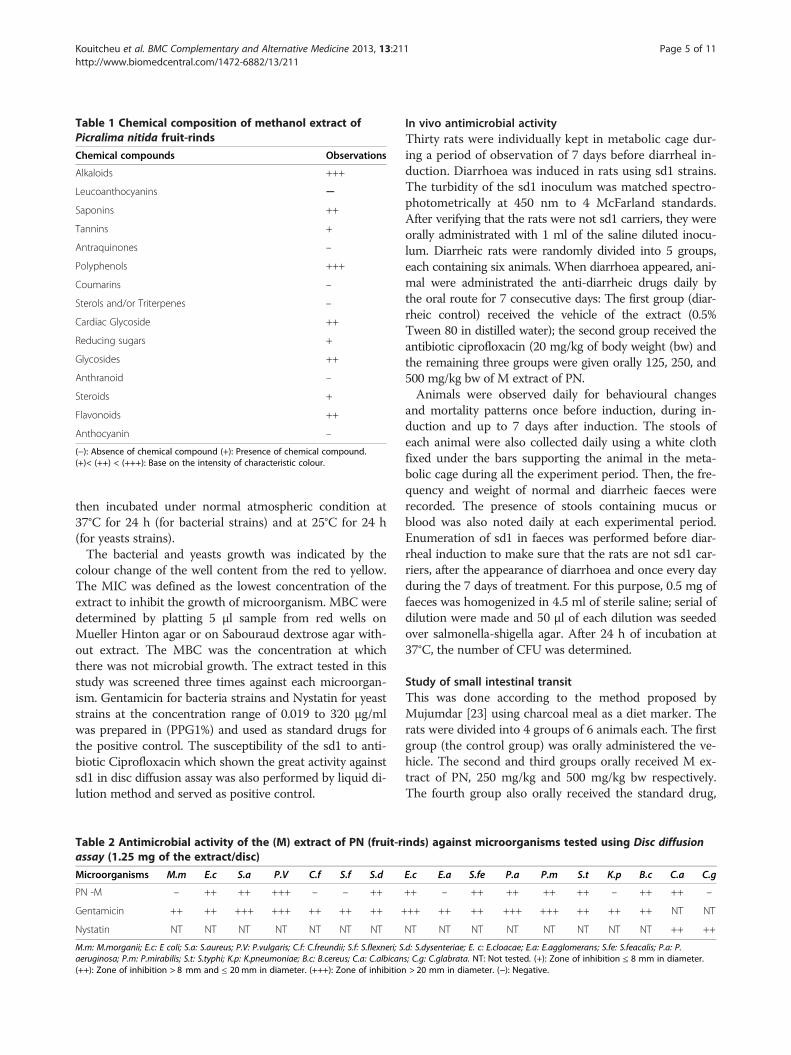
Table 1 Chemical composition of methanol extract ofPicralima nitida fruit-rinds
Chemical compounds Observations
Alkaloids +++
Leucoanthocyanins ─
Saponins ++
Tannins +
Antraquinones –
Polyphenols +++
Coumarins –
Sterols and/or Triterpenes –
Cardiac Glycoside ++
Reducing sugars +
Glycosides ++
Anthranoid –
Steroids +
Flavonoids ++
Anthocyanin –
(−): Absence of chemical compound (+): Presence of chemical compound.(+)< (++) < (+++): Base on the intensity of characteristic colour.
Kouitcheu et al. BMC Complementary and Alternative Medicine 2013, 13:211 Page 5 of 11http://www.biomedcentral.com/1472-6882/13/211
then incubated under normal atmospheric condition at37°C for 24 h (for bacterial strains) and at 25°C for 24 h(for yeasts strains).The bacterial and yeasts growth was indicated by the
colour change of the well content from the red to yellow.The MIC was defined as the lowest concentration of theextract to inhibit the growth of microorganism. MBC weredetermined by platting 5 μl sample from red wells onMueller Hinton agar or on Sabouraud dextrose agar with-out extract. The MBC was the concentration at whichthere was not microbial growth. The extract tested in thisstudy was screened three times against each microorgan-ism. Gentamicin for bacteria strains and Nystatin for yeaststrains at the concentration range of 0.019 to 320 μg/mlwas prepared in (PPG1%) and used as standard drugs forthe positive control. The susceptibility of the sd1 to anti-biotic Ciprofloxacin which shown the great activity againstsd1 in disc diffusion assay was also performed by liquid di-lution method and served as positive control.
Table 2 Antimicrobial activity of the (M) extract of PN (fruit-rassay (1.25 mg of the extract/disc)
Microorganisms M.m E.c S.a P.V C.f S.f S.d
PN -M – ++ ++ +++ – – ++
Gentamicin ++ ++ +++ +++ ++ ++ ++ +
Nystatin NT NT NT NT NT NT NT
M.m: M.morganii; E.c: E coli; S.a: S.aureus; P.V: P.vulgaris; C.f: C.freundii; S.f: S.flexneri; Saeruginosa; P.m: P.mirabilis; S.t: S.typhi; K.p: K.pneumoniae; B.c: B.cereus; C.a: C.albican(++): Zone of inhibition > 8 mm and ≤ 20 mm in diameter. (+++): Zone of inhibition
In vivo antimicrobial activityThirty rats were individually kept in metabolic cage dur-ing a period of observation of 7 days before diarrheal in-duction. Diarrhoea was induced in rats using sd1 strains.The turbidity of the sd1 inoculum was matched spectro-photometrically at 450 nm to 4 McFarland standards.After verifying that the rats were not sd1 carriers, they wereorally administrated with 1 ml of the saline diluted inocu-lum. Diarrheic rats were randomly divided into 5 groups,each containing six animals. When diarrhoea appeared, ani-mal were administrated the anti-diarrheic drugs daily bythe oral route for 7 consecutive days: The first group (diar-rheic control) received the vehicle of the extract (0.5%Tween 80 in distilled water); the second group received theantibiotic ciprofloxacin (20 mg/kg of body weight (bw) andthe remaining three groups were given orally 125, 250, and500 mg/kg bw of M extract of PN.Animals were observed daily for behavioural changes
and mortality patterns once before induction, during in-duction and up to 7 days after induction. The stools ofeach animal were also collected daily using a white clothfixed under the bars supporting the animal in the meta-bolic cage during all the experiment period. Then, the fre-quency and weight of normal and diarrheic faeces wererecorded. The presence of stools containing mucus orblood was also noted daily at each experimental period.Enumeration of sd1 in faeces was performed before diar-rheal induction to make sure that the rats are not sd1 car-riers, after the appearance of diarrhoea and once every dayduring the 7 days of treatment. For this purpose, 0.5 mg offaeces was homogenized in 4.5 ml of sterile saline; serial ofdilution were made and 50 μl of each dilution was seededover salmonella-shigella agar. After 24 h of incubation at37°C, the number of CFU was determined.
Study of small intestinal transitThis was done according to the method proposed byMujumdar [23] using charcoal meal as a diet marker. Therats were divided into 4 groups of 6 animals each. The firstgroup (the control group) was orally administered the ve-hicle. The second and third groups orally received M ex-tract of PN, 250 mg/kg and 500 mg/kg bw respectively.The fourth group also orally received the standard drug,
inds) against microorganisms tested using Disc diffusion
E.c E.a S.fe P.a P.m S.t K.p B.c C.a C.g
++ – ++ ++ ++ ++ – ++ ++ –
++ ++ ++ +++ +++ ++ ++ ++ NT NT
NT NT NT NT NT NT NT NT ++ ++
.d: S.dysenteriae; E. c: E.cloacae; E.a: E.agglomerans; S.fe: S.feacalis; P.a: P.s; C.g: C.glabrata. NT: Not tested. (+): Zone of inhibition ≤ 8 mm in diameter.> 20 mm in diameter. (−): Negative.
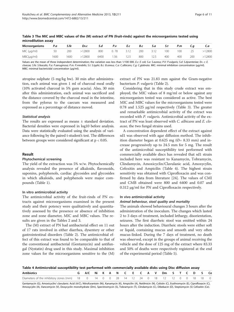
Table 3 The MIC and MBC values of the (M) extract of PN (fruit-rinds) against the microorganisms tested usingmicrodilution assay
Microorganisms P.a S.fe En.c S.d P.v E.c B.c S.a S.t P.m C.g C.a
MIC (μg/ml) 50 200 >12800 800 0. 78 3.12 200 3.12 100 100 25 >12800
MBC(μg/ml) 1600 1600 >12800 6400 1.56 12.5 800 12.5 400 400 200 >12800
Values are the mean of three independent determination; the variation was less than 1/100 000. E.c: E coli; S.a: S.aureus; P.V: P.vulgaris; S.d: S.dysenteriae; En. c: E.cloacae; S.fe: S.feacalis; P.a: P.aeruginosa; P.m: P.mirabilis; S.t: S.typhi; B.c: B.cereus; C.a: C.albicans; C.g: C.glabrata. MIC: minimal inhibition concentration (μg/ml).MBC: minimal bactericidal concentration (μg/ml).
Kouitcheu et al. BMC Complementary and Alternative Medicine 2013, 13:211 Page 6 of 11http://www.biomedcentral.com/1472-6882/13/211
atropine sulphate (5 mg/kg bw). 30 min after administra-tion, each animal was given 1 ml of charcoal meal orally(10% activated charcoal in 5% gum acacia). Also, 30 minafter this administration, each animal was sacrificed andthe distance covered by the charcoal meal in the intestine,from the pylorus to the caecum was measured andexpressed as a percentage of distance moved.
Statistical analysisThe results are expressed as means ± standard deviation.Bacterial densities were expressed in log10 before analysis.Data were statistically evaluated using the analysis of vari-ance following by the paired t-student’s test. The differencesbetween groups were considered significant at p < 0.05.
ResultPhytochemical screeningThe yield of the extraction was 5% w/w. Phytochemicallyanalysis revealed the presence of alkaloids, flavonoids,saponins, polyphenols, cardiac glycosides and glycosidesin which alkaloids, and polyphenols were major com-pounds (Table 1).
In vitro antimicrobial activityThe antimicrobial activity of the fruit-rinds of PN ex-tracts against microorganisms examined in the presentstudy and their potency were qualitatively and quantita-tively assessed by the presence or absence of inhibitionzone and zone diameter, MIC and MBC values. The re-sults are given in the Tables 2 and 3.The (M) extract of PN had antibacterial effect on 11 out
of 17 rats involved in either diarrhea, dysentery or othergastrointestinal disorders (Table 2). The antimicrobial ef-fect of this extract was found to be comparable to that ofthe conventional antibacterial (Gentamycin) and antifun-gal (Nystatin) drug used in this study. Maximal inhibitionzone values for the microorganisms sensitive to the (M)
Table 4 Antimicrobial susceptibility test performed with com
Antibiotics G A/C Ni K A N
Diameters of the inhibitory zones (mm) 18 0 14 0 0 28
Gentamycin (G), Amoxicyclin/ clavulanic Acid (A/C), Nitrofurantoin (Ni), Kanamycin (Amoxycylin (A), Vancomycin (V), Doxycyclin monohydrate (Dm), Spectinomycin (S),
extract of PN was 21.83 mm against the Gram-negativebacterium P. vulgaris (Table 2).Considering that in this study crude extract was em-
ployed, the MIC values of 8 mg/ml or below against anymicroorganism tested was considered as active. The bestMIC and MBC values for the microorganisms tested were0.78 and 1.525 μg/ml respectively (Table 3). The greaterand remarkable antimicrobial activity of the extract wasrecorded with P. vulgaris. Antimicrobial activity of the ex-tract of PN was least observed with C. albicans and E. clo-acae, the two fungal strains used.A concentration dependent effect of the extract against
sd1 was observed with agar diffusion method. The inhib-ition diameter began at 0.625 mg (Ø= 8.33 mm) and in-crease progressively up to 24.5 mm for 5 mg. The resultof the antimicrobial susceptibility test performed withcommercially available discs has revealed that sd1 strainincluded here was resistant to Kanamycin, Tobramycin,Clindamycin, Amoxicyclin/Clavulanic acid, Amoxycylin,Cefoxitin and Ampicilin (Table 4). The highest strainsensitivity was obtained with Ciprofloxacin and was con-firmed by data from literature [24]. The values of CMIand CMB obtained were 800 and 6400 and 0.07 and0.312 μg/ml for PN and Ciprofloxacin respectively.
In vivo antimicrobial activityAnimal behaviour, stool quality and mortalityThe animals showed behavioural changes 3 hours after theadministration of the inoculum. The changes which lasted2 to 3 days of treatment, included lethargy, disorientation,seizures. The first diarrheic stool was emitted within 24hours after the induction. Diarrheic stools were either softor liquid, containing mucus and smooth and very oftenmucus-linked. During the 7 days of treatment, no deathwas observed, except in the groups of animal receiving thevehicle and the dose of 125 mg of the extract where 83.33and 50% of deaths were respectively registered at the endof the experimental period (Table 5).
mercially available disks using Disc diffusion assay
C E C A V Dm S T C D S Ce
14 12 24 0 18 12 12 0 0 18 18 0
K), Ampicilin (A), Netilmicin (N), Colistin (C), Erythromycin (E), Ciprofloxacin (C),Tobramycin (T), Clindamycin (C), Dibekacin (D), Steptomycin (S) Cefoxitin (Ce).
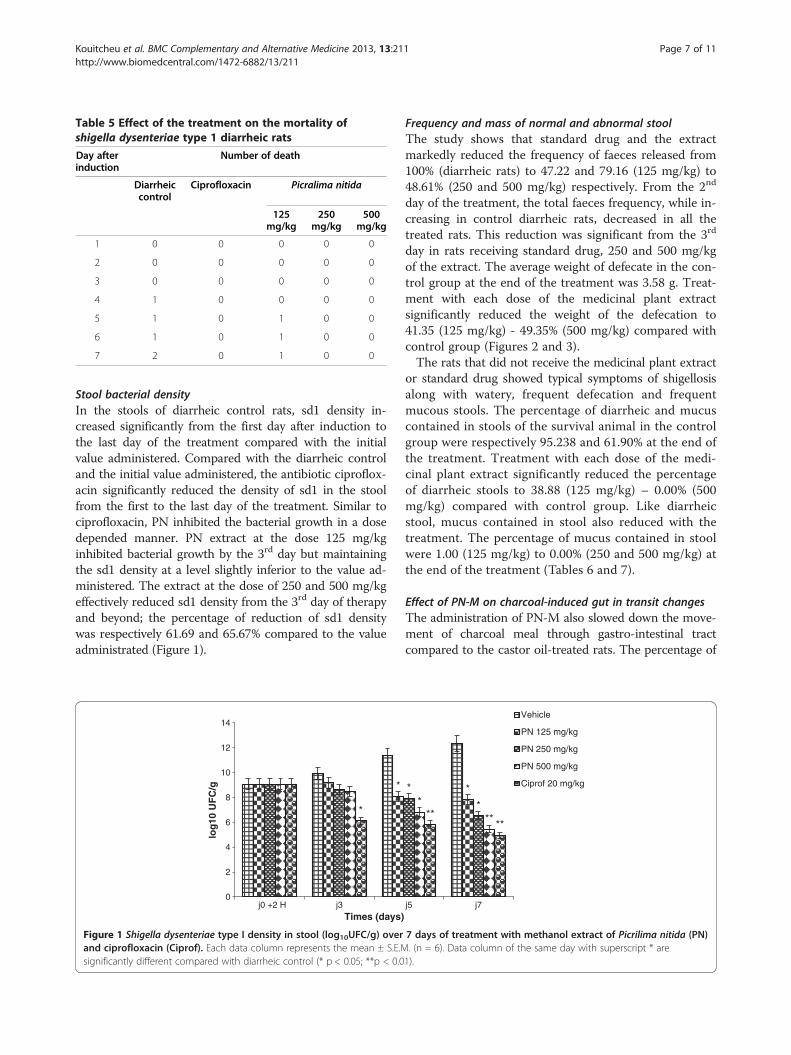
Table 5 Effect of the treatment on the mortality ofshigella dysenteriae type 1 diarrheic rats
Day afterinduction
Number of death
Diarrheiccontrol
Ciprofloxacin Picralima nitida
125mg/kg
250mg/kg
500mg/kg
1 0 0 0 0 0
2 0 0 0 0 0
3 0 0 0 0 0
4 1 0 0 0 0
5 1 0 1 0 0
6 1 0 1 0 0
7 2 0 1 0 0
Kouitcheu et al. BMC Complementary and Alternative Medicine 2013, 13:211 Page 7 of 11http://www.biomedcentral.com/1472-6882/13/211
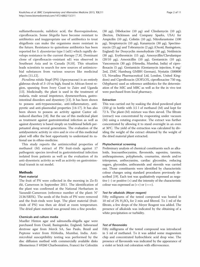
Stool bacterial densityIn the stools of diarrheic control rats, sd1 density in-creased significantly from the first day after induction tothe last day of the treatment compared with the initialvalue administered. Compared with the diarrheic controland the initial value administered, the antibiotic ciproflox-acin significantly reduced the density of sd1 in the stoolfrom the first to the last day of the treatment. Similar tociprofloxacin, PN inhibited the bacterial growth in a dosedepended manner. PN extract at the dose 125 mg/kginhibited bacterial growth by the 3rd day but maintainingthe sd1 density at a level slightly inferior to the value ad-ministered. The extract at the dose of 250 and 500 mg/kgeffectively reduced sd1 density from the 3rd day of therapyand beyond; the percentage of reduction of sd1 densitywas respectively 61.69 and 65.67% compared to the valueadministrated (Figure 1).
*
*
0
2
4
6
8
10
12
14
j0 +2 H j3
log
10 U
FC
/g
Times (days)
Figure 1 Shigella dysenteriae type I density in stool (log10UFC/g) overand ciprofloxacin (Ciprof). Each data column represents the mean ± S.E.Msignificantly different compared with diarrheic control (* p < 0.05; **p < 0.0
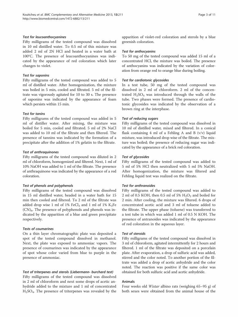
Frequency and mass of normal and abnormal stoolThe study shows that standard drug and the extractmarkedly reduced the frequency of faeces released from100% (diarrheic rats) to 47.22 and 79.16 (125 mg/kg) to48.61% (250 and 500 mg/kg) respectively. From the 2nd
day of the treatment, the total faeces frequency, while in-creasing in control diarrheic rats, decreased in all thetreated rats. This reduction was significant from the 3rd
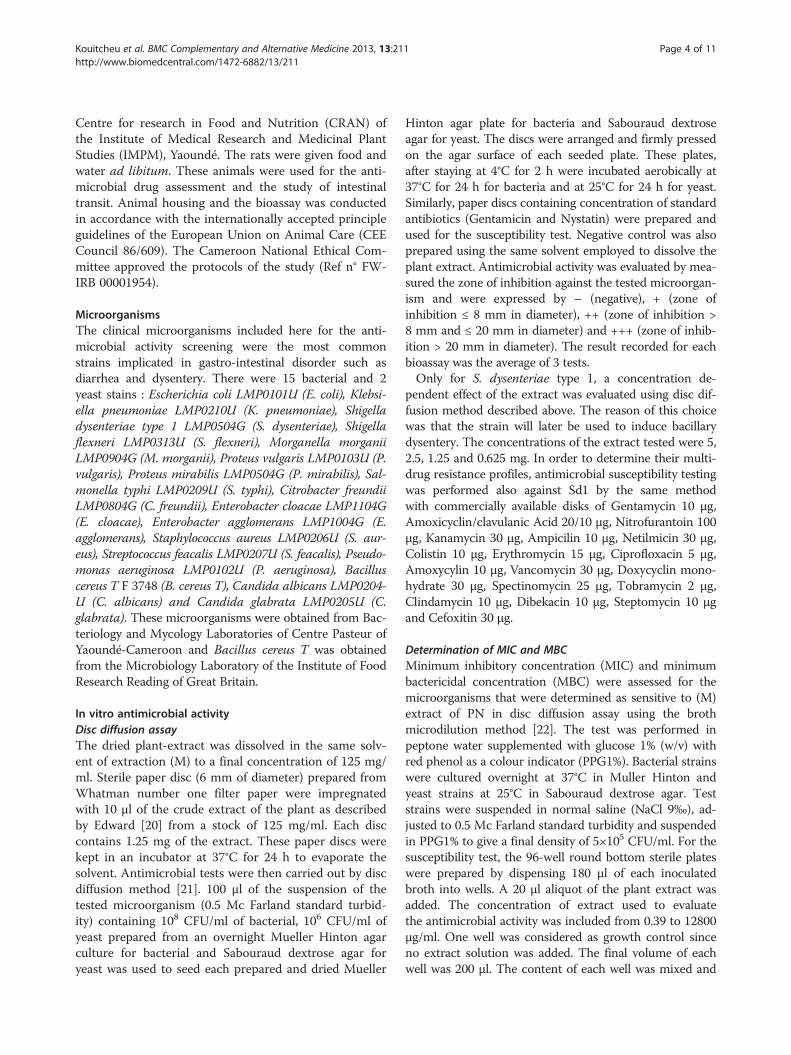
day in rats receiving standard drug, 250 and 500 mg/kgof the extract. The average weight of defecate in the con-trol group at the end of the treatment was 3.58 g. Treat-ment with each dose of the medicinal plant extractsignificantly reduced the weight of the defecation to41.35 (125 mg/kg) - 49.35% (500 mg/kg) compared withcontrol group (Figures 2 and 3).The rats that did not receive the medicinal plant extract
or standard drug showed typical symptoms of shigellosisalong with watery, frequent defecation and frequentmucous stools. The percentage of diarrheic and mucuscontained in stools of the survival animal in the controlgroup were respectively 95.238 and 61.90% at the end ofthe treatment. Treatment with each dose of the medi-cinal plant extract significantly reduced the percentageof diarrheic stools to 38.88 (125 mg/kg) – 0.00% (500mg/kg) compared with control group. Like diarrheicstool, mucus contained in stool also reduced with thetreatment. The percentage of mucus contained in stoolwere 1.00 (125 mg/kg) to 0.00% (250 and 500 mg/kg) atthe end of the treatment (Tables 6 and 7).
Effect of PN-M on charcoal-induced gut in transit changesThe administration of PN-M also slowed down the move-ment of charcoal meal through gastro-intestinal tractcompared to the castor oil-treated rats. The percentage of
**
**
******
j5 j7
Vehicle
PN 125 mg/kg
PN 250 mg/kg
PN 500 mg/kg
Ciprof 20 mg/kg
7 days of treatment with methanol extract of Picrilima nitida (PN). (n = 6). Data column of the same day with superscript * are1).
**
* *21 *
* * **** **
**
0
5
10
15
20
25
30
35
40
j-1 j0 j1 j2 j3 j4 j5 j6 j7
To
tal s
too
l fre
qu
ency
(n
br/
day
)
Times (days)
vehicle
PN 125 mg/kg
PN 250 mg/kg
PN 500 mg/kg
Ciprof 20mg/kg
Figure 2 Total stool frequency before and during treatment of Shigella dysenteriae type I diarrheic rats with methanol extract ofPicrilima nitida (PN) and ciprofloxacin (Ciprof). Each data column represents the mean ± S.E.M. (n = 6). Data column of the same day withsuperscript * are significantly different compared with diarrheic control (* p < 0.05; **p < 0.01).
Kouitcheu et al. BMC Complementary and Alternative Medicine 2013, 13:211 Page 8 of 11http://www.biomedcentral.com/1472-6882/13/211
intestinal length covered by charcoal meal in PN-M pre-treated (250 and 500 mg/kg) and castor oil-treated ratswas 58.27, 60.54 and 74.57 respectively. Atropine on itspart, produced a marked decrease in the propulsive move-ment and the intestinal length covered by charcoal mealwas 40.33 (Table 8).
0
0.5
1
1.5
2
2.5
3
3.5
4
j-1 j0 j1 j2
To
tal s
too
l wei
gh
t (g
/day
)
Times
Figure 3 Total stool weight before and during treatment of Shigella dnitida (PN) and ciprofloxacin (Ciprof). Each data column represents the mare significantly different compared with diarrheic control (* p < 0.05; **p <
DiscussionThe (M) extract of PN fruit-rinds showed a promisingbroad spectrum antibacterial property, inhibiting 80%of bacterial strains tested, especially P. vulgaris, E. coli andS. aureus with MBC ranging between 0.78 and 1.56 μg/ml.The presence of flavonoids, saponins, tannins and alkaloids
** *
** * *
*
**
****
** *
j3 j4 j5 j6 j7
(days)
vehicle
PN 125 mg/kg
PN 250 mg/kg
PN 500 mg/kg
Ciprof 20 mg/kg
ysenteriae type I diarrheic rats with methanol extract of Picrilimaean ± S.E.M. (n = 6). Data column of the same day with superscript *0.01).
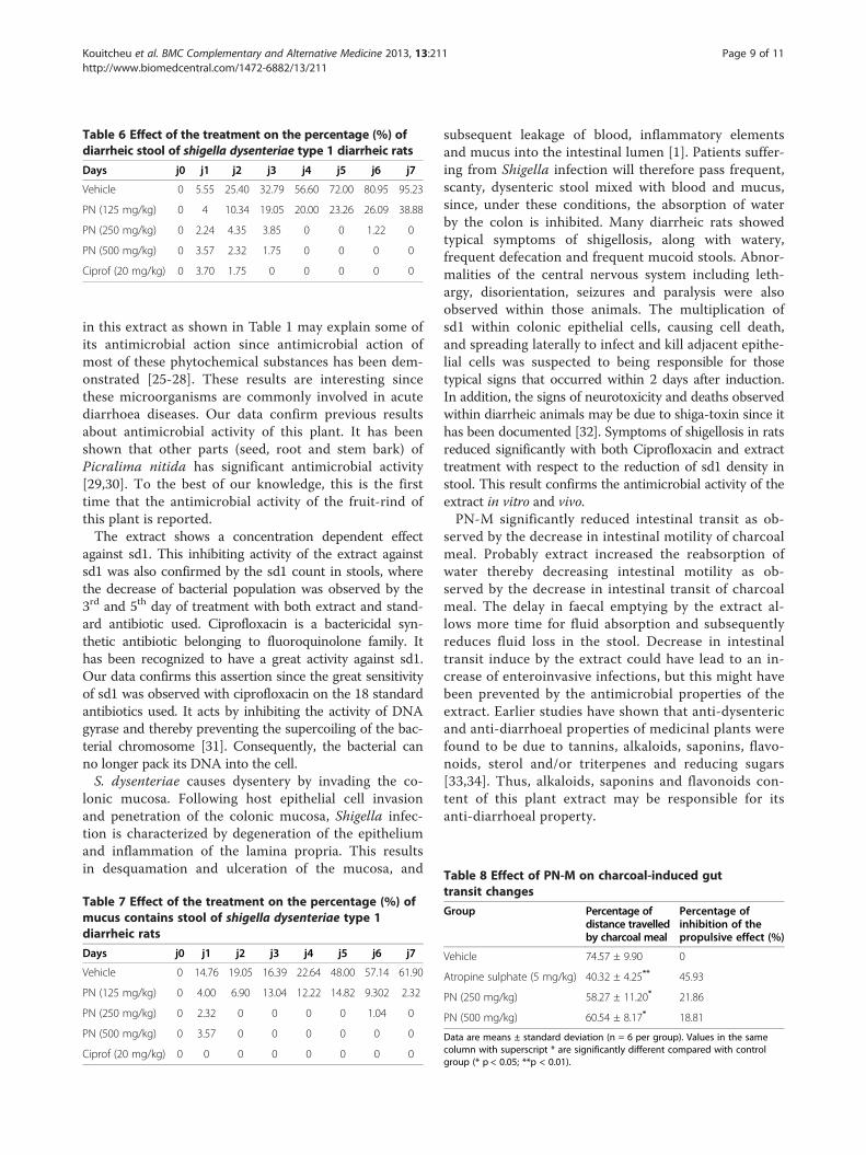
Table 6 Effect of the treatment on the percentage (%) ofdiarrheic stool of shigella dysenteriae type 1 diarrheic rats
Days j0 j1 j2 j3 j4 j5 j6 j7
Vehicle 0 5.55 25.40 32.79 56.60 72.00 80.95 95.23
PN (125 mg/kg) 0 4 10.34 19.05 20.00 23.26 26.09 38.88
PN (250 mg/kg) 0 2.24 4.35 3.85 0 0 1.22 0
PN (500 mg/kg) 0 3.57 2.32 1.75 0 0 0 0
Ciprof (20 mg/kg) 0 3.70 1.75 0 0 0 0 0
Table 8 Effect of PN-M on charcoal-induced gut
Kouitcheu et al. BMC Complementary and Alternative Medicine 2013, 13:211 Page 9 of 11http://www.biomedcentral.com/1472-6882/13/211
in this extract as shown in Table 1 may explain some ofits antimicrobial action since antimicrobial action ofmost of these phytochemical substances has been dem-onstrated [25-28]. These results are interesting sincethese microorganisms are commonly involved in acutediarrhoea diseases. Our data confirm previous resultsabout antimicrobial activity of this plant. It has beenshown that other parts (seed, root and stem bark) ofPicralima nitida has significant antimicrobial activity[29,30]. To the best of our knowledge, this is the firsttime that the antimicrobial activity of the fruit-rind ofthis plant is reported.The extract shows a concentration dependent effect
against sd1. This inhibiting activity of the extract againstsd1 was also confirmed by the sd1 count in stools, wherethe decrease of bacterial population was observed by the3rd and 5th day of treatment with both extract and stand-ard antibiotic used. Ciprofloxacin is a bactericidal syn-thetic antibiotic belonging to fluoroquinolone family. Ithas been recognized to have a great activity against sd1.Our data confirms this assertion since the great sensitivityof sd1 was observed with ciprofloxacin on the 18 standardantibiotics used. It acts by inhibiting the activity of DNAgyrase and thereby preventing the supercoiling of the bac-terial chromosome [31]. Consequently, the bacterial canno longer pack its DNA into the cell.S. dysenteriae causes dysentery by invading the co-
lonic mucosa. Following host epithelial cell invasionand penetration of the colonic mucosa, Shigella infec-tion is characterized by degeneration of the epitheliumand inflammation of the lamina propria. This resultsin desquamation and ulceration of the mucosa, and
Table 7 Effect of the treatment on the percentage (%) ofmucus contains stool of shigella dysenteriae type 1diarrheic rats
Days j0 j1 j2 j3 j4 j5 j6 j7
Vehicle 0 14.76 19.05 16.39 22.64 48.00 57.14 61.90
PN (125 mg/kg) 0 4.00 6.90 13.04 12.22 14.82 9.302 2.32
PN (250 mg/kg) 0 2.32 0 0 0 0 1.04 0
PN (500 mg/kg) 0 3.57 0 0 0 0 0 0
Ciprof (20 mg/kg) 0 0 0 0 0 0 0 0
subsequent leakage of blood, inflammatory elementsand mucus into the intestinal lumen [1]. Patients suffer-ing from Shigella infection will therefore pass frequent,scanty, dysenteric stool mixed with blood and mucus,since, under these conditions, the absorption of waterby the colon is inhibited. Many diarrheic rats showedtypical symptoms of shigellosis, along with watery,frequent defecation and frequent mucoid stools. Abnor-malities of the central nervous system including leth-argy, disorientation, seizures and paralysis were alsoobserved within those animals. The multiplication ofsd1 within colonic epithelial cells, causing cell death,and spreading laterally to infect and kill adjacent epithe-lial cells was suspected to being responsible for thosetypical signs that occurred within 2 days after induction.In addition, the signs of neurotoxicity and deaths observedwithin diarrheic animals may be due to shiga-toxin since ithas been documented [32]. Symptoms of shigellosis in ratsreduced significantly with both Ciprofloxacin and extracttreatment with respect to the reduction of sd1 density instool. This result confirms the antimicrobial activity of theextract in vitro and vivo.PN-M significantly reduced intestinal transit as ob-
served by the decrease in intestinal motility of charcoalmeal. Probably extract increased the reabsorption ofwater thereby decreasing intestinal motility as ob-served by the decrease in intestinal transit of charcoalmeal. The delay in faecal emptying by the extract al-lows more time for fluid absorption and subsequentlyreduces fluid loss in the stool. Decrease in intestinaltransit induce by the extract could have lead to an in-crease of enteroinvasive infections, but this might havebeen prevented by the antimicrobial properties of theextract. Earlier studies have shown that anti-dysentericand anti-diarrhoeal properties of medicinal plants werefound to be due to tannins, alkaloids, saponins, flavo-noids, sterol and/or triterpenes and reducing sugars[33,34]. Thus, alkaloids, saponins and flavonoids con-tent of this plant extract may be responsible for itsanti-diarrhoeal property.
transit changes
Group Percentage ofdistance travelledby charcoal meal
Percentage ofinhibition of thepropulsive effect (%)
Vehicle 74.57 ± 9.90 0
Atropine sulphate (5 mg/kg) 40.32 ± 4.25** 45.93
PN (250 mg/kg) 58.27 ± 11.20* 21.86
PN (500 mg/kg) 60.54 ± 8.17* 18.81
Data are means ± standard deviation (n = 6 per group). Values in the samecolumn with superscript * are significantly different compared with controlgroup (* p < 0.05; **p < 0.01).
Kouitcheu et al. BMC Complementary and Alternative Medicine 2013, 13:211 Page 10 of 11http://www.biomedcentral.com/1472-6882/13/211
ConclusionS. dysenteria type 1 strain was resistant to all antibi-otics from the beta-lactam family used in this study.The great sensibility of the strain was obtained withciprofloxacin, a synthetic antibiotic belonging to fluo-roquinolone family. The (M) extract of PN had anti-microbial activity on 11 out of 17 microorganismstested amongst witch S. dysenteriae type 1. The valuesof CMI and CMB obtain against this strain were 800and 6400 μg/ml respectively. This anti-Shigella activityof PN was also shown in vivo. The administration ofPN extract to diarrheic rats significantly reduced thebacterial population in stools; thereby reduce thesymptoms of shigellosis on rats.At present, our group is concerned with the fraction-
ation and the isolation of pure compounds of the crudeextracts and the elucidation of their structures in orderto better evaluate their pharmacological activity in vitroand in vivo.
Competing interestsThe authors declare that they have no competing interests.
Authors’ contributionsJK conducted experiments on phytochemical screening and LBKM onantimicrobial and anti-shigellosis activity of tested plant extract. JLTparticipated in design of the study and preparation of the manuscript. Allthe authors read and approved the final manuscript.
AcknowledgementWe are grateful to the Bacteriology and Mycology Laboratories of CentrePasteur Yaoundé and the Microbiology Laboratory of the Institute of FoodResearch Reading of Great Britain for giving us microorganisms. We are alsograteful to Mr. Nelson SONELA for reading through the manuscripts andmaking some language corrections.
Author details1Microbiology and Pharmacology Laboratory, Department of Biochemistry,Faculty of Science, University of Dschang-Cameroon, P. O. Box 67, Dschang,Cameroon. 2Department of Biological Sciences Higher Teacher’s TrainingCollege, University of Yaoundé I, P.O. Box: 47, Yaoundé, Cameroon.3Department of Organic Chemistry, Faculty of Sciences, University ofYaoundé I, P.O. Box 812, Yaoundé, Cameroon.
Received: 31 January 2013 Accepted: 2 August 2013Published: 17 August 2013
References1. Niyogi S: Shigellosis. J Microbiol 2005, 43:133–143.2. Meicler C, Cerf M: Diarrhées a shigelles, a colibacilles entero-invasifs,
entero-hémorragiques et a colibacilles. In diarrhées aigues infectieuses.Dion Paris; 1993:9–98.
3. Keusch GT: Shigella infections. Clin Gastroenterol 1979, 8:645–662.4. Griffin PM, Tauxe RV: The epidemiology of infections caused by Echerichia
coli 0157: H7 and the associated hemolytic uremic syndrome.Epidemiol Rev 1991, 13:60–98.
5. Struelens MJ, Patte D, Kabir I, Salam A, Nath SK, Butler T: Shigellasepticemia: prevalence, presentation, risk factors and outcome. J InfectDis 1985, 152:784–790.
6. Lipsky PE, Fosmire S, Stieglitz H: Bacterial epitopes involved in inductionof reactive arthritis. Am J Med 1988, 85:56–58.
7. Dutta S, Ghosh A, Ghosh K, Dutta D, Bhattacharya SK, Nair GB: Newly emergedmultiple antibiotic-resistant Shigella dysenteriae type 1 strains in andaround Kolkatta, India, are clonal. J Clin Microbiol 2003, 41:5833–5834.
8. Sivapalasingam S, Nelson JM, Joyce K, Hoekstra M, Angula FJ, Mintz ED:High Prevalence of Antimicrobial Resistance among Shigella Isolates inthe Unated States Tested by the National Antimicrobial ResistanceMonitoring System from 1999 to 2002. Antimicrob Agents Chemother2006, 50:49–54.
9. Dutta S, Dutta D, Dutta P, Matsushita S, Bhattacharya SK, Yoshida SI: Shigelladysenteriae type1, Kolkatta, India. Emerg Infect Dis 2003, 9:1471–1474.
10. Pazhani GP, Sarkar B, Ramamurthy T, Bhattacharya SK, Takeda Y, Niyogi SK:Clonal multidrug-resistant Shigella dysenteriae type 1 strains associatedwith epidemic and sporadic dysenteries in eastern Indian.Antimicrob Agents Chemother 2004, 48:681–684.
11. Clark AM: Natural products as resource for new drugs.Pharmaceutical research 1996, 13:1133–1141.
12. Cordell GA: Biodiversity and drug discovery a symbiotic relationship.Phytochemistry 2000, 55:463–480.
13. Adjanohoun JE, Aboubakar N, Dramane K, Ebot ME, Ekpere JA, Enow-OrockEG, Focho D, Gbile ZO, Kamanyi A, Kamsu Kom J, Kieta A, Mbenkum T, MbiCN, Mbiele L, Mbome IL, Mubiru NK, Nancy WL, Nkongmeneck B, Satabie B,Sofowora A, Tamze V, Wirmum CK: Contribution to Ethnobotanical andFloristic Studies in Cameroon: Traditional Medicine and Pharmacopoeia. Lagos:Technical and Research Commission of Organisaion of African Unity (OAU/STRC); 1996:60–61.
14. Wosu LO, Ibe CC: Use of extract of Picralima nitida bark in the treatmentof experimental trypanosomiasis: a preliminary study. J Ethnopharmacol1989, 25:263–268.
15. Ezeamuzie K, Ojinnaka MC, Uzogara EO, Oji SE: Anti-inflammatory,antipyretic and antimalarial activities of a West African medicinal plant:Picralima nitida. Afr J Med Med Sci 1994, 23:85–90.
16. Iwu MM, Klayman Daniel L, Jackson Joan E, Tally John D, Andersen StevenL: Alkaloids of Picralima nitida used for treatment of protozoal diseases.United States Patent 1994, 5:553.
17. François G, Aké Assi L, Holenz J, Bringmann G: Constituents of Picralimanitida display pronounced inhibitory activities against asexualerythrocytic forms of plasmodium. J. ethnopharmacol 1996,54:165–168.
18. Kouitcheu Mabeku LB, Penlap Beng V, Kouam J, Ngadjui BT, Fomum ZT,Etoa FX: Evaluation of Antidiarrhoeal Activity of the fruit-rind of Picralimanitida (Apocynaceae). Afr J Trad CAM 2006, 3:66–73.
19. Harbone JB: Phytochimical methods: a guide to modern techniques of plantsanalysis. London: Chapman and Hall; 1988:1–867.
20. Edward DI: Antimicrobial Action. 1st edition. London and Basingstoke: TheMacmillan Press; 1980:8–30.
21. Murray PR, Baron EJ, Pfaller MA, Tenover FC, Yolken RH: Manual of ClinicalMicrobiology. 6th edition. Washington, DC: ASM Press; 1995:1327–1341.
22. NCCLS: Methods for determining bactericidal activity of antimicrobial agents.Wayne Pa; 1999. Approved guideline, M26-A, National Committee forClinical Laboratory Standards.
23. Mujumdar AM: Antidiarrhoeal activity of Azadiachta indica leaf extract.Indian Drugs 1998, 35:417–420.
24. OMS: Directives pour la lutte contre les shigelloses y compris lors des épidémiesdues a shigella dysenteriae type I. Genève Switzerland: bull de l’OMS; 2005:69.
25. Lewis WH, Elvin-Lewis MPF: Plants Affecting Man’s Health. In MedicinalBotany. London: Wiley; 1977:515.
26. Plasuntherum C, Iyer K: Antimicrobial activity of Coscinium fenetratumColebr. against clostridium tetani. Indian J Med Res 1982, 76:71–76.
27. Palacious P, Gutkind G, Randina RVD, De Torres R, Coussio JD: Antimicrobialactivity of B. crispa and B. notosergila. Genus Baccharis II. Planta Med 1983,49:128.
28. Ahmad A, Khan HA, Ahmad YU, Qazi S: Antibacterial activity of juliflorineisolated from Prosopis juliflora. Planta Med 1986, 4:285–288.
29. Fakeye TO, Itiola OA, Odelola HA: Evaluation of the antimicrobialproperty of the stem barks of Picralima nitida (Apocynaceae).Phytother Res 2000, 14:368–370.
30. Nkere CK, Iroegbu CU: Antibacterial screening of the root, seed andstem bark extracts of Picralima nitida. Afr J Biotechnol 2005,4:522–526.
31. Alcamo Edward I: Fundamentals of Microbiology: Chemotherapeutic Agentsand Antibiotics. Addison-Wesley Publishing Company, Inc; 1983:669–691.
32. Cherla R, Lee S, Vernon T: Shiga toxins and apoptosis. FEMS Microbiol Lett2003, 228:159–166.
Kouitcheu et al. BMC Complementary and Alternative Medicine 2013, 13:211 Page 11 of 11http://www.biomedcentral.com/1472-6882/13/211
33. Galvez J, Zarzuelo A, Crespo ME: Antidiarrhoeic activity of Scleroaryabirrea bark extract and its active tannin constituent in rats. Phytother Res1991, 5:276–278.
34. Vercruysse A, Foriers A, Longanga Otshudi A: Contribution to theethnobotanical, phytochemical and pharmacological studies oftraditionally used medicinal plants in the treatment of dysentery anddiarrhoea in Lomela area, Democratic Republic of Congo (DRC).J Ethnopharmacol 2000, 71:411–423.
doi:10.1186/1472-6882-13-211Cite this article as: Kouitcheu et al.: The anti-shigellosis activity of themethanol extract of Picralima nitida on Shigella dysenteriae type Iinduced diarrhoea in rats. BMC Complementary and Alternative Medicine2013 13:211.
Submit your next manuscript to BioMed Centraland take full advantage of:
• Convenient online submission
• Thorough peer review
• No space constraints or color figure charges
• Immediate publication on acceptance
• Inclusion in PubMed, CAS, Scopus and Google Scholar
• Research which is freely available for redistribution
Submit your manuscript at www.biomedcentral.com/submit

![PHYTOCHEMISTRY AND ANTIBACTERIAL ACTIVITY OF ......antibacterial compounds in the leaf extract [28]. Methanol extract showed a higher activity because it was able to extract much constituents](https://img.pdfslide.us/doc/110x75/60b4d809c28542624d73722b/phytochemistry-and-antibacterial-activity-of-antibacterial-compounds-in.jpg)