Embed Size (px)
Citation preview
IntroductionCardiac growth during development occurs in twophases, initially by myocyte hyperplasia, but primarilyby myocyte hypertrophy after the early postnatal peri-od. Postnatal cardiac hypertrophy is a normal physio-logical process that increases heart size to maintain cardiac output to the growing organism (1). Catecho-lamines such as norepinephrine are one signal for car-diac hypertrophy, and their role in pathological hyper-trophy in disease is well characterized (2, 3), yet theirrole in physiological hypertrophy during developmentis less well studied. Knockout of the enzymes that syn-
thesize catecholamines leads to death in utero fromcardiac defects, indicating that catecholamines arerequired for prenatal cardiac development (4). Cardiacadrenergic innervation increases from birth to wean-ing (1), and thus, increased norepinephrine releasefrom adrenergic nerves might be involved in postnatalheart growth as well.
Norepinephrine stimulates all adrenergic receptors(ARs), α1, α2, and β, and alters hemodynamic loading.However, experiments in cultured neonatal rat cardiacmyocytes show clearly that catecholamines inducehypertrophy via α1-ARs, independent of loading (2, 5,6). Of the three α1-AR subtypes, α1A/C, α1B, and α1D,culture studies suggest that the α1A/C-AR mediatesmyocyte hypertrophy (7).
Experiments in transgenic mice partly support the cul-ture results and the idea that α1-ARs can be sufficient toinduce hypertrophy during development. An activatedmutant of the α1B-AR subtype causes hypertrophy whenoverexpressed in cardiac myocytes with the α-myosinheavy chain (α-MyHC) promoter (8). The WT α1B over-expressed by an α1B promoter also increases heart size (9).
On the other hand, heart size is not changed by α-MyHC–directed overexpression of either the WT α1B
or the WT α1A/C (10, 11). Furthermore, growth of the
The Journal of Clinical Investigation | June 2003 | Volume 111 | Number 11 1783
The α1A/C- and α1B-adrenergic receptors are required for physiological cardiac hypertrophy in the double-knockout mouse
Timothy D. O’Connell,1 Shinji Ishizaka,2 Akihiro Nakamura,3 Philip M. Swigart,1
M.C. Rodrigo,1 Gregory L. Simpson,1 Susanna Cotecchia,4 D. Gregg Rokosh,5
William Grossman,2 Elyse Foster,2 and Paul C. Simpson1
1Cardiology Division, San Francisco Veterans Affairs Medical Center, and the Cardiovascular Research Institute andDepartment of Medicine, University of California San Francisco (UCSF), San Francisco, California, USA
2Cardiology Division, Department of Medicine, UCSF, San Francisco, California, USA3Iwate Prefectural Miyako Hospital, Iwate, Japan4Institut de Pharmacologie et Toxocologie, Universite de Lausanne, Switzerland5Cardiology Division, Department of Medicine, University of Louisville, Louisville, Kentucky, USA
Catecholamines and α1-adrenergic receptors (α1-ARs) cause cardiac hypertrophy in cultured myocytesand transgenic mice, but heart size is normal in single KOs of the main α1-AR subtypes, α1A/C and α1B.Here we tested whether α1-ARs are required for developmental cardiac hypertrophy by generatingα1A/C and α1B double KO (ABKO) mice, which had no cardiac α1-AR binding. In male ABKO mice,heart growth after weaning was 40% less than in WT, and the smaller heart was due to smallermyocytes. Body and other organ weights were unchanged, indicating a specific effect on the heart.Blood pressure in ABKO mice was the same as in WT, showing that the smaller heart was not due todecreased load. Contractile function was normal by echocardiography in awake mice, but the small-er heart and a slower heart rate reduced cardiac output. α1-AR stimulation did not activate extracel-lular signal–regulated kinase (Erk) and downstream kinases in ABKO myocytes, and basal Erk activ-ity was lower in the intact ABKO heart. In female ABKO mice, heart size was normal, even afterovariectomy. Male ABKO mice had reduced exercise capacity and increased mortality with pressureoverload. Thus, α1-ARs in male mice are required for the physiological hypertrophy of normal post-natal cardiac development and for an adaptive response to cardiac stress.
J. Clin. Invest. 111:1783–1791 (2003). doi: 10.1172/JCI200316100.
Received for publication June 6, 2002, and accepted in revised form March 25, 2003.
Address correspondence to: Paul C. Simpson, Veterans AffairsMedical Center 111C8, 4150 Clement Street, San Francisco,California 94121, USA. Phone: (415) 221-4810 ext. 3200; Fax: (415) 750-6950; E-mail: [email protected] of interest: The authors have declared that no conflict ofinterest exists.Nonstandard abbreviations used: adrenergic receptor (AR);myosin heavy chain (MyHC); α1A/C- and α1B-AR double KO(ABKO); extracellular signal–regulated kinase (Erk); ribonucleaseprotection assay (RPA); heart rate (HR); transverse aorticconstriction (TAC); heart weight (HW); body weight (BW); leftventricular (LV); phenylephrine (PE).

heart is normal in the individual α1B- and α1A/C-subtypeKO mice (12, 13). Similarly, heart size is not reduced ina double β-AR KO (14) and is increased in a double α2-AR KO due to enhanced norepinephrine release(15). Therefore, although catecholamines and α1-ARsare implicated in cardiac hypertrophy, none of the indi-vidual ARs, particularly α1-ARs, appears to be essentialfor postnatal cardiac growth in vivo.
One explanation for normal cardiac development inthe single α1-AR KO mice is functional redundancy ofthe different α1-AR subtypes. Therefore, to test furtherthe hypothesis that α1-ARs are required for hypertrophy,we generated a double KO — ABKO — of the two mainα1-AR subtypes in the heart, the α1A/C and α1B. We stud-ied cardiac structure and function during the hypertro-phy of normal postnatal development. Hypertrophyduring development is the most common type of cardiachypertrophy and is an example of physiological hyper-trophy, in which cardiac function remains normal orimproves, in contrast with pathological hypertrophy, inwhich function deteriorates eventually. Our results showa load-independent and sex-specific requirement for α1-ARs in developmental hypertrophy and in the cardiacresponse to stress, and they implicate extracellular sig-nal–regulated kinase (Erk) signaling in this effect.
MethodsGeneration of α1A/C and α1B double KO mice. α1B KO mice(C57BL/6, 129Sv) (12) were mated with α1A/C KO mice(FVB, 129Sv) (13) to produce F1 mice heterozygous forboth KOs. F1 heterozygous mice were mated to produceF2 WT and ABKO mice, and breeding pairs from thesetwo lines produced offspring used in most experi-ments. Concurrently, mice heterozygous for both KOswere backcrossed with C57BL/6 mice to produce fifth-generation congenic mice.
Ribonuclease protection assay. Ribonuclease protectionassay (RPA) used 25 µg total ventricular RNA (Trizol,GIBCO BRL; Life Technologies Inc., Gaithersburg,Maryland, USA); bands were quantified using Image-Quant (Molecular Dynamics, Sunnyvale, California,USA) and normalized to β-actin (Ambion Inc., Austin,Texas, USA) (7, 16).
Radioligand binding assay. Saturation binding in heart100,000 g membranes used 0.04–1.2 nM [3H]-prazosin
and 10 µM phentolamine (RBI, Natick,Massachusetts, USA) to define nonspe-cific binding (17). Total receptor number(Bmax) and binding affinity (KD) were cal-
culated by nonlinear regression using GraphPad Prism(GraphPad Software Inc., San Diego, California, USA).
Echocardiography. Echocardiography was done with anAcuson Sequoia C256 (Acuson, Mountain View, Cali-fornia, USA) with a 15-MHz linear array transducer.Mice were under anesthesia with isoflurane or wereawake and gently restrained (18).
Blood pressure and heart rate. Systolic BP and heart rate(HR) were measured using a noninvasive computerizedtail cuff system (BP-2000; Visitech Systems, Apex,North Carolina, USA) (13). Mice were trained for 3days, and recordings were made on the next 5 days,with at least 15 of 20 successful readings each day.
Heart histology. Excised hearts were rinsed in PBS, fixedwith 4% paraformaldehyde, embedded in paraffin, andsectioned at 10 µm. Alternately, hearts were fixed in situafter arrest in diastole with 40 mM KCl, or embedded inTissue-Tek O.C.T. compound (Miles Laboratories Inc.,Elkhart, Indiana, USA) and frozen. Paraffin-embeddedcoronal sections were stained with fluorescein-conju-gated wheat germ agglutinin and Hoechst 33342, andmyocyte cross-sectional area was measured by fluores-cence microscopy and NIH Image. Heart cross-sectionalarea was measured from 10-µm frozen sagittal sections.
Myocyte isolation and volume. Ventricular myocytes wereisolated as described (19), except that collagenase perfu-sion was at 4 ml/min. Isolated myocytes were fixed with5% formaldehyde in PBS, and myocyte volume was meas-ured using a Coulter Multisizer with Accucomp software(Coulter Instruments Inc., Hialeah, Florida, USA) (20).
Myocyte surface area and nucleation. Isolated myocyteswere plated in two-well slides, fixed with 30% acetone/70%ethanol, and viewed under phase contrast (Eclipse E600;Nikon Inc., Melville, New York, USA). Surface area wasmeasured with NIH Image software from digital images(SPOT digital camera; Diagnostic Instruments Inc., Ster-ling Heights, Michigan, USA). Myocyte nuclei were count-ed in cells stained with Hoechst 33342 and visualized byfluorescence microscopy (Eclipse E600; Nikon Inc.).
Culture of adult mouse myocytes and Western blot. Isolatedmyocytes were plated at 50 rod-shaped cells per mm2 onlaminin-coated 35-mm dishes in 2% CO2 at 37°C (19).After overnight culture in MEM with HBSS, 1 mg/mlBSA, and 10 mM butanedione monoxime, myocyteswere treated with the agonists shown in Figure 5. Medi-
1784 The Journal of Clinical Investigation | June 2003 | Volume 111 | Number 11
Figure 1α1-AR subtype mRNA levels and receptor binding inhearts from ABKO and WT mice. (a) α1-AR subtypemRNAs by RPA. RNA was from ventricles of 10-week-old male mice. (b) α1-AR saturation binding.[3H]-prazosin was used with ventricular membranesfrom 10-week-old male and female mice; n = 4 ofeach sex and genotype.
um was aspirated after 15 minutes, and lysates werecollected in 100 µl 1× Laemmli sample buffer (Bio-RadLaboratories Inc., Hercules, California, USA), separat-ed on 10% polyacrylamide gels (Bio-Rad LaboratoriesInc.), and transferred to nitrocellulose membranes(Bio-Rad Laboratories Inc.). Blots were blocked with 5%nonfat dry milk and probed with antibodies to the fol-lowing phosphorylated (activated) and total signalingproteins: p38, Akt, p70S6K, p90RSK, and p44/42MAPK/Erk1/2 (all from Cell Signaling TechnologyInc., Beverly, Massachusetts, USA), and Ca2+/calmod-ulin kinase II (CaMKII) (Affinity BioReagents Inc.,Golden, Colorado, USA). Bands were detected byEnhanced Chemiluminescence Reagent (AmershamPharmacia Biotech, Piscataway, New Jersey, USA).
Erk1/2 activity in intact heart. Activated Erk1/2 duallyphosphorylated on T202/Y204 was immunoprecipitatedfrom ventricular lysates and used to phosphorylate anElk1 fusion protein in vitro; phospho-Elk1 (S383) wasdetected by Western blot (Cell Signaling Technology Inc.).
Ovariectomy. Bilateral ovariectomy or sham operationwas done at weaning (3 weeks). Mice were fed anisoflavone-free diet to prevent the ingestion of phytoe-strogens and were sacrificed at 12 weeks.
Exercise testing. Exercise capacity was quantified by twoapproaches. For voluntary exercise, individual mice wereplaced in a cage containing a freely spinning runningwheel attached to a chronometer. For forced exercise,individual mice were placed on a motorized treadmillwith a mild motivational shock bar (Eco-6M Treadmill;Columbus Instruments, Columbus, Ohio, USA).
Transverse aortic constriction. Transverse aortic con-striction (TAC) was done without intubation underanesthesia with isoflurane, and the pressure gradientacross the stenosis was estimated by echocardiographyin awake mice (18). The surgeon and the echocardio-grapher were both blinded to genotype.
Data analysis. Results are mean ± SEM. Mean valueswere compared by unpaired two-tailed Student’s t test,and regression curves were compared by F test. P < 0.05was considered significant.
Resultsα1A/C and α1B double KO mice. To generate ABKO mice,α1B KO mice were mated with α1A/C KO mice to pro-duce mice heterozygous for both KOs. Initial matingsof double-heterozygous mice produced 235 mice (116males, 119 females, average litter size 8), and ABKOmice were born in expected mendelian frequency (15expected, 17 observed, 8 males, 9 females). ABKO micewere viable and free from obvious disease to 1 year.
α1-ARs in the heart. To confirm the double KO, wemeasured α1-AR subtype mRNA levels (A/C, B, and D)by RPA. In male ABKO hearts, only the α1D-AR mRNAwas expressed, whereas in WT hearts, all three sub-types were present (Figure 1a). In ABKO hearts, α1-ARbinding was not detected in males or females by satu-ration analysis with [3H]-prazosin (Figure 1b). Thus,the α1D mRNA is not translated in the heart, or the α1D
protein is below the level of detection in the bindingassay. These results confirmed the double KO andindicated that the α1A/C and α1B subtypes accountedfor all α1-AR binding in the WT heart.
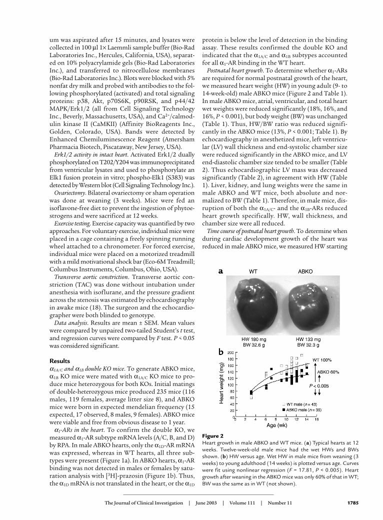
Postnatal heart growth. To determine whether α1-ARsare required for normal postnatal growth of the heart,we measured heart weight (HW) in young adult (9- to14-week-old) male ABKO mice (Figure 2 and Table 1).In male ABKO mice, atrial, ventricular, and total heartwet weights were reduced significantly (18%, 16%, and16%, P < 0.001), but body weight (BW) was unchanged(Table 1). Thus, HW/BW ratio was reduced signifi-cantly in the ABKO mice (13%, P < 0.001; Table 1). Byechocardiography in anesthetized mice, left ventricu-lar (LV) wall thickness and end-systolic chamber sizewere reduced significantly in the ABKO mice, and LVend-diastolic chamber size tended to be smaller (Table2). Thus echocardiographic LV mass was decreasedsignificantly (Table 2), in agreement with HW (Table1). Liver, kidney, and lung weights were the same inmale ABKO and WT mice, both absolute and nor-malized to BW (Table 1). Therefore, in male mice, dis-ruption of both the α1A/C- and the α1B-ARs reducedheart growth specifically. HW, wall thickness, andchamber size were all reduced.
Time course of postnatal heart growth. To determine whenduring cardiac development growth of the heart wasreduced in male ABKO mice, we measured HW starting
The Journal of Clinical Investigation | June 2003 | Volume 111 | Number 11 1785
Figure 2Heart growth in male ABKO and WT mice. (a) Typical hearts at 12weeks. Twelve-week-old male mice had the wet HWs and BWsshown. (b) HW versus age. Wet HW in male mice from weaning (3weeks) to young adulthood (14 weeks) is plotted versus age. Curveswere fit using nonlinear regression (F = 17.81, P < 0.005). Heartgrowth after weaning in the ABKO mice was only 60% of that in WT;BW was the same as in WT (not shown).
at weaning (3 weeks) and continuing through youngadulthood (14 weeks; Figure 2b). Litter sizes werematched to eliminate any impact on development. In WTmice, HW approximately doubled from weaning toyoung adulthood (Figure 2b). In ABKO mice, HW andHW/BW ratio were the same as in WT at weaning. At 3
weeks, HW was 64.1 ± 3.5 mg in WT and 72.6 ± 1.4 mg inABKO mice, and HW/BW was 5.1 ± 0.1 in WT and 5.2 ± 0.2 in ABKO mice (n = 5 WT, 4 ABKO, P = NS).However, growth of the ABKO hearts was significantlyless from weaning onward, with HW reaching only 84%of WT in the young adult (P < 0.05; Table 1 and Figure
1786 The Journal of Clinical Investigation | June 2003 | Volume 111 | Number 11
Table 1Heart and organ weights, myocyte size, and mRNA levels in ABKO mice
Sex Male Female Male/FemaleGenotype WT ABKO ABKO/WT WT ABKO ABKO/WT WT ABKO
(%) (%) (%) (%)
BW (g) 31.2 ± 0.5 (33) 29.8 ± 0.7 (27) 96 26.0 ± 0.9 (16) 25.4 ± 0.5 (15) 98 120B 118C
HWAtria (mg) 9.7 ± 0.3 (31) 7.9 ± 0.4 (25) 82A 6.3 ± 0.3 (16) 6.6 ± 0.5 (15) 105 154B 120C
Ventricles (mg) 136 ± 3 (31) 115 ± 3 (25) 84A 102 ± 3 (16) 96 ± 2 (15) 94A 133B 120C
Heart (mg) 147 ± 3 (33) 122 ± 3 (27) 84A .109 ± 3 (16) 103 ± 2 (15) 94 135B 118C
HW/BW (× 10–3) 4.7 ± 0.1 (33) 4.1 ± 0.1 (27) 87A 4.2 ± 0.1 (16) 4.1 ± 0.1 (15) 98 112B 100Myocyte size and mRNA
Myocyte volume (µm3) 19,647 ± 722 (6) 14,688 ± 1,111 (7) 75A 11,513 ± 529 (4) 11,371 ± 264 (3) 99 170B 128Myocyte surface area (µm2) 3,266 ± 40 (3) 2,505 ± 180 (3) 77A ND NDMyocyte csa (µm2) 210 ± 24 (2) 141 ± 10 (2) 67A ND NDMyHC (α+β) (MyHC/actin) 1.80 ± 0.05 (6) 1.57 ± 0.07 (6) 87A ND NDANF (ANF/actin) 0.35 ± 0.02 (6) 0.21 ± 0.02 (6) 60A ND ND
LiverLiver weight (g) 1.4 ± 0.1 (18) 1.4 ± 0.1 (14) 100 1.2 ± 0.1 (11) 1.1 ± 0.1 (8) 92 117B 127C
Liver weight/BW (× 10–2) 4.6 ± 0.1 (18) 4.6 ± 0.1 (14) 100 4.8 ± 0.1 (11) 4.4 ± 0.2 (8) 92 96 109Lung
Lung weight (mg) 177 ± 4 (18) 164 ± 5 (14) 93 .165 ± 6 (11) 157 ± 7. (8) 95 107 104Lung weight/BW (× 10–3) 5.8 ± 0.2 (18) 5.6 ± 0.2 (14) 97 6.7 ± 0.4 (11) 6.2 ± 0.3 (8) 93 87 90
KidneyRight kidney (mg) 213 ± 7 (18) 210 ± 10 (14) 99 .135 ± 6 (11) 158 ± 5 (8) 117A 158B 133C
Kidney weight/BW (× 10–3) 6.9 ± 0.2 (18) 7.1 ± 0.3 (14) 103 5.5 ± 0.3 (11) 6.2 ± 0.2 (8) 113A 125B 114C
Heart and organ weights, myocyte size, and mRNA levels were measured in young adult (9- to 14-week-old) ABKO and WT mice of both sexes. Chamber weightsare combined right and left. Values are mean ± SEM with the numbers of mice in parentheses. HW, heart weight; BW, body weight; csa, cross-sectional area;ANF, atrial natriuretic factor; ND, not determined. P < 0.05; AABKO vs. WT; Bmale vs. female WT; Cmale vs. female ABKO.
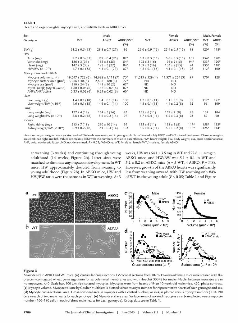
Figure 3Myocyte size in ABKO and WT mice. (a) Ventricular cross sections. LV coronal sections from 10- to 11-week-old male mice were stained with flu-orescein-conjugated wheat germ agglutinin for sarcolemmal membranes and with Hoechst 33342 for nuclei. Nuclei between myocytes are innonmyocytes. ×40. Scale bar, 100 µm. (b) Isolated myocytes. Myocytes were from hearts of 9- to 10-week-old male mice. ×20, phase contrast.(c) Myocyte volume. Myocyte volume by Coulter Multisizer is plotted versus myocyte number for representative hearts of each genotype and sex.(d) Myocyte cross-sectional area. Cross-sectional area in myocytes with a central nucleus, as in a, is plotted versus myocyte number (110–190cells in each of two male hearts for each genotype). (e) Myocyte surface area. Surface areas of isolated myocytes as in b are plotted versus myocytenumber (160–190 cells in each of three male hearts for each genotype). Group data are in Table 1.
2b). Growth curves modeled by nonlinear regressionshowed that the increase in heart size after weaning in theABKO mice was only 60% of that in the WT (P < 0.005;Figure 2b). BW of WT and ABKO mice did not differ sig-nificantly at any time. Thus, the α1A/C and α1B subtypeswere required for normal cardiac growth after weaning.
Myocyte size. The mechanism of the smaller heart inthe ABKO mice could have been either smallermyocytes or fewer myocytes. Reduced myocyte hyper-trophy was more likely, because myocyte proliferationis largely complete by weaning (21). To test for a reduc-tion in myocyte size in the intact heart, we measuredmyocyte cross-sectional area (Figure 3a). Heart andbody weights of the ABKO and WT mice were matchedto approximate the averages in Table 1. Myocyte cross-sectional area was reduced by 33% in the male ABKOheart (P < 0.05; Figure 3d and Table 1).
To confirm the results from ventricular sections, wemeasured volume and surface area of myocytes isolat-ed from hearts of ABKO and WT mice matched for ageand BW (Figure 3b). Myocyte volumes were quantifiedusing a Coulter Multisizer, and surface area was meas-ured by phase microscopy. In male ABKO mice, medi-an myocyte volume was reduced by 25% and medianmyocyte surface area was reduced by 23% (Figure 3, cand e, and Table 1). Therefore, reduced myocyte hyper-trophy could explain reduced cardiac hypertrophy afterweaning in male ABKO mice.
Myocyte binucleation. During the first 2 weeks of nor-mal postnatal development, mouse myocytes undergoa final round of DNA synthesis and become binucleateby nuclear division without cell division (21). A delay interminal differentiation can increase the number ofsmaller, mononuclear myocytes (22) and was a poten-tial explanation for the reduced myocyte hypertrophyobserved in the male ABKO mice. However, the majori-ty of isolated adult male myocytes were binucleate inboth genotypes (ABKO, 91% ± 0.2%; WT, 89% ± 0.7%; n = 2 hearts per genotype, 100–120 myocytes per heart),suggesting normal terminal differentiation.
Myocyte mRNA levels. To test whether α1-ARs were re-quired for normal transcription of cardiac genes dur-ing postweaning hypertrophy, we measured MyHCand atrial natriuretic factor (ANF) mRNA levels byRPA. In adult male ABKO ventricles, total MyHC andα-MyHC mRNA was reduced by 13% (P < 0.05; Table1), and β-MyHC mRNA (6% of total MyHC) tended tobe lower (not shown). ANF mRNA was reduced by 40%(P < 0.05; Table 1). Thus, KO of both α1-ARs reducedmyocyte mRNA levels.
Heart and myocyte size in congenic ABKO mice. To con-firm that the small-heart phenotype observed in theABKO mouse was not dependent on mixed strains, wemeasured heart and myocyte size in congenic C57BL/6mice. In congenic male ABKO mice, the small-heartphenotype was preserved (Figure 4). Heart cross-sec-
The Journal of Clinical Investigation | June 2003 | Volume 111 | Number 11 1787
Table 2Echocardiography in anesthetized ABKO mice
Sex Male Female Male/FemaleGenotype WT ABKO ABKO/WT WT ABKO ABKO/WT WT ABKO
(%) (%) (%) (%)
n 16 11 8 8BW (g) 33.4 ± 1.3 31.0 ± 1.3 93 25.9 ± 0.9 26.6 ± 1.4 103 129B 117C
HR (beats/min) 432 ± 11 435 ± 22 101 441 ± 19 438 ± 25 99 98 99M-mode
LV posterior wall thickness, D (mm) 0.61 ± 0.02 0.50 ± 0.01 82A 0.49 ± 0.02 0.47 ± 0.02 96 124B 106LV anterior wall thickness, D (mm) 0.74 ± 0.03 0.58 ± 0.01 78A 0.59 ± 0.02 0.56 ± 0.02 95 125B 104LV internal dimension, D (mm) 4.46 ± 0.07 4.28 ± 0.07 96 4.04 ± 0.08 4.13 ± 0.12 102 110B 104LV posterior wall thickness, S (mm) 0.77 ± 0.02 0.63 ± 0.02 82A 0.62 ± 0.02 0.58± 0.02 94 124B 109LV anterior wall thickness, S (mm) 0.84 ± 0.03 0.68 ± 0.02 81A 0.66 ± 0.02 0.64 ± 0.01 97 127B 106LV internal dimension, S (mm) 3.19 ± 0.07 2.97 ± 0.10 93A 2.870 ± 0.13 2.83 ± 0.07 99 113B 105Fractional shortening (%) 28 ± 2 31 ± 1 111A .29 ± 2.1 31 ± 1 107 96 100End-diastolic volume (µl) 92 ± 4 .83 ± 4 90 .69 ± 4 75 ± 7 109 133 111End-systolic volume (µl) 34 ± 2 28 ± 3 82A 26 ± 4 24 ± 2 92 131 117Stroke volume (µl) 58 ± 4 55 ± 3 95 43 ± 2 51 ± 6 119 135B 108Ejection fraction (%) 62 ± 3 67 ± 2 108A 64 ± 3 68 ± 2 106 96 99Cardiac output (ml/min) 25 ± 2 24 ± 2 96 19 ± 2 22 ± 3 116 131B 109
2DLV mass (mg) 113 ± 6 79 ± 4 70A .71 ± 4 70 ± 4 99 159B 113LV mass index (mg/g × 10–3) 3.4 ± 0.1 2.6 ± 0.2 76A 2.8 ± 0.2 2.7 ± 0.1 96 121B 96
Pulsed-wave DopplerAortic flow rate (m/s) 1.05 ± 0.03 1.05 ± 0.04 100 0.90 ± 0.05 0.96 ± 0.05 107 117 109
LV wall stress (g/cm2) (n = 8) 140 ± 8 146 ± 8 104 ND ND
Echocardiography was performed on anesthetized young adult (9- to 14-week-old) ABKO and WT mice of both sexes. 2D long axis and 2D-guided M-modemeasurements were recorded. Calculations were made for LV mass by the truncated-ellipsoid method from 2D recordings, and from M-mode measurements.Calculations were made for ventricular volumes by the cubed method [volume = 1.047 × (LV internal dimension)3]. Stroke volume (SV) = end-diastolic vol-ume (EDV) – end-systolic volume (ESV). Cardiac output = HR × SV. Fractional shortening = [(LVIDD – LVIDS)/LVIDD] × 100, where LVID is LV internal dimen-sion, D is diastole, and S is systole. Ejection fraction = [(EDV – ESV)/EDV] × 100. Wall stress = [(1.35)(SysBP)(LVIDS)]/{(4)(LVPWS)[1 + (LVPWS)/(LVIDS)]},where SysBP is systolic BP from tail cuff measurements as in Table 3, and LVPWS is LV posterior wall thickness in systole. Values are mean ± SEM. P < 0.05; AABKO vs. WT; Bmale vs. female WT; Cmale vs. female ABKO.
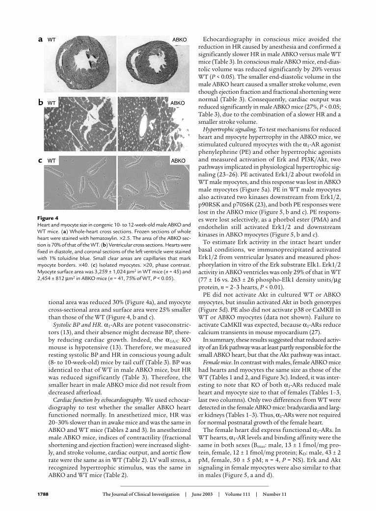
tional area was reduced 30% (Figure 4a), and myocytecross-sectional area and surface area were 25% smallerthan those of the WT (Figure 4, b and c).
Systolic BP and HR. α1-ARs are potent vasoconstric-tors (13), and their absence might decrease BP, there-by reducing cardiac growth. Indeed, the α1A/C KOmouse is hypotensive (13). Therefore, we measuredresting systolic BP and HR in conscious young adult(8- to 10-week-old) mice by tail cuff (Table 3). BP wasidentical to that of WT in male ABKO mice, but HRwas reduced significantly (Table 3). Therefore, thesmaller heart in male ABKO mice did not result fromdecreased afterload.
Cardiac function by echocardiography. We used echocar-diography to test whether the smaller ABKO heartfunctioned normally. In anesthetized mice, HR was20–30% slower than in awake mice and was the same inABKO and WT mice (Tables 2 and 3). In anesthetizedmale ABKO mice, indices of contractility (fractionalshortening and ejection fraction) were increased slight-ly, and stroke volume, cardiac output, and aortic flowrate were the same as in WT (Table 2). LV wall stress, arecognized hypertrophic stimulus, was the same inABKO and WT mice (Table 2).
Echocardiography in conscious mice avoided thereduction in HR caused by anesthesia and confirmed asignificantly slower HR in male ABKO versus male WTmice (Table 3). In conscious male ABKO mice, end-dias-tolic volume was reduced significantly by 20% versusWT (P < 0.05). The smaller end-diastolic volume in themale ABKO heart caused a smaller stroke volume, eventhough ejection fraction and fractional shortening werenormal (Table 3). Consequently, cardiac output wasreduced significantly in male ABKO mice (27%, P < 0.05;Table 3), due to the combination of a slower HR and asmaller stroke volume.
Hypertrophic signaling. To test mechanisms for reducedheart and myocyte hypertrophy in the ABKO mice, westimulated cultured myocytes with the α1-AR agonistphenylephrine (PE) and other hypertrophic agonistsand measured activation of Erk and PI3K/Akt, twopathways implicated in physiological hypertrophic sig-naling (23–26). PE activated Erk1/2 about twofold inWT male myocytes, and this response was lost in ABKOmale myocytes (Figure 5a). PE in WT male myocytesalso activated two kinases downstream from Erk1/2,p90RSK and p70S6K (23), and both PE responses werelost in the ABKO mice (Figure 5, b and c). PE respons-es were lost selectively, as a phorbol ester (PMA) andendothelin still activated Erk1/2 and downstreamkinases in ABKO myocytes (Figure 5, b and c).
To estimate Erk activity in the intact heart underbasal conditions, we immunoprecipitated activatedErk1/2 from ventricular lysates and measured phos-phorylation in vitro of the Erk substrate Elk1. Erk1/2activity in ABKO ventricles was only 29% of that in WT(77 ± 16 vs. 263 ± 26 phospho-Elk1 density units/µgprotein, n = 2–3 hearts, P < 0.01).
PE did not activate Akt in cultured WT or ABKOmyocytes, but insulin activated Akt in both genotypes(Figure 5d). PE also did not activate p38 or CaMKII inWT or ABKO myocytes (data not shown). Failure toactivate CaMKII was expected, because α1-ARs reducecalcium transients in mouse myocardium (27).
In summary, these results suggested that reduced activ-ity of an Erk pathway was at least partly responsible for thesmall ABKO heart, but that the Akt pathway was intact.
Female mice. In contrast with males, female ABKO micehad hearts and myocytes the same size as those of theWT (Tables 1 and 2, and Figure 3c). Indeed, it was inter-esting to note that KO of both α1-ARs reduced maleheart and myocyte size to that of females (Tables 1–3,last two columns). Only two differences from WT weredetected in the female ABKO mice: bradycardia and larg-er kidneys (Tables 1–3). Thus, α1-ARs were not requiredfor normal postnatal growth of the female heart.
The female heart did express functional α1-ARs. InWT hearts, α1-AR levels and binding affinity were thesame in both sexes (Bmax: male, 13 ± 1 fmol/mg pro-tein, female, 12 ± 1 fmol/mg protein; KD: male, 43 ± 2pM, female, 50 ± 5 pM; n = 4, P = NS). Erk and Aktsignaling in female myocytes were also similar to thatin males (Figure 5, a and d).
1788 The Journal of Clinical Investigation | June 2003 | Volume 111 | Number 11
Figure 4Heart and myocyte size in congenic 10- to 12-week-old male ABKO andWT mice. (a) Whole-heart cross sections. Frozen sections of wholeheart were stained with hematoxylin. ×2.5. The area of the ABKO sec-tion is 70% of that of the WT. (b) Ventricular cross sections. Hearts werefixed in diastole, and coronal sections of the left ventricle were stainedwith 1% toluidine blue. Small clear areas are capillaries that markmyocyte borders. ×40. (c) Isolated myocytes. ×20, phase contrast.Myocyte surface area was 3,259 ± 1,024 µm2 in WT mice (n = 45) and2,454 ± 812 µm2 in ABKO mice (n = 41, 75% of WT, P < 0.05).

To test whether female sex hormones protected fromKO of α1-ARs, we did ovariectomy at weaning (3 weeks)and studied the mice at age 12 weeks. Male heart growthwas reduced in the ABKO mice over this same 3- to 12-week interval (Figure 2b). Ovariectomy reduced uterineweight to 6–7% of that of sham-operated mice in bothWT and ABKO mice (n = 3–6 in each group, P < 0.025).However, ovariectomy had no effect in either genotypeon BW, tibia length, HW, BP, or HR (data not shown).These same variables did not differ significantly between
WT and ABKO mice after ovariectomy, except thatHR was 15% lower in the ABKO mice (data notshown), as observed in nonoperated female mice(Table 3). Thus, female sex hormones did not explainwhy the female heart was unaffected by the ABKO.
Response of the ABKO to stress. We used exercise and aor-tic banding to test whether the small male ABKO hearthad an adaptive response to cardiac stress. Voluntaryexercise was tested with a running wheel. WT mice rana nightly average of about 6 km over about 4 hours at22 m/min, and ABKO mice ran significantly less by allmeasures (Figure 6a). To test whether this result sim-ply reflected motivational factors, we used forced exer-
cise on a motorized treadmill. ABKO mice ran signifi-cantly less on the treadmill, measured either at a singlesession, or over 20 consecutive daily training sessions (Fig-ure 6b, left and right). Thus, exercise capacity by two inde-pendent assays was reduced significantly in ABKO mice.
We used TAC to test the response to pressure overload.The gradient across the stenosis at 14 days was similar inWT and ABKO mice (99 ± 6 mmHg, n = 14, in WT vs.105 ± 8 mmHg, n = 8, in ABKO, P = NS). However, sur-vival to 14 days after TAC was only 56% in the ABKO ver-
The Journal of Clinical Investigation | June 2003 | Volume 111 | Number 11 1789
Figure 5Hypertrophic signaling in myocytes. Cultured myocytes from heartsof 10- to 11-week-old congenic WT and ABKO mice were treatedfor 15 minutes with PE (20 µM, plus 2 µM timolol); phorbol 12-myristate, 13-acetate (PMA, 100 nM); sorbitol (Sor, 1 M);endothelin (ET, 10 nM); insulin (Ins, 6 µM); or vehicle. Westernblots were done for (a) phospho-Erk1/2 (pErk) and total Erk1/2,(b) phospho-p90RSK (pp90RSK), (c) phospho-p70S6K (pp70S6K),and (d) phospho-Akt (pAkt) and total Akt. In a, the bar graphssummarize Erk1/2 data from six hearts in each group. In b, a non-specific (NS) band is indicated with anti–pp90RSK. In d, a secondpAkt antibody (S473) gave identical results (not shown). Totalp90RSK (b) and total p70S6K (c) were the same in WT and ABKOmice (not shown). *P < 0.05 vs. Veh.
Table 3Cardiac function in conscious ABKO mice
Sex Male Female Male/FemaleGenotype WT ABKO ABKO/WT (%) WT ABKO ABKO/WT (%) WT (%) ABKO (%)
Tail cuffn 14 11 11 11Systolic BP (mmHg) 111 ± 2 112 ± 3 101 111 ± 3 111 ± 4 100 100 101HR (beats/min) 622 ± 10 568 ± 15 91A 604 ± 15 578 ± 13 96 103 98Echocardiographyn 4 5 5 4BW (g) 36 ± 2 33 ± 2 92 25 ± 1 23 ± 1 92 144B 143C
HR (beats/min) 561 ± 10 496 ± 6 88A 575 ± 13 531 ± 13 92A 98 93M-mode
Fractional shortening (%) 54 ± 2 58 ± 3 107 56 ± 1 54 ± 3 96 96 107End-diastolic volume (µl) 60 ± 3 48 ± 3 80A 41 ± 2 49 ± 7 120 146B 98End-systolic volume (µl) 6 ± 1 4 ± 1 67 4 ± 0 6 ± 1 150 150 67Stroke volume (µl) 54 ± 2 44 ± 3 81A 38 ± 2 44 ± 6 116 142B 100Ejection fraction (%) 90 ± 1 92 ± 2 102 92 ± 1 90 ± 2 98 98 102Cardiac output (ml/min) 30 ± 1 22 ± 3 73A 23 ± 1 22 ± 4 96 130B 100
Cardiac function was measured in young adult ABKO and WT mice of both sexes (8–10 weeks old for tail cuff systolic BP and HR, 14 weeks old for echocardio-graphy). Echocardiographic calculations were as in Table 2. Values are mean ± SEM. P < 0.05, AABKO vs. WT, Bmale vs. female WT, Cmale vs. female ABKO.

sus 100% in the WT mice (P < 0.002; Figure 6c). ABKOmice died at 5–8 days after TAC (Figure 6c), and heartfailure was evident in autopsied ABKO mice by fluid inthe pleural and peritoneal cavities and thrombi in theatria. In the few ABKO mice that survived to 2 weeks,overall hypertrophy after TAC was the same as observedin WT mice. TAC increased HW/BW by 153% in ABKOmice, as compared with a 150% increase in WT mice (P = NS versus ABKO). Thus, α1-ARs were not requiredfor the increase in heart size after pressure overload, buttheir absence caused death due to heart failure.
DiscussionHere we characterized a double α1-AR KO to show thatthe α1A/C and α1B subtypes are together required for nor-mal cardiac hypertrophy during postnatal life. Only themale ABKO mice had reduced heart growth, but bothmales and females had normal BP and bradycardia. Themechanism of the small heart was reduced Erk signal-ing, at least in part. These results indicate a sex-specificand load-independent role for catecholamines and α1-ARs in physiological hypertrophy, a role not revealed inearlier single KOs (12, 13).
The ABKO eliminated cardiac α1-AR binding andcaused a 40% reduction in growth of the male ABKOheart after weaning. The smaller heart was explained bysmaller myocytes, and terminal differentiation was nor-mal. The following supported a direct effect of α1-ARsignaling on myocyte hypertrophy in vivo: myocyte-spe-cific mRNAs and Erk1/2 signaling were reduced, otherorgans were normal, and BP and ventricular wall stresswere unchanged. A direct effect was shown initially bystudies in myocyte culture (2, 5, 6).
Cardiac adrenergic innervation increases greatly frombirth to weaning (1), the α1A/C subtype is first detectedin the mouse heart at weaning (28), and the ABKO car-diac phenotype became evident after weaning. By wean-ing, myocyte DNA synthesis is largely complete, andsubsequent growth of the heart is due to myocyte hyper-trophy (1, 21, 22). Taken together, the findings suggesta model in which increased cardiac sympathetic inner-vation after weaning, increased norepinephrine releaseduring daily life, and subsequent activation of α1-ARsplay an important role in the physiological myocytehypertrophy of normal postnatal development.
The validity of this model would seem to be challengedby the normal growth of the female ABKO heart. How-ever, a body of evidence indicates that sympathetic acti-vation is significantly lower in females than in males (29,30). Lower sympathetic tone in females can explainfemale protection from hypertension (29) and couldreadily account for lesser dependence of female heartgrowth on α1-AR stimulation. Indeed, it was notable thatan overall effect of the ABKO was to reduce male heartand myocyte size indices to those of the female. It wasalso notable that myocyte α1-AR levels and signaling werethe same in WT males and females. Thus, the ABKO canbe seen as defining a sympathetic contribution to normaldevelopmental hypertrophy. Ovariectomy did not un-mask a small heart in ABKO females, so the sex differencewas not determined by female sex hormones.
We did not see any sex differences in other cardiovascu-lar parameters, as both males and females had normalresting BP and bradycardia. These were unexpected find-ings. As a class, α1-ARs are potent vasoconstrictors, andthe single α1A/C KO is hypotensive (13). On the other hand,the single α1B KO is normotensive (12), and overexpres-sion of the α1B with its own promoter causes hypotension(9). Thus, the ABKO might combine offsetting effects onBP. Alternately, reduced cardiac output in the ABKOmouse might stimulate a reflex increase in BP through thevascular α1D subtype (31). HR is not altered significantlyin the single α1A/C or α1B KOs (12, 13), and α1-AR stimu-lation does not change HR in the mouse when the barore-flex is blocked (13). The mechanisms of normotensionand bradycardia require further study, but the small maleABKO heart was not explained by altered loading.
Insulin/PI3K/Akt and Erk are two pathways so farimplicated in physiological hypertrophic signaling (23–26,32). Reduced Erk signaling was likely one mechanism forreduced hypertrophy in the ABKO mouse, because Erkactivity was reduced in the intact ABKO heart, and α1-AR
1790 The Journal of Clinical Investigation | June 2003 | Volume 111 | Number 11
Figure 6Response of the ABKO mice to stress. Ten- to twelve-week-old maleABKO and WT mice were used. (a) Exercise: free wheel running. Micewere given access to a running wheel for 12 hours on each of 30 con-secutive nights, and distance, duration, and speed were recorded bya chronometer attached to the wheel (n = 4–5 mice each group). (b)Exercise: motorized treadmill. Mice were placed on a motorized tread-mill set at the speed and for the duration indicated, and the numberof times the motivational bar was touched, indicating failure to main-tain treadmill speed, was recorded as breaks per minute. The leftpanel shows a single session (n = 8–9 mice per group), and the rightpanel shows 20 consecutive daily training sessions (n = 9–10). (c) TACwas done in congenic mice, and survival was recorded over 14 days.

stimulation of Erk was lost in isolated ABKO myocytes.However, reduced Erk activity might not explain the entireABKO phenotype. α1-AR stimulation of Erk is also lost inthe single α1A/C KO heart (data not shown), but heart sizeis normal in the single α1A/C KO (13). On the other hand,PKCε activation is lost in the single α1B KO (33), and PKCεcan stimulate a physiological signaling in the mouse heart(34). Thus, the small-heart phenotype might be seen onlyin the double-subtype KO because the subtypes couple todistinct signaling pathways, which together are essentialfor developmental hypertrophy.
It was notable in this regard that endothelin andinsulin had normal Erk and Akt signaling in ABKOmyocytes. These results implied that α1-AR effects onpostnatal heart growth could not be compensated forby these other hypertrophic agonists, or by the manyother Gq-linked systems implicated in hypertrophy.Indeed, it seems likely that α1-ARs/Erk and insulin/PI3K/Akt (24–26, 32) represent independent pathwaysfor developmental hypertrophy.
Finally, the male ABKO heart not only was small butalso had abnormal function. Contractile function invivo at rest was intact, with normal ejection fraction andfractional shortening by echocardiography. However,cardiac output was decreased significantly in consciousmice, due to the combination of a smaller heart and aslower HR. With stress, exercise capacity was reducedsignificantly, most likely by an impaired increase in car-diac output. Furthermore, pressure overload causeddeath due to heart failure in the ABKO mice that wasnot observed in WT mice. Thus, α1-ARs were requiredfor an adaptive response to cardiac stress. Preliminarydata suggest abnormal remodeling in ABKO mice afterTAC, and therefore the ABKO mouse might help definea physiological contribution to the hypertrophic re-sponse that follows a pathological stimulus.
AcknowledgmentsThis work was supported by the NIH (P.C. Simpson andE. Foster), the Swiss National Science Foundation(grant 31-51043.97, to S. Cotecchia), the Department ofVeteran Affairs (P.C. Simpson), fellowships from theCanadian Heart and Stroke Foundation and the Amer-ican Heart Association, Western States Affiliate (to D.G.Rokosh) and from the Cardiovascular Research Insti-tute at UCSF (T32HL07731, to T.D. O’Connell).
1. Rakusan, K. 1984. Cardiac growth, maturation and aging. In Growth of theheart in health and disease. R. Zak, editor. Raven Press. New York, New York,USA. 131–164.
2. Simpson, P.C., Kariya, K., Karns, L.R., and Long, C.S. 1990. The α1-adrener-gic receptor in left ventricular hypertrophy. Journal of Vascular Medicine andBiology. 2:236–246.
3. Rapacciuolo, A., et al. 2001. Important role of endogenous norepinephrineand epinephrine in the development of in vivo pressure-overload cardiachypertrophy. J. Am. Coll. Cardiol. 38:876–882.
4. Thomas, S.A., Matsumoto, A.M., and Palmiter, R.D. 1995. Noradrenaline isessential for mouse fetal development. Nature. 374:643–646.
5. Simpson, P. 1983. Norepinephrine-stimulated hypertrophy of cultured ratmyocardial cells is an alpha1-adrenergic response. J. Clin. Invest. 72:732–738.
6. Simpson, P. 1985. Stimulation of hypertrophy of cultured neonatal rat heartcells through an α1-adrenergic receptor and induction of beating throughan α1- and β1-adrenergic receptor interaction: evidence for independent reg-ulation of growth and beating. Circ. Res. 56:884–894.
7. Rokosh, D.G., et al. 1996. α1-Adrenergic receptor subtype mRNAs are dif-ferentially regulated by α1-adrenergic and other hypertrophic stimuli in car-diac myocytes in culture and in vivo: repression of α1B and α1D but inductionof α1C. J. Biol. Chem. 271:5839–5843.
8. Milano, C.A., et al. 1994. Myocardial expression of a constitutively activeα1B-adrenergic receptor in transgenic mice induces cardiac hypertrophy.Proc. Natl. Acad. Sci. U. S. A. 91:10109–10113.
9. Zuscik, M.J., et al. 2001. Hypotension, autonomic failure, and cardiac hyper-trophy in transgenic mice overexpressing the α1B-adrenergic receptor. J. Biol.Chem. 276:13738–13743.
10. Akhter, S.A., et al. 1997. Transgenic mice with cardiac overexpression of α1B-adrenergic receptors. In vivo α1-adrenergic receptor-mediated regulation ofβ-adrenergic signaling. J. Biol. Chem. 272:21253–21259.
11. Lin, F., et al. 2001. Targeted α1A-adrenergic receptor overexpression inducesenhanced cardiac contractility but not hypertrophy. Circ. Res. 89:343–350.
12. Cavalli, A., et al. 1997. Decreased blood pressure response in mice deficientof the α1b-adrenergic receptor. Proc. Natl. Acad. Sci. U. S. A. 94:11589–11594.
13. Rokosh, D.G., and Simpson, P.C. 2002. Knockout of the α1A/C-adrenergicreceptor subtype: the α1A/C is expressed in resistance arteries and is requiredto maintain arterial blood pressure. Proc. Natl. Acad. Sci. U. S. A. 99:9474–9479.
14. Rohrer, D.K., Chruscinski, A., Schauble, E.H., Bernstein, D., and Kobilka,B.K. 1999. Cardiovascular and metabolic alterations in mice lacking bothβ1- and β2-adrenergic receptors. J. Biol. Chem. 274:16701–16708.
15. Hein, L., Altman, J.D., and Kobilka, B.K. 1999. Two functionally distinct α2-adrenergic receptors regulate sympathetic neurotransmission. Nature.402:181–184.
16. Deng, X.-F., Rokosh, D.G., and Simpson, P.C. 2000. Autonomous andgrowth factor-induced hypertrophy in cultured neonatal mouse cardiacmyocytes: comparison with rat. Circ. Res. 87:781–788.
17. Stewart, A.F.R., et al. 1994. Cloning of the rat α1C-adrenergic receptor fromcardiac myocytes: α1C, α1B, and α1D mRNAs are present in cardiac myocytes,but not in cardiac fibroblasts. Circ. Res. 75:796–802.
18. Nakamura, A., et al. 2001. LV systolic performance improves with develop-ment of hypertrophy after transverse aortic constriction in mice. Am. J. Phys-iol. Heart Circ. Physiol. 281:H1104–H1112.
19. Zhou, Y.Y., et al. 2000. Culture and adenoviral infection of adult mouse car-diac myocytes: methods for cellular genetic physiology. Am. J. Physiol. HeartCirc. Physiol. 279:H429–H436.
20. Chang, K.C., et al. 1997. Thyroid hormone improves function and Ca2+ han-dling in pressure overload hypertrophy. Association with increased sar-coplasmic reticulum Ca2+-ATPase and α-myosin heavy chain in rat hearts. J. Clin. Invest. 100:1742–1749.
21. Soonpaa, M.H., et al. 1997. Cyclin D1 overexpression promotes cardiomy-ocyte DNA synthesis and multinucleation in transgenic mice. J. Clin. Invest.99:2644–2654.
22. Liao, H.S., et al. 2001. Cardiac-specific overexpression of cyclin-dependentkinase 2 increases smaller mononuclear cardiomyocytes. Circ. Res.88:443–450.
23. Bueno, O.F., and Molkentin, J.D. 2002. Involvement of extracellular signal-regulated kinases 1/2 in cardiac hypertrophy and cell death. Circ. Res.91:776–781.
24. Shioi, T., et al. 2000. The conserved phosphoinositide 3-kinase pathwaydetermines heart size in mice. EMBO J. 19:2537–2548.
25. Shioi, T., et al. 2002. Akt/protein kinase B promotes organ growth in trans-genic mice. Mol. Cell. Biol. 22:2799–2809.
26. Shiojima, I., et al. 2002. Akt signaling mediates postnatal heart growth inresponse to insulin and nutritional status. J. Biol. Chem. 277:37670–37677.
27. McCloskey, D.T., et al. 2002. α1-Adrenoceptor subtypes mediate negativeinotropy in myocardium from α 1A/C-knockout and wild type mice. J. Mol.Cell. Cardiol. 34:1007–1017.
28. O’Connell, T.D., Rokosh, D.G., and Simpson, P.C. 2001. Cloning and char-acterization of the mouse α1C/A-adrenergic receptor gene and analysis ofan α1C promoter in cardiac myocytes: role of an MCAT element that bindstranscriptional enhancer factor-1 (TEF-1). Mol. Pharmacol. 59:1225–1234.
29. Hinojosa-Laborde, C., Chapa, I., Lange, D., and Haywood, J.R. 1999. Genderdifferences in sympathetic nervous system regulation. Clin. Exp. Pharmacol.Physiol. 26:122–126.
30. Evans, J.M., et al. 2001. Gender differences in autonomic cardiovascular reg-ulation: spectral, hormonal, and hemodynamic indexes. J. Appl. Physiol.91:2611–2618.
31. Tanoue, A., et al. 2002. The α1D-adrenergic receptor directly regulates arte-rial blood pressure via vasoconstriction. J. Clin. Invest. 109:765–775.doi:10.1172/JCI200214001.
32. Belke, D.D., et al. 2002. Insulin signaling coordinately regulates cardiac size,metabolism, and contractile protein isoform expression. J. Clin. Invest.109:629–639. doi:10.1172/JCI200213946.
33. Deng, X.-F., Rokosh, D.G., and Simpson, P.C. 1999. α1-Adrenergic receptorsubtypes activate different PKC isoforms in mouse heart. Circulation.100:I-566. (Abstr.)
34. Mochly-Rosen, D., et al. 2000. Cardiotrophic effects of protein kinase C ε:analysis by in vivo modulation of PKCε translocation. Circ. Res.86:1173–1179.
The Journal of Clinical Investigation | June 2003 | Volume 111 | Number 11 1791