Embed Size (px)
Citation preview
337
The Anatomy and Phytogeny of Spondylus, with a 'particular reference to the Lamellibranch Nervous System .
By W illiam J ohn D a k in , D.Sc., F.Z.S., Professor of Zoology, University ofLiverpool.*
(Communicated by Prof. E. W. McBride, F.R.S.—Received June 15, 1928.)
A study of the anatomy of the rather rare Lamellibranch, Spondylus, has always attracted the author, owing to the fact that at a comparatively early date the striking resemblance between the eyes of this creature and the remarkable eyes of the genus Pecten had been noted. In structure the eyes of Pecten stand practically alone amongst invertebrate visual organs, and since, by reason of their complexity as well as number, their evolution has been associated with the faculty of swimming, it was a matter of considerable interest to determine the relationships of Pecten with Spondylus, for whilst Pecten can swim, Spondylus lives attached to rocks.
The investigation was made purely from the above view-point, and hence minute details of histology, etc., have not been given. Resemblances to Pecten were expected. One very unexpected morphological anomaly turned up, however ; and in the midst of a remarkable agreement with the genus Pecten, one system, the nervous system, presented a condition altogether unique amongst Lamellibranchs. To obviate repetition, reference should be made to an account of the anatomy of the genus Pecten by the author (Dakin, 1909) and also to a paper, following this, which deals with the eye.
As pointed out by Ridewood (1903), the Lamellibranchiata have ever been a most troublesome group to classify ; witness the inadequacy of differences in such features as the shell, hinge, teeth, ligament, pallial sinus, siphons, adductor muscles, etc., all of which have been utilised in attempts at classification. Ridewood adopted, with some modifications dependent on his own extensive work, the classification of Pelseneer, based largely upon the gill characters. In this scheme there is a sub-order, Pectinacea (the Order Eleuthe- rorhabda, to which it belongs, is characterised by gill filaments arranged on the two sides of the gill axis ; adjacent filaments are held in position by stiff cilia disposed in patches), which contains the families Spondylidae, Pectinidae, and Aviculidae. The two genera Spondylus and Pecten are therefore closely
* The author wishes to express his thanks to the Authorities of the Natural History Museum for permission to examine material, and also to the American Natural History Museum, Washington, for gift of a rare specimen.
on May 19, 2018http://rspb.royalsocietypublishing.org/Downloaded from
338 W. J. Dakin.
associated, but Amussium is not even put in the same sub-order. The older grouping of Pelseneer, in which the genera Amussium, Spondylus, Pecten, Chlamys and Pedum are all brought close together, is nearer the truth, but even here Spondylus is placed in a different family from Pecten, a feature which seems rather unnecessary.
The shell of Spondylus is very different from that of Pecten. I t is well known, some long-spined species of great beauty having been particularly favoured by shell collectors in the past. Other species are more coarse, rough, and with shorter spines. The valves are usually heavier than those of Pecten, inequivalve —one projecting considerably dorsal to the hinge line in a very characteristic manner, and frequently one valve is much flatter than the other. In S. gcederopus, from the Mediterranean, one valve is definitely flat, the other convex. In S. Americana both valves are convex.
Now it is worthy of note (for reasons to be given later) that the flat valve, which is the right one in S. gcederopus, is the attached valve. Pecten maximus has one valve very convex, whilst the other is quite fla t; but the animal always rests on the convex valve. At a first glance, therefore, the two shells are not only rather unlike, but the orientation of the creatures appears different. The innate identity is realised, however, when one finds that, notwithstanding such diversities in valve shape, all the species of the two genera examined tend to rest on the same valve—the right.
Again, the convex rigid valve of Pecten maximus actually overlaps the flat left valve by inch ; we have noted above that the flat right valve of Spondylus greatly overlaps the left valve. We have two points indicating fundamental resemblances in orientation to put against differences in shell form. These resemblances become of greater importance when one remembers that there is no rule in the Lamellibranchiata regarding the orientation, and that in one genus the right valve may overlap the left whilst in another it is the left valve which is the larger. In fixed species the fixed valve may be the right valve or the left.
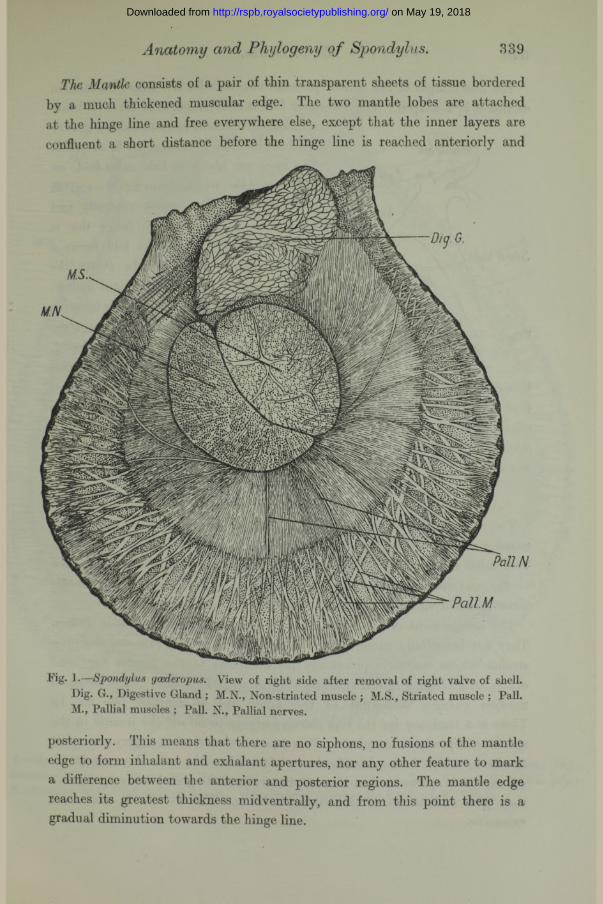
Mantle and General Organisation.With the removal of one of the valves, the resemblance between the animals,
Spondylus (fig. 1) and Pecten, is seen at a glance. There is the same single and large adductor muscle situated centrally and obviously made up of two parts (histologically different, fig. 1, M.S. and M.N.) and the same type of mantle, the edge of which is so characteristic in the genus Pecten, where it is closely correlated with the power of swimming.
on May 19, 2018http://rspb.royalsocietypublishing.org/Downloaded from
Anatomy and Phytogeny o f aSpondylus. 339
The Mantle consists of a pair of thin transparent sheets of tissue bordered by a much thickened muscular edge. The two mantle lobes are attached at the hinge line and free everywhere else, except that the inner layers are confluent a short distance before the hinge line is reached anteriorly and
Fig. 1.—Spondylus gcederopus. View of right side after removal of right valve of shell. Dig. G., Digestive Gland ; M.N., Non-striated muscle ; M.S., Striated muscle ; Pall. M., Pallial muscles ; Pall. N., Pallial nerves.
posteriorly. This means that there are no siphons, no fusions of the mantle edge to form inhalant and exhalant apertures, nor any other feature to mark a difference between the anterior and posterior regions. The mantle edge reaches its greatest thickness midventrally, and from this point there is a gradual diminution towards the hinge line.
on May 19, 2018http://rspb.royalsocietypublishing.org/Downloaded from
340 W. J. Dakin.
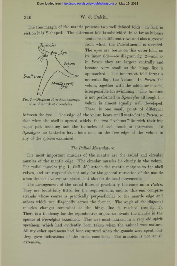
The free margin of the mantle presents two well-defined folds ; in fact, in section it is Y-shaped. The outermost fold is subdivided, in so far as it bears
tentacles in different rows and also a groove from which the Periostracum is secreted. The eyes are borne on this outer fold, on its inner side—see diagram fig. 2—and as in Pecten they are largest vent-rally and become very small as the hinge line is approached. The innermost fold forms a muscular flap, the Velum. In Pecten the velum, together with the adductor muscle, is responsible for swimming. This function is not performed in Spondylus although the velum is almost equally well developed. There is one small point of difference
between the tvro. The edge of the velum bears small tentacles in Pecten, so that vflien the shell is opened widely the two “ velums ” lie with their free edges just touching and the tentacles of each touch or intercross. In Spondylus no tentacles have been seen on the free edge of the velum in any of the species examined.
The Pallial Musculature.The most important muscles of the mantle are the radial and circular
muscles of the mantle edge. The circular muscles lie chiefly in the velum. The radial muscles (fig. 1 , Pall. M.) attach the mantle margins to the shell valves, and are responsible not only for the general retraction of the mantle when the shell valves are closed, but also for its local movements.
The arrangement of the radial fibres is practically the same as in Pecten. They are beautifully fitted for the requirements, and to this end comprise strands whose course is practically perpendicular to the mantle edge and others which run diagonally across the former. The angle of the diagonal muscles changes somewhat as the hinge line is reached (see fig. 1). There is a tendency for the reproductive organs to invade the mantle in the species of Spondylus examined. This was most marked in a very old spirit- specimen, which had evidently been taken when the animal was mature. All my other specimens had been captured when the gonads were spent, but they gave indications of the same condition. The invasion is not at all extensive.
Tentacles
Velum
Shell side
Mantle cavity Side
Fig. 2.—Diagram of section through edge of mantle of Spondylus.
on May 19, 2018http://rspb.royalsocietypublishing.org/Downloaded from

Anatomy and Phytogeny o f Spondylus. 341
Fig. 3.—Spondylus gcederopus. View of right side after removal of right mantle lobe. Ct., Ctenidium ; Dig., Digestive gland; F., F oot; Ht., Heart; Int., Intestine ; K., Kidney; Lip., Lip ; L.P., Labial palp ; M., Mantle ; M.N., Non-striated muscle ; M.S., Striated muscle ; R., Rectum ; V., Velum.
on May 19, 2018http://rspb.royalsocietypublishing.org/Downloaded from

342 W. J. Dakin.
The Adductor Muscle.As in Pecten, one might describe the organs of Spondylus as slung round the
single large adductor muscle, which runs across from one valve to the other —eccentrically. I t is situated nearer to the posterior margin of the shell. In consequence of this position there is most space left on the dorsal and on the anterior side of the muscle, and it is here that one finds the digestive gland and the visceral mass.
One can see at a glance that, as in Pecten, the adductor muscle consists of two parts. These are distinct in both fresh and preserved specimens. The line dividing the two parts runs obliquely, as indicated in the figures, so that one many speak of an anterior and a posterior portion. The fibres of the posterior part (figs. 1 and 3, M.N.) are more opaque—white in fresh specimens —whilst the anterior part is semi-translucent. The interesting feature of the “ bipartite ” composition of the adductor muscle has already been described in detail for the genus Pecten. The translucent fibres are cross-striped, and are alone responsible for the rhythmical contractions which close the valves and make swimming possible. The white opaque fibres do not contract rhythmically, and when the muscle is cut away from the shell they remain in a tonic state and are shorter than the other fibres, which are easily excited by contact. The opaque fibres are responsible for the sustained closure of the shell valves.
Now, just as the eyes and mantle edge of Spondylus reveal the ancestry of Pecten, so does the adductor muscle. So far as I am aware, no Spondylus swims in the adult stages, yet the adductor muscle still presents the two separately specialised sorts of fibres. But there is this interesting point of difference. In Spondylus, the volume of the white opaque fibres is relatively greater than that of the translucent fibres when compared with species of Pecten. The sectional area in S. americana, for example, is about one-half that of the translucent portion. In P. maximus the area of the opaque fibres is only about a quarter that of the translucent fibres.
Thus the long period during which Sp&ndylus has given up its swimming habits has been accompanied by a relative increase in that part of the adductor which is responsible for the ordinary shell closure in Pecten, or, putting it the other way, by a decrease of the muscle fibres responsible for rhythmic contractions in that genus. The translucent striped fibres are, however, not only still present, but the volume of this kind of muscle still exceeds that of the unstriped fibres. There are apparently no histological differences between the striped fibres of the two genera.
on May 19, 2018http://rspb.royalsocietypublishing.org/Downloaded from

Anatomy and Pliylogeny of Spondylus. 343
The large central adductor muscle of Spondylus and Pecten corresponds to the posterior adductor of those Lamellibranchs possessing two. The anterior has completely disappeared in the adult Pecten (although present in the early embryo) and it is altogether absent in adult Spondylus also.
Retractor Muscles.
Usually one finds retractor muscles (four in the majority of Lamellibranchs) attached to the foot. In Pecten the foot is very small and the retractor muscles have been reduced to one : this retractor is the left posterior. Now, it is interesting that even within the genus Pecten signs of still further reduction of this retractor are to be found, for whilst it is moderately developed in P. opercularis, it is vestigial in P. maximus.
In Spondylus all the retractors are missing. This fits in exactly with what might be expected if one assumes that Spondylus has evolved from Pecten, and that P. maximus represents one of the end types in the line of evolution in the Pectinidae. We shall see that the structure of Spondylus can be interpreted perfectly on this hypothesis.
Instead of retractor muscles proper, there are considerable developments of muscle fibres in the tissue of the visceral mass between the foot and the sides of the body and the lips. These muscles run into the mantle.
The Foot (fig. 3) is a small organ situated in the same position as in Pecten, very high up on the anterior surface of the visceral mass. One often assumes that this small structure is a rather rudimentary organ. The free end, however, negatives this. I t is sucker-like, widely expanded, and possesses a peculiar grooved surface. Sections indicate that it has a most extensive nerve supply. Whether these nerves are necessary for the intrinsic muscle fibres of the foot, or whether sensory endings are present in numbers in the epithelium, it is impossible to say at present. I t would appear, however, that the foot performs actively some functions, possibly that of cleaning the gills? or as an aid in nutrition. Sections failed to reveal any signs of byssal gland in the depths of the foot, and one must therefore conclude that again in this feature Spondylus has evolved a stage further along the line already indicated by the different species of Pecten.
The pedal ganglia are situated at the base of the foot; statocysts, as they should be called, often present in the foot, are situated as in Pecten, outside the foot, between its base and the mouth region (see later).
on May 19, 2018http://rspb.royalsocietypublishing.org/Downloaded from

344 W. J. Dakin.
Ctenidia.The Gills or Ctenidia (fig. 3, Ct.), structures which have played a very
important role in the classification of the Lamellibranchiata, are very conspicuous organs. Like the gills of Pecten they are slung round the adductor muscle. Each ctenidium consists of a supporting ctenidial axis, from which depend the two lamellae of filaments. The ctenidium is plicate and hetero- rhabdic, exactly like the ctenidium of Pecten, and so far as morphology and histology is concerned there is close similarity again between the two genera. For example, the principal filaments of Pecten bear a curious expansion, which the author called a Respiratory Expansion. I t is a plate of tissue with blood vessels running in its margins, and with the intervening surface thrown into folds. The same structure exactly is found on the principal filaments of Spondylus.
The “ gills ” of Lamellibranchs are primarily organs for producing currents which subserve both respiration and nutrition. The mantle lobes are highly vascular, and are the most important respiratory organs. I t is difficult, however, to understand how the ctenidia can be refused all participation in the function of respiration, as has lately been asserted. At all events, in Pecten and Spondylus parts of the ctenidia are certainly likely to function in this manner. There are some slight differences between the ctenidia of Pecten and Spondylus, but they are very minor points, and so far as can be seen differences of the same type exist between species of Pecten.
The Alimentary Canal and the Digestive Gland.The alimentary canal of Spondylus is comparatively simple in form and closely
resembles that of Pecten. The mouth opening is hidden, in the characteristic manner seen in Pecten, by the convoluted margins of the lips (fig. 3, Lip) and their entanglement. Thus, by the interlocking of the upper and lower lips, the entrance to the mouth is reduced to numerous fine passages. The margins of the lips are pigmented with the same orange yellow pigment so characteristic of this region in Pecten. The Labial Palps, which are continuations of the lips, are rectangular in shape, with the long side at right angles to the direction of the numerous typical grooves. Details of their ciliation have not been worked out.
From the mouth a short oesophagus leads into the “ stomach,” which is situated in the midst of the digestive gland. This occupies a position between the adductor muscle and the hinge line, and extends down the visceral mass towards the foot. The intestine (fig. 3) passes through the digestive
on May 19, 2018http://rspb.royalsocietypublishing.org/Downloaded from
Anatomy and Phylogeny of Spondylus. 345
gland ventrally, and then through the visceral mass to its extreme tip (as* in Pecten maximus in contradistinction to P. opercularis). I t then bends abruptly, and returns as an ascending limb close to the adductor muscle,, plunging through the digestive gland and leaving it to traverse the pericardium and heart in the manner characteristic of so many Lamellibranchs. No other coiling of the alimentary canal is present, and there is no separate sac for the* crystalline style.
Nervous System.
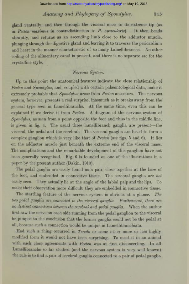
Up to this point the anatomical features indicate the close relationship of Pecten and Spondylus, and, coupled with certain palaeontological data, make it extremely probable that Spondylus arose from Pecten ancestors. The nervous system, however, presents a real surprise, inasmuch as it breaks away from the general type seen in Lamellibranchs. At the same time, even this can be explained if we derive it from Pecten. A diagram of the nervous system of Spondylus, as seen from a point opposite the foot and thus in the middle line, is given in fig. 4. The usual three lamellibranch ganglia are present—the visceral, the pedal and the cerebral. The visceral ganglia are fused to form a complex ganglion which is very like that of Pecten (see figs. 5 and 6). It lies on the adductor muscle just beneath the extreme end of the visceral mass. The complications and the remarkable development of this ganglion have not been generally recognised. Fig. 6 is founded on one of the illustrations in a paper by the present author (Dakin, 1910).
The pedal ganglia are easily found as a pair, close together at the base of the foot, and embedded in connective tissue. The cerebral ganglia are not easily seen. They actually lie at the angle of the labial palp and the lips. To make their observation more difficult they are embedded in connective tissue.
The startling feature of the nervous system is obvious at a glance. The two pedal ganglia are connected to the visceral ganglia. Furthermore, there are no distinct connectives between the cerebral and pedal ganglia. When the author first saw the nerve on each side running from the pedal ganglion to the visceral he jumped to the conclusion that the former ganglia could not be the pedal at all, because such a connection would be unique in Lamellibranchiata.
Had such a thing occurred in Teredo or some other more or less highly modified form it would not have been surprising. To meet it in an animal with such close agreements with Pecten was at first disconcerting. In all Lamellibranchs so far studied (and the nervous system is very well known) the rule is to find a pair of cerebral ganglia connected to a pair of pedal ganglia
on May 19, 2018http://rspb.royalsocietypublishing.org/Downloaded from

346 W. J. Dakin.
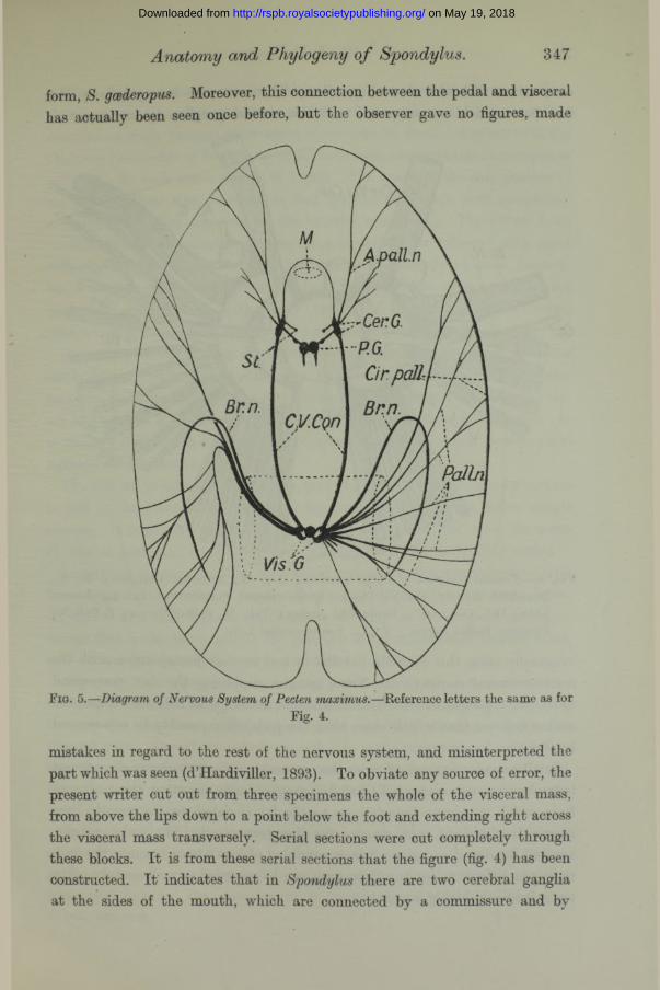
and also connected to a pair of visceral ganglia. This state of affairs is illustrated in the diagram of the nervous system of Pecten (fig. 5). I t is
Fig. 4.—Diagram of Nervous System of Spondylus. A. pall.n., Anterior pallial nerve, Add.M., Adductor muscle; Br.n., Branchial nerve; C.V.Con., Cerebro-visceral connective; Cer. G., Cerebral Ganglion; Cir. pall., Circum-pallial nerve; M.,Mouth ; P.G., Pedal Ganglion ; P.Y.C., Pedo-visceral connectives ; Pall, n., Pallial nerves ; St., Statocyst; Vis. G., Visceral Ganglion.
perfectly well known to all students who study Anodon as a class type and complete their studies later with other typical Lamellibranchs.
There can be no doubt about this extraordinary state of affairs in Spondylus. The author has dissected out the pedo-visceral connective in an American species as well as in a considerable number of specimens of the Mediterranean
on May 19, 2018http://rspb.royalsocietypublishing.org/Downloaded from
Anatomy and Phylogeny o f Spondylus. 347
form, S. gcederopus. Moreover, this connection between the pedal and visceral has actually been seen once before, but the observer gave no figures, made
A palln
Fig. 5.—Diagram of Nervous System of Pecten maximus.—Reference letters the same as forFig. 4.
mistakes in regard to the rest of the nervous system, and misinterpreted the part which was seen (d’Hardiviller, 1893). To obviate any source of error, the present writer cut out from three specimens the whole of the visceral mass, from above the lips down to a point below the foot and extending right across the visceral mass transversely. Serial sections were cut completely through these blocks. I t is from these serial sections that the figure (fig. 4) has been constructed. I t indicates that in Spondylus there are two cerebral ganglia at the sides of the mouth, which are connected by a commissure and by
on May 19, 2018http://rspb.royalsocietypublishing.org/Downloaded from
348 W. J. Dakin.
connectives with the visceral ganglion. There are also two ganglia in the foot base, which are connected with the visceral ganglion by nerves which
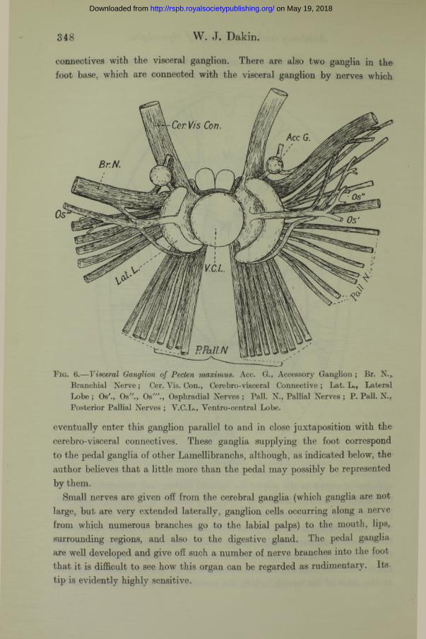
Fig. 6.— Visceral Ganglion of Pecten maximus. Acc. G., Accessory Ganglion; Br. N., Branchial Nerve; Cer. Vis. Con., Cerebro-visceral Connective ; Lat. L., Lateral Lobe ; Os'., Os"., Os"'., Osphradial Nerves ; Pall. N., Pallial Nerves ; P. Pall. N., Posterior Pallial Nerves ; V.C.L., Ventro-central Lobe.
eventually enter this ganglion parallel to and in close juxtaposition with the cerebro-visceral connectives. These ganglia supplying the foot correspond to the pedal ganglia of other Lamellibranchs, although, as indicated below, the author believes that a little more than the pedal may possibly be represented by them.
Small nerves are given off from the cerebral ganglia (which ganglia are not large, but are very extended laterally, ganglion cells occurring along a nerve from which numerous branches go to the labial palps) to the mouth, lips, surrounding regions, and also to the digestive gland. The pedal ganglia are well developed and give off such a number of nerve branches into the foot that it is difficult to see how this organ can be regarded as rudimentary. Its tip is evidently highly sensitive.
on May 19, 2018http://rspb.royalsocietypublishing.org/Downloaded from

Anatomy and Phylogeny o f Spondylus. 349
There are no separate cerebro-pedal connectives of the usual type. This is unique, and since d’Hardiviller stated that he saw them, the present writer searched carefully through three series of sections for them. Any nerve of the thickness just visible in dissections would have been unmistakable in microscope sections. No such nerve was to be found. The result of the examination of the serial sections was, however, to find the two statocysts, both completely enclosed vesicles (no aperture to the exterior remaining). The nerves from the statocysts were exceedingly minute and only to be followed with great difficulty in the serial sections. Their course is naturally of great interest, for, if cerebro-pedal connectives had been present, these nerves would most likely have connected up with them. Instead of that, the statocyst nerves, after a rather long course, run into the cerebro-visceral connectives.
I t is not possible to deny altogether the passage of nerve fibres from the cerebral to the pedal ganglia other than by the long path through the visceral ganglion, because extremely fine nerve branches, consisting of a few fibres only, are to be found with the microscope here and there in the visceral mass. It is unlikely that such a connection exists, but in any case typical cerebro-pedal connectives are absent.
The visceral ganglion (fig. 7) is, like that of Pecten, a most unusual structure to find in the Lamellibranchs, where the ganglia are as a rule rather simple masses. I t might be termed the “ brain ” of the animal, for, from the point of view of both size, complexity and functions, it is the controlling centre.
Looking at the ganglion from the ventral surface one sees a prominent central lobe occupying the region of greatest thickness and two lateral crescentic lobes. (It will be seen that fundamentally this is the same as Pecten maximus, fig. 6, except that in the latter species the ganglion is asymmetrical, a feature which may be associated with the asymmetry of the Mollusc.)
Anteriorly (or perhaps one might better put it, orally) two large nerves leave the ganglion on each side. These nerves arise from the central mass and pass towards the hinge line on the surface of the adductor muscle just below the visceral mass. One of the nerves on each side is the pedo-visceral connective, the other is a nerve which splits later on, part going to the cerebral ganglion and the other part continuing as the Anterior Pallial nerve to enter the mantle margin near the hinge line.
In Pecten only one large nerve on each side is to be found, the cerebro-visceral connective (fig. 6, Cer. Vis. Con.). Between the cerebro-visceral connectives as they leave the visceral ganglion are two smaller ganglionic lobes, usually pigmented brown. The same region is present in Spondylus. The figure
VOL. CIII.—B. 2 C
on May 19, 2018http://rspb.royalsocietypublishing.org/Downloaded from

350 W. J. Dakiu.
of the visceral ganglion of Spondylus given in the text is not, however, of such accuracy as that of Pecten. The present research was not carried out as a
J i ^
Pig. 7.— Visceral Ganglion of Spondylus. Acc. G., Accessory Ganglion ; Br. N. ; Branchial Nerve ; C.P.V.C., Cerebropallial-visceral connective ; Lat. L., Lateral Lobe ; Os. Os'., Osphradial Nerves ; Pall. N., Pallial Nerves ; P. Pall. N., Posterior Pallial Nerves ; Pe. V.O., Pedo-visceral connective ; V.C.L., Ventro-central Lobe.
special investigation of the visceral ganglion, and the rarity of the material has made matters difficult. The Spondylus figure is the result of several very careful dissections. The Pecten figure resulted from a long study of serial sections and dissections. The fundamental features of the ganglion of Spondylus are, however, accurately depicted.
Two other small accessory ganglia remain to be mentioned ; they are visible in both genera. In Pecten they are connected both with the visceral ganglion and also with the branchial nerves. Another branch leaves them for the viscera. In Spondylus, the small connection with the branchial nerve has not been observed.
The following nerves leave the visceral ganglion, apart from the cerebro- visceral connectives (and in Spondylus the pedo-visceral connectives) referred to above—the branchial nerves, the osphradial nerves and the pallial nerves.
on May 19, 2018http://rspb.royalsocietypublishing.org/Downloaded from

Anatomy and Phytogeny o f Spondylus. 351
The branchial nerves leave the ganglion in both Pecten and Spondylus near the point of entry of the cerebro-visceral connectives.
The osphradial nerves are peculiar and very characteristic. In both genera they arise from the face of the ganglion exposed to view and from what is apparently the central lobe. As a result each nerve crosses over the lateral lobe of its side. A little way from the ganglion it sends a branch into the branchial nerve and later still branches leave this nerve for the osphradium.
The pallial nerves of Pecten and Spondylus are particularly well represented, a fact to be correlated with the highly developed structure, including the eyes, tentacles and musculature, of the mantle edges. From the visceral ganglion there are posterior pallial nerves and lateral pallial nerves, and in Spondylus there is also an anterior pallial nerve, although at its commencement it is not distinguishable from the cerebro-visceral. connective. All these pallial nerves are connected to a circular nerve which runs round in the mantle margins (see figs. 4 and 5, Cir. pall.).
Finally, nerves to the adductor muscle leave the visceral ganglion from the surface apposed to the muscle. They cannot be seen in the figures of the ganglion.
The internal structure of this complex ganglion has been described by Dakin (1910), and it has been shown that various tracts of fibres can be clearly made out in it. Reference should be made to the original paper for details. The structure of the lateral lobes of the visceral ganglion deserves mention, however, for these bodies appear to be characteristic of Pecten and Spondylus. The pallial nerves arise from their under surface and although some of the fibres of these nerves come from the more central parts of the ganglion and some actually from the cerebro-visceral connective, a considerable part comes from the lateral lobes. In fact, one may go further, and correlate the lateral lobes with the presence of the eyes. This might explain the difference between the size of the lobes of the right and left sides in both Pecten maximus and P. jacobceus, where the eyes are much more numerous on one mantle lobe than on the other.
A Possible Explanation of the Nervous System of Spondylus.The question which now remains is the most interesting. Is it possible to
explain the unusual features of the nervous system of Spondylus and to bring it in line with that of other Lamellibranchs ? In my opinion this is not difficult, if the conditions found in Pecten be used as the key.
If the diagram of the Pecten nervous system (fig. 5) is studied, it will be noted, 2 c 2
on May 19, 2018http://rspb.royalsocietypublishing.org/Downloaded from
352 W. J. Dakin.
that the cerebral ganglia are very close indeed to the pedal ganglia, and at the same time some distance away from their more usual position at the sides of the mouth. In consequence of this, the cerebral commissure is unusually long, and has to travel some distance to pass over the oesophagus. This feature has frequently been pointed out (Pelseneer and others) as rather characteristic of Pecten. Now in Spondylus, we must either explain the pedo-visceral connective as something new, which has suddenly appeared in this mollusc, which in so many other respects is a Pecten, or else we must look for the nerve in Pecten itself.
The clues are, to my mind, provided by the facts that in Spondylus (1) the cerebral ganglia are to be found in their normal position at the sides of the mouth and reduced in size ; (2) a considerable part of the cerebro-visceral connective splits off just before reaching the cerebral ganglion and proceeds straight on, as an anterior pallial nerve to the mantle, near the hinge line. This latter fact points to the conclusion that in Pecten the pallial nerve from the cerebral ganglion does not really arise there at a ll; it only appears to do so because its fibres run from the visceral ganglion together with fibres which belong to the cerebral ganglion.
In Spondylus, however, for some reason or other, there is a tendency for nerves to split into their components. If we suppose that the cerebral ganglion itself has split (on each side), and that one part has remained with the pedal ganglion, whilst the other part has receded to the more normal position at the mouth, everything can be clearly explained. Each pedal ganglion would then have to be regarded as including part of the cerebral, whilst the cerebral ganglion proper would only be part of the ganglion bearing that name in Pecten.
It would then follow quite naturally that the “ pedal ” ganglion of Spondylus would be connected with the visceral, for the pedo-visceral connective would really be part of the cerebro-visceral connective of Pecten and all other Lamellibranchs. At first, this may seem to be rather difficult to understand, but the more one looks into the Lamellibranch system, and the more familiar one is with the nervous system of Pecten, the more plausible does it become.
This is not all however. In the Gastropoda there is a distinct ganglion, termed the pleural. This is usually supposed to be part of the cerebral of the Lamellibranchiata, and there is strong evidence for such an assumption. (In Pecten the cerebral ganglion on each side consists of two obvious lobes.) Now in some Gastropods, the pleural remains close to the cerebral ganglion. In others, it is situated closer to the pedal, and the pleuro-visceral connective
on May 19, 2018http://rspb.royalsocietypublishing.org/Downloaded from

Anatomy and Phylogeny o f Spondylus. 353
is then almost a nerve from the pedal to the visceral ganglion. I t is possible that the splitting of the cerebral (or cerebro-pleural) ganglion, which I believe has taken place in Spondylus, has left the pleural part of this nerve centre attached to the pedal ganglion. What we must call the pedo-visceral connective of Spondylus is then to be regarded as a strand of nerve fibres represented in Pecten and other Lamellibranchs by part of the cerebro-visceral connective.
One other feature of the Spondylus nervous system remains to be explained. There is no typical direct connection (cerebro-pedal connectives) between the cerebral ganglia at the lip-mouth angle and the ganglia of the foot. The only hypothesis which I can suggest is that such a direct connection may be quite unnecessary, if part of the cerebral (or the pleural) ganglion is really present in this form as part of the large foot ganglion. That is my theory. I t supports most strongly the suggestions that the cerebro-visceral connective of Pecten has split, giving rise to the two connectives of Spondylus ; and if such were not the case, i.e., if the pedo-visceral connective of Spoyidylus were a new thing, a connection not even represented in the Mollusca, then one would also have to explain the entire absence of a cerebro-pedal connective. Thus, instead of this unique disposition of a Lamellibranch nervous system being a puzzle, it provides most interesting evidence for the evolution of the Spondylidae.
Sense organs.The eyes of Spondylus are discussed in the paper following. The other
sense organs consist of the Osphradia, the Statocysts, the Abdominal Sense Organ and sensory cells and free nerve endings on gills, palps, foot and probably other parts of the surface of the body. The only organs of this group it is necessary to comment upon here are the statocysts. Although very small they are both present. This is quite in accordance with the fact that they are present in certain other fixed Lamellibranchs. I t has been generally assumed that, although the Lamellibranch statocysts are often connected to the pedal ganglia, or to the cerebro-pedal connectives near the pedal ganglia, their nerve supply comes really from the cerebral ganglia. The state of affairs in Spondylus lends support to this view, for, had the nerve supply been from the pedal ganglia, one might have expected the statocyst nerves to run directly into them.
The Excretory and Reproductive Organs.There is no reason to go into details regarding the structure of these two
systems. They are not essentially very different morphologically from the corresponding organs of Pecten. Unfortunately, all the specimens except one
on May 19, 2018http://rspb.royalsocietypublishing.org/Downloaded from
354 Anatomy and Phylogeny of Spondylus.
were taken in non-breeding seasons and the gonads were thus in the reduced state. They lie in the visceral mass beneath the foot and, as stated previously, slightly invade the mantle in the region where they abut on the digestive gland. The animals are probably unisexual, but I should not like to be dogmatic upon this point. I t is worthy of note that some species of Pecten are unisexual (P. tenuicostatus of America), whilst P. maximus and P. opercularis, our common English species, are hermaphrodite.
Conclusion.
The anatomy of Spondylus has been investigated in order to obtain evidence of the phylogeny of the genus, with a view to a discussion of the evolution of the eyes of Pecten and Spondylus. Jackson, in his classic work on the Phylogeny of the Pelecypoda, concluded that Spondylus had been evolved from Pecten. This theory was based entirely upon a study of the shell and upon palaeontological discoveries—Pecten dates at least from Carboniferous times, Spondylus from the Triassic beds. The present work has not only shown that the anatomical evidence is overwhelmingly in support of this view, but it has indicated the line which has been followed, and shown how the more equivalve species of Pecten, like P. opercularis, represent the earlier and more primitive type, and that the direction which culminated in Spondylus was that which can be seen in the Pectinidae, leading from P. opercularis through P. jacobceus and P. maximus.
The surprising discovery in relation to the nervous system shows that Spondylus must be regarded as the extreme of an evolutionary line, which probably commenced with the Aviculopectens of Palaeozoic times.
Literature.
Dakin, W. J., 1909. “ Memoir on Pecten,” 4 Liverpool Marine Biology Committee Memoir XVII,’ Liverpool.
Dakin, W. J., 1910. “ The visceral ganglion of Pecten, with notes on the physiology of the system, etc.,” * Mitt. Zool. Stat. z. Neapel,’ Band 20, Heft i.
Fischer, P., 1887. 4 Manuel de Conchyliologie,’ Paris.d’Hardiviller, A., 1893. 44 Sur quelques faits qui permettent de rapprocher le systeme
nerveux des Lamellibranches de celui des Gasteropodes,” 4 C. R. Acad, des Sci.,’ T. 117.
Jackson, R. T. J., 1889. 44 Phylogeny of the Pelecypoda,” 4 Boston Journal of NaturalHistory,’ New Ser., vol. 4 (1886-1893).
Ridewood, W. G., 1903. 44 Gills of Lamellibranchiata,” 4 Phil. Trans.,’ B, vol. 195.
on May 19, 2018http://rspb.royalsocietypublishing.org/Downloaded from