Embed Size (px)
Citation preview
83
In years past, the prevailing approach to providing pain control was focused on identifying underlying etiologies or pathologic syndromes, e.g., low back pain, trigeminal neuralgia, and cancer pain, that produce the pain. While treating the pre-sumed source of the pain, attempts to improve the accompanying discomfort relied largely on the use of non-opioid medications and the limited use of opioid and adju-vant analgesics. Over the past 25 years, however, there has been a dramatic increase
H. J. Gould III , MD, PhD (*) Department of Neurology and Neuroscience, Louisiana State University Health Sciences Center , New Orleans , LA 70112 , USAe-mail: [email protected]
A. D. Kaye , MD, PhD Interventional Pain Management, University Hospital and Ochsner Kenner Hospital , Kenner , LA 70112 , USA
Department of Anesthesiology, Louisiana State University School of Medicine , New Orleans , LA 70112, USA
Department of Pharmacology, Louisiana State University School of Medicine , New Orleans , LA 70112, USA
The Anatomy of Pain
Harry J. Gould III • Alan David Kaye
A.D. Kaye et al. (eds.), Essentials of Regional Anesthesia, DOI 10.1007/978-1-4614-1013-3_4, © Springer Science+Business Media, LLC 2012
4
Contents
Considerations of General Organization .................................................................................... 84Stimulus Transduction and Transmission .................................................................................. 90Stimulus Modulation .................................................................................................................. 101Stimulus Perception and Interpretation ...................................................................................... 105Stimulus Modulation and Behavioral Response ........................................................................ 107Pathway Alterations Following Injury ....................................................................................... 110Multiple-Choice Questions ........................................................................................................ 116References .................................................................................................................................. 119
84 H.J. Gould III and A.D. Kaye
in our understanding of the nervous system and how stimuli associated with actual or potential tissue injury are transduced, transmitted, modulated, perceived, and interpreted to form the basis for initiating appropriate evasive or protective behav-ior, thereby avoiding or limiting injury. Our current bank of knowledge has led to the recognition that (1) pain in the chronic state is in itself a disease deserving con-sideration, assessment, and management, (2) pain is not a single entity but a com-plex, multifaceted experience that warrants detailed and comprehensive evaluation to elucidate symptoms that may refl ect specifi c associated mechanisms amenable to targeted treatment [ 1, 2 ] , and (3) treatment modalities and management approaches not heretofore considered can be effective and can improve the quality of life for those suffering with pain. This chapter will provide a brief overview of the anatomy of pain that forms the basis for current practice.
Considerations of General Organization
The somatosensory system provides the means through which living organisms explore and monitor the body’s external and internal environment in order to recognize changes that may be benefi cial and embraced or detrimental to survival and avoided.
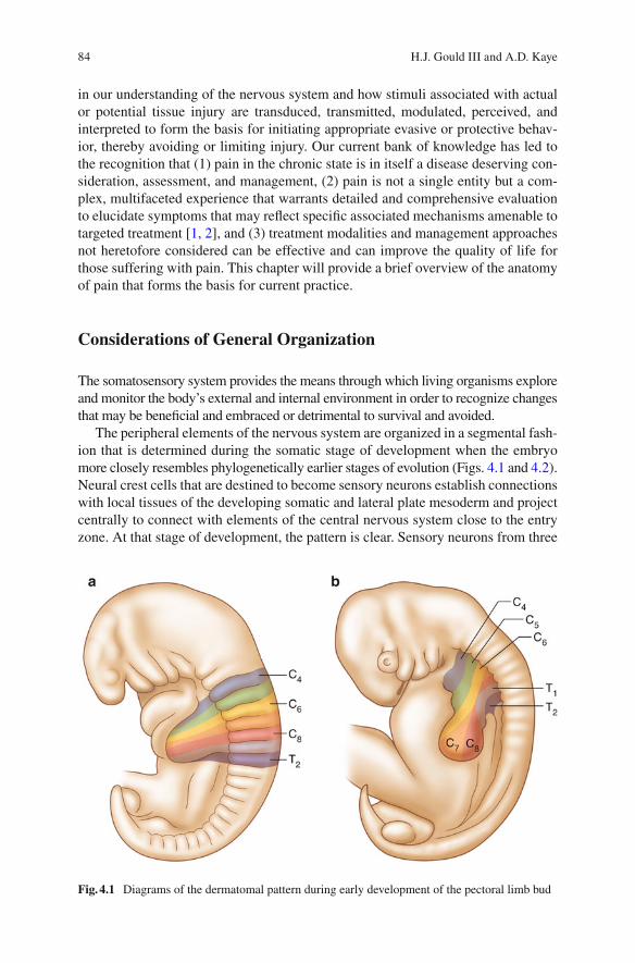
The peripheral elements of the nervous system are organized in a segmental fash-ion that is determined during the somatic stage of development when the embryo more closely resembles phylogenetically earlier stages of evolution (Figs. 4.1 and 4.2 ). Neural crest cells that are destined to become sensory neurons establish connections with local tissues of the developing somatic and lateral plate mesoderm and project centrally to connect with elements of the central nervous system close to the entry zone. At that stage of development, the pattern is clear. Sensory neurons from three
Fig. 4.1 Diagrams of the dermatomal pattern during early development of the pectoral limb bud
854 The Anatomy of Pain
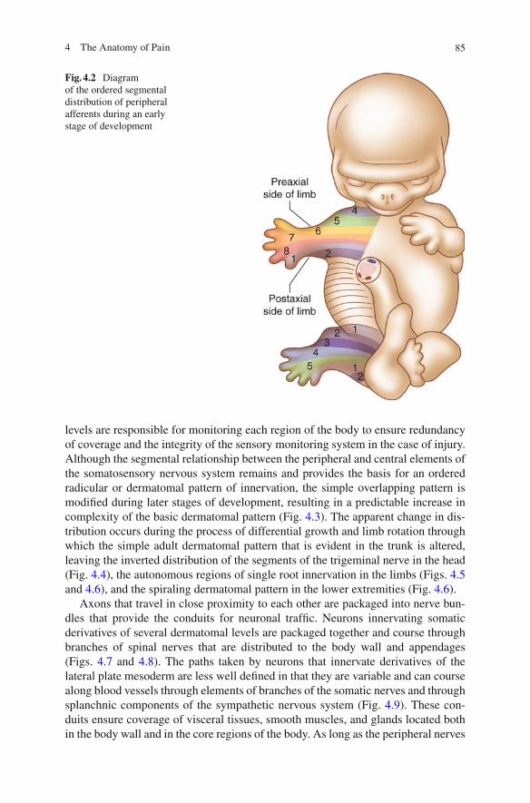
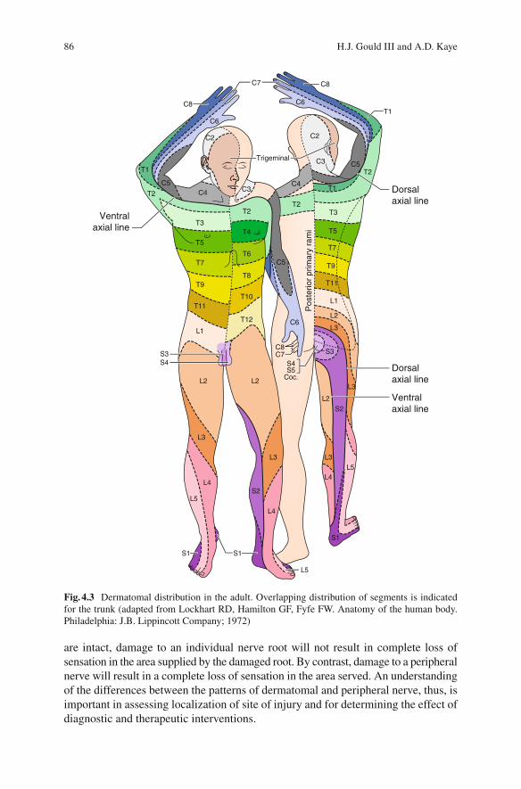
levels are responsible for monitoring each region of the body to ensure redundancy of coverage and the integrity of the sensory monitoring system in the case of injury. Although the segmental relationship between the peripheral and central elements of the somatosensory nervous system remains and provides the basis for an ordered radicular or dermatomal pattern of innervation, the simple overlapping pattern is modifi ed during later stages of development, resulting in a predictable increase in complexity of the basic dermatomal pattern (Fig. 4.3 ). The apparent change in dis-tribution occurs during the process of differential growth and limb rotation through which the simple adult dermatomal pattern that is evident in the trunk is altered, leaving the inverted distribution of the segments of the trigeminal nerve in the head (Fig. 4.4 ), the autonomous regions of single root innervation in the limbs (Figs. 4.5 and 4.6 ), and the spiraling dermatomal pattern in the lower extremities (Fig. 4.6 ).
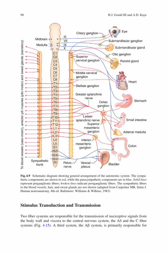
Axons that travel in close proximity to each other are packaged into nerve bun-dles that provide the conduits for neuronal traffi c. Neurons innervating somatic derivatives of several dermatomal levels are packaged together and course through branches of spinal nerves that are distributed to the body wall and appendages (Figs. 4.7 and 4.8 ). The paths taken by neurons that innervate derivatives of the lateral plate mesoderm are less well defi ned in that they are variable and can course along blood vessels through elements of branches of the somatic nerves and through splanchnic components of the sympathetic nervous system (Fig. 4.9 ). These con-duits ensure coverage of visceral tissues, smooth muscles, and glands located both in the body wall and in the core regions of the body. As long as the peripheral nerves
Fig. 4.2 Diagram of the ordered segmental distribution of peripheral afferents during an early stage of development
86 H.J. Gould III and A.D. Kaye
are intact, damage to an individual nerve root will not result in complete loss of sensation in the area supplied by the damaged root. By contrast, damage to a peripheral nerve will result in a complete loss of sensation in the area served. An understanding of the differences between the patterns of dermatomal and peripheral nerve, thus, is important in assessing localization of site of injury and for determining the effect of diagnostic and therapeutic interventions.
Ventralaxial line
Trigeminal
Dorsalaxial line
Dorsalaxial line
Pos
terio
r pr
imar
y ra
mi
Ventralaxial line
T2
T2
T2T2
T4
T6
T8
T10
T12
C5
C5
C6
C5
T1
T1
T3
T3
T5
T5
T7
T9
T11
L1
L2
L3
T7
T9
T11
L1
S3S4
L2L2
L3
L3 L3
L2
L3
S3
S4S5
Coc.
S2
L4
L4
L4L5
L5
L5S2
S1
S1
S1
T1
C2 C2
C3
C3C4
C4
C6
C6
C7
C8
C7C8
C8
Fig. 4.3 Dermatomal distribution in the adult. Overlapping distribution of segments is indicated for the trunk (adapted from Lockhart RD, Hamilton GF, Fyfe FW. Anatomy of the human body. Philadelphia: J.B. Lippincott Company; 1972)
874 The Anatomy of Pain
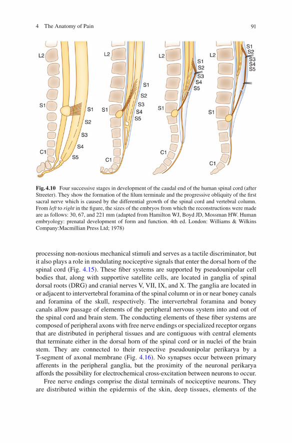
Differential growth also results in an important disparity between boney vertebral levels, the location of the dorsal root ganglia, the location of the caudal end of the spinal cord, and the dorsal root entry zone of the spinal cord observed at different stages of development and in the adult. Figure 4.10 depicts the changes in the rela-tive relationship between neural and boney elements from early stages in develop-ment (30, 67, and 111 mm) to shortly after birth (221 mm). In the adult, the relative
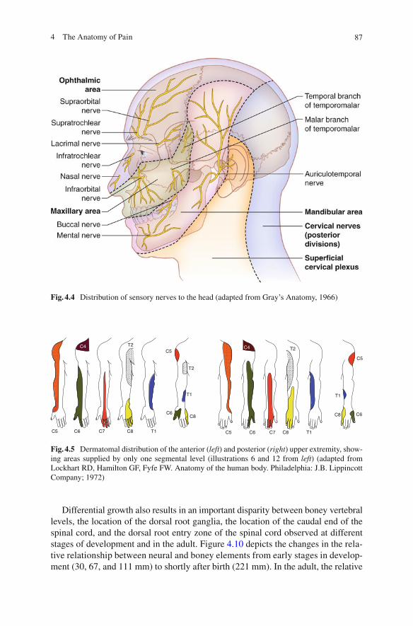
Fig. 4.4 Distribution of sensory nerves to the head (adapted from Gray’s Anatomy, 1966)
C5 C6 C7 C8
C6C8
C5 C6 C7 C8 T1
C8
T1
C6
C5
T1
T2
C5C4 T2
T2
T1
C4
Fig. 4.5 Dermatomal distribution of the anterior ( left ) and posterior ( right ) upper extremity, show-ing areas supplied by only one segmental level (illustrations 6 and 12 from left ) (adapted from Lockhart RD, Hamilton GF, Fyfe FW. Anatomy of the human body. Philadelphia: J.B. Lippincott Company; 1972)
88 H.J. Gould III and A.D. Kaye
L2
L2
L5
L5
L4
L3
L2
S2
S1
S1
S2
L3
L4
S3
L5
L2
L4
S1
S2
S1
S2
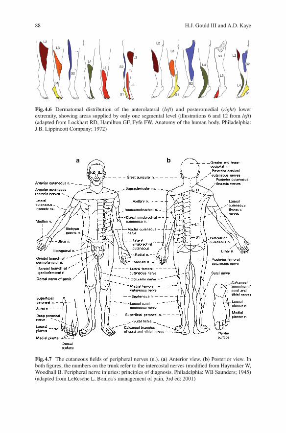
Fig. 4.6 Dermatomal distribution of the anterolateral ( left ) and posteromedial ( right ) lower extremity, showing areas supplied by only one segmental level (illustrations 6 and 12 from left ) (adapted from Lockhart RD, Hamilton GF, Fyfe FW. Anatomy of the human body. Philadelphia: J.B. Lippincott Company; 1972)
Fig. 4.7 The cutaneous fi elds of peripheral nerves (n.). ( a ) Anterior view. ( b ) Posterior view. In both fi gures, the numbers on the trunk refer to the intercostal nerves (modifi ed from Haymaker W, Woodhall B. Peripheral nerve injuries: principles of diagnosis. Philadelphia: WB Saunders; 1945) (adapted from LeResche L, Bonica’s management of pain, 3rd ed; 2001)
894 The Anatomy of Pain
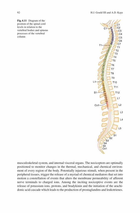
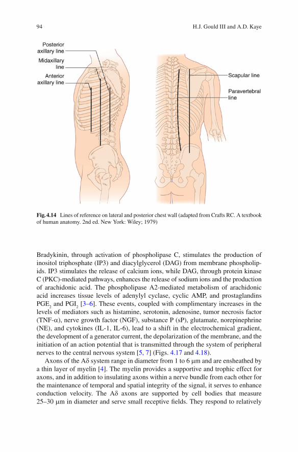
disparity between level of the spinal nerve and its respective entry into the spinal cord generally follows the following formula: vertebral level (vertebral spinous pro-cess) + n = spinal cord level, where n = 0 for the upper cervical region, 1 between the lower cervical and upper thoracic region (vertebral prominence), 2 between T3 and T9, and 3 between T9 and T11 (Fig. 4.11 ). The conus medullaris is located between the spinous processes of the T12–L2 vertebrae. Figures 4.12 – 4.14 depict boney landmarks and lines of reference to aid in identifying vertebral levels. An under-standing of the disparity and knowledge of superfi cial landmarks is important for guiding and determining the best approaches for performing interventions on indi-vidual nerve roots and spinal cord levels. For example, the knowledge that the adult spinal cord extends inferiorly only to the L2–L3 vertebrae offers a degree of safety when inserting needles for obtaining spinal fl uid from the lumbar cistern when the approach is made below the L3 vertebral level.
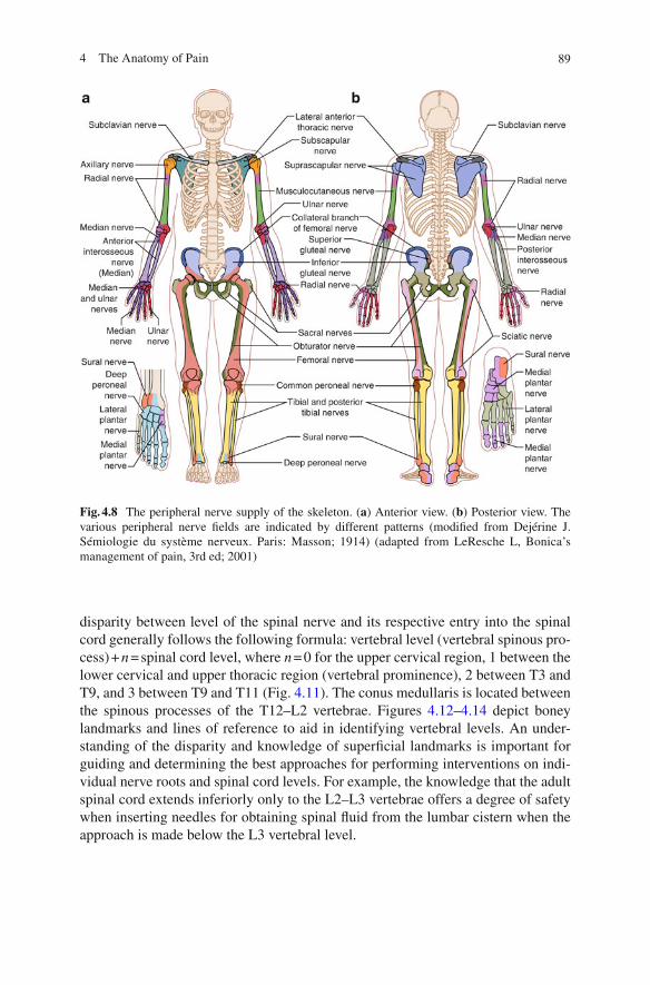
Fig. 4.8 The peripheral nerve supply of the skeleton. ( a ) Anterior view. ( b ) Posterior view. The various peripheral nerve fi elds are indicated by different patterns (modifi ed from Dejérine J. Sémiologie du système nerveux. Paris: Masson; 1914) (adapted from LeResche L, Bonica’s management of pain, 3rd ed; 2001)
90 H.J. Gould III and A.D. Kaye
Stimulus Transduction and Transmission
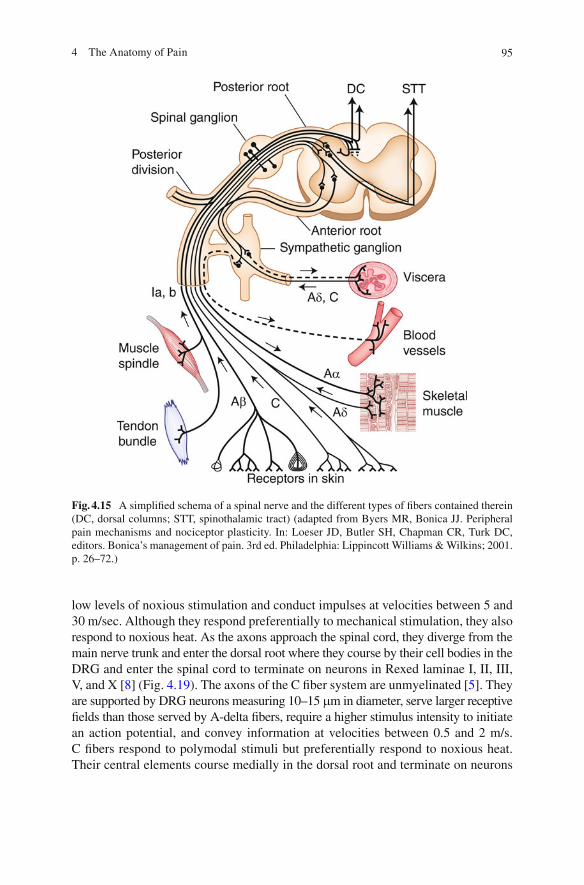
Two fi ber systems are responsible for the transmission of nociceptive signals from the body wall and viscera to the central nervous system, the A d and the C fi ber systems (Fig. 4.15 ). A third system, the A b system, is primarily responsible for
Fig. 4.9 Schematic diagram showing general arrangement of the autonomic system. The sympa-thetic components are shown in red , while the parasympathetic components are in blue . Solid lines represent preganglionic fi bers; broken lines indicate postganglionic fi bers. The sympathetic fi bers to the blood vessels, hair, and sweat glands are not shown (adapted from Carpenter MB, Sutin J. Human neuroanatomy. 8th ed. Baltimore: Williams & Wilkins; 1983)
914 The Anatomy of Pain
processing non-noxious mechanical stimuli and serves as a tactile discriminator, but it also plays a role in modulating nociceptive signals that enter the dorsal horn of the spinal cord (Fig. 4.15 ). These fi ber systems are supported by pseudounipolar cell bodies that, along with supportive satellite cells, are located in ganglia of spinal dorsal roots (DRG) and cranial nerves V, VII, IX, and X. The ganglia are located in or adjacent to intervertebral foramina of the spinal column or in or near boney canals and foramina of the skull, respectively. The intervertebral foramina and boney canals allow passage of elements of the peripheral nervous system into and out of the spinal cord and brain stem. The conducting elements of these fi ber systems are composed of peripheral axons with free nerve endings or specialized receptor organs that are distributed in peripheral tissues and are contiguous with central elements that terminate either in the dorsal horn of the spinal cord or in nuclei of the brain stem. They are connected to their respective pseudounipolar perikarya by a T-segment of axonal membrane (Fig. 4.16 ). No synapses occur between primary afferents in the peripheral ganglia, but the proximity of the neuronal perikarya affords the possibility for electrochemical cross-excitation between neurons to occur.
Free nerve endings comprise the distal terminals of nociceptive neurons. They are distributed within the epidermis of the skin, deep tissues, elements of the
Fig. 4.10 Four successive stages in development of the caudal end of the human spinal cord (after Streeter). They show the formation of the fi lum terminale and the progressive obliquity of the fi rst sacral nerve which is caused by the differential growth of the spinal cord and vertebral column. From left to right in the fi gure, the sizes of the embryos from which the reconstructions were made are as follows: 30, 67, and 221 mm (adapted from Hamilton WJ, Boyd JD, Mossman HW. Human embryology: prenatal development of form and function. 4th ed. London: Williams & Wilkins Company:Macmillian Press Ltd; 1978)
92 H.J. Gould III and A.D. Kaye
musculoskeletal system, and internal visceral organs. The nociceptors are optimally positioned to monitor changes in the thermal, mechanical, and chemical environ-ment of every region of the body. Potentially injurious stimuli, when present in the peripheral tissues, trigger the release of a myriad of chemical mediators that set into motion a constellation of events that alters the membrane permeability of afferent nerve terminals to charged ions. Among the inciting nociceptive events are the release of potassium ions, protons, and bradykinin and the initiation of the arachi-donic acid cascade which leads to the production of prostaglandins and leukotrienes.
Fig. 4.11 Diagram of the position of the spinal cord levels in relation to the vertebral bodies and spinous processes of the vertebral column
934 The Anatomy of Pain
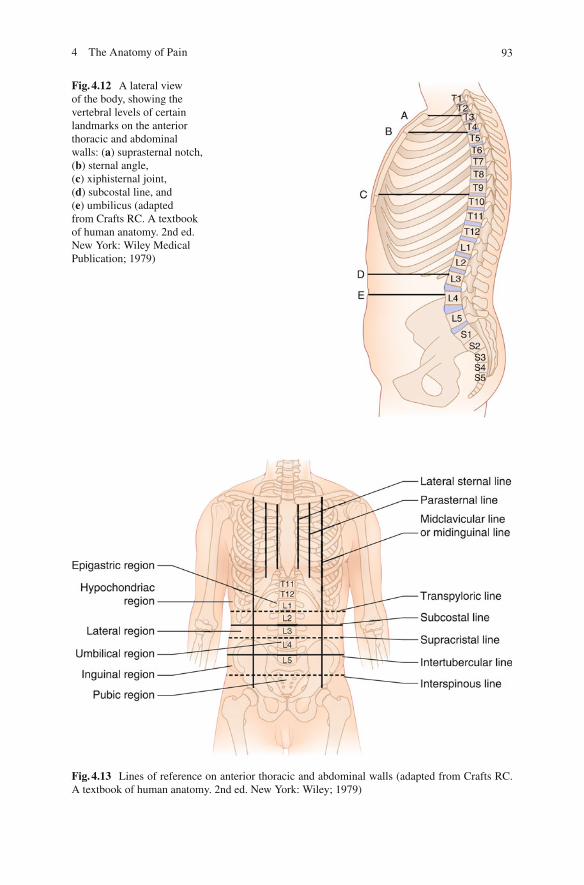
Fig. 4.12 A lateral view of the body, showing the vertebral levels of certain landmarks on the anterior thoracic and abdominal walls: ( a ) suprasternal notch, ( b ) sternal angle, ( c ) xiphisternal joint, ( d ) subcostal line, and ( e ) umbilicus (adapted from Crafts RC. A textbook of human anatomy. 2nd ed. New York: Wiley Medical Publication; 1979)
Fig. 4.13 Lines of reference on anterior thoracic and abdominal walls (adapted from Crafts RC. A textbook of human anatomy. 2nd ed. New York: Wiley; 1979)
94 H.J. Gould III and A.D. Kaye
Bradykinin, through activation of phospholipase C, stimulates the production of inositol triphosphate (IP3) and diacylglycerol (DAG) from membrane phospholip-ids. IP3 stimulates the release of calcium ions, while DAG, through protein kinase C (PKC)-mediated pathways, enhances the release of sodium ions and the production of arachidonic acid. The phospholipase A2-mediated metabolism of arachidonic acid increases tissue levels of adenylyl cyclase, cyclic AMP, and prostaglandins PGE
2 and PGI
2 [ 3– 6 ] . These events, coupled with complimentary increases in the
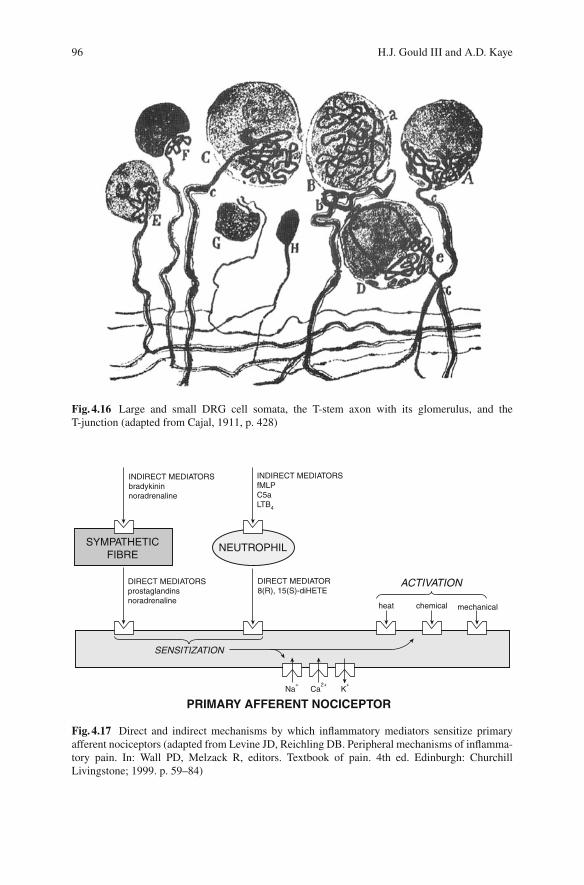
levels of mediators such as histamine, serotonin, adenosine, tumor necrosis factor (TNF- a ), nerve growth factor (NGF), substance P (sP), glutamate, norepinephrine (NE), and cytokines (IL-1, IL-6), lead to a shift in the electrochemical gradient, the development of a generator current, the depolarization of the membrane, and the initiation of an action potential that is transmitted through the system of peripheral nerves to the central nervous system [ 5, 7 ] ( Figs. 4.17 and 4.18 ).
Axons of the A d system range in diameter from 1 to 6 m m and are ensheathed by a thin layer of myelin [ 4 ] . The myelin provides a supportive and trophic effect for axons, and in addition to insulating axons within a nerve bundle from each other for the maintenance of temporal and spatial integrity of the signal, it serves to enhance conduction velocity. The A d axons are supported by cell bodies that measure 25–30 m m in diameter and serve small receptive fi elds. They respond to relatively
Fig. 4.14 Lines of reference on lateral and posterior chest wall (adapted from Crafts RC. A textbook of human anatomy. 2nd ed. New York: Wiley; 1979)
954 The Anatomy of Pain
low levels of noxious stimulation and conduct impulses at velocities between 5 and 30 m/sec. Although they respond preferentially to mechanical stimulation, they also respond to noxious heat. As the axons approach the spinal cord, they diverge from the main nerve trunk and enter the dorsal root where they course by their cell bodies in the DRG and enter the spinal cord to terminate on neurons in Rexed laminae I, II, III, V, and X [ 8 ] (Fig. 4.19 ). The axons of the C fi ber system are unmyelinated [ 5 ] . They are supported by DRG neurons measuring 10–15 m m in diameter, serve larger receptive fi elds than those served by A-delta fi bers, require a higher stimulus intensity to initiate an action potential, and convey information at velocities between 0.5 and 2 m/s. C fi bers respond to polymodal stimuli but preferentially respond to noxious heat. Their central elements course medially in the dorsal root and terminate on neurons
Fig. 4.15 A simplifi ed schema of a spinal nerve and the different types of fi bers contained therein (DC, dorsal columns; STT, spinothalamic tract) (adapted from Byers MR, Bonica JJ. Peripheral pain mechanisms and nociceptor plasticity. In: Loeser JD, Butler SH, Chapman CR, Turk DC, editors. Bonica’s management of pain. 3rd ed. Philadelphia: Lippincott Williams & Wilkins; 2001. p. 26–72.)
96 H.J. Gould III and A.D. Kaye
Fig. 4.16 Large and small DRG cell somata, the T-stem axon with its glomerulus, and the T-junction (adapted from Cajal, 1911, p. 428)
INDIRECT MEDIATORSbradykininnoradrenaline
Na+
Ca2+
K+
DIRECT MEDIATORSprostaglandinsnoradrenaline
DIRECT MEDIATOR8(R), 15(S)-diHETE
ACTIVATION
SENSITIZATION
NEUTROPHIL
PRIMARY AFFERENT NOCICEPTOR
SYMPATHETICFIBRE
INDIRECT MEDIATORSfMLPC5aLTB4
heat chemical mechanical
Fig. 4.17 Direct and indirect mechanisms by which infl ammatory mediators sensitize primary afferent nociceptors (adapted from Levine JD, Reichling DB. Peripheral mechanisms of infl amma-tory pain. In: Wall PD, Melzack R, editors. Textbook of pain. 4th ed. Edinburgh: Churchill Livingstone; 1999. p. 59–84)
974 The Anatomy of Pain
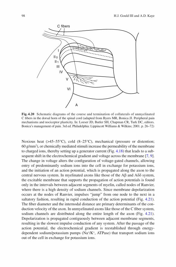
in Rexed lamina I, the outer portion of lamina II, and lamina V [ 8 ] (Fig. 4.20 ). Upon entering the spinal cord, the axons of the primary nociceptors ascend and descend in the zone of Lissauer. The majority of these fi bers ascend approximately two spinal levels before terminating in the dorsal horn.
In the resting state, the free nerve endings of nociceptive afferents maintain a polarized membrane with a higher concentration of sodium ions outside the cell.
Propagatingcompartment
Encodingcompartment
Generatorcompartment
Generatorcurrent
Spike trainStimulus
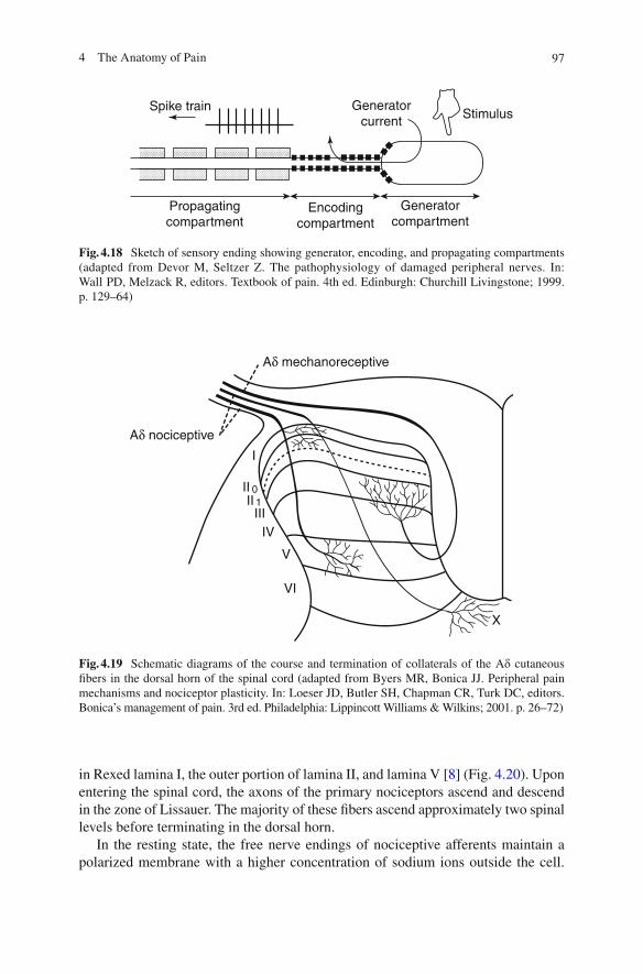
Fig. 4.18 Sketch of sensory ending showing generator, encoding, and propagating compartments (adapted from Devor M, Seltzer Z. The pathophysiology of damaged peripheral nerves. In: Wall PD, Melzack R, editors. Textbook of pain. 4th ed. Edinburgh: Churchill Livingstone; 1999. p. 129–64)
Aδ mechanoreceptive
Aδ nociceptive
I
IIII
IIIIV
V
VI
X
01
Fig. 4.19 Schematic diagrams of the course and termination of collaterals of the A d cutaneous fi bers in the dorsal horn of the spinal cord (adapted from Byers MR, Bonica JJ. Peripheral pain mechanisms and nociceptor plasticity. In: Loeser JD, Butler SH, Chapman CR, Turk DC, editors. Bonica’s management of pain. 3rd ed. Philadelphia: Lippincott Williams & Wilkins; 2001. p. 26–72)
98 H.J. Gould III and A.D. Kaye
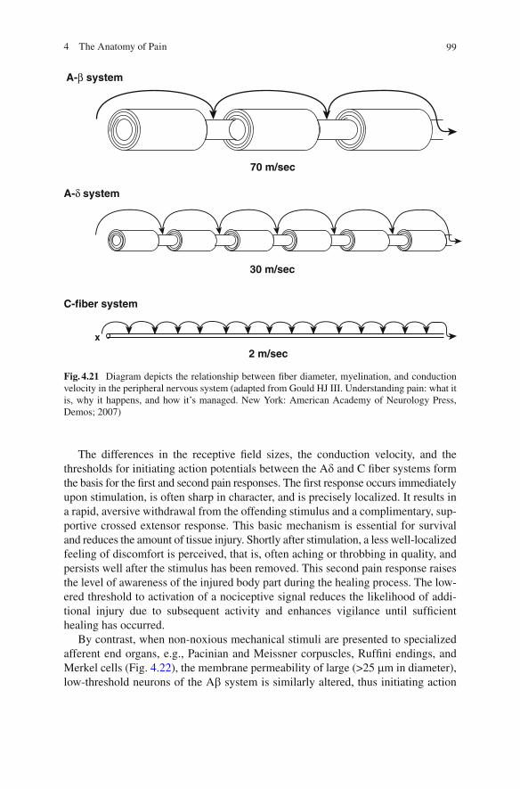
Noxious heat (>45–55°C), cold (8–25°C), mechanical (pressure or distention; 60 g/mm 2 ), or chemically mediated stimuli increase the permeability of the membrane to charged ions, thereby setting up a generator current (Fig. 4.18 ) that leads to a sub-sequent shift in the electrochemical gradient and voltage across the membrane [ 7, 9 ] . The change in voltage alters the confi guration of voltage-gated channels, allowing entry of predominantly sodium ions into the cell in exchange for potassium ions, and the initiation of an action potential, which is propagated along the axon to the central nervous system. In myelinated axons like those of the A b and A d d system, the excitable membrane that supports the propagation of action potentials is found only in the intervals between adjacent segments of myelin, called nodes of Ranvier, where there is a high density of sodium channels. Since membrane depolarization occurs at the nodes of Ranvier, impulses “jump” from one node to the next in a saltatory fashion, resulting in rapid conduction of the action potential (Fig. 4.21 ). The fi ber diameter and the internodal distance are primary determinants of the con-duction velocity of the axon. In unmyelinated axons like those of the C fi ber system, sodium channels are distributed along the entire length of the axon (Fig. 4.21 ). Depolarization is propagated contiguously between adjacent membrane segments, resulting in the slowest impulse conduction of any system. After the passage of the action potential, the electrochemical gradient is reestablished through energy-dependent sodium/potassium pumps (Na + /K + , ATPase) that transport sodium ions out of the cell in exchange for potassium ions.
C fibers
I
III
IV
V
VI
A
II 0
II 1
Fig. 4.20 Schematic diagrams of the course and termination of collaterals of unmyelinated C fi bers in the dorsal horn of the spinal cord (adapted from Byers MR, Bonica JJ. Peripheral pain mechanisms and nociceptor plasticity. In: Loeser JD, Butler SH, Chapman CR, Turk DC, editors. Bonica’s management of pain. 3rd ed. Philadelphia: Lippincott Williams & Wilkins; 2001. p. 26–72)
994 The Anatomy of Pain
The differences in the receptive fi eld sizes, the conduction velocity, and the thresholds for initiating action potentials between the A d and C fi ber systems form the basis for the fi rst and second pain responses. The fi rst response occurs immediately upon stimulation, is often sharp in character, and is precisely localized. It results in a rapid, aversive withdrawal from the offending stimulus and a complimentary, sup-portive crossed extensor response. This basic mechanism is essential for survival and reduces the amount of tissue injury. Shortly after stimulation, a less well-localized feeling of discomfort is perceived, that is, often aching or throbbing in quality, and persists well after the stimulus has been removed. This second pain response raises the level of awareness of the injured body part during the healing process. The low-ered threshold to activation of a nociceptive signal reduces the likelihood of addi-tional injury due to subsequent activity and enhances vigilance until suffi cient healing has occurred.
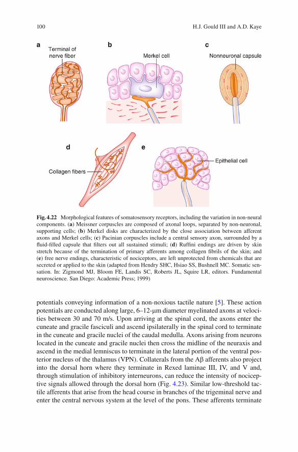
By contrast, when non-noxious mechanical stimuli are presented to specialized afferent end organs, e.g., Pacinian and Meissner corpuscles, Ruffi ni endings, and Merkel cells (Fig. 4.22 ), the membrane permeability of large (>25 m m in diameter), low-threshold neurons of the A b system is similarly altered, thus initiating action
A-β system
A-δ system
C-fiber system
70 m/sec
30 m/sec
2 m/sec
x
Fig. 4.21 Diagram depicts the relationship between fi ber diameter, myelination, and conduction velocity in the peripheral nervous system (adapted from Gould HJ III. Understanding pain: what it is, why it happens, and how it’s managed. New York: American Academy of Neurology Press, Demos; 2007)
100 H.J. Gould III and A.D. Kaye
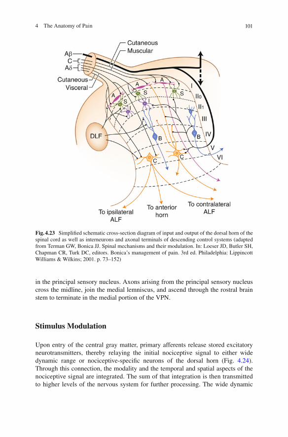
potentials conveying information of a non-noxious tactile nature [ 5 ] . These action potentials are conducted along large, 6–12- m m diameter myelinated axons at veloci-ties between 30 and 70 m/s. Upon arriving at the spinal cord, the axons enter the cuneate and gracile fasciculi and ascend ipsilaterally in the spinal cord to terminate in the cuneate and gracile nuclei of the caudal medulla. Axons arising from neurons located in the cuneate and gracile nuclei then cross the midline of the neuraxis and ascend in the medial lemniscus to terminate in the lateral portion of the ventral pos-terior nucleus of the thalamus (VPN). Collaterals from the A b afferents also project into the dorsal horn where they terminate in Rexed laminae III, IV, and V and, through stimulation of inhibitory interneurons, can reduce the intensity of nocicep-tive signals allowed through the dorsal horn (Fig. 4.23 ). Similar low-threshold tac-tile afferents that arise from the head course in branches of the trigeminal nerve and enter the central nervous system at the level of the pons. These afferents terminate
Fig. 4.22 Morphological features of somatosensory receptors, including the variation in non-neural components. ( a ) Meissner corpuscles are composed of axonal loops, separated by non-neuronal, supporting cells; ( b ) Merkel disks are characterized by the close association between afferent axons and Merkel cells; ( c ) Pacinian corpuscles include a central sensory axon, surrounded by a fl uid-fi lled capsule that fi lters out all sustained stimuli; ( d ) Ruffi ni endings are driven by skin stretch because of the termination of primary afferents among collagen fi brils of the skin; and ( e ) free nerve endings, characteristic of nociceptors, are left unprotected from chemicals that are secreted or applied to the skin (adapted from Hendry SHC, Hsiao SS, Bushnell MC. Somatic sen-sation. In: Zigmond MJ, Bloom FE, Landis SC, Roberts JL, Squire LR, editors. Fundamental neuroscience. San Diego: Academic Press; 1999)
1014 The Anatomy of Pain
in the principal sensory nucleus. Axons arising from the principal sensory nucleus cross the midline, join the medial lemniscus, and ascend through the rostral brain stem to terminate in the medial portion of the VPN.
Stimulus Modulation
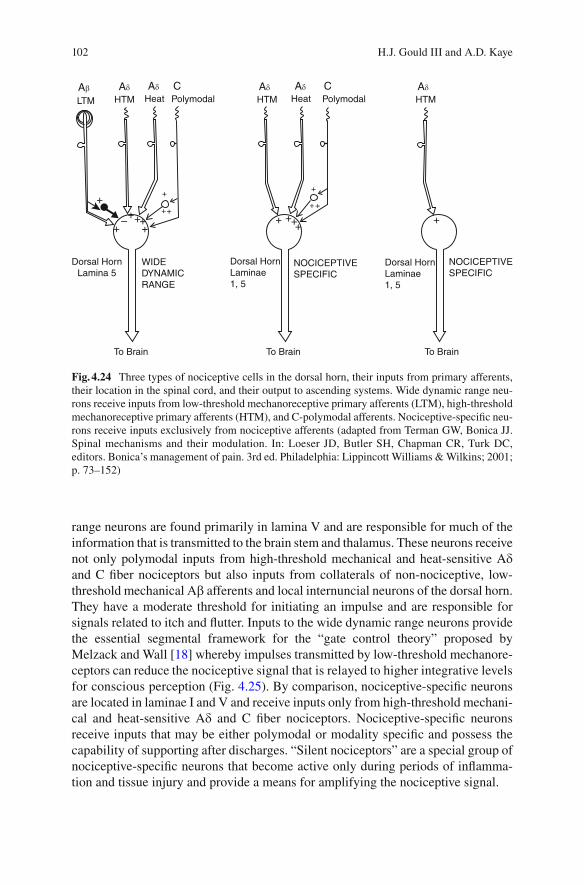
Upon entry of the central gray matter, primary afferents release stored excitatory neurotransmitters, thereby relaying the initial nociceptive signal to either wide dynamic range or nociceptive-specifi c neurons of the dorsal horn (Fig. 4.24 ). Through this connection, the modality and the temporal and spatial aspects of the nociceptive signal are integrated. The sum of that integration is then transmitted to higher levels of the nervous system for further processing. The wide dynamic
Fig. 4.23 Simplifi ed schematic cross-section diagram of input and output of the dorsal horn of the spinal cord as well as interneurons and axonal terminals of descending control systems (adapted from Terman GW, Bonica JJ. Spinal mechanisms and their modulation. In: Loeser JD, Butler SH, Chapman CR, Turk DC, editors. Bonica’s management of pain. 3rd ed. Philadelphia: Lippincott Williams & Wilkins; 2001. p. 73–152)
102 H.J. Gould III and A.D. Kaye
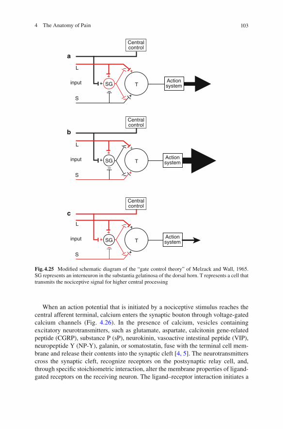
range neurons are found primarily in lamina V and are responsible for much of the information that is transmitted to the brain stem and thalamus. These neurons receive not only polymodal inputs from high-threshold mechanical and heat-sensitive A d and C fi ber nociceptors but also inputs from collaterals of non-nociceptive, low-threshold mechanical A b afferents and local internuncial neurons of the dorsal horn. They have a moderate threshold for initiating an impulse and are responsible for signals related to itch and fl utter. Inputs to the wide dynamic range neurons provide the essential segmental framework for the “gate control theory” proposed by Melzack and Wall [ 18 ] whereby impulses transmitted by low-threshold mechanore-ceptors can reduce the nociceptive signal that is relayed to higher integrative levels for conscious perception (Fig. 4.25 ). By comparison, nociceptive-specifi c neurons are located in laminae I and V and receive inputs only from high-threshold mechani-cal and heat-sensitive A d and C fi ber nociceptors. Nociceptive-specifi c neurons receive inputs that may be either polymodal or modality specifi c and possess the capability of supporting after discharges. “Silent nociceptors” are a special group of nociceptive-specifi c neurons that become active only during periods of infl amma-tion and tissue injury and provide a means for amplifying the nociceptive signal.
++
+
++++
++++
+– +++
+ +
Ab
LTM HTM HTM HTM
Dorsal HornLamina 5
Dorsal HornLaminae1, 5
Dorsal HornLaminae1, 5
WIDEDYNAMICRANGE
To Brain To Brain To Brain
NOCICEPTIVESPECIFIC
NOCICEPTIVESPECIFIC
Heat HeatPolymodal PolymodalAd Ad AdAd AdC C
Fig. 4.24 Three types of nociceptive cells in the dorsal horn, their inputs from primary afferents, their location in the spinal cord, and their output to ascending systems. Wide dynamic range neu-rons receive inputs from low-threshold mechanoreceptive primary afferents (LTM), high-threshold mechanoreceptive primary afferents (HTM), and C-polymodal afferents. Nociceptive-specifi c neu-rons receive inputs exclusively from nociceptive afferents (adapted from Terman GW, Bonica JJ. Spinal mechanisms and their modulation. In: Loeser JD, Butler SH, Chapman CR, Turk DC, editors. Bonica’s management of pain. 3rd ed. Philadelphia: Lippincott Williams & Wilkins; 2001; p. 73–152)
1034 The Anatomy of Pain
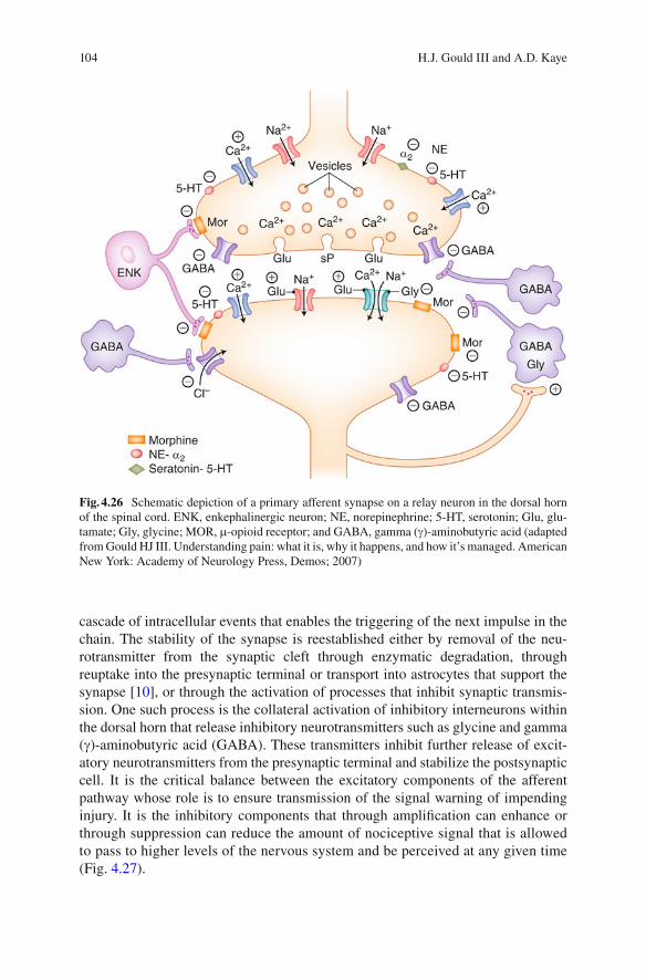
When an action potential that is initiated by a nociceptive stimulus reaches the central afferent terminal, calcium enters the synaptic bouton through voltage-gated calcium channels (Fig. 4.26 ). In the presence of calcium, vesicles containing excitatory neurotransmitters, such as glutamate, aspartate, calcitonin gene-related peptide (CGRP), substance P (sP), neurokinin, vasoactive intestinal peptide (VIP), neuropeptide Y (NP-Y), galanin, or somatostatin, fuse with the terminal cell mem-brane and release their contents into the synaptic cleft [ 4, 5 ] . The neurotransmitters cross the synaptic cleft, recognize receptors on the postsynaptic relay cell, and, through specifi c stoichiometric interaction, alter the membrane properties of ligand-gated receptors on the receiving neuron. The ligand–receptor interaction initiates a
L
S
SG T
Centralcontrol
Actionsystem
input
L
S
SG T
Centralcontrol
Actionsystem
input
L
S
SG T
Centralcontrol
Actionsystem
input
a
b
c
Fig. 4.25 Modifi ed schematic diagram of the “gate control theory” of Melzack and Wall, 1965. SG represents an interneuron in the substantia gelatinosa of the dorsal horn. T represents a cell that transmits the nociceptive signal for higher central processing
104 H.J. Gould III and A.D. Kaye
cascade of intracellular events that enables the triggering of the next impulse in the chain. The stability of the synapse is reestablished either by removal of the neu-rotransmitter from the synaptic cleft through enzymatic degradation, through reuptake into the presynaptic terminal or transport into astrocytes that support the synapse [ 10 ] , or through the activation of processes that inhibit synaptic transmis-sion. One such process is the collateral activation of inhibitory interneurons within the dorsal horn that release inhibitory neurotransmitters such as glycine and gamma ( g )-aminobutyric acid (GABA). These transmitters inhibit further release of excit-atory neurotransmitters from the presynaptic terminal and stabilize the postsynaptic cell. It is the critical balance between the excitatory components of the afferent pathway whose role is to ensure transmission of the signal warning of impending injury. It is the inhibitory components that through amplifi cation can enhance or through suppression can reduce the amount of nociceptive signal that is allowed to pass to higher levels of the nervous system and be perceived at any given time ( Fig. 4.27 ).
Fig. 4.26 Schematic depiction of a primary afferent synapse on a relay neuron in the dorsal horn of the spinal cord. ENK, enkephalinergic neuron; NE, norepinephrine; 5-HT, serotonin; Glu, glu-tamate; Gly, glycine; MOR, m -opioid receptor; and GABA, gamma ( g )-aminobutyric acid (adapted from Gould HJ III. Understanding pain: what it is, why it happens, and how it’s managed. American New York: Academy of Neurology Press, Demos; 2007)
1054 The Anatomy of Pain
Stimulus Perception and Interpretation
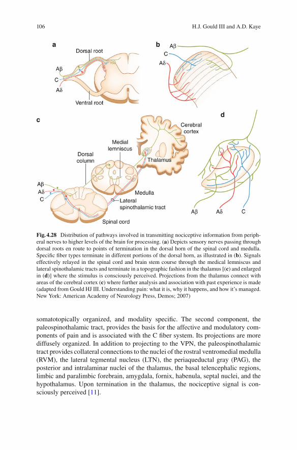
Axons en route to the thalamus from the spinal cord course through the ventral white commissure of the spinal cord, cross the midline, and enter the contralateral lateral spinothalamic tract where they project rostrally through the central nervous system to terminate in the VPN. Similar projections that subserve the territory of the trigeminal nerve receive inputs from axons that, upon entering the pons, descend in the spinal trigeminal tract and terminate on neurons in the spinal trigeminal nucleus. The projections that arise from the relay neurons in the spinal trigeminal nucleus cross the midline and join the spinothalamic tract en route to the VPN (Fig. 4.28 ). There are two components of the lateral spinothalamic pathway. The fi rst compo-nent, the neospinothalamic tract, provides for discriminative functions and is related to the A d system. It projects directly to the VPN and is rapidly conducting, precisely
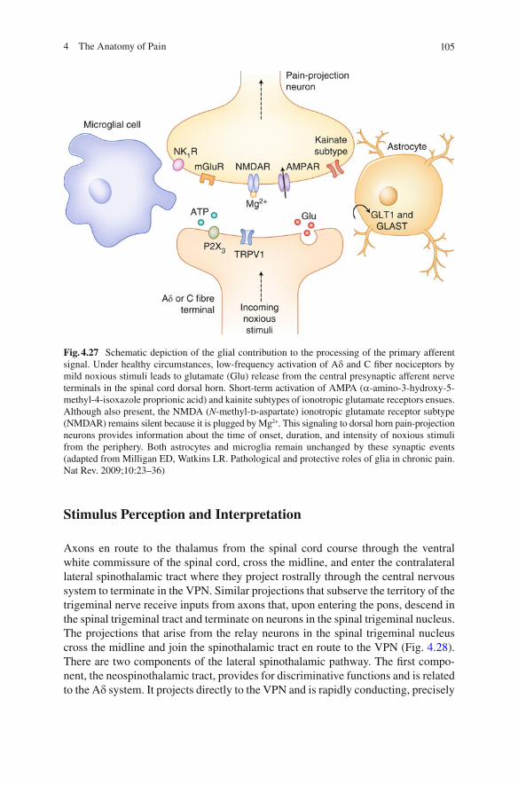
Fig. 4.27 Schematic depiction of the glial contribution to the processing of the primary afferent signal. Under healthy circumstances, low-frequency activation of A d and C fi ber nociceptors by mild noxious stimuli leads to glutamate (Glu) release from the central presynaptic afferent nerve terminals in the spinal cord dorsal horn. Short-term activation of AMPA ( a -amino-3-hydroxy-5-methyl-4-isoxazole proprionic acid) and kainite subtypes of ionotropic glutamate receptors ensues. Although also present, the NMDA ( N -methyl- d -aspartate) ionotropic glutamate receptor subtype (NMDAR) remains silent because it is plugged by Mg 2+ . This signaling to dorsal horn pain-projection neurons provides information about the time of onset, duration, and intensity of noxious stimuli from the periphery. Both astrocytes and microglia remain unchanged by these synaptic events (adapted from Milligan ED, Watkins LR. Pathological and protective roles of glia in chronic pain. Nat Rev. 2009;10:23–36)
106 H.J. Gould III and A.D. Kaye
somatotopically organized, and modality specifi c. The second component, the paleospinothalamic tract, provides the basis for the affective and modulatory com-ponents of pain and is associated with the C fi ber system. Its projections are more diffusely organized. In addition to projecting to the VPN, the paleospinothalamic tract provides collateral connections to the nuclei of the rostral ventromedial medulla (RVM), the lateral tegmental nucleus (LTN), the periaqueductal gray (PAG), the posterior and intralaminar nuclei of the thalamus, the basal telencephalic regions, limbic and paralimbic forebrain, amygdala, fornix, habenula, septal nuclei, and the hypothalamus. Upon termination in the thalamus, the nociceptive signal is con-sciously perceived [ 11 ] .
Fig. 4.28 Distribution of pathways involved in transmitting nociceptive information from periph-eral nerves to higher levels of the brain for processing. ( a ) Depicts sensory nerves passing through dorsal roots en route to points of termination in the dorsal horn of the spinal cord and medulla. Specifi c fi ber types terminate in different portions of the dorsal horn, as illustrated in ( b ). Signals effectively relayed in the spinal cord and brain stem course through the medical lemniscus and lateral spinothalamic tracts and terminate in a topographic fashion in the thalamus [( c ) and enlarged in ( d )] where the stimulus is consciously perceived. Projections from the thalamus connect with areas of the cerebral cortex ( c ) where further analysis and association with past experience is made (adapted from Gould HJ III. Understanding pain: what it is, why it happens, and how it’s managed. New York: American Academy of Neurology Press, Demos; 2007)
1074 The Anatomy of Pain
Neurons in the VPN relay the nociceptive signal to the primary and secondary somatosensory cortices for the processing of location, intensity, and stimulus characterization and to the inferotemporal and frontal cortices for cognitive and contextual content and for cognitive, affective, and executive responses, respec-tively (Fig. 4.28 ). In cortex, nociceptive signals are integrated and compared with past experience, emotions, mood, and current status for interpretation and imple-mentation of a behavioral response. It is in this integrative process that the initial nociceptive signal is transformed into the complex, uncomfortable sensory and emotional experience that we call pain. It is the dynamic relationship between the thalamic neurons and the cortical modulating cells that determines the intensity of the unique painful experience perceived by each individual at any moment in time. Following the integration of the discriminative and affective components of the pain pathway, corticofugal projections return to VPN and surrounding thalamic association nuclei, to the hypothalamus, and to brain stem nuclei. These projec-tions can either augment or diminish the level of pain that is perceived for facilita-tion of a fi ght-or-fl ight response, depending on the state of the individual.
Stimulus Modulation and Behavioral Response
The hypothalamus monitors basal body functions, such as thirst, hunger, satiety, sexual function, blood pressure, temperature, and emotion, and infl uences behavior based on conscious and subconscious information sent from cortex and from vari-ous body organs to maintain normal body function. Hypothalamic modulation of the behavioral response can be affected through the release of several hormones, including vasopressin, corticotropin-releasing factor (CRF), and pituitary adreno-corticotropic hormone (ACTH), that act centrally or peripherally to produce direct or indirect activity on pain-transmitting neurons. The process of modulation occurs through direct projections that affect the activity of enkephalinergic neurons of the PAG, the norepinephrine-containing neurons of the LTN, the serotonergic neurons of the RVM, and the neurons in the entry zones that receive primary afferent input [ 8, 12 ] . Projections from the RVM and the LTN descend through the brain stem and the dorsolateral funiculus of the spinal cord and synapse on the terminals of the primary afferent neurons and on inhibitory enkephalinergic and GABAergic interneurons of the dorsal horn, thereby indirectly affecting the transmission of nociceptive signals through the dorsal horn (Fig. 4.29 ). These projections can block the release of neurotransmitter from the primary afferent terminals, stimulate local inhibitory interneurons, or stabilize the membrane of the relay neurons and thus suppress the amount of nociceptive signal that is allowed to pass through the dorsal horn en route to higher integrative centers. Depending on the state of the individual, modulation of these descending systems can produce the opposite effect through
108 H.J. Gould III and A.D. Kaye
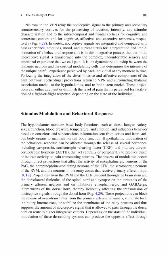
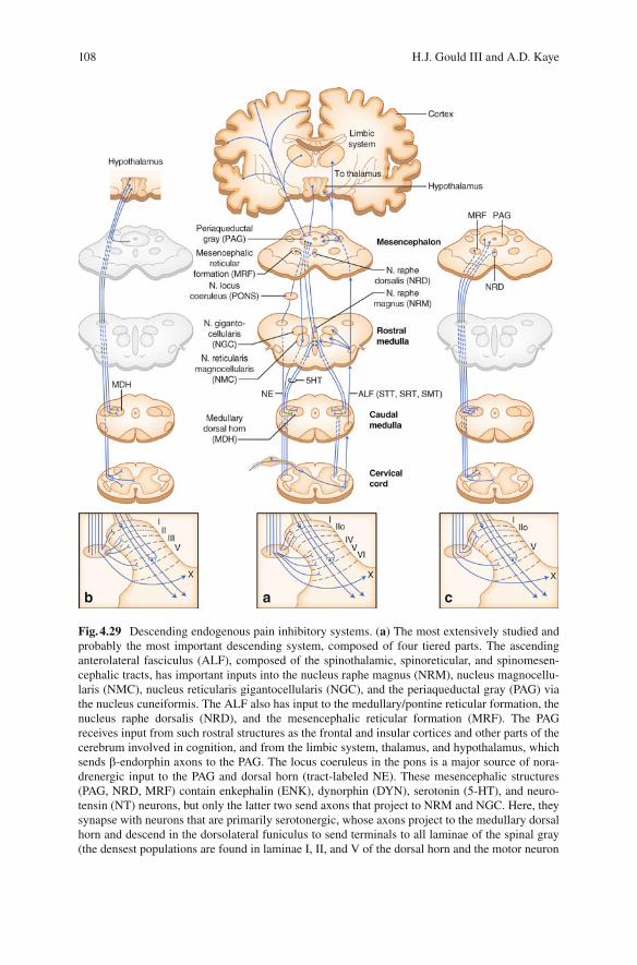
Fig. 4.29 Descending endogenous pain inhibitory systems. ( a ) The most extensively studied and probably the most important descending system, composed of four tiered parts. The ascending anterolateral fasciculus (ALF), composed of the spinothalamic, spinoreticular, and spinomesen-cephalic tracts, has important inputs into the nucleus raphe magnus (NRM), nucleus magnocellu-laris (NMC), nucleus reticularis gigantocellularis (NGC), and the periaqueductal gray (PAG) via the nucleus cuneiformis. The ALF also has input to the medullary/pontine reticular formation, the nucleus raphe dorsalis (NRD), and the mesencephalic reticular formation (MRF). The PAG receives input from such rostral structures as the frontal and insular cortices and other parts of the cerebrum involved in cognition, and from the limbic system, thalamus, and hypothalamus, which sends b -endorphin axons to the PAG. The locus coeruleus in the pons is a major source of nora-drenergic input to the PAG and dorsal horn (tract-labeled NE). These mesencephalic structures (PAG, NRD, MRF) contain enkephalin (ENK), dynorphin (DYN), serotonin (5-HT), and neuro-tensin (NT) neurons, but only the latter two send axons that project to NRM and NGC. Here, they synapse with neurons that are primarily serotonergic, whose axons project to the medullary dorsal horn and descend in the dorsolateral funiculus to send terminals to all laminae of the spinal gray (the densest populations are found in laminae I, II, and V of the dorsal horn and the motor neuron
1094 The Anatomy of Pain
reduction of the level of direct inhibitory input or through the disinhibition of local inhibitory circuits, thus amplifying nociceptive signals and augmenting the likeli-hood that additional signals of a painful nature will be transmitted to the thalamus for perception [ 8 ] .
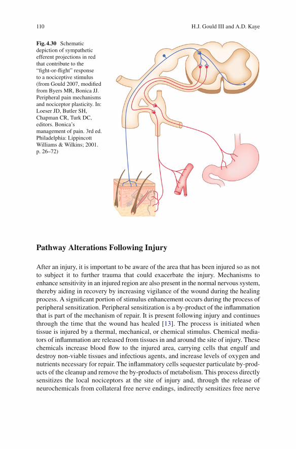
For optimum survival, it is important to prepare the organism for an appropriate behavioral response and return the monitoring system to optimum levels of func-tioning in anticipation of additional warnings. This function is built into the nervous system. Since pain may well signal a threat to the survival of at least a part of an individual, painful stimuli automatically prepare the individual for rapid assessment of the afferent stimulus and the initiation of defensive “fi ght-or-fl ight” behavior through activation of the sympathetic nervous system (Fig. 4.30 ). The sympathetic nervous system controls blood pressure, heart and breathing rate, and the volume of blood that fl ows to specifi c tissues – more to voluntary muscles, heart, and lungs and less to the intestinal system and skin. The neurotransmitter that is released to pro-duce these responses is norepinephrine. When released in the vicinity of peripheral afferent nerve terminals, impulse generation is made easier. The sympathetic tone is modulated through descending cortical and hypothalamic projections that deter-mine the fi ring frequency of preganglionic sympathetic neurons located in the inter-mediolateral cell column of the spinal gray matter from C8 (T1) to L1–2 levels of the spinal cord.
After a nociceptive signal has been effectively relayed to the thalamus for further processing, the mechanisms responsible for receiving the nociceptive signals must be reset in the event that additional noxious stimuli requiring assessment arrive at the dorsal horn. To accomplish this, active relay neurons send axon collaterals to local inhibitory neurons in the dorsal horn that project back to the primary afferent terminal and to the initiating relay neuron to inhibit further activity and thus reduce the likelihood that multiple impulses will be sent to higher levels of analysis. The primary transmitters utilized by these inhibitory neurons are GABA and glycine.
Fig. 4.29 (continued) pools of lamina IX). The projection from NRM is bilateral, whereas the projection from NGC is ipsilateral. Noradrenergic fi bers descend and project to the medullary dorsal horn and then descend in the dorsolateral funiculus of the spinal cord to send terminals to laminae I, II, IV through VI, and X. ( b ) A simplistic schema to show the direct hypothalamospinal descending control system, which originates in the medial and paraventricular hypothalamic nuclei. This descending system consists of vasopressin and oxytocin neurons (and perhaps some enkephalinergic neurons), which not only send terminals predominantly to laminae I and X but also provide sparse input into laminae II and III and the lateral part of lamina V, as well as the homologous area in the medullary dorsal horn. ( c ) Direct PAG-spinal projection system, which bypasses the medullary nuclei and projects directly to the medullary dorsal horn and then descends in the dorsolateral funiculus to send terminals to laminae I, II
o , V, and X. Most of the axons are
serotonergic and noradrenergic (adapted from Terman GW, Bonica JJ. Spinal mechanisms and their modulation. In: Loeser JD, Butler SH, Chapman CR, Turk DC, editors. Bonica’s management of pain. 3rd ed. Philadelphia: Lippincott Williams & Wilkins; 2001. p. 73–152)
110 H.J. Gould III and A.D. Kaye
Pathway Alterations Following Injury
After an injury, it is important to be aware of the area that has been injured so as not to subject it to further trauma that could exacerbate the injury. Mechanisms to enhance sensitivity in an injured region are also present in the normal nervous system, thereby aiding in recovery by increasing vigilance of the wound during the healing process. A signifi cant portion of stimulus enhancement occurs during the process of peripheral sensitization. Peripheral sensitization is a by-product of the infl ammation that is part of the mechanism of repair. It is present following injury and continues through the time that the wound has healed [ 13 ] . The process is initiated when tissue is injured by a thermal, mechanical, or chemical stimulus. Chemical media-tors of infl ammation are released from tissues in and around the site of injury. These chemicals increase blood fl ow to the injured area, carrying cells that engulf and destroy non-viable tissues and infectious agents, and increase levels of oxygen and nutrients necessary for repair. The infl ammatory cells sequester particulate by-prod-ucts of the cleanup and remove the by-products of metabolism. This process directly sensitizes the local nociceptors at the site of injury and, through the release of neurochemicals from collateral free nerve endings, indirectly sensitizes free nerve
Fig. 4.30 Schematic depiction of sympathetic efferent projections in red that contribute to the “fi ght-or-fl ight” response to a nociceptive stimulus (from Gould 2007, modifi ed from Byers MR, Bonica JJ. Peripheral pain mechanisms and nociceptor plasticity. In: Loeser JD, Butler SH, Chapman CR, Turk DC, editors. Bonica’s management of pain. 3rd ed. Philadelphia: Lippincott Williams & Wilkins; 2001. p. 26–72)
1114 The Anatomy of Pain
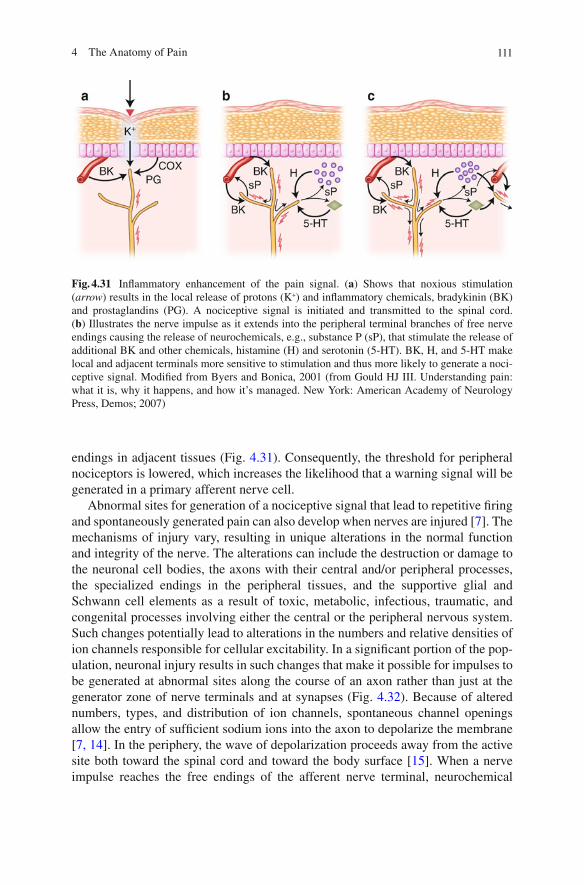
endings in adjacent tissues (Fig. 4.31 ). Consequently, the threshold for peripheral nociceptors is lowered, which increases the likelihood that a warning signal will be generated in a primary afferent nerve cell.
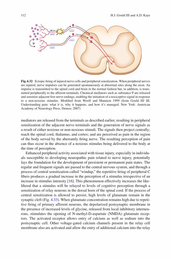
Abnormal sites for generation of a nociceptive signal that lead to repetitive fi ring and spontaneously generated pain can also develop when nerves are injured [ 7 ] . The mechanisms of injury vary, resulting in unique alterations in the normal function and integrity of the nerve. The alterations can include the destruction or damage to the neuronal cell bodies, the axons with their central and/or peripheral processes, the specialized endings in the peripheral tissues, and the supportive glial and Schwann cell elements as a result of toxic, metabolic, infectious, traumatic, and congenital processes involving either the central or the peripheral nervous system. Such changes potentially lead to alterations in the numbers and relative densities of ion channels responsible for cellular excitability. In a signifi cant portion of the pop-ulation, neuronal injury results in such changes that make it possible for impulses to be generated at abnormal sites along the course of an axon rather than just at the generator zone of nerve terminals and at synapses (Fig. 4.32 ). Because of altered numbers, types, and distribution of ion channels, spontaneous channel openings allow the entry of suffi cient sodium ions into the axon to depolarize the membrane [ 7, 14 ] . In the periphery, the wave of depolarization proceeds away from the active site both toward the spinal cord and toward the body surface [ 15 ] . When a nerve impulse reaches the free endings of the afferent nerve terminal, neurochemical
Fig. 4.31 Infl ammatory enhancement of the pain signal. ( a ) Shows that noxious stimulation ( arrow ) results in the local release of protons (K + ) and infl ammatory chemicals, bradykinin (BK) and prostaglandins (PG). A nociceptive signal is initiated and transmitted to the spinal cord. ( b ) Illustrates the nerve impulse as it extends into the peripheral terminal branches of free nerve endings causing the release of neurochemicals, e.g., substance P (sP), that stimulate the release of additional BK and other chemicals, histamine (H) and serotonin (5-HT). BK, H, and 5-HT make local and adjacent terminals more sensitive to stimulation and thus more likely to generate a noci-ceptive signal. Modifi ed from Byers and Bonica, 2001 (from Gould HJ III. Understanding pain: what it is, why it happens, and how it’s managed. New York: American Academy of Neurology Press, Demos; 2007)
112 H.J. Gould III and A.D. Kaye
mediators are released from the terminals as described earlier, resulting in peripheral sensitization of the adjacent nerve terminals and the generation of nerve signals as a result of either noxious or non-noxious stimuli. The signals then project centrally; reach the spinal cord, thalamus, and cortex; and are perceived as pain in the region of the body served by the aberrantly fi ring nerve. The resulting perception of pain can thus occur in the absence of a noxious stimulus being delivered to the body at the time of perception.
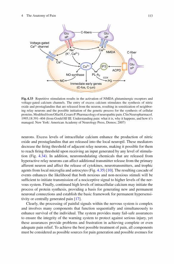
Enhanced peripheral activity associated with tissue injury, especially in individu-als susceptible to developing neuropathic pain related to nerve injury, potentially lays the foundation for the development of persistent or permanent pain states. The regular and frequent signals are passed to the central nervous system, and through a process of central sensitization called “windup,” the repetitive fi ring of peripheral C fi bers produces a gradual increase in the perception of a stimulus irrespective of an increase in stimulus intensity [ 16 ] . This phenomenon effectively increases the like-lihood that a stimulus will be relayed to levels of cognitive perception through a sensitization of relay neurons in the dorsal horn of the spinal cord. If the process of central sensitization is allowed to persist, high levels of glutamate remain in the synaptic cleft (Fig. 4.33 ). When glutamate concentration remains high due to repeti-tive fi ring of primary afferent neurons, the depolarized postsynaptic membrane in the presence of increased levels of glycine, released from local inhibitory interneu-rons, stimulates the opening of N-methyl-D-aspartate (NMDA) glutamate recep-tors. The activated receptor allows entry of calcium as well as sodium into the postsynaptic cell. Other voltage-gated calcium channels present in the relay cell membrane also are activated and allow the entry of additional calcium into the relay
Fig. 4.32 Ectopic fi ring of injured nerve cells and peripheral sensitization. When peripheral nerves are injured, nerve impulses can be generated spontaneously at abnormal sites along the axon. An impulse is transmitted to the spinal cord and brain in the normal fashion but, in addition, is trans-mitted peripherally to the afferent terminals. Chemical mediators such as substance P are released and sensitize adjacent free nerve endings, enabling the initiation of a nociceptive signal in response to a non-noxious stimulus. Modifi ed from Woolf and Mannion 1999 (from Gould HJ III. Understanding pain: what it is, why it happens, and how it’s managed. New York: American Academy of Neurology Press, Demos; 2007)
1134 The Anatomy of Pain
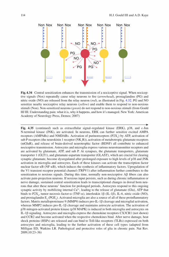
neurons. Excess levels of intracellular calcium enhance the production of nitric oxide and prostaglandins that are released into the local neuropil. These mediators decrease the fi ring threshold of adjacent relay neurons, making it possible for them to reach fi ring threshold upon receiving an input generated by any level of stimula-tion (Fig. 4.34 ). In addition, neuromodulating chemicals that are released from hyperactive relay neurons can affect additional transmitter release from the primary afferent neuron and affect the release of cytokines, neurotransmitters, and trophic agents from local microglia and astrocytes (Fig. 4.35 ) [ 10 ] . The resulting cascade of events enhances the likelihood that both noxious and non-noxious stimuli will be suffi cient to initiate transmission of a nociceptive signal to higher levels of the ner-vous system. Finally, continued high levels of intracellular calcium may initiate the process of protein synthesis, providing a basis for generating new and permanent neuronal connections and establish the basic framework for permanent hypersensi-tivity or centrally generated pain [ 17 ] .
Clearly, the processing of painful signals within the nervous system is complex and involves many components that function sequentially and simultaneously to enhance survival of the individual. The system provides many fail-safe assurances to ensure the integrity of the warning system to protect against serious injury, yet these assurances provide problems and frustration in achieving complete or even adequate pain relief. To achieve the best possible treatment of pain, all components must be considered as possible sources for pain generation and possible avenues for
Fig. 4.33 Repetitive stimulation results in the activation of NMDA glutaminergic receptors and voltage-gated calcium channels. The entry of excess calcium stimulates the synthesis of nitric oxide and prostaglandins that are released from the neuron, resulting in sensitization of neighbor-ing relay neurons and the possible initiation of the genetic process for the synthesis of cellular proteins. Modifi ed from Ollat H, Cesaro P. Pharmacology of neuropathic pain. Clin Neuropharmacol. 1995;18:391–404 (from Gould HJ III. Understanding pain: what it is, why it happens, and how it’s managed. New York: American Academy of Neurology Press, Demos; 2007)
114 H.J. Gould III and A.D. Kaye
Fig. 4.34 Central sensitization enhances the transmission of a nociceptive signal. When nocicep-tive signals (Nox) repeatedly cause relay neurons to fi re ( arrowhead ), prostaglandins (PG) and nitric oxide (NO) are released from the relay neuron ( red ), as illustrated in Fig. 4.32 . PG and NO sensitize nearby nociceptive relay neurons ( yellow ) and enable them to respond to non-noxious stimuli (Non). Non-sensitized neurons ( green ) do not respond to non-noxious stimuli (from Gould HJ III. Understanding pain: what it is, why it happens, and how it’s managed. New York: American Academy of Neurology Press, Demos; 2007)
Fig. 4.35 (continued) such as extracellular signal–regulated kinase (ERK), p38, and c-Jun N-terminal kinase (JNK), are activated. In neurons, ERK can further sensitize excited AMPA receptors (AMPARs) and NMDARs. Activation of purinoreceptors (P2X
3 ) by ATP, activation of
sub P receptors (the neurokinin 1 receptor (NK 1 R)), activation of metabotropic glutamate receptors
(mGluR), and release of brain-derived neurotrophic factor (BDNF) all contribute to enhanced nociceptive transmission. Astrocytes and microglia express various neurotransmitter receptors and are activated by glutamate, ATP, and sub P. At synapses, the glutamate transporters, glutamate transporter 1 (GLT1), and glutamate-aspartate transporter (GLAST), which are crucial for clearing synaptic glutamate, become dysregulated after prolonged exposure to high levels of p38 and JNK activation in microglia and astrocytes. Each of these kinases can activate the transcription factor nuclear factor k B (NF- k B), which induces the synthesis of infl ammatory factors. Upregulation of the V1 transient receptor potential channel (TRPV1) after infl ammation further contributes to the sensitization to noxious signals. During this time, normally non-nociceptive A b fi bers can also activate pain-projection neurons. If noxious input persists, such as during chronic infl ammation or nerve damage, sustained central sensitization leads to transcriptional changes in dorsal horn neu-rons that alter these neurons’ function for prolonged periods. Astrocytes respond to this ongoing synaptic activity by mobilizing internal Ca 2+ , leading to the release of glutamate (Glu), ATP that binds to P2X
4 , tumor necrosis factor- a (TNF- a ), interleukin 1 b (IL-1 b ), IL-6, nitric oxide (NO),
and prostaglandin E 2 (PGE
2 ). Activated microglia are also a source of all of these proinfl ammatory
factors. Matrix metalloproteinase 9 (MMP9) induces pro-IL-1 b cleavage and microglial activation, whereas MMP2 induces pro-IL-1 b cleavage and maintains astrocyte activation. The activation of p38 mitogen-activated protein kinase (p38 MAPK) is induced in both microglia and astrocytes on IL-1 b signaling. Astrocytes and microglia express the chemokine receptors CX3CR1 (not shown) and CCR2 and become activated when the respective chemokines bind. After nerve damage, heat shock proteins (HSPs) are released and can bind to Toll-like receptors (TLRs) expressed on both astrocytes and microglia, leading to the further activation of these cell types (adapted from Milligan ED, Watkins LR. Pathological and protective roles of glia in chronic pain. Nat Rev. 2009;10:23–36)

1154 The Anatomy of Pain
Fig. 4.35 Schematic depiction of the role of glia in processing repetitive nociceptive input and pain processing during infl ammation. After repetitive synaptic communication, which can occur after a short barrage of nociceptive afferent input, there is an increase in the responsiveness of dorsal horn pain-projection neurons to subsequent stimuli (known as central sensitization). A co-release of glutamate and neurotransmitters such as substance P (sub P) and calcitonin gene-related peptide (CGRP) mediates NMDAR activation, leading to voltage-gated Ca 2+ currents (VGCCs). In addition, inositol-1,4,5-triphosphate (Ins(1,4,5,)P
3 ) signaling and mitogen-activated protein kinases,
116 H.J. Gould III and A.D. Kaye
pain control. Knowledge of the anatomical and physiological basis for nociceptive processing and an understanding of the most likely sites where damage and inter-vention can occur is essential for providing optimum care for your patients.
Multiple-Choice Questions
1. Inhibitory interneurons within the dorsal horn release inhibitory neurotransmitters such as: (a) Glycine and gamma ( g )-aminobutyric acid (GABA) (b) Glutamate and aspartate (c) Calcitonin gene-related peptide (CGRP), galanin, and substance P (sP) (d) Neurokinin, vasoactive intestinal peptide (VIP), and neuropeptide Y (NP-Y)
2. There are two components of the lateral spinothalamic pathway: (a) Neospinothalamic tract and paleospinothalamic tract (b) Subthalamic tract and cerebellar vermis tract (c) Anterior and posterior longitudinal tract (d) Neocerebellar and tuberculum tract
3. When glutamate concentration remains high due to repetitive fi ring of primary afferent neurons, the depolarized postsynaptic membrane in the presence of increased levels of glycine, released from local inhibitory interneurons, stimu-lates the opening of: (a) Serotonin receptors (b) Bradykinin receptors (c) Muscarinic receptors (d) N -Methyl- d -aspartate (NMDA) glutamate receptors
4. Inputs to the wide dynamic range neurons provide the essential segmental framework for the “gate control theory” proposed by: (a) Melzack and Wall (1965) (b) Racz and Raj (1971) (c) Bonica (1958) (d) Lema (1986)
5. The gate control theory: (a) Is completely false. (b) States that impulses transmitted by low-threshold mechanoreceptors can
reduce the nociceptive signal that is relayed to higher integrative levels for conscious perception.
(c) Explains the mechanism of the gamma refl ex loop. (d) Is the basis of our understanding of saltatory conduction.
1174 The Anatomy of Pain
6. The regular and frequent signals which can be passed to the central nervous system and through a process of central sensitization are called: (a) “Windup” (b) Diffusion (c) Archicerebellum redundancy (d) Schmidt–Lanterman syndrome
7. The consequences of “windup” include: (a) Quicker refl exes. (b) Increased micturition and defecation. (c) The repetitive fi ring of peripheral C fi bers which produces a gradual
increase in the perception of a stimulus irrespective of an increase in stimu-lus intensity.
(d) The sequential discharge of b fi bers which produces g -mediated pain.
8. Unique structures, which are depolarized by stimuli in response to tissue damage: (a) Touch receptors (b) Nociceptors (c) Temperature receptors (d) Chloride channels
9. As the axons approach the spinal cord, they diverge from the main nerve trunk and enter the dorsal root where they course by their cell bodies in the DRG and enter the spinal cord to terminate on neurons in: (a) Rexed laminae I and II (b) Rexed laminae III and V (c) Rexed laminae X (d) All of the above
10. The axons of the C fi ber system: (a) Are unmyelinated. (b) Are myelinated. (c) Are never found in the peripheral nerves of the somatic sensory system. (d) Have fast conduction velocity of over 20 m/s.
11. Neurons in the ventral posterior nucleus of the thalamus (VPN) relay the noci-ceptive signal to: (a) The primary somatosensory cortex (b) The secondary somatosensory cortex (c) The inferotemporal and frontal cortices (d) All of the above
12. After an injury, a signifi cant portion of stimulus enhancement can occur during the process of peripheral sensitization and is limited to injury by: (a) Thermal stimulus (b) Mechanical stimulus (c) Chemical stimulus (d) All of the above
118 H.J. Gould III and A.D. Kaye
13. Which is false regarding wide dynamic range neurons? (a) They are found primarily in lamina V. (b) They are responsible for much of the information that is transmitted to the
brain stem and thalamus. (c) These neurons receive polymodal inputs. (d) One limitation is that they do not receive inputs from collaterals of non-
nociceptive, low-threshold mechanical A b afferents and local internuncial neurons of the dorsal horn.
14. C fi bers: (a) Respond to polymodal stimuli but preferentially respond to noxious heat. (b) Their central elements course medially in the dorsal root and terminate on
neurons in Rexed lamina I, the outer portion of lamina II, and lamina V. (c) Upon entering the spinal cord, the axons of the primary nociceptors ascend
and descend in the zone of Lissauer. (d) The majority of these fi bers ascend approximately two spinal levels before
terminating in the dorsal horn.
15. In myelinated axons, the excitable membrane that supports the propagation of action potentials found only in the intervals between adjacent segments of myelin is called: (a) Nodes of Ranvier (b) Basilar sulci (c) Nervus intermedius (d) Riopelle lipofuscin
Answers: 1. a 2. a 3. d 4. a 5. b 6. a 7. c 8. b 9. d 10. a 11. d 12. d 13. d 14. d 15. a
Acknowledgements The authors wish to thank Dr. Dennis Paul for his helpful comments and suggestions in the preparation of this manuscript.
1194 The Anatomy of Pain
References
1. Woolf CJ, Decosterd I. Implications of recent advances in the understanding of pain pathophysiology for the assessment of pain in patients. Pain. 1999;6(Suppl):S141–7.
2. Woolf CJ, Max MB. Mechanism-based pain diagnosis. Issues for analgesic drug development. Anesthesiology. 2001;95:241–9.
3. Bevan S. Nociceptive peripheral neurons: cellular properties. In: Wall PD, Melzack R, editors. Textbook of pain. 4th ed. Edinburgh: Churchill Livingstone; 1999. p. 85–104.
4. Raja SN, Meyer RA, Ringkamp M, Campbell JN. Peripheral neural mechanisms of nociception. In: Wall PD, Melzack R, editors. Textbook of pain. 4th ed. Edinburgh: Churchill Livingstone; 1999. p. 11–57.
5. Byers MR, Bonica JJ. Peripheral pain mechanisms and nociceptor plasticity. In: Loeser JD, Butler SH, Chapman CR, Turk DC, editors. Bonica’s management of pain. 3rd ed. Philadelphia: Lippincott Williams & Wilkins; 2001. p. 26–72.
6. Rang HP, Bevan S, Dray A. Nociceptive peripheral neurons: cellular properties. In: Wall PD, Melzack R, editors. Textbook of pain. 3rd ed. Edinburgh: Churchill Livingstone; 1994. p. 57–78.
7. Devor M, Seltzer Z. The pathophysiology of damaged peripheral nerves. In: Wall PD, Melzack R, editors. Textbook of pain. 4th ed. Edinburgh: Churchill Livingstone; 1999. p. 129–64.
8. Terman GW, Bonica JJ. Spinal mechanisms and their modulation. In: Loeser JD, Butler SH, Chapman CR, Turk DC, editors. Bonica’s management of pain. 3rd ed. Philadelphia: Lippincott Williams & Wilkins; 2001. p. 73–152.
9. Meyer RA, Campbell JN, Raja SN. Peripheral neural mechanisms of nociception. In: Wall PD, Melzack R, editors. Textbook of pain. 3rd ed. Edinburgh: Churchill Livingstone; 1994. p. 13–44.
10. Milligan ED, Watkins LR. Pathological and protective roles of glia in chronic pain. Nat Rev. 2009;10:23–36.
11. Adams RD, Victor M. Principles of neurology. 4th ed. New York: McGraw Hill; 1989. 12. Basbaum AI, Fields HL. Endogenous pain control systems: brain stem spinal pathways and
endorphin circuitry. Annu Rev Neurosci. 1984;7:309–38. 13. Levine JD, Reichling DB. Peripheral mechanisms of infl ammatory pain. In: Wall PD, Melzack
R, editors. Textbook of pain. 4th ed. Edinburgh: Churchill Livingstone; 1999. p. 59–84. 14. Amir R, Devor M. Spike-evoked suppression and burst patterning in dorsal root ganglion neurons.
J Physiol (London). 1997;501:183–96. 15. Woolf CJ, Mannion RJ. Neuropathic pain: aetiology, symptoms, mechanisms, and management.
Lancet. 1999;353:1959–64. 16. Wall PD, Woolf CJ. The brief and the prolonged facilitatory effects of unmyelinated afferent
input on the rat spinal cord are independently infl uenced by peripheral nerve section. Neuroscience. 1986;17:1199–205.
17. Woolf CJ, Shortland P, Coggeshall RE. Peripheral nerve injury triggers central sprouting of myelinated afferents. Nature. 1992;355:75–8.
18. Melzack R, Wall PD. Pain mechanisms: a new theory. Science. 1965;150:971–9.