Embed Size (px)
Citation preview

The American College of Radiology Imaging Network, in conjunction with the Avon
Foundation, presents: ACRIN 6666: SCREENING BREAST ULTRASOUND IN HIGH-RISK WOMEN
Study Chair Wendie A. Berg, MD, PhD (410) 252-2332 [email protected]
Co- Investigator Ellen B. Mendelson, MD Northwestern University School of Medicine
Co- Investigator—Reader Studies Christopher R.B. Merritt, MD Thomas Jefferson University School of Medicine
Statistician Jeffrey Blume, PhD Center for Statistical Sciences Brown University (401) 863-9968 Fax #(401) 863-9182
Cost-Effectiveness Analysis Mark Schleinitz, MD Brown University 401-444-3830 Fax #(401) 444-5040 Version Date: November 9, 2007 Administrative Update: November 30, 2007 Activation Date: April 19, 2004 Including Amendments: 1 - 6
This protocol was designed and developed by the American College of Radiology Imaging Network (ACRIN). Funding from the Avon Foundation made this work possible. It is intended to be used only in conjunction with institution-specific IRB approval for study entry. No other use or reproduction is authorized by ACRIN, nor does ACRIN assume any responsibility for unauthorized use of this protocol.
PARTIAL PROTOCOL—CONTACT ACRIN PROTOCOL DEVELOPMENT AND REGULATORY COMPLIANCE FOR A COMPLETE PROTOCOL

ACRIN 6666 2 November 9, 2007 (11/30/07 administrative update)
INDEX
Schema………………………………………………………………………. 4
1.0 Abstract ……………………………………………………………… 10
2.0 Background and Significance ………………………………………. 11
3.0 Specific Aims/Objectives ………………………………………….. . 20
4.0 Methods ……………………………………………………………... 22
5.0 Participant Selection ………………………………………………… 48
6.0 Site Selection ………………………………………………………… 55
7.0 Online Registration ………………………………………………….. 58
8.0 Data Collection and Management …………………………………… 58
9.0 Data Collection, Adverse Events, and Auditing……………………… 61
10.0 Image Submission …………………………………………………….. 76
11.0 Communication of Results and Participant Follow-up Procedures……. 78
12.0 Cost-Effectiveness Assessment………………………………………… 80
13.0 Statistical Considerations………………………………………………. 87
References ……………………………………………………………..………. 101
Appendix I: BI-RADS® Ultrasound Lexicon…………………………...…… 110
Appendix IA: Summary Breast Imaging Reporting and Data System
(BI-RADS®): MRI Lexicon1…………………………………... 113
Appendix II: Phantom Development…………………………………………. 117
Appendix IIA: References …………………………………………………….. 122
Appendix III: Sample Informed Consent Form……………………………….. 123
Appendix IIIA: Supplemental Sample Informed Consent Form to
ACRIN 6666 MRI SubStudy………………………..………..… 128
Appendix IV: Eligibility Checklist……………………………………………. 133
Appendix IVA: Eligibility Checklist: MRI Amendment ……………………….. 136
Appendix V: HIPAA Research Authorization………………………………… 140
ACRIN 6666 3 November 9, 2007 (11/30/07 administrative update)
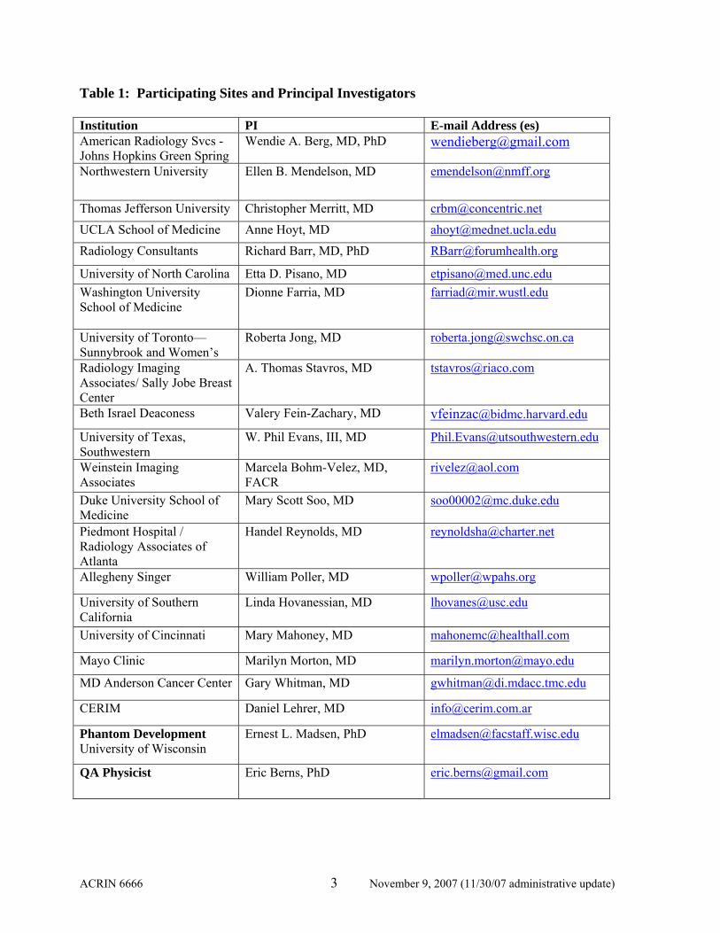
Table 1: Participating Sites and Principal Investigators Institution PI E-mail Address (es) American Radiology Svcs - Johns Hopkins Green Spring
Wendie A. Berg, MD, PhD [email protected]
Northwestern University Ellen B. Mendelson, MD [email protected]
Thomas Jefferson University Christopher Merritt, MD [email protected] UCLA School of Medicine Anne Hoyt, MD [email protected] Radiology Consultants Richard Barr, MD, PhD [email protected]
University of North Carolina Etta D. Pisano, MD [email protected] Washington University School of Medicine
Dionne Farria, MD
University of Toronto—Sunnybrook and Women’s
Roberta Jong, MD [email protected]
Radiology Imaging Associates/ Sally Jobe Breast Center
A. Thomas Stavros, MD [email protected]
Beth Israel Deaconess Valery Fein-Zachary, MD [email protected]
University of Texas, Southwestern
W. Phil Evans, III, MD [email protected]
Weinstein Imaging Associates
Marcela Bohm-Velez, MD, FACR
Duke University School of Medicine
Mary Scott Soo, MD [email protected]
Piedmont Hospital / Radiology Associates of Atlanta
Handel Reynolds, MD [email protected]
Allegheny Singer William Poller, MD [email protected]
University of Southern California
Linda Hovanessian, MD [email protected]
University of Cincinnati Mary Mahoney, MD [email protected]
Mayo Clinic Marilyn Morton, MD [email protected]
MD Anderson Cancer Center Gary Whitman, MD [email protected]
CERIM Daniel Lehrer, MD [email protected]
Phantom Development University of Wisconsin
Ernest L. Madsen, PhD [email protected]
QA Physicist
Eric Berns, PhD [email protected]

ACRIN 6666 4 November 9, 2007 (11/30/07 administrative update)
Determine eligibility Informed consent QOL instruments
Women at High Risk of Breast Cancer
Randomize order of initial screening modality (Mammogram or US), independently read, All patients to receive both exams
Routine mammogram performed
Physician-performed bilateral whole-breast US
Negative or Benign Findings: Screen again 12 mos, 24 mos Mammogram and US as above
Incomplete
Additional mammographic views Targeted US as needed
Negative or Benign Findings: Screen again 12 mos, 24 mos Mammogram and US as above
Probably Benign Findings: 6-mo follow-up (See details a)
Suspicious or Highly Suggestive of Malignancy: Biopsy (See detailsb)
+
or
or
or
(Integration Interpretation if mammo or US other than negative or benign)

ACRIN 6666 5 November 9, 2007 (11/30/07 administrative update)
Probably Benign Findings: Short-interval follow-up (6 mo) Targeted mammographic views and/or Targeted US, diagnostic exama
Negative or Benign: Return to annual screening at 12 and 24 mo
Probably Benign Findings: Diagnostic exam in another 6 mo (12 mo from initial screen) Includes bilateral screening US and Mammogram and targeted exam prn
Findings gone or clearly benign: Return to annual screening at 24 mos
Findings stable at 12 months: Diagnostic exam at 24 months Includes bilateral screening US and Mammogram and targeted exam prn
Findings increasing/Suspicious: Biopsy recommended
Suspicious Abnormality or Findings Highly Suggestive of Malignancy: Biopsy recommendedb
or
or
or
or
ACRIN 6666 6 November 9, 2007 (11/30/07 administrative update)
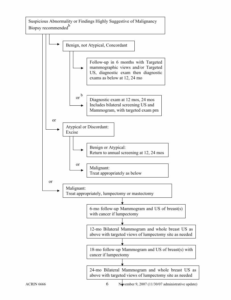
Suspicious Abnormality or Findings Highly Suggestive of Malignancy Biopsy recommendedb
Benign, not Atypical, Concordant
Follow-up in 6 months with Targeted mammographic views and/or Targeted US, diagnostic exam then diagnostic exams as below at 12, 24 mo
Diagnostic exam at 12 mos, 24 mos Includes bilateral screening US and Mammogram, with targeted exam prn
Atypical or Discordant: Excise
or b
Benign or Atypical: Return to annual screening at 12, 24 mos
Malignant: Treat appropriately as below
Malignant: Treat appropriately, lumpectomy or mastectomy
6-mo follow-up Mammogram and US of breast(s) with cancer if lumpectomy
12-mo Bilateral Mammogram and whole breast US as above with targeted views of lumpectomy site as needed
18-mo follow-up Mammogram and US of breast(s) with cancer if lumpectomy
24-mo Bilateral Mammogram and whole breast US as above with targeted views of lumpectomy site as needed
or
or
or

ACRIN 6666 7 November 9, 2007 (11/30/07 administrative update)
Magnetic Resonance Imaging (MRI) of the Breast
a If probably benign findings are identified, a 6 month follow-up diagnostic unilateral mammogram with or without spot compression and/or magnification views and/or targeted ultrasound will be performed as appropriate. Each annual examination will include both breasts in their entirety. Acceptable follow-up of a probably benign finding would be one of the following: 2 year stability, biopsy (or aspiration if appropriate), or decrease beyond experimental error or resolution at any follow-up. b If the lesion is amenable to percutaneous core (14-g) or directional vacuum-assisted (11-g) biopsy, it is anticipated that this will be the preferred method of initial biopsy, though inaccessible or poorly visualized lesions may require direct needle localization and excision. Lesions that may be complicated cysts but are felt to require intervention may be aspirated in lieu of core biopsy if they resolve completely. With a specific benign, concordant diagnosis of fibroadenoma, fat necrosis, or lymph node, the participant may resume annual screening. A concordant result of fibrocystic changes, sclerosing adenosis, or other benign result will require a 6 month follow-up diagnostic unilateral mammogram and/or targeted ultrasound directed to the abnormality biopsied. Atypical results on core biopsy or aspiration will prompt needle localization and excision as described in Section 4.10.1. Initial prevalence screen and annual incidence screens are planned for 2 subsequent consecutive years for all participants (at 0 months, 12 months and 24 months). Mammography and physician-performed US will be conducted independently at each annual screen. The order in which these exams are given will be randomly determined at the initial prevalence screen and that same order will be carried forward for all other screens. A “screening” examination is defined as a whole breast bilateral ultrasound and bilateral CC and MLO view mammogram in an asymptomatic woman with no known current breast problems, supplemented as needed by additional projections necessary to cover the tissue. For participants
Eligible participants (1200 women) from a subset of the ACRIN 6666 protocol will undergo a single screening contrast-enhanced breast MRI examination after completion of, and within 8 weeks of, the 24 month screening US and mammogram.
Additional suspicious lesions seen only on MRI will undergo second-look targeted US for biopsy guidance or MRI-guided vacuum-assisted biopsy after completion of any biopsies or additional views prompted by the 24-month screening US and mammogram visit. NOTE: Results of MRI will not be used to deter additional views prompted by screening mammography and/or US.
A six month follow-up MRI may be needed in some participants for probably benign findings seen only on MRI.
Clinical follow-up of cancer status of all participants at 36-38 months after initial study entry will conclude the follow-up.

ACRIN 6666 8 November 9, 2007 (11/30/07 administrative update)
who are status post mastectomy, these are unilateral examinations. For annual follow-up of participants who are status post lumpectomy for cancer, this may include magnification views of the lumpectomy site. For purposes of the study, a “diagnostic” examination is one targeted to a specific area of concern. A final assessment of negative (BI-RADS® 1) or benign (BI-RADS® 2) may result from the screening or diagnostic examinations. Final assessments of probably benign (BI-RADS® 3), suspicious (BI-RADS® 4), or highly suggestive of malignancy (BI-RADS® 5) are expected after screening or diagnostic US or diagnostic mammography. It is expected that most abnormalities on screening mammography will receive a BI-RADS® assessment of 0, requiring additional evaluation on the clinical report; similarly, calcifications seen sonographically will likely be coded as BI-RADS® 0 on the clinical report and require comparison to mammography and possibly additional mammographic views. In order to facilitate further analysis, investigators will be asked for their rating of likelihood of malignancy in the (hypothetical) absence of further work-up for those findings requiring additional evaluation. Participants will have mammography and physician-performed bilateral whole breast ultrasound examinations at each annual “screen.” It is suggested that the clinical mammographic report be addended to indicate the results of the study screening sonographic report as detailed in Section 4.6.6. The order of those examinations will be randomized to avoid bias that may result from additionally requested workup due to either modality. For each participant, the order of the examinations will be the same for each annual screen. Such randomization may prove to be a barrier to accrual and burdensome to sites. If we find accrual is deficient (defined in Section 6.3), we will consider dropping the randomization after discussion with the Data Safety and Monitoring Board. If randomization is discontinued, participants will undergo initial mammography then independently performed and interpreted sonography. Eligibility: Original Screening US protocol (see Section 5.3 for details; accrual closed 2/3/06): • Women ≥ 25 years of age; • High-risk of breast cancer (at least one of the following):
o Known to have a mutation in BRCA-1 or -2; o Personal history of breast cancer (with conserved breast analyzed separately; after
mastectomy, the breast reconstructed with autologous tissue or implant[s] will not be imaged, but the other breast will be eligible for imaging);
o History of prior biopsy showing ADH, ALH, or atypical papilloma not receiving chemoprevention [i.e. not on Tamoxifen, Evista (Raloxifene), Arimidex (Anastrazole), Aromasin (Exemestane), or any other aromatase inhibitor]; or, any of these atypical lesions (including phyllodes tumors) and a first degree relative diagnosed with breast cancer under age 50 even if the patient is on chemoprevention;
o History of prior biopsy showing LCIS; o History of prior chest and/or mediastinal and/or axillary irradiation ≤ age 30 and at least 8 years
previously; o Lifetime risk of breast cancer by Gail or Claus models ≥ 25%; o Five-year risk of breast cancer by Gail model ≥ 2.5%; o Five-year risk of breast cancer by Gail model ≥ 1.7% and known to have extremely dense breasts
(at least 75% dense) by most recent prior mammogram; • Heterogeneously dense or extremely dense breasts (see Section 5.3) or unknown breast density due to
no prior mammogram;

ACRIN 6666 9 November 9, 2007 (11/30/07 administrative update)
• The participant agrees, in principle, to return for the required two-year follow-up and/or biopsy if necessary;
• Most recent mammogram (if any) was interpreted as negative, benign and/or remarkable only for post-treatment changes; this is a routine annual visit (i.e. at least 11 full months have elapsed since the prior routine annual mammogram, per Section 4.5);
• Signed study-specific informed consent prior to study entry; • No present signs or symptoms of breast cancer (no palpable breast mass(es), bloody or spontaneous
clear nipple discharge, axillary mass, or abnormal skin changes in the breast(s) or nipple(s); • No medical or psychiatric conditions that would preclude biopsy; • No prior malignancy other than:
o Breast cancer at least one year earlier (12 full months have elapsed since the last treatment surgery) with no known distant metastases and no known residual tumor, or
o Basal or squamous cell skin cancer or in situ cervical cancer, or o Other cancer for which the patient has been disease free for ≥ 5 years, with no recurrence of
cancer in the last five years and no residual disease detected in the last five years. • Not pregnant or breast-feeding, or planning to become pregnant within 2 years of study entry; • No breast implant(s) currently in the study breast(s); • No breast procedures (fine needle aspiration, core biopsy, surgical procedure) within one year prior to
study entry; • No participation in other breast cancer screening trials; • Has not undergone contrast-enhanced breast MR within one year prior to study; • Has not undergone whole breast bilateral sonography within one year (i.e. at least 11 full months
have elapsed) prior to study; • Has not undergone injection of sonographic or mammographic contrast agents or tomosynthesis
within one year prior to study entry; • No participation in studies of breast MR, sonographic or mammographic contrast agents, or
tomosynthesis during the trial period (entry and 2 years of follow-up). Note: If the participant is diagnosed with breast cancer during the trial period, it is then acceptable for the participant to undergo contrast-enhanced breast MR to evaluate the extent of disease for treatment planning.
MRI of the Breast at 24 Months (See section 5.4 for details): Study participants who have completed three annual rounds of screening with both mammography and US as part of ACRIN 6666 protocol by February 10, 2008 are potentially eligible for participation in the MRI component of the study. In addition to women with prior negative (BI-RADS 1) mammogram and US examinations, women undergoing surveillance of findings which are considered benign (BI-RADS 2) or probably benign (BI-RADS 3) on prior breast imaging (i.e. not including the results of the 24 month screening mammogram or US examinations) are eligible. Required Sample Size: 2808 participants (with 2809 enrolled as of 2/3/06 and accrual closed at that time). For the MRI component of the trial, the estimated sample size is 1200 participants.
ACRIN 6666 10 November 9, 2007 (11/30/07 administrative update)
1.0 ABSTRACT Early detection is currently the most effective strategy to reduce deaths from breast cancer. Mammographic screening is highly effective in identifying calcifications due to ductal carcinoma in situ. Invasive cancer, which can spread to the lymph nodes and ultimately metastasize, is usually well seen in fatty breasts but is often mammographically subtle or occult when the breast tissue is dense. Ultrasound requires no ionizing radiation, no discomfort to the breasts, and is not limited by breast density. In several single-center studies, screening ultrasound allowed detection of small nonpalpable invasive breast cancers not visible on mammography. It is easy to perform a needle biopsy of lesions found on ultrasound. The full potential of ultrasound in screening for breast cancer will not be realized, however, unless these promising results can be generalized across investigators and institutions. Ultrasound is highly dependent on the operator and on the equipment and technique used. Further, many incidental solid masses and complicated cystic lesions are found on screening ultrasound. While criteria have been proposed that will allow many of these lesions to be followed rather than biopsied, these criteria have not been validated at multiple centers and it is not clear that they will be generalizable. Improved ultrasound technology such as spatial compounding may help in margin analysis and thereby in reliably identifying lesions that can be followed. We propose a multicenter trial of screening whole breast ultrasound using standardized technique and interpretation criteria in women at high risk of breast cancer. We will perform annual sonographic screening for three years (at 0, 12, and 24 months) independently, and in addition to, mammography screening. The number of cancers seen on the initial screen (prevalent cancers) as well as each of two subsequent screens (incident cancers) will be assessed (see Section 4.10). We will collect follow up information as to cancer status through 36 months after study entry. It is hoped that the results of this trial will provide guidance to participants and practitioners alike on the role, if any, of screening breast ultrasound and the associated risk of an unnecessary biopsy. If the results are favorable, a larger study to evaluate all women of screening age with dense breasts may be necessary to allow more generalized recommendations. Consortium A consortium of sites emphasizing centers within the Avon Foundation Breast Cancer Research and Care Network as well as additional university and private practice settings with recognized expertise in breast imaging, (specifically mammography and sonography) have agreed to participate in this study conducted by American College of Radiology Imaging Network. At each site, at least two investigators have agreed to be trained in study protocol for both mammographic interpretation and sonographic performance and interpretation. Lead investigators and the sites in the consortium are summarized in Table 1. Magnetic Resonance Imaging (MRI) of the Breast There remains uncertainty as to the most appropriate method(s) to screen high risk women for breast cancer. Annual surveillance with both ultrasound (US) and mammography may allow detection of the vast majority of cancers when they remain minimal. Contrast-enhanced magnetic resonance imaging (MRI) is limited by high cost, reduced patient tolerance, and access issues. US is inexpensive, well tolerated by patients, and widely available. Widespread implementation of screening MRI, even limited to high-risk women, is problematic. Private carriers and Medicare are often reimbursing for screening MRI in women at high genetic risk of breast cancer, at considerable costs to the health care system.

ACRIN 6666 11 November 9, 2007 (11/30/07 administrative update)
With the women who have completed three rounds of annual screening US and mammography as part of ACRIN 6666 protocol, this study provides a unique opportunity to estimate the role of MRI, if any, above and beyond combined US and mammography. The use of US has been carefully controlled in protocol 6666, with extensive training and qualification of investigators, high quality equipment, and strict interpretive criteria. Combined US and mammography has the potential to be far more cost effective in screening than MRI. If, however, even after three rounds of annual screening with US and mammography, MRI retains the potential to significantly increase the cancer detection yield (as has been seen in three smaller prior studies [1-3]), this study would provide additional support for current use and future studies of screening MRI in high-risk women with dense breasts. While digital mammography shows improved sensitivity over film in denser breast tissue and in younger women [4], a large percentage of cancers remain undetected. Indeed, in a multicenter study of digital mammography, at least 30% of cancers were mammographically occult even with digital mammography [4]. As of September 2005, 34% of the participants in ACRIN 6666 have received digital mammograms, and this percentage is expected to increase. 2.0 BACKGROUND AND SIGNIFICANCE Screening mammography has yielded significant reduction in mortality from breast cancer within and outside of multiple randomized controlled trials, ranging from 23 to 65% [5, 6], and there is a shift toward detection of smaller, lower grade tumors with better prognosis [7, 8]. The sensitivity of mammography is as high as 98% in women over 50 with fatty breasts, 84% with dense breast tissue, and 69% in women under 50 with a family history of breast cancer [9, 10]. Recently published work by Kolb et al [11] suggests the sensitivity of mammography may be as low as 48% in extremely dense breasts and that age < 50 may be an independent factor lowering mammographic sensitivity. The use of US for screening has also been proposed. Previous studies in the 1980s of screening US failed to demonstrate a benefit [12-14], and indeed only 18% of nonpalpable mammographically depicted lesions going to biopsy could be seen sonographically in one small series [12]. Technology has improved dramatically since that time, however, and systematic reevaluation is merited. In the Radiological Diagnostic Oncology Group V trial that accrued from 1994 through 1996, 551/719 (77%) of nonpalpable, mammographically depicted masses going to biopsy could be seen sonographically [15]. More recent studies of whole breast sonography include that of Gordon and Goldenberg in 1995 [16], who documented 1575 solid masses including 44 cancers seen only on US in 12,706 (0.3%) women undergoing breast sonography for other reasons. In 1998, Kolb et al [17] evaluated 3626 women with non-fatty breasts and normal mammograms and clinical breast exam. Two hundred fifteen solid masses were found on US only, of which 11 (5.1%) proved malignant [17]. Another 974 women (27%) had cysts, and 132 (3.6%) had complicated cysts [17]. Follow-up or aspiration was performed for those with complicated cysts and no malignancies were found in that group [17]. Buchberger et al [18] screened 6113 asymptomatic women with non-fatty breasts with US and found 23 cancers in 21 women, though another 353 incidental masses required aspiration or biopsy. In an update of those results [19], the average size of cancer depicted only by US was 9 mm, the same as that of cancers found at mammographic screening. Kaplan [20] reported results on 1862 women with negative clinical exam, heterogeneously dense or dense parenchyma, and bilateral screening
ACRIN 6666 12 November 9, 2007 (11/30/07 administrative update)
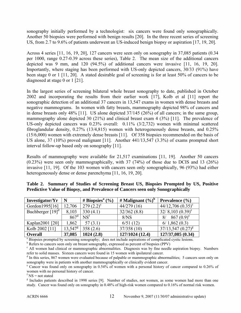
sonography initially performed by a technologist: six cancers were found only sonographically. Another 50 biopsies were performed with benign results [20]. In the three recent series of screening US, from 2.7 to 9.6% of patients underwent an US-induced benign biopsy or aspiration [17, 19, 20]. Across 4 series [11, 16, 19, 20], 127 cancers were seen only on sonography in 37,085 patients (0.34 per 1000, range 0.27-0.39 across these series), Table 2. The mean size of the additional cancers depicted was 9 mm, and 120 (94.5%) of additional cancers were invasive [11, 16, 19, 20]. Importantly, where staging has been performed with US-only depicted cancers, 30/33 (91%) have been stage 0 or 1 [11, 20]. A stated desirable goal of screening is for at least 50% of cancers to be diagnosed at stage 0 or 1 [21]. In the largest series of screening bilateral whole breast sonography to date, published in October 2002 and incorporating the results from their earlier work [17], Kolb et al [11] report the sonographic detection of an additional 37 cancers in 13,547 exams in women with dense breasts and negative mammograms. In women with fatty breasts, mammography depicted 98% of cancers and in dense breasts only 48% [11]. US alone depicted 37/145 (26%) of all cancers; in the same group, mammography alone depicted 30 (21%) and clinical breast exam 4 (3%) [11]. The prevalence of US-only depicted cancers was 0.23% overall: 0.11% (3/2,732) women with minimal scattered fibroglandular density, 0.27% (13/4,815) women with heterogeneously dense breasts, and 0.25% (15/6,000) women with extremely dense breasts [11]. Of 358 biopsies recommended on the basis of US alone, 37 (10%) proved malignant [11]. Another 441/13,547 (3.3%) of exams prompted short interval follow-up based only on sonography [11]. Results of mammography were available for 21,517 examinations [11, 19]. Another 50 cancers (0.23%) were seen only mammographically, with 37 (74%) of those due to DCIS and 13 (26%) invasive [11, 19]. Of the 103 women with cancers seen only sonographically, 96 (93%) had either heterogeneously dense or dense parenchyma [11, 16, 19, 20]. Table 2. Summary of Studies of Screening Breast US, Biopsies Prompted by US, Positive Predictive Value of Biopsy, and Prevalence of Cancers seen only Sonographically Investigator/Yr N # Biopsiesa (%) # Malignant (%)b Prevalence (%) Gordon1995[16] 12,706 279 (2.2)c 44/279 (16) 44/12,706 (0.35)c
8,103 330 (4.1) 32/362 (8.8) 32/ 8,103 (0.39)e Buchberger [19]d 867d NSf 8/NS 8/ 867 (0.9)e
Kaplan2001 [20] 1,862 57 (3.1) 6/51 (12) 6/ 1,862 (0.3) Kolb 2002 [11] 13,547g 358 (2.6) 37/358 (10) 37/13,547 (0.27)g
Overall 37,085 1024 (2.8) 127/1024 (12.4) 127/37,085 (0.34) a Biopsies prompted by screening sonography; does not include aspirations of complicated cystic lesions. b Refers to cancers seen only on breast sonography, expressed as percent of biopsies (PPV) c All women had clinical or mammographic abnormalities. Diagnosis was by fine needle aspiration biopsy. Numbers refer to solid masses. Sixteen cancers were found in 15 women with ipsilateral cancer. d In this series, 867 women were evaluated because of palpable or mammographic abnormalities; 5 cancers seen only on sonography were in patients with another mammographically or clinically evident cancer. e Cancer was found only on sonography in 0.54% of women with a personal history of cancer compared to 0.26% of women with no personal history of cancer. f NS = not stated g Includes patients described in 1998 series [9]. Number of studies, not women, as some women had more than one study. Cancer was found only on sonography in 0.48% of high-risk women compared to 0.16% of normal risk women.

ACRIN 6666 13 November 9, 2007 (11/30/07 administrative update)
The large number of incidental solid masses points to the need for reliable lesion characterization. Indeed, across the above single institution series [11, 16-18, 20], the positive predictive value of a recommendation for biopsy or aspiration ranged from 3.1 to 10.5%. Stavros et al [22] proposed criteria for assessing solid masses on US. In his series [22], uniformly echogenic masses and those with two or three gentle lobulations, ellipsoid, and lacking any suspicious features could be considered probably benign with < 2% risk of malignancy, though further multicenter validation is needed. Unfortunately, Rahbar et al [23] and Baker et al [24] found that not all readers could effectively apply these criteria. The need for generalizable criteria for following incidental masses seen only on sonography remains great, and validation of specific criteria is needed. Complicated cysts have been defined as masses with homogeneous low-level internal echoes throughout that otherwise meet the criteria of a simple cyst [25]. Venta et al [26] recently found only 1/308 (0.3%) of complicated cysts to be malignant, containing a 3 mm focus of ductal carcinoma in situ (DCIS). None of the 132 complicated cysts in the series of Kolb et al [17] proved malignant, nor did any of the 127 in the series of Buchberger et al [18]. It has recently been suggested that circumscribed masses with posterior enhancement and a fluid-debris level or mobile internal echoes without a discrete solid component would also appropriately be considered a complicated cyst [27]. Thus it appears that in the absence of a mural mass, thick wall or thick septations, cysts with homogeneous low-level internal echoes can be considered probably benign and followed, with a positive predictive value of 0.2% across these several series [17, 18, 26]. Complex cystic lesions with a discrete solid component, thick wall, thick (≥ 0.5 mm) septations, or intracystic mass merit biopsy, with 18/79 (23%) of such lesions proving malignant in one series [27]. Excluding aspirations of complicated cystic lesions, biopsies were recommended in 2.2 to 4.1% of sonographically-detected masses (overall 2.8%), with a positive predictive value of biopsy of 8.8 to 16% (12.4% overall) (Table 2 [11, 16, 19, 20]). The accuracy of sonography for characterizing simple cysts approaches 100% [28] provided strict adherence to classical criteria are observed: a circumscribed round, oval, or gently lobulated, anechoic mass, with posterior enhancement. Simple cysts can be dismissed as benign. Very small simple cysts (< 4 to 5 mm, depending on depth in breast and equipment) may appear as solid masses or complicated cysts. Round lesions that appear solid would remain indeterminate. Oval or gently lobulated, circumscribed masses with posterior enhancement or no posterior features, which might be small cysts or solid masses, would appear to be appropriately classified as probably benign provided such lesions are incidental findings, with short interval follow-up sonogram (in 6 months) appropriate. Nonpalpable lesions composed entirely of clusters of microcysts with thin (< 0.5 mm) septations are often due to apocrine metaplasia [29] or other fibrocystic changes. This may be another class of lesions, which can be considered probably benign. In the series of Berg et al [27], all 16 lesions with this appearance proved benign. In an overlapping series [30] of 66 such lesions with 2-year follow-up (n=48) or biopsy (n=18), no malignancies have been identified. Ultrasound has widespread acceptance as a diagnostic tool for the evaluation of palpable and nonpalpable abnormalities and the combined diagnostic yield of mammography and sonography has been shown to be greater than mammography alone in women with palpable lumps or abnormal screening examinations [31]. It is easy to guide interventions with US, and US can be used in

ACRIN 6666 14 November 9, 2007 (11/30/07 administrative update)
evaluation of problems associated with breast implants [32, 33]. As with any test, an abnormality must be recognized by the observer. Unlike many other examinations, double reading is not readily accomplished with US, as real-time information is needed to determine the presence of an abnormality and, at times, to appropriately analyze its features. Skaane et al [34] reported slightly lower interobserver agreement for ultrasound than for mammography or combined readings, with mean kappa of 0.48 for hard-copy ultrasound images, compared to 0.58 for mammography and 0.71 for the combined readings. Baker et al [24] reported kappa of 0.51 for management based on sonographic images. Despite these multiple potential sources of variability, Bosch et al [35] found high interexamination agreement in both detection and classification across three observers independently performing real time whole breast sonography in 58 patients and 113 breasts; 60% of breasts had a lesion and 10% had cancer. Kappas were 0.72-0.75 between pairs of observers indicating excellent reliability [35], decreasing slightly to a mean of 0.65 when normal breasts were excluded, and further decreasing to 0.55 in the 32 dense breasts evaluated (compared to 0.82 in non-dense breasts). Importantly, these kappas exceeded those of mammography across the same observers in the same patients [35]. Note that in the study of Bosch et al [35], a resident with experience performing 500 sonographic examinations performed on par with more senior investigators. These results suggest that ultrasound is indeed reliable enough to evaluate its performance in a multi-institutional screening study. Standardization of technique with respect to transducer frequency, positioning the patient, scan planes, setting of focal zones, and even specifics of labeling have not been established previously. Investigators will be specifically trained in these technical aspects prior to initiating the study (Section 4.2). To establish that our investigators meet a standard of performance in lesion detection, we have established experience requirements (Section 6.1.1) as well as a qualification task in phantoms, as detailed in Section 4.3. Further evaluation of the factors that affect reliability may be warranted in separate reliability studies. Based on the evidence produced thus far, such evaluations, while of scientific interest themselves, are not critical to the conduct or interpretation of our proposed screening trial. Professional guidelines for the performance of breast US have been published by the American College of Radiology [33] and include the following:
1) At least one set of images of a lesion should be obtained without calipers. The maximal dimensions of a mass should be included. If volume analysis is needed, three-dimensional measurements should be obtained.
2) Label images as to right or left breast, lesion location (specified by quadrant, clock position, distance from the nipple, or shown on a diagram of the breast), and orientation of the probe.
3) Linear array transducer greater than 7 MHz should be used. 4) Set the focal zone at the depth of the lesion. 5) Gain settings should be adjusted to allow simple cysts to be distinguished from solid
masses. 6) Patient should be positioned supine for the inner breast and supine oblique to evaluate the
upper outer quadrant and lateral breast (with the ipsilateral shoulder elevated by a pillow or wedge).

ACRIN 6666 15 November 9, 2007 (11/30/07 administrative update)
7) Permanent identification label for each study should include the patient’s first and last names, identification number and/or date of birth, facility name and location, examination date, and the sonographer’s identification.
Baker and Soo [36] evaluated static images from 152 examinations at 86 institutions and found 60.5% of cases failed to comply with at least one of these guidelines. Errors in interpretation were identified in 23/152 (15%) of cases [36]. To further ensure standardization of interpretation in this protocol, interpretive criteria will be reviewed with investigators as will a set of 70 proven US cases chosen to emphasize the threshold of intervention. As described in Section 4.3, investigators will be required to qualify for study participation based on their performance recommending biopsy appropriately in that test set of cases developed specifically for the trial (as well as a set of 50 mammographic lesions previously evaluated [37]). Factors that influence the performance of breast US have not been systematically studied to date. These may include the size of the breast, “depth” of the breast from the skin to the chest wall, and depth of any lesions. The ability to distinguish a < 5 mm complicated cyst from a solid mass may be especially problematic, and even simple cysts can be difficult to characterize when deep. Phantoms will be constructed to assure that consistent performance in identifying small simple cysts can be demonstrated on the equipment used across the multiple sites in this trial. Indeed, as of March 2003, the first phantom is available for testing. Composition of the breast may also be a factor. It has been suggested that masses may be more difficult to identify in fatty breasts. Normal interfaces at the edge of fatty lobules can cause posterior acoustic shadowing that may be mistaken for a lesion. There are breasts with diffusely heterogeneous echotexture, which may obscure detail and lower the sensitivity (and perhaps also specificity) of sonography; this has not been addressed in prior studies, but heterogeneity of echotexture will be systematically recorded in this trial. Screening with US is problematic also at this time due to its requirement of considerable physician resources. In Kaplan’s study [20], technologists performed the initial sonogram, with verification by the physician. Dennis et al [38] also report success with technologist-performed breast sonography. This remains an area for further validation and would indeed be necessary to implement widespread sonographic screening. However, this is beyond the scope of this trial. Kolb et al [17] reported the mean time for performing a complete bilateral screening US examination was 3 min 59 sec, with a range of 1 min 28 sec to 9 min 46 sec. This may be optimistic and requires further validation. This does not include the time to complete the dictation and interpretation. We will monitor these times as the study progresses. The full costs of screening US must include calculation of the induced costs of follow-up, aspirations, and biopsies. In addition to the rates of induced procedures above, short interval follow-up was recommended in another 3-10% of patients in the above series [11, 16, 18, 20]. It is doubtful that US will replace mammography in the depiction of DCIS, as the vast majority of DCIS is manifest as microcalcifications [39]. Due to the speckle artifact normally present in breast tissue, most calcifications remain occult sonographically unless present within a mass. Spatial compounding reduces speckle artifact and may improve DCIS detection. Moon et al [40] performed prebiopsy US in a series of 100 mammographically depicted foci of calcifications and found 45 (45%) were visible. Calcifications were far more likely to be seen when in a malignant mass, with 31/38 (82%) of such lesions visible sonographically compared to 14/62 (23%) of those in benign
ACRIN 6666 16 November 9, 2007 (11/30/07 administrative update)
processes [40]. In the series of Skaane and Sauer [41], only 1/18 (6%) of DCIS foci were seen sonographically and recommended for biopsy. Another 9/18 (50%) of DCIS were seen as focal abnormalities but not recommended for biopsy, and 8/18 (44%) of DCIS were not seen [41]. In the series of Berg and Gilbreath [38], 7/16 (44%) of DCIS foci were seen sonographically. In nonoverlapping results presented by Berg et al [43] at the Radiologic Society of North America 2001, mammography depicted 17/28 (61%) of DCIS foci, sonography 15/28 (54%) and magnetic resonance (MR) imaging 25/28 (89%). As stated above, of the 127 cancers seen only sonographically in the four summary single center series to date [11, 16, 19, 20], 120 (94.5%) were invasive and 7 (5.5%) were DCIS [44]. Indeed, one criticism of mammographic screening is its high sensitivity for detecting noninvasive disease (DCIS) manifest as microcalcifications. The benefit of detecting DCIS is not clear in every case, particularly in women over age 70. The need for aggressive treatment of all DCIS remains controversial [45]. From autopsy series, up to 15% of women have undiagnosed DCIS at the time of death [46]. It would appear that a large number of cases of DCIS do not come to clinical relevance. Review of pathologic specimens has occasionally demonstrated foci of (low-grade) DCIS initially classified as benign. In these series, invasive cancer developed in from 11-60% of cases with 10-24 years of follow-up, and 75% of these cancers were at the original site of DCIS (reviewed in [47]). At this time we have no reliable method to distinguish when a cancer has become invasive: detection and treatment of DCIS is currently sought. There is, however, the potential that US will depict the vast majority of clinically significant DCIS (e.g. larger foci of DCIS, potentially higher grade DCIS). If screening US is to be offered routinely, clear understanding of the false negative rate and sources of false negatives will be necessary. Patients seek an alternative to mammography and require accurate information. Assessment of the sensitivity of US to detection of breast cancer independent of mammography is an important secondary aim. The sensitivity of US to invasive cancer indeed may exceed that of mammography, with 45/48 (94%) sensitivity of US and 39/48 (81%) sensitivity of mammography in the series of Berg and Gilbreath [42] evaluating patients newly diagnosed with cancer. In the series of Skaane and Sauer [41], 223/246 (90.6%) of invasive ductal cancers were classified as indeterminate or malignant sonographically; another 8/246 (3.3%) were seen but not recognized and 9/246 (3.7%) were not seen on sonography. In the recently presented work of Berg et al [43], of 97 foci of invasive ductal carcinoma, mammography depicted 75 (77%), sonography 92 (95%), and MR imaging 90 (93%). The sensitivity of mammography to invasive lobular carcinoma is particularly low and it is overrepresented among missed cancers [48]. In the series of Butler et al [49], 81/208 (39%) of invasive lobular carcinomas were considered mammographically occult or subtle. Of those 81, 71 (88%) were depicted sonographically [49]. In the series of Berg and Gilbreath [42], 7/11 (64%) of foci of invasive lobular carcinoma were depicted mammographically and 9/11 (81%) sonographically, though 2/11 (19%) were occult on both. In the recently presented work of Berg et al [43], mammography depicted 9/26 (35%) of invasive lobular carcinoma, sonography 21/26 (81%), and MR imaging 26/26 (100%). In the series of Skaane and Sauer [41], 35/39 (90%) of invasive lobular carcinomas were depicted sonographically, though one was misclassified as benign.

ACRIN 6666 17 November 9, 2007 (11/30/07 administrative update)
Invasive lobular cancer usually lacks microcalcifications, is frequently manifest as a focal asymmetric density, and often is seen in only one mammographic view [50-52]. Asymmetric densities are commonly seen, however, in approximately 3% of mammograms, as a normal variant [53]. Malignancies due to focal asymmetries are therefore not surprisingly among the most common cause of false negative mammographic interpretations [54, 55]. Anecdotally, US can be very helpful in evaluating persistent asymmetric densities [56]. A secondary endpoint of this study will be determination of the negative predictive value of a negative sonogram in areas of focal asymmetric density mammographically. It is unlikely we will have a sufficient number of cancers manifest as asymmetries to fully address the impact of sonography in this setting, but these results will likely provide important preliminary data assessing the utility of sonography in further evaluation of asymmetries seen mammographically. It may be more cost-effective and facilitate earlier detection of true positives if patients with focal asymmetries undergo sonography as immediate evaluation rather than several short-interval follow-ups. The combination of mammography and sonography may be particularly effective in depicting breast cancer. In the study of Kolb et al [11], mammography alone depicted only 48% of breast cancers in dense breasts, whereas mammography and sonography together depicted 97%. Similarly, in a study of 374 women with 2-year follow-up information and/or linkage with a state cancer registry, Moy et al [57] reported only 6 (2.6%) of women had cancer not seen on either mammography or sonography. In a matched pairs analysis of 240 consecutive symptomatic women who underwent both mammography and sonography at a breast clinic in Sydney, Australia, Houssami et al [58] reported combined sensitivity of mammography and sonography of 96% and specificity of 79%. Sonography was more sensitive than mammography in women under age 46 [58]. Thus the primary aim of this study is to determine the performance (sensitivity, specificity, positive and negative predictive values) of combined mammography with sonography to that of the current standard of mammography alone. As noted, MR imaging is highly sensitive to breast cancer and is currently being proposed as a screening supplement to mammography in high-risk women [59-62]. Across several series evaluating high-risk women [59-61], after a normal mammogram and clinical breast examination, approximately 3/100 will have cancer found on the first MR screening. Across several series, high yields of cancers seen only on MRI have persisted on subsequent screening rounds, even among women screened with mammography combined with US. Specifically, Kuhl et al [3] found 2.6% cancer detection rate across modalities in year one, and 2.5% in years 2-6 of screening, with 44% of all cancers seen only on MRI and the same additional yield of MRI in each year (C. Kuhl personal communication, October 2005). In the series of Warner et al [2], MRI-only detection rates in BRCA-1 or -2 mutation carriers were 4.7% in year 1, 2% in year two, 1% in year three, and 3% in year four (R. Jong, personal communication 5/06). In the series of Kriege et al [63], in women with 15% lifetime risk of breast cancer undergoing only mammography, clinical breast examination, and MRI, the yield of MRI was 10-12 per 1000 in years one and two, and 3-7 per 1000 in subsequent years, and was always at least double the detection rate of mammography. Unfortunately, MR requires injection of intravenous contrast, is approximately 10 times as costly as US, less available, and, compared to US, is hampered by challenges in biopsying and confirming successful biopsy of lesions depicted only on MR imaging. US is attractive as a supplement to mammographic screening. It is widely available, and sonographically-guided aspiration and/or core biopsy is readily performed [64-66]. Of note, the

ACRIN 6666 18 November 9, 2007 (11/30/07 administrative update)
combination of mammography and US was shown to be equal in sensitivity to MR in one series [67]. In our experience, as noted, the combined performance of mammography and US was the same as that of MR for invasive ductal cancer but was slightly less than MR for invasive lobular carcinoma [43]. The potential benefit of any supplement to mammography is greatest in women at high risk and in those with dense breasts. Only one recent study of whole breast US after mammography included women with fatty breasts, and no benefit to US was found in such patients [68]. As stated, across four series, [11, 16, 19, 20], of the 103 women with cancer seen only sonographically, 96 (93%) had heterogeneously dense or extremely dense breasts. Precise definition of the meaning of heterogeneously dense or extremely dense parenchyma is lacking. We describe specific scenarios where the mammographic density is felt to be sufficient to obscure small masses in at least one quadrant of the breast as a threshold criterion (Section 5.3). Across these same series [11, 16, 19, 20], of 103 women with cancer depicted only sonographically, 51 (50%) were women at high risk of breast cancer. Of 478 women at “very high risk” in the series of Kolb et al [17], 5 (1%) had cancer found only on US. In the more recent overlapping series of Kolb et al [11], of 3,588 women with a high risk because of a first degree relative with breast cancer or personal history of breast cancer, 15 (0.42%) had cancer found only on US. In women with newly diagnosed breast cancer, evaluation of the contralateral breast is receiving increasing attention. In a series of 405 patients with newly diagnosed cancer evaluated with mammography, clinical breast examination, sonography, and MR, Fischer et al [69] found 19 (4.7%) with synchronous bilateral cancer. Of the 19 contralateral cancers, 15 (79%) were seen only on MR. In the series of Kuhl et al [70], and also in the series of Woo et al [71], 6% of patients with newly diagnosed cancer had unsuspected contralateral cancer seen on MR. In our experience [43], 10/97 (10%) of patients with newly diagnosed cancer had bilateral synchronous cancer: 7/10 (70%) were depicted mammographically, and 3/10 (30%) were seen only on MR and US. Another patient suspected of cancer in the right breast proved to have a 5 mm tubular cancer in the left breast seen only on MR. We propose to systematically evaluate screening US in a controlled, multicenter trial. By limiting the initial protocol to high-risk women, we are selecting a population enriched with cancers where disparities between mammography, clinical breast examination, and sonography will be readily apparent. As described, there are many issues in the performance and reproducibility of screening breast US that need to be addressed. The impact of the large number of false positive tests on quality of life and costs of medical care will need to be evaluated as well. As such, mortality is not an endpoint of this trial. Surrogate measures such as lesion size [72] and nodal status [73] and diagnostic yield will be evaluated. If the results of this study are favorable, a broader study of screening breast US, which may include mortality as an endpoint, will be needed prior to widespread implementation. Magnetic Resonance Imaging (MRI) of the Breast In women at high risk of breast cancer and particularly those with dense breasts, there has been an increasing interest in supplemental screening with MRI or US in addition to mammography. Fewer than half of cancers are seen on mammography in such women [1, 11, 74]. MRI and US have both been shown to depict small invasive cancers < 2 cm in size, with negative nodes, which are not seen

ACRIN 6666 19 November 9, 2007 (11/30/07 administrative update)
on mammography, and the detection of such cancers should reduce morbidity and mortality from breast cancer.
The current ACRIN 6666 protocol seeks to determine the yield of combined US and mammography in such women. In single center studies, there is reported to be a high yield of cancers seen only on MRI in high risk women, averaging 1.8% across 8 series, compared to 0.48-1.3% for US [75]. Investigators all completed training in standardized technique and interpretive criteria for both US and mammography, and state of the art US equipment has been used. Detailed information on risk factors, breast density, benign lesions seen on mammography and/or US, accompanying images and pathology reports where appropriate, as well as clinical follow-up, is in the ACRIN database for each patient, together with cost effectiveness data for US and mammography on these women. In three screening series of women at high genetic risk of breast cancer, each including fewer than 600 women to date (C Kuhl and F Sardanelli, personal communications 10/05), where MRI was performed in addition to US and mammography, the overall sensitivity of US was only 30%, compared to MRI at 96% [76]. Even after combined US and mammography, another 33% [1], 36% [2], or 42% [3] of all cancers respectively were seen only on MRI (including both invasive and intraductal carcinomas). A preponderance of grade III invasive ductal cancers was observed across all series among cancers seen only on MRI. Importantly, rates of cancer detection in high risk women appear comparable across prevalence and incidence screens [1, 3].
The series of Kriege et al [77], which compared mammography combined with MRI to mammography alone, found significant downstaging of cancers in the group of women screened also with MRI. Conflicting results have been reported in both the diagnostic [57, 78] and screening [11] settings, even where supplemental MRI has been used: Cancer detection rates of 92-97% have been reported after combined US and mammography in a broader population not limited to those at high genetic risk of breast cancer. As such, it is not clear that supplemental MRI after combined US and mammography would be of clinical benefit. Small invasive cancers < 1 cm in size, usually with negative nodes, are well seen on US [44]. The vast majority of the mortality reduction benefit due to breast screening is attributed to early detection of node negative invasive cancer. US is relatively insensitive to ductal carcinoma in situ (DCIS), whereas 24% of cancers seen only on MRI are DCIS [75]. The majority of cancers seen only on MRI after combined US and mammography might be DCIS, a result of uncertain significance. The 2809 participants in ACRIN 6666 will have received annual US and mammography for three screening rounds (total of 24 months), with the first of the 24 month screening examinations due in May 2006. Participants were selected based on a variety of criteria to define high risk (www.acrin.org), not limited to women at high genetic risk. All participants have at least heterogeneously dense parenchyma. With this group of women, this study provides an ideal opportunity to determine the additional cancer detection yield, if any, of contrast-enhanced breast MRI, above and beyond annual screening with combined US and mammography. In this amendment of the ACRIN 6666 protocol, eligible participants will undergo a single screening MRI examination after completion of the 24 month screening US and mammogram. Fewer than 2% of participants in ACRIN 6666 have had screening MRI during the study period or 12 months prior

ACRIN 6666 20 November 9, 2007 (11/30/07 administrative update)
to enrollment, and women having had a screening MRI during the 24 month study period are excluded from participation in the MRI substudy (Section 5.6). As such, this will be a prevalence screen for the yield of MRI above and beyond mammography combined with US. In prior MRI screening trials (C Kuhl and R Jong, personal communications), high risk women had been routinely screened with mammography and often (but not systematically) with US prior to initiation of MRI. If after three rounds of annual screening with US and mammography, MRI retains the potential to significantly increase the cancer detection yield by a clinically meaningful amount (as has been seen in three smaller prior studies [1-3]), this study would provide additional support for current use and future studies of screening MRI in high-risk women with dense breasts. Given this, broader population-based studies or registries of screening MRI may be warranted prior to widespread implementation. It will be particularly important to know the stage and grade of cancers found only on MRI, if any. False positives are a known limitation to any screening test, and the rate of false positives on MRI in this population will be determined. This information will greatly inform our approach to screening these women. Cost-effectiveness analyses will further inform public policy. While mortality will not be an endpoint of this study design, the size and nodal status of cancers depicted are validated measures of efficacy of a breast imaging screening examination [72, 79, 80]. In order to complete data collection for the MRI component of the study, the 36 month follow-up will be completed by February 2009 (i.e. clinical follow-up 36 months after study entry, which is 12 months after the screening MRI), allowing for forms collection and recommended biopsies to be performed. Another 6 months will be required for data analysis for this component of the study. 3.0 SPECIFIC AIMS/OBJECTIVES We anticipate that systematic scanning of the breast with high resolution ultrasound (US) imaging is capable of detecting nonpalpable breast cancers occult to mammography in women at high risk of breast cancer. Further, we expect this result to be relatively constant across multiple institutions. 3.1 Primary Specific Aim 3.1.1 Aim 1
To assess the diagnostic yield of integrated whole breast bilateral screening sonography combined with mammography compared to mammography alone in the detection of breast cancer in high-risk women with dense breasts.
3.2 Secondary Specific Aims 3.2.1 Aim 2
Determine the sensitivity and specificity of screening whole breast sonography and mammography independently in high-risk women and characterize the degree to which the performance of the screening modalities (screening mammography and ultrasound) depends on selected participant characteristics, such as breast density and heterogeneity of the parenchyma, respectively (here screening performance will primarily be measured by the area under the ROC curve, but other measures such as sensitivity, specificity, and diagnostic yield will be considered).

ACRIN 6666 21 November 9, 2007 (11/30/07 administrative update)
3.2.2 Aim 3 Validate the sonographic classification of certain lesions as “probably benign” and estimate the rate of malignancy in that classification after both sonographic and mammographic examinations.
3.2.3 Aim 4 Estimate the costs of screening breast ultrasound in terms of radiologist and resource time performing the exam and the induced costs of screening ultrasound (follow-up, biopsy). Assess the cost-effectiveness of screening breast US (see Section 12). Prior to the involvement in the screening trial, investigators wishing to participate must attend a training/qualification session or complete the specified qualification criteria. Specifically, investigators must (1) scan a phantom and correctly identify a certain number of lesions in the phantom and (2) correctly evaluate a (large) proportion of pre-compiled training cases. Although the primary goal of these activities is to minimize sources of variability in detection and interpretation for the main screening study, we will collect these data and analyze them, with the intention of identifying broad patterns that may be of interest in future trials.
3.2.4 Aim 5: Analysis of Qualification Data To examine and estimate the reproducibility of lesion identification, measurement of lesion diameters and volume and recording of location of lesions on sonography across multiple observers in a phantom. We will also examine and estimate the agreement among multiple examiners in sonographic feature analysis (using terms from the BI-RADS® lexicon) and final assessment (e.g., estimated probability of malignancy and/or recommendation for biopsy) in the enriched set of diagnostic training cases compared to consensus and histopathologic reference standards. Agreement in mammographic feature analysis and final assessments will also be analyzed across observers.
3.3 MRI of the Breast 3.3.1 Primary Aim:
Estimate the cancer detection yield of a single contrast-enhanced MRI examination after three rounds of annual screening with US and mammography, if any.
3.4 Secondary Aims 3.4.1 Aim 1:
Describe the size, type, grade, and nodal status of cancers seen only on MRI, if any. 3.4.2 Aim 2:
Estimate the rate of benign biopsies and short interval follow-up induced only by MRI in this population.
3.4.3 Aim 3:
Estimate the cost effectiveness of MRI in this setting, including induced costs of unnecessary biopsies and follow-up.

ACRIN 6666 22 November 9, 2007 (11/30/07 administrative update)
The rate of induced benign biopsies and short interval follow-up prompted only by MRI may be unacceptable to patients and/or add excessive cost to screening such women.
3.4.4 Aim 4: Analysis of Qualification Data Examine and estimate the agreement among multiple examiners in MRI feature analysis (using terms from the BI-RADS® lexicon) and final assessment (e.g., estimated probability of malignancy and/or recommendation for biopsy) in the enriched set of diagnostic training cases compared to consensus and histopathologic reference standards. ROC curves will be determined for investigator performance.
4.0 METHODS 4.1 Clinical Breast Examination
At all sites, prior to study entry, the participant will be asked the same questions asked in routine mammography practice: has she or her primary care provider noted a lump or nipple discharge (and if so, is it spontaneous or only with stimulation, bloody, clear, or milky), has she noted any other abnormal change in her breast to her own exam and was any other abnormality noted on her most recent doctor’s exam. At the time of performing the mammogram, the mammographic technologist will be asked to record any scars or suspicious findings to her routine inspection or abnormalities evident on further questioning the participant, including lumps or nipple discharge, as would be standard practice. If bloody nipple discharge occurs during compression of the mammogram, this will also be noted. The following findings either by patient report or on technologist’s routine evaluation will preclude patient participation in study: any palpable breast mass (es), bloody nipple discharge, spontaneous clear nipple discharge, axillary mass, or abnormal skin changes in the breast(s) or nipple(s). The following are eligible for study participation: prior surgical biopsy scar with clinical findings consistent with those expected from the surgical history; focal pain (as no greater risk of malignancy has been found in that setting [81]); milky nipple discharge or clear nipple discharge only with stimulation.
4.2 Standardization of Ultrasound Technique and Interpretation
As mentioned, one of the limitations to widespread application of freehand screening breast US may be operator dependence. As such, a review of standardized technique and interpretive criteria is required of investigators prior to initiating this study. Experience in both performing and interpreting breast sonography is critical. Only investigators with a minimum experience of 500 breast sonograms performed and interpreted per year for at least 2 years prior to study will be eligible for participation. At each site, at least two investigators must participate in performance and interpretation of breast sonography and (independently) in mammographic interpretation. Investigators will have to demonstrate adequate performance in lesion identification in phantoms and in interpretation in a set of enriched diagnostic cases in order to qualify for study participation (Section 4.3.1).
4.2.1 Ultrasound The study will be performed with commercially available ultrasound equipment meeting the following requirements:

ACRIN 6666 23 November 9, 2007 (11/30/07 administrative update)
1. A broad bandwidth linear array transducer with maximum frequency of at least 12 MHz, center frequency of at least 7 MHz, and footprint of at least 38 mm.
2. Capability for high resolution imaging at depths of from 2 to 45 mm. 3. Capability for labeling of image plane location and orientation. 4. Power and color Doppler capability. 5. Spatial compounding is required on all ultrasound units used in the study.
Note: Computer-assisted detection and/or diagnosis is not permitted on study mammograms nor is double reading of study mammograms or sonograms. Tissue harmonic imaging may ALSO be performed at the discretion of the investigator and its use should be documented both on images of the lesion(s) and on the IS form. Consistency in image quality among scanners employed will be confirmed by phantom studies prior to initiation of patient studies. The software version, make of equipment, and transducer frequency and footprint utilized will be recorded for each study. Design and construction of the phantom is included in the protocol, per Appendix II, under the direction of Dr. Ernie Madsen at University of Wisconsin. As of March 2003, the first phantom was available for use in quality assurance. An additional five phantoms have been made and used in the training sessions in June 2003. A range of lesion types and sizes is included in the phantom. Documentation of the ability to identify, accurately measure, and characterize lesions in the phantom will be required by each radiologist investigator and of each ultrasound unit used in the trial. The phantom will also be used in initial reproducibility studies as described in Section 4.3.1. Accreditation per ACR or AIUM breast ultrasound accreditation is required of all facilities. As of 10/03, most US units do not allow the removal of patient identifying information from the digital images. As such, when entering “new patient” data into the US unit prior to scanning, the following should be used in lieu of the participant’s name, with no other patient identifying information:
Institution number, Study number (6666), and Study participant identifier (case number, without leading zeroes) assigned at registration Participant initials: L, F (last, first)
Images will be transferred over the web to the ACRIN Image Archive bearing only this study identifier (see Section 10.1.1). The patients’ initials (last, first) can be included in the identifying information. For clinical purposes, images can subsequently be labeled with the patient name and other standard identifiers used at the facility (e.g. using a comment field in the PACS, or permanent ink marker or adhesive label for film images). If the site PACS will not accept images labeled as above, and the site will need to use patient name and/or history number, and this information will remain embedded in the US images, then the site consent form must be modified (and approved by the site IRB) to include statements to the effect that such identifiers will be on images sent to ACRIN and thereby seen by other investigators in reader studies, by ACRIN and Brown University staff, and potentially in any government or IRB audit.
ACRIN 6666 24 November 9, 2007 (11/30/07 administrative update)
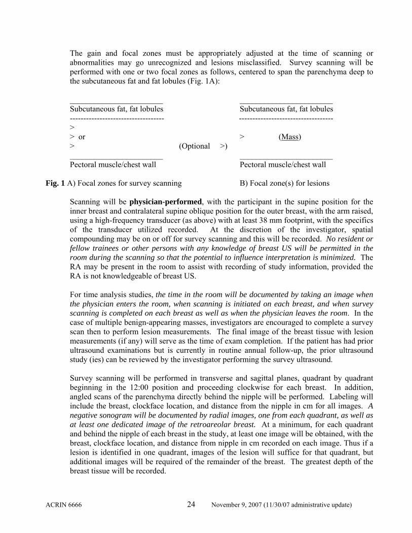
The gain and focal zones must be appropriately adjusted at the time of scanning or abnormalities may go unrecognized and lesions misclassified. Survey scanning will be performed with one or two focal zones as follows, centered to span the parenchyma deep to the subcutaneous fat and fat lobules (Fig. 1A): _______________________ _______________________
Subcutaneous fat, fat lobules Subcutaneous fat, fat lobules ----------------------------------- ----------------------------------- >
> or > (Mass) > (Optional >) _______________________ _______________________ Pectoral muscle/chest wall Pectoral muscle/chest wall Fig. 1 A) Focal zones for survey scanning B) Focal zone(s) for lesions
Scanning will be physician-performed, with the participant in the supine position for the inner breast and contralateral supine oblique position for the outer breast, with the arm raised, using a high-frequency transducer (as above) with at least 38 mm footprint, with the specifics of the transducer utilized recorded. At the discretion of the investigator, spatial compounding may be on or off for survey scanning and this will be recorded. No resident or fellow trainees or other persons with any knowledge of breast US will be permitted in the room during the scanning so that the potential to influence interpretation is minimized. The RA may be present in the room to assist with recording of study information, provided the RA is not knowledgeable of breast US. For time analysis studies, the time in the room will be documented by taking an image when the physician enters the room, when scanning is initiated on each breast, and when survey scanning is completed on each breast as well as when the physician leaves the room. In the case of multiple benign-appearing masses, investigators are encouraged to complete a survey scan then to perform lesion measurements. The final image of the breast tissue with lesion measurements (if any) will serve as the time of exam completion. If the patient has had prior ultrasound examinations but is currently in routine annual follow-up, the prior ultrasound study (ies) can be reviewed by the investigator performing the survey ultrasound.
Survey scanning will be performed in transverse and sagittal planes, quadrant by quadrant beginning in the 12:00 position and proceeding clockwise for each breast. In addition, angled scans of the parenchyma directly behind the nipple will be performed. Labeling will include the breast, clockface location, and distance from the nipple in cm for all images. A negative sonogram will be documented by radial images, one from each quadrant, as well as at least one dedicated image of the retroareolar breast. At a minimum, for each quadrant and behind the nipple of each breast in the study, at least one image will be obtained, with the breast, clockface location, and distance from nipple in cm recorded on each image. Thus if a lesion is identified in one quadrant, images of the lesion will suffice for that quadrant, but additional images will be required of the remainder of the breast. The greatest depth of the breast tissue will be recorded.

ACRIN 6666 25 November 9, 2007 (11/30/07 administrative update)
It is critical that both the study mammogram and US be interpreted independently, and sites are responsible to assure compliance with this. Receipt of results by participants may occur the same day as the examinations were performed in person and/or by telephone, in writing, or by mail, as is the standard procedure of the institution for notifying women of their screening mammogram results, provided results of the annual screening study mammogram are not provided to the study investigator performing the annual screening US or vice versa prior to study interpretation. Short-interval follow-up examinations are performed as diagnostic examinations, with integration by one study physician, and results given to the patient at the time of her examination(s). Permanent images will be stored on film or electronically on a PACS. Records at sites will be kept in locked file cabinets and/or password-protected databases. A live, hands-on demonstration of technique will be included in the training course for investigators. When assessing lesions, the more anterior of the two focal zones will be set in the mid portion of the lesion (Fig. 1B), or a single focal zone will be set centered in the mid portion of the lesion. The largest simple cyst will be documented in each breast, with its largest diameter recorded. When multiple simple cysts are present, only representative images are required. All lesions other than simple cysts will be documented with measurements in at least three planes. The lesion will be documented initially in the plane in which it has its largest horizontal diameter. The orientation of the image, location by breast, clockface, distance (in cm) from the nipple, and depth from the skin surface (in cm) of the center of the lesion will be recorded. Lesion measurements will be recorded as largest horizontal diameter (parallel to the skin surface, d1, in mm) by anteroposterior (vertical) diameter on that same image (d2, in mm) by perpendicular horizontal diameter (d3, in mm). Images of all lesions other than simple cysts will be recorded both with and without spatial compounding, and with and without power Doppler flow (4.4.4). At the investigator’s discretion, harmonic imaging can also be used to evaluate lesions and the use of harmonic imaging will be recorded both by documenting images of the lesion(s) with tissue harmonic imaging and by so indicating on the IS form. When a discrete mass other than a cyst is identified sonographically, the investigator will perform a targeted clinical breast exam to ascertain if a lesion is palpable. This vague palpability may influence the risk of malignancy for lesions that would otherwise be considered probably benign. Targeted clinical breast exam will thus be performed during sonography when discrete lesions other than simple cysts are found. If the lesion is palpable in retrospect, “vaguely palp” will be recorded on at least one image of the lesion.
Participants will undergo initial mammography and US, with initial sonography paid by study. If sonographic results are abnormal, or the mammogram prompts targeted ultrasound or other additional testing, such additional testing will be the responsibility of the participant and her insurance until such time as the participant would be returned to routine follow-up. Such additional testing should be performed at the study institution (i.e. participants whose insurance precludes additional testing at the study site should not be recruited). If any mammographic or breast sonographic studies are performed on participants at an institution not in the study, every effort should be made to obtain the original images and a study radiologist should perform a study interpretation (using IM, F6, or other appropriate study
ACRIN 6666 26 November 9, 2007 (11/30/07 administrative update)
forms). It is acceptable for targeted additional evaluation to be performed by non-study radiologists provided the study interpretation and forms completion is performed by a study radiologist. When results are benign or negative, the participant will undergo another screening round at 12 and 24 months with both sonography and mammography, with routine annual sonography paid by study.
4.3 Training in Scanning Technique and Interpretation 4.3.1 Qualification Task A: Detection, Lesion Characterization, and Measurement in Phantoms
A training course is planned prior to opening the trial. This will include training in scanning technique and interpretation criteria, validation of the reproducibility of lesion identification and measurement, and measures of observer performance in interpretation in a phantom. Phantoms containing multiple (n=16) masses (described in Appendix II) are available, and all radiologist participants will be asked to perform ultrasound on the phantom for reproducibility analysis after initial instruction in scanning technique. The ability of each radiologist to identify the same lesions and record the location will be determined. Lesion diameters will be recorded rounded to the nearest millimeter (mm). Reproducibility of lesion depth will be measured. Radiologists not able to attend the training course will need to scan one of the phantoms prior to study entry and submit results of lesion identification, measurement, and location in the phantom as well as general description of the lesion (cyst, complicated cyst, solid circumscribed, irregular solid) prior to participating in protocol. 4.3.1.1 Identification of Lesions
Preliminary experience with the phantom by Drs. Berg and Mendelson indicates that 13-14 of the 16 lesions can be readily identified. The others are deep. A threshold of detecting 12 lesions in the phantom has been proposed. Those who do not meet this requirement will undergo additional training in scanning technique. Until the investigator can document a minimum of 12 lesions, he/she will not be eligible to participate in the trial. This has been validated at training sessions during two weekends in June 2003 at Northwestern wherein all 32 investigators completing the phantom scanning were able to identify at least 12 lesions (median 14 lesions identified).
4.3.1.2 Lesion Characterization
The investigators will be asked to describe the shape, echogenicity, and posterior features of lesions in the phantom.
4.3.1.3 Measurement of Lesion Size
Consistent measurement of lesion size (maximal diameter to the nearest millimeter) and volume (calculated as [d1 x d2 x d3]/2) is critical to following solid masses considered probably benign. That is, if the apparent “growth” of a lesion is within experimental error (20%, as described in 4.6.3), the lesion can be reasonably followed providing the morphologic features continue to meet the criteria of a probably benign or benign lesion.

ACRIN 6666 27 November 9, 2007 (11/30/07 administrative update)
4.3.2 Qualification Task B: Observer Performance in Interpretation Pre-study validation of interpretive skills and measurement of agreement on feature analysis for both mammography and US will also be performed at the training session prior to beginning the main study. Training in BI-RADS® for mammography using a proven set of 54 cases has been shown to improve agreement in feature analysis, assessment, and, most importantly, improve biopsy rates of cancers [37]. This training set was developed by Dr. Berg and is available for use in this study. All cases have a defined reference standard of biopsy or four years of follow-up (histopathologic truth), as well as a consensus reference standard of experienced breast imagers. A similar set of 70 proven lesions representative of standard sonographic features has been developed and will serve as the basis for a qualification session for investigators in this protocol. These case sets are enriched in malignancies and benign findings in an effort to adequately measure agreement across the range of expected lesions. While this introduces “context bias” and tends to improve sensitivity and decrease specificity [82], we are most interested in demonstrating agreement in description and management. Investigators will receive an initial 1 hour training session reviewing BI-RADS® feature analysis and assessment in both mammography and sonography using cases that do not overlap with the test set. Observers will be then tested on final assessments in mammography and sonography initially without, then with, immediate feedback. Description of major features will be recorded (microcalcification morphology and distribution, mass margins). Kappa values and intraclass correlation coefficients will be calculated as a measure of agreement where appropriate (see statistical section for further details) [83]. Biopsy performance (sensitivity and specificity) without feedback will be compared to that after immediate feedback. If an investigator shows inadequate performance with feedback (to be determined as an outlier relative to the group), consideration will be given to either excluding that investigator from protocol or requiring additional training for that investigator.
These training materials will also be available on CD-ROM, and review of these materials will be required of additional investigators added to protocol. Further, the performance (sensitivity and specificity) of additional investigators will be determined on the training set, as with the initial group of radiologists in the training study. Again, any outliers will be identified with considerations as above.
4.3.3 Qualification Task: Breast MRI Interpretation: A set of 30 breast MRI cases with histopathologic proof or definitive follow-up (e.g. resolution) has been developed, representative of the spectrum of benign and malignant findings on MRI. Potential investigators who meet experience requirements detailed in Section 6.1.2 are required to describe major MRI features (e.g. foci, mass or non-mass features, using the BI-RADS: MRI lexicon [84] terminology) and record their final assessment on the expanded BI-RADS scale (1, negative; 2, benign; 3, probably benign; 4a, low suspicion; 4b, intermediate suspicion; 4c, moderate suspicion; 5, highly suggestive of malignancy). Kinetic analysis of lesions will not specifically be tested, though investigators will be given standard definitions (Appendix IA) and kinetic behavior of the lesion to be interpreted when appropriate. Feedback will be given. Kappa values and intraclass correlation coefficients will be calculated as a measure of agreement where appropriate (see statistical section for further details) [83]. Biopsy performance (sensitivity and specificity) will be determined as will ROC curves as a function of investigator experience. If an

ACRIN 6666 28 November 9, 2007 (11/30/07 administrative update)
investigator shows inadequate performance (to be determined as an outlier relative to the group), consideration will be given to either excluding that investigator from protocol or requiring additional training for that investigator.
4.4 Variables Affecting Image Quality and Interpretation 4.4.1 Heterogeneity of Parenchyma
The heterogeneity of the parenchyma on sonography may affect the sensitivity of sonography and will be recorded. Classification proposed is homogeneous, focally heterogeneous (< one quadrant), or diffusely heterogeneous (> one quadrant). Examples of this approach are included in the training session materials.
4.4.2 Breast Size
Breast size will be recorded both by initial recording of bra cup size and by recording the greatest depth of the breast (in cm) while scanning sonographically.
4.4.3 Spatial Compounding
The influence of spatial compounding on margin assessment for probably benign lesions will also be evaluated. Spatial compounding provides a multidirectional US beam, which may facilitate margin analysis. Posterior shadowing is less often seen with spatial compounding, simply because the off-perpendicular beams are able to penetrate more of the mass. Initial survey scanning will be with or without spatial compounding per investigator choice as in 4.2.1. All lesions other than cysts will be documented with images with and without spatial compounding and with and without flow. To evaluate the influence of spatial compounding on sonographic interpretation, investigators are asked to document lesions they consider other than outright benign both with and without spatial compounding. They will be asked to rate the influence (if any) of spatial compounding on margin analysis, assessment of internal structure, posterior features, and final assessment. The influence of spatial compounding will be independently (blindly) reviewed at the conclusion of the study in the overreading studies. The subset of lesions rated probably benign with and without use of spatial compounding will be compared with respect to rates of malignancy (ideally < 2%).
4.4.4 Flow
The presence or absence of flow within a sonographically depicted lesion will be recorded and may influence the rate of malignancy [85], particularly in lesions, which might otherwise be classified as probably benign. Teh et al [86] found power Doppler facilitated identification of the most suspicious areas in 8/37 (22%) of areas of calcifications sampled sonographically. Gain will be set at the maximum at which there is no diffuse background artifactual signal noted, with sensitivity set to detect low flow at velocities < 5 cm/sec. Compression will be the minimum necessary to maintain adequate image quality. Flow within the lesion detected with either color flow or power Doppler will be considered “flow,” though power Doppler is generally more sensitive and will be used preferentially. For lesions otherwise considered probably benign, the presence of flow within the lesion, and possibly immediately adjacent to the lesion, may portend a higher risk of malignancy. The presence or absence of flow in and/or immediately adjacent to the lesion will be recorded prospectively. For lesions otherwise considered probably benign, the rates of malignancy in those lesions with and those without flow will be compared on masked overread at the conclusion of the study.

ACRIN 6666 29 November 9, 2007 (11/30/07 administrative update)
4.4.5 Post-Surgical Changes Architectural distortion due to post-surgical changes may be difficult to distinguish from recurrent tumor both on mammography and sonography. Similarly, calcifications, which are dystrophic or due to fat necrosis can develop at the scar and be difficult to distinguish from recurrence; these can be a source of false positive biopsy recommendations for mammography. These issues have not been systematically studied for sonography, and current information on the performance of mammography in this setting is desirable as well. Recurrent or residual tumor is generally seen within 2 cm of the lumpectomy site. For each lesion, we will record if it is felt to be at the lumpectomy site, and performance of both mammography and sonography will be analyzed separately for such lesions.
4.5 Mammography The participant should be due for a routine annual mammogram. The definition of “annual” will comply with that used in routine practice, i.e. if the participant’s insurance would normally cover the costs of a mammogram after 11 full months (e.g. Medicare) that will suffice. For women diagnosed with breast cancer at least one year earlier (i.e. at least 12 full months have elapsed since the last treatment surgery), the participant may have had a unilateral mammogram of a treated breast within the past year, or additional views of one or both breasts in the interim, provided the current visit is routine and bilateral. Similarly, a bilateral survey ultrasound should not have been performed less than 11 full months earlier.
Routine mammographic views will be performed at the same site as the ultrasound within two weeks of the ultrasound examination on all study participants. Mammography examinations can be performed on either FDA-approved digital or film-screen systems. For digital mammograms, the institution number, study number (6666), and study participant identifier (case number)—NOT the patient name or social security number— should appear on the images. Digital mammographic images are to be submitted electronically to the ACRIN Image Archive. For film images, the participant name must be masked with labels provided to the site or generated at the site that will include the institution number, study number (6666), and study participant identifier (case number). Film images should be sent by overnight express service to the ACRIN Image Archive and will be digitized at ACRIN Headquarters and returned to the site within 3-5 business days. Participants will be randomly assigned to receive routine mammography or bilateral whole-breast physician-performed ultrasound as their initial examination. The same order of tests will be followed for that participant at each annual screen. Such randomization may prove to be a barrier to accrual and burdensome to sites. If we find accrual is deficient (defined in Section 6.3), we will consider dropping the randomization after discussion with the Data Safety and Monitoring Board. If randomization is discontinued, participants will undergo initial mammography then independently performed and interpreted sonography. Mammographic technologists or the Research Assistant (or a radiology resident or fellow involved with the clinical reading of that patient’s mammogram but not her study sonogram) will review prior mammograms or reports when available to ascertain that the participant has heterogeneously dense or extremely dense breasts (Section 5.3) prior to approaching the participant for study eligibility. Women who have not had a prior mammogram will still be considered for study if they otherwise meet eligibility criteria. It is anticipated that very few women at high risk will not have had a prior mammogram and that those who have not will generally be under age 40. The likelihood of such young women having fatty breasts is small. If such participants are accrued to study and prove to have fatty breasts, a sensitivity analysis will be

ACRIN 6666 30 November 9, 2007 (11/30/07 administrative update)
performed by removing these cases from the analysis. If a participant is enrolled in study based on reported density on prior mammograms and the current mammograms are not felt to show heterogeneously dense or extremely dense parenchyma (Section 5.3), the participant will continue on study.
All study sites must be accredited by the Food and Drug Administration and must meet the requirements of the Mammography Quality Standards Act (MQSA) (or equivalent). Mammograms must include CC and MLO views. Additional views determined to be necessary by the technologist to complete the routine evaluation of the breast(s) should also be included (such as laterally exaggerated CC views or well-compressed views of the anterior portions of the breasts). Films should be labeled according to ACR/MQSA standardized labeling criteria. The interpreting physician must meet the requirements of the MQSA. In addition, it is proposed that investigators will have a minimum experience of interpreting 2500 mammograms per year for at least 2 years.
Note: Computer-assisted detection and/or diagnosis is not permitted on study mammograms nor is double reading of study mammograms or sonograms. Reports will be dictated using ACR BI-RADS® terminology to describe the lesion(s). Lesion(s) should be measured in medial-lateral, anterior-posterior, and superior-inferior planes. Lesion location(s) will be described by quadrant, retroareolar, central (if directly behind the nipple in both views), or axillary, and distance from the nipple. Asymmetric densities will be noted. The results of the initial routine mammogram will be recorded separately from the results of interpretation after additional targeted diagnostic work-up. 4.6 Interpretation Criteria Features will be recorded for each lesion as described, using the BI-RADS® lexicons for mammography and sonography. Interpretation and management will be based on the worst features present.
4.6.1 Final Assessments
Both a BI-RADS® category final assessment and a risk of malignancy based on a 100-point probability of malignancy scale will be recorded (see Section 4.10). Management recommendations will be recorded separately. An area of confusion in the application of BI-RADS® in clinical practice has been the distinction of level of suspicion of malignancy and final recommendation. For example, there are lesions judged by the radiologist to have < 2% risk of malignancy yet to merit biopsy based on inability to follow the lesion, ipsilateral to a breast cancer, or participant desires biopsy. In a high-risk population, such as the one proposed, lesions otherwise considered probably benign may be recommended for biopsy more often because of a perceived increased risk of malignancy on the part of the participant or radiologist. For this reason, we have asked the investigators to record their assessment and likelihood of malignancy separate from their management recommendation. If the lesion is judged probably benign by imaging features (or to have < 2% risk of malignancy), yet biopsy is recommended, the reason(s) for recommending biopsy will also be recorded.
4.6.2 BI-RADS® Final Assessment 2: Benign Findings The following will be considered benign findings:

ACRIN 6666 31 November 9, 2007 (11/30/07 administrative update)
1. Multiple bilateral circumscribed masses seen mammographically (at least 3 total, at least 1 in each breast, per Leung and Sickles [87]), provided the participant has no history of malignancy outside the breast, including multiple bilateral cysts and complicated cyst(s) as seen sonographically;
2. Mammographically stable circumscribed masses;
3. Circumscribed masses that clearly contain fat;
4. Intensely and uniformly hyperechoic circumscribed masses on US [22];
5. Typically benign [88] calcifications, including macrocalcifications (> 0.5 mm) on sonography;
6. Diffuse, scattered, bilateral punctate and amorphous calcifications [89];
7. Simple cysts [28];
8. Round or oval masses with imperceptible wall, posterior enhancement, and mobile internal echoes or mobile fluid-debris level with NO evidence of intracystic mass, thick wall, or thick septations;
9. Siliconomas [90];
10. Lymph nodes under 2 cm that retain a fatty hilum, without focally or diffusely thickened cortex;
11. Post-surgical scar, not known to be increasing compared to prior studies;
Note: Post-surgical scar within the first two years following lumpectomy for cancer or other benign surgery may be considered a benign or probably benign finding on either sonography or mammography at the discretion of the investigator.
12. Masses within the skin.
4.6.3 BI-RADS® Final Assessment 3: Probably Benign Findings
Lesions considered probably benign sonographically must be nonpalpable and must not have any suspicious features (below). These will include the following when identified on baseline screening:
1. Oval masses (parallel to the skin in orientation) hypoechoic to fat with circumscribed
borders and no posterior features or minimal posterior enhancement, including multiple bilateral masses with these features if seen only sonographically;
2. Hyperechoic masses with central hypoechoic to anechoic components suggesting fat necrosis;
3. Hypoechoic oval masses with homogeneous low-level internal echoes that otherwise meet the criteria for simple cysts (circumscribed, acoustic enhancement) (See 4.7.2 point #1 above if multiple with associated simple cysts.);
4. Microlobulated or oval masses composed entirely of clustered microcysts with or without layering microcalcifications;
5. Probably artifactual posterior shadowing at the interface of fat lobules without any associated mass, which changes appearance on changing the angle of insonation;

ACRIN 6666 32 November 9, 2007 (11/30/07 administrative update)
6. Architectural distortion felt to be due to post-surgical scar can be classified as probably benign or benign at the discretion of the investigator.
Such lesions will be followed sonographically at 6 months, 12 months, and 24 months and stability recorded. Any abnormal interval change (defined as an increase in volume by more than 20% or development of suspicious features) should prompt aspiration or biopsy. (The benchmark volume change of 20% was determined by statistical simulation and provides a change in volume that most often indicates a real change as opposed to an apparent change because of measurement error. Specifically, suppose that, without any loss of generalizability, all three dimensions are measured independently and those measurements have a normal distribution with mean 10mm and standard deviations 1mm. Thus the true volume is 1000 cubic mm. Simulations indicate that random increases in volume of more than 20% occur about 15% of the time and increases of 30% only occur about 5% of the time. Hence increases in volumes by 20% or more are likely due to real tumor growth and not measurement error.) A lesion that decreases in volume by more than 20% or resolves on any follow-up will not require further follow-up.
Lesions considered probably benign mammographically have been shown to have < 2% risk of malignancy [91-94] and will include the following:
1. A circumscribed nonpalpable mass of any size on initial mammogram (after full
diagnostic mammographic work-up); final characterization sonographically is encouraged unless it is known to be stable compared to prior mammograms. For those circumscribed masses on baseline mammogram not visible sonographically, short-interval follow-up is proposed.
2. A focal asymmetric density on baseline mammogram that partially effaces on spot compression and has no sonographic correlate.
3. A cluster (≥ 5) or multiple clusters of uniformly round (punctate) microcalcifications < 0.5 mm in diameter [89] on baseline mammogram.
4. Architectural distortion felt to be due to post-surgical scar can be classified as probably benign or benign at the discretion of the investigator.
Again, such lesions will be followed mammographically at 6 months, 12 months, and 24 months and stability recorded. Any abnormal interval change (increase in size or calcifications or development of suspicious features) should prompt aspiration or biopsy. A lesion that decreases or resolves on any follow-up will not require further follow-up. For participants in whom probably benign findings are being followed at the time of their annual examination, the mammographic and sonographic interpretations will be recorded with reference only to prior studies of the same modality initially. Reference to prior reports will also be necessary for good clinical practice and may reference both mammographic and sonographic findings. It is understood that usual clinical practice requires integration of both studies in this diagnostic setting: an integration reading will be required in this situation even if the finding being followed is considered stable.

ACRIN 6666 33 November 9, 2007 (11/30/07 administrative update)
Note: Lesions which morphologically would be considered probably benign but are new or enlarging should be considered suspicious, with intervention performed. Similarly, if a lesion appears suspicious by either mammography or sonography, final management should be predicated on the most suspicious features present unless the lesion is clearly benign on either imaging modality. For example, a mass may appear indistinctly marginated on mammography and be proven to be a simple cyst. This would be appropriately classified as benign. A mass, which appears mostly circumscribed mammographically but is noted to have partially angular margins on sonography, would be classified as suspicious.
4.6.4 BI-RADS® Final Assessment 4: Suspicious Findings, BI-RADS® Final Assessment 5: Highly Suggestive of Malignancy Lesions in these categories require intervention with biopsy or possibly aspiration if they resolve. Lesions, which are considered to have greater than 95% risk of malignancy, are appropriately classified as category 5. Lesions appropriately classified as category 5 include a new spiculated mass or new branching, fine linear microcalcifications.
Findings suspicious for or highly suggestive of malignancy on sonography include the following [22, 25]:
1. Irregular shape;
2. Microlobulated, indistinct, angular, or spiculated margin;
3. Posterior acoustic shadowing (excludes refractive edge shadowing) not felt to be artifactual at the interface of fat lobules;
4. Round shape and solid;
5. Cystic lesions with any of the following: intracystic mass, thick septations (> 0.5 mm), thick wall, discrete solid components (excludes lesions composed entirely of microcysts) [27];
6. Intraductal mass;
7. Microcalcifications (≤ 0.5 mm) within a mass;
8. Duct extension;
9. Antiparallel orientation relative to skin (taller than wide);
10. Architectural distortion in the absence of a history of trauma or surgery;
11. Skin retraction or skin thickening (>2 mm) in the absence of a history of infection, radiation, trauma, or surgery;
12. Any new or enlarging mass which would otherwise be considered probably benign as in 4.6.3;
13. Any combination of the above features;
Findings suspicious for or highly suggestive of malignancy on mammography include the following [88]:

ACRIN 6666 34 November 9, 2007 (11/30/07 administrative update)
1. Focal developing asymmetry in the absence of history of trauma or signs or symptoms of infection or hormonal therapy;
2. Mass with indistinct or spiculated margins;
3. Mass with microlobulated margins not corresponding to a cluster of microcysts on sonography;
4. A new or enlarging circumscribed mass that is solid on US;
5. Amorphous or indistinct microcalcifications in a clustered, linear, or segmental distribution [95];
6. Pleomorphic calcifications;
7. Branching or fine linear calcifications;
8. Punctate calcifications in a linear or segmental distribution;
9. Architectural distortion in the absence of a history of trauma or surgery;
10. Skin retraction or skin thickening in the absence of a history of infection, radiation, trauma, or surgery;
11. Any combination of the above features. 4.6.5 BI-RADS® Final Assessment 6: Known Malignancy
If imaging evaluation is performed prior to definitive surgery but after tissue diagnosis (such as following neoadjuvant chemotherapy), a final assessment of category 6, known malignancy, can be used for clinical reports.
4.6.6 Mammograms Obtained After Clip Placement For mammograms performed only to document clip placement following percutaneous sampling of a lesion, no numeric BI-RADS assessment is required for clinical reports. The final assessment would read, “post procedure mammograms for marker placement,” and is used only for post procedure mammograms to confirm the deployment and position of a breast tissue marker. The lay summary, which must be provided to the patient, must be specific to the procedure. If the facility makes this post procedure examination part of the interventional study instead of a separately charged examination, then it does not fall under MQSA, and this FDA-approved alternative requirement does not apply.
4.6.7 Overall Final Assessment by Breast Final assessments and recommendations will be recorded for each lesion considered clinically significant. In addition, investigators will give a final assessment for the breast as a whole. The latter should be based on the most suspicious finding(s) present.
4.7 Reliability of Interpretation 4.7.1 Clinical Interpretations and Masking
Interpretation of the mammogram and performance and interpretation of the US will be without knowledge of the results of the other study. This will require two study investigators be involved in each study case at each site. Once the initial results of each study are recorded, either of these investigators can complete a third integration reading (required only when the study mammogram or sonogram results are other than negative or benign). It is

ACRIN 6666 35 November 9, 2007 (11/30/07 administrative update)
encouraged that, when possible, the integrated reading be performed by the study radiologist performing the participant’s survey US.
Clinical reports should reflect the overall final assessment by breast, taking together findings from both ultrasound and mammography. If the tracking system used at an institution permits only a single final assessment category, the overall assessment given in clinical reports should reflect the most suspicious findings present in either breast. It is suggested that the clinical mammographic report be addended to indicate the results of the study screening sonographic report. Alternatively, sites can issue a separate study sonographic report provided doing so will not generate billing to the patient. In general sites may find that addending the mammographic or sonographic reports is best performed at the time of the integration interpretation (Form ID) when one is required. In any event, ideally such an integration reading will be evident as a separate paragraph in the mammographic or sonographic clinical report. Such an addendum or separate paragraph is only necessary when it would be clinically appropriate, e.g. a mass seen on mammography which would require recall if it was not a cyst or otherwise benign on US. If no integration interpretation is needed (in the case of negative or benign findings only on the study screening mammogram and sonogram), use of a macro such as the following is suggested: “Addendum: The patient underwent (bilateral/right/left) breast screening ultrasound as part of a research protocol (ACRIN 6666), with scanning performed by Dr. X and there were no findings of significance.” Such an addendum can only be generated once blinded interpretation of initial study mammogram (IA form) and sonogram (IS form) have been completed. The RA can assist with the process of assuring that such an addendum is generated, with final verification of the clinical report by either of the study radiologists involved in the patient’s examinations. The clinical mammographic report will serve as source documentation for both IA and IS forms.
Note: Should there be visualization of a clip due to a prior biopsy, the clip should be mentioned in the mammographic and (when seen sonographically) US clinical reports as well as indicated in the “comments” sections of the IA and IS forms. If the original lesion which was biopsied is no longer seen, the lesion number should be reported as “gone” and that lesion number is then retired, not to be reused in the future. If there is a scar at the biopsy site, it will be assigned a new lesion number. If the patient is recalled after integrated interpretation, a clinical report will be generated based on additional mammographic views and/or a repeat targeted US as needed. Form IM is to be completed at the time of the additional evaluation. The clinical report will serve as source documentation for the IM form. Note that results of the initial screening interpretations will be collected (and analyzed) separately from those after the integration interpretation. The interpretations after the targeted diagnostic evaluation will also be considered separately or in combination with the initial screen. Readers will also be asked to rate whether the final assessment was based primarily on mammography, sonography, or both, for each lesion. As described below, a masked overread will be performed at the conclusion of the study.

ACRIN 6666 36 November 9, 2007 (11/30/07 administrative update)
4.7.2 Overreading Study
As mentioned, Baker et al [24] found substantial variability in sonographic feature analysis and assessment across five observers in the same practice, with kappa for final assessment of 0.51 across readers. A BI-RADS® lexicon for breast ultrasound has been developed (Appendix I, [25]) and preliminarily tested at the Society of Breast Imaging May 2001. Agreement was high for anechogenicity, and fair to good (κ 0.4 – 0.75) for shape, orientation, echo pattern, posterior acoustic features, and special cases such as siliconomas and lymph nodes. Kappa for circumscribed margins ranged from 0.11 to 0.46 between readers (EB Mendelson, BI-RADS® Committee meeting, National Conference on Breast Cancer, Dallas, TX 4/19/02). Assessment of the circumscribed nature of a mass’ margin is critical in making final assessment and management recommendations. It is considered likely that real-time analysis is critical in such a determination. It is also critical that the criteria for determining a mass to be circumscribed be applied as uniformly as possible across investigators both for the study and generally due to the high prevalence of incidental solid (usually benign) masses as described. The reliability of clinical study interpretations will be assessed by central overreads of all abnormal sonograms (final assessment completed is BI-RADS® 3, 4A, 4B, 4C, or 5 with recommendation for short interval follow-up, biopsy, or additional evaluation) and 10% of negative and benign interpretations. Original mammographic images will be reviewed, as will digital US images for negative and benign cases. Similar overreads are planned for all abnormal mammograms (final interpretation completed is BI-RADS® 3, 4A, 4B, 4C, or 5, with recommendation for short interval follow-up, biopsy, or additional evaluation) and 10% of negative and benign interpretations. Central overreads will be performed at the ACR Headquarters in Philadelphia by Drs. Merritt (Thomas Jefferson University School of Medicine), Berg, Mendelson, Bassett and Valerie Jackson (Indiana University), with an RA assigned to assist in data collection.
These reader studies will assess interpretation rather than detection. We encourage the numbering of lesions on mammographic images in order to facilitate consistent tracking of lesions over time. As there is no simple way to remove those marks (indicating lesion numbers) in the course of digitizing film images, the marks will remain on images in the course of digitization. Images originally acquired electronically, i.e. digital mammograms and sonograms, will not have “marks” on the images submitted to ACRIN headquarters. Readers will be precluded from re-reviewing cases performed at their own institution and will be masked to palpability, histopathologic truth, and follow-up for each case. As stated, lesions that are palpable may be more likely malignant: in the overread study, the readers will be asked to give their recommendations for the lesion assuming it is not palpable then assuming that it is palpable in retrospect. The rates of malignancy in lesions classified as probably benign on overread will be determined as will agreement levels (kappa) within overreading investigators and agreement levels with the site PI. Classifications with and without review of flow and spatial compounding images will be reviewed (with random initial review of flow or spatial compounded images vs. those without). Agreement on feature analysis for both

ACRIN 6666 37 November 9, 2007 (11/30/07 administrative update)
mammographic and sonographic features and final assessments will be assessed among overreaders and compared to site PI interpretations.
4.8 Data Collection 4.8.1 Historical information
a) Prior screening history including date of last mammogram, any prior breast ultrasound (including the area of the breast(s) previously evaluated), any prior contrast-enhanced breast MRI (right, left, or both breasts evaluated);
b) Risk factors including: prior breast cancer, BRCA-1 or –2 status if known, prior atypical ductal or lobular hyperplasia, radial sclerosing lesion, or lobular carcinoma in situ, detailed family history of breast or ovarian cancer including age at diagnosis and relationship to participant, hormonal status (premenopausal, last menstrual period < 1 year ago, postmenopausal, surgical menopause, on exogenous hormones to include estrogen or progesterone preparations, Tamoxifen, Raloxifene, “natural” hormonal preparations, aromatase inhibitors such as Arimidex), age at first childbirth, prior radiation to the chest and/or mediastinum and/or axilla;
c) Bra cup size.
4.8.2 Results of Imaging Studies
For mammograms, comparison to prior mammograms is recommended, as is standard clinical practice. Comparison to prior breast sonograms, however, will not be permitted at the time of initial annual mammogram interpretation (IA form). All comparison studies will be reviewed when integration interpretation is needed (assessment on initial mammogram and/or survey ultrasound is other than negative or benign). For mammograms, the breast density will be recorded, together with the size (in three planes) and location (clockface and distance from nipple in cm) of any discrete abnormalities with features described using BI-RADS®. The “size” of areas of calcifications will be “measured” by the greatest dimensions (in mm) over which similar calcifications are seen mammographically in three planes. This is most easily accomplished by marking on the films at the extremes of the area of calcifications then measuring between marks. Final assessments will be recorded for each lesion and by breast using BI-RADS®, together with an estimated likelihood of malignancy to allow receiver operating characteristic curve analysis. As above, management recommendations will be recorded separate from the final assessment and likelihood of malignancy ratings. For breast sonograms, comparison to prior breast sonograms is recommended. Prior mammograms should not be reviewed prior to performing the survey sonogram or at the time of survey sonogram interpretation (IS form). As above, all comparison studies will be reviewed when integration interpretation is needed (assessment on initial mammogram and/or survey ultrasound is other than negative or benign). The heterogeneity of the parenchyma will be recorded as above. The size (in three planes, in mm) and location (clockface and distance from the nipple in cm) of any discrete abnormalities will be recorded with features described and final assessments recorded using BI-RADS® as well as an estimated likelihood of malignancy as above. The assessments and separate recommendations will be made for each lesion and by breast. Palpability in retrospect will be recorded for any discrete lesion other than simple cysts.

ACRIN 6666 38 November 9, 2007 (11/30/07 administrative update)
The results of the mammogram and the sonogram will be reported on study forms separately. Thus a different radiologist will read the mammogram from the radiologist who performs and interprets the sonogram. A reconciliation integration interpretation will be recorded for all studies where either the study sonogram or mammogram is interpreted as other than negative or benign and for all studies requiring additional evaluation. Once the study IS and IA forms are completed, the clinical report should reflect that a screening sonogram has been performed and indicate its results as detailed in Section 4.7.1. It is possible that there will be differences of interpretation at the time of the integration reading compared to the original screening reading of either ultrasound or mammography or both. The worst original screening interpretation and recommendations will take precedence for the management of the patient unless the following circumstances apply:
(1) If the original mammographic reading recommends ultrasound for a mass, which is
clearly shown at integration reading to be a cyst on survey ultrasound, then no recall will be necessary for that lesion.
(2) If the original mammographic reading recommends ultrasound for a mass, which is clearly shown to be suspicious on survey ultrasound, then targeted ultrasound will not be necessary and the patient should proceed directly to biopsy.
(3) If the survey ultrasound demonstrates calcifications not in a mass for which mammography is recommended, and screening views demonstrate diffuse, scattered, punctate and amorphous calcifications bilaterally with no suspicious calcifications and the finding was interpreted as benign mammographically, then no recall will be necessary for that finding.
Yet another reading will be required if additional views or targeted sonogram need to be performed based on the initial mammographic interpretation (and after the integration reading). The reconciliation interpretation and targeted ultrasound and interpretation can be performed by either of the initial study radiologists or by a third radiologist, provided all these radiologists undergo training and validation in study protocol. See Section 4.8 regarding clinical reporting.
4.9 Degree of Suspicion and Quasi-continuous Probability Scale To facilitate the statistical analysis, in addition to BI-RADS® final assessments both by lesion and by breast, examiners will provide an assessment using the new BI-RADS final assessment categories (1=negative; 2= benign; 3=probably benign; 4A=low suspicion; 4B=intermediate suspicion; 4C=moderate suspicion; 5=highly suggestive of malignancy, Appendix I). For lesions requiring further evaluation (category 0), examiners will be asked for their assessment in the absence of further evaluation to facilitate analysis. We may find that there are insufficient numbers of lesions in some of the subcategories of suspicious lesions: these may be collapsed if needed to facilitate analysis. The second measurement will simply be the reader’s estimated probability or likelihood of malignancy from 0% to 100% for each lesion as well as for the entire breast. This latter scale is referred to as the likelihood of presence (or malignancy) scale or the quasi-continuous probability scale in the statistics section of this protocol (Section 13.0). There are limited data on which to base an absolute risk of malignancy for specific lesions going to biopsy. Liberman et al [96] describe the rate of malignancy for lesions with particular features in a

ACRIN 6666 39 November 9, 2007 (11/30/07 administrative update)
series of 492 nonpalpable lesions going to biopsy. Berg et al [95] reported a 20% rate of malignancy for amorphous calcifications. Rates of malignancy among complex cystic lesions have also recently been described in a series of 150 biopsy-proven lesions [27]. Note, however, the most important quality of the likelihood of malignancy scale is that the reader is internally consistent. This is because the statistical ROC analysis depends on the relative orderings of these outcomes and not their absolute magnitude.
The final assessment of any given lesion considered other than benign will require the integration of both sonographic and mammographic features for lesions seen on both modalities, as described in Section 4.6. 4.10 Reference Standard Definitive information about the presence of malignancy will be obtained by biopsy directed by the imaging method best depicting the lesion: 14-g core or 11-g vacuum-assisted needle biopsy, or by surgery after wire-guided localization for women undergoing these procedures. Biopsies should not be performed by devices for which the goal is percutaneous (non-surgical) removal and/or ablation as this may interfere with measurements of tumor size and thereby staging information. All study participants will receive annual mammography and sonography for a period of two years after the initial prevalence screen by both mammography and sonography. We will also obtain cancer status information (Form F1) on all participants at 12, 24, and 36 months after study entry. As detailed in Section 13.2.2, the lack of malignancy after 12 months will serve as reference standard for patients with negative or benign results. The lack of malignancy at 24 months will serve as reference standard for lesions classified as probably benign. If a “probably benign” lesion decreases or resolves on any follow-up, it will be considered benign. We do not expect to see new lesions classified as probably benign at 12 month or 24 month screens, but if there are such lesions, they will require either biopsy or follow-up for either 2 years of stability or interval decrease or resolution at any subsequent follow-up. Histopathologic overread is not deemed necessary due to the high (> 96%) rates of agreement seen in the Radiologic Diagnosis Oncology Group V trial [97] and the International Breast MR Consortium (IBMC) trial between central and local pathologists for both core and excisional histopathology. Further, experience in both those trials as well as DMIST has shown that routinely sending pathology material for overread presents a burden to sites. Central pathology overread will be available at no cost to sites when standard clinical practice would include a second opinion. To request an overread, please contact the ACRIN 6666 project manager (Cynthia Olson; [email protected]). Dr. Olga Ioffe at University of Maryland has agreed to serve as the central overread pathologist. For any disagreements of local and central overread, Dr. Shahla Masood will serve as a third opinion.
4.10.1 Biopsy Technique
Investigators performing core biopsies for the trial must meet the CME and experience requirements analogous to ACR accreditation for breast biopsy (i.e. 3 hr CME category 1 in US-guided biopsy, 3 hr CME specific to stereotactic biopsy, and have performed at least 12 of each procedure in the past year). It is acceptable for non-study physicians to perform biopsies on study participants, provided a study radiologist completes the appropriate case report forms. For 14-g core biopsy under sonographic guidance, a minimum of 3 samples will be obtained [98], and or stereotactically guided 14-g core biopsy of masses or

ACRIN 6666 40 November 9, 2007 (11/30/07 administrative update)
asymmetric densities, a minimum of 5 samples will be obtained [99] unless the lesion can no longer be identified after fewer passes. For sonographically-guided biopsies, post-fire images will be obtained documenting the needle through the lesion on each pass. Additional passes may be warranted if the lesion is not felt to have been adequately sampled after 3 passes. For calcifications seen only mammographically, 11-g directional vacuum-assisted biopsy (DVAB) will be accepted provided a minimum of 10 specimens are obtained [100]. Specimen radiography will be performed on all lesions biopsied that contain calcifications, electively on core biopsy specimens of noncalcified lesions, and on all excisional specimens. The specimens must be deemed to contain adequate and representative material. Placement of a clip or other suitable marker at the biopsy site is strongly recommended whenever there is concern that the site may be difficult to identify should excision be needed. Post-clip placement unilateral mammograms are recommended. The following results on core biopsy or DVAB will prompt needle localization and excision [101]:
1. Any malignancy;
2. Atypical ductal hyperplasia;
3. Atypical lobular hyperplasia (ALH) or LCIS if this is the most significant finding at histopathology;
4. Radial scar or radial sclerosing lesion;
5. Papillary lesion with atypia;
6. Columnar alteration with cytologic atypia;
7. Discordant imaging and histopathologic results;
8. Lack of adequate retrieval of calcifications (as judged by the radiologist) when calcifications are targeted.
DCIS with cancerization of the lobules can mimic LCIS or even atypical lobular hyperplasia. E-cadherin, a cell adhesion molecule is lost in lobular lesions, and staining for e-cadherin can be used to differentiate DCIS from ALH or LCIS [102-104]. DCIS would, of course, be considered malignant and require excision, and would be expected to show e-cadherin staining, whereas lobular lesions would not. To avoid this potential source of error in pathology interpretation, e-cadherin immunohistochemical staining is recommended on all lesions interpreted as ALH or LCIS on core biopsy. If the only finding at histopathology after core biopsy or DVAB is ALH or LCIS, excision should be performed. A result of atypical lobular hyperplasia or lobular carcinoma in situ on core biopsy remains controversial: excision is recommended even if a benign concordant result is obtained (such as fibroadenoma) and the ALH or LCIS is considered incidental with no remaining suspicious findings on imaging [105]. Ultimately, this is left to the discretion of the site. Follow-up after a benign, concordant, core or vacuum-assisted biopsy diagnosis will be on an annual basis for simple fibroadenomas, fat necrosis, and benign lymph nodes. All other diagnoses will generally be followed with an initial 6-month short-interval follow-up of the biopsied breast by the imaging technique(s) best depicting the area biopsied.

ACRIN 6666 41 November 9, 2007 (11/30/07 administrative update)
4.10.2 Central Overread Central pathology overread will not be performed except in the rare instance when a second opinion is requested, as in standard clinical practice. To request an overread, please contact the ACRIN 6666 project manager (Cynthia Olson; [email protected]). In this setting, the BX, NL, or S1 form should not be submitted until overread has been completed as detailed below. The original histopathology reports will be required. When central overread second opinion is performed, at least 2 representative H&E slides of core biopsies will be sent to Dr. Olga Ioffe at University of Maryland ([email protected]). All representative H&E slides will be sent for excisional specimens when central overread is desired. Pathology specimens will be labeled with the ACRIN study number and the case number of the participant. The pathology report should have the participant identifiers replaced with the study and case numbers. Slides should be sent via Federal Express (or equivalent) to:
Dr. Olga Ioffe Department of Pathology University of Maryland 22 S. Greene St. Baltimore, MD 21201
Slides will be returned to the sites within 4 weeks. If there is substantial disagreement between the local pathologist and the first consulting pathologist (defined as a disagreement that changes a participant’s breast cancer status), then the pathology material will be sent to the second consultant (Dr. Shahla Masood) for another interpretation. The true pathologic diagnosis will be considered that diagnosis that is agreed upon by two out of three interpreters. When there is disagreement of local and central pathologists on final overread, the local pathologist will be notified by telephone as will the site PI by telephone or e-mail and ACRIN headquarters ([email protected]) by e-mail. The final diagnosis agreed to by two pathologists will be considered the result and should be entered on the BX or NL or S1 form (as appropriate to core biopsy, excisional biopsy, or treatment surgery respectively) to be sent to ACRIN headquarters. It is the responsibility of the local pathologist and site PI to contact the participant should an atypical or malignant result be found only on central overread. Excision is recommended in that scenario. In the case of a benign diagnosis on central overread only, the central overread will be considered the reference standard reading if the (expected) excision also proves benign.
4.10.3 Fine Needle Aspiration of Complicated Cysts Many complicated cysts will be followed at 6 month intervals per protocol. If there is concern on the part of the investigator, fine needle aspiration using a 20-g or 18-g needle may be performed under sonographic guidance, with the needle documented to be within the lesion. Fine needle aspiration will only be accepted for complicated cysts that resolve completely on aspiration. Cytology will not be sent unless the fluid is bloody or otherwise heme-positive. Cultures and gram stain will be sent if the fluid appears purulent. All cytology and/or culture reports are required.

ACRIN 6666 42 November 9, 2007 (11/30/07 administrative update)
4.11 Reference Standards - Diagnostic End Points 1. All imaging-detected abnormalities judged to be suspicious for malignancy or highly
suggestive of malignancy will be biopsied with 14-g or 11-g needle devices or needle localized excision as above.
2. Linkage with a regional tumor registry or clinical and imaging follow-up for at least two years after study imaging is required to identify all undetected cancers. Note that linkage with a regional tumor registry is preferred over clinical and imaging follow-up, but the combination of linkage, clinical, and imaging follow-up is preferred over any method alone. Cases without at least 23 months of follow-up after the initial screen will be rejected from analysis.
3. Breast cancer is defined as the histopathologic diagnosis of ductal carcinoma in situ (DCIS) or any invasive breast cancer, lymphoma, sarcoma, or metastasis to the breast from distant primary.
4. In addition to histopathologic tumor type, data should be collected on established surrogate markers for breast cancer prognosis, including, but not limited to: tumor size, lymph node status, tumor grade, and UICC / AJCC tumor stage [106].
4.12 MRI of the Breast On February 3, 2006, ACRIN protocol 6666 completed enrollment of 2809 women at high risk for breast cancer. Based on a current compliance rate of 85% follow-up at each annual examination by the end of 14 months (based on forms received), 1529 women are expected to be potentially eligible for the MRI examination from May 1, 2006 through October 31, 2007. Only ACRIN 6666 participants who have completed their 24 month US and mammography screenings by February 10, 2008 (allowing for slight variations in appointment scheduling) will be eligible for the additional MRI. From May 2006 through May 2008, the third round (24 month) annual screening US and mammogram will be completed, including additional targeted work-up and induced biopsies. The 24 month screening US and mammography examinations will be completed per protocol prior to the MRI. It is the responsibility of the site to assure that interpretation of the 24 month screening US and mammography examinations and any needed additional views is performed blinded to each other and to the MRI. Twenty (20) sites in ACRIN 6666 were surveyed on their equipment, software, and experience with breast MRI examinations as well as MRI-guided vacuum-assisted biopsies. All but five (5) sites have met the proposed requirements, with at least a potential third MRI-qualified investigator, and will be qualified to participate. These sites account for 84% of accrual, resulting in 1280 participants potentially eligible for MRI. It has been estimated that 1200 of these women may be eligible and choose to participate in the MRI component of the study. We do not expect any systematic selection bias in the subsample of women participating in the MRI substudy relative to the main study population. However we will examine the covariate profiles of these groups to validate this assumption. As detailed in Sections 4.3.3 and 6.1.2, investigators with a minimum experience interpreting 50 breast MRI examinations and performing 5 vacuum-assisted MRI guided biopsies will be eligible to participate. All MRI interpreting radiologists will be required to review a set of training cases and achieve adequate performance in interpretation of those cases prior to qualifying as a breast MRI

ACRIN 6666 43 November 9, 2007 (11/30/07 administrative update)
interpreting investigator. The MRI interpreting physician need not be one of the same radiologists who is qualified to interpret study mammograms and breast US examinations. Investigators interpreting the MRI will be blinded to the mammographic and/or US findings from the 24 month screen. If the clinical report would be delayed more than one week due to such a constraint, the mammography interpreting physician could read the study MRI examination if that interpreting physician is qualified as a study investigator for both mammographic and MRI interpretations. After rendering the MRI interpretation, clinical integration with mammographic and US findings will be performed, with a short form recording any changes in interpretation by breast and biopsies or follow-up prompted only by MRI. The integration reading is expected to reduce false positives but is otherwise unlikely to affect interpretation of the MRI. MRI should be scheduled within 8 weeks of the 24 month annual routine US and mammogram visit. The MRI examination should be scheduled 7-14 days after the onset of menses in premenopausal participants when possible. Sites will record date of last menstrual period where applicable (i.e. if within past 30 days). 4.12.1 Timeline for Receiving MRI Component of Trial • At the time of the routine 24 month US and mammography examinations, eligible women
enrolled in ACRIN 6666 protocol will be asked to consent to participate in this substudy. Participants may be enrolled to the MRI component up to 14 days prior to their 24 month US and mammography examinations, provided that eligibility is verified at the time of their 24 month visit. Participants will agree to undergo a contrast-enhanced MRI of their breast(s) within 8 weeks of the 24 month US and mammography examinations, using a standardized protocol with simultaneous bilateral breast acquisition, optimal timing in the menstrual cycle (when applicable), standardized interpretive criteria, terminology [107], and data collection forms similar to those used in ACRIN protocol 6667. If the MRI examination has to be rescheduled, it must be completed within 3 full months of the 24-month US and mammography examinations.
• Participants will undergo MRI prior to performance of any biopsies recommended based on mammography or US. In general, a lesion which appears suspicious on any modality should be biopsied unless it is clearly benign on integration with the other modalities. It is unlikely that a lesion considered suspicious on US will be downgraded to benign solely on the basis of MRI results: the MRI is unlikely to affect any of the original ACRIN 6666 study aims.
• When necessary, additional suspicious lesions will undergo MRI-guided vacuum-assisted biopsy (and clip placement) [[107-109]], if they are not visible on second-look US [110-112], or at the discretion of the investigator.
• The histopathologic results will be collected (BX, NL, S1 forms).
• A six-month follow-up MRI may be needed in some participants for probably benign findings seen only on the MRI. Interval decrease or resolution at six months will be considered benign.
• Clinical follow-up of cancer status of all participants at 36-38 months (after entry into ACRIN protocol 6666, i.e. 11-14 months after the MRI examination) will conclude the follow-up.
ACRIN 6666 44 November 9, 2007 (11/30/07 administrative update)
There will be a few participants with incomplete MRI follow-up, though it is expected that the impact of this will be minimized by knowledge of benign lesions seen on US and mammography for a minimum of 24 months prior to the MRI examination. Indeed, across 5 published series of MRI screening, 466/5544 (8%) of examinations were classified as probably benign, with 12 (2.6%) of the lesions followed proving malignant [2, 59, 77, 113, 114]. The vast majority of malignancies initially followed were evident by growth or other suspicious features on the initial short-interval follow-up examination. The participant’s insurance will be billed for the initial MRI examination and any MRI-guided biopsy (ies), or short interval follow-up. Preliminary discussion with ACRIN 6666 protocol sites indicates that, while some private payers may not be automatically reimbursing for high-risk screening MRI at this time, the screening MRI examinations will be covered by insurance with prior approval. Fewer than 0.5% of participants in ACRIN 6666 protocol within the United States are self-pay or uninsured. For participants with inadequate insurance coverage, ACRIN has set aside up to $500 each to cover the cost of the initial screening MRI examination, including both technical and professional components and contrast injection. 4.13 MRI Technique All but six (6) sites have participated in ACRIN 6667 protocol (MRI Evaluation of the Contralateral Breast in Women with a Recent Diagnosis of Breast Cancer) and have undergone image quality control (QC) for breast MRI. Each site, regardless of prior participation in ACRIN 6667, must submit de-identified images of a breast MRI examination to Dr. R. Edward Hendrick at Northwestern University for review prior to being approved to participate in this component of the protocol. MRI should be scheduled within 8 weeks of the 24 month annual routine US and mammogram visit. This visit should occur after completing additional views and/or targeted US workup prompted by 24 month routine annual US and/or mammogram visit but prior to performance of any recommended biopsies based on mammography or US. When possible, the MRI examination should be scheduled 7-14 days after the onset of menses in premenopausal women (women in whom last menstrual period occurred within prior 30 days) [115]. Simultaneous bilateral contrast-enhanced breast MRI will be performed in a 1.5T scanner using a dedicated phased array breast coil. Axial or sagittal T1 and fat-suppressed T2 or inversion recovery images will be obtained prior to contrast injection. A three-dimensional spoiled gradient echo volume acquisition with fat suppression will be obtained through both breasts both prior to and a minimum of three times following the intravenous injection of 0.1 mmol/kg Gd-DTPA followed by a 20 cc saline flush. The entirety of both breasts must be imaged within 3 minutes of contrast injection, with delayed imaging for a total of at least 6 minutes after injection (with at least 3 post-contrast acquisitions through both breasts). Ideally, a power injector will be used, with contrast injected at a rate of at least 2 cc/sec, and scanning starting at the conclusion of contrast injection. Maximum voxel dimensions for the three-dimensional volume acquisition will be no greater than 1.0 x 1.0 in-plane x 3 mm slice thickness. Images will be viewed with subtraction technique and maximum intensity pixel projection technique.

ACRIN 6666 45 November 9, 2007 (11/30/07 administrative update)
Use of computer-assisted processing (CAD, e.g. CADstream, Confirma, Kirkland, WA, or DynaCad, In Vivo, Orlando, FL) for kinetic analysis will be recorded. The investigator is asked to record description of findings prior to applying the CAD algorithms, and to use CAD only for kinetic analysis of MRI, not detection of lesions. Where CAD is not available, manual drawing of regions of interest, to include 4 pixels over the most suspicious area of the lesion, will be used to determine kinetic contrast behavior [116] (Appendix IA). 4.14 Breast MRI Interpretive Criteria Initially the MRI will be interpreted together with clinical information and prior comparison mammographic and US examinations from earlier examinations only, i.e. blinded to the current recent annual screening mammography and US examinations from the 24 month time point. Any identified technical issues with the imaging will be noted (e.g. failed injection, motion, other artifacts, etc.) in the clinical report and on the M3 form. M3 form(s) will be completed for each lesion (one form for each lesion), with a minimum of one M3 form per study breast. Interpretation will follow the BI-RADS: MRI lexicon (Appendix IA) [84]. Investigators will record both the BI-RADS features and final assessments [84] by lesion (1, negative; 2, benign; 3, probably benign; 4a, low suspicion; 4b, intermediate suspicion; 4c, moderate suspicion; 5, highly suggestive of malignancy) together with a likelihood of malignancy (0-100%) and management recommendation for each lesion. Nonenhancing cysts and nonenhancing scars can be noted in the comments on the M3 form and do not need to be specifically numbered. 4.14.1 BI-RADS 2, Benign Findings on MRI (routine follow-up):
1. Cysts and complicated cysts; 2. Cysts with thin (≤ 3 mm) smooth, persistent rim-enhancement typical of a ruptured cyst.
(Note: Electively these can be further evaluated with US, followed, or dismissed as benign, depending on experience at the site);
3. Clustered microcysts with slow, persistent or no enhancement; 4. Multiple (at least 3) bilateral (at least one in each breast) smooth oval or gently lobulated
enhancing masses without suspicious kinetics; 5. Post-surgical changes including architectural distortion, skin retraction and skin
thickening (> 2 mm) without enhancement, and smooth, ≤ 4 mm thick rim enhancement around the seroma cavity;
6. Nipple enhancement not directly contiguous with suspicious findings; 7. Lymph nodes that retain a fatty hilum, without focally or diffusely thickened cortex or
other suspicious findings; 8. Multiple (at least 3), bilateral (at least one in each breast), scattered foci of enhancement; 9. Smooth oval or gently lobulated mass with plateau or persistent kinetics and non-
enhancing internal septations (suggesting a fibroadenoma) [117]; 10. Patchy symmetric regional enhancement felt to be due to inflow phenomena bilaterally; 11. Fat necrosis or hamartoma (bright internally on non fat-suppressed T1); 12. Diffuse bilateral parenchymal enhancement; 13. Dilated ducts; 14. Edema; 15. Clip and other artifacts;

ACRIN 6666 46 November 9, 2007 (11/30/07 administrative update)
16. Mass or other findings accepted as benign based on integration with results of prior US, mammography, or prior core biopsy.
4.14.2 BI-RADS 3, Probably Benign Findings on MRI (short interval follow-up
recommended): 1. Solitary enhancing focus (5 mm or smaller) with persistent or plateau kinetics [118]; 2. Patchy regional enhancement with persistent kinetics likely due to normal variant, with
no US correlate; 3. One or two smooth, oval or gently lobulated mass(es) with plateau or persistent kinetics
and no suspicious findings on correlation with mammography and US; 4. Findings listed as benign where there is diagnostic uncertainty (e.g. fat necrosis or rim-
enhancing cysts, clustered microcysts with thin septations and enhancement). 4.14.3 BI-RADS 4, Suspicious Findings or BI-RADS 5, Highly Suggestive of Malignancy on
MRI (Biopsy): 1. Linear, ductal, or segmental enhancement; 2. Spiculated mass not corresponding to post-surgical scar, even if no enhancement
demonstrated; 3. Washout kinetics in mass other than a morphologically normal lymph node; 4. Mass with irregular shape and/or margins; 5. Rapid, intense, regional enhancement; 6. Markedly asymmetric enhancement in one breast compared to the other breast without
any known clinical explanation (e.g. radiation to the nonenhancing breast); 7. Skin enhancement or retraction not related to prior surgery, keloid, or other known
benign finding; 8. Focus (5 mm or smaller) which appears to be a satellite lesion to a more suspicious mass
or known cancer; 9. Intraductal enhancing mass. 10. Nodular or irregular enhancement at the edge of post-surgical scar in patient with close or
positive margins post lumpectomy for cancer. 4.14.4 Integration Interpretation including MRI
Initially, study screening MRI will be interpreted blinded to the 24-month routine annual mammography and US images and results. Ideally, the MRI interpretation (M3) will be performed by a third study interpreting radiologist, different from those who had interpreted each of the study mammogram and US examinations. If the clinical report would be delayed more than one week due to such a constraint, the mammography interpreting physician could read the study MRI examination if that interpreting physician is qualified as a study investigator for both mammographic and MRI interpretations. Once the initial MR interpretation has occurred, an integration interpretation (MX) with current mammography and US will be performed. The 24-month mammogram and US images and reports will be made available for the integration interpretation (MX) together with any related additional mammographic views and/or targeted US, however results from biopsies prompted by 24-month US or mammography should not be made available at the time of integration reading.

ACRIN 6666 47 November 9, 2007 (11/30/07 administrative update)
The integration should be performed by an ACRIN 6666 main protocol or MRI substudy qualified study investigator, provided the investigator is qualified as an MRI-interpreting physician. Ideally, an addendum or separate paragraph in the MRI clinical report will detail comparison with the 24 month mammography and US, particularly if there are findings seen across imaging modalities. The results of the MRI may not be used to avoid additional mammographic views or targeted US based on integrated reading (ID) of US and mammographic findings from the main ACRIN 6666 protocol. It is unlikely that MRI would obviate the need for biopsy of a mammographically and/or sonographically suspicious abnormality.
4.15 MRI-Guided Biopsy For suspicious (BI-RADS 4a, 4b, or 4c) findings or those highly suggestive of malignancy (BI-RADS 5) initially seen only on MRI, an initial targeted US may be performed at the discretion of the investigator. If a corresponding abnormality is identified, US-guided core biopsy may be performed according to the ACRIN 6666 protocol. Under all circumstances, when a biopsy is prompted initially by an MRI, a clip should be placed at the biopsy site. For lesions not visible or clearly benign under US guidance, MRI-guided biopsy should generally be performed due to a 6-14% risk of malignancy among lesions not visible at US [110, 111]. For MRI-guided biopsy, the breast should be immobilized using MRI-compatible grid compression plates. A marker is placed over the area of interest and a dynamic three-dimensional volume acquisition is performed of the breast of interest both prior to and following i.v. injection of 0.1 mmol/kg Gd-DTPA.
• If the lesion(s) of concern appears to be decreasing or resolved and biopsy is cancelled, a
six month follow-up MRI is recommended [119].
After confirming the location of the lesion to be biopsied, using sterile technique, and following local anesthesia, an obturator is placed. The position of the obturator is confirmed to be at the edge of the lesion to be biopsied.
• A minimum of 5 samples should be obtained of the lesion using at least an 11-g vacuum-
assisted biopsy device. • A clip should be placed and confirmed on post-procedure MR. • Mammograms should be obtained following the procedure to document clip position..
Any atypical or high-risk lesion result, including atypical ductal or lobular hyperplasia, lobular carcinoma in situ, papillary lesion with atypia, radial scar and radial sclerosing lesion or cellular atypia, should prompt excision, as should any malignant or discordant benign result. A specific benign, concordant result (e.g. fibroadenoma, fat necrosis) can be followed routinely. A nonspecific benign result (e.g. fibrosis or fibrocystic changes) should be followed by MRI at 6 months if probably concordant.
4.16 RA and Investigator Training ACRIN will provide research associate training at the time the study opens and on an ongoing basis as needed. Training will also be provided to research associates and investigators at ACRIN semi-

ACRIN 6666 48 November 9, 2007 (11/30/07 administrative update)
annual meetings. Training will include use of the ACRIN computer for transfer of images, patient selection, the consent process, accrual issues, source documentation, and protocol compliance. 5.0 PARTICIPANT SELECTION 5.1 Approaching Participants Sites will approach all eligible patients to participate in the study. As in Section 4.5, mammographic technologists will generally be asked to review the standard risk factor information collected as part of general mammographic practice and identify potential candidates for trial. High-risk clinics may also serve as referral sources for patients as outlined in Section 5.1.1. When prior mammograms are available, these will be reviewed by the mammographic technologist and/or research assistant prior to study entry to determine that the breasts are heterogeneously dense or extremely dense (Section 5.3). When no prior mammograms are available, and the patient otherwise meets the risk criteria defined in Section 5.3, the patient will be approached for study entry. If accrual fails to meet projections (described in Section 6.3), a log of eligible patients, without unique patient identifiers but including race, age, and reason for not enrolling, will be kept for a two-week period each year at each site in order to exclude potential racial or other bias in accrual (see Section 5.5). The log will be kept at the sites. The information in this log will be summarized on a separate form and faxed to the ACRIN Biostatistics Center at Brown University (401-863-9182), but no information that could identify a patient will leave the site. Potential candidates will be approached, study consent obtained, eligibility forms will be completed, and then on-line registration will occur if the patient/participant is eligible.
5.1.1 HIPAA Considerations
All participants must be given a Notice of Privacy Practices (NPP) by each site at their first encounter. In addition to the research consent form (Appendix III & IIIA), a HIPAA authorization may be required by the site IRB.
Under the preparatory research provisions of the Health Insurance Portability and Accountability Act of 1996 (HIPAA), “covered entities” are permitted to use or disclose protected health information for purposes preparatory to research, such as to aid study recruitment. As such, a researcher who is an employee or a member of the covered entity’s workforce could use protected health information to contact prospective research subjects. The preparatory research provision would allow such a researcher to identify prospective research participants for purposes of seeking their authorization to use or disclose protected health information for a research study. In addition, the Rule permits a covered entity to disclose protected health information to the individual who is the subject of the information. See 45 Code of Federal Regulations 164.502(a)(1)(i). Therefore, covered health care providers and patients may continue to discuss the option of enrolling in a clinical trial without patient authorization, and without an Institutional Review Board (IRB) or Privacy Board waiver of the authorization. However, a researcher who is not a part of the covered entity may not use the preparatory research provision to contact prospective research subjects. Rather, the outside researcher could obtain contact information through a partial waiver of individual authorization by an IRB or Privacy Board as permitted at 45 CFR 164.512 (i)(1)(i). The IRB or Privacy Board

ACRIN 6666 49 November 9, 2007 (11/30/07 administrative update)
waiver of authorization permits the partial waiver of authorization for the purposes of allowing a researcher to obtain protected health information as necessary to recruit potential research subjects. For example, even if an IRB does not waive informed consent and individual authorization for the study itself, it may waive such authorization to permit the disclosure of protected health information as necessary for the researcher to be able to contact and recruit individuals into the study. Researchers should submit their recruitment strategies to their site IRB for approval. All advertising materials including brochures, posters, letters to referring physicians, and press releases should be approved by the site IRB.
5.2 Background for Inclusion Criteria The definition of populations at high risk for breast cancer has received much attention, particularly with the identification of mutations in BRCA-1 and –2, which are implicated in 5-10% of breast and ovarian cancers [120]. Several models have been proposed to calculate a woman’s risk of developing breast cancer. The Gail model [121-123] is widely applied and incorporates age at menarche, age at first live birth, number (but not age at diagnosis) of first-degree relatives with breast cancer, number of biopsies, and participant age. A lifetime risk of breast cancer of at least 25% in the Gail model has been applied as a category of women at high risk in the Breast Cancer Prevention Trial and in the International Breast Magnetic Resonance Imaging Consortium. The Gail model is most accurate in white women undergoing annual mammography without a strong family history of breast cancer [124]. The Gail model is not used for women < 35 years of age or with a personal history of DCIS or LCIS, and is not relevant for women with a personal history of cancer. The Claus model [125] is based on data from the Cancer and Steroid Hormone (CASH) study data set of a population with limited breast cancer screening and can be used to predict risk of breast cancer in women with a moderate family history of breast cancer. Cumulative risk of developing breast cancer at specific ages is estimated based on age of onset of affected first- and second-degree relatives. Again, a lifetime risk of at least 25% in this model has been used to define high-risk women. The Claus model is only applicable if there is a family history of breast cancer. For purposes of the model, first degree relatives are only the participant’s mother and sisters. For purposes of the trial, calculation of Claus model risk is not applicable if the participant has a personal history of cancer, DCIS, or LCIS.
The risk of breast cancer increases with increasing patient age. Indeed, in the NSABP-P1 prevention trial, all women over age 60 were considered high risk [126]. The Gail and Claus models calculate absolute risk of breast cancer as a function of patient age. As a woman gets older, her lifetime risk of developing breast cancer decreases due to intervening other-cause mortality. If only the lifetime risk of breast cancer is considered, then women over age 60 will not meet eligibility based on these models and lifetime risk estimates. The Gail model generates a five-year absolute risk calculation in addition to the lifetime risk. In the NSABP-P1 trial, women aged 35-39 were considered high risk with a five-year risk by the Gail model of 1.7% or more [126]. For a woman at age 60 with menarche at 12-13, no family history, and first child at age 25-29, the five-year risk is 2.5%. Breast density is receiving increasing attention as a risk factor as well [127], with increasing risk seen with increasing density. A conservative estimate is an increase in risk of a minimum of 1.8-fold with extremely dense parenchyma (at least 75% of the tissue is dense [128]) [127]. The HallsMD website (www.halls.md/breast/risk.htm) includes breast density as an optional addition to the Gail model risk calculation, and uses a polynomial function to compute absolute risk. Empirically, at a minimum, the risk increases by a factor of 1.5 with extremely dense parenchyma

ACRIN 6666 50 November 9, 2007 (11/30/07 administrative update)
(www.halls.md/breast/risk.htm). A woman aged 60 with no other risk factors (menarche at 14, first child by 19) and extremely dense breasts has a calculated five-year risk of 2.5%. Thus we will include participation of women who have a Gail model five year risk of 2.5%. If a woman is known by most recent prior mammography report or review of films to have extremely dense parenchyma, and their Gail model risk is at least 1.7%, we will consider these women at high risk as well (1.7% x 1.5 = 2.55% risk). Women with prior biopsies showing atypical ductal hyperplasia (ADH) or atypical lobular hyperplasia (ALH) are at 4- to 5-fold increased risk of breast cancer [129]. This risk nearly doubles with a family history of breast cancer in a first-degree relative [130]. Such high-risk women are candidates for chemoprevention with agents such as tamoxifen. The NSABP P-1 chemoprevention trial demonstrated that tamoxifen lowered the rate of invasive breast cancer by 49% in women at high risk [127]. At that lowered rate, women with prior atypical hyperplasia without a family history of breast cancer would be expected to have rates of breast cancer only 2- to 2.5-fold times those of patients without atypical hyperplasia and would no longer qualify as “high-risk” according to protocol entrance criteria. Similarly, in the MORE study, postmenopausal women on raloxifene for 3 years experienced a 76% reduction in invasive breast cancer [131]. Thus, in the absence of a family history of breast cancer or other additional risk factors, women with prior atypical hyperplasia on chemoprevention (such as participants in the Study of Tamoxifen and Raloxifene or STAR trial, NSABP P-2) will not be eligible for protocol.
Women with prior lobular carcinoma in situ (LCIS) are also at high risk of breast cancer, with rates of 8- to 10-fold those of women without such risk [132]. Women with a personal history of breast cancer are also at high risk of similar magnitude. There is concern that scarring from breast conservation therapy may adversely affect the performance of US, though this is unproven. While both breasts will be scanned in the conserved participant, data from the conserved breast will be analyzed separately. Women with a history of prior axillary, chest and/or mediastinal irradiation, usually for Hodgkin’s disease, are another group at high risk of developing breast cancer [133-135]. The relative risk of breast cancer is approximately 7-fold in women irradiated between 20 and 30 years of age and as high as 56-fold if exposure was after puberty and under age 20 [133-135]. Increased rates of breast cancer are seen within 8 years of treatment, with median time to diagnosis of breast cancer about 15 years after initial radiotherapy [135]. Thus women 25 and older with radiation to the chest and/or mediastinum or axilla at least 8 years earlier and irradiated before age 31 will be included as a high-risk population. 5.3 Inclusion Criteria To summarize, women of at least 25 years of age and with heterogeneously dense or extremely dense parenchyma mammographically will be considered eligible for study if they are also considered to be at “high risk” of breast cancer. Women whose breast density is not known because they have never had a mammogram are also eligible. Heterogeneously dense parenchyma (or greater) is defined as the equivalent of at least one quadrant (or the anterior portion) of the breast where the tissue is at least 50% dense and difficult to penetrate mammographically with at least scattered fibroglandular densities in the remainder of the breast(s). Extremely dense parenchyma is defined as at least 75% tissue density (not fatty) throughout the entire breast [128]. If at least one

ACRIN 6666 51 November 9, 2007 (11/30/07 administrative update)
breast meets either definition of breast density, the patient is considered eligible for this criterion. A woman is considered to be at high risk if any one of the following criteria is satisfied: 1) Known to have a mutation in BRCA-1 or -2;
2) Personal history of cancer (with conserved breast analyzed separately; after mastectomy, the breast reconstructed with autologous tissue will not be imaged, but the other breast will be eligible for imaging);
3) History of prior biopsy showing LCIS;
4) History of prior biopsy showing ADH, ALH, or atypical papillary lesion, not on chemoprevention, [i.e. not on Tamoxifen, Evista (Raloxifene), Arimidex (Anastrazole), Aromasin (Exemestane) or any other aromatase inhibitor]; or, any of these atypical lesions (including phyllodes tumor) and a first degree relative diagnosed with breast cancer under age 50 even if the patient is on chemoprevention;
5) History of prior chest and/or mediastinal and/or axillary irradiation ≤ age 30 and at least 8 years previously;
6) Lifetime risk of breast cancer by Gail or Claus models of at least 25%;
7) Five-year risk of breast cancer by Gail model ≥ 2.5%;
8) Five-year risk of breast cancer by Gail model ≥ 1.7% and known to have extremely dense breasts (at least 75% dense) by most recent prior mammogram.
Women will be recruited to participate without regard to race, religion, or ethnicity. Local Institutional Review Board approval of protocol and informed consent will be required of all participants. 5.4 Inclusion Criteria for MRI of the Breast Study participants who have completed three annual rounds of screening with both mammography and US as part of ACRIN 6666 protocol by February 10, 2008 are eligible for participation in the MRI component of the study. The study participant will be informed of the MRI component of the study when she presents for her routine annual 24-month follow-up mammogram and US visit. In addition to women with prior negative (BI-RADS 1) mammogram and US examinations, women who are undergoing surveillance of findings which are considered benign, BI-RADS 2, or probably benign, BI-RADS 3, on prior breast imaging (based on clinical reports) are eligible.
5.4.1 Inclusion Criteria: MRI of the Breast 1. Currently eligible, active and enrolled in ACRIN 6666 protocol, including:
• Meets definitions of high risk; • Has not had bilateral mastectomy; • No known metastatic disease; • Not pregnant or lactating and does not plan to become pregnant within 14 months of MRI
substudy entry; • No present signs or symptoms of breast cancer [no palpable mass(es), bloody or spontaneous
clear nipple discharge, axillary mass, or abnormal skin changes in the breast(s) or nipple(s)].

ACRIN 6666 52 November 9, 2007 (11/30/07 administrative update)
2. Has no contraindications to MRI: • No pacemaker, aneurysm clip, or other implanted magnetic device; • No claustrophobia that cannot be controlled by medication with valium, ativan, or other
sedative under her physician’s orders; • Have intravenous access; • Weight < 300 lbs; • Physically able to tolerate positioning in the MRI scanner.
3. Able to undergo contrast-enhanced MRI within 8 weeks after completing both study US and mammogram at 24 month time point (to be scheduled when possible in 7-14 days after onset of menses in premenopausal women);
4. Agreed to undergo follow-up MRI at 6 months and/or MRI-guided vacuum-assisted biopsy or US-guided core biopsy, if needed based on results of the MRI examination;
5. Obtained a signed MRI study specific informed consent form. 5.5 Exclusion Criteria 1. Male;
Reason: Men present for imaging only when symptomatic and are therefore excluded from study. Further, male breast cancer represents <1% of all newly diagnosed breast cancer.
2. Less than 25 years of age;
Reason: The prevalence of breast cancer is sufficiently low in women < 25 years of age that screening is not likely to be recommended at this age. Further, there is a higher risk of causing breast cancer due to the ionizing radiation of mammography in women < 25.
3. Women symptomatic with palpable breast mass (es), abnormal (bloody or spontaneous clear) nipple discharge, axillary mass, or abnormal skin changes in the breast(s) or nipple(s) are excluded;
Note: Clinical breast abnormalities are self-reported, those noted on their most recent clinical breast examination by their primary care provider, or those noted on the mammographic technologist’s positioning and routine inspection of the breasts. Women with noncyclical, discrete, focal pain (able to be indicated with one finger pointing to the area of interest) are not excluded (because no higher prevalence of breast cancer has been observed in this setting [81]).
4. Women who are unable to provide informed consent;
5. Woman who cannot undergo adequate mammography or unable to cooperate with breast ultrasound;
6. Pregnant or breast-feeding women or women who plan to become pregnant within two years of study entry;
7. Women < 1 year following diagnosis of breast cancer (less than 12 full months have elapsed since the last treatment surgery) or with known distant metastases from breast cancer and/or known residual tumor;
8. Women with cancer other than:
• breast cancer (see item #7);

ACRIN 6666 53 November 9, 2007 (11/30/07 administrative update)
• basal or squamous cell skin cancer, or in situ cervical cancer;
• other cancer for which the patient has been disease free for ≥ 5 years, with no recurrence of cancer in the last five years and no residual disease detected in the last five years.
9. Women with fatty breasts or only minimal scattered fibroglandular density (not meeting the definition of at least heterogeneously dense breasts mammographically, Section 5.3);
10. Women with breast implants;
Note: If a woman has a breast implant on only one side and she would otherwise be eligible for participation in the trial, she may be enrolled for evaluation of the breast without an implant.
Reason: Mammography may be more limited in these women.
11. Women who are participating or plan to participate in other breast cancer screening trials at study entry or within 2 years after study entry;
12. Women who have undergone contrast-enhanced breast MR within one year prior to enrollment on this study or who plan to undergo contrast-enhanced breast MR within 2 years after study entry;
13. Women who have had bilateral whole breast sonography performed within one year (11 full months, per Section 4.5) prior to study entry;
14. Women who have had a breast procedure (fine needle aspiration, core biopsy, surgical procedure) performed within one year prior to this study. (Note: this exclusion does not apply to cyst aspiration.)
15. Women who have had an injection of sonographic or mammographic contrast agents, or tomosynthesis, within one year prior to study entry or who plan to participate in any such study within 2 years after study entry.
16. Women who know they will be unable to return for the required two-year follow-up and/or biopsy if necessary.
17. Women whose most recent prior mammogram and/or breast US recommended additional imaging evaluation or short-interval follow-up for anything other than expected post-operative changes.
Reason: This is to be a routine mammographic visit. It is standard practice at some facilities to recommend close surveillance of the breast in which cancer was identified for years after the treatment surgery. Provided this is a routine annual visit except for follow-up of the surgical site, the woman would be eligible.
Note: The study mammogram and sonogram must be performed at the same site and within 2 weeks of each other. Women with a mammogram performed at another facility must be willing to undergo repeat mammography for study entry.
5.6 Exclusion Criteria for MRI of the Breast 1. Had a screening contrast-enhanced breast MRI within the past 24 months or diagnostic contrast-
enhanced MRI on any study breasts within the past 12 months; 2. Had breast surgery on the study breast(s) performed within the prior 12 months and/or a core
biopsy on the study breast(s) performed within the prior 5 months;

ACRIN 6666 54 November 9, 2007 (11/30/07 administrative update)
3. Currently receiving chemotherapy [with exception to participant with personal history of cancer, and on chemoprevention with Tamoxifen, Evista (Raloxifene), Arimidex (Anastrosole), Aromasin (Exemestane) or other aromatase inhibitor];
4. Participant with severely impaired renal function with estimated glomerular filtration rate (GFR) < 30 mL/min/1.73m2 and/or on dialysis.
Note: Sites may calculate GFR using institutional standards. A web calculator for GFR is available at: http://www.nkdep.nih.gov/professionals/gfr_calculators/.
5.7 Log of Eligible Participants As described, participants will be randomized to initial sonogram or mammogram, which should correct for most potential sources of bias. If accrual falls below targets described in Section 6.3, we will institute a log to record potentially eligible patients, including those who do not enroll in the study, for a period of two weeks each year at each site. Only patient race, age, and reason for not enrolling will be collected; no unique patient identifiers will be recorded. This log will allow us to ascertain potential recruitment biases and will meet will all HIPAA requirements. The log will be kept at the sites. A summary of the log will be faxed to the ACRIN Biostatistics Center at Brown University (401-863-9182). No information that could identify a patient will leave the site. Note: Accrual targets were met as of 2/3/06. 5.8 Protocol Violations and Deviations Protocol violations and deviations will be reported on Form PR. This section identifies potential ways in which participants may be non-compliant with this protocol. Noncompliant participants will be identified and may be analyzed separately if their (collective) number is large. However, once enrolled, participants who are noncompliant with this protocol will not be excluded or dropped from this study (unless the participant requests such a course of action). Every effort will be made to keep the participants on study and to obtain their data. This list is not meant to be exhaustive and may be modified during the course of this study.
1. Women on study may not undergo screening breast MR prior to the 24 month study
mammogram and US interpretation visit.
Reason: Estimation of the incident rate of cancers that would have been found at mammography or US will not be knowable if they are identified on MR prior to completion of the initial study. Obtaining an MR at the conclusion of the study may help to identify false negatives, though, as described, few false negatives are expected after the combination of mammography and US. If a participant undergoes MR at the completion of the 24 months of mammography and US screening, either as part of the MRI substudy or independently, results of any ensuing biopsies are requested and will serve as reference standard information.
Note: If the participant is diagnosed with breast cancer during the trial period, it is then acceptable for the participant to undergo contrast-enhanced breast MR to evaluate the extent of disease for treatment planning.
2. Women who have a breast procedure (fine needle aspiration, core biopsy, surgical procedures) performed between the time of the initial mammographic and sonographic screens.
3. No injection of sonographic or mammographic contrast agents, or tomosynthesis during the two years of the study.

ACRIN 6666 55 November 9, 2007 (11/30/07 administrative update)
4. Screening mammogram and sonogram not performed within 2 weeks of each other at the study site.
5. Annual study sonogram not performed by an investigator qualified in the protocol.
6. Results of screening mammogram or sonogram become known to the investigator prior to performance/interpretation of the other examination.
7. Registration for the US portion of the study more than 2 business days after the time of initial consent.
8. Imaging not performed per randomization sequence.
9. Participant withdrew study consent.
10. Mammographic or sonographic images lost, unable to be archived.
11. Site not current with study requirements or certifications.
12. Recommended additional imaging evaluation or biopsy not performed.
13. Incorrect or inadequate imaging performed.
14. Imaging ID numbering changed from one exam to the next. (Lesion numbers are not to be reused, and must be retired if a lesion has resolved or been biopsied and removed.) If a scar is noted at that site, it should be assigned a new lesion number.
15. For women participating in the MRI substudy, screening MRI performed prior to MRI substudy consent.
16. For women participating in the MRI substudy, breast surgery < 12 months earlier or core biopsy < 5 months earlier on the study breast(s).
6.0 SITE SELECTION 6.1 Institution Requirements To participate, an institution must:
• Be approved as an ACRIN institution through a General Qualifying Application (GQA);
• Submit protocol-specific application to ACRIN (Appendix V) including:
o Specifications of mammography and sonography equipment to be used;
o Qualifications of participating radiologists (below);
o Documentation of certifications below;
• Have the participation of a Research Associate;
• Have internet access for entry and transfer of data.
6.1.1 Investigator Qualifications
• Investigators will have a minimum experience of interpreting 2500 mammograms per year for at least 2 years;
• Investigators will have a minimum experience of performing and interpreting 500 breast sonograms per year for at least 2 years;

ACRIN 6666 56 November 9, 2007 (11/30/07 administrative update)
• Investigators must scan a phantom (Appendix II) and demonstrate adequate identification of lesions;
• Investigators must review a set of proven cases of mammographic and sonographic findings (specific to ACRIN 6666, Powerpoint presentation on CD-ROM) and demonstrate adequate interpretive skills (described in Section 13.2.4) in recommending biopsy for malignant lesions;
• Investigators performing breast biopsies must have at least 3 hr CME category 1 specific to the guidance method and have performed at least 12 such procedures in the past year (US-guided biopsy and/or stereotactic breast biopsy);
• Investigators must have human subject research training certification.
6.1.2 Investigator Qualifications for MRI of the Breast
• For the MRI component, investigators will have a minimum experience interpreting 50 breast MRI examinations. At least one MRI investigator per site will also have experience performing 5 vacuum-assisted MRI-guided biopsies, and any MRI-guided biopsies required will be performed by that investigator (NOTE: The MRI investigator does not need to be a study investigator for mammography and/or breast US as above All MRI-guided biopsies will be performed by individuals meeting both the interpretive and MR-biopsy experience requirements);
• Investigators must review a set of training images and achieve adequate interpretive performance of those cases prior to qualifying as a breast MRI interpreting investigator;
• Investigators must have human subject research training certification;
NOTE: For sites participating in the MRI component of the study (in order to keep study mammogram, US, and MRI interpretations separate), it may be necessary to qualify additional investigators to read study mammograms. Such investigators will need to meet the above mammographic experience requirement (2500 mammograms interpreted per year for at least 2 years) and review mammographic proven cases with demonstration of adequate interpretive skills.
6.1.3 Equipment Requirements
• MQSA certification of mammography facilities (or equivalent) and personnel
• AIUM or ACR accreditation of breast ultrasound (or equivalent)
• Ultrasound equipment must meet the following requirements:
o A broad bandwidth linear array transducer with maximum frequency of at least 12 MHz, center frequency of at least 7 MHz, and footprint of at least 38 mm.
o Capability for high resolution imaging at depths of from 2 to 45 mm.
o Capability for labeling of image plane location and orientation.
o Power and color Doppler capability.
o Spatial compounding is required of all ultrasound units used in the study.

ACRIN 6666 57 November 9, 2007 (11/30/07 administrative update)
6.1.4 Equipment Requirements for the MRI of the Breast
• 1.5 T scanner using a dedicated phased array breast coil; • Power injector strongly recommended; • Set of images reviewed and acceptable to Dr. R. Edward Hendrick per Section 4.13.
6.2 IRB Approval and Informed Consent Individual sites must obtain full-board Institutional Review Board approval prior to accruing participants and must fax a copy of their IRB approval and IRB-approved institutional consent(s) to 215-717-0936. The fax cover sheet should include the Study # (6666), protocol version date, site contact person with telephone and fax number, and site #. Study-specific written informed consent must be obtained from each participant prior to performing the study-specific breast ultrasound. A separate and/or new consent for the MRI substudy is required from each participant prior to undergoing any study screening MRI. The informed consent(s) must be maintained on file for a period not less than six years after the completion of the study. A copy of the Federal-Wide Assurance for each site also must be on file at ACRIN headquarters. For the MRI component of the study, the participant can be consented once the 24-month US and mammographic visits have been scheduled, up to 14 days prior to the 24-month examinations, or at the time of the 24-month screening US and mammogram visit or shortly thereafter. Telephone determination of eligibility and verbal agreement to consent can be obtained but not more than 2 weeks in advance of the 24-month study US and mammogram visit. This discussion may facilitate scheduling of the MRI appointment. The participant must sign the site specific IRB-approved MRI informed consent form and be registered for the MRI substudy prior to undergoing the study screening MRI. NOTE: The participant must have completed the 24-month screening US and mammogram visit prior to undergoing the study screening MRI. 6.3 Participant Accrual Issues A total of 20 sites will enroll 2808 women, or an average of 140 participants per site. We expect accrual can be completed in the first year of the study and that we will follow those participants each year for the following two years. The mammographic technologists will play an important role in helping to identify asymptomatic women with heterogeneously dense or extremely dense breasts and elevated risk of breast cancer based on the routine participant questionnaires completed at the time of mammography. High-risk clinics may also serve as referral sources as described in Section 5.1.1. The Research Associate will then follow-up with potential participants and determine interest in the study and eligibility. Since women with a personal history of breast cancer are eligible to participate, in addition to women at high risk, each site is expected to readily accrue their quota in the first 12 months on study. Sites failing to accrue at least 40 participants in the first year of the trial will be suspended from accruing additional participants though follow-up of existing patients will be required from any suspended site through two years after study entry. We project a six-month ramp up period in initiating the study and bringing sites on protocol. If, in the first eighteen months after opening the protocol, we cannot accrue a minimum of 1000 patients, we will consider two modifications. First, randomization may prove to be a barrier to accrual and burdensome to sites. If we find accrual is deficient, we will consider dropping the randomization after discussion with the Data Safety and Monitoring Board, if indeed randomization can be shown to be a significant barrier to recruitment (this will have to be assessed from site PI, RA, and participant

ACRIN 6666 58 November 9, 2007 (11/30/07 administrative update)
feedback). If randomization is discontinued, participants will undergo initial mammography then independently performed and interpreted sonography. Second, additional sites will be added to the protocol once investigators complete the required training in feature analysis and assessments on CD-ROM and perform phantom scanning as described in Section 4.3.1. As of 2/26/04, we have twelve additional sites interested in joining the protocol. NOTE: Accrual opened 4/19/04 and 2809 patients were accrued from 5/1/04 through 2/3/06 at 20 sites. Accrual is now closed to new participants. 7.0 ONLINE REGISTRATION 7.1 Using the Online Registration System 7.1.1 Once a participant has completed the eligibility form and been found to be eligible, the
participant may be consented. The RA will register the participant by logging onto the ACRIN web site (www.acrin.org) and selecting the link for new participant registrations. The system triggers a program to verify that all regulatory requirements (OHRP assurance, IRB approval) have been met by the institution. The registration screens begin by asking for the date on which the eligibility checklist was completed, the identification of the person who completed the checklist, whether the participant was found to be eligible on the basis of the checklist, and the date the study-specific informed consent form was signed.
7.1.2 Once the system has verified that the participant is eligible and that the institution has met regulatory requirements, it assigns a participant-specific case number. The system then moves to a screen that confirms that the participant has been successfully enrolled. This screen can be printed so that the registering site will have a copy of the registration for the participant’s record. Two e-mails are generated and sent to the registering site: the Confirmation of Eligibility and the participant-specific calendar. The system creates a case file in the study’s database at the DMC (Data Management Center) and generates a data submission calendar listing all data forms, images, and reports and the dates on which they are due.
7.2 Unsuccessful Registrations 7.2.1 If either the participant is ineligible or the institution has not met regulatory requirements, the
system switches to a screen that includes a brief explanation for the failure to register the participant. This screen can be printed.
7.2.2 In the unlikely event that the ACR web registration site is not accessible, participating sites
may still register a participant by faxing the completed eligibility checklist to the DMC at the ACR (215-574-0300, ATTN: PARTICIPANT REGISTRATION). ACR staff will fax a response to the registering site with the confirmation of registration and participant case number as soon as possible.
8.0 DATA COLLECTION AND MANAGEMENT 8.1 General 8.1.1 The ACRIN web address is www.acrin.org.

ACRIN 6666 59 November 9, 2007 (11/30/07 administrative update)
8.1.2 Data collection and management will be performed by the Biostatistics and Data Management Center (BDMC) of ACRIN under the direction of Dr. Constantine Gatsonis. The Biostatistics Center (BC) is located at Center for Statistical Sciences at Brown University in Providence, RI, and the Data Management Center (DMC) is located at the American College of Radiology’s Data Management Department in Philadelphia.
8.1.3 Participant enrollment and data collection occurs through a series of programmed screens accessed through the ACRIN web site to register/randomize participants, collect participant data, and maintain calendars of data submissions for each participant. By using the World Wide Web, ACRIN has made participant registration, data entry, and updated calendar information available to clinical sites 24 hours a day, seven days a week. Each successful case registration is confirmed through receipt of an e-mail containing a registration/randomization confirmation and a case specific calendar identifying timelines for data and image submission. If the confirmation e-mail is not received, the enrolling person should contact the Data Management Center before attempting a re-registration.
8.2 Clinical Data Submission 8.2.1 Upon successful participant registration, a confirmation e-mail containing the registration
and case specific calendar is sent to the research staff enrolling the participant via the web. In addition, the investigator-designated research staff may download the participant specific data submission calendar, which lists all forms and designated reports required by protocol, along with the form due dates at the DMC. These calendars will be updated as the study proceeds to reflect data that have been received, reply deadlines for queries about unclear data, deadlines for follow-up reports of adverse events, or changes in the protocol that change the data being collected or the timeframe. Updated calendars for each participant can be obtained 24 hours a day from the ACRIN website. The research associate may use the calendar as a case management tool for data submission and follow-up scheduling.
8.2.2 The investigative site is required to submit data according to protocol as detailed on each participant’s calendar, as long as the case status is designated as open/alive or until the study is terminated. The case is closed when all data have been received, reviewed and no outstanding data query exists for the case.
8.2.3 To submit data via the ACRIN website, the appropriate investigator-designated research staff will log onto the ACRIN web site and supply the pre-assigned user name and password. Case report forms will be available on the web site through a series of links. Each web form is separated into modules; each module must be completed sequentially in order for the internal programming to be accurate. The user selects the link to the appropriate form and enters data directly into the web-based form. As information is entered into the web form application, various logic checks will be performed. These logic checks look for missing data, data that are out of range, and data that are in the wrong format (e.g. character data in a field requiring numeric responses). Such errors will be detected as soon as the user attempts to either submit the form or move to the next data element. They must be corrected before the form is transmitted to the DMC. The user will not be able to finalize form transmission to the DMC until all data entered pass these logic checks. Forms that are not completed in one sitting can still be submitted and completed at a later date. The form will remain available on the web until the “Complete Form Submission” button is depressed.

ACRIN 6666 60 November 9, 2007 (11/30/07 administrative update)
8.2.4 Once data entry of a form is complete, and the summary form reviewed for completeness and
accuracy, the investigator or the research staff presses the “Complete Form Submission” button on the form summary screen and the data is transferred into the clinical database. No further direct revision of the submitted data is allowed after this point. E-mail confirmation of web data entry is automatically generated and sent to the site investigator or research associate listing all of the data completed and just submitted. Should a problem occur during transmission and the e-mail confirmation of data submission is not received, the investigator or research associate should contact the Data Management Center for resolution of the submission.
8.2.5 If a temporary problem prevents access to the Internet, all sites are notified of the event and estimated down time through an ACRIN broadcast message. The investigative site should wait until access is restored to submit data. The site RA or investigator should notify the DMC of the problem and the DMC will give an estimated time when access will be restored. If access will be unavailable for an extended period, sites must seek another Internet Service Provider (ISP). On a short-term basis, the ACR can serve as an ISP.
8.3 Data Security The registration and data collection system has a built-in security feature that encrypts all data for transmission in both directions, preventing unauthorized access to confidential participant information. Access to the system will be controlled by a sequence of identification codes and passwords.
8.4 Electronic Data Management 8.4.1 Data received from the web-based forms are electronically stamped with the date and time of
receipt by the ACRIN server. The data are then entered into the database. A protocol-specific validation program is used to perform more extensive data checks for accuracy and completeness. Complimentary validation programs are initiated at the Brown Biostatistics Center and the Data Management Center. The logic checks performed on the data at this point are more comprehensive than those built into the web-based data entry screens. They include checking that answers are logical, based on data entered earlier in the current form and the more thorough checks. Data elements that fail validation are followed up by the DMC research associate. The validation program generated by BC produces a log of errors, which is sent to the DMC Research Associate (RA) for resolution. The program is frequently updated to incorporate exceptions to rules so that subsequent validity checks minimize the time the DMC RA at the DMC needs to spend resolving problems. Additional data review will take place once the data is transferred to the BC. The BC will run thorough cross-form validations, frequency distributions to look for unexpected patterns in data, and other summaries needed for study monitoring. Any errors found at the BC will be reported to the DMC RA for resolution. All BDMC communication with the participating sites is normally done through the Data Management Center.
8.4.2 If checks at DMC or BC detect missing or problematic data, the DMC RA sends a Request
for Information (Z1 query letter) to the site RA or investigator specifying the problem and requesting clarification. The DMC RA updates the participant’s data submission calendar with the due date for the site RA or investigator’s response.
ACRIN 6666 61 November 9, 2007 (11/30/07 administrative update)
8.5 Missing and Delinquent Data Submission
In addition to providing the investigator a data collection calendar for each case, the DMC periodically prompts institutions for timely submission of data through the use of a Forms Due Report. Distributed at intervals via the electronic mail system directly to both the RA and the investigator at each site, this report lists data items (e.g. forms, reports, and images) that are delinquent and those that will be due before the next report date. In addition to prompting clinicians to submit overdue data, the Forms Due Report helps to reconcile the DMC’s case file with that of the RA and/or investigator. Future Due Forms Report may be sent on an as needed basis in addition to past due reports. The site investigator or research associate may use the Forms Due and Future Due Reports as a case management tool.
8.6 Data Quality Assurance 8.6.1 The ACRIN Quality Assurance staff will review case report forms and source documents on
several initial study participants enrolled at each site, including a few cases defined as positive. This educational process is to provide clarification in completion of the case report forms in order to minimize any inconsistencies or misunderstandings.
8.6.2 The Biostatistical Center (BC) at Brown University will maintain a study database at its
site for monitoring data quality and for performing analyses. These data are drawn directly from the permanent database of the Data Management Center (DMC). The transfer of data between the DMC and the BC has been validated through a series of checks consisting of roundtrip data verification in which data are sent back and forth to verify that the sent data are equivalent to the received data. These checks are repeated at random intervals during the course of a given study. Any discrepancies and other data quality issues will be referred to DMC for resolution, since only the DMC can correct the data file. No changes to the data will be made at the BC.
8.6.3 A goal of the monitoring of data is to assess compliance with the protocol and to look for
unforeseen trends that may be indicative of procedural differences among clinical sites. If patterns are discovered in the data that appear to arise from causes specific to an institution, the Biostatistical and Data Management Center (BDMC) will apprise the ACRIN Headquarters and the site of the problem, and work with the site, along with ACRIN Protocol Development and Regulatory Compliance Department, until the problem has been resolved. If the BDMC, along with the Audit Group, cannot find a resolution to the problem, it will be brought to the Steering Committee for further discussion and resolution.
9.0 DATA COLLECTION, ADVERSE EVENTS, AND AUDITING 9.1 Data Collection Forms
These are the forms to be used in ACRIN 6666 Trial of Screening Breast Ultrasound in High-Risk Women. Many of these forms will be completed in part by the RA, the mammographic technologist, and the Investigator Radiologist, and some will be completed in part by the participant. The RA will verify the completeness of the information, and the Investigator Radiologist is responsible for ensuring both completeness and correct usage of study forms. It is strongly recommended that site PIs also review each paper CRF for completeness and accuracy prior to data being entered onto the web until co-investigators at the site become

ACRIN 6666 62 November 9, 2007 (11/30/07 administrative update)
comfortable with all aspects of form completion. The PI is encouraged to sign and date paper versions of the forms at the time of review. The goal of this policy is to promote completeness and correct usage of study forms. Under no circumstances should the reviewing PI change any data elements. Potential revisions or clarifications should be circled and sent back to the radiologist responsible for filling out the form for their clarification/revision. If changes are needed, the original radiologist should draw a single line through the response to be changed, initial the change, and indicate the correct response. The validity of the responses should always be assumed correct. Diagnostic interpretations and similar elements requiring the radiologist’s judgment should not be modified as part of this process. These elements may only be reviewed for completeness (i.e., to be sure an answer has been given to the question). The reviewing PI should not use this review as an opportunity to teach or assess a radiologist’s interpretive skills or sonographic technique. We are hoping to catch errors such as missing data elements, measurements reported on the wrong scale (e.g., cm instead of mm), and incorrect usage of forms. The validity of these answers will be assessed by ACRIN data management to avoid the appearance of on-site bias. All data are submitted electronically via the ACRIN web site by the RA. Any missing data elements are to be completed before proceeding to other data forms and/or questionnaires. Unless otherwise stated, the completed forms are to be kept securely (locked file cabinets and/or password protected computers) at the site. 1. Eligibility Checklist/Registration-Appendix IV. This online registration form provides a unique case number for each participant. At the time of registration, assuming proper responses, informed consent is to be obtained and dated. In the event of online registration failure, the site RA can call ACRIN headquarters, or this form can be faxed to ACRIN headquarters at 215-574-0300, ATTN: PARTICIPANT REGISTRATION. 2. I1 Initial Evaluation Form. This form details the participant’s medical history and risk factors for breast cancer and is to be completed following informed consent and enrollment. A worksheet based on this form may be completed in part by the mammographic technologist and/or over the telephone in order to ascertain that patient risk factors meet protocol definition of eligibility; however the worksheet is to be kept on site and will not be submitted to ACRIN headquarters. Only those interested women who meet the protocol definition of high-risk are eligible to be registered to protocol. 3. Contact Information Form. This form is completed at the Enrollment Visit. The form collects information used to maintain contact with the participant over the course of the trial as well as the name of a primary (or other) physician to whom results can be communicated. This form is retained at the site and is not submitted to the ACRIN master database. The form is faxed to the ACRIN 6666 QOL at Rhode Island Hospital/ Brown University (401-444-0325). The contact information IS NOT linked to the master database. 4. PR Protocol Variation Form. This form documents all variations to the protocol on variance and case-specific instance. The form is initiated either by the site RA to report a case-specific variance and/or the form may be initiated by ACRIN personnel, i.e. data

ACRIN 6666 63 November 9, 2007 (11/30/07 administrative update)
management, imaging, auditing, or QC. This form is to be faxed to ACRIN headquarters when initiated by the site RA. In the case of headquarters initiation, the form will be sent to the site via U.S. mail. 5. IA Mammography Interpretation. This form is to be completed by the qualified Investigator Radiologist, with the assistance of the RA. This form must be completed by a different radiologist than the one performing and interpreting the study US, without knowledge of the results of the study US. 6. IS Ultrasound Interpretation. This form is to be completed by the qualified Investigator Radiologist, with the assistance of the RA, and refers to the annual survey whole breast ultrasound. This form must be completed by a different radiologist than the one interpreting the most recent mammograms on participant, without knowledge of the results of the study mammogram. 7. ID Integration Interpretation. This form is to be completed by either of the above radiologists, or a third radiologist knowledgeable and qualified in study protocol, with the assistance of the RA, upon completion of both IA (mammography) and IS (ultrasound) forms for cases where either the IA or IS shows a final assessment of other than negative (BI-RADS 1) or benign (BI-RADS 2). 8. IM Additional Views/Targeted US Follow-Up. This form is to be completed if either or both of the ID, or for the MRI substudy, MX, forms indicates the need for additional evaluation. This may include additional mammographic views, targeted ultrasound, or both. The IM form is also completed when a study participant returns for additional evaluation “off study,” i.e. not prompted by the annual screening examination(s). Examples of “off study events” (Q4b) include: a) participant presents to the study site with a new clinical abnormality between annual screenings and requires additional evaluation; b) participant has an MRI performed and presents to the study site with abnormalities requiring additional evaluation. The IM form can be completed by any of the study investigator radiologists with the assistance of the RA. The clinical performance and interpretation of the additional evaluation can follow the usual clinical practice of the site and does not need to be performed by a study radiologist; however, a study radiologist must complete the IM form and take responsibility for the study interpretation. A clinical report is expected and will serve as source documentation. A final assessment and management recommendation should result, and all additional evaluation is expected to be completed at the same participant visit. 9. BX Diagnostic Biopsy-Pathology. This form is to be completed by the Study Radiologist performing the image-guided aspiration or core biopsy, with the assistance of the RA. If central overread is requested, please complete form and submit after the pathology results have been obtained from the central overread pathologist (see Sections 4.10 and 4.10.2 for details). 10. NL Diagnostic Needle Localized Biopsy. This form is to be completed by the Study Radiologist with the assistance of the RA when needle localized excisional biopsy is performed as a diagnostic procedure (e.g. core not able to be performed, prior atypical result, discordant result). If central overread is requested, please complete form and submit after the
ACRIN 6666 64 November 9, 2007 (11/30/07 administrative update)
pathology results have been obtained from the central overread pathologist (see Sections 4.10 and 4.10.2 for details). If this will be the only surgical procedure for what proves to be a malignant result, then completion of S1 form is also required. 11. S1 Therapeutic Surgery. This form is to be used in the event that any prior study biopsy showed malignancy and surgery is performed for therapy. The S1 form details the staging for malignant results. This form is to be completed by the RA with assistance from a Study Radiologist within 4 weeks of therapeutic surgery unless central overread is requested. If central overread is necessary, please complete form and submit after the pathology results have been obtained from the central overread pathologist (see Sections 4.10 and 4.10.2 for details). 12. F6 Short Interval Follow-Up Form. The F6 form is to be completed if the ID form indicates the need for short interval follow up or if a previous IM form indicated the need for a short interval follow-up. The F6 form should be completed by the radiologist who performs the targeted US. If no targeted US is performed, or a non-study radiologist performed the targeted US, then any study radiologist may complete the form. 13. QA Breast Phantom. To be completed by a minimum of each Study Radiologist at least once and by at least one radiologist on each ultrasound unit to be used in study prior to performing patient studies. A minimum of 12 lesions must be identified (see Section 4.3.1.1). 14. F1 Annual Follow-up Form. This form records the participant’s breast cancer status at annual time points (12, 24, and 36 months) post study enrollment. It is completed by the RA to document changes in participant contacts, interval health status, and medical diagnoses such as breast cancer, medical interventions performed, and the impetus to receive those interventions. The form is administered to all participants at each annual follow-up visit. If the participant does not return for her imaging, the form is administered by the RA by mail or telephone. 15. CC Cost Effectiveness Coversheet for all CEA forms. Submitted for all cases as outlined in Section 12.0. 16. Cost Effectiveness Forms. These forms are Q1, Q2, Q3, Q4, Q5, T1, T2, T3, T4, T5, V1, V2, V3, V4, V5 TL, TM, and TS. See Section 12.0 for details. 17. QC Clinical Image Quality Form. This form is to be completed by the Quality Control Readers, assigned by the Study Principal Investigator, upon reviewing the individual institution’s cases for quality assurance and protocol compliance.
18. A2 MRI Eligibility Checklist. This is a second step online registration form that will register the participant to the MRI component of the study. 19. M3 MRI Interpretation. This form is to be completed by the qualified investigator radiologist with the assistance of the RA, blinded to the current (24 month) study mammogram and ultrasound examinations and their results. This form must be completed by

ACRIN 6666 65 November 9, 2007 (11/30/07 administrative update)
a different radiologist than the one interpreting the most recent mammograms or ultrasounds. If this requirement will delay interpretation of the MRI by one week or more, then the study radiologist who interpreted the mammogram can interpret the MRI if that radiologist is qualified as an MRI investigator.
20. MX MRI Integration Interpretation. This form is to be completed by a qualified investigator radiologist after the IA, IS, and M3 forms have been completed, if the M3 reports findings recommended for other than routine follow-up. The 24 month study mammograms, US, and screening MR are interpreted together, along with any additional views or targeted US performed based on the 24 month study mammogram and US, but without knowledge of any biopsy results prompted by the 24-month study US or mammogram. Additional workup prompted by the MRI should be reported on an IM form. 21. M4 MRI Follow-up Interpretation. The M4 is to be completed if the MX form indicates the need for short interval follow-up MRI.
22. AE Adverse Events. Medical record documentation of the event and AE form signed and dated by the Radiologist, RA, or both. 23. Cost Effectiveness Forms. These forms are Q1, Q2, Q3, Q4, Q5, T1, T2, T3, T4, T5, T6, T7, V1, V2, V3, V4, V5 TL, TM, and TS. See Section 12.0 for details. 24. F2 Post 36 Month Follow-up Form. This form is designed to capture the results of screening and any follow-up imaging performed at 36 months after study entry. This form is to be completed by the RA or a study radiologist based on images and/or reports.
9.2 Data Collection Timetable Participant calendars will be provided at study enrollment. Data forms to be completed for follow-up studies will be determined by the results of screening examinations.
Form Submission Due Date Appendix IV (A0) Registration At time of registration Contact Information Form At time of registration I1 Initial Evaluation Form Within 2 weeks of registration IA Mammography Interpretation
Form Within 2 weeks of imaging
C4 Mammography Images Within 2 weeks of imaging per section 10.0
IS Annual Ultrasound Interpretation Form
Within 2 weeks of imaging
H1 Ultrasound Images Within 2 weeks of imaging per section 10.0
ID Integration Interpretation –Mammogram & Ultrasound
Within 4 weeks of imaging if IA or IS show other than benign or negative results

ACRIN 6666 66 November 9, 2007 (11/30/07 administrative update)
IM Additional Views / Targeted US / Follow-Up
Within 2 weeks of imaging if recommended on ID and/or MX
BX* Diagnostic Breast Biopsy and Pathology Form
Within 4 weeks of biopsy procedure
NL* Diagnostic Needle Localized Surgical Biopsy Form
Within 4 weeks of surgical biopsy
S1* Therapeutic Surgery Form Within 4 weeks of therapeutic surgery
QC Clinical Image Quality Form Completed by QC Reader
PR Protocol Variation Form Submission is on an as-needed basis.
F6 Short Interval Follow-up Form
Within 2 weeks of an interval visit.
F1 Annual Follow-up Form Within 2 weeks of each annual visit
F2 Post 36 Month Follow-up Form
Within 2 weeks of imaging
CC Cost Effectiveness Coversheet See Section 12.0
A2 MRI Eligibility Checklist At time of MRI registration
M3 MRI Interpretation Within 2 weeks of imaging
MX MRI Integration Interpretation Within 4 weeks of imaging if M3 reports findings recommended for other than routine follow-up.
M4 MRI Follow up Interpretation Within 2 weeks of interval imaging
AE Adverse Events Due at first knowledge of the adverse event
Q1, Q2, Q3, Q4, Q5, V1, V2, V3, V4, V5, T1, T2, T3, T4, T5,T6, T7, TS, TL, TM
Cost-Effectiveness Forms
See Section 12.0
*If central overread is requested, please complete form and submit after the pathology results have been obtained from the central overread pathologist (see Sections 4.10 and 4.10.2 for details).
9.3 Adverse Event Reporting 9.3.1 Definition of an Adverse Event
An Adverse Event (AE) is any untoward medical occurrence in a participant that does not necessarily have a causal relationship with the study intervention. An AE can therefore be

ACRIN 6666 67 November 9, 2007 (11/30/07 administrative update)
any unfavorable and unintended sign (including an abnormal laboratory finding), symptom, or disease temporally associated with the use of a medical treatment or procedure, regardless of whether it is considered related to the medical treatment or procedure (attribution of unrelated, unlikely, possible, probable, or definite).
The following are defined as serious adverse events (SAE): • Death; • Threat to life; • Inpatient hospitalization or prolongation of any existing hospitalization; • Persistent or significant disability/incapacity; • Congenital anomaly or birth defect.
9.3.2 Adverse Event Grading
Grade is used to denote the severity of the adverse event. 1 - Mild: AE is noticeable to the participant but does not interfere with routine activity.
2 – Moderate: AE interferes with routine activity but responds to symptomatic therapy/rest 3 - Severe: AE significantly limits the subject’s ability to perform routine activities despite symptomatic therapy
4 - Life-threatening or disabling: 5 - Fatal
9.3.3 Adverse Event Attribution
Attribution is the determination of whether an adverse event is related to a study treatment or procedure. Attribution categories are:
Definite – AE is clearly related to the study treatment or procedure. Probable – AE is likely related to the study treatment or procedure. Possible – AE may be related to the study treatment or procedure. Unlikely – AE is doubtfully related to the study treatment or procedure. Unrelated – AE is clearly NOT related to the study treatment or procedure.
9.3.4 Expected Adverse Events from Screening Ultrasound Approximately 2 to 10% risk of an aspiration or biopsy, which does not prove to be
cancer. A 1-3% risk of hematoma from the induced core biopsy. Remote risk of reaction to local anesthetic (<1 in 1000) Infection (< 1 in 1000). Induced surgical biopsies carry the additional risk of reaction:
• To additional anesthesia, • Bleeding, • Infection.
9.3.5 Expected Adverse Events from MRI of the Breast

ACRIN 6666 68 November 9, 2007 (11/30/07 administrative update)
Any adverse events for the MRI study with attribution of possible, probable, or definite require reporting (see section 9.3.8 below).
MRI Scan:
Anxiety/Stress; Claustrophobia; Discomfort.
Contrast Agent - Gadolinium
Headache; Nausea; Vomiting; Hives; Temporary low blood pressure; Allergic reaction.
Precautions should be exercised for patients with severely impaired renal function or hemolytic anemia. The very unlikely possibility of a reaction, including anaphylactic or cardiovascular reactions, should be considered, especially for patients with a known sensitivity to Gd or history of asthma. Nephrogenic Systemic Fibrosis (NSF) or Nephrogenic Fibrosing Dermopathy (NFD), kidney disorders, may occur in patients with moderate to end-stage kidney disease (glomerular filtration rate <30mL/min/1.73m2) and in patients with renal dysfunction due to the hepatorenal syndrome or in the perioperative liver transplantation period after they have had a MRI scan with gadolinium-based MR contrast agents (GBMCA). NSF causes fibrosis of the skin and connective tissues throughout the body. Patients develop skin thickening that may prevent bending and extending joints, resulting in decreased mobility of joints. NSF usually starts in the lower extremities. Fibrosis can also develop in the diaphragm, muscles in the thigh and lower abdomen, and lung vessels. Reference: FDA/Center for Drug Evaluation and Research. May 23, 2007 http://www.fda.gov/cder/drug/infopage/gcca/qa_200705.htm
9.3.6 Expected Adverse Events from IV Needle Placement
Hemorrhage (hematoma at the injection site); Infection (catheter related infection) at the injection site; Minor discomfort; Bleeding; Infection; Bruising; Venous thrombosis.
9.3.7 Recording of Adverse Events
At each contact (site visit and/or telephone) with the study participant, the investigator or investigator-designee must seek information on adverse events through discussion and, as appropriate, by examination. Information on adverse events from biopsy procedures will be

ACRIN 6666 69 November 9, 2007 (11/30/07 administrative update)
recorded (form BX and the AE CRF). The most severe events expected from percutaneous biopsy would be hematomas and less likely infection requiring antibiotic treatment (Grade 2 adverse events). Induced surgical biopsies carry additional risks as above (Section 9.3.4), as expected in usual clinical practice. Adverse events should be recorded immediately into the source document, e.g. adverse event log and/or progress notes of the study participant’s chart and retained at the site. All adverse events will be recorded in the AE CRF and reviewed by the investigator in real time to determine grade and attribution of the event. A pre-existing condition is one that is present at the start of the study. A pre-existing medical condition is defined as an adverse event if the frequency, intensity, or character of the medical condition worsens during the study period. At screening visit, any clinically significant findings/abnormalities should be recorded as a pre-existing condition. At the end of study, any new clinically significant findings/abnormalities that meet the definition of an adverse event must also be recorded and documented as adverse events.
9.3.8 Reporting of Adverse Events Prompt reporting of all adverse events is the responsibility of each investigator, clinical research associate, and nurse engaged in clinical research. Routine reporting is defined as any adverse events that are documented in the AE CRF and submitted to ACRIN for preparation of a report for Data and Safety Monitoring Board (DSMB) review and annual reports and final study report to the appropriate federal regulatory agencies. Expedited reporting is defined as any adverse events that meet the criteria of serious and severity as indicated in either the protocol or the ACRIN Adverse Event Reporting Manual and require immediate notification to NCI and ACRIN in a specified timeframe. Since this is a diagnostic study that does not involve any experimental forms of cancer therapy, adverse event reporting will be minimal. ACRIN will collect and report only those adverse events considered possibly, probably, or definitely related to the Ultrasound or MRI that occur during study participation and within 30 days after of the last study procedure. Local IRBs and/or institutions may stipulate additional adverse events reporting based upon their review of the protocol. All adverse events and serious adverse events will be documented in the study participant’s chart and AE CRFs, in addition to meeting all study-specific reporting requirements of ACRIN, National Cancer Institute (NCI)/Cancer Imaging Program (CIP), and the local IRB (per local IRB policy).
The reporting of AEs in this protocol will conform to the following:
1. Grade 3 Expected and Unexpected AEs with attribution of possible, probable, or definite will be reported by routine reporting procedures.
2. All hospitalization (or prolongation of existing hospitalization) for medical events equivalent to CTC Grade 3, 4, 5 which precipitated hospitalization must be reported
ACRIN 6666 70 November 9, 2007 (11/30/07 administrative update)
within ten (10) working days of first knowledge of the event. Routine reporting procedures also apply.
3. Grade 4 Expected AEs with attribution of possible, probable, or definite will be reported by routine reporting procedures.
4. Grade 4 Unexpected AEs with attribution of possible, probable, or definite will be reported within ten (10) days of first knowledge of the event by Expedited Written Report. Routine reporting procedures also apply.
5. Grade 5 AEs, or Deaths with attribution of possible, probable, or definite will be reported within 24 hours of first knowledge of the event by Telephone Report to ACRIN and NCI-CIP and followed by Expedited Written Report within ten (10) days of first knowledge of the event. Routine reporting procedures also apply.
9.3.9 Expedited Reporting to NCI and ACRIN
1. Investigator or investigator-designee must use expedited adverse event reporting for all deaths with attribution of possible, probable, or definite occurring during study participation and up to 30 days after the last study procedure. Deaths should be reported by telephone to NCI and ACRIN within 24 hours of first knowledge of the event and followed by an expedited written report within ten (10) days.
2. All life-threatening/disabling (Grade 4) unexpected adverse events (considered possibly,
probably, or definitely related) occurring during study participation and up to 30 days after the last study procedure will reported within ten (10) working days. These reports should be sent to ACRIN, NCI/CIP, and the local Institutional Review Board (IRB).
3. All hospitalizations (or prolongation of existing hospitalization) for medical events
equivalent to CTCAEv3.0 Grade 3, 4, 5 with attribution of possible, probable, or definite must be reported within ten (10) working days of first knowledge of the event.
4. Significant new information and/or follow-up information (e.g., test results, autopsy,
discharge summary) on any on-going serious adverse events should be promptly reported to ACRIN.
9.3.10 How To Report
1. All serious adverse event and adverse event meeting the requirement for expedited reporting: an expedited adverse event report requires submission to the NCI/CIP and ACRIN using the paper templates “Adverse Event Expedited Report (AdEERS)—Single Agent” available on both the ACRIN and CTEP home page (www.acrin.org and http://ctep.info.nih.gov).
Protocols involving only imaging procedures must be submitted using a paper version. Investigators following those protocols should omit the Course Information section and the Protocol Agent section, even though the template indicates those as mandatory. (Do not try to send the form via the web site; it will not accept a form without those fields filled in.)

ACRIN 6666 71 November 9, 2007 (11/30/07 administrative update)
General questions regarding completion of the AdEERS report or submission can be sent to [email protected]. AdEERSMD helpline is available for any questions via phone at 301-897-7497.
2. To make an expedited telephone reports to NCI/CIP, contact TRI staff at (301) 897-1704,
available 24 hours a day (recorder after hours from 7:30 PM to 7:30 AM Eastern Time). 3. An expedited adverse event report must be sent with the above-mentioned timeframe to
NCI/CIP by fax at (301) 897-7402. All fatal adverse events should be reported by telephone within 24 hours of the event.
4. A copy of all expedited adverse event reports should be sent to ACRIN by fax at (215)717-
0936. All fatal adverse events should be reported by telephone within 24 hours of the first knowledge of the event. To make a telephone report to ACRIN, call (215)717-2763, available 24 hours a day (ACRIN telephone reporting is available 24 hours a day. Voice mail reporting is in effect from 4:30 PM to 8:30 AM EST.)
5. A copy of all expedited adverse event reports should be sent to ACRIN by fax at (215) 717-
0936 and the original signed and dated report must be sent to ACRIN.
ACRIN 6666 Adverse Event Attn: ACRIN 6666 AE Coordinator
1818 Market Street, 16th Floor Philadelphia, PA 19103
6. All expedited adverse event reports should be sent to your local Institutional Review Board
(IRB). Adverse events not requiring expedited reporting are normally reported to your local IRB in an annual report and/or continuing review. Please refer to your local institution’s IRB policies regarding adverse events and serious adverse events and safety reports.
9.4 Institutional Audits The investigator will permit study-related auditing and inspections of all study-related documents by the Ethics Committee/Institutional Review Board, government regulatory agencies, and ACRIN. The investigator will ensure the capability for inspection of all the participating site’s study-related facilities (e.g. imaging center, satellite sites). The investigator will allocate adequate time for these activities, allow access to all study-related documents and facilities, and provide adequate space to conduct these visits. The investigator and/or RA will be available throughout the audit process for consultation and/or inquiry as needed. Participating institutions will be eligible for on-site audits when their accrual has reached 25 and/or three positive cases have been identified. Positive cases are defined as any case triggering an ID form, i.e. on the annual study mammogram (form IA) or sonogram (form IS), a final assessment of other than benign or negative was rendered or the recommendation was for other than routine annual follow-up. If an audit is scored as acceptable, a subsequent audit will be scheduled for 12 to 18 months after the initial audit date. If an audit is scored as unacceptable, a follow-up audit will be scheduled as per the ACRIN Audit Manual. The audits will be conducted per procedures established by the Cancer Imaging Program (CIP) of the NCI. Instructions for preparation for the audit visit will

ACRIN 6666 72 November 9, 2007 (11/30/07 administrative update)
be sent to the site prior to the scheduled audit visit. These instructions will specify which participant case records will be reviewed during the audit. On-site records will be verified against the submitted form, and the findings will be recorded on specially prepared audit reports. Major discrepancies will be forwarded to the appropriate oversight body within ACRIN. IRB procedures, approvals, and consent forms will also be reviewed at the time of the audit visit. The ACRIN Audit Manual is available online at www.acrin.org.
Cases to be audited will be stratified by positivity: a random sample of negative cases and a separate random sample of positive cases will be audited. To help sites prepare for audits and assure that the investigator and the research staff maintain records appropriately, the ACRIN data management and auditing departments will offer education to sites. This information will cover all aspects of data collection, including special instructions to obtain and file the various source documents needed to verify the accuracy of submitted data for this trial. Please refer to Table 3, Summary of Source Documentation Required, for details.
9.4.1 Source Documents
Source data are found in all information, original records of findings, observations, or other activities in a clinical trial necessary for the reconstruction and evaluation of the trial. Source data are contained in source documents. Source documents represent the first recording of any observations made or data generated about a study participant while he or she is enrolled in a clinical trial. Source documents for each study participant substantiate the data that are submitted to ACRIN.
Source documents must verify the eligibility criteria and data submitted on all case report forms (CRFs). If an item is not mentioned (e.g., history and physical with no mention of a psychological condition), it will be assumed it is not present.
Research records for each case should contain copies of the source documents for the data reported to ACRIN. If data are abstracted from medical charts that are not filed at the investigative sites (e.g. hospital charts), copies of these records should be filed in the research chart. However, every attempt must be made to obtain all records/charts that were used to abstract any study data for this protocol at the time of the audit visit. This will prevent any discrepancies and the inability to verify the document and the data reported.
9.4.2 Case Report Forms Case report forms (CRFs) are the primary data collection instruments for the study. All data requested on the CRFs must be recorded, and any missing data must be explained. If a space is left blank because the procedure was not done or the question was not asked, “N/D” must be noted. If the item is not applicable to the individual case, “N/A” must be noted. All entries must be printed legibly in black or blue ink on the paper case report forms. In the event of any entry errors, corrections must be made by drawing a single straight line through the incorrect entry, writing the initials of the person making the correction, recording the date when the correction is being made, and entering the correct data above the strike through. Do not use white out or an eraser. Data elements that are extracted from the medical record (such as participant history or official clinical interpretations of images, pathology, or surgery results) and recorded on the

ACRIN 6666 73 November 9, 2007 (11/30/07 administrative update)
case report forms (CRFs) will be audited against the appropriate component of the medical record. Data elements gathered from signed participant questionnaires may be documented on the CRF. If and when image interpretation data required by the study is a more detailed extraction of information from the image(s) than is typically documented in the standard radiology report, the data as recorded on the CRF will be accepted as source documentation if the CRF is signed by the Investigator. At the time of audit, the auditor will verify the occurrence of the imaging examination, the reader, and the date on which the exam took place from the medical record. Any use of an approved CRF as source documentation requires that the CRF be signed and dated and refer to the source of the information (participant questionnaire, CT, MR, etc.). Any use of CRFs as source documentation when the protocol has designated the source data to be medical record documentation will be considered a deficiency. It is strongly recommended that site PIs review each paper CRF for accuracy prior to data being entered onto the web. The PI is encouraged to sign and date paper versions of the forms at the time of review.
9.4.3 Secure Digital Signatures
The utility for Secure Digital Signatures will be available at all sites for use when data are entered directly into the web application. ACRIN will provide the RAs, PIs, and participants at the institutions the resources to create verifiable signatures that are entered in a digital format. This capability will be accomplished by means of a software application developed at ACRIN. These digital signatures will be associated with specific data forms and will be transmitted as structured XML data across the web to be saved at the ACRIN server. The digital signature can be captured in a web browser by utilizing various input devices such as a mouse or a pen pad. This technique of capturing signatures is designed to improve the functionality of the web by providing more flexible and adaptable information identification. Data integrity and message authentication will be accomplished by using digital certificates.
9.4.4 Institutional Review Board
Sites must obtain local IRB initial approval. Prior to subject registration, a copy of the IRB approval letter for the protocol and the informed consent form must be sent to ACRIN, along with a copy of IRB-approved informed consent form. The Investigator will provide copies of IRB approval letters for any amendments, and copies of annual renewals, and such materials should be put in a regulatory binder, along with a copy of the site’s current Federal Wide Assurance or Multiple Project Assurance.
9.4.5 Consent Form The informed consent form(s) must be signed and dated by participants prior to implementation of any study procedures. The consent(s) must contain all signatures as requested by the local IRB. If the site consent form(s) require PI signature, a letter from the IRB must state a timeline for date of the PI’s signature. If no timeline is specified, all consents should be signed within two weeks of the participant’s signature and date. The witness and participant will have signed the informed consent on the same date, and within 3 business days prior to study registration.

ACRIN 6666 74 November 9, 2007 (11/30/07 administrative update)
9.4.6 Table 3. Summary of Source Documentation Required A file containing the following forms and source documents should be maintained for each participant. Source documents must be retained in locked filing cabinets for minimum of six (6) years after study entry. Form Data Collection Source Documentation
Consent Form(s) A0 Registration
(Appendix IV) At time of registration via the ACRIN web site
Consists of Eligibility Checklist (Appendix IV) and Participant Information (i.e., participant hospital medical records or participant clinic chart or breast imaging questionnaire or prior biopsy reports, prior mammography or other breast imaging reports, prior pathology reports, or Gail or Claus print-outs sufficient to document “high risk” as defined in Protocol sections 5.3 & 5.4)
Participant Information and Completed, signed (Research Associate and Participant) and dated A0 form, after signed consent (worksheet/Appendix IV).
I1 Initial Evaluation Form Completed after consent & registration, only “high risk” women are eligible for Study
Details Participant’s risk factors for breast cancer. Participant Information (i.e., participant hospital medical records or participant clinic chart or breast imaging questionnaire or prior biopsy reports, prior mammography or other breast imaging reports, prior pathology reports, or Gail or Claus print-outs sufficient to document “high risk” as defined in Protocol sections 5.3 & 5.4) Note: The site will utilize a worksheet version of this to determine risk eligibility. The worksheet will be kept on site if the patient does not meet eligible risk definitions.
Participant Information and Completed, signed (Research Associate) and dated I1 form. Note: Participant signature is required on the study worksheet or I1 if the I1 information has been obtained through participant interview or participant self-completion.
Randomization Form Randomization confirmation at the time of registration
ACRIN randomization confirmation. Randomization confirmation containing printed name, signature (Technologist or Research Associate or Radiologist) and date.
IA Mammography Interpretation Form
Mammography protocol specific time-point: Performed at same site as Study US, within 2 weeks of each other. Interpretation to be performed without knowledge of screening US results.
Completed, signed (Radiologist) and dated IA form or direct web entry with Radiologist’s digital signature. Mammography Clinical Report to be available upon request.
IS Survey Ultrasound Interpretation Form
Ultrasound protocol specific points: Annual Whole Breast US, to be done by a different Radiologist from Mammogram Interpreter, within 2 weeks of mammogram and without knowledge of recent mammography results.
Completed, signed (Radiologist) and dated IS form or direct web entry with Radiologist’s digital signature. Ultrasound Clinical Report to be available upon request

ACRIN 6666 75 November 9, 2007 (11/30/07 administrative update)
ID Integration
Interpretation – Mammogram & Ultrasound
May be completed by Radiologist who read Mammogram or US, or third Radiologist familiar with Protocol, after the IA and IS Forms are completed and if the IA or IS show other than negative or benign results.
Completed, signed (Radiologist) and dated ID form or direct web entry with Radiologist’s digital signature. and Reports: Integrated clinical report for Mammogram & Ultrasound, clearly stating the dates of the exams, the date of integration interpretation, the integration reader, and the overall BI-RADS final assessment and recommendations.
IM Additional Views / Targeted US / Follow-Up
If additional evaluation recommended on ID form. Completed by Radiologist with assistance of RA, may include additional mammography views, targeted ultrasound or both. Final assessment and management should result at same participant visit.
Completed, signed (Radiologist) and dated IM form or direct web entry with Radiologist’s digital signature. and Reports: Mammogram, Ultrasound or both reports.
F6 Short Term Interval Follow-up
If short interval follow-up is recommended on ID form.
Completed, signed (Radiologist) and dated F6 form or direct web entry with Radiologist’s digital signature. and Reports: Mammogram, Ultrasound or both reports.
F1 Interval Follow-Up Form
Completed by Research Associate at the time of interim and annual imaging.
Completed, signed (Radiologist or Research Associate), and dated F1 form.
F2 Post 36 Month Follow-Up Form
Completed by Research Associate or Radiologist at 36 months after study entry.
Completed, signed (Radiologist or Research Associate), and dated F2 form.
BX Diagnostic Breast Biopsy and Pathology Form
Completed by Radiologist performing biopsy procedure, Image-guided core biopsy (or rarely aspiration).
Completed, signed (Radiologist & Research Associate) and dated BX form or direct web entry with Radiologist’s digital signature. and Reports: Diagnostic Procedure and Pathology (or rarely Cytology or Microbiology)
NL Diagnostic Needle Localized Surgical Biopsy Form
Completed by Radiologist / RA if needle localized excisional biopsy is performed. * S1 Form is also required if this is the only surgical procedure with malignant results.
Completed, signed (Radiologist & Research Associate) and dated NL form or direct web entry with Radiologist’s digital signature. and Reports: Diagnostic Procedure and Pathology report.
S1 Therapeutic Surgery Form
Completed by RA or study Radiologist. Study biopsy with malignancy and surgery for therapy. Details staging for malignant results.
Completed, signed (Research Associate or Radiologist) and dated SI form or direct web entry with Radiologist’s digital signature. and Reports: Pathology report from surgery and Operative report to be available upon request.
A2 MRI Registration Form
Eligibility checklist and related source documentation
A2 form completed, signed, and dated by the research associate or radiologist.

ACRIN 6666 76 November 9, 2007 (11/30/07 administrative update)
M3 MRI
Interpretation
Details results of MRI screening for participants in the MR substudy. Completed by study Radiologist who interprets the study MRI (qualified as an MRI investigator and different from the radiologist who read the 24 month study US-IS), assisted by RA, for participants in the MR substudy. Consent for the MR substudy in file, signed prior to the MRI examination.
Completed, signed (Radiologist), and dated M3 form or direct web entry with Radiologist’s digital signature and Reports: Clinical report of MRI interpretation
MX MRI Integration Interpretation
Completed by any study Radiologist investigator qualified to interpret breast MRI, if the study MRI shows findings recommended for other than routine follow-up. Clinical reports of MRI, 24-month mammography, and 24-month US.
Completed, signed (Radiologist), and dated MX form or direct web entry with Radiologist’s digital signature and Reports: Clinical report of MRI interpretation and 24-month mammogram and US interpretations
M4 Short-Interval Follow-up MRI Interpretation
Completed by study Radiologist qualified as an MRI investigator for patients undergoing a short-interval follow-up MRI. Details results of short-interval follow-up MRI when needed for MRI substudy participants.
Completed, signed (Radiologist), and dated M4 form or direct web entry with Radiologist’s digital signature and Reports: Clinical report of MRI interpretation
PR Protocol Variation Form
Completed by RA, site PI, or headquarters staff, clearly documenting the protocol variation
Completed, signed (Research Associate or headquarters staff) and dated. All e-mails and correspondence pertaining to case.
9.4.7 MRI of the Breast
For the MRI component of the study, participating sites will again be audited to ascertain protocol compliance.
10.0 IMAGE SUBMISSION 10.1 Wherever possible, all images for this protocol (ultrasounds and mammograms) are requested
to be provided in digital format. ACRIN has developed software (“Preview”) that allows for electronic transmission to the Imaging Management Center (IMC) image archive of images that have been scrubbed of all participant identifiers. Individual PC computers with this software installed will be supplied to each participating site. ACRIN will be contacting each site individually to determine their readiness and ability to work with this system. Once readiness has been determined, imaging personnel from ACRIN will coordinate the shipment and installation of the PC computers and train all operating staff on use of the system.
10.1.1 When digitizing and direct transfer or electronic media (e.g. CD or DVD) of any
required mammogram film images are not available, original films must be submitted via mail for digitization at the IMC and subsequent entry to the image archive. For film submissions, all unique patient identifiers must be removed from the film, and the identity of the participant will be reflected as follows: Institution ID, ACRIN Case #, study #. All original films will be returned to the site within 3 to 5 business days. All media will be retained at ACRIN Headquarters unless otherwise requested, and return packaging and postage is provided.

ACRIN 6666 77 November 9, 2007 (11/30/07 administrative update)
10.1.2 Digitally generated image files in DICOM v3.0 shall be transmitted to the ACRIN
Image Management Center (IMC) via FTP directly to the image archive. ACRIN has developed software that allows for electronic transmission to the image archive images that have been scrubbed of all patient identifiers. Individual PC computers with this software installed will be supplied to each participating site. ACRIN will be contacting each site individually to determine their readiness and ability to work with this system. If you have preliminary questions, you may contact Fraser Wilton (215-574-3215) for information about this system.
10.1.3 If DICOM is being used, please note that the header record on DICOM formatted
image data, which often contains information identifying the patient by name, MUST be scrubbed before the image is transferred. This involves replacing the Patient Name tag with the ACRIN patient number, and putting the study number (6666) into the other Patient ID tag. This can be performed using a customized software program or using a program available from ACRIN.
10.1.4 In the event that either DICOM capability or transfer of scrubbed image headers is
not available, digital files may also be sent on a CD or other electronic medium for the ACRIN IMC to transfer to the image archive. If you have any questions, please contact Fraser Wilton ([email protected]; 215-574-3215) or Anthony Levering ([email protected]; 215/574-3244).
Mailed film images or images on CD should be addressed and sent as follows:
ACRIN Image Archive ACRIN 6666 Images American College of Radiology 1818 Market Street, Suite 1600 Philadelphia, PA 19103-3604 Attn: ACRIN 6666 Imaging Specialist
As described in Section 4.0, for both US and MRI examinations, the only identifying information on images sent to headquarters should be “[Institution #], 6666, [case number]”.
10.1.5 Where required, images stored in the ACRIN Headquarters image archive may then be
routed to other sites involved, using either FTP or CD-ROM where appropriate, for purposes of secondary review.
10.2 Image Quality Control
Mammography Quality Control (QC) will be performed per the Mammography Quality Standards Act (MQSA) or the equivalent (for Canadian sites). The ACRIN 6666 QC Manual describes procedures designed to evaluate the image quality and mechanical performance of each individual ultrasound unit and associated transducer.
ACRIN 6666 78 November 9, 2007 (11/30/07 administrative update)
The manual lists the quality control procedures, their frequencies, and recommended action limits.
Ultrasound and MR QC results will need to be kept at each site and a copy of all results will need to be submitted to Dr. Eric Berns at Northwestern University. Ultrasound and MR QC is only effective if the procedures are performed correctly, results are compared to previous results and to action limits as data are collected, and appropriate corrective actions are taken when needed. To aid in recognizing when corrective actions should be taken, specific recommended action limits are given for all QC test results and a sample QC data sheet is provided in the QC manual for reference. 10.2.1 A review of a sampling of imaging procedures will be performed in order to ascertain
the quality of image processing at the contributing institutions for adequate quality. Mammography and ultrasound images from the fourth, fifth, and sixth cases from each institution will be sent to the ACRIN image archive in Philadelphia.
10.2.2 The image studies will by transmitted from the ACRIN image archive for remote
review by Drs. Ellen Mendelson or Eric Berns at Northwestern University or by Dr. Wendie Berg. After that time, a Radiologist and/or Physicist will review a random sample of all standard imaging studies for quality control purposes: five percent of all cases (both mammograms and US images) will be reviewed by one of Drs. Mendelson, Berns, or Berg, as will the first 5 positive cases from each site (final assessment on ID form other than negative or benign). After the review of an initial anonymized MR test study at each site, a percentage of all MR studies will be periodically reviewed. A QC form will be completed by the reviewer and faxed to ACRIN Headquarters (attn: ACRIN 6666 Imaging Specialist) together with a summary form, supplied by Headquarters, within 30 days of receipt of images.
In cases of suboptimal quality ratings, the study in question will be reviewed and a decision will be made regarding eligibility for the study. The site PI will be contacted by one of the QC reviewers by telephone or e-mail with specifics as to deficiencies and a plan for correction; a record of these communications will be kept on file at ACRIN Headquarters.
11.0 COMMUNICATION OF RESULTS AND PARTICIPANT FOLLOW-UP PROCEDURES As required by the Mammography Quality Standards Act, all sites are expected to have a system in place for sending letters to all participants with the results of the screening mammogram and screening US results, detailing the follow-up recommendations in lay language, as well as sending the reports to the participant’s physicians. The randomization of participants to initial mammographic or sonographic screening should minimize biases that may result from the participant knowing the results of the initial study prior to performance of the second screening exam. Whenever possible, the participant will not be told the results of either examination until after both screening exams have been completed. All participants requiring biopsy will be told directly, preferably in person, or less often by telephone, of the recommendation for biopsy and this will be documented in the report. It is also required by MQSA that a system be in place to obtain the results of all biopsies recommended. Participants requiring short-interval follow-up will be sent reminder

ACRIN 6666 79 November 9, 2007 (11/30/07 administrative update)
letters to schedule their appointments. Reminder letters will also be sent for annual screening examinations for each of the subsequent 2 years of incidence scans. For study participants, it is anticipated that the vast majority of biopsies will be performed at the study site. When performed at an outside institution, the pathology report and slides may be reviewed at the study site and/or submitted for central overread as detailed in Section 4.10.2. Results of the screening MRI examination should be communicated in person, by telephone, or by mail to the participant and procedures should be in place to assure compliance with any recommended follow-up or biopsy. 11.1 Adherence with Follow-Up or Biopsy Recommendations
The cost-effectiveness of screening mammography and screening ultrasound is dependent upon women with suspicious lesions receiving appropriate medical follow-up, and women who do not require biopsy not obtaining this procedure. In this study we will monitor study participants’ adherence with follow-up recommendations. Women may be non-adherent by failing to return for recommended immediate additional imaging evaluation, failing to return for short interval follow-up or failing to return for biopsy. We will consider women non-adherent if they fail to return for recommended additional imaging evaluation after integrated mammographic and sonographic interpretation within 2 months. We will consider women with a BI-RADS® category 3 assessment, who have been instructed to return for a follow-up mammogram and/or sonogram in 6 months, as non-adherent if they fail to return for additional imaging within 8 months. We will consider women with a BI-RADS® category 4 or 5 assessment, who have been directed to obtain a biopsy, to be non-adherent if they do not obtain the recommended biopsy within 2 months. Women may choose biopsy of a lesion, which was deemed probably benign, and we will be ascertaining reasons for this, however this will not be considered non-compliance, particularly in this high-risk population. Follow up is not always a reasonable option medically even when the imaging features suggest benign etiology. This would include the following scenarios: newly diagnosed ipsilateral cancer; lesion is palpable but appears benign; investigator uncertainty (as experience with follow-up of solid lesions on sonography is less broadly established); or interval growth of more than 20% in six months’ time. Further, it is possible that women with a personal history of cancer will have different behavior from other high-risk women. We will monitor the first 500 women in the study for their characteristics (i.e. how many have a personal history of cancer) and adherence with recommendations. We anticipate that at least 20% of participants will receive a recommendation for immediate additional evaluation on integrated interpretation (ID) and/or a BI-RADS® assessment of 3 or higher after integrated interpretation (ID) or additional evaluation (IM). If we find that more than 20% of these women are non-adherent, we will, beginning with the 12 month examination, investigate the factors associated with non-adherence in this study population including patient characteristics, perceived risk, familial influences, physician influences, physician specialty, health care system factors, and characteristics of the lesions identified. If more than half of participants have a personal history of cancer, assessing perceived risks may not be generalizable and we may exclude questions on perceived risks from the questionnaires.

ACRIN 6666 80 November 9, 2007 (11/30/07 administrative update)
12.0 COST-EFFECTIVENESS ASSESSMENT 12.1 Screening Breast Ultrasound
12.1.1 Rationale Rising medical costs have fostered an interest in the incremental cost-effectiveness of new technology. Incremental cost-effectiveness is defined as the difference in societal lifetime expenditure, in dollars, between two options for medical care divided by the difference in societal benefit, in life expectancy or quality adjusted life expectancy, between the same options [135]. Interventions with a low incremental cost-effectiveness ratio are more attractive than those with larger ratios, particularly when health-care dollars are limited. We will therefore evaluate the incremental cost-effectiveness of ultrasound plus mammography, compared to the current standard of care, mammography alone.
12.1.2 Analysis Plan
Assessment of the cost-effectiveness of each strategy will require knowledge of both effectiveness as well as costs for each strategy. As the study duration is finite, this will also require modeling of events beyond the termination of the trial. We will therefore utilize a combination of primary trial data and computer modeling to determine both the average quality adjusted life expectancy and average lifetime costs for each strategy. Model inputs will include probabilities, costs, and utility values.
The effectiveness of these strategies is the primary objective of this study and will include assessment of the true positive, true negative, false positive and false negative rates. As all participants will be managed based on the result of the combination of tests, we will use existing breast cancer natural history models to account for stage shift caused by the delayed diagnosis in false negative cases. Also important in determining the effectiveness of these screening strategies is their effect on quality of life. Our preliminary evaluation has identified the disutility associated with each screening modality as the most significant contributors to the incremental cost-effectiveness ratio. Also important is the disutility associated with additional diagnostic evaluation, which may be either ultrasound or mammography, and biopsy, which may be ultrasound guided, stereotactic, or excisional. As we anticipate further diagnostic testing, and biopsies, to occur more frequently in strategies that include screening ultrasound, these values will be critical in determining the cost-effectiveness of this screening strategy. We will therefore assess women’s values for these health states as described in Section 12. We will estimate costs for each outcome and for each diagnostic strategy based on resource utilization during the trial, which may include further imaging, biopsies, pathological evaluation, surgery, and treatment for any breast cancers detected. We will track utilization at each study site, completing detailed forms for further imaging (Form IM), biopsy including pathology (Forms BX and NL), and therapeutic surgery including pathology (Form S1). We will ask subjects to identify utilization occurring

ACRIN 6666 81 November 9, 2007 (11/30/07 administrative update)
away from a study institution (Form F1). We will track utilization for 8 months following each screening examination to avoid overlap with the subsequent year’s screen. We will use the independent, masked interpretations of each screening strategy to determine if this utilization is attributable to mammography alone, mammography plus ultrasound or both strategies. We will use cost estimates based on Medicare reimbursement rates, and reports from the literature. Beyond the time frame of the trial we will utilize a cohort-based stochastic simulation model [135, 136] to project the ongoing performance of each strategy as a function of diagnostic accuracy parameters. We will incorporate data from the trial, including incident rates and performance characteristics of each diagnostic strategy, as well as data on disease progression and survival rates from breast cancer registries. Use of this model will allow us to calculate lifetime costs as well as life expectancy for patients screened with each diagnostic strategy. We will report results as life expectancy in quality adjusted life years and in absolute terms, and lifetime costs in dollars. We will calculate the incremental cost-effectiveness ratio by dividing the difference in costs by the difference in quality adjusted life expectancy. To assess the robustness of these findings we will perform both one-way and probabilistic sensitivity analysis. We will perform one-way sensitivity analyses for each model input by analyzing the results of the model at both extremes of the 95% confidence interval for that variable. The model will be deemed sensitive to any variable that changes the cost-effectiveness frontier, and in this case incremental cost-effectiveness ratios will be recalculated for each strategy. We will perform probabilistic sensitivity analysis using Monte Carlo simulation [137]. In each of 10,000 simulations the value for each model input will be selected at random from its 95% confidence interval. We will compute lifetime costs and life expectancy in QALYs for each screening strategy in each simulation. We will assess the results of this analysis in two ways. We will first calculate the average cost and average life expectancy for each strategy and use these values to calculate the incremental cost-effectiveness ratio. In the second method, we will construct a cost-effectiveness acceptability curve by calculating the average net monetary benefit for each strategy in each simulation over a range of potential cost-effectiveness thresholds, ranging from no additional expenditure relative to the least expensive therapy to $50,000 for each quality adjusted life year gained. We will then determine the proportion of the 10,000 simulations for which mammography plus ultrasound results in the greatest net monetary benefit at each cost-effectiveness threshold. To further characterize the difference in efficacy between screening strategies we will use the model to determine the number of breast cancer deaths averted by the addition of ultrasound to mammography. To assess the degree to which earlier detection affects the efficacy of this strategy, we will also calculate the number and proportion of breast cancers detected at each stage of disease with mammography plus ultrasound compared to mammography alone.
12.2 Patient Preferences and Value to Patients
ACRIN 6666 82 November 9, 2007 (11/30/07 administrative update)
12.2.1 Disutility of Tests Our preliminary analysis has identified the disutility associated with the different screening modalities, diagnostic modalities, and biopsy techniques as significant determinants of the cost-effectiveness of each strategy. We define disutility as a transient decrement in quality of life, including both psychological components, such as anxiety, and physical components, such as physical discomfort. As screening and diagnostic tests in the different strategies are not performed in a one-to-one ratio, we must capture the total disutility of each screening method, rather than the difference in disutility between ultrasound and mammography. We anticipate that more biopsies will be performed when screening ultrasound is added to mammography, and therefore also need to evaluate the disutility of breast biopsy. As this can be performed in different ways, guided by either ultrasound or stereotactic mammography, or surgically, each of which require differing amounts of time, discomfort and patient cooperation, the disutility associated with each technique will be different. We will assess disutility for each biopsy method independently. We propose two assessments of the disutility, waiting time trade-off, and willingness to pay.
12.2.2 Waiting Time Trade-off
In this technique, developed by Swan [138], we will assess women’s disutility, due mainly to physical discomfort, but also including anxiety, by asking them to compare the test or tests they have experienced to a hypothetical, perfect test. We will ask women whether they would prefer the test they experienced, or a perfect test, that did not require them to change into a gown, was instantaneous and pain free. We will quantify disutility by asking them how long they would be willing to wait for its results, and still prefer the hypothetical test. Subjects will express increased willingness to wait when comparing the hypothetical test to tests that invoke more anxiety or discomfort. As this assessment has high discriminating power, we will assess 100 women for each analysis. For the evaluation of ultrasound and mammography as screening tests, participants will be selected at random from the trial population and we will evaluate both ultrasound and mammography via a single telephone interview (Forms TL and TM). We will randomize the order in which the evaluation is made. For both ultrasound and mammography as diagnostic tests, we will randomly select 100 women from the subset of trial participants who experience these tests. As each subject who requires further diagnostic evaluation may not undergo both modalities, we will evaluate the disutility of ultrasound and mammography in separate phone calls (Forms T1 and T2). The 100 subjects contacted for diagnostic ultrasound, therefore, are unlikely to be the same subjects contacted for diagnostic mammography. Biopsies will occur infrequently in comparison to screening and diagnostic tests, particularly as we will consider each biopsy technique independently. We will therefore contact each subject who has a biopsy, with a goal of 100 women for each biopsy technique. As each woman is likely to experience only a single biopsy technique, these evaluations will necessarily be done in distinct groups of subjects (Forms T3, T4, and T5).

ACRIN 6666 83 November 9, 2007 (11/30/07 administrative update)
In each evaluation, women will be asked the following question: “Imagine a breast cancer (screening/diagnostic/biopsy) test that is instantaneous, painless and risk-free. It takes time to get the results of this test, however. If you could choose between (the test experienced by the subject) with immediate results and treatment or waiting for both results and treatment after this imaginary test, how long would you be willing to wait and still choose the new test?” Women will be asked to identify their point of indifference, or the waiting time for which the two options are identical to them. We will compare the waiting time for ultrasound with that for mammography, and expect that ultrasound, as a less uncomfortable examination, would have a shorter waiting time than mammography (less different from the idealized test), both as a screening and diagnostic modality. When multiplied by women’s utilities for awaiting test results these values result in the disutility toll, or decrement in quality adjusted life expectancy, that will be used in our models for each imaging and biopsy modality.
12.2.3 Willingness To Pay With this technique we will ask women to make an economic assessment of the difference between tests. We will again ask women to compare each test to a hypothetical, pain-free, instantaneous test, but with results available in the identical time frame to the screening test, diagnostic test, or biopsy that they actually experienced. We will ask them how much money they would be willing to pay to undergo this hypothetical test rather than the test they experienced. There is much greater variability in this method than the waiting time trade-off, as responses will depend on factors distinct from the actual test itself, such as socioeconomic status. We will address this in two ways. First, we will ask this information of all subjects with regard to ultrasound and mammography as screening tests. Second, to avoid potential ceiling or floor effects, we will use five distinct scales to elicit willingness to pay for each screening modality. To establish the appropriate scale, we will pilot this assessment with an open-ended question in 50 participants administered via telephone. We will then scale the questionnaires so that less than 1% of participants have a willingness to pay that is greater than the largest upper bound. We will randomize each study participant, excluding those included in the pilot evaluation, to one of five scaled questionnaires for screening ultrasound (Forms V1-V5) and for screening mammography (Forms Q1-Q5), which will be administered by mail. The values and confidence intervals generated by this analysis will be used directly in the cost-effectiveness analysis. The number of women who experience diagnostic tests or biopsies will not allow for scaling. For these procedures, we will pair an open-ended willingness to pay question with the waiting time trade-off evaluation in a single telephone call to the subset of women who experience each procedure (Forms T1-T5). Dr. Mark Schleinitz and his RA, Dina DePalo, at Rhode Island Hospital will be responsible for conducting these cost effectiveness studies. The participant contact information sheet should be faxed

ACRIN 6666 84 November 9, 2007 (11/30/07 administrative update)
to Mark Schleinitz, MD/Dina DePalo, Study Coordinator, at: (401) 444-0325. For any questions, contact either Dina DePalo at (401) 316-7520 or Mark Schleinitz, MD at (401) 444-3830.
12.3 Cost-Effectiveness Form Collection- Screening Breast Ultrasound
FORM After First Mammogram
After First Ultrasound
After Additional Tests
TS 1st 50 subjects 1st 50 subjects V1 1/5 of subjects
(n=600)
V2 1/5 of subjects (n=600)
V3 1/5 of subjects (n=600)
V4 1/5 of subjects (n=600)
V5 1/5 of subjects (n=600)
Q1 1/5 of subjects (n=600)
Q2 1/5 of subjects (n=600)
Q3 1/5 of subjects (n=600)
Q4 1/5 of subjects (n=600)
Q5 1/5 of subjects (n=600)
TL 100 subjects selected at random
TM 100 subjects selected at random (same subjects as TL)
T1 100 subjects T2 100 subjects T3 100 subjects T4 100 subjects T5 100 subjects
12.4 Cost Effectiveness: MRI of the Breast
12.4.1 Analysis Plan As part of ACRIN 6666, the cost effectiveness component of the study will assess the disutility of additional diagnostic evaluation, either ultrasound or mammography, and

ACRIN 6666 85 November 9, 2007 (11/30/07 administrative update)
biopsy, which may be ultrasound guided, stereotactic, or excisional. The MRI substudy introduces two new study procedures: breast MRI and MRI guided biopsy. Disutility assessments for these procedures are described below. The effectiveness of these strategies is the primary objective of this study and will include assessment of true positive, true negative, false positive and false negative rates. All participants will be managed based on the result of the combination of tests. The existing breast cancer natural history models will be used to account for stage shift caused by the delayed diagnosis in false negative cases.
As in the current ACRIN 6666 cost effectiveness protocol, this supplemental study will estimate costs for each outcome and for each diagnostic strategy based on resource utilization during the trial. This may include further imaging, biopsies, pathological evaluation, surgery, and treatment for any breast cancers detected. In addition to utilization data collected for ACRIN 6666, this supplement will also collect data on utilization induced by MRI (Forms M3, M4, and MX in addition to previously identified forms). Independent, masked interpretations of each screening strategy will be used to determine if this utilization is attributable to mammography plus ultrasound additional MRI or both. Cost estimates from Medicare and reports from the literature will be used. Beyond the time frame of the trial, a cohort-based stochastic simulation model [139, 140] will be used to project the ongoing performance of each strategy as a function of diagnostic accuracy parameters. Data from the trial, including incident rates and performance characteristics of each diagnostic strategy, will be incorporated, as will data on disease progression and survival rates from breast cancer registries. Use of this model will allow for calculation of lifetime costs as well as life expectancy for participants screened with each diagnostic strategy. Results will be reported as life expectancy in quality adjusted life years (QALYs) in absolute terms and lifetime costs in dollars. The incremental cost-effectiveness ratio will be calculated by dividing the difference in costs by the difference in quality adjusted life expectancy. Both one-way and probabilistic sensitivity analysis will be performed to assess the robustness of these findings. One-way sensitivity analyses for each model input will be performed by analyzing the results of the model at both extremes of the 95% confidence interval for that variable. The model will be deemed sensitive to any variable that changes the cost-effectiveness frontier. In this case, incremental cost-effectiveness ratios will be recalculated for each strategy. The probabilistic sensitivity analysis will be performed by using Monte Carlo simulation [141]. In each of the 5,000 simulations, the value for each model input will be selected at random from its 95% confidence interval. The lifetime costs and life expectancy in QALYs will be computed for each screening strategy in each simulation. The results of this analysis will be assessed in two ways. In the first method, the average cost and average life expectancy will be calculated for each strategy. With this calculation, these values will be used to calculate the incremental cost-effectiveness ratio. In the second method, a cost-effectiveness acceptability

ACRIN 6666 86 November 9, 2007 (11/30/07 administrative update)
curve will be constructed by calculating the average net monetary benefit for each strategy in each simulation over a range of potential cost-effectiveness thresholds. These thresholds will range from no additional expenditure relative to the least expensive therapy to $50,000 for each quality adjusted life year gained. The study will determine the proportion of the 5,000 simulations with the addition of MRI to a screening regimen that includes both mammography and ultrasound, resulting in the greatest net monetary benefit at each cost-effectiveness threshold. To further characterize the difference in efficacy between screening strategies, the model will be used to determine the number of breast cancer deaths averted by using MRI with ultrasound and mammography. The number and proportion of breast cancers detected at each stage of disease will also be calculated for all three modalities and compared to mammography plus ultrasound in order to assess the degree to which earlier detection affects the efficacy of this strategy.
12.4.2 Disutility Assessment
In preparatory work for ACRIN 6666, it was determined that the disutility of screening and diagnostic studies (which will occur with greater frequency following screening modalities with lower specificity, such as MRI) play a critical role in the cost-effectiveness of new screening technologies. Whereas the disutility assessment for mammography, ultrasound, and breast biopsy methods are part of the current 6666 protocol, this supplement introduces two new procedures, MRI and MRI-guided biopsy. Both warrant disutility assessment. Disutility is defined as a transient decrement in quality of life, including both psychological components, such as anxiety, and physical components, such as physical discomfort. MRI differs from other screening modalities in that it is typically more time consuming, involves an intravenous contrast injection, and may induce claustrophobia for some women. All participants in this supplement to ACRIN 6666 will undergo screening MRI. As for other procedures, disutility will be assessed from a randomly selected sample of 100 participants among those completing MRI screens prior to 1/31/07. MRI-guided biopsy will occur less frequently. Therefore, disutility will be assessed for all participants requiring this procedure prior to 1/31/07. All evaluations will be performed from Brown University / Rhode Island Hospital, where the repository of participant contact information is maintained per 6666 protocol. As in the original ACRIN 6666 protocol, this component of the study will assess disutility with waiting time trade-off [142] and willingness to pay. These assessments will be obtained in a single telephone interview by a research assistant. The waiting time trade-off is a two-stage procedure, based on the time-tradeoff utility assessment technique [143]. The willingness to pay will be assessed with a single open-ended question. The participant will be asked how much money they would be willing to pay to undergo a hypothetical, pain-free, risk-free procedure rather than the procedure they actually underwent. The values and confidence intervals generated by this analysis will be used directly in the cost-effectiveness analysis.

ACRIN 6666 87 November 9, 2007 (11/30/07 administrative update)
12.4.4 Cost-Effectiveness Form Collection-MRI Component
FORM After Screening MRI After MRI biopsy
T6 100 participants (randomly selected from those undergoing MRI prior to 1/31/07)
T7 All who undergo (up to 100) 13.0 STATISTICAL CONSIDERATIONS
This section has been intentionally left blank.

ACRIN 6666 88 November 9, 2007 (11/30/07 administrative update)
This section has been intentionally left blank.

ACRIN 6666 89 November 9, 2007 (11/30/07 administrative update)
This section has been intentionally left blank.

ACRIN 6666 90 November 9, 2007 (11/30/07 administrative update)
This section has been intentionally left blank.

ACRIN 6666 91 November 9, 2007 (11/30/07 administrative update)
This section has been intentionally left blank.

ACRIN 6666 92 November 9, 2007 (11/30/07 administrative update)
This section has been intentionally left blank.

ACRIN 6666 93 November 9, 2007 (11/30/07 administrative update)
This section has been intentionally left blank.

ACRIN 6666 94 November 9, 2007 (11/30/07 administrative update)
This section has been intentionally left blank.

ACRIN 6666 95 November 9, 2007 (11/30/07 administrative update)
This section has been intentionally left blank.

ACRIN 6666 96 November 9, 2007 (11/30/07 administrative update)
This section has been intentionally left blank.

ACRIN 6666 97 November 9, 2007 (11/30/07 administrative update)
This section has been intentionally left blank.

ACRIN 6666 98 November 9, 2007 (11/30/07 administrative update)
This section has been intentionally left blank.

ACRIN 6666 99 November 9, 2007 (11/30/07 administrative update)
This section has been intentionally left blank.

ACRIN 6666 100 November 9, 2007 (11/30/07 administrative update)
This section has been intentionally left blank.

ACRIN 6666 101 November 9, 2007 (11/30/07 administrative update)
REFERENCES 1. Podo F, Sardanelli F, Canese R, et al.: The Italian multi-centre project on evaluation of MRI
and other imaging modalities in early detection of breast cancer in subjects at high genetic risk. J Exp Clin Cancer Res 2002; 21(3 Suppl): 115-24.
2. Warner E, Plewes DB, Hill KA, et al.: Surveillance of BRCA1 and BRCA2 mutation carriers with magnetic resonance imaging, ultrasound, mammography, and clinical breast examination. Jama 2004; 292(11): 1317-25.
3. Kuhl CK, Schrading S, Leutner CC, et al.: Mammography, breast ultrasound, and magnetic resonance imaging for surveillance of women at high familial risk for breast cancer. J Clin Oncol 2005; 23(33): 8469-76.
4. Pisano ED, Gatsonis C, Hendrick E, et al.: Diagnostic performance of digital versus film mammography for breast-cancer screening. N Engl J Med 2005; 353(17): 1773-83.
5. Smart CR, Byrne C, Smith RA, et al.: Twenty-year follow-up of the breast cancers diagnosed during the Breast Cancer Detection Demonstration Project [see comments]. CA Cancer J Clin 1997; 47(3): 134-49.
6. Tabar L, Vitak B, Tony HH, Yen MF, Duffy SW, Smith RA: Beyond randomized controlled trials: organized mammographic screening substantially reduces breast carcinoma mortality. Cancer 2001; 91(9): 1724-31.
7. Tabar L, Vitak B, Chen HH, et al.: The Swedish Two-County Trial twenty years later. Updated mortality results and new insights from long-term follow-up. Radiol Clin North Am 2000; 38(4): 625-51.
8. Carlson KL, Helvie MA, Roubidoux MA, et al.: Relationship between mammographic screening intervals and size and histology of ductal carcinoma in situ. AJR 1999; 172(2): 313-7.
9. Kerlikowske K, Carney PA, Geller B, et al.: Performance of screening mammography among women with and without a first-degree relative with breast cancer. Ann Intern Med 2000; 133(11): 855-863.
10. Kerlikowske K, Grady D, Barclay J, Sickles EA, Ernster V: Effect of age, breast density, and family history on the sensitivity of first screening mammography [see comments]. JAMA 1996; 276(1): 33-8.
11. Kolb TM, Lichy J, Newhouse JH: Comparison of the performance of screening mammography, physical examination, and breast US and evaluation of factors that influence them: An analysis of 27,825 patient evaluations. Radiology 2002; 225(1): 165-75.
12. Sickles EA, Filly RA, Callen PW: Breast cancer detection with sonography and mammography: comparison using state-of-the-art equipment. AJR 1983; 140(5): 843-5.
13. Weber WN, Sickles EA, Callen PW, Filly RA: Nonpalpable breast lesion localization: limited efficacy of sonography. Radiology 1985; 155(3): 783-4.
14. Fung HM, Jackson FI: Clinically and mammographically occult breast lesions demonstrated by ultrasound. J R Soc Med 1990; 83(11): 696-8.
15. Berg WA, Fajardo LL, Pisano ED, Caudry DJ, Gatsonis CA, McNeil BJ: Correlation of breast sonographic features with risk of malignancy: the Radiological Diagnostic Oncology Group V experience. Radiology 2002; in preparation.
16. Gordon PB, Goldenberg SL: Malignant breast masses detected only by ultrasound. A retrospective review [see comments]. Cancer 1995; 76(4): 626-30.
17. Kolb TM, Lichy J, Newhouse JH: Occult cancer in women with dense breasts: detection with screening US-- diagnostic yield and tumor characteristics. Radiology 1998; 207(1): 191-9.

ACRIN 6666 102 November 9, 2007 (11/30/07 administrative update)
18. Buchberger W, DeKoekkoek-Doll P, Springer P, Obrist P, Dunser M: Incidental findings on sonography of the breast: clinical significance and diagnostic workup. AJR 1999;173: 921-927.
19. Buchberger W, Niehoff A, Obrist P, DeKoekkoek-Doll P, Dunser M: Clinically and mammographically occult breast lesions: detection and classification with high-resolution sonography. Semin Ultrasound CT MR 2000; 21(4): 325-36.
20. Kaplan SS: Clinical utility of bilateral whole-breast US in the evaluation of women with dense breast tissue. Radiology 2001; 221(3): 641-9.
21. AgencyforHealthCarePolicyResearch: Quality Determinants of Mammography, Vol. Pub No 95-0632. Rockville, MD: US Department of Health and Human Services, 1994. (Bassett LW, Hendrick R, eds.
22. Stavros AT, Thickman D, Rapp CL, Dennis MA, Parker SH, Sisney GA: Solid breast nodules: Use of sonography to distinguish between benign and malignant lesions. Radiology 1995; 196:123-134.
23. Rahbar G, Sie AC, Hansen GC, et al.: Benign versus malignant solid breast masses: US differentiation. Radiology 1999; 213(3): 889-94.
24. Baker JA, Kornguth PJ, Soo MS, Walsh R, Mengoni P: Sonography of solid breast lesions: observer variability of lesion description and assessment. AJR Am J Roentgenol 1999; 172(6): 1621-5.
25. Mendelson EB, Berg WA, Merritt CR: Toward a standardized breast ultrasound lexicon, BI-RADS:Ultrasound. Semin Roentgenol 2001; 36: 217-225.
26. Venta LA, Kim JP, Pelloski CE, Morrow M: Management of complex breast cysts. AJR 1999; 173(5): 1331-6.
27. Berg W, Campassi C, Ioffe O: Cystic lesions of the breast: sonographic-pathologic correlation. Radiology 2003; 227: 183-191.
28. Hilton SV, Leopold GR, Olson LK, Willson SA: Real-time breast sonography: application in 300 consecutive patients. AJR Am J Roentgenol 1986; 147(3): 479-86.
29. Warner JK, Kumar D, Berg WA: Apocrine metaplasia: mammographic and sonographic appearances. AJR Am J Roentgenol 1998; 170(5): 1375-9.
30. Berg W: Can clusters of microcysts appropriately be followed? Radiology 2002; 224(P): abstr. 31. Flobbe K, Bosch AM, Kessels AG, et al.: The additional diagnostic value of ultrasonography
in the diagnosis of breast cancer. Arch Intern Med 2003; 163(10): 1194-9. 32. Bassett LW: Imaging of breast masses. Radiol Clin North Am 2000; 38(4): 669-691. 33. AmericanCollegeofRadiology: ACR Standards 2000-2001. Reston, VA: American College of
Radiology, 2000. 34. Skaane P, Engedal K, Skjennald A: Interobserver variation in the interpretation of breast
imaging. Comparison of mammography, ultrasonography, and both combined in the interpretation of palpable noncalcified breast masses. Acta Radiol 1997; 38(4 Pt 1): 497-502.
35. Bosch AM, Kessels AG, Beets GL, et al.: Interexamination variation of whole breast ultrasound. Br J Radiol 2003; 76(905): 328-331.
36. Baker JA, Soo MS: Breast US: assessment of technical quality and image interpretation. Radiology 2002; 223: 229-238.
37. Berg W, D'Orsi C, Jackson V, et al.: Does training in the Breast Imaging Reporting and Data System (BI-RADS) improve biopsy recommendations or feature analysis agreement with experienced breast imagers at mammography? Radiology 2002; 224: 871-880.
38. Dennis MA, Parker SH, Klaus AJ, Stavros AT, Kaske TI, Clark SB: Breast biopsy avoidance: the value of normal mammograms and normal sonograms in the setting of a palpable lump. Radiology 2001; 219(1): 186-91.
ACRIN 6666 103 November 9, 2007 (11/30/07 administrative update)
39. Dershaw DD, Abramson A, Kinne DW: Ductal carcinoma in situ: mammographic findings and clinical implications. Radiology 1989; 170(2): 411-5.
40. Moon WK, Im JG, Koh YH, Noh DY, Park IA: US of mammographically detected clustered microcalcifications. Radiology 2000; 217(3): 849-54.
41. Skaane P, Sauer T: Ultrasonography of malignant breast neoplasms. Analysis of carcinomas missed as tumor. Acta Radiol 1999; 40(4): 376-82.
42. Berg WA, Gilbreath PL: Multicentric and multifocal cancer: whole-breast US in preoperative evaluation. Radiology 2000; 214(1): 59-66.
43. Berg W, Nguyen T, Gutierrez L, Ioffe O: Local extent of disease: preoperative evaluation of the breast cancer patient with mammography, ultrasound, and MRI. Radiology 2001; 221(P): 230.
44. Berg WA: Rationale for a trial of screening breast ultrasound: American College of Radiology Imaging Network (ACRIN) 6666. AJR, American J Roentgenology 2003; 180: 1225-1228.
45. Stahl L: DCIS. 60 Minutes, 2002. 46. Welch HG, Black WC: Using autopsy series to estimate the disease "reservoir" for ductal
carcinoma in situ of the breast: how much more breast cancer can we find? Ann Intern Med 1997; 127(11): 1023-8.
47. Feig SA: Ductal carcinoma in situ. Implications for screening mammography. Radiol Clin North Am 2000; 38(4): 653-68, vii.
48. Ma L, Fishell E, Wright B, Hanna W, Allan S, Boyd NF: Case-control study of factors associated with failure to detect breast cancer by mammography. J Natl Cancer Inst 1992; 84(10): 781-5.
49. Butler RS, Venta LA, Wiley EL, Ellis RL, Dempsey PJ, Rubin E: Sonographic evaluation of infiltrating lobular carcinoma. AJR 1999; 172(2): 325-30.
50. Hilleren DJ, Andersson IT, Lindholm K, Linnell FS: Invasive lobular carcinoma: mammographic findings in a 10-year experience [see comments]. Radiology 1991; 178(1): 149-54.
51. Le Gal M, Ollivier L, Asselain B, et al.: Mammographic features of 455 invasive lobular carcinomas. Radiology 1992; 185(3): 705-8.
52. Helvie MA, Paramagul C, Oberman HA, Adler DD: Invasive lobular carcinoma. Imaging features and clinical detection. Invest Radiol 1993; 28(3): 202-7.
53. Sickles EA: Findings at mammographic screening on only one standard projection: outcomes analysis. Radiology 1998; 208(2): 471-5.
54. Bird RE, Wallace TW, Yankaskas BC: Analysis of cancers missed at screening mammography. Radiology 1992; 184(3): 613-7.
55. Harvey JA, Fajardo LL, Innis CA: Previous mammograms in patients with impalpable breast carcinoma: retrospective vs blinded interpretation. 1993 ARRS President's Award. AJR Am J Roentgenol 1993; 161(6): 1167-72.
56. Brenner J: Asymmetric densities of the breast: strategies for imaging evaluation. Semin Roentgenol 2001; 36: 201-216.
57. Moy L, Slanetz PJ, Moore R, et al.: Specificity of mammography and US in the evaluation of a palpable abnormality: retrospective review. Radiology 2002; 225(1): 176-81.
58. Houssami N, Irwig L, Simpson JM, McKessar M, Blome S, Noakes J: Sydney Breast Imaging Accuracy Study: Comparative sensitivity and specificity of mammography and sonography in young women with symptoms. AJR Am J Roentgenol 2003; 180(4): 935-40.

ACRIN 6666 104 November 9, 2007 (11/30/07 administrative update)
59. Kuhl CK, Schmutzler RK, Leutner CC, et al.: Breast MR imaging screening in 192 women proved or suspected to be carriers of a breast cancer susceptibility gene: preliminary results. Radiology 2000; 215(1): 267-79.
60. Tilanus-Linthorst MM, Obdeijn IM, Bartels KC, de Koning HJ, Oudkerk M: First experiences in screening women at high risk for breast cancer with MR imaging. Breast Cancer Res Treat 2000; 63(1): 53-60.
61. Warner E, Plewes DB, Shumak RS, et al.: Comparison of breast magnetic resonance imaging, mammography, and ultrasound for surveillance of women at high risk for hereditary breast cancer. J Clin Oncol 2001; 19(15): 3524-31.
62. Schnall MD: Application of magnetic resonance imaging to early detection of breast cancer. Breast Cancer Res 2001; 3(1): 17-21.
63. Kriege M, Brekelmans CT, Boetes C, et al.: Differences between first and subsequent rounds of the MRISC breast cancer screening program for women with a familial or genetic predisposition. Cancer 2006; 106(11): 2318-26.
64. Parker SH, Jobe WE, Dennis MA, et al.: US-guided automated large-core breast biopsy. Radiology 1993; 187(2): 507-11.
65. Mendelson EB, Tobin CE: Critical pathways in using breast US. Radiographics 1995; 15(4): 935-45.
66. Liberman L, Feng TL, Dershaw DD, Morris EA, Abramson AF: US-guided core breast biopsy: use and cost-effectiveness. Radiology 1998; 208(3): 717-23.
67. Malur S, Wurdinger S, Moritz A, Michels W, Schneider A: Comparison of written reports of mammography, sonography and magnetic resonance mammography for preoperative evaluation of breast lesions, with special emphasis on magnetic resonance mammography. Breast Cancer Res 2001; 3(1): 55-60.
68. Leconte I, Feger C, Galant C, et al.: Mammography and subsequent whole-breast sonography of nonpalpable breast cancers: the importance of radiologic breast density. AJR Am J Roentgenol 2003; 180(6): 1675-9.
69. Fischer U, Kopka L, Grabbe E: Breast carcinoma: effect of preoperative contrast-enhanced MR imaging on the therapeutic approach. Radiology 1999; 213(3): 881-8.
70. Kuhl C, Schmiedel A, Morakkabati N, Leutner C, Schild H: Breast MR imaging of the asymptomatic contralateral breast in the work-up or follow-up of patients with unilateral breast cancer. Radiology 2000; 217(P): 268 (abstr).
71. Woo I, Orel S, Schnall M, Desiderio L, Czerniecki B, Tomaszewski J: MR imaging of the contralateral breast in patients with newly diagnosed breast cancer. Radiology 2000; 217(P): 526 (abstr).
72. Michaelson JS, Silverstein M, Wyatt J, et al.: Predicting the survival of patients with breast carcinoma using tumor size. Cancer 2002; 95(4): 713-23.
73. Page DL, Jensen RA, Simpson JF: Routinely available indicators of prognosis in breast cancer. Breast Cancer Res Treat 1998; 51(3): 195-208.
74. Kuhl CK: High-risk screening: multi-modality surveillance of women at high risk for breast cancer (proven or suspected carriers of a breast cancer susceptibility gene). J Exp Clin Cancer Res 2002; 21(3 Suppl): 103-6.
75. Berg WA: Supplemental screening sonography in dense breasts. Radiol Clin North Am 2004; 42(5): 845-51, vi.
76. Liberman L: Breast cancer screening with MRI--what are the data for patients at high risk? N Engl J Med 2004; 351(5): 497-500.

ACRIN 6666 105 November 9, 2007 (11/30/07 administrative update)
77. Kriege M, Brekelmans CT, Boetes C, et al.: Efficacy of MRI and mammography for breast-cancer screening in women with a familial or genetic predisposition. N Engl J Med 2004; 351(5): 427-37.
78. Berg WA, Gutierrez L, Nessaiver MS, et al.: Diagnostic Accuracy of Mammography, Clinical Examination, US, and MR Imaging in Preoperative Assessment of Breast Cancer. Radiology 2004; 233(3): 830-49.
79. Smith RA, Duffy SW, Gabe R, Tabar L, Yen AM, Chen TH: The randomized trials of breast cancer screening: what have we learned? Radiol Clin North Am 2004; 42(5): 793-806, v.
80. Tabar L, Smith RA, Duffy SW: Update on effects of screening mammography. Lancet 2002; 360(9329): 337; author reply 339-40.
81. Leung JW, Kornguth PJ, Gotway MB: Utility of targeted sonography in the evaluation of focal breast pain. J Ultrasound Med 2002; 21(5): 521-6; quiz 528-9.
82. Egglin TK, Feinstein AR: Context bias. A problem in diagnostic radiology. Jama 1996; 276(21): 1752-5.
83. Landis JR, Koch GG: The measurement of observer agreement for categorical data. Biometrics 1977; 33: 159-174.
84. Ikeda DM, Hylton NM, Kuhl CK, et al.: Breast Imaging Reporting and Data System, BI-RADS: Magnetic Resonance Imaging. Reston: American College of Radiology, 2003.
85. Cosgrove DO, Kedar RP, Bamber JC, et al.: Breast diseases: color doppler US in differential diagnosis. Radiology 1993; 189: 99-104.
86. Teh WL, Wilson AR, Evans AJ, Burrell H, Pinder SE, Ellis IO: Ultrasound guided core biopsy of suspicious mammographic calcifications using high frequency and power Doppler ultrasound. Clin Radiol 2000; 55(5): 390-4.
87. Leung JW, Sickles EA: Multiple bilateral masses detected on screening mammography: assessment of need for recall imaging. AJR Am J Roentgenol 2000; 175(1): 23-9.
88. American College of Radiology: Illustrated Breast Imaging Reporting and Data System (BI-RADS), 3rd Ed. Reston, VA: American College of Radiology, 1998.
89. Sickles E: Breast calcifications: mammographic evaluation. Radiology 1986; 160: 289-293. 90. Harris KM, Ganott MA, Shestak KC, Losken HW, Tobon H: Silicone implant rupture:
Detection with US. Radiology 1993; 187: 761-768. 91. Sickles EA: Periodic mammographic follow-up of probably benign lesions: results in 3184
consecutive cases. Radiology 1991; 179: 463-468. 92. Sickles EA: Nonpalpable, circumscribed, noncalcified solid breast masses: likelihood of
malignancy based on lesion size and age of patient. Radiology 1994; 192: 439-442. 93. Varas X, Leborgne F, Leborgne JH: Nonpalpable, probably benign lesions: role of follow-up
mammography. Radiology 1992; 184: 409-414. 94. Varas X, Leborgne JH, Leborgne F, Mezzera J, Jaumandreu S: Revisiting the mammographic
follow-up of BI-RADS category 3 lesions. AJR Am J Roentgenol 2002; 179(3): 691-5. 95. Berg WA, Arnoldus CL, Teferra E, Bhargavan M: Biopsy of amorphous breast calcifications:
pathologic outcome and yield at stereotactic biopsy. Radiology 2001; 221(2): 495-503. 96. Liberman L, Abramson AF, Squires FB, Glassman JR, Morris EA, Dershaw DD: The breast
imaging reporting and data system: positive predictive value of mammographic features and final assessment categories. AJR 1998; 171(1): 35-40.
97. Collins LC, Connolly JL, Page DL, et al.: Diagnostic agreement in the evaluation of image-guided breast core needle biopsies: results from a randomized clinical trial. Am J Surg Pathol 2003; in press.

ACRIN 6666 106 November 9, 2007 (11/30/07 administrative update)
98. Fishman JE, Milikowski C, Ramsinghani R, Velasquez MV, Aviram G: US-guided Core-Needle Biopsy of the Breast: How Many Specimens Are Necessary? Radiology 2003; 226(3): 779-82.
99. Liberman L, Dershaw DD, Rosen PP, Abramson AF, Deutch BM, Hann LE: Stereotaxic 14-gauge breast biopsy: how many core biopsy specimens are needed? Radiology 1994; 192: 793-795.
100. Jackman RJ, Burbank F, Parker SH, et al.: Atypical ductal hyperplasia diagnosed at stereotactic breast biopsy: improved reliability with 14-gauge, directional, vacuum-assisted biopsy. Radiology 1997; 204(2): 485-8.
101. Liberman L: Clinical management issues in percutaneous core breast biopsy. Radiol Clin North Am 2000; 38(4): 791-807.
102. Lehr HA, Folpe A, Yaziji H, Kommoss F, Gown AM: Cytokeratin 8 immunostaining pattern and E-cadherin expression distinguish lobular from ductal breast carcinoma. Am J Clin Pathol 2000; 114(2): 190-6.
103. Acs G, Lawton TJ, Rebbeck TR, LiVolsi VA, Zhang PJ: Differential expression of E-cadherin in lobular and ductal neoplasms of the breast and its biologic and diagnostic implications. Am J Clin Pathol 2001; 115(1): 85-98.
104. Goldstein NS, Bassi D, Watts JC, Layfield LJ, Yaziji H, Gown AM: E-cadherin reactivity of 95 noninvasive ductal and lobular lesions of the breast. Implications for the interpretation of problematic lesions. Am J Clin Pathol 2001; 115(4): 534-42.
105. Berg WA, Mrose HE, Ioffe OB: Atypical lobular hyperplasia or lobular carcinoma in situ at core-needle breast biopsy. Radiology 2001; 218(2): 503-9.
106. Singletary SE, Allred C, Ashley P, et al.: Revision of the American Joint Committee on Cancer staging system for breast cancer. J Clin Oncol 2002; 20(17): 3628-36.
107. Liberman L, Morris EA, Dershaw DD, Thornton CM, Van Zee KJ, Tan LK: Fast MRI-guided vacuum-assisted breast biopsy: initial experience. AJR Am J Roentgenol 2003; 181(5): 1283-93.
108. Lehman CD, Deperi ER, Peacock S, McDonough MD, Demartini WB, Shook J: Clinical experience with MRI-guided vacuum-assisted breast biopsy. AJR Am J Roentgenol 2005; 184(6): 1782-7.
109. Perlet C, Heywang-Kobrunner SH, Heinig A, et al.: Magnetic resonance-guided, vacuum-assisted breast biopsy: results from a European multicenter study of 538 lesions. Cancer 2006; 106(5): 982-90.
110. LaTrenta LR, Menell JH, Morris EA, Abramson AF, Dershaw DD, Liberman L: Breast lesions detected with MR imaging: utility and histopathologic importance of identification with US. Radiology 2003; 227(3): 856-61.
111. Sim LS, Hendriks JH, Bult P, Fook-Chong SM: US correlation for MRI-detected breast lesions in women with familial risk of breast cancer. Clin Radiol 2005; 60(7): 801-6.
112. Beran L, Liang W, Nims T, Paquelet J, Sickle-Santanello B: Correlation of targeted ultrasound with magnetic resonance imaging abnormalities of the breast. Am J Surg 2005; 190(4): 592-4.
113. Stoutjesdijk MJ, Boetes C, Jager GJ, et al.: Magnetic resonance imaging and mammography in women with a hereditary risk of breast cancer. J Natl Cancer Inst 2001; 93(14): 1095-102.
114. Liberman L, Morris EA, Benton CL, Abramson AF, Dershaw DD: Probably benign lesions at breast magnetic resonance imaging: preliminary experience in high-risk women. Cancer 2003; 98(2): 377-88.
ACRIN 6666 107 November 9, 2007 (11/30/07 administrative update)
115. Kuhl CK, Bieling HB, Gieseke J, et al.: Healthy premenopausal breast parenchyma in dynamic contrast-enhanced MR imaging of the breast: normal contrast medium enhancement and cyclical- phase dependency. Radiology 1997; 203(1): 137-44.
116. Kuhl CK, Mielcareck P, Klaschik S, et al.: Dynamic breast MR imaging: are signal intensity time course data useful for differential diagnosis of enhancing lesions? [see comments]. Radiology 1999; 211(1): 101-10.
117. Nunes LW, Schnall MD, Orel SG: Update of breast MR imaging architectural interpretation model. Radiology 2001; 219(2): 484-94.
118. Liberman L, Mason G, Morris EA, Dershaw DD: Does size matter? Positive predictive value of MRI-detected breast lesions as a function of lesion size. AJR Am J Roentgenol 2006; 186(2): 426-30.
119. Hefler L, Casselman J, Amaya B, et al.: Follow-up of breast lesions detected by MRI not biopsied due to absent enhancement of contrast medium. Eur Radiol 2003; 13(2): 344-6.
120. Weber B, Couch F, Farid L: Genetic analysis of susceptibility to inherited breast cancer with emphasis on BRCA1 and BRCA2. Adv Pathol 1996; 9: 361-391.
121. Gail MH, Brinton LA, Byar DP, et al.: Projecting individualized probabilities of developing breast cancer for white females who are being examined annually. J Natl Cancer Inst 1989; 81(24): 1879-86.
122. Gail MH, Benichou J: Validation studies on a model for breast cancer risk. J Natl Cancer Inst 1994; 86(8): 573-5.
123. Gail MH, Greene MH: Gail model and breast cancer. Lancet 2000; 355(9208): 1017. 124. Bondy ML, Lustbader ED, Halabi S, Ross E, Vogel VG: Validation of a breast cancer risk
assessment model in women with a positive family history. J Natl Cancer Inst 1994; 86(8): 620-5.
125. Claus EB, Risch N, Thompson WD: Autosomal dominant inheritance of early-onset breast cancer. Implications for risk prediction. Cancer 1994; 73(3): 643-51.
126. Fisher B, Costantino JP, Wickerham DL, et al.: Tamoxifen for prevention of breast cancer: report of the National Surgical Adjuvant Breast and Bowel Project P-1 Study. J Natl Cancer Inst 1998; 90(18): 1371-88.
127. Harvey JA, Bovbjerg VE: Quantitative assessment of mammographic breast density: relationship with breast cancer risk. Radiology 2004; 230(1): 29-41.
128. D'Orsi CJ, Bassett LW, Berg WA, et al.: Breast Imaging Reporting and Data System, BI-RADS: Mammography, 4th edition. Reston: American College of Radiology, 2003.
129. Page DL, Dupont WD, Rogers LW, Rados MS: Atypical hyperplastic lesions of the female breast. A long-term follow- up study. Cancer 1985; 55(11): 2698-708.
130. Cummings SR, Eckert S, Krueger KA, et al.: The effect of raloxifene on risk of breast cancer in postmenopausal women: results from the MORE randomized trial. Multiple Outcomes of Raloxifene Evaluation. Jama 1999; 281(23): 2189-97.
131. Page DL, Kidd TE, Jr., Dupont WD, Simpson JF, Rogers LW: Lobular neoplasia of the breast: higher risk for subsequent invasive cancer predicted by more extensive disease. Hum Pathol 1991; 22(12): 1232-9.
132. Hancock SL, Tucker MA, Hoppe RT: Breast cancer after treatment of Hodgkin's disease. J Natl Cancer Inst 1993; 85(1): 25-31.
133. Aisenberg AC, Finkelstein DM, Doppke KP, Koerner FC, Boivin JF, Willett CG: High risk of breast carcinoma after irradiation of young women with Hodgkin's disease. Cancer 1997; 79(6): 1203-10.
ACRIN 6666 108 November 9, 2007 (11/30/07 administrative update)
134. Clemons M, Loijens L, Goss P: Breast cancer risk following irradiation for Hodgkin's disease. Cancer Treat Rev 2000; 26(4): 291-302.
135. Gold M, Siegel J, Russell L, Weinstein M: Cost-Effectiveness in Health and Medicine. New York: Oxford University Press, 1996.
136. Sonnenberg FA, Beck JR: Markov models in medical decision making: a practical guide. Med Decis Making 1993; 13(4): 322-38.
137. Doubilet P, Begg CB, Weinstein MC, Braun P, McNeil BJ: Probabilistic sensitivity analysis using Monte Carlo simulation. A practical approach. Med Decis Making 1985; 5(2): 157-77.
138. Swan JS, Fryback DG, Lawrence WF, Sainfort F, Hagenauer ME, Heisey DM: A time-tradeoff method for cost-effectiveness models applied to radiology. Med Decis Making 2000; 20(1): 79-88.
139. Sonnenberg FA, Beck JR: Markov Models in Medical Decision Making: A Practical Guide. Med Decis Making 1993; 13(4): 322-338.
140. Gold MR, Siegel JE, Russell LB, Weinstein MC: Cost-Effectiveness in Health and Medicine. New York: Oxford University Press, 1996.
141. Doubilet P, Begg CB, Weinstein MC, Braun P, McNeil BJ: Probabilistic sensitivity analysis using monte carlo simulation: a practical approach. Med Decis Making 1985; 5(2): 157-177.
142. Swan J, Fryback D, Lawrence W, Sainfort F, Hagenauer M, Heisey D: A time-tradeoff method for cost-effectiveness models applied to radiology. Med Decis Making 2000; 20(1): 79-88.
143. Torrance G: Social preferences for health states: an empirical evaluation of three measurement techniques. Socio-Economic Planning Science 1976; 10(3): 129-36.
144. Begg CB, Greenes RA: Assessment of diagnostic tests when disease verification is subject to selection bias. Biometrics 1983; 39(1): 207-15.
145. Toledano AY, Gatsonis C: Generalized estimating equations for ordinal categorical data: arbitrary patterns of missing responses and missingness in a key covariate. Biometrics 1999; 55(2): 488-96.
146. Breslow NE, Day NE: Statistical methods in cancer research. Volume II--The design and analysis of cohort studies. IARC Sci Publ 1987(82): 1-406.
147. Beam CA: Analysis of clustered data in receiver operating characteristic studies. Stat Methods Med Res 1998; 7(4): 324-36.
148. Ishwaran H, Gatsonis CA: A general class of hierarchical ordinal regression models with applications to correlated ROC analysis. Can J Stat 2000; 28: 731-750.
149. Gatsonis CA: Random-effects models for diagnostic accuracy data. Acad Radiol 1995; 2 Suppl 1: S14-21; discussion S57-67, S61-4 pa
150. Dorfman DD, Berbaum KS, Metz CE: Receiver operating characteristic rating analysis. Generalization to the population of readers and patients with the jackknife method. Invest Radiol 1992; 27(9): 723-31.
151. Beiden SV, Wagner RF, Campbell G: Components-of-variance models and multiple-bootstrap experiments: an alternative method for random-effects, receiver operating characteristic analysis. Acad Radiol 2000; 7(5): 341-9.
152. Leisenring W, Alonzo T, Pepe MS: Comparisons of predictive values of binary medical diagnostic tests for paired designs. Biometrics 2000; 56(2): 345-51.
153. Toledano AY, Gatsonis C: Ordinal regression methodology for ROC curves derived from correlated data. Stat Med 1996; 15(16): 1807-26.
154. Becker MP, Agresti A: Log-linear modelling of pairwise interobserver agreement on a categorical scale. Stat Med 1992; 11(1): 101-14.
ACRIN 6666 109 November 9, 2007 (11/30/07 administrative update)
155. Goodman SN: Multiple comparisons, explained. Am J Epidemiol 1998; 147(9): 807-12; discussion 815.
156. Boetes C, Stoutjesdijk M: MR imaging in screening women at increased risk for breast cancer. Magn Reson Imaging Clin N Am 2001; 9(2): 357-72, vii.
157. Miettinen OS: The matched pairs design in the case of all-or-none responses. Biometrics 1968; 24(2): 339-52.
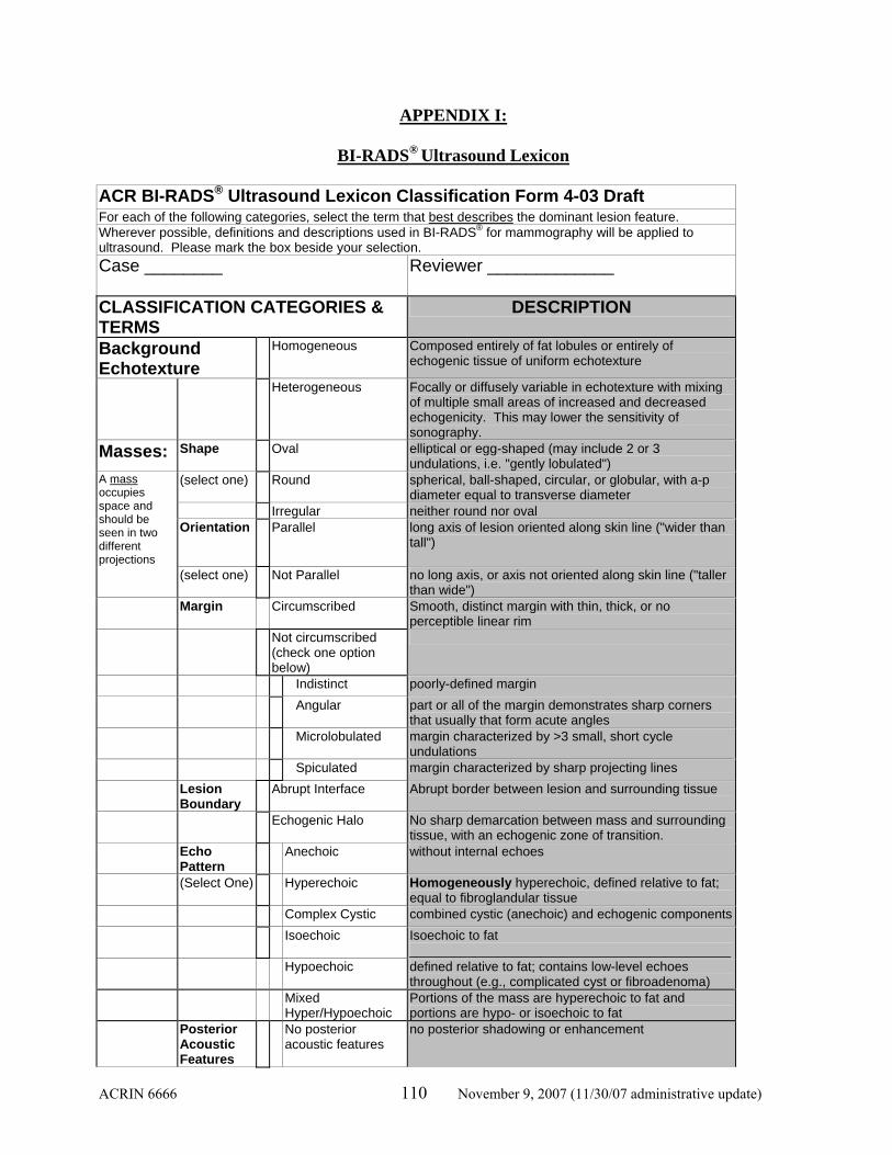
ACRIN 6666 110 November 9, 2007 (11/30/07 administrative update)
APPENDIX I:
BI-RADS® Ultrasound Lexicon ACR BI-RADS® Ultrasound Lexicon Classification Form 4-03 Draft For each of the following categories, select the term that best describes the dominant lesion feature. Wherever possible, definitions and descriptions used in BI-RADS® for mammography will be applied to ultrasound. Please mark the box beside your selection. Case ________ Reviewer _____________
CLASSIFICATION CATEGORIES & TERMS
DESCRIPTION
Background Echotexture
Homogeneous Composed entirely of fat lobules or entirely of echogenic tissue of uniform echotexture
Heterogeneous Focally or diffusely variable in echotexture with mixing of multiple small areas of increased and decreased echogenicity. This may lower the sensitivity of sonography.
Masses: Shape Oval elliptical or egg-shaped (may include 2 or 3 undulations, i.e. "gently lobulated")
(select one) Round spherical, ball-shaped, circular, or globular, with a-p diameter equal to transverse diameter
Irregular neither round nor oval
A mass occupies space and should be seen in two different projections
Orientation Parallel long axis of lesion oriented along skin line ("wider than tall")
(select one) Not Parallel no long axis, or axis not oriented along skin line ("taller than wide")
Margin Circumscribed Smooth, distinct margin with thin, thick, or no perceptible linear rim
Not circumscribed (check one option below)
Indistinct poorly-defined margin
Angular part or all of the margin demonstrates sharp corners that usually that form acute angles
Microlobulated margin characterized by >3 small, short cycle undulations
Spiculated margin characterized by sharp projecting lines
Lesion Boundary
Abrupt Interface Abrupt border between lesion and surrounding tissue
Echogenic Halo No sharp demarcation between mass and surrounding tissue, with an echogenic zone of transition.
Echo Pattern
Anechoic without internal echoes
(Select One) Hyperechoic Homogeneously hyperechoic, defined relative to fat; equal to fibroglandular tissue
Complex Cystic combined cystic (anechoic) and echogenic components
Isoechoic Isoechoic to fat ____________________________________________
Hypoechoic defined relative to fat; contains low-level echoes throughout (e.g., complicated cyst or fibroadenoma)
Mixed Hyper/Hypoechoic
Portions of the mass are hyperechoic to fat and portions are hypo- or isoechoic to fat
Posterior Acoustic Features
No posterior acoustic features
no posterior shadowing or enhancement

ACRIN 6666 111 November 9, 2007 (11/30/07 administrative update)
(Select One) Enhancement increased posterior echoes
Shadowing decreased posterior echoes; excluding edge shadows
Combined pattern both shadowing and enhancement
Surrounding Tissue
No effect surrounding tissue unaffected by lesion
Identifiable effect (select all that apply)
Duct changes abnormal caliber and/or arborization
Cooper's ligament* changes
straightening or thickening of Cooper's ligaments (curvilinear connective tissue bands providing support for the breasts)
Edema increased echogenicity of surrounding tissue, reticulation: includes angular hypoechoic lines
Architectural distortion
disruption of normal anatomic planes
Skin Thickening focal/diffuse skin thickening. Normal skin is < 2mm in thickness except in the periareolar area and lower breasts.
Skin retraction/ irregularity
skin surface is concave or ill-defined, appears pulled in
Calcifications: None seen no calcifications seen Calcifications are poorly characterized with ultrasound but can be recognized, particularly in a mass If present, (select all that apply) Macrocalcifications ≥ 0.5 mm in diameter Microcalcifications
out of mass Microcalcifications in
mass Special cases are those with a unique diagnosis or finding None Special case present Mass in or on skin including sebaceous or epidermal inclusion cyst; keloid,
etc. Complicated Cyst Nonpalpable incidental cyst with imperceptible wall,
mobile internal echoes and/or fluid-debris level Clustered microcysts Without discrete solid component Intraductal mass Foreign body including clip, coil, wire, catheter sleeve, silicone, etc. Lymph nodes-
intramammary in breast, including axillary tail
Lymph nodes-axilla Post-surgical scar Area of architectural distortion with or without
shadowing, extending to the skin surface and corresponding to the site of prior surgery
Vascularity (select all that apply)
Cannot assess vascularity
color flow not done or inadequate for interpretation
None no color flow
Present in lesion
Present immediately adjacent to lesion
Increased in surrounding tissue
There are limited data to support management recommendations for solid masses based on ultrasound findings at this time.
However, what would be your best assessment and management recommendation in each case? Incomplete 0-Incomplete Additional evaluation needed before final assessment

ACRIN 6666 112 November 9, 2007 (11/30/07 administrative update)
Assessment Final Assessment Category
1-Negative No lesion found (routine follow-up)
2-Benign finding No malignant features; e.g. cyst (routine follow-up)
3-Probably benign
Low probability of cancer, e.g. asymptomatic fibroadenoma or complicated cyst (short interval follow-up in 6 months)
4-Suspicious abnormality
intermediate probability of cancer (tissue sampling)
4A – Low Suspicion
Lesion is judged to have a low probability of malignancy , such as intraductal mass, probable abscess, or symptomatic complicated cyst. Aspiration or biopsy is recommended.
4B – IntermediateSuspicion
Lesion is of intermediate suspicion of malignancy, such as complex cystic lesions, ovoid indistinctly marginated masses. Biopsy is recommended.
4C – Moderate Suspicion
Lesion is of moderately high suspicion of malignancy such as a microlobulated mass with calcifications
5-Highly suggestive of malignancy
High probability of cancer (take appropriate action, biopsy)
6-Known malignancy
Take appropriate action
Modified from Copyright 2001 American College of Radiology Based on Final Report of Expert Working Group Developed Under Contract 282-97-0016 - Between U.S. Public Health Service Office on Women's Health,
U.S. Department of Health, and Human Services and the American

ACRIN 6666 113 November 9, 2007 (11/30/07 administrative update)
APPENDIX IA
Summary Breast Imaging Reporting and Data System (BI-RADS®): MRI Lexicon1 Indication: Describe clinical problems, history of biopsies (date and results), risk factors, phase of menstrual cycle (if relevant) Comparison: Prior breast imaging, including prior breast MRI should be reviewed, with the dates and types of prior studies reported Technical Factors: Describe magnet field strength, coil, use of compression, scan orientation (e.g. axial, sagittal) and types of sequences (e.g. T1WI, T2WI with fat suppression or STIR, 3D SPGR with fat suppression pre and post injection), which breast(s) scanned, amount and type of contrast, number of post-contrast acquisitions over what time period, and type of post-processing (e.g. subtraction technique and MIP reconstructions). Use of CAD, pharmacokinetic or other parametric mapping, should be reported. Limitations: If applicable, describe severity of image artifacts, motion, problems with injection Classification Categories and Terms
Description
Focus Note: If this is only finding, proceed to associated findings
Punctate, nonspecific enhancement, too small to characterize morphologically, usually < 5 mm
Mass Shape Round Spherical, ball-shaped, circular Oval Elliptical or egg-shaped Lobulated Undulating contour, scalloped Irregular Uneven shape, not round, oval, or
lobulated Margin Smooth Well circumscribed, well-defined,
sharply demarcated Irregular Uneven, neither smooth or spiculated;
may be ill-defined or indistinct Spiculated Radiating lines extend from margins Internal
Enhancement Characteristics
Homogeneous Confluent, uniform enhancement of the mass
Heterogeneous Nonuniform enhancement, variable signal intensity
Rim Enhancement More pronounced at periphery of mass
Dark Internal Septations
Dark, nonenhancing lines within a mass
Enhancing Internal Septations
Enhancing lines within a mass
Central Enhancement
Enhancement more pronounced at center of mass
Non-Mass-Like
Enhancement of an area, not a mass, including small or large regions, and whose internal enhancement results in a pattern discrete from normal surrounding parenchyma. Usually has interspersed spots of normal glandular tissue or fat

ACRIN 6666 114 November 9, 2007 (11/30/07 administrative update)
between enhancing areas. Distribution Focal Area < 25% of quadrant, in a confined area,
with interspersed fat or normal glandular tissue
Linear In a line, not definitely a duct. May be sheet-like in 3D.
Ductal In a line pointing toward nipple, can be branching, conforming to a duct
Segmental Triangular region or cone with apex pointing to nipple, suggesting a duct and its branches, can be ductal in areas
Regional Geographic enhancement of a large volume (≥ 25% of quadrant) not conforming to a ductal distribution
Multiple Regions 2 or more regional areas of enhancement; patchy
Diffuse Distributed uniformly and evenly throughout the breast
Internal Enhancement Patterns
Homogeneous Confluent, uniform
Heterogeneous Nonuniform in random pattern, separated by areas of normal breast parenchyma or fat
Stippled/Punctate Round, tiny, similar-appearing spots, sand-like or dot-like
Clumped Cobblestone-like, with occasional confluent areas; may resemble bunch of grapes in focal or segmental area or look beaded, like string of pearls when in linear distribution
Reticular/Dendritic Seen in involuted breasts: strand-like, finger-like projections of enhancing parenchyma separated by fat, extending toward nipple. Abnormal when associated with trabecular thickening and distortion: angulated, distorted at parenchyma-fat interface, with the enhancing areas truncated, thickened, stubby.
Symmetry (if bilateral scan)
Symmetric Mirror image, both breasts
Asymmetric More in one breast than the other Associated Findings
Nipple Retraction or Inversion Nipple is pulled in abnormally
Pre-Contrast High Duct Signal Bright signal in ducts before contrast,

ACRIN 6666 115 November 9, 2007 (11/30/07 administrative update)
on T1WI Skin Retraction Skin is pulled in abnormally Skin Thickening > 2 mm, focal or diffuse Skin Invasion Extension of abnormal enhancement
to include skin, which is usually thickened
Edema Trabecular thickening on T2WI, usually with associated skin thickening
Lymphadenopathy Enlarged, rounded lymph nodes, usually with loss of fatty hila
Pectoralis Muscle Invasion Extension of abnormal enhancement into adjacent pectoralis muscle; not sufficient to abut the muscle
Chest Wall Invasion Extension of abnormal enhancement into ribs or intercostal spaces
Hematoma/Blood Bright signal before contrast on T1WI due to blood
Abnormal Signal Void Absence of signal due to artifact Cyst Well-circumscribed, round or oval
fluid-filled structure with imperceptible wall, bright on T2WI
Location Breast Describe right, left, or bilateral Location Quadrant, subareolar, central, axillary tail Depth Distance from nipple, skin, or chest wall (in cm) as appropriate Kinetics Sample for and report the most rapidly enhancing or most suspicious area of the
lesion, avoiding less than 3 pixel ROI size Signal Intensity
(SI)/Time Curve Description
Initial Phase Enhancement within first two minutes after injection (when curve starts to change)
Slow < 50-60% increase in SI within 2 minutes
Medium 60-100% increase in SI within minutes
Rapid >100% increase in SI within minutes Delayed Phase Enhancement pattern after two minutes
(when curve starts to change) Persistent Progressive, continued increase in
signal over time Plateau SI does not change over time after
initial rise; flat (+/- 10%) Washout SI decreases after peaking Assessment Categories Incomplete Assessment
0-Incomplete Additional evaluation needed before final assessment
Final Assessment 1-Negative No lesion found (routine follow-up)

ACRIN 6666 116 November 9, 2007 (11/30/07 administrative update)
Category 2-Benign finding No malignant features; e.g. cyst
(routine follow-up) 3-Probably benign Very low probability of cancer, (short
interval follow-up in 6 months) 4-Suspicious abnormality Intermediate probability of cancer
(tissue sampling) 4A – Low
Suspicion Lesion is judged to have a low
probability of malignancy; biopsy is recommended.
4B – Intermediate Suspicion
Lesion is of intermediate suspicion of malignancy. Biopsy is recommended.
4C – Moderate Suspicion Lesion is of moderately high suspicion of malignancy
5-Highly suggestive of malignancy
High probability of cancer (take appropriate action, biopsy)
6-Known malignancy Take appropriate action 1 Adapted from Ikeda DM et al, Breast Imaging and Reporting Data System – Magnetic Resonance Imaging (BI-RADS® - MRI), 1st ed. Reston, VA: American College of Radiology, 2003. Note: Proposed changes as of 12/20/05 include addition of categories for background breast tissue enhancement: 1) Background Enhancement:
No/Minimal, Mild, Moderate, Marked
2) Subdivision of category 3 Probably benign, Short-term follow-up: 3A: Probably benign, possibly hormonal Recommend 1-3 mo f/u 3B: Probably benign Recommend 6 month follow-up

ACRIN 6666 117 November 9, 2007 (11/30/07 administrative update)
APPENDIX II
PROPOSAL TO MAKE ANTHROPOMORPHIC COMPRESSED BREAST PHANTOMS WHICH ARE TISSUE-MIMICKING WITH RESPECT TO ULTRASOUND AND X-RAYS
WITH PHOTON ENERGIES IN THE MAMMOGRAPHY RANGE Submitted by Ernest L. Madsen, Professor of Medical Physics, University of Wisconsin Introduction It is proposed that four anthropomorphic compressed phantoms be produced from materials that mimic breast tissues in terms of ultrasound and x-rays in the mammographic range. Similar compressed breast phantoms have been produced in the past at the University of Wisconsin 1,2 although, regarding target masses, no attempt was made to test the tissue-mimicking (TM) extent for x-ray mammography. The earlier versions have been used extensively for training sonographers in breast imaging as well as for comparing different versions of ultrasound breast imagers. Realistic beam distortions occur at the interface between the simulated subcutaneous fat layer and simulated glandular parenchyma. Subtle variations in ultrasonic properties between masses and simulated glandular parenchyma are represented; e.g., low contrast masses are present, and shadowing and enhancement will occur. All masses are within 4.5 cm of the scanning window; hence, visualization with 10 MHz systems will be tested. The phantoms will also find use in testing 3-D capabilities of ultrasound systems. Excellent mimicking of x-ray absorption characteristics was found for the materials used to mimic the ultrasonic properties of breast glandular parenchyma and breast fat.3 Because the various masses will have different compositions than the TM glandular parenchyma surrounding them, they should be similar in detectability to masses in real breast, appropriate for comparison between mammography sites. Anthropomorphic compressed phantoms produced in the past at our lab1,2 involved positioning of simulated masses by impaling them on very thin (0.1 micrometer diameter) stainless steel wires before introducing the molten TM glandular parenchyma and then, after congealing of the TM glandular material, withdrawing the wires. Tracks in the gel left after withdrawal of the wires were seldom detectable with scanners of the 1980s and the early 1990s. However, ultrasound scanners have apparently advanced in sophistication in the last decade to the extent that these tracks are rather easily detected in low echo materials such as simulated cysts. We have another technique for positioning the masses, which will not leave any tracks in the masses because there is no invasion of the mass material. This new technique requires considerably more effort and time, however, and adaptations will need to be made to produce the anthropomorphic phantoms proposed here. Phantom configuration The anthropomorphic compressed breast phantom, which we propose producing, is diagrammed in Figs 1, 2 and 3. The composition and ultrasonic properties of the TM fat to be used have been described previously1,4 as they have for the TM glandular parenchyma.1 The direct-contact interface (see Figs. 2 and 3) between the TM subcutaneous fat and the TM glandular parenchyma will have a scalloped shape with interconnected, randomly positioned, spherical subsurfaces each having a

ACRIN 6666 118 November 9, 2007 (11/30/07 administrative update)
radius of curvature of 1.5 cm. This uneven surface simulates the corresponding interface in real breast, challenging the focusing of ultrasound scan heads. The 16 masses are depicted and described in Fig. 1. For masses designated as (ultrasonically) low scatter or high scatter, the appropriated object contrast will be determined by making small test samples consisting of spheres of prospective mass material surrounded by TM glandular parenchyma and having them assessed by Drs. Wendie A. Berg, PI, and Ellen Mendelson at Northwestern University. The thickness of the anthropomorphic compressed breast phantoms will be 6 cm. (The bottom acrylic plate below the muscle layer shown in Figs. 2 and 3 will be removable for minimizing thickness during x-ray mammography exposures; a 100-µm thick sheet with a very low permeability for water will cover the bottom of the muscle layer.) The double-ended conical structure shown in Fig. 1 (mass no. 7) will consist of two cones joined at the base, the cones having the same diameter base, but different heights (3 mm and 5 mm). The base diameter will be 3 mm. This structure will challenge imagers first to detect it and then to determine its orientation. The retromammary fat layer will be 5 mm thick and consist of the same material as that used in the subcutaneous fat layer. The pectoral muscle layer will replicate that in phantom #1 in reference 1; i.e., randomly positioned but closely packed high attenuation, 2 mm diameter graphite-in-agar cylinders will be surrounded by gelatin.

ACRIN 6666 119 November 9, 2007 (11/30/07 administrative update)
1 2 3
4 56
7 8 9 10
11 12 13
14 15 16
1 : 10mm, high scatter, high attenuation
2
3 : 10mm, low scatter, backgd attenuation
2 : 10mm, low scatter, high attenuation
4 : 3mm, high scatter, backgd attenuation
5 : 3mm, low scatter, backgd attenuation
6 : 10mm cyst
7 : double cone, 4mm diameter at base
8 : 5mm diam. fat- mimicking sphere
9 : 6mm cyst in subareolar zone
10 : 2mm very high attenuation mass
11 : 6mm cyst close to retromammary fat
12 : 3mm very high attenuation mass
13 : 6mm cyst close to subcutaneous fat
14 : 3mm cyst near retromammary fat
15 : 3mm cyst midway between subcut. and retromamm. fat
16 : 3mm cyst near subcutaneous fat
1 cm thick acrylic wall
GLANDULAR
subareolar zone
10 cm
15 cm
Fig. 1. SIXTEEN MASS BREAST PHANTOM FOR ACR (TOP VIEW THROUGH SCANNING WINDOW)

ACRIN 6666 120 November 9, 2007 (11/30/07 administrative update)
acrylic wall
retromammary fat
glandular parenchyma
subareolar zonesubcutaneous fatskin
cysts
fat sphere
scanning window
Fig. 2. SIXTEEN MASS BREAST PHANTOM FOR ACR (END VIEW WITH ONLY FIVE MASSES DEPICTED)
6 cm
10 cm
muscle
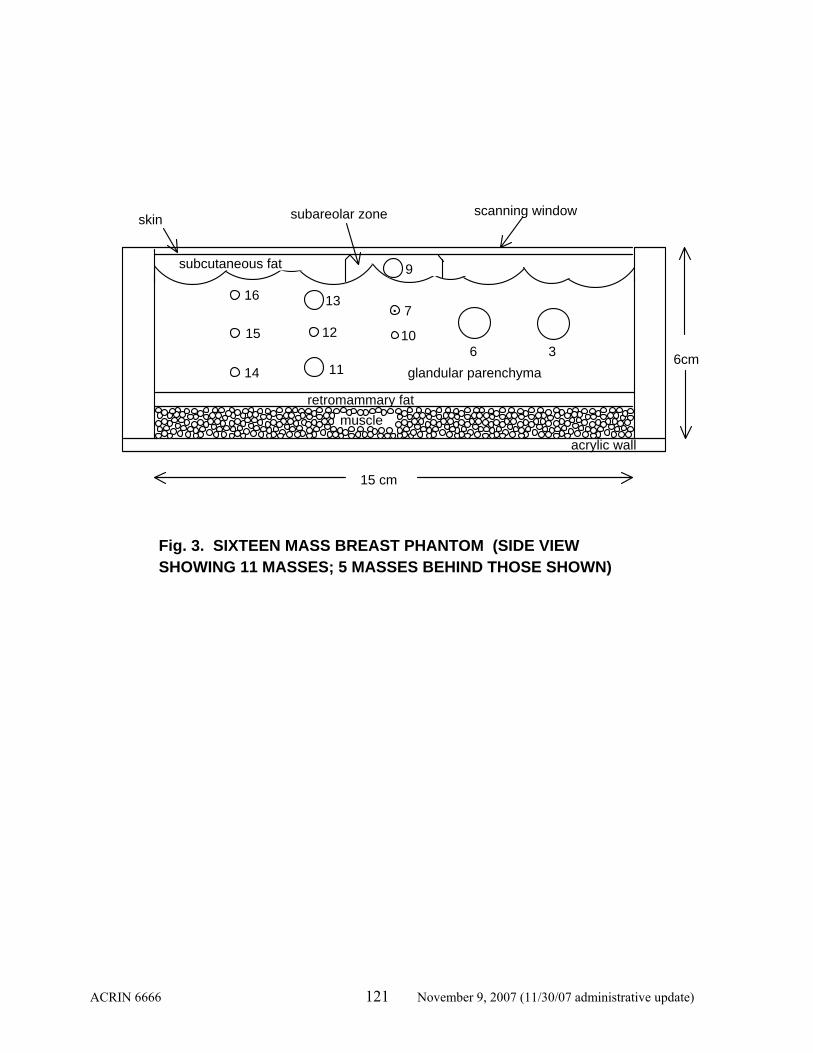
ACRIN 6666 121 November 9, 2007 (11/30/07 administrative update)
subcutaneous fat
.
36
9
10
713
12
1114
15
16
glandular parenchyma
acrylic wall
scanning windowskin subareolar zone
15 cm
6cm
Fig. 3. SIXTEEN MASS BREAST PHANTOM (SIDE VIEW SHOWING 11 MASSES; 5 MASSES BEHIND THOSE SHOWN)
retromammary fatmuscle

ACRIN 6666 122 November 9, 2007 (11/30/07 administrative update)
APPENDIX IIA
REFERENCES
1 EL Madsen, E Kelly-Fry and GR Frank, “Anthropomorphic phantoms for assessing systems used in ultrasound imaging of the compressed breast,” Ultrasound in Med & Biol 14, Sup. 1, pp. 183-201 (1998). 2 E Kelly-Fry, EL Madsen & GR Frank, “Use of anthropomorphic breast phantoms for comparing ultrasound breast imagers,”Archives of Acoustics (Poland) 16 pp. 501-511 (1991). 3 TM Burke, EL Madsen and JA Zagzebski, “X-ray linear attenuation coefficients in the mammographic range for ultrasonic breast phantom materials,” Radiology 142 pp. 755-757 (1982). 4 EL Madsen, JA Zagzebski and GR Frank, “Oil-in-gelatin dispersions for use as ultrasonically tissue-mimicking materials,” Ultrasound in Med & Biol 8 pp. 277-287 (1982).

ACRIN 6666 123 November 9, 2007 (11/30/07 administrative update)
APPENDIX III
AMERICAN COLLEGE OF RADIOLOGY IMAGING NETWORK
ACRIN 6666: SCREENING BREAST ULTRASOUND IN HIGH-RISK WOMEN
SAMPLE CONSENT FORM
[Note: ACRIN does not monitor compliance with the Health Insurance Portability and Accountability Act (HIPAA); that is the responsibility of local IRBs. Information on ACRIN’s HIPAA policy, as well as a template for HIPAA authorization, can be found at www.acrin.org.] You are being asked to read this consent form because you are eligible to enroll in a clinical trial (a type of research study). Clinical trials include only participants who choose to take part. Please take time to make your decision. You may want to discuss this with your friends, family, or doctor. This trial, which is conducted through the American College of Radiology Imaging Network (ACRIN), is sponsored by the National Cancer Institute and The Avon Foundation. The Avon Foundation (www.avoncompany.com/women/avonfoundation/) works to improve the lives of women and their families; one way they do this is by funding medical research on breast cancer. You are being asked to participate in this study because you have partially dense (non-fatty) breasts and are considered to be at increased risk of breast cancer. WHY IS THE STUDY BEING DONE? Finding breast cancer early has been shown to lower the chance of dying of breast cancer. Mammography helps find breast cancer early. Some breast cancers, however, are not seen on mammography. A cancer’s chance of not being seen on mammography is higher when the tissue in a woman’s breasts is dense (not fatty). It is possible that ultrasound may help to find breast cancers that are not seen on mammography in women with dense breasts. This study is being done to see if screening whole breast ultrasound can find cancers not seen on mammography. We are also interested in women’s experience with the screening tests, and will measure this by asking women how much they might be willing to pay to get the same information about breast cancer without having to have the test. HOW MANY PEOPLE WILL TAKE PART IN THIS STUDY? About 2808 women across the country will take part in this study. About 140 women from this institution will take part. WHAT IS INVOLVED IN THE STUDY? • You will be “randomized” to have either a mammogram first or an ultrasound exam first.
Randomization means that you are put into a group by chance. It is like flipping a coin. Neither you nor the study doctor will be able to choose which exam you will have first. No matter which you have first, you will have both a standard mammogram and a standard ultrasound exam within two weeks of each other. They will be interpreted in the usual way, but you will not be told the results of either exam until both have been completed.

ACRIN 6666 124 November 9, 2007 (11/30/07 administrative update)
• In addition, you will be asked a series of questions about any lumps, abnormal nipple discharge,
or skin changes in the breast or under your arm. If abnormalities are found with either ultrasound or mammography, additional tests, such as the following, may be required: • additional mammographic views. • additional ultrasound imaging. • needle aspiration (removing a small amount of breast fluid through a needle). • biopsy (removing a small amount of breast tissue). Any tissue removed will be analyzed in the
usual way. You will return for additional mammograms and ultrasound exams at: • One year after your first exams. • Two years after your first exams. Each year, for three years after your first exams, you will be asked questions about your breast health and any procedures you may have had on your breasts. You will also be asked to complete some short surveys in person and by mail or telephone. These surveys will take from 10 to 30 minutes to complete depending on which survey you are selected to receive. If you are selected and do not respond to the mailed surveys, you may be interviewed by telephone. At 12, 24, and 36 months after your first exams, you will be asked questions about any other breast imaging or breast biopsies you may have had and their results. If you undergo any procedures on your breasts over the next three years, you should send the results to the Research Associate at this facility. Copies of your images and records will be stored at ACRIN headquarters for later review. Pathology slides from any biopsies may be reviewed by ACRIN researchers at the University of Florida and/or the University of Maryland. All results will be kept confidential. HOW LONG WILL I BE IN THE STUDY? You are being asked to participate in the study for at least 3 full years after your first exam. If we find abnormalities in your breasts we may ask you to participate for up to 4 years so that we can continue to monitor your breasts. The study doctor has the right to take you off the study at any time, especially if you become too ill to participate. You can withdraw from the study at any time. If you decide to stop taking part in the study, we encourage you to talk to the study doctor or a member of the ACRIN staff and your regular doctor first. WHAT ARE THE RISKS OF THE STUDY? • Any screening test has the potential to identify areas of concern. Most of these will not be
cancer. From 2 to 10 of every 100 women who have a screening breast ultrasound will need a biopsy (removing a small amount of breast tissue using a needle) or aspiration (removing a small amount of breast fluid through a needle). Of those procedures, on average, 12 in 100 will show cancer and 88 will not. From 2 to 10 in 100 women may also need more tests and follow-up beyond what would normally result from mammography alone.
ACRIN 6666 125 November 9, 2007 (11/30/07 administrative update)
• There is also a risk that even after clinical breast examination, mammography, and ultrasound of your breast(s), that you will have a breast cancer that will not be found by these tests. Even when breast cancer is found early, before it can be felt, some women will still die of the disease.
ARE THERE POTENTIAL BENEFITS TO TAKING PART IN THE STUDY? If you agree to take part in this study, there may or may not be direct medical benefit to you. The possible benefits of taking part in this study are the same as being screened with breast ultrasound in addition to mammography and clinical breast examination without being part of the study. These benefits include the opportunity to have screening ultrasounds at no charge. This may result in: • Providing you and your doctor with baseline readings of your normal breasts. • The earlier diagnosis of any breast cancer, which could lead to:
Prevention or delay of death from breast cancer; Prevention of, or reduction in, symptoms from breast cancer; Milder treatment, leading to fewer side effects, from treatment of breast cancer.
It is hoped that the information learned from this study will eventually help you and other women who are at risk for breast cancer. WHAT OTHER OPTIONS ARE THERE? You may choose not to take part in this study. Other screening options you may consider include: • screening with mammography; • screening with clinical breast examination, with or without self breast examination; • screening with ultrasound at your own expense; • screening with a contrast-enhanced magnetic resonance imaging (MRI) study of the breast(s). Please talk with your regular doctor about these and other options. WILL MY RECORDS BE CONFIDENTIAL? Although all efforts will be made to keep your personal information confidential, we cannot guarantee absolute confidentiality. Your personal information may be disclosed if required by law. All records, including any imaging on file, will be kept in a confidential form at this institution and in a computer file at the headquarters of the American College of Radiology Imaging Network (ACRIN) and the Center for Statistical Sciences at Brown University. Your contact information will be sent to researchers from the Rhode Island Hospital at Brown University so that researchers working there can administer surveys to you by mail and telephone; this information will not be used for any other purpose and will not be entered into the main ACRIN database. The screening exams performed in this study and representative images will be kept for at least 2 years after the study is over. Pathology slides from any biopsies may be reviewed by ACRIN researchers at the University of Florida and/or the University of Maryland. Images of the pathology tissue may be obtained and kept for at least two years after the study is over. Organizations that may inspect and/or copy your research records for quality assurance and data analysis include groups such as ACRIN, the National Cancer Institute (NCI), the Avon Foundation, the Food and Drug Administration (FDA), and the Institutional Review Board of [institution name].

ACRIN 6666 126 November 9, 2007 (11/30/07 administrative update)
Information gathered from screening exams and pathological specimens in this study may be used by these or other researchers in the future for other studies of research questions related to breast cancer. Your name or other identifying information about you will never be used in any reports of the results of these studies. WHAT ARE THE COSTS OF THE STUDY? The yearly screening ultrasound and clinical breast examination at the study site will be paid for by Avon/NCI through ACRIN. You and your insurance company are responsible for the costs of your mammogram(s). You and your insurance company are responsible for all costs associated with diagnostic tests, including additional mammographic views, ultrasound directed to areas of concern on the screening studies, and other follow-up tests and/or treatments that result from screening. If you do not have adequate insurance coverage to pay for these procedures, we will try to find additional resources to help you. In the case of injury or illness resulting from this study, emergency medical treatment is available, but it will be provided at the usual charge. No funds have been set aside to compensate you in the event of injury. WHAT ARE MY RIGHTS AS A PARTICIPANT? Taking part in the study is voluntary. You will not be paid for your participation. If you choose not to take part in the study or to leave the study at any time, your medical care will not be affected. A Data Safety and Monitoring Board, an independent group, will be reviewing the data from this research throughout the study. We will tell you about the new information from this or other related studies that may affect your health, welfare, or willingness to stay in this study. WHOM DO I CALL IF I HAVE QUESTIONS OR PROBLEMS? (Individual sites must complete this information.) For information about your screening or participation, or about the study, you may contact: Name, Title, Site Principal Investigator Phone number For information about your rights as a research participant, you may contact the Institutional Review Board of : Name Phone number

ACRIN 6666 127 November 9, 2007 (11/30/07 administrative update)
WHERE CAN I GET MORE INFORMATION? Visit the NCI’s Web sites for comprehensive clinical trials information http://cancertrials.nci.nih.gov, http://cancernet.nci.nih.gov, or the American College of Radiology Imaging Network’s website www.acrin.org. The Avon Foundation’s Web site is http://www.avoncompany.com/women/avonfoundation. PERMISSION TO REVIEW MEDICAL RECORDS By agreeing to participate, I give permission for my health care providers and hospitals where I have been seen to release my medical records to the study doctors. SIGNATURE I have read all the above and/or had it explained to me. I have had the opportunity to ask questions and have received satisfactory answers. I willingly give my consent to participate in this study. Upon signing this form I will receive a copy. I may also request a copy of the protocol (full study plan). ______________________________________ ______________________ Participant (or Legal Representative) Signature Date Witnessed by: Study Investigator/Research Associate Date

ACRIN 6666 128 November 9, 2007 (11/30/07 administrative update)
APPENDIX IIIA
SUPPLEMENTAL SAMPLE INFORMED CONSENT TO ACRIN 6666 STUDY
YIELD OF MRI AFTER COMBINED SCREENING WITH ULTRASOUND AND MAMMOGRAPHY IN HIGH RISK WOMEN: AN AMENDMENT TO ACRIN PROTOCOL
6666
You are being asked to be in this part of the study because you are a participant in the ACRIN 6666 Screening Breast Ultrasound Study. This study involves a screening magnetic resonance imaging (MRI) scan to see if the images obtained during the MRI scan are able to find cancers that are not found by mammography and/or ultrasound. This research study is managed by the American College of Radiology Imaging Network (ACRIN) and funded by the Avon Foundation and the National Cancer Institute (NCI). The purpose of this study is to investigate whether MRI can provide additional information above and beyond mammography and ultrasound. An MRI uses powerful magnets and radio waves linked to a computer to create cross-sectional images of the breasts. This study involves an MRI scan and the collection and review of health care information including information from your medical records, MRI images, questions about your hormonal and family history, and any abnormal results from the removal of breast tissue or surgery. You are being asked to give your permission to have a breast MRI scan, to document your medical and family history, for review of your medical records, and to allow submission of computer images and reports from your MRI scan and to have any further biopsies if necessary. If you agree to participate in this trial, you will have the MRI scan within 8 weeks of the 24 month annual routine US and mammogram visit. You will not receive any payment for taking part in this study. HOW MANY PEOPLE WILL TAKE PART IN THE STUDY? About 1200 people will take part in the MRI part of the study. HOW LONG WILL I BE IN THE STUDY? You may be contacted up to 14 months after your MRI scan for additional follow-up. Depending on your initial MRI screening, you may receive a 6-month follow-up MRI scan. WHAT IS INVOLVED IN THIS STUDY? If the exams, tests, and procedures show that you can be in the study, have an MRI scan, and you choose to take part, you will have (not need) the following procedure. MRI examinations are part of regular medical care. For the MRI scan, you will change into a hospital gown and lie on your stomach on the scanning table with your breasts through an opening in the table. Wire coils within a plastic mold will be placed on either side of your breasts to receive very weak radio signals from the breasts. Gentle compression may be applied to the breasts. A needle attached to a small thin tube (called a catheter)

ACRIN 6666 129 November 9, 2007 (11/30/07 administrative update)
will be put into the vein of your arm. The table will slide into a tube-like machine that contains a magnet. The MRI machine sends a strong magnetic field that passes through your body. The strong magnetic field is produced by passing an electric current through wire coils which are located inside the scanner. Other coils in the machine send and receive radio waves. When in the machine, your body produces very faint signals in response to the radio waves. These signals are detected by the machine. The collected signals create 3-D pictures of your breasts. During the scan you will need to remain very still for several minutes at a time. You will hear tapping or loud thumping during the scan. After some initial sets of pictures, you will receive an MRI contrast agent (a dye-like liquid called Gadolinium) through the needle in your arm. Gadolinium is considered safe and is routinely used for MRI scans. This contrast agent helps to improve the images of your breasts, making any breast tumors easier to see. The MRI scan is painless, will not require hospitalization, withholding or delaying of treatments, blood tests, or special preparation. If a lesion is found on your MRI, additional procedures may be performed. This includes mammography, ultrasound, and/or additional MRI scans. In addition, a biopsy may be recommended for certain types of lesions by your study doctor. You may be asked to come in for a 6 month follow-up MRI visit up to 12 months after the first MRI visit. WHAT ARE THE POSSIBLE RISKS OR DISCOMFORTS OF THE STUDY? You may have side effects while on the study. Everyone taking part in the study will be watched carefully for any side effects. However, doctors do not know all the side effects that may happen. Side effects may be mild or serious. Your doctor may give you medicines to help lessen side effects. Medications may be given to make side effects less serious and uncomfortable. RISKS OF MRI SCAN Because of the powerful magnetic force of the MRI scanner, you may not be able to participate in the study if you have:
• metallic or other surgical implants (for example: pacemaker, heart valves, aneurysm clips, metal plates or pins and some orthopedic prostheses)
• metal pieces in your eye(s) or other body part • difficulty lying still or inability to lay on your stomach.
Notify your doctor if any of the above relate to you. Also, carefully read the information you should receive at the MRI facility about other risks. You may experience certain side effects due to the MRI scan.
Anxiety/stress; Discomfort due to the loud noise; Claustrophobia due to being in a confined space.

ACRIN 6666 130 November 9, 2007 (11/30/07 administrative update)
RISKS OF CONTRAST AGENT: GADOLINIUM Approximately two percent of patients experience some side effects with the use of Gadolinium; however, they are mostly mild. Serious side effects are very rare. Less likely:
Headaches; Nausea, vomiting; Burning, itching or tingling sensation; Hives; Temporary low blood pressure.
Rare, but serious:
Major allergic reaction; Nephrogenic systemic fibrosis (NSF)/nephrogenic fibrosing dermopathy (NFD): In rare cases,
some patients who have severe kidney disease developed symptoms of tightening or scarring of the skin and organ failure called nephrogenic systemic fibrosis (NSF) and nephrogenic fibrosing dermopathy (NFD) after they have had an MRI scan with gadolinium-based contrast agent.
NSF has not been seen in patients with normal working kidneys or mild problems in kidney function. If there is concern about your kidney function, you may be asked to have a blood test to determine if your kidneys are working properly before you have the MRI. NSF causes fibrosis of the skin and connective tissues throughout the body. Patients develop skin thickening that may prevent bending and extending joints, resulting in decreased mobility of joints. NSF usually starts in the lower extremities. It can also develop in the diaphragm, muscles in the thigh and lower abdomen, and lung vessels. In very rare cases, it can be deadly. Reference: FDA/Center for Drug Evaluation and Research. May 23, 2007, http://www.fda.gov/cder/drug/infopage/gcca/qa_200705.htm.
RISKS ASSOCIATED WITH INTRAVENOUS CATHETER (IV) PLACEMENT Likely
Minor pain at the placement site. Less likely
Low risk of bleeding, infection, bruising, and venous thrombosis (clot in your vein).
RISKS ASSOCIATED WITH BIOPSIES Likely
Minor discomfort.
Less likely Low risk of minor pain and bleeding; Infection; Bruising; Collection of air or gas in the chest cavity (pneumothorax).
REPRODUCTIVE RISKS

ACRIN 6666 131 November 9, 2007 (11/30/07 administrative update)
You must not be pregnant or plan on becoming pregnant within the next 14 months. If you think you might be pregnant, you must tell your study doctor at this time. You may need to take a pregnancy test before you can take part in this study. WHAT ARE THE POSSIBLE BENEFITS OF TAKING PART IN THE STUDY? There may be no direct benefit to you from being in the MRI study. We hope that the results of this study may help patients with breast cancer in the future. WILL I HAVE TO PAY FOR ANYTHING? Taking part in this study may lead to added costs to you or your insurance company. Your insurance company will be billed for the initial MRI scan and any MRI-prompted biopsy(ies), or follow-up. In the case of injury or illness resulting from this study, emergency medical treatment is available but will be provided at the usual charge. No funds have been set aside to compensate you in the event of injury. You or your insurance company will be billed for continuing medical care and/or hospitalization. Please ask about any unexpected added costs or insurance problems. WHAT ABOUT CONFIDENTIALITY? Your records will be identified only by a study identification number at the headquarters of the American College of Radiology Imaging Network (ACRIN) in Philadelphia, PA and at the Statistical Center at Brown University in Providence, RI. Only the researchers, the Avon Foundation, the National Cancer Institute (NCI), the Institutional Review Board (IRB), and ACRIN will have access to information about you. During their required reviews, representatives of NCI, ACRIN, the Statistical Center at Brown University, IRB, or other organizations involved in this study may have access to your medical records.
Your questionnaire results and MRI images will be kept permanently on file at ACRIN and may be used for future research. All personal identifiers are removed and replaced with a unique identifying number. Your name will never be used in any reports of these studies. WHOM DO I CALL IF I HAVE QUESTIONS OR PROBLEMS? For questions and information about your screening or participation, or about the study, you may contact: Name of Site Principal Investigator Phone number For questions and information about your rights as a research participant, you may contact the Institutional Review Board of :

ACRIN 6666 132 November 9, 2007 (11/30/07 administrative update)
Name Phone number WHAT OTHER CHOICES DO I HAVE IF I DO NOT WANT TO PARTICIPATE? You may choose not to participate in this study. If you choose not to participate in this study, your care will not be affected. WHAT ARE MY RIGHTS AS A PARTICIPANT? Your participation in the MRI study is voluntary. If you do not participate, you will not be contacted again for the study. You may withdraw from this study at any time. You will continue to receive your usual medical care whether or not you decide to participate in this study. ACKNOWLEDGEMENT When you sign this document, you are agreeing to take part in the MRI part of the study. This means you have read all the above information, asked questions regarding your participation, and received answers that you understand to all your questions. You have also had the opportunity to take this consent form home for review or discussion if you want to. A copy of this signed consent form will be given to you. Printed Name of Study Participant/ Legal Representative __________ Signature Date Printed Name of Person Obtaining Consent __________ Signature Date

ACRIN 6666 133 November 9, 2007 (11/30/07 administrative update)
APPENDIX IV: 6666 Eligibility Checklist (Page 1 of 3)
The following questions will be asked at study registration: __________ 1. Institutional person randomizing case (Name of individual randomizing case) __________ 2. (Y) Has the eligibility checklist (worksheet) been completed? __________ 3. (Y) Patient eligible for this study? (Participant meets at least one of the six high-
risk criteria defined in Section 5.3.) ___-___-___ 4. Date the study-specific consent form was signed (mm-dd-yyyy; must be prior to
study entry) __________ 5. Participant’s initials (Last, First; L., F.) __________ 6. Verifying physician __________ 7. Participant’s ID # (Optional; this is an institution's method of internally tracking a
participant to a protocol case number; may code a series of 9's)
___-___-___ 8. Date of birth (mm-dd-yyyy; must be > 25 years old) __________ 9. Ethnic Category:
1 Hispanic or Latino 2 Not Hispanic or Latino 9 Unknown
(10. Omitted)
__________ 11. Gender: 2 Female __________ 12. Participant’s Country of Residence (if country of residence is other, complete
Q18): 1 United States 2 Canada 3 Other 9 Unknown
__________ 13. Zip Code (US residents 5-digit zip code)

ACRIN 6666 134 November 9, 2007 (11/30/07 administrative update)
Eligibility Checklist, Page 2 of 3 __________ 14. Participant’s Insurance Status:
0 Other 1 Private Insurance 2 Medicare 3 Medicare and Private Insurance 4 Medicaid 5 Medicaid and Medicare 6 Military or Veteran’s Administration 7 Self Pay 8 No means of payment 9 Unknown/Decline to answer
__________ 15. Any care at VA or military hospital
1 No 2 Yes 9 Unknown
___-___-___ 16. Calendar base date (First study imaging scheduled date) (mm-dd-yyyy) ___-___-___ 17. Randomization date (mm-dd-yyyy) ___________ 18. Other country, specify (complete Q18 if Q12 is other) ___________ 19. (N/Y) Race: American Indian or Alaskan Native ___________ 20. (N/Y) Race: Asian ___________ 21. (N/Y) Race: Black or African-American ___________ 22. (N/Y) Race: Native Hawaiian or other Pacific Islander ___________ 23. (N/Y) Race: White ___________ 24. (N/Y) Race: Unknown __________ 25. (N) Is participant enrolled in first year of Digital Mammography Imaging
Screening Trial (DMIST), any contrast-enhanced breast MRI trials, tomosynthesis trial, any other trial of breast ultrasound or breast ultrasound agents, or any breast cancer screening trial?
__________ 26. (N) Has the participant undergone contrast-enhanced breast MRI or bilateral
whole breast ultrasound within the past 12 months? __________ 27. (N) Has the participant had any breast procedures (FNAB other than cyst
aspiration, core biopsy, or other breast surgical procedure) within the past 12 months?

ACRIN 6666 135 November 9, 2007 (11/30/07 administrative update)
Eligibility Checklist, Page 3 of 3 __________ 28. (N) Is the participant aware of any palpable abnormality in the breast(s), abnormal
skin changes of the breast(s) and/or nipple(s), bloody discharge, or spontaneous nipple discharge?
__________ 29. (Y) Does the participant meet one of the high-risk criteria as defined in Section
5.3 of the protocol? __________ 30. (N) Has the participant had breast cancer diagnosed within the prior 12 months or
have known distant metastases from breast cancer or have known residual cancer? __________ 31. (N) Excluding breast cancer, basal cell or squamous cell skin cancer, and in situ
cervical cancer, has the participant been diagnosed with cancer in the last five years or has the participant had a recurrence of cancer in the last five years or has residual disease been detected in the last five years?
__________ 32. (N) Does the participant have breast implant(s) in the study breast(s)? __________ 33. (N) Is the participant pregnant, nursing, or does she have any reason to believe
she may be pregnant or does she plan to become pregnant within the next 2 years? __________ 34. (Y) Does the participant understand and agree to the follow-up requirements as
outlined in Section 4.10 of the protocol? ___-___- ___ 35. Date* study mammogram scheduled (mammogram and sonogram must be within
2 weeks of each other and performed at the same site) (mm-dd-yyyy) ___-___-____ 36. Date* study sonogram scheduled (sonogram and mammogram must be within 2
weeks of each other and performed at the same site) (mm-dd-yyyy)
__________ 37. (N/Y) Is this participant’s first mammogram? (If yes, answer Q38 and skip Q39, if no, answer Q38 and Q39.)
___________ 38. (Y) Is this a routine annual mammogram visit? ___________ 39. (Y) Are the breast(s) heterogeneously dense or dense mammographically as
defined in Section 5.3 of the protocol? (leave blank if no prior mammogram)
Participant signature ______________________________________________
Signature of person responsible for the data: _____________________ (Research Associate or Principal Investigator)
Date form completed (mm-dd-yyyy): ______________________________
Signature of person entering data on the web: ______________________________ *If the study mammogram and/or sonogram have been scheduled, please provide the dates. If the imaging appointments have not been scheduled, please leave the question blank.

ACRIN 6666 136 November 9, 2007 (11/30/07 administrative update)
APPENDIX IVA 6666 ELIGIBILITY CHECKLIST: MRI AMENDMENT
Eligibility Checklist worksheet: MRI Substudy of ACRIN 6666 __________ (Y) 1. Is the participant currently eligible, active and enrolled in ACRIN 6666 protocol,
including: __________ (Y) 1a. Meets definitions of high risk?
__________ (N) 1b. Had bilateral mastectomy?
__________ (N) 1c. Is the participant pregnant or lactating and/or plan
to become pregnant within 14 months of MRI study entry?
__________ (N) 1d. Does the participant present with signs or
symptoms of breast cancer (palpable mass(es), bloody or spontaneous clear nipple discharge, axillary mass, or abnormal skin changes in the breast(s) or nipple(s))?
__________ (N) 1e. Is the participant enrolled in any other breast
screening trials? __________ (N) 1f. Has the participant been diagnosed with
metastatic cancer of any type since entering ACRIN 6666 protocol?
__________ (Y) 2. Is this a routine annual mammogram visit?
Note: Women who are undergoing surveillance of findings considered benign or probably benign on prior breast imaging are still eligible
__________ (Y) 3. Will the participant have completed three annual rounds of screening with both
mammography and US as part of ACRIN 6666 protocol by 02/10/2008? ___-___ -___ 3a. Date 24 month mammogram scheduled ___-___ -___ 3b. Date 24 month US scheduled
__________ (N) 4. Does the participant have contraindications to MRI:
__________ (N) 4a. Pacemaker, aneurysm clip, or other implanted magnetic device?
__________ (N) 4b. Claustrophobia not able to be controlled by premedication
with valium or ativan, or other sedative under her physician’s orders?
__________ (N) 4c. Lack of intravenous access?

ACRIN 6666 137 November 9, 2007 (11/30/07 administrative update)
__________ (N) 4d. Weight > 300 lbs?
__________ (N) 4e. Physically unable to tolerate positioning in the MRI
scanner? __________ (N) 4f. Impaired renal function, with estimated glomerular filtration
rate (GFR) < 30 mL/min/1.73 m2 and/or on dialysis?
__________ (N) 5. Has the participant had screening contrast-enhanced breast MRI within the past
24 months performed on all study breasts (usually bilateral, or unilateral in the case of women s/p mastectomy) or diagnostic MRI on any study breast(s) within the past 12 months?
__________ (N) 6. Has the participant had breast surgery performed < 12 months earlier on any
study breast(s)? __________ (N) 7. Has the participant had core biopsy performed < 5 months earlier on any study
breast(s)? __________ (N) 8. Is the participant currently receiving chemotherapy (excluding personal history
of cancer, on chemoprevention with Tamoxifen, Evista (Raloxifene), Arimidex (Anastrosole), Aromasin (Exemestane) or other aromatase inhibitor)?
__________ (Y) 9. Has a study specific consent been signed?
___-___ -___ 9a. Date the MRI study-specific Consent Form was
signed (must be prior to MRI substudy registration). __________ (Y) 10. Is the participant able to undergo contrast-enhanced MRI within 8 weeks after
completing both study US and mammogram at 24 month time point?
To be scheduled when possible in days 7-14 after onset of menses in premenopausal women. ___-___ -___ 10a. Date of last menstrual period or enter N/A at Q10b
if> 30 days ago or unknown
__________ (N/A) 10b. Last menstrual period > 30 days ago __________ (Y) 11. Has the participant agreed to undergo follow-up MRI at 6 months if needed and
to undergo MRI-guided vacuum-assisted biopsy or US-guided core biopsy if needed based on results of the MRI examination?
__________ (Y) 12. Has the participant agreed to provide clinical follow-up information 11-14
months after completing the MRI examination?
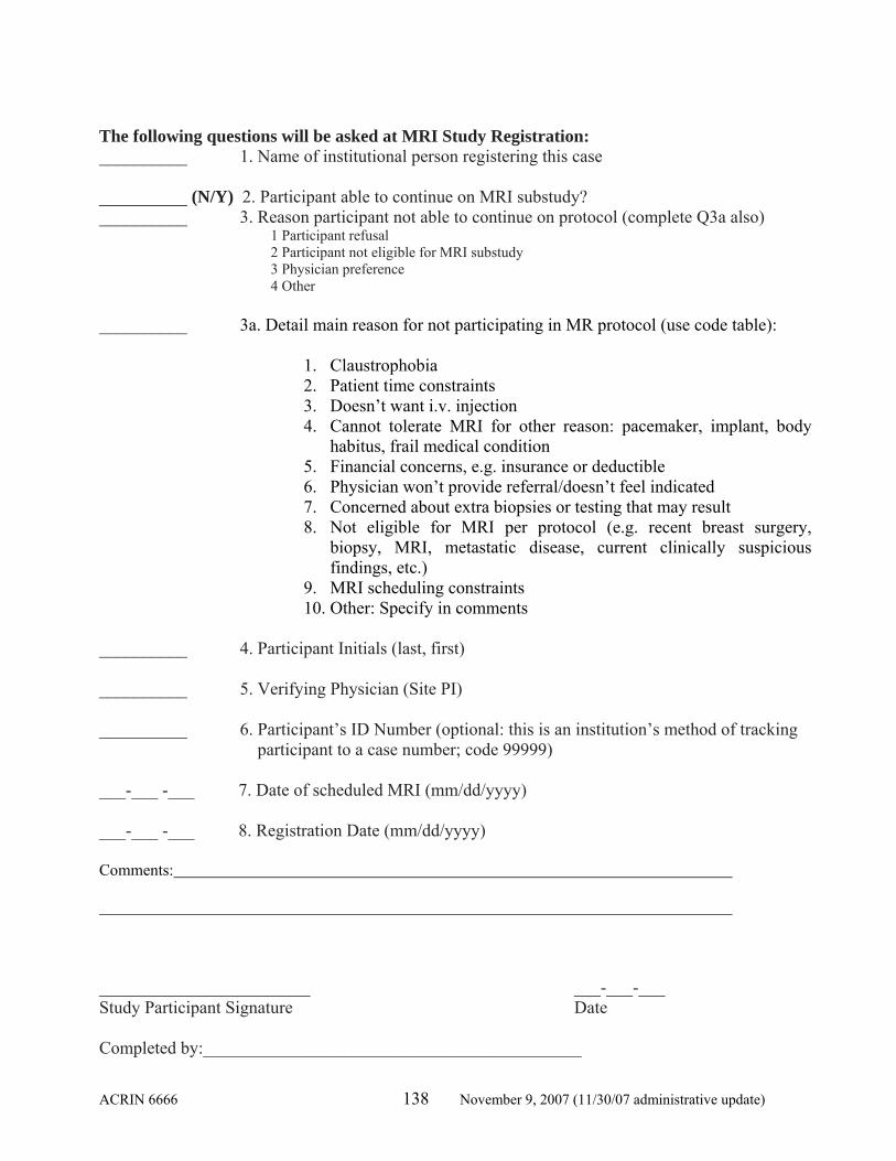
ACRIN 6666 138 November 9, 2007 (11/30/07 administrative update)
The following questions will be asked at MRI Study Registration: __________ 1. Name of institutional person registering this case __________ (N/Y) 2. Participant able to continue on MRI substudy? __________ 3. Reason participant not able to continue on protocol (complete Q3a also)
1 Participant refusal 2 Participant not eligible for MRI substudy 3 Physician preference 4 Other
__________ 3a. Detail main reason for not participating in MR protocol (use code table):
1. Claustrophobia 2. Patient time constraints 3. Doesn’t want i.v. injection 4. Cannot tolerate MRI for other reason: pacemaker, implant, body
habitus, frail medical condition 5. Financial concerns, e.g. insurance or deductible 6. Physician won’t provide referral/doesn’t feel indicated 7. Concerned about extra biopsies or testing that may result 8. Not eligible for MRI per protocol (e.g. recent breast surgery,
biopsy, MRI, metastatic disease, current clinically suspicious findings, etc.)
9. MRI scheduling constraints 10. Other: Specify in comments
__________ 4. Participant Initials (last, first) __________ 5. Verifying Physician (Site PI) __________ 6. Participant’s ID Number (optional: this is an institution’s method of tracking
participant to a case number; code 99999) ___-___ -___ 7. Date of scheduled MRI (mm/dd/yyyy) ___-___ -___ 8. Registration Date (mm/dd/yyyy) Comments: ________________________ ___-___-___ Study Participant Signature Date Completed by:___________________________________________
ACRIN 6666 139 November 9, 2007 (11/30/07 administrative update)
(Research Associate, Investigator Designee, or Principal Investigator) _____________________________________ ___-___-___ Signature of person entering data onto the Web Date form completed
ACRIN 6666 140 November 9, 2007 (11/30/07 administrative update)
APPENDIX V
Health Insurance Portability and Accountability Act (HIPAA) Research Authorization (Optional)
ACRIN does not monitor compliance with the HIPAA. It is the responsibility of local Institutional Review Boards (IRBs). Information on ACRIN’s HIPAA policy, as well as a template for HIPAA authorization, can be found at www.acrin.org.