Embed Size (px)
Citation preview
MANUSCRIP
T
ACCEPTED
ACCEPTED MANUSCRIPT1
1 Eugene and Marilyn Glick Eye Institute, Indiana University School of Medicine, Indianapolis, IN 2 Retina Service, Midwest Eye Institute, Indianapolis, IN
The acute and chronic effects of intravitreal anti-vascular endothelial growth factor injections on
intraocular pressure: A Review
Peter Bracha, MD1
Nicholas A Moore, MD1
Thomas A Ciulla, MD, MBA1,2
Darrell WuDunn, MD, PhD1
Louis B Cantor, MD1
___________________________________________________________________
This is the author's manuscript of the article published in final edited form as:Bracha, P., Moore, N. A., Ciulla, T. A., WuDunn, D., & Cantor, L. B. (2017). The acute and chronic effects of intravitreal anti-vascular endothelial growth factor injections on intraocular pressure: A Review. Survey of Ophthalmology. https://doi.org/10.1016/j.survophthal.2017.08.008

MANUSCRIP
T
ACCEPTED
ACCEPTED MANUSCRIPT2
[Type here]
Outline
I. Introduction
II. Short-term ocular hypertensive effects of intravitreal anti-VEGF injections
A. Intraocular pressure trend following anti-VEGF injection
B. Risk factors for acute ocular hypertension following injection
C. Studies supporting medical prophylaxis for acute ocular hypertension following intravitreal
anti-VEGF injections
1. Apraclonidine
2. Timolol
3. Dorzolamide-timolol
4. Brimonidine-timolol
5. Brinzolamide-timolol
D. Studies not supporting medical prophylaxis for acute ocular hypertension following
intravitreal anti-VEGF injections
E. Other prophylactic measures for acute ocular hypertension following intravitreal anti-VEGF
injections
1. Anterior chamber paracentesis
2. Ocular decompression
F. Summary of studies investigating prophylaxis against acute ocular hypertension following
intravitreal injections
III. Long-term effects of intravitreal anti-VEGF injections
A. Studies associating sustained ocular hypertension with chronic anti-VEGF injections
B. Studies not associating sustained ocular hypertension with chronic anti-VEGF injections
C. Effect of repeated injections on the retinal nerve fiber layer
IV. Macular disease and the risks of forgoing anti-VEGF therapy
V. Conclusion

MANUSCRIP
T
ACCEPTED
ACCEPTED MANUSCRIPT3
[Type here]
Key Words: Intraocular Pressure; Intravitreal Injection; Anti-vascular endothelial growth factor;
Glaucoma; Macular degeneration

MANUSCRIP
T
ACCEPTED
ACCEPTED MANUSCRIPT4
[Type here]
Abstract
The acute and chronic effects of repeated intravitreal anti-vascular endothelial growth factor (VEGF)
injections on intraocular pressure (IOP) have not been fully characterized and the development of
sustained ocular hypertension could adversely affect patients who are at risk of glaucomatous optic
neuropathy. As expected, volume-driven, acute ocular hypertension immediately follows intravitreal
injection, but this pressure elevation is generally transient and well-tolerated. Several medications have
been investigated to limit acute ocular hypertension following anti-VEGF therapy, but the benefits of
pretreatment are not conclusive. Chronic, sustained ocular hypertension, distinct from the short-term
acute ocular hypertension following each injection, has also been associated with repeated intravitreal
anti-VEGF injections. Risk factors for chronic ocular hypertension include the total number of injections,
a greater frequency of injection, and pre-existing glaucoma. Proposed mechanisms for chronic ocular
hypertension include microparticle obstruction, toxic or inflammatory effects on trabecular meshwork,
as well as alterations in outflow facility by anti-VEGF agents. Although limiting anti-VEGF therapy could
minimize the risk of both acute and chronic ocular hypertension, foregoing anti-VEGF therapy risks
progression of various macular diseases with resulting permanent central vision loss. While definitive
evidence of damage to the retinal nerve fiber layer is lacking, patients receiving repeated injections
should be monitored for ocular hypertension and those who subsequently develop sustained ocular
hypertension should be periodically monitored for glaucomatous changes with an optic nerve optical
coherence tomography (OCT) and static visual fields.
MANUSCRIP
T
ACCEPTED
ACCEPTED MANUSCRIPT5
[Type here]
I. Introduction
Anti-vascular endothelial growth factor (anti-VEGF) therapy has proliferated over the past
decade. In 2004, intravitreal pegaptanib (Macugen®) was the first anti-VEGF agent approved for any
ophthalmic indication after administration every 6 weeks was shown to decrease vision loss by half
compared to sham in the treatment of neovascular AMD (nAMD).41 Subsequently, in Genentech’s
MARINA (Minimally Classic/Occult Trial of the Anti-VEGF Antibody Ranibizumab in the Treatment of
nAMD) and ANCHOR (Anti-VEGF Antibody for the Treatment of Predominantly Classic Choroidal
Neovascularization in Age-Related Macular Degeneration Study) trials investigating monthly intravitreal
ranibizumab (Lucentis®) in nAMD, subjects experienced a mean improvement in visual acuity of 7.2 and
11.3 Early Treatment of Diabetic Retinopathy Study (ETDRS) letters, respectively, at 1 year, compared to
a mean loss of 10.4 and 9.5 ETDRS letters in the sham and verteforpin control groups, respectively.16, 96
Ranibizumab 0.5 mg was approved for the treatment of nAMD in 2006, a major milestone because, for
the first time, a treatment improved vision in nAMD, as opposed to simply ameliorating decline. Several
years later, Regeneron’s VIEW (Vascular Endothelial Growth Factor VEGF Trap-Eye: Investigation of
Efficacy and Safety in Wet Age-Related Macular Degeneration) randomized control trials (RCTs) of
intravitreal aflibercept (Eylea®) demonstrated monthly and bimonthly regimens of aflibercept were non-
inferior to monthly ranibizumab over a 1-year time period,44 and led to the approval of aflibercept 2 mg
for the treatment of nAMD in 2011. Simultaneously, off-label intravitreal bevacizumab 1.25 mg
(Avastin®) was found to be noninferior to ranibizumab in the Comparison of AMD Treatments Trials
(CATT), a National Institutes of Health sponsored RCT.77
Anti-VEGF therapy for the treatment of diabetic macular edema (DME) and macular edema due
to retinal vein occlusions was subsequently adopted, after several studies demonstrated a significant
benefit for these indications.15, 18, 27 Because of the remarkable benefit of anti-VEGF therapies in these
disorders, their use has exploded from essentially 0 in 2004 to over 16 million intravitreal anti-VEGF
injections performed in 2016, comprising a global market of over $8 billion annually (www.market-
scope.com). Coinciding with this burst in anti-VEGF therapy, reports of associated sustained ocular
hypertension surfaced. Confounding this issue, however, is an aging population at risk of both
developing glaucoma and acquiring pathologies necessitating anti-VEGF therapy. In addition, both vein
occlusion and diabetes are known risk factors for glaucoma. Consequently, the acute and chronic ocular
hypertensive effects of repeated anti-VEGF therapy has not been effectively characterized.55, 56, 63 We
review the short- and long-term effects of intravitreal anti-VEGF injections on IOP and the optic nerve,
as well as the prophylactic measures that have been investigated to reduce immediate post-injection
IOP spikes. In addition, this review presents the theoretical mechanisms for anti-VEGF-related chronic
ocular hypertension.
II. Short-term ocular hypertensive effects of intravitreal anti-VEGF injections
Anti-VEGF regimens typically involve long-term monthly or periodic intravitreal injections (IVI) of
50 µl of therapy via a small gauge needle, most often without paracentesis. As expected, ocular
hypertension immediately follows from a volume effect, but this is generally transient and well-
tolerated in the vast majority of patients.
A. Intraocular pressure trend following anti-VEGF injection
A significant, transient rise in IOP occurs following anti-VEGF injection of 50 µl into an
average vitreous volume of 4.5 -5.0 ml. Figure 1 demonstrates the IOP trend immediately
MANUSCRIP
T
ACCEPTED
ACCEPTED MANUSCRIPT6
[Type here]
following injection and it is a weighted average of 14 studies that are summarized in
Supplemental Material Table 1.25, 30, 31, 33, 38, 51, 62, 66, 71, 73, 82, 85, 101, 113 IOP rises to an average of 46
mmHg and then quickly decreases to pre-injection measurements within 1 hour in eyes without
significant abnormalities in trabecular meshwork outflow facility.
B. Risk factors for acute ocular hypertension following injection
Numerous risk factors for the severity of acute ocular hypertension following IVI have
been investigated, including the absence of post-injection subconjunctival reflux, smaller
needles, tunneled injection techniques, and a prior diagnosis of glaucoma.
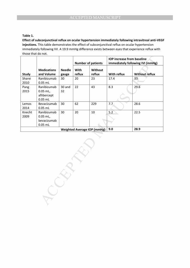
The greatest risk factor for acute ocular hypertension is the absence of subconjunctival
reflux. Table 1 presents four studies that investigated the effect of subconjunctival reflux on
immediate post-injection IOP and reveals that the IOP immediately following IVI averages 20
mmHg lower in patients with reflux.69, 72, 88, 101 The occurrence of reflux is often overlooked by
busy clinicians and is difficult to quantitate, but represents a potentially important risk factor for
ocular hypertension following IVI. Reflux depends on the injection technique and size of the
needle. Larger bore needles create a wider needle track through the sclera, resulting in a greater
chance of reflux. Several studies have demonstrated that smaller bore needles have less post-
injection reflux and a higher IOP.50, 69, 87, 88 Similarly, tunneled injection techniques allow the
needle track to be pinched off by the edge of the sclera, reducing the amount of reflux and
increasing post-injection IOP.69, 87
A second important risk factor for severe, acute ocular hypertension and delayed
recovery is a prior history of glaucoma.10, 66 Bakri et al. found that 10 minutes after IVI of
bevacizumab, triamcinolone, or pegaptanib, only 75% of glaucomatous patients recovered to an
IOP of less than 35 mmHg in contrast with 95.5% of patients without glaucoma.10 This
observation supports the notion that glaucomatous eyes suffer from pathologically
compromised aqueous humor outflow; however, studies demonstrating a correlation between
the diagnosis of glaucoma and the severity of ocular hypertension following IVI are primarily
limited by small sample sizes. Further investigations are necessary to confirm and quantify this
observation.
Another suspected risk factor for acute ocular hypertension is a smaller vitreous volume
manifested by a short axial length. The literature supporting this hypothesis is mixed.17, 38, 39, 47, 71,
87 Surprisingly, the volume of injected drug has not been confirmed to be a risk factor. Bakri et
al. assessed three different volumes (triamcinolone 0.1 mL, pegaptanib 0.09 mL and
bevacizumab 0.05 mL) and found no difference in the post-injection IOP change;10 however, this
observation is likely confounded by the variety of needle gauges used for the different
medications. Triamcinolone is generally injected with a 27-gauge needle, resulting in a greater
incidence of reflux and counteracting the hypertensive effects of the greater volume injected. It
seems intuitive that larger volumes would likely result in greater IOP change when controlling
for confounding variables.
C. Studies supporting medical prophylaxis for acute ocular hypertension following IVI
Several medications have been investigated to limit acute ocular hypertension following
anti-VEGF therapy, and five studies, discussed in detail below, have found topical agents to be
mildly effective at prophylactically decreasing IOP (Figure 3).26, 65, 86, 91, 107 A major limitation of

MANUSCRIP
T
ACCEPTED
ACCEPTED MANUSCRIPT7
[Type here]
these studies is the lack of recording and control for subconjunctival reflux. Additionally, each
study uses different IOP cutoffs and observation times following injection, making comparison
between studies difficult. Finally, the clinical benefit of a mild decrease in a short-duration IOP
spike remains unclear.
1. Apraclonidine
Apraclonidine decreases aqueous production and increases both trabecular and
uveoscleral outflow.108 These pressure-dependent and pressure-independent
mechanisms are effective at reducing the IOP spike following anterior segment laser
procedures, cataract surgery and vitrectomy, making apraclonodine a promising
investigational agent to limit acute ocular hypertension following IVI.95 El Chehab et al.
prospectively assessed the effect of 1 drop of 1% apraclonidine administered
prophylactically 2 hours prior to injection. The study included 250 patients who were
randomized to five study arms, receiving either prophylactic apraclonidine, oral
acetazolamide, brimonidine-timolol, dorzolamide-timolol, or no prophylaxis (control).
Immediately following injection, the average IOP measured 37.3 mmHg in the
apraclonidine group in comparison to 46.4 mmHg in the control group. The proportion
of patients with an IOP of >45 mmHg was 65.5% in the control group and 7.7% in the
apraclonidine group. At 15 minutes, the proportion of patients with an IOP of >25
mmHg was 0.0% in the apraclonidine group and 36.6% in the control group.26
Consequently, apraclonidine appeared more effective than oral acetazolamide at
decreasing acute ocular hypertension following IVI, and appeared similarly effective to
the brimonidine-timolol and dorzolamide-timolol topical formulations.
2. Timolol
Timolol decreases aqueous production and is well tolerated by patients.14 Pece
et al. prospectively randomized 150 patients to receive either no prophylactic
medication, timolol 0.1% gel the evening before injection, or timolol 0.1% gel 2 hours
prior to injection. Five minutes following injection, the use of timolol gel 2 hours prior to
injection resulted in an average pressure of 25.5 mmHg in comparison to 29.3 mmHg in
the control group. The proportion of patients with an IOP spike of > 40 mmHg was
reduced from 18% to 2% with the use of timolol 2 hours prior to injection. The use of
timolol 0.1% gel the evening before injection had no effect on IOP control.91 While direct
comparison with other protocols is challenging, these results are suggestive that timolol
gel is slightly less effective than the apraclonidine, dorzolamide-timolol and
brimonidine-timolol formulations evaluated by Chehab et al.26
3. Dorzolamide-Timolol
Timolol decreases aqueous production through sympathetic blockade of the
nerve endings in the ciliary epithelium 14 and dorzolamide reduces aqueous production
by blocking carbonic anhydrase.75 Three studies have assessed the prophylactic effect of
the dorzolamide-timolol combination prior to IVI. A summary of the studies and their
IOP-lowering effects are displayed in table 2.26, 65, 86 All three studies demonstrated a
mild decline in IOP immediately following injection. Kim et al. observed that the

MANUSCRIP
T
ACCEPTED
ACCEPTED MANUSCRIPT8
[Type here]
dorzolamide-timolol group had a modest decrease in IOP in comparison to the control
group at 5 minutes (14.1 mmHg versus 28.2 mmHg) and at 1 hour (10.7 mmHg versus
18.7 mmHg), but dorzolamide-timolol was not more effective than the brinzolamide-
timolol combination.65 These studies suggest that dorzolamide-timolol has similar
efficacy to apraclonidine and brimonidine-timolol at mildly lowering the severity of post-
injection acute ocular hypertension.
4. Brimonidine-timolol
Brimonidine suppresses aqueous production, increases uveoscleral outflow and
has possible neuroprotective properties.42, 97 Two studies have investigated the
prophylactic effect of brimonidine-timolol drops.
Theoulakis et al. prospectively investigated the prophylactic effect of 1 drop of
brimonidine-timolol given twice a day, the day prior to and the day of injection. Eighty-
eight patients were randomized to equally sized groups, one receiving brimonidine-
timolol and the other receiving no drops. At 5 minutes the treated group had an average
IOP of 28.4 mmHg compared to an average pressure of 34.1 mmHg in the control group.
At 15 minutes, none of the patients in the treatment group had a pressure of >20
mmHg, as compared to 34% in the control group.107
El Chehab et al., as previously discussed, assessed the effect of 1 drop of
brimonidine-timolol given 2 hours prior to injection. Fifty patients were randomized to
receive either brimonidine-timolol or four other investigational arms (control,
apraclonidine, oral acetazolamide, dorzolamide-timolol). Immediately following
injection, the IOP was 38 mmHg in the brimonidine-timolol group and 46.4 mmHg in the
control group. At 15 minutes, the percentage of patients with an IOP of >25mmHg was
0% in the brimonidine-timolol group and 36.6% in the control group.26 Brimonidine-
timolol showed a greater prophylactic effect than control, and was similarly effective to
apraclonidine and dorzolamide-timolol topical formulations.
5. Brinzolamide-timolol
Brinzolamide-timolol combines the aqueous suppression from carbonic
anhydrase inhibition with the aqueous suppression from ciliary epithelial sympathetic
pathway blockage.14, 46 Kim et al. prospectively investigated the effects of 1 drop of
brinzolamide-timolol given 1 hour prior to injection. Patients were divided into three
groups: 84 in the brinzolamide-timolol arm, 53 in the dorzolamide-timolol arm and 29 in
the control arm. After 5 minutes, the IOP in the brinzolamide-timolol group averaged
14.87 mmHg as compared to 28.21 mmHg in the control arm. After 1 hour, the average
IOP in the brinzolamide-timolol group was 13.61 mmHg, in comparison to 18.72 mmHg
in the control arm. Topical brinzolamide-timolol showed similar effectiveness to
dorzolamide-timolol.
D. Studies not supporting medical prophylaxis for acute ocular hypertension following IVI
Oral acetazolamide is a potent carbonic anhydrase inhibitor 98, and its
effectiveness in angle-closure glaucoma makes it a promising agent in the prophylaxis
against acute ocular hypertension following IVI. Surprisingly, two studies have assessed

MANUSCRIP
T
ACCEPTED
ACCEPTED MANUSCRIPT9
[Type here]
oral acetazolamide prior to injection and noted no IOP lowering effect, although it is
possible that the dosing and timing were not optimized for this indication.
Murray et al. compared 12 patients receiving 500 mg PO acetazolamide 60-90
minutes prior to injection with 12 control patients. There was no statistically significant
difference in pressures immediately after, 5 minutes after or 10 minutes after the
injection. At 30 minutes, the average IOP in the treatment arm was 15.7 mmHg and in
the control arm was 20.6 mmHg, which was statistically significant.83
El Chehab et al. compared 50 patients receiving 250 mg of oral acetazolamide
given 2 hours prior to injections with three other treatment arms (apraclonidine,
dorzolamide-timolol, brominidine-timolol) and 50 patients in a control arm. No
difference in IOP was found between the control arm and the oral acetazolamide arm.26
E. Other prophylactic measures for acute ocular hypertension following IVI
1. Anterior chamber paracentesis
More effective than topical and oral treatments, anterior chamber (AC)
paracentesis effectively prevents a post-injection IOP spike, but introduces additional
risks of infection and iatrogenic injury. Knip et al. demonstrated the effectiveness of an
AC paracentesis, showing that the average immediate post-injection IOP was 15.3
mmHg in the paracentesis group as compared to 47.1 mmHg in the control group. At 2
minutes and at 30 minutes there was no statistically significant difference between
groups.70
Despite the effectiveness, there are risks of performing an AC paracentesis. A
study assessing 560 paracenteses showed 4 complications (0.7%).109 Two patients
experienced inadvertent injection of sterile air into the anterior chamber which
spontaneously resolved without adverse consequences. One patient experienced a
small anterior lens capsule laceration that was self-sealing, but left a localized opacity
and another patient experienced an allergic reaction to povidone iodine.109 Other case
studies have demonstrated complications such as endophthalmitis and infectious
keratitis.5, 45, 57
Although AC paracentesis involves some minimal risk, in patients at risk of
glaucomatous progression who are known to experience transient vision loss related to
severe acute ocular hypertension following IVI, an anterior chamber paracentesis could
be appropriate. In these patients, the transient vision loss is likely from severely
elevated pressures that result in hypoperfusion to the retina.
2. Ocular decompression
One study assessed the effect of ocular decompression by cotton swabs on
post-injection acute ocular hypertension. Forty-eight patients were divided into two
groups. One group was anesthetized with a cotton swab soaked in 4% lidocaine and
received ocular decompression, and a control group received 3.5% lidocaine gel
anesthetic without ocular decompression. The immediate post-injection IOP was 25.7
mmHg in the decompression group and 30.9 mmHg in the control group. The
proportion of patients with IOP of >50 mmHg was 10% in the decompression group and
35% in the control group.43 These findings support the beneficial effect of moderate

MANUSCRIP
T
ACCEPTED
ACCEPTED MANUSCRIPT10
[Type here]
ocular pressure prior to injection. A similar principle has been applied using the Honan
Intraocular Pressure Reducer, which applies gentle pressure to an eye before a
procedure. One study supported the prophylactic anti-hypertensive effect of this device,
while another study showed no benefit.52, 67
F. Summary of studies investigating prophylaxis against acute ocular hypertension following
intravitreal injection
Numerous medications have been investigated to prophylactically reduce acute ocular
hypertension following anti-VEGF IVIs, and most topical drops have a similar mild
effectiveness at decreasing IOP. Surprisingly, oral acetazolamide has little effect at
lowering IOP based on material currently published. Overall, the benefits of
pretreatment with ocular anti-hypertensive agents prior to IVI is not conclusive, mainly
because of the questionable clinical benefit in slightly decreasing IOP over the short
duration before IOP normalizes. However, it can be argued that patients with advanced
glaucomatous optic neuropathy may benefit from a slight reduction in pressure and
prophylactic treatment can be considered, particularly because these patients are more
susceptible to further damage from increased pressures and may be at increased risk of
higher IOP spikes over a greater duration of time due to underlying outflow pathology.
An AC paracentesis is a more effective, albeit riskier, method at preventing acute ocular
hypertension in at-risk patients.
III. Long-term effects of intravitreal anti-VEGF injections
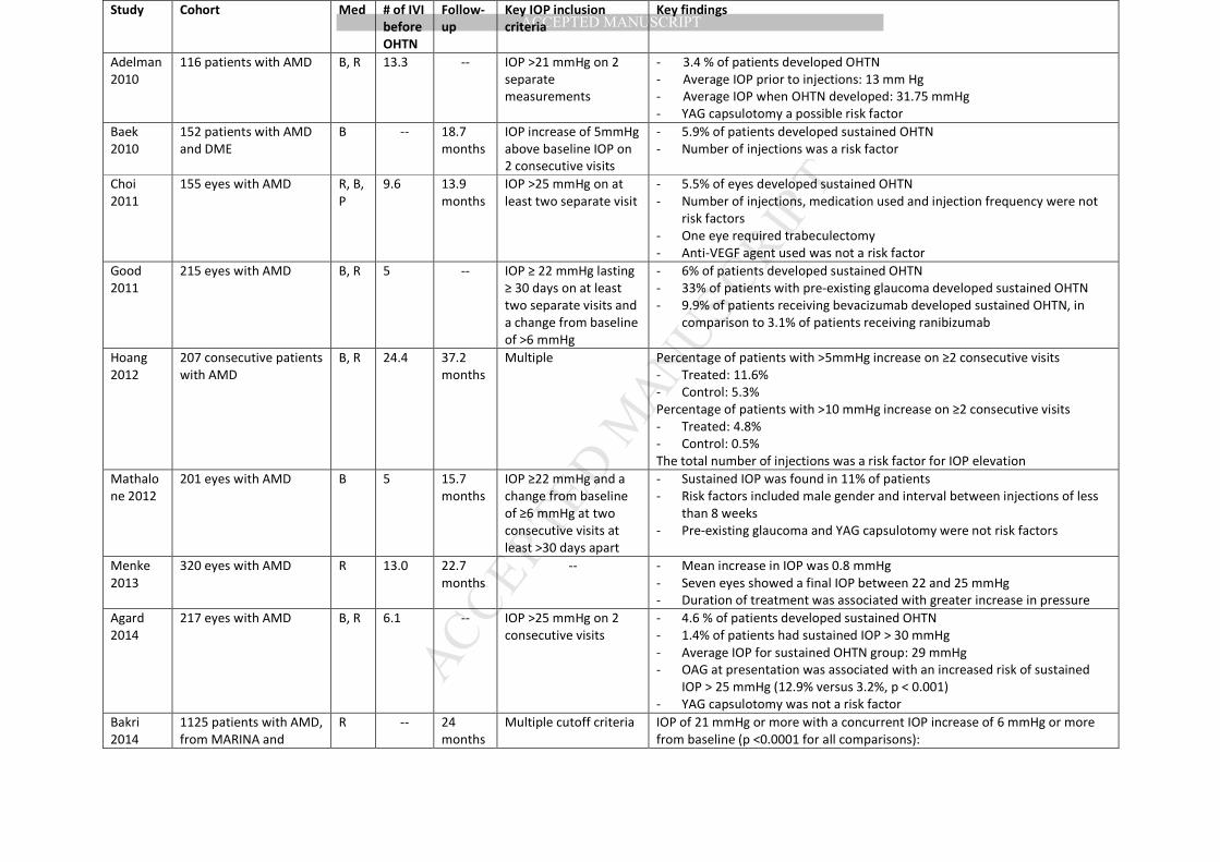
A. Studies associating sustained ocular hypertension with chronic anti-VEGF injections
Repeated intravitreal anti-VEGF injections have been associated with chronic ocular
hypertension, distinct from the short-term acute ocular hypertension following each injection, in
a subset of patients. Numerous case reports and case series have suggested this phenomenon
and those with incidence rates are summarized in Table 3.2-4, 6, 19, 34, 59, 76, 79, 80, 93, 99, 110 In some cases,
the ocular hypertension was severe enough to warrant surgical filtration.23, 104
Post hoc analyses of major anti-VEGF trials have provided the best evidence in support
of this phenomenon. While numerous retrospective case series have similarly supported this
finding, as demonstrated in Table 3, their nonrandomized nature and typically smaller cohort
provide inferior quality of evidence. On analysis of the MARINA and ANCHOR studies, Bakri et
al. noted the proportion of patients who had at least one visit with an increase in pre-injection
IOP of 6 mmHg or more from baseline with a concurrent IOP ≥21 mmHg was 26.1% in the 0.5
mg ranibizumab group, 23.6% in the 0.3 mg Ranibizumab group and 13.6% in the sham/PDT
groups.9 The proportion of patients with at least one IOP measurement of 21 mmHg or more
was 39.9% in the 0.5 mg ranibizumab group, 37.0% in the 0.3 mg ranibizumab group and 29.1%
in the sham/PDT groups.9 These findings are suggestive that a subgroup of patients develop
sustained ocular hypertension due to chronic anti-VEGF therapy and are also suggestive of a
possible dose-related response. A post-hoc analysis of DRCR data found that 9.5% of patients in
the ranibizumab plus prompt or deferred focal/grid laser group experienced sustained IOP
elevation versus 3.5% of patients in the sham injection plus focal/grid laser treatment group.13
The difference in the percentage of affected patients between the DRCR and MARINA/ANCHOR
trials is due to different definitions of sustained ocular hypertension. Similar findings were
presented in a post-hoc analysis of the IVAN study.32 Additionally, a recent population-based,
MANUSCRIP
T
ACCEPTED
ACCEPTED MANUSCRIPT11
[Type here]
nested, case-control study in Canada found that patients receiving 7 or more annual injections
had a greater odds ratio of undergoing glaucoma drainage surgery than controls.23 The major
limitations of these and other studies listed in Table 3 are their retrospective nature and their
uncontrolled methods of IOP measurement, as IOPs were variably measured with the use of a
tonopen or Goldmann applanation tonometer. Glaucoma trials typically utilize standardized
protocol-driven IOP measuring regimens, such as requiring two masked individuals to measure
IOP with only Goldmann applanation tonometry, to minimize the risk of bias or error.
Risk factors for chronic ocular hypertension in these studies are intuitive. The total
number of injections was demonstrated to be a risk factor,11, 40, 48, 49, 93, 99 as was a greater
frequency of injections79 and a pre-existing diagnosis of glaucoma prior to initiation of IVIs.22, 40
Fruend et al. observed that ranibizumab, as compared to aflibercept, had higher rates of ocular
hypertension, with 8.4% of patients on monthly ranibizumab having an IOP of greater than
21mmHg on two consecutive visits compared to 3.2% and 2.7% of patients who received 2 mg
and 0.5 mg of aflibercept every month, respectively.34 While the Freund et al. analysis has been
the only study to directly compare aflibercept with ranibizumab, ranibizumab was not
associated with greater rates of sustained OHTN in comparison to bevacizumab in other
studies.2, 3, 40, 48, 68 In fact, Good et al. observed that patients receiving ranibizumab had lower
rates of sustained OHTN compared to bevacizumab (3.1% versus 9.9%).40 It remains to be seen if
one medication is consistently associated with higher rates of sustained OHTN. A history of
cataract extraction or a posterior capsulotomy, with a theoretical increased rate of diffusion of
medication to the anterior chamber, were not observed to be risk factors.
Recently, Wen et al. investigated the conventional outflow facility of patients receiving
chronic anti-VEGF therapy, utilizing electronic Schiøtz tonography. They found a small but
statistically significant decrease in outflow facility in patients with a greater number of IVIs
(≥20). Additionally, they found eyes with contralateral ocular hypertension had a two-fold
reduction in outflow facility following anti-VEGF therapy as compared to the contralateral
eye.112 This functional study supports the observation that the total number of injections is a risk
factor for sustained ocular hypertension. It also supports the notion that chronic anti-VEGF
therapy can reduce outflow capacity. Finally, it is suggestive of a two-hit hypothesis, where
patients with underlying ocular hypertension and pathological outflow capacity, are at increased
risk of further outflow reduction due to chronic anti-VEGF therapy. A two-hit hypothesis may
also explain why only a fraction of patients developed sustained ocular hypertension.
A multitude of theoretical mechanisms have been described in an effort to explain the
reduced outflow capacity and the sustained rise in IOP observed in a subset of patients receiving
recurrent intravitreal anti-VEGF injections. One of the proposed mechanisms involves
microparticle obstruction of the trabecular meshwork.20 Some studies have demonstrated that
silicone microdroplets from syringes and protein aggregates from medication packaging or
delivery equipment can obstruct aqueous outflow.7, 60, 74 This is of particular relevance with the
advent of prefilled ranibizumab syringes, and patients receiving this therapy should be observed
for the development of sustained ocular hypertension due to possible microparticle obstruction
from materials that become dissolved in the anti-VEGF solution. Kahook et al. reported
variations in the concentration of high-molecular-weight protein aggregates in different samples
of compounded and repackaged bevacizumab.60 In addition, Good et al. reported a sustained
IOP elevation in 9.9% of patients receiving bevacizumab compared with 3.1% in patients
MANUSCRIP
T
ACCEPTED
ACCEPTED MANUSCRIPT12
[Type here]
receiving ranibizumab. Given that bevacizumab (149 kDa) is approximately 3 times larger than
ranibizumab (48 kDa), it is thought that the high-molecular-weight proteins may accumulate in
the trabecular meshwork and obstruct aqueous outflow.2, 8, 40
Another proposed mechanism involves the direct effects on trabecular meshwork cells
by intravitreal anti-VEGF agents. In vitro studies by Kahook et al. demonstrated that 4 mg/mL of
bevacizumab slows the metabolism and replication of trabecular meshwork cells.58 This was not
demonstrated with lower concentrations of bevacizumab (2 mg/mL) or with ranibizumab;58
However, Kernt et al. reported no toxic effects to the trabecular meshwork with bevacizumab at
the 1.25 mg/0.05 mL concentration used in intravitreal injections.61 Others have proposed that
monomer antibodies, aggregated proteins, or other high molecular weight molecules may incite
an underlying inflammatory reaction leading to trabeculitis with impaired aqueous humor
outflow.37, 81, 105
In addition to the mechanical obstruction and the potential toxic and inflammatory
effects on the trabecular meshwork, some have proposed alterations in outflow facility by anti-
VEGF agents. VEGF receptors have been found to be expressed on the trabecular meshwork
and Schlemm’s canal endothelial cells. Schlemm’s canal cells express vascular endothelial (VE)
cadherin, an endothelial cell adhesion molecule.92 VEGF stimulation promotes the
endocytosis of VE cadherin, and VEGF blockade can disrupt this barrier function and reduce
endothelial cell permeability.36 Other in vitro models have similarly demonstrated that VEGF
induces endothelial fenestrations.29 In addition, nitric oxide has been shown to increase anterior
chamber aqueous outflow through a reduction in trabeculocyte size and smooth muscle
contractility with Schlemm’s canal vasodilation. Anti-VEGF therapy disrupts the
normal nitric oxide signaling pathway by inhibition of nitric oxide synthase.94 Animal studies
have demonstrated that VEGF increases aqueous humor outflow facility and that blockage of
VEGF receptors leads to ocular hypertension.35 Anti-VEGF inhibition of the disassembly of the
endothelial intercellular junctions, decrease in the endothelial fenestrations, and inhibition of
nitric oxide production are a few of the biochemical mechanisms that may explain the impaired
outflow and sustained rise in IOP.
Although there is no clear consensus on which of these proposed mechanisms
contributes most to the rise in IOP, various studies support that anti-VEGF injections may have
an effect on the trabecular meshwork pressure-dependent outflow system.
B. Studies not associating sustained ocular hypertension with chronic anti-VEGF
injections
The main limitation of post-hoc analyses supporting the ocular hypertensive effects of
anti-VEGF IVIs is that IOP was not the primary outcome measured. Consequently, IOP
measurement was not standardized and both applanation and tonopen methods were used to
varying degrees.54 Publication bias may also limit the submission and acceptance of studies
observing no increase in rates of sustained ocular hypertension. Another source of bias is the
inclusion of the cases that initiated a study, which may artificially elevate observed incidence
rates of ocular hypertension. Finally, other studies have demonstrated conflicting results. Wehrli
et al. did not observe an increased risk of sustained ocular hypertension in patients receiving
frequent intraocular injections as compared to fellow untreated eyes, and similar results were
demonstrated in other studies summarized in Table 4.12, 64, 68, 84, 111, 114 The strengths of the
MANUSCRIP
T
ACCEPTED
ACCEPTED MANUSCRIPT13
[Type here]
Wehrli et al. and Kim et al. studies are their fellow-eye control and large patient number.68, 111
Interestingly, Kim et al. found higher rates of sustained ocular hypertension in patients with
underlying glaucoma and a history of retinal vein occlusion.68 Even though not correspondingly
observed by Wehrli et al., these findings may support the notion that patients with underlying
outflow pathology are susceptible to the effects of repeated anti-VEGF therapy. While the
studies by Kim et al. and Wehrli et al. are well-constructed retrospective analyses, the evidence
provided by the retrospective analysis of the MARINA, ANCHOR and DRCR studies, all of which
support the hypothesis of a subset of patients developing sustained OHTN, provide better
quality evidence due to their larger size and lower likelihood for selection bias.
C. Effect of repeated injections on the retinal nerve fiber layer
Average retinal nerve fiber layer (aRNFL) thinning on optic nerve OCT is expected if anti-
VEGF agents and associated ocular hypertension are harmful to ganglion cells and their axons.
Most studies have found no correlation between repeated injections and aRNFL thickness.21, 24,
28, 53, 100, 102, 106 One study, in contrast, found an aRNFL thinning of -5.5 μm following a mean of 4.8
injections.78 Two studies found an aRNFL thinning in injected eyes but found a similar thinning in
the control eyes.90, 103 Overall, these studies suggest a minimal average effect of repeated anti-
VEGF injections on aRNFL thickness in patients receiving multiple injections. However, there are
major limitations in the interpretation of these results. All of the studies mentioned above
excluded patients with a history of glaucoma and therefore cannot allow for any conclusions
about repeat injections on aRNFL thickness in patients with glaucoma – though the disease
process itself would be expected to show progressive aRNFL thinning. In addition, 6 of 8 studies
that investigated the effect of injection frequency had fewer than 7 mean injections, and 5 of 8
studies that investigated duration of follow-up observed patients for less than 15 months. These
studies may be missing patients who develop aRNFL thinning after a greater number of
injections given over a longer period of time. Additionally, as observed by the post hoc analysis
of the MARINA, ANCHOR and DRCR studies, only a subset of patients develop sustained ocular
hypertension, and analysis of patients as a whole who receive anti-VEGF IVIs may miss the
proportion of affected patients.
One study has evaluated the effect of an underlying diagnosis of glaucoma on the aRNFL
thinning. Park et al. found that a greater number of injections was associated with aRNFL
thinning in glaucomatous patients compared to those without glaucoma.89 These results are
consistent with studies finding a greater IOP rise in patients with an underlying diagnosis of
glaucoma and repeated injections;22, 40 however, an obvious limitation of the Park study is the
lack of control for natural glaucoma progression, as these patients may have had RNFL thinning
regardless of IVI. Overall, glaucomatous patients may be more susceptible to RNFL thinning with
repeated anti-VEGF injections given the underlying damage to the ganglion cells, their axons,
the trabecular meshwork and downstream outflow pathways; however, available evidence does
not definitively support this hypothesis.
IV. Macular disease and the risks of forgoing anti-VEGF therapy
Foregoing anti-VEGF therapy risks progression of various macular diseases. The MARINA study
demonstrated a dramatic improvement in mean visual acuity of 7.2 ETDRS letters after one year of
ranibizumab therapy compared to a loss of 10.4 ETDRS letters without treatment (for a net 17.6 ETDRS
letter benefit); at one year, monthly treatment decreased the risk of losing 15 ETDRS letters from 37.8%
to 5.5%.96 Unfortunately, a decreased intensity of anti-VEGF treatment in an as-needed regimen may

MANUSCRIP
T
ACCEPTED
ACCEPTED MANUSCRIPT14
[Type here]
yield an inferior result, as the CATT study demonstrated a mean difference of 2.4 ETDRS letters in favor
of the monthly versus the as-needed regimen after two years.77
The benefit of anti-VEGF therapy has been similarly demonstrated in macular edema in diabetic
retinopathy and retinal vein occlusions.15, 18, 27 For example, in the DRCR protocol I study of diabetic
macular edema (DME), subjects in the ranibizumab/deferred laser group experienced a mean gain of 9
ETDRS letters after 2 years, in contrast to those in the prompt laser group who experience a mean
improvement of only 3 ETDRS letters.27
In the BRAVO study of macular edema due to branch retinal vein occlusion, subjects in the
ranibizumab group experienced a mean gain of 18.3 ETDRS letters at 6 months in contrast to those in
the sham group who experienced a mean improvement of only 7.3 ETDRS letters at 6 months.18 In the
CRUISE study of macular edema associated with central retinal venous occlusions, subjects in the
ranibizumab group experienced a mean gain of 14.9 ETDRS letters at 6 months in contrast to those in
the sham group who experienced a mean improvement of only 0.8 ETDRS letters.15
For macular edema with diabetic retinopathy or branch vein occlusion, an alternative to anti-
VEGF medications could be macular laser photocoagulation, which generally reduces vision loss by 50%.1
Unfortunately, for exudative AMD, no effective alternative therapy exists and forgoing anti-VEGF
therapy places patients at a significantly increased risk of vision loss.
V. Conclusion
Although anti-VEGF therapy is well tolerated in the vast majority of patients, acute and chronic
ocular hypertension following treatment merits consideration. We have discussed the degree and
timing of ocular hypertension immediately following anti-VEGF IVIs. IOP typically rises acutely following
IVI with normalization within 30-60 minutes. In glaucomatous patients, this ocular hypertension is more
dramatic and of longer duration. Numerous medications have been investigated to reduce
prophylactically acute ocular hypertension, and all topical drops have a similar mild effect in decreasing
IOP following IVI. Surprisingly, oral acetazolamide has little effect on lowering IOP following IVI based on
material currently published. Overall, the benefits of pretreatment with ocular anti-hypertensive agents
prior to IVI is not conclusive, mainly because of the questionable clinical benefit in slightly decreasing
IOP over the short duration before IOP normalizes; however, it can be argued that patients with
advanced glaucomatous optic neuropathy may benefit from a slight reduction in pressure, and
prophylactic treatment can be considered because these patients are more susceptible to further
damage from increased pressures and may be at increased risk of higher IOP spikes over a greater
duration of time. An AC paracentesis is a more effective, albeit riskier, intervention to prevent acute
ocular hypertension in at-risk patients.
Chronically, a subset of patients likely develop persistent ocular hypertension. Several studies
did not find a correlation between chronic anti-VEGF therapy and sustained OHTN. However, post hoc
analyses of the MARINA, ANCHOR and DRCR studies provides the best quality evidence that a subset of
patients develop a clinically significant elevation in intraocular pressure following chronic anti-VEGF
intravitreal therapy. Evaluation of these studies are suggestive that patients who have underlying
outflow pathology, as manifested by an underlying diagnosis of glaucoma, ocular hypertension or a
history of retinal vein occlusion, are at particular risk of developing sustained ocular hypertension and
these patients should be closely monitored for the development of sustained pressure elevation. A
history of cataract extraction or posterior capsulotomy were not consistently identified as risk factors,
MANUSCRIP
T
ACCEPTED
ACCEPTED MANUSCRIPT15
[Type here]
and the particular anti-VEGF utilized was not conclusively observed as a risk factor. Further studies are
necessary to clarify which particular anti-VEGF therapy, if any, has increased rates of sustained ocular
hypertension. Although definitive evidence of damage to the retinal nerve fiber layer is lacking, patients
receiving repeated injections should be monitored for the development of ocular hypertension and
those who develop sustained ocular hypertension should be periodically monitored for glaucomatous
changes with an optic nerve OCT and static visual field. Referrals to a glaucoma specialist should be
considered in patients with concerning features.
This review allows for a better risk-benefit analysis for clinicians providing frequent intravitreal
anti-VEGF injections. Future studies, potentially assessing subconjunctival reflux, are needed to further
clarify the role of prophylactic medications prior to IVI for acute ocular hypertension. In addition, longer-
duration prospective studies or larger, population-based retrospective studies focusing on progression
of glaucomatous optic neuropathy following IVI could help clarify the long-term risk of anti-VEGF
therapy and aid in identifying which subset of patients are at risk of developing sustained ocular
hypertension.
Literature Search
Prospective randomized trials, prospective cohort studies, and retrospective studies that
reported on IOP following intravitreal injections of anti-VEGF agents were searched using Medline
through November, 2017. Key words included in the search included intraocular pressure, IOP, optical
coherence tomography, OCT, intravitreal, intraocular, anti-VEGF, VEGF, vascular endothelial growth
factor, Lucentis, ranibizumab, Avastin, bevacizumab, Eylea, aflibercept, Macugen, pegaptanib, injection,
and injections. Inclusion criteria included prospective randomized trials, retrospective case series,
retrospective case reports and injection of anti-VEGF medications. Exclusion criteria included literature
reviews, summaries, editorials, letters, and steroid injections. Those publications deemed eligible
following review of the abstract were obtained in full. In addition, references were reviewed for possible
publications missed by the initial review.
Funding: This study was supported in part by an unrestricted grant from Research to Prevent Blindness,
Inc.
Acknowledgment/Disclosures: All authors made a substantial contribution to the acquisition and
interpretation of the data. Each author participated in drafting or revising the manuscript and approved
submission of this version for publication. The authors report no commercial or proprietary interest in
any product or concept discussed in this article.
Supplemental Material
References
1. Argon laser photocoagulation for macular edema in branch vein occlusion. The Branch Vein
Occlusion Study Group. American journal of ophthalmology. 1984;98(3):271-82.
2. Adelman RA, Zheng Q, Mayer HR. Persistent ocular hypertension following intravitreal
bevacizumab and ranibizumab injections. Journal of ocular pharmacology and therapeutics : the official
journal of the Association for Ocular Pharmacology and Therapeutics. 2010;26(1):105-10.
3. Agard E, Elchehab H, Ract-Madoux G, et al. Repeated intravitreal anti-vascular endothelial
growth factor injections can induce iatrogenic ocular hypertension, especially in patients with open-

MANUSCRIP
T
ACCEPTED
ACCEPTED MANUSCRIPT16
[Type here]
angle glaucoma. Canadian journal of ophthalmology Journal canadien d'ophtalmologie. 2015;50(2):127-
31.
4. Al-Abdullah AA, Nowilaty SR, Asghar N, et al. Intraocular pressure trends after intravitreal
injections of anti-vascular endothelial growth factor agents for diabetic macular edema. Retina
(Philadelphia, Pa). 2015;35(3):440-8.
5. Azuara-Blanco A, Katz LJ. Infectious keratitis in a paracentesis tract. Ophthalmic surgery and
lasers. 1997;28(4):332-3.
6. Baek SU, Park IW, Suh W. Long-term intraocular pressure changes after intravitreal injection of
bevacizumab. Cutaneous and ocular toxicology. 2016;35(4):310-4.
7. Bakri SJ, Ekdawi NS. Intravitreal silicone oil droplets after intravitreal drug injections. Retina
(Philadelphia, Pa). 2008;28(7):996-1001.
8. Bakri SJ, McCannel CA, Edwards AO, Moshfeghi DM. Persisent ocular hypertension following
intravitreal ranibizumab. Graefe's archive for clinical and experimental ophthalmology = Albrecht von
Graefes Archiv fur klinische und experimentelle Ophthalmologie. 2008;246(7):955-8.
9. Bakri SJ, Moshfeghi DM, Francom S, et al. Intraocular pressure in eyes receiving monthly
ranibizumab in 2 pivotal age-related macular degeneration clinical trials. Ophthalmology.
2014;121(5):1102-8.
10. Bakri SJ, Pulido JS, McCannel CA, et al. Immediate intraocular pressure changes following
intravitreal injections of triamcinolone, pegaptanib, and bevacizumab. Eye (London, England).
2009;23(1):181-5.
11. Beato J, Pedrosa AC, Pinheiro-Costa J, et al. Long-Term Effect of Anti-VEGF Agents on Intraocular
Pressure in Age-Related Macular Degeneration. Ophthalmic research. 2016;56(1):30-4.
12. Boyer DS, Goldbaum M, Leys AM, Starita C. Effect of pegaptanib sodium 0.3 mg intravitreal
injections (Macugen) in intraocular pressure: posthoc analysis from V.I.S.I.O.N. study. The British journal
of ophthalmology. 2014;98(11):1543-6.
13. Bressler SB, Almukhtar T, Bhorade A, et al. Repeated intravitreous ranibizumab injections for
diabetic macular edema and the risk of sustained elevation of intraocular pressure or the need for
ocular hypotensive treatment. JAMA ophthalmology. 2015;133(5):589-97.
14. Brooks AM, Gillies WE. Ocular beta-blockers in glaucoma management. Clinical pharmacological
aspects. Drugs & aging. 1992;2(3):208-21.
15. Brown DM, Campochiaro PA, Singh RP, et al. Ranibizumab for macular edema following central
retinal vein occlusion: six-month primary end point results of a phase III study. Ophthalmology.
2010;117(6):1124-33.e1.
16. Brown DM, Kaiser PK, Michels M, et al. Ranibizumab versus verteporfin for neovascular age-
related macular degeneration. N Engl J Med. 2006;355(14):1432-44.
17. Cacciamani A, Oddone F, Parravano M, et al. Intravitreal injection of bevacizumab: changes in
intraocular pressure related to ocular axial length. Japanese journal of ophthalmology. 2013;57(1):63-7.
18. Campochiaro PA, Heier JS, Feiner L, et al. Ranibizumab for macular edema following branch
retinal vein occlusion: six-month primary end point results of a phase III study. Ophthalmology.
2010;117(6):1102-12.e1.
19. Choi DY, Ortube MC, McCannel CA, et al. Sustained elevated intraocular pressures after
intravitreal injection of bevacizumab, ranibizumab, and pegaptanib. Retina (Philadelphia, Pa).
2011;31(6):1028-35.
20. Dedania VS, Bakri SJ. Sustained Elevation of Intraocular Pressure After Intravitreal Anti-VEGF
Agents: What is the evidence? . Retina (Philadelphia, Pa). 2015;35(5):841-58.
21. Demirel S, Batioglu F, Ozmert E, Erenler F. The effect of multiple injections of ranibizumab on
retinal nerve fiber layer thickness in patients with age-related macular degeneration. Current eye
research. 2015;40(1):87-92.

MANUSCRIP
T
ACCEPTED
ACCEPTED MANUSCRIPT17
[Type here]
22. Demirel S, Yanik O, Batioglu F, Ozmert E. Intraocular pressure changes related to intravitreal
injections of ranibizumab: analysis of pseudophakia and glaucoma subgroup. International
ophthalmology. 2015;35(4):541-7.
23. Eadie BD, Etminan M, Carleton BC, et al. Association of Repeated Intravitreous Bevacizumab
Injections With Risk for Glaucoma Surgery. JAMA ophthalmology. 2017.
24. El-Ashry MF, Lascaratos G, Dhillon B. Evaluation of the effect of intravitreal ranibizumab
injections in patients with neovascular age related macular degeneration on retinal nerve fiber layer
thickness using optical coherence tomography. Clinical ophthalmology (Auckland, NZ). 2015;9:1269-74.
25. El Chehab H, Agard E, Russo A, et al. Intraocular Pressure Spikes after Aflibercept Intravitreal
Injections. Ophthalmologica Journal international d'ophtalmologie International journal of
ophthalmology Zeitschrift fur Augenheilkunde. 2016;236(1):43-7.
26. El Chehab H, Le Corre A, Agard E, et al. Effect of topical pressure-lowering medication on
prevention of intraocular pressure spikes after intravitreal injection. European journal of ophthalmology.
2013;23(3):277-83.
27. Elman MJ, Bressler NM, Qin H, et al. Expanded 2-year follow-up of ranibizumab plus prompt or
deferred laser or triamcinolone plus prompt laser for diabetic macular edema. Ophthalmology.
2011;118(4):609-14.
28. Entezari M, Ramezani A, Yaseri M. Changes in Retinal Nerve Fiber Layer Thickness after Two
Intravitreal Bevacizumab Injections for Wet Type Age-related Macular Degeneration. Journal of
ophthalmic & vision research. 2014;9(4):449-52.
29. Esser S, Wolburg K, Wolburg H, et al. Vascular endothelial growth factor induces endothelial
fenestrations in vitro. The Journal of cell biology. 1998;140(4):947-59.
30. Falkenstein IA, Cheng L, Freeman WR. Changes of intraocular pressure after intravitreal injection
of bevacizumab (avastin). Retina (Philadelphia, Pa). 2007;27(8):1044-7.
31. Farhood QK, Twfeeq SM. Short-term intraocular pressure changes after intravitreal injection of
bevacizumab in diabetic retinopathy patients. Clinical ophthalmology (Auckland, NZ). 2014;8:599-604.
32. Foss AJ, Scott LJ, Rogers CA, et al. Changes in intraocular pressure in study and fellow eyes in the
IVAN trial. The British journal of ophthalmology. 2016.
33. Frenkel RE, Mani L, Toler AR, Frenkel MP. Intraocular pressure effects of pegaptanib (Macugen)
injections in patients with and without glaucoma. American journal of ophthalmology.
2007;143(6):1034-5.
34. Freund KB, Hoang QV, Saroj N, Thompson D. Intraocular Pressure in Patients with Neovascular
Age-Related Macular Degeneration Receiving Intravitreal Aflibercept or Ranibizumab. Ophthalmology.
2015;122(9):1802-10.
35. Fujimoto T, Inoue T, Maki K, et al. Vascular Endothelial Growth Factor-A Increases the Aqueous
Humor Outflow Facility. PloS one. 2016;11(9):e0161332.
36. Gavard J, Gutkind JS. VEGF controls endothelial-cell permeability by promoting the beta-
arrestin-dependent endocytosis of VE-cadherin. Nature cell biology. 2006;8(11):1223-34.
37. Georgopoulos M, Polak K, Prager F, et al. Characteristics of severe intraocular inflammation
following intravitreal injection of bevacizumab (Avastin). The British journal of ophthalmology.
2009;93(4):457-62.
38. Gismondi M, Salati C, Salvetat ML, et al. Short-term effect of intravitreal injection of
Ranibizumab (Lucentis) on intraocular pressure. Journal of glaucoma. 2009;18(9):658-61.
39. Goktas A, Goktas S, Atas M, et al. Short-term impact of intravitreal ranibizumab injection on
axial ocular dimension and intraocular pressure. Cutaneous and ocular toxicology. 2013;32(1):23-6.
40. Good TJ, Kimura AE, Mandava N, Kahook MY. Sustained elevation of intraocular pressure after
intravitreal injections of anti-VEGF agents. The British journal of ophthalmology. 2011;95(8):1111-4.
MANUSCRIP
T
ACCEPTED
ACCEPTED MANUSCRIPT18
[Type here]
41. Gragoudas ES, Adamis AP, Cunningham ET, Jr., et al. Pegaptanib for neovascular age-related
macular degeneration. The New England journal of medicine. 2004;351(27):2805-16.
42. Greenfield DS, Liebmann JM, Ritch R. Brimonidine: a new alpha2-adrenoreceptor agonist for
glaucoma treatment. Journal of glaucoma. 1997;6(4):250-8.
43. Gregori NZ, Weiss MJ, Goldhardt R, et al. Randomized clinical trial of two anesthetic techniques
for intravitreal injections: 4% liquid lidocaine on cotton swabs versus 3.5% lidocaine gel. Expert opinion
on drug delivery. 2012;9(7):735-41.
44. Heier JS, Brown DM, Chong V, et al. Intravitreal aflibercept (VEGF trap-eye) in wet age-related
macular degeneration. Ophthalmology. 2012;119(12):2537-48.
45. Helbig H, Noske W, Kleineidam M, et al. Bacterial endophthalmitis after anterior chamber
paracentesis. The British journal of ophthalmology. 1995;79(9):866.
46. Herkel U, Pfeiffer N. Update on topical carbonic anhydrase inhibitors. Current opinion in
ophthalmology. 2001;12(2):88-93.
47. Hoang QV, Jung JJ, Mrejen S, Freund KB. Influence of axial length and postinjection reflux on
sustained intraocular pressure elevation as a result of intravitreal anti-vascular endothelial growth factor
therapy. Retina (Philadelphia, Pa). 2014;34(3):519-24.
48. Hoang QV, Mendonca LS, Della Torre KE, et al. Effect on intraocular pressure in patients
receiving unilateral intravitreal anti-vascular endothelial growth factor injections. Ophthalmology.
2012;119(2):321-6.
49. Hoang QV, Tsuang AJ, Gelman R, et al. Clinical predictors of sustained intraocular pressure
elevation due to intravitreal anti-vascular endothelial growth factor therapy. Retina (Philadelphia, Pa).
2013;33(1):179-87.
50. Hohn F, Mirshahi A. Impact of injection techniques on intraocular pressure (IOP) increase after
intravitreal ranibizumab application. Graefe's archive for clinical and experimental ophthalmology =
Albrecht von Graefes Archiv fur klinische und experimentelle Ophthalmologie. 2010;248(10):1371-5.
51. Hollands H, Wong J, Bruen R, et al. Short-term intraocular pressure changes after intravitreal
injection of bevacizumab. Canadian journal of ophthalmology Journal canadien d'ophtalmologie.
2007;42(6):807-11.
52. Hong SW, Jee D. Effect of the Honan intraocular pressure reducer to prevent vitreous reflux
after intravitreal bevacizumab injection. European journal of ophthalmology. 2012;22(4):615-9.
53. Horsley MB, Mandava N, Maycotte MA, Kahook MY. Retinal nerve fiber layer thickness in
patients receiving chronic anti-vascular endothelial growth factor therapy. American journal of
ophthalmology. 2010;150(4):558-61.e1.
54. Jampel H, Kalenak J. Anti-Vascular Endothelial Growth Factor Injections and Intraocular Pressure
Measurement: Should We Throw the Baby out with the Bath Water? Ophthalmology. 2015;122(9):1735-
6.
55. Jonas JB, Schlichtenbrede F. Visual acuity and intraocular pressure after high-dose intravitreal
triamcinolone acetonide in selected ocular diseases. Eye (London, England). 2008;22(7):869-73.
56. Jones R, 3rd, Rhee DJ. Corticosteroid-induced ocular hypertension and glaucoma: a brief review
and update of the literature. Current opinion in ophthalmology. 2006;17(2):163-7.
57. Joondeph BC, Joondeph HC. Purulent anterior segment endophthalmitis following paracentesis.
Ophthalmic surgery. 1986;17(2):91-3.
58. Kahook MY, Ammar DA. In vitro effects of antivascular endothelial growth factors on cultured
human trabecular meshwork cells. Journal of glaucoma. 2010;19(7):437-41.
59. Kahook MY, Kimura AE, Wong LJ, et al. Sustained elevation in intraocular pressure associated
with intravitreal bevacizumab injections. Ophthalmic surgery, lasers & imaging : the official journal of
the International Society for Imaging in the Eye. 2009;40(3):293-5.
MANUSCRIP
T
ACCEPTED
ACCEPTED MANUSCRIPT19
[Type here]
60. Kahook MY, Liu L, Ruzycki P, et al. High-molecular-weight aggregates in repackaged
bevacizumab. Retina (Philadelphia, Pa). 2010;30(6):887-92.
61. Kernt M, Welge-Lussen U, Yu A, et al. [Bevacizumab is not toxic to human anterior- and
posterior-segment cultured cells]. Der Ophthalmologe : Zeitschrift der Deutschen Ophthalmologischen
Gesellschaft. 2007;104(11):965-71.
62. Kiddee W, Montriwet M. Intraocular Pressure Changes in Non-Glaucomatous Patients Receiving
Intravitreal Anti-Vascular Endothelial Growth Factor Agents. PloS one. 2015;10(9):e0137833.
63. Kiddee W, Trope GE, Sheng L, et al. Intraocular pressure monitoring post intravitreal steroids: a
systematic review. Survey of ophthalmology. 2013;58(4):291-310.
64. Kim D, Nam WH, Kim HK, Yi K. Does intravitreal injections of bevacizumab for age-related
macular degeneration affect long-term intraocular pressure? Journal of glaucoma. 2014;23(7):446-8.
65. Kim GN, Han YS, Chung IY, et al. Effect of Dorzolamide/Timolol or Brinzolamide/Timolol
prophylaxis on intravitreal anti-VEGF injection-induced intraocular hypertension. Seminars in
ophthalmology. 2013;28(2):61-7.
66. Kim JE, Mantravadi AV, Hur EY, Covert DJ. Short-term intraocular pressure changes immediately
after intravitreal injections of anti-vascular endothelial growth factor agents. American journal of
ophthalmology. 2008;146(6):930-4.e1.
67. Kim KS, Jee D. Effect of the Honan intraocular pressure reducer on intraocular pressure increase
following intravitreal injection using the tunneled scleral technique. Japanese journal of ophthalmology.
2011;55(6):632-7.
68. Kim YJ, Sung KR, Lee KS, et al. Long-term effects of multiple intravitreal antivascular endothelial
growth factor injections on intraocular pressure. American journal of ophthalmology. 2014;157(6):1266-
71.e1.
69. Knecht PB, Michels S, Sturm V, et al. Tunnelled versus straight intravitreal injection: intraocular
pressure changes, vitreous reflux, and patient discomfort. Retina (Philadelphia, Pa). 2009;29(8):1175-81.
70. Knip MM, Valimaki J. Effects of pegaptanib injections on intraocular pressure with and without
anterior chamber paracentesis: a prospective study. Acta ophthalmologica. 2012;90(3):254-8.
71. Kotliar K, Maier M, Bauer S, et al. Effect of intravitreal injections and volume changes on
intraocular pressure: clinical results and biomechanical model. Acta ophthalmologica Scandinavica.
2007;85(7):777-81.
72. Lemos V, Cabugueira A, Noronha M, et al. Intraocular Pressure in Eyes Receiving Intravitreal
Antivascular Endothelial Growth Factor Injections. Ophthalmologica Journal international
d'ophtalmologie International journal of ophthalmology Zeitschrift fur Augenheilkunde. 2015;233(3-
4):162-8.
73. Lim HB, Kim MS, Jo YJ, Kim JY. Short-Term Visual Acuity and Intraocular Pressure Changes and
Their Correlation after Anti-Vascular Endothelial Growth Factor Injection. Ophthalmologica Journal
international d'ophtalmologie International journal of ophthalmology Zeitschrift fur Augenheilkunde.
2016;236(1):36-42.
74. Liu L, Ammar DA, Ross LA, et al. Silicone oil microdroplets and protein aggregates in repackaged
bevacizumab and ranibizumab: effects of long-term storage and product mishandling. Investigative
ophthalmology & visual science. 2011;52(2):1023-34.
75. Loftsson T, Jansook P, Stefansson E. Topical drug delivery to the eye: dorzolamide. Acta
ophthalmologica. 2012;90(7):603-8.
76. Loukianou E, Brouzas D, Apostolopoulos M. Sustained ocular hypertension following intravitreal
injections of 0.5 mg/0.05 ml ranibizumab. International ophthalmology. 2011;31(3):211-3.
77. Martin DF, Maguire MG, Fine SL, et al. Ranibizumab and bevacizumab for treatment of
neovascular age-related macular degeneration: two-year results. Ophthalmology. 2012;119(7):1388-98.

MANUSCRIP
T
ACCEPTED
ACCEPTED MANUSCRIPT20
[Type here]
78. Martinez-de-la-Casa JM, Ruiz-Calvo A, Saenz-Frances F, et al. Retinal nerve fiber layer thickness
changes in patients with age-related macular degeneration treated with intravitreal ranibizumab.
Investigative ophthalmology & visual science. 2012;53(10):6214-8.
79. Mathalone N, Arodi-Golan A, Sar S, et al. Sustained elevation of intraocular pressure after
intravitreal injections of bevacizumab in eyes with neovascular age-related macular degeneration.
Graefe's archive for clinical and experimental ophthalmology = Albrecht von Graefes Archiv fur klinische
und experimentelle Ophthalmologie. 2012;250(10):1435-40.
80. Matsubara H, Miyata R, Kobayashi M, et al. A Case of Sustained Intraocular Pressure Elevation
after Multiple Intravitreal Injection of Ranibizumab and Aflibercept for Neovascular Age-Related Macular
Degeneration. Case reports in ophthalmology. 2016;7(1):230-6.
81. Menke MN, Salam A, Framme C, Wolf S. Long-term intraocular pressure changes in patients with
neovascular age-related macular degeneration treated with ranibizumab. Ophthalmologica Journal
international d'ophtalmologie International journal of ophthalmology Zeitschrift fur Augenheilkunde.
2013;229(3):168-72.
82. Mojica G, Hariprasad SM, Jager RD, Mieler WF. Short-term intraocular pressure trends following
intravitreal injections of ranibizumab (Lucentis) for the treatment of wet age-related macular
degeneration. The British journal of ophthalmology. 2008;92(4):584.
83. Murray CD, Wood D, Allgar V, et al. Short-term intraocular pressure trends following intravitreal
ranibizumab injections for neovascular age-related macular degeneration-the role of oral acetazolamide
in protecting glaucoma patients. Eye (London, England). 2014;28(10):1218-22.
84. Nariani A, Williams B, Hariprasad SM. Long-term effect of anti-vascular endothelial growth
factor injections on intraocular pressure. Indian journal of ophthalmology. 2016;64(9):643-7.
85. Omay E, Elgin U, Sen E, Yilmazbas P. The early effects of intravitreal anti vascular endothelial
growth factor agents on intraocular pressure and central corneal thickness. International
ophthalmology. 2016;36(5):665-70.
86. Ozcaliskan S, Ozturk F, Yilmazbas P, Beyazyildiz O. Effect of dorzolamide-timolol fixed
combination prophylaxis on intraocular pressure spikes after intravitreal bevacizumab injection.
International journal of ophthalmology. 2015;8(3):496-500.
87. Ozkaya A, Alkin Z, Celik U, et al. Comparing the effects of three different intravitreal injection
techniques on vitreous reflux and intraocular pressure. Journal of ocular pharmacology and therapeutics
: the official journal of the Association for Ocular Pharmacology and Therapeutics. 2013;29(3):325-9.
88. Pang CE, Mrejen S, Hoang QV, et al. Association between needle size, postinjection reflux, and
intraocular pressure spikes after intravitreal injections. Retina (Philadelphia, Pa). 2015;35(7):1401-6.
89. Park CH, Lee KI, Park HY, et al. Changes in the Retinal Nerve Fiber Layer after Intravitreal
Injections of Bevacizumab in Glaucoma Patients. J Korean Ophthalmol Soc. 2014;55(5):693-701.
90. Parlak M, Oner FH, Saatci AO. The long-term effect of intravitreal ranibizumab on retinal nerve
fiber layer thickness in exudative age-related macular degeneration. International ophthalmology.
2015;35(4):473-80.
91. Pece A, Allegrini D, Montesano G, Dimastrogiovanni AF. Effect of prophylactic timolol 0.1% gel
on intraocular pressure after an intravitreal injection of ranibizumab: a randomized study. Clinical
ophthalmology (Auckland, NZ). 2016;10:1131-8.
92. Perkumas KM, Stamer WD. Protein markers and differentiation in culture for Schlemm's canal
endothelial cells. Experimental eye research. 2012;96(1):82-7.
93. Pershing S, Bakri SJ, Moshfeghi DM. Ocular hypertension and intraocular pressure asymmetry
after intravitreal injection of anti-vascular endothelial growth factor agents. Ophthalmic surgery, lasers
& imaging retina. 2013;44(5):460-4.
94. Ricca AM, Morshedi RG, Wirostko BM. High intraocular pressure following anti-vascular
endothelial growth factor therapy: proposed pathophysiology due to altered nitric oxide metabolism.
MANUSCRIP
T
ACCEPTED
ACCEPTED MANUSCRIPT21
[Type here]
Journal of ocular pharmacology and therapeutics : the official journal of the Association for Ocular
Pharmacology and Therapeutics. 2015;31(1):2-10.
95. Robin AL. The role of alpha-agonists in glaucoma therapy. Current opinion in ophthalmology.
1997;8(2):42-9.
96. Rosenfeld PJ, Brown DM, Heier JS, et al. Ranibizumab for neovascular age-related macular
degeneration. The New England journal of medicine. 2006;355(14):1419-31.
97. Saylor M, McLoon LK, Harrison AR, Lee MS. Experimental and clinical evidence for brimonidine
as an optic nerve and retinal neuroprotective agent: an evidence-based review. Archives of
ophthalmology (Chicago, Ill : 1960). 2009;127(4):402-6.
98. Scozzafava A, Supuran CT. Glaucoma and the applications of carbonic anhydrase inhibitors. Sub-
cellular biochemistry. 2014;75:349-59.
99. Segal O, Ferencz JR, Cohen P, et al. Persistent elevation of intraocular pressure following
intravitreal injection of bevacizumab. The Israel Medical Association journal : IMAJ. 2013;15(7):352-5.
100. Sengul EA, Artunay O, Kumral ET, et al. Retinal Nerve Fiber Layer Thickness Changes in Age-
Related Macular Degeneration Treated with Multiple Intravitreal Ranibizumab. Journal of ocular
pharmacology and therapeutics : the official journal of the Association for Ocular Pharmacology and
Therapeutics. 2016;32(10):665-70.
101. Sharei V, Hohn F, Kohler T, et al. Course of intraocular pressure after intravitreal injection of
0.05 mL ranibizumab (Lucentis). European journal of ophthalmology. 2010;20(1):174-9.
102. Shin HJ, Kim SN, Chung H, et al. Intravitreal Anti-Vascular Endothelial Growth Factor Therapy and
Retinal Nerve Fiber Layer Loss in Eyes With Age-Related Macular Degeneration: A Meta-Analysis.
Investigative ophthalmology & visual science. 2016;57(4):1798-806.
103. Shin HJ, Shin KC, Chung H, Kim HC. Change of retinal nerve fiber layer thickness in various retinal
diseases treated with multiple intravitreal antivascular endothelial growth factor. Investigative
ophthalmology & visual science. 2014;55(4):2403-11.
104. Skalicky SE, Ho I, Agar A, Bank A. Glaucoma filtration surgery following sustained elevation of
intraocular pressure secondary to intravitreal anti-VEGF injections. Ophthalmic surgery, lasers & imaging
: the official journal of the International Society for Imaging in the Eye. 2012;43(4):328-34.
105. Sniegowski M, Mandava N, Kahook MY. Sustained intraocular pressure elevation after
intravitreal injection of bevacizumab and ranibizumab associated with trabeculitis. The open
ophthalmology journal. 2010;4:28-9.
106. Sobaci G, Gungor R, Ozge G. Effects of multiple intravitreal anti-VEGF injections on retinal nerve
fiber layer and intraocular pressure: a comparative clinical study. International journal of
ophthalmology. 2013;6(2):211-5.
107. Theoulakis PE, Lepidas J, Petropoulos IK, et al. Effect of brimonidine/timolol fixed combination
on preventing the short-term intraocular pressure increase after intravitreal injection of ranibizumab.
Klinische Monatsblatter fur Augenheilkunde. 2010;227(4):280-4.
108. Toris CB, Gleason ML, Camras CB, Yablonski ME. Effects of brimonidine on aqueous humor
dynamics in human eyes. Archives of ophthalmology (Chicago, Ill : 1960). 1995;113(12):1514-7.
109. Trivedi D, Denniston AK, Murray PI. Safety profile of anterior chamber paracentesis performed
at the slit lamp. Clinical & experimental ophthalmology. 2011;39(8):725-8.
110. Tseng JJ, Vance SK, Della Torre KE, et al. Sustained increased intraocular pressure related to
intravitreal antivascular endothelial growth factor therapy for neovascular age-related macular
degeneration. Journal of glaucoma. 2012;21(4):241-7.
111. Wehrli SJ, Tawse K, Levin MH, et al. A lack of delayed intraocular pressure elevation in patients
treated with intravitreal injection of bevacizumab and ranibizumab. Retina (Philadelphia, Pa).
2012;32(7):1295-301.

MANUSCRIP
T
ACCEPTED
ACCEPTED MANUSCRIPT22
[Type here]
112. Wen JC, Reina-Torres E, Sherwood JM, et al. Intravitreal Anti-VEGF Injections Reduce Aqueous
Outflow Facility in Patients With Neovascular Age-Related Macular Degeneration. Investigative
ophthalmology & visual science. 2017;58(3):1893-8.
113. Wu L, Evans T. [Immediate changes in intraocular pressure after an intravitreal injection of 2.5
mg of bevacizumab]. Archivos de la Sociedad Espanola de Oftalmologia. 2010;85(11):364-9.
114. Yu AL, Seidensticker F, Schaumberger M, et al. Evaluation of intraocular pressure elevation after
multiple injections of intravitreal ranibizumab. Clinical ophthalmology (Auckland, NZ). 2014;8:743-7.
MANUSCRIP
T
ACCEPTED
ACCEPTED MANUSCRIPT
Table 1.
Effect of subconjunctival reflux on ocular hypertension immediately following intravitreal anti-VEGF
injections. This table demonstrates the effect of subconjunctival reflux on ocular hypertension
immediately following IVI. A 19.9 mmHg difference exists between eyes that experience reflux with
those that do not.
Study
Medications
and Volume
Needle
gauge
Number of patients
IOP increase from baseline
immediately following IVI (mmHg)
With
reflux
Without
reflux With reflux Without reflux
Sharei
2010
Ranibizumab
0.05 mL
30 20 23 17.4 33
Pang
2015
Ranibizumab
0.05 mL,
aflibercept
0.05 mL
30 and
32
22 43 8.3 29.6
Lemos
2014
Bevacizumab
0.05 mL
30 62 229 7.7 28.6
Knecht
2009
Ranibizumab
0.05 mL,
bevacizumab
0.05 mL
30 20 10 5.2 22.5
Weighted Average IOP (mmHg) 9.0 28.9

MANUSCRIP
T
ACCEPTED
ACCEPTED MANUSCRIPT
Table 2.
The
effec
t of
dorz
olam
ide-
timo
lol at
redu
cing
acut
e
post-
IVI
ocul
ar
hype
rtens
ion.
Study
IVI drug and
volumes
Dorzolamide-
timolol
regimen
Number of
patients Key findings
El
Chehab
2013
Ranibizumab
0.05 mL
1 drop of
dorzolamide-
timolol given 2
hours before
injection
- 50 patients in
dorzolamide-
timolol arm, - 50
patients in each
of 4 other arms:
- Control
- Apraclonidine
- Oral
acetazolamide
- Brimonidine-
timolol
Immediately following
injection, the IOP was 36.9
mmHg in the dorzolamide-
timolol arm and 46.4 mmHg
in the control arm; an IOP
spike of >45 mmHg occurred
in 20.0% of the dorzolamide-
timolol arm and 65.5% in
control arm. Dorzolamide-
timolol was not more
effective than apraclonidine
or brimonidine-timolol.
Kim 2013 Ranibizumab
0.05 mL or
Bevacizumab
0.05 mL
1 drop of
dorzolamide-
timolol given 1
hour before
injection
- 53 patients in
the dorzolamide-
timolol arm - 84
in the
brinzolamide-
timolol arm
- 29 in the
control arm
At 5 minutes, the
dorzolamide-timolol group
had an IOP of 14.1 mmHg
compared to control group
of 28.2 mmHg. At 1 hour, the
average IOP was 10.7 mmHg
in the dorzolamide-timolol
group and 18.7 mmHg in the
control group. Dorzolamide-
timolol was not more
effective than brinzolamide-
timolol.
Ozcaliskin
2015
Bevacizumab
0.05 mL
1 drop of
dorzolamide-
timolol given 2
hours before
injection
- 75 patients in
treatment arm
- 76 patients in
control arm
At 1 minute, average IOP of
was 29.8 mmHg in the
treated arm and 34.4 mmHg
in the control arm. At 30
minutes, the average
pressure equalized.

MANUSCRIP
T
ACCEPTED
ACCEPTED MANUSCRIPT
Table 3. Studies findings a subgroup of patients experiencing sustained ocular hypertension following repeated intravitreal injections.
MANUSCRIP
T
ACCEPTED
ACCEPTED MANUSCRIPTStudy Cohort Med # of IVI
before
OHTN
Follow-
up
Key IOP inclusion
criteria
Key findings
Adelman
2010
116 patients with AMD B, R 13.3 -- IOP >21 mmHg on 2
separate
measurements
- 3.4 % of patients developed OHTN
- Average IOP prior to injections: 13 mm Hg
- Average IOP when OHTN developed: 31.75 mmHg
- YAG capsulotomy a possible risk factor
Baek
2010
152 patients with AMD
and DME
B -- 18.7
months
IOP increase of 5mmHg
above baseline IOP on
2 consecutive visits
- 5.9% of patients developed sustained OHTN
- Number of injections was a risk factor
Choi
2011
155 eyes with AMD R, B,
P
9.6 13.9
months
IOP >25 mmHg on at
least two separate visit
- 5.5% of eyes developed sustained OHTN
- Number of injections, medication used and injection frequency were not
risk factors
- One eye required trabeculectomy
- Anti-VEGF agent used was not a risk factor
Good
2011
215 eyes with AMD B, R 5 -- IOP ≥ 22 mmHg lasting
≥ 30 days on at least
two separate visits and
a change from baseline
of >6 mmHg
- 6% of patients developed sustained OHTN
- 33% of patients with pre-existing glaucoma developed sustained OHTN
- 9.9% of patients receiving bevacizumab developed sustained OHTN, in
comparison to 3.1% of patients receiving ranibizumab
Hoang
2012
207 consecutive patients
with AMD
B, R 24.4 37.2
months
Multiple Percentage of patients with >5mmHg increase on ≥2 consecutive visits
- Treated: 11.6%
- Control: 5.3%
Percentage of patients with >10 mmHg increase on ≥2 consecutive visits
- Treated: 4.8%
- Control: 0.5%
The total number of injections was a risk factor for IOP elevation
Mathalo
ne 2012
201 eyes with AMD B 5 15.7
months
IOP ≥22 mmHg and a
change from baseline
of ≥6 mmHg at two
consecutive visits at
least >30 days apart
- Sustained IOP was found in 11% of patients
- Risk factors included male gender and interval between injections of less
than 8 weeks
- Pre-existing glaucoma and YAG capsulotomy were not risk factors
Menke
2013
320 eyes with AMD R 13.0 22.7
months
-- - Mean increase in IOP was 0.8 mmHg
- Seven eyes showed a final IOP between 22 and 25 mmHg
- Duration of treatment was associated with greater increase in pressure
Agard
2014
217 eyes with AMD B, R 6.1 -- IOP >25 mmHg on 2
consecutive visits
- 4.6 % of patients developed sustained OHTN
- 1.4% of patients had sustained IOP > 30 mmHg
- Average IOP for sustained OHTN group: 29 mmHg
- OAG at presentation was associated with an increased risk of sustained
IOP > 25 mmHg (12.9% versus 3.2%, p < 0.001)
- YAG capsulotomy was not a risk factor
Bakri
2014
1125 patients with AMD,
from MARINA and
R --
24
months
Multiple cutoff criteria IOP of 21 mmHg or more with a concurrent IOP increase of 6 mmHg or more
from baseline (p <0.0001 for all comparisons):
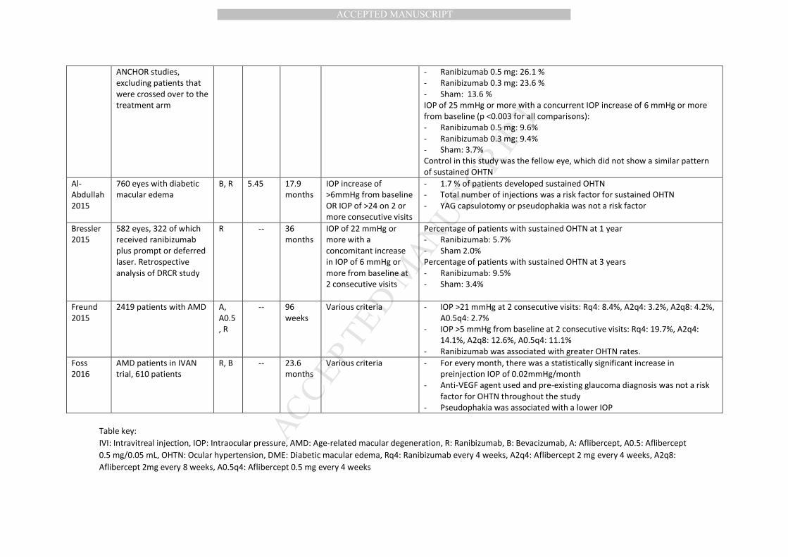
MANUSCRIP
T
ACCEPTED
ACCEPTED MANUSCRIPT
Table key:
IVI: Intravitreal injection, IOP: Intraocular pressure, AMD: Age-related macular degeneration, R: Ranibizumab, B: Bevacizumab, A: Aflibercept, A0.5: Aflibercept
0.5 mg/0.05 mL, OHTN: Ocular hypertension, DME: Diabetic macular edema, Rq4: Ranibizumab every 4 weeks, A2q4: Aflibercept 2 mg every 4 weeks, A2q8:
Aflibercept 2mg every 8 weeks, A0.5q4: Aflibercept 0.5 mg every 4 weeks
ANCHOR studies,
excluding patients that
were crossed over to the
treatment arm
- Ranibizumab 0.5 mg: 26.1 %
- Ranibizumab 0.3 mg: 23.6 %
- Sham: 13.6 %
IOP of 25 mmHg or more with a concurrent IOP increase of 6 mmHg or more
from baseline (p <0.003 for all comparisons):
- Ranibizumab 0.5 mg: 9.6%
- Ranibizumab 0.3 mg: 9.4%
- Sham: 3.7%
Control in this study was the fellow eye, which did not show a similar pattern
of sustained OHTN
Al-
Abdullah
2015
760 eyes with diabetic
macular edema
B, R 5.45 17.9
months
IOP increase of
>6mmHg from baseline
OR IOP of >24 on 2 or
more consecutive visits
- 1.7 % of patients developed sustained OHTN
- Total number of injections was a risk factor for sustained OHTN
- YAG capsulotomy or pseudophakia was not a risk factor
Bressler
2015
582 eyes, 322 of which
received ranibizumab
plus prompt or deferred
laser. Retrospective
analysis of DRCR study
R -- 36
months
IOP of 22 mmHg or
more with a
concomitant increase
in IOP of 6 mmHg or
more from baseline at
2 consecutive visits
Percentage of patients with sustained OHTN at 1 year
- Ranibizumab: 5.7%
- Sham 2.0%
Percentage of patients with sustained OHTN at 3 years
- Ranibizumab: 9.5%
- Sham: 3.4%
Freund
2015
2419 patients with AMD A,
A0.5
, R
-- 96
weeks
Various criteria - IOP >21 mmHg at 2 consecutive visits: Rq4: 8.4%, A2q4: 3.2%, A2q8: 4.2%,
A0.5q4: 2.7%
- IOP >5 mmHg from baseline at 2 consecutive visits: Rq4: 19.7%, A2q4:
14.1%, A2q8: 12.6%, A0.5q4: 11.1%
- Ranibizumab was associated with greater OHTN rates.
Foss
2016
AMD patients in IVAN
trial, 610 patients
R, B -- 23.6
months
Various criteria - For every month, there was a statistically significant increase in
preinjection IOP of 0.02mmHg/month
- Anti-VEGF agent used and pre-existing glaucoma diagnosis was not a risk
factor for OHTN throughout the study
- Pseudophakia was associated with a lower IOP

MANUSCRIP
T
ACCEPTED
ACCEPTED MANUSCRIPT
Table 4. Studies not finding sustained ocular hypertension following repeated intravitreal injections.
Study key: P: pegaptanib, B: Bevacizumab, R: Ranibizumab, IVI: Intravitreal injection, IOP: Intraocular pressure, mmHg: millimeters of mercury,
OHTN: ocular hypertension, AMD: Age-related macular degeneration
Study Cohort Medication Follow-up Key IOP inclusion criteria Key findings
Wehrli
2012
302 eyes
with AMD
and 226
control
fellow eyes
B, R 2 years IOP ≥22 mmHg on 2
consecutive visits with
an increase from
baseline of >6 mmHg
or IOP > 26 mmHg on a
single visit
- 0.5% incidence per eye-year of sustained OHTN in
injected eyes compared to 1.0% of control eyes
- For patients with glaucoma, 3.1% of patients treated
with anti-VEGF developed sustained OHTN,
compared to 5.7% of control eyes
Boyer
2014
221 eyes
with AMD,
114 treated
with P
P 24
months
≥2 measurements of
≥22 mmHg
- 5.3% of patients receiving pegaptanib versus 9.3% in
the sham group
Kim 2014
AJO
629 eyes with
AMD and 95
eyes with RVO
B, R 35.5
months
IOP increase of >5mmHg
over baseline on 2
consecutive visits
- 3.1% of control fellow eyes developed sustained OHTN
- 3.0% of patients with AMD developed sustained OHTN
- 7.4% of patients with RVO developed sustained OHTN
Kim 2014
J Glauc
83 patients
with AMD
B 24
months
Average IOP - No difference in IOP between groups
- No comment on the percentage of patients with an
increase in IOP, as only averages were presented
- Average of only 3.71 injections

MANUSCRIP
T
ACCEPTED
ACCEPTED MANUSCRIPT
Figure 1.
Weighted IOP average following intravitreal injection. This graph presents the acute change in IOP
following intravitreal injection. The data for this graph was extracted from material presented in
Supplemental Material Table 1. This data was then used to create weighted IOP averages based on the
number of patients included in each study for each of the plotted time points. Error bars were created
from the weighted standard deviation of the means.
0
5
10
15
20
25
30
35
40
45
50
0 10 20 30 40 50 60
Intr
ao
cula
r P
ress
ure
(m
mH
g)
Time Following Injection (minutes)

MANUSCRIP
T
ACCEPTED
ACCEPTED MANUSCRIPT
Figure 2.
Prophylactic effect of topical drops given prior to intravitreal injection. The data for this graph was
extracted from published material presented in Supplemental Material Table 2. This data was used to
create weighted IOP averages based on the number of patients included at each time point. Error bars
were not included in this graph due to the paucity of data available and to not give an erroneous
impression of statistical significance.
0.0
5.0
10.0
15.0
20.0
25.0
30.0
35.0
40.0
45.0
50.0
0 5 10 15 20 25 30
Intr
ao
cula
r P
ress
ure
(m
mH
g)
Time Following Injection (Minutes)
Prophylactic Drops No Prophylaxis


![Skin Inflammation, [Acute, Suppurative, Chronic, Chronic ... · Skin – Inflammation, [Acute, Suppurative, Chronic, Chronic Active, Granulomatous] presence of mononuclear cells (lymphocytes,](https://img.pdfslide.us/doc/110x75/5f0eb0c97e708231d44075f1/skin-inflammation-acute-suppurative-chronic-chronic-skin-a-inflammation.jpg)