Embed Size (px)
Citation preview

J. Am. Chem. SOC. 1994,116, 11961-11968 11961
The Active Site of Hydroxylamine Oxidoreductase from Nitrosomonas: Evidence for a New Metal Cluster in Enzymes
Michael P. Hendrich,**+ Michael Logan,’ Kristoffer K. Andersson,s Dave M. Arciero,l John D. Lipscomb,” and Alan B. Hooper-L
Contribution from the Department of Biochemistry, University of Minnesota, Minneapolis, Minnesota 55455, and the Department of Genetics and Cell Biology, University of Minnesota, St. Paul, Minnesota 55108
Received June 15, 1994@
Abstract: Hydroxylamine oxidoreductase (HAO) from Nitrosomonas europaea catalyzes the oxidation of NH2OH to NOz-. The enzyme contains eight hemes per subunit (63 m a ) which participate in catalytic function and electron transport. In the resting ferric state of the enzyme, we find that these hemes can be categorized on the basis of quantitative integer-spin and multifrequency EPR data as follows: (a) Four hemes are noninteracting, ferric C-type hemes. (b) Two hemes are low-spin and weakly spin-coupled through an interaction which is best described as dipolar on the basis of frequency-dependent g values. (c) The final two hemes are exchange-coupled; this cluster is the origin of a new integer-spin EPR signal which can be quantitatively associated with the reactive P m heme of HAO. Accordingly, treatments that affect the optical properties of P m and catalysis of the enzyme, such as substrates, CN-, or H202, all caused the integer-spin resonance to vanish. For reduced HAO, a second integer-spin EPR signal is observed which derives from one high-spin ferrous heme; previous Mossbauer studies have shown that the only such heme is Pm. An analysis of potential electronic models for the exchange-coupled cluster indicated that the interacting spins are both S = l/2. It thus appears that the active site of HA0 contains a pair of low-spin hemes which are coupled in a manner not previously observed in proteins. The structure of the diheme cluster and in the presence of substrates or inhibitors, and its bearing on catalysis by HA0 is discussed.
Introduction
The autotrophic bacterium Nitrosomonas europaea obtains energy for growth by the oxidation of ammonia to nitrite. Hydroxylamine oxidoreductase (HAO),’ a soluble enzyme in the periplasmic region of the bacterium, catalyzes the oxidation of the intermediate hydroxylamine: NHzOH + H2O - NOz- + 4 e- + 5H+.213 HA0 is a trimer of polypeptides with each polypeptide (63 containing eight covalently bound hemes? Seven of these are C-type cytochromes, and the eighth is an unusual prosthetic group, heme P4m, which has a Soret band at 463 nm for the reduced form of HAO. Six of the c-hemes have a-band absorption maxima at 553 nm in the ferrous form. These have midpoint redox potential values at +288, -10, -162, -192, -265, and -412 mV (vs NHE at pH 7).6 The midpoint potentials for the seventh c-heme, which has an a-band at 559 nm, and the P w heme are at +11 and -260 mV, respectively.
The function of most of the c-hemes is believed to be the transfer of electrons from the catalytic active site to exogenous
t Current address: Department of Chemistry, Camegie Mellon University, Pittsburgh, PA 15213. * Current address: Exxon Research, Annandale, NJ 08801.
Current address: Department of Biochemistry, University of Oslo, Oslo,
Department of Genetics and Cell Biology. Department of Biochemistry.
Norway.
@Abstract published in Advance ACS Absrracts, November 15, 1994. (1) Abbreviations: HAO, hydroxlyamine oxidoreductase; TPP, tetraphen-
(2) Hooper, A. B.; Nason., A. J. Biol. Chem. 1965, 240, 4044-4057. (3) Andersson, K. K.; Hwper, A. B. FEBS Lett. 1983, 164, 236-240. (4) Terry, K.; Hwper; A. B. Biochemistry 1981, 20, 7026-7032. (5)Arciero, D. M.; Hooper, A. B. J. Biol. Chem. 1993, 268, 14645-
(6) Collins, M.; Arciero, D. M.; Hooper, A. B. J. Biol. Chem. 1993,
0002-7863/94/1516-11961$04.50/0
ylporphyrin.
14654.
268, 14655-14662.
electron acceptor^.^*^ Several observations indicate that the P460 heme is a component of that active site. Ferrous P460 is the only heme of HA0 which binds CO and thus may be the only heme capable of binding exogenous molecules?JO In the ferrous state, the hemes of HA0 are rapidly oxidized by 0 2 or HzOz in a process which is inhibited by C0.9911 Incubation of oxidized HA0 with Hz02 causes loss of hydroxylamine reactivity and concomitant irreversible loss of the absorbancy of ferrous Pm, whereas the optical spectra of the c-hemes are unaffected.12 Oxidized HA0 binds cyanide in a 1 : 1 stoichiometry with P460 to form an optically-detectable c0mp1ex.l~ Formation of the cyanide-HA0 complex results in noncompetitive inhibition of hydroxylamine oxidation, inhibition of the reaction of ferric HA0 with H202, and inhibition of the inactivation of HA0 by organohydrazine suicide substrate^.'^
Mossbauer spectroscopy of fully reduced HA0 shows ferrous PM to be high spin and to exhibit an unusually large quadrupole splitting (AEQ = 4.21 mm/s).14 The chemical models which mimic the Mossbauer and optical properties of ferrous P w are pentacoordinate heme complexes where the fifth ligand is an anion and the iron is substantially out of the porphyrin plane.15 Resonance Raman spectroscopy of ferrous P460 confirms that
(7) Hooper, A. B. In Autotrophic Bacteria; Schlegel, H. G., Bowien,
(8) Arciero, D. M.; Balny, C.; Hooper, A. B. Biochemisrry 1991, 30,
(9) H o o p , A. B.; Debye, P.; Andersson, K. K.; Balny, C. B. Eur. J.
(10) Lipscomb, J. D.; Andersson, K. K.; Miinck, E.; Kent, T. A.; Hooper,
(11) Hooper, A. B.; Balny, C. FEBS Lett. 1982, 144, 299-303. (12) Hwper, A. B.; Terry, K. R. Biochemisrry 1977, 16, 455-459. (13) Logan, M. S., Ph.D. Thesis, 1991, University of Minnesota. (14) Andersson, K. K.; Kent, T. A.; Lipscomb, J. D.; Hooper, A. B.;
B., Eds.; Science Tech Publishers: Madison, WI, 1989, pp; 239-265.
11466-11472.
Biochem. 1983, 134, 83-87.
A. B. Biochemistry 1982, 21, 3973-3976.
Miinck, E. J. Biol. Chem. 1984, 259, 6833-6840.
0 1994 American Chemical Society

11962 J. Am. Chem. SOC., Vol. 116, No. 26, 1994 Hendrich et al.
the band at 463 nm is a Soret band and that it is a unique heme with low symmetry.16 Upon proteolytic digestion of HAO, heme P460 has been shown to have a covalent linkage between a meso-carbon of protoporphyrin IX and a ring carbon of a tyrosyl residue of the protein.17
The properties of the active site of oxidized HA0 are much less understood, in part because the absorption spectrum of oxidized heme Pm are not easily distinguished from the other hemes. The EPR spectra of oxidized HA0 show many low- spin heme resonances, which have allowed some differentiation between the c-hemes; however, none of those resonances have been associated with the active site.18 Reductive titration studies in conjunction with EPR have given some insight into this problem. When the system potential of HA0 is set so that the C-type heme with J?o = -192 mV becomes reduced, a new EPR signal near g = 6 is observed which may originate from heme P4so.18919 The g = 6 signal disappears at still lower potentials where heme Pm is reduced. These results suggest that ferric Pm may be high-spin and electronically coupled to another paramagnetic center of HAO.
In the present work, integer-spin EPR spectroscopy has been applied to HA0 in both its fully oxidized and reduced states and in the presence of substrates and other small molecules. We find a new EPR signal which originates from a spin-coupled pair of hemes in oxidized HAO, both of which must be low- spin. The properties of this cluster and the associated EPR signal in the presence of substrate indicate that the cluster must contain the enzymatic center of the protein, which in its reduced form is the origin of the characteristic P4w Soret absorption. This is the first spectroscopic identification of the active center of HA0 in its resting state. HA0 is believed to be the first example of an enzyme containing a diheme cluster.
Materials and Methods
Purification of Enzyme. Nitrosomonas europaea (Schmidt strain) was either grown in 15 L carboys as previously describedZ or grown in a pH-controlled, continuous culture apparatus operated at a dilution rate of 0.32 day-L; the reactor volume was 55 L. HA0 was purified as described previously.*J3 The form of enzyme used for these experiments was HAO-A. All experiments were performed in 50 mM potassium phosphate buffer, pH 7.5, unless otherwise noted. The concentration of HA0 was determined spectrophotometrically for the oxidized state (e = 700 mM-' cm-' at 408 nm) and is given in terms of the subunit concentration (63 kDa) throughout this paper. The amount of P4m in HA0 was determined from the difference spectrum of reduced HA0 plus CO minus reduced HA0 ( E = 76 mM-' cm-' for &Z minus &).I3 For the two EPR samples used in spin quantitation, the accuracy of the extinction coefficients was checked by submitting aliquots for amino acid content determination. The extinction coefficients were found to be in agreement with the previous determinations. The subunit concentration of HA0 in the EPR studies was nominally 1 mM. All chemicals were reagent grade or better. Double-distilled or Millipore Super Q water was used throughout.
For the experiments with substrates, hydroxylamine or hydrazine were added to samples of HA0 at a 7-fold excess relative to the subunit concentration. For the inactivation experiment, HA0 was inactivated by incubation of 1 mM enzyme with a 5-fold excess of peroxide for 1 h, followed by dialysis overnight with three changes of 50 mM
(15) (a) Nasri, H.; Fischer, J.; Weiss, R.; Bi1,E.; Trautwein, A. J. Amer. Chem. SOC. 1987, 109, 2549-2550. (b) Shaevitz, B. A.; Lang, G.; Reed, C. A. lnorg. Chem. 1988,27,4607-4613. (c) Latos-Grazynski, L.; Lisowski, J.; Olmstead, M. M.; Balch, A. L. Inorg. Chem. 1989, 28, 1183-1188.
(16) Andersson, K. K.; Babcock, G. T.; Hooper, A. B. Biochem. Biophys. Res. Commun. 1991, 174. 358-363.
(17) Arciero, D. M.; Hooper, A. B.; Cai, M.; Timkovich, R. Biochemistry
(18) Liuscomb, J. D.; Hoouer, A. B. Biochemistry 1982,21,3965-3972. 1993, 32, 9370-9378.
(19) Prince, R.; Hooper, A. B. Biochemistry 1987, 26, 970-974.
h 11.6 , 1y.. . .
I I I I I I ' I ~ ' I 0 100 200 300 400 500
B (mT)
Figure 1. X-band EPR spectra (-) and simulations (- - -) of oxidized HA0 (A-C) and dithionite-reduced HA0 (D), all pH 7.5. The oscillating microwave field is oriented either perpendicular (A, B) or parallel (C, D) to the static magnetic field. Simulation parameters: (C) gefi = 4.8, A0 = 0.25 cm-', UA = 0.036 cm-', and (D) gee = 8.0, A0
= 0.7 cm-l, UA = 0.2 cm-'. Instrumental parameters: temperature, 2.5 K; microwave frequency, 9.14 GHz (A, B) or 9.07 GHz (C, D). All intensities have been adjusted for equal gain, power, and protein concentration (0.87 mM); the spectra are additionally magnified as given in the figure.
potassium phosphate buffer, pH 7.5. There was no remaining P a chromophore in the sample, as assayed by the carbon monoxide difference spectrum of the dithionite-reduced enzyme. The titration of oxidized HA0 with KCN was monitored by the increase in the 434 nm ( E = 3 cm-') peak in optical difference spectra for a 1.1 mm path length cell on a HF' 8452a spectrophotometer. Both the UV filter no. 3 supplied with the instrument and a glass plate were used to absorb wavelengths of light less than 300 nm.
EPR Instrumentation. X-band EPR spectra were recorded on a Varian E109 spectrometer equipped with an Oxford ESR-910 liquid helium cryostat and a Varian E236 bimodal cavity. For the variable- temperature study, the temperature was measured in several sets of experiments with a calibrated carbon-glass resistor (Lakeshore Cry- otronics, CGR-1-1000) and in several other sets of experiments using the inverse temperature dependence of the low-spin EPR signals in the region 1.7 < g 3.4. The quantifications of the integer-spin resonances are relative to the spin standard Zn fluosilicate (1.5% Fe2+).m Daily variations in the spectrometer sensitivity were compensated relative to a [CuEDTA]*- spin standard in 10% glycerol. The half- integer-spin quantifications are relative to the copper standard. The published spectra were obtained with field modulation of 1 mTpp at 100 kHz, unless otherwise noted. The magnetic field was calibrated with an NMR gaussmeter, and the microwave frequency was measured with a counter.
S-band data where collected at the National Biomedical ESR Center in the Medical College of Wisconsin. The spectra were recorded on a Varian spectrometer using a homemade microwave bridge and cavity and an Air Products liquid helium cryostat. The P-band data were collected at the Biophysical Research Division of the University of Michigan. The spectra were recorded on a homemade spectrometer using a Air Products liquid helium cryostat.
Results Oxidized HAO. EPR spectra of HA0 as isolated (all irons
ferric) in microwave fields oscillating parallel (B1 II B) or perpendicular (B1 I B) to the static magnetic field are shown in Figure 1A-C. The groups of resonances in Figure 1A at g(3.09, 2.19, 1.4) and g(3.00, 2.25, 1.4) have been assigned to two different types of low-spin Fe3+ heme sites with bis-histidine
(20) Hendrich, M. P.; Debrunner, P. G. Biophys. J. 1989,56,489-506.
Integer-Spin EPR of Hydroxylamine Oxidoreductase J. Am. Chem. SOC., Vol. 11 6, No. 26, 1994 11963
of the doublet, respectively. Note that geff does not necessarily equal g. The latter is not a physically meaningful number; it simply denotes some conspicuous spectral feature at a field B = hv/g/?, which is shifted to a field position lower than hvl g,& due to a nonzero value of A. The values of A and geff cannot yet be independently determined; however, reasonably accurate simulations of the spectrum in Figure 1C can only be achieved for geg < 6. The simulation shown in this figure assumes g,ff = 4.8. The simulation also assumes that the dominate source of line broadening is due to a spread in the values of which is modeled by a Gaussian distribution with a center value A0 and one standard deviation width of OA. Based on simulations of the g = 7.7 resonance, the ratio of the spin concentration to the P4w concentration (determined spectro- photometrically) was found to be between 0.6 (g,ff = 2)23 and 1.2 (ges = 6); for geff = 4.8, this ratio is one. Thus, the resonance at g = 7.7 originates from a species having a concentration close to that of heme P4a.
Reduced HAO. HA0 was reduced anaerobically with a 25- fold stoichiometric excess of NazSz04 under argon. The EPR spectrum of reduced HA0 is shown in Figure 1D. The resonance at g = 7.7 is absent, and a new resonance is present at g = 1 1.6. The temperature dependence of the g = 1 1.6 signal (not shown) indicates that it also originates from a ground spin doublet. Its shape and position are consistent with that expected for a high-spin Fez+ center. The analysis of this signal follows previous studies of Fez+ complexes.z0 If we assume that the resonance at g = 11.6 originates from the ground doublet of an S = 2 quintet with geff = 8, then the zero-field parameters of this doublet obtained from the simulation of Figure 1D are A0 = 0.7 cm-' and UA = 0.2 cm-'. Based on this simulation, quantification of the g = 11.6 signal gave an Fez+ concentration equal, within experimental error, to heme P ~ M ) determined spectrophotometrically .z4 Approximately 5% of these high-spin Fez+ sites have A < hv FZ 0.3 cm-', and thus are observable with an X-band EPR spectrometer. For the 95% of sites with A > hv, eq 1 cannot be satisfied for any B, and no resonance is observed.
Addition of Substrate CN- or Inactivation of HA0 with HzOz. The addition of a stoichiometric excess relative to the concentration of P460 of substrates NHzOH or Nz&, or the inhibitor CN-, resulted in the loss of the resonance at g = 7.7. This resonance also vanished after HzOz inactivation of HAO. No other EPR signals were affected by the addition of CN- or HzOz, notably, the loss of the resonance at g = 7.7 was not accompanied by a change in the frequency dependent resonances of Figure 1A (g = 3.42, 2.74, 1.86, 1.67). Consistent with the previous observation that NHzOH reduces 35% of the hemes of HA0,l8 but not heme P460?~,~~ no integer-spin signal like that of Figure 1D was observed.
Inhibition and binding studies indicate that CN- is an active site inhibitor.13 Figure 3 shows the result of titrations of HA0 with CN-, which were monitored optically at 434 nm and with EPR from the intensity of the resonance at g = 7.7. The
+ + I 0.5
2 6 10 14 i a TW)
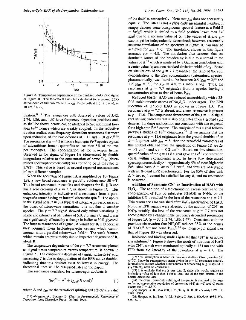
Figure 2. Temperature dependence of the oxidized HA0 EPR signal of Figure IC. The theoretical lines are calculated for a ground EPR- active doublet and two excited energy levels both at 1 (-), 3 (. - *), or 10 cm-I (- - -).
l i g a t i ~ n . ~ ~ , ' ~ The resonances with observed g values of 3.42, 2.74, 1.86, and 1.67 have frequency dependent positions and, as shall be shown below, can be assigned to two additional low- spin Fe3+ hemes which are weakly coupled. In the reductive titration studies, these frequency-dependent resonances disappear upon reduction of the two c-hemes at +11 and -10 mV.18,19 The resonance at g = 4.3 is from a high-spin Fe3+ species typical of adventitious iron; it quantifies to less than 5% of the iron per monomer. The concentration of the low-spin hemes observed in the signal of Figure 1A (determined by double integration) relative to the concentration of heme P4.50 (deter- mined spectrophotometrically) was found to be in the ratio of 5.7(2). This value is based on several repeated measurements of two different samples.
W e n the spectrum of Figure 1A is amplified by 10 (Figure lB), a new broad resonance is partially evident near 50 mT. This broad resonance intensifies and sharpens for B1 1 1 B and has a zero crossing of g = 7.7, as shown in Figure 1C. This enhanced intensity is indicative of a resonance from a para- magnetic center having an integral electronic spin.*O The upturn in the signal near B = 0 is typical of integer-spin resonances at the onset of microwave saturation; it is not due to another species. The g = 7.7 signal showed only minor variations in shape and intensity at pH values of 5.0,7.0, and 9.0, and it was not significantly affected by a change in buffer to 50% glycerol. The intense resonances of Figure 1A vanish for B1 I I B because they originate from half-integer-spin centers which cannot interact with a parallel microwave field.21 The weak features which remain are presumably due to imperfect alignment of B1 along B.
The temperature dependence of the g = 7.7 resonance, plotted as signal times temperature versus temperature, is shown in Figure 2. The continuous decrease of (signal intensity).T with increasing Tis due to depopulation of the EPR-active doublet, indicating that this doublet must be lowest in energy. The theoretical lines will be discussed later in the paper.
The resonance condition for integer-spin doublets is
where A and geff are the zero-field splitting and effective g value
(21) Abragam, A.; Bleaney B. Electron Paramagnetic Resonance of Transition lons; Clarendon Press: Oxford, 1970.
(22) This assumption is based on previous studies of iron proteins(cf. ref 20). Since the paramagnetic center giving the g = 7.7 resonance is novel, it remains to be seen whether other sources of broadening (e.g., a spread in gee values), must be considered.
(23) It is unlikely that gee is less than 2, since this would require an intrinsic g value of less than 1 for at least one of the spin centers in the cluster discussed later.
(24) The overall zero-field splitting of the quintet is assumed to be large so that no appreciable population of the excited 1+1) f 1-1) and (0) states occurs for T = 2.5 K.
(25) Hooper, A. B.; Maxwell, P. C.; Terry, K. R. Biochemisty 1978,17,
(26) Hooper, A. B.; Tran, V. M.; Balny, C. Eur. J. Biochem. 1984, 141, 2984-2989.
565-571.

11964 J. Am. Chem. SOC., Vol. 116, No. 26, 1994 Hendrich et al.
[KCN]/[subunit HAO] (mole ratio) Figure 3. Titration of oxidized HA0 with KCN. Shown are the progressive loss of the g = 7.7 EPR signal and increase in the 434 minus 470 nm difference optical signal. The dashed lines are guides showing the stoichiometric titration of HAO, assuming that binding of cyanide goes to completion. HA0 was present at a subunit concentration of 1.2 mM in 0.25 mL (EPR) or 30 pM in 0.10 mL (optical). Other conditions are given in Materials and Methods.
g = 9 I !
I I 0 100 200 300
0 (mT)
Figure 4. X-band EPR spectrum of oxidized HAO, pH 7.5, plus 2-fold excess KCN per subunit. The dashed line is oxidized HA0 at an equal concentration, but displayed at one-fi i the intensity. Instrumental parameters: temperature, 2.5 K, microwave frequency, 9.09 GHz.
resonance disappeared after addition of one equivalent of CN- per heme Pm. The dissociation constant for the binding of CN- to HA0 was found to be 10 pM. Under the conditions used for Figure 3, the total amount of CN- remaining unbound was calculated to be less than 5%.
In many samples of oxidized HAO, additional sharp reso- nances were superimposed on the resonance at g = 7.7. The addition of CN- or HzOz resulted in a large decrease in the intensity of the resonance at g = 7.7, but these additional sharp features and an associated broad signal were not affected. Figure 4 shows a spectrum of H A 0 in the presence of a 2-fold excess of CN-. The signal at g = 7.7 is absent, leaving a spectrum with a zero crossing at g = 9 which contains four hyperfine peaks, each separated by approximately 7.5 mT. The peak at highest magnetic field of the four was near the noise level, but it was reproducible in several samples. This hype f i e spectrum had enhanced intensity for B1 I I B and lost intensity with increasing temperature, indicating that the signal originates from an integer-spin doublet which is lowest in energy. The signal strength of the hyperfine spectrum is highly preparation dependent and is most intense for samples having low specific activity. The largest intensity of this signal we have observed is 5-fold weaker than the resonance at g = 7.7 (overlaid in Figure 4). These observations suggest that the four line
3.09
I I A
m 1.86 1.67 I I P
H P
3'09 2.30
1.55
v i I
B + Figure 5. P-, X-, and S-band EPR spectra of oxidized HAO, pH 7.5. Instrumental parameters: temperature, 12 K, microwave frequency, 14.77 (A), 9.23 (B), 3.05 GHz (C).
hyperfine signal originates from a minority species of HA0 which contains an atom with a nuclear spin of Z = 3/2, such as cu*+.
Frequency Dependent, Half-Integer-Spin EPR Reso- nances. P-, X-, and S-band EPR spectra of oxidized HA0 are shown in Figure 5 recorded with B1 I B. The spectra are plotted for equal effective microwave frequency" so that signal features which do not obey the standard resonance condition (g = hv/ PB) are made readily apparent. It is evident that many changes occur in the spectra as a function of microwave frequency. In this study, we focus only on those resonances that show clear changes in g values irrespective of line broadening effects and baseline drifts. This is true for the resonances having X-band g values (Figure 5B) of 3.42 and 2.74. At P-band (Figure 5A), these resonances have shifted to g = 3.29 and 2.82, respectively. The positions of these resonances at S-band (Figure 5C) are not clear.
Discussion
An understanding of the active site of HA0 has been hindered by the presence of many iron atoms (24-27) per trimeric unit, several of which are interacting as will be demonstrated here. Many previous studies have laid a foundation for disentangling this complexity. The number of iron atoms in HA0 has been determined to be either eight or nine per monomeric unit. The value of eight is based on a counting of the peptide fragments containing the sequence -C-a-b-C-H-, which is known to be associated with heme center^.^ The value of nine is based on a previous comparison of optical and EPR data.'* The observation of frequency dependent g values in oxidized HA0 (Figure 5 ) suggests that the value of nine estimated in the EPR study is open for reinterpretation. Analysis of Mijssbauer spectra of H A 0 are consistent with either eight or nine iron atoms per monomer.I4
We find that the half-integer-spin EPR signals of Figure 1A integrate to six Fe3+ atoms per monomer, rather than seven as previously estimated.I8 Four of these centers, with g, = 3.09 or 3.00, have been previously assigned to low-spin c-hemes with bis-histidine l i g a t i ~ n . ' ~ J ~ The other two iron centers, with gz = 3.42 or 2.74 at X-band, have frequency dependent g values and can be associated with two weakly interacting low-spin hemes as will be shown below. None of these six hemes have
(27) The magnetic field value of each point in a spectrum is multiplied by the factor ~ ~ ~ ~ / v ~ ~ , j . with no adjustment to the signal value,

Integer-Spin EPR of Hydroxylamine Oxidoreductase
been assigned to the active site of HAO; rather, they may be involved in the transport of electrons from the active site or redox poising of the enzyme?,8
At least two observations have led to the conclusion that the site containing heme P460 is responsible for catalytic oxidation of hydroxylamine. First, selective destruction of P4m with stoichiometric amounts of H202 results in loss of enzymatic activity.12 Second, the reduced form of P m can bind C0,9,14*28 indicating that the Fe2+ site in P m has an open coordination site for substrate binding. The previous lack of a EPR signal which reflects these observations and the presence of only Fe3+ in the Mossbauer spectrum of oxidized HAO’O has led to the proposal that the P m center is composed of one or two iron atoms which are rendered “EPR silent” due to either spin coupling or fast electronic spin relaxation. The Mossbauer spectroscopy of oxidized HA0 indicates that the active site must contain one or two Fe3+ atoms and suggest that these iron(s> are low-spin.lo An indication that a high-spin Fe3+ center might be associated with the active site of HA0 comes from EPR studies in which HA0 is poised at various reduction poten- t i a l ~ . ~ ~ ~ ~ ~ A signal near g = 6 (typical of high-spin Fe3+) is observed at a reduction potential of -217 mV (Pdm, -260 mV) and vanishes at still lower potentials at which the P m heme is reduced. Furthermore, in reduced HAO, P m is known to be high-spin.14 These observations require that we initially consider clusters which may contain high-, intermediate-, or low-spin iron states andor a radical species.
In the discussion to follow, we show that the integer-spin resonance at g = 7.7 is associated with heme P m and that it originates from an exchange coupling between the two iron centers, both of which are low-spin. Using the current best estimate of eight hemes per monomeric unit of HAO, we show that HA0 contains 4 hemes which are not interacting, two hemes which are weakly interacting, and two hemes which are exchange-coupled.
Two Weakly Interacting Low-Spin Hemes. The above results indicate that the hemes giving the frequency dependent g values of Figure 1A (g, = 3.42 and 2.74) are not associated with the PW heme. The resonances associated with the frequency dependent g values of Figure 1A are unchanged for HA0 in the presence of CN-, whereas the g = 7.7 resonance of Figure 1C vanishes and is associated with the P m heme as discussed in the next section. The observation that the former resonances are frequency dependent is an indication of spin coupling between two paramagnetic centers. Furthermore, these frequency-dependent resonances have g values similar to those observed for low-spin hemes, indicating that the magnitude of the spin coupling is weak compared to the Zeeman interaction of the magnetic field, i.e., IJ( << gpB 0.3 cm-’. This can occur for two spatially close, low-spin iron atoms which interact via a magnetic dipole coupling. Figure 6 shows calculated g, values at 15, 9, and 3 GHz as a function of the distance r between the two hemes. The calculations assume: (1) that the heme planes are parallel, (2) that the vector joining the two iron atoms is along the heme normal, and (3) that the intrinsic g, values of both hemes are 3.06. The horizontal lines mark the observed gz values at 15 GHz (dashed lines, gz = 3.29 and 2.82) and 9 GHz (solid lines, gz = 3.42 and 2.74) from Figure 5 . We find that the calculated and observed gz values ap- proximately match at a distance of 5.2 A, for both microwave frequencies. These resonances are predicted from Figure 6 to shift to g, = 4.7 and 2.3 at 3 GHz. Although resonances are observed at approximately these values, overlap with other
(28) Erickson, R. H.; Hooper, A. B. Biochem. Biophys. Acta 1972,275, 23 1-244.
J. Am. Chem. SOC., Vol. 11 6, No. 26, 1994 11965
r(A) Figure 6. Plot of observed g, value as a function of the distance r between two coplanar hemes experiencing a magnetic dipole interaction. The theoretical lines are for microwave frequencies of 14.77 (. -*),9.23 (-), and 3.05 GHz (- - -). The horizontal arrows mark the observed gz values taken from Figure 5. The vertical arrow marks the distance at which the calculated and observed gz values approximately match. See text for other assumptions.
resonances in the spectrum prevents a def i t ive assignment from the S-band data alone.
The iron-iron distance suggests that a weak exchange interaction may also exist between the hemes, in addition to the dipolar coupling. In a study of inter-heme spin coupling, an exchange interaction was observed between the n-clouds of face-to-face heme pairs separated by 4.6 A.29 At present, it is not possible to separate the exchange and dipolar contributions to the spin coupling, thus, the separation of 5.2 A between hemes should be considered an estimate.
The short iron-iron distance also raises structural questions concerning the axial ligands. These hemes are clearly both low- spin and therefore probably 6-coordinate. The intrinsic gz value appears to be close to 3.06, since this value allows agreement of the theoretical and experimental g values displayed in Figure 6. This intrinsic g, value is typical for bis-histidine coordination in hemes, which is believed to be the ligation of the noninter- acting hemes.18 However, the space between the weakly- coupled hemes is probably insufficient for two axial histidines, and one bridging histidine would likely give rise to an exchange- coupling which would overwhelm the dipolar interaction and thus breakdown the agreement seen in Figure 6. While unlikely, it is possible that the agreement with a dipolar model is fortuitous and that a very weak exchange interaction is present from partial heme overlap in an edge-to-edge rather than a face- to-face arrangement. An edge-to-edge overlap could presum- ably accommodate axial ligands. Further insight into this question, and an improved distance calculation, may come from a more detailed analysis of the entire EPR spectrum.
Two Exchange-Coupled Low-Spin Hemes. Whereas a magnetic dipole coupling can approximately account for the frequency shifts of Figure 5 , it cannot be the dominant interaction which gives rise to the integer-spin resonance at g = 7.7. The largest component of the exchange interaction between the pair of spin centers giving the latter resonance must be greater than gpB, as indicated by an integer-spin EPR signal with no associated resonances for either B1 I B or B1 1 I B. The integer-spin resonance must arise from a pair of spins because all the irons of oxidized HA0 are femc with half-integer-spin. Thus, we now consider system spin states, S, resulting from an electronic exchange coupling between individuals spins Si and S,. The magnetic properties of this coupled system are described by the spin Hamiltonian
(29) Gupta, G. P.; Lang, G.; Scheidt, W. R.; Geiger, D. K.; Reed, C. A. J . Chem. Phys. 1985, 83, 5945-5952.

11966 J. Am. Chem. SOC., Vol. 116, No. 26, 1994
H , = Sl*JS2 + (S;D;Si + ,l?Bp.Si) (2) i=1,2
Hendrich et al.
where the first term is the exchange interaction and the second and third terms are the zero-field and Zeeman interactions, respectively, for each site.
The assignment of the resonance at g = 7.7 now proceeds with an examination of all possible origins of this type of signal. There are three essential properties of the associated doublet that must be simultaneously reproduced by a coupled pair of spins, namely, geff < 6 and A0 < 0.3 cm-*. Further, it must be a ground doublet. The ground doublet of a spin S multiplet will have geff % 2geS where the intrinsic g-value of the paramagnetic center is ge % 2.0. From this relation and the range of allowed values of g,ff, the integer-spin resonance must originate from within an S = 1 multiplet or possibly an S = 2 multiplet if the spin coupling reduces the value of g, to lower then 1.5. In addition, as mentioned above, the cluster may contain either high-, intermediate-, or low-spin Fe3+ or a radical species. These results restrict the problem to four possible cases for SI and SZ: (1) I/z, l/2; (2) l/z, 3 / ~ ; (3) l/*, V2; (4) 3 / ~ , ?2.
A common element of cases 2-4 is the presence of at least one iron atom with a large intrinsic zero-field splitting. The axial zero-field parameter, D, for intermediate- or high-spin hemes is positive and generally greater than +5 cm-1.30*31 We have examined these three cases with full diagonalization of eq 2, with at least one Di value of greater than +5 cm-l, and for broad ranges of the exchange coupling and other zero-field parameters. In all cases, the isolated EPR-active spin doublets which have geff < 6 and A < 0.3 cm-' are high in energy, and thus cannot give the resonance at g = 7.7 because it originates from a ground doublet. Therefore, we conclude that the cluster associated with this pair of spins does not contain a typical intermediate- or high-spin heme. The possibility remains, however, that the cluster contains a novel heme center with a small, negative D value. If so, the spectrum of this novel heme center must then have been obscured by other low-spin heme doublets in the Mossbauer spectra.
Case 1 yields system spin states of S = 0 and 1 as shown in Figure 7. The energy splitting between the singlet and triplet state is determined by the isotropic component of the exchange coupling, J . The temperature dependence of the EPR signal (Figure 2) indicates that the paramagnetic triplet state must be lowest in energy, therefore, the exchange coupling between the Si = l/z sites is ferromagnetic (J < 0). The temperature dependence of the EPR signal is a function of the energies El 10)
and Elm) of the two excited levels of Figure 7. We have attempted to fit the data by arbitrarily varying these energies, but the uncertainties in the data do not allow an unambiguous fit. The theoretical curves shown in Figure 7 indicate that E11p and Elm> are both greater than 1 cm-'; better fits are obtained with Elm> 2 E110> 1 3 cm-'.
The zero-field splitting, Dex, of levels within the triplet state of Figure 7 is a function of both the magnetic dipole coupling, and the exchange coupling with higher orbital states mixed into the ground orbital state through the spin-orbit in te rac t i~n .~~ The former is small ( < O . l cm-l), whereas the latter can be much larger when metal centers are involved. Transitions between the magnetic spin states I+l) - IO) or 1-1) - IO) are not
(30) (a) Palmer, G. In The Porphyrins; Dolphin, D., Ed.; Academic Press: New York, 1979; Vol. 4, pp 313-354. (b) Miinck, E. Zbid, pp. 379- 423.
(3 1) Emptage, M. H.; Zimmerman, R.; Que, L., Jr.; Miinck, E.; Hamilton, W. D.; he-Johnson, W. H. Biochem. Biophys. Acta 1977,495, 12-23.
(32) Bencini, A.; Gatteschi, D., In EPR of Spin Coupled Systems; Springer-Verlag: New York, 1991.
IS=O, mgO>
J
11, o>
57 Figure 7. Energy diagram of two ferromagnetically coupled S = l12
centers giving the triplet state low in energy. The EPR-active doublet is split in zero-field by an energy A.
observed in the HA0 X-band spectra, indicating that the magnitude of Dex is larger than the microwave energy (Dex > hv 0.3 cm-I). Moreover, the temperature dependence indicates that Dex =- 1 cm-l. The large value of Dex and presence of an integer-spin resonance indicate a large anisotropic exchange interaction, which must originate from a strong spin- orbit coupling, as is present in low-spin heme complexes.
A Spin-Coupled Species Containing a Cu2+ Atom. Oxi- dized HA0 in some preparations contains a species which is unaffected by CN- or H202. The similarity of its EPR spectrum (Figure 4) with that of the majority species which vanishes in the presence of these molecules, suggests that it also originates from an exchange-coupled metal center. Previous metal analyses of HA0 have found 0.1 copper atoms per subunit.25 This amount is consistent with the observed intensity of a 4-line hyperfine spectrum (IcU = 3/2) relative to the majority species. The copper signal is probably associated with a minor and inactive enzyme species; it is thus not of great biological significance. However, the origin of this signal may be of general interest for several reasons: the copper might substitute for an iron in the active site cluster or bind near to it, resulting in inactivation; the presence of the hyperfiie signal provides an indication of protein homogeneity; integer-spin signals do not often display hyperfine features.
The position of the copper signal indicates that it may originate from either an S = 1 or 2 multiplet. The splitting between two consecutive hyperfine resonances, a (in mT), in relation to the intrinsic hyperfine value, A (in cm-l), for system spin states S = 1 or 2 is
A = ge,p1/2140 (S = 1) (3)
A = 1.5ge@/2140 (S = 2)
For the S = 1 case, the Cu2+ atom would be ferromagnetically coupled to a SI = l/2 center, whereas for S = 2 it would be antiferromagnetically coupled to a high-spin Fe3+ atom. The hyperfine spacing determined from Figure 4 is a = 7.5 mT. The range of allowed g,rvalues determined from simulations of Figure 4 is geR < 6 (S = 1) or gcff = 6 (S = 2). Substituting these values into eq 3 gives A < 0.0210 cm-' for S = 1, or A = 0.0315 cm-l for S = 2.
A values for copper porphyrins are largest in the direction perpendicular to the porphyrin plane and cover a range of 0.0180 < A < 0.0210 cm-1;33 A values for nonporphyrin copper centers range lower, 0.0140 A < 0.0210 cm-1.34 In comparison with
(33) Lin, W. C. In The Porphyrins; Dolphin, D., Ed.; Academic Press: New York, 1979; Vol. 4, pp 355-377.
(34) Solomon, E. I.; Penfield, K. W.; Wilcox, D. E. In Structure and Bonding; Springer-Verlag: New York, 1983; Vol. 53, pp 3-57.

Integer-Spin EPR of Hydroxylamine Oxidoreductase
the A values derived for HAO, the origin of the copper hyperfine is consistent with a system spin state of S = 1, but not S = 2. Thus, the presence of a high-spin Fe3+ atom coupled to the copper atom is not indicated.
The Active Site of HAO. The enzymatic active site of HA0 contains an iron center which, in its reduced form, gives rise to the characteristic PW optical spectrum. Two observations indicate that the integer-spin resonance of Figure 1C is associated with that center. First, the addition of NHzOH, N2H4, or CN-, or inactivation with H202 all cause the EPR signal to vanish. The inactivation of HA0 with H202 is known to selectively affect the PW heme. Second, quantification of the integer-spin EPR signal at g = 7.7 indicates that it originates from a majority species of oxidized HAO. Upon reduction, this signal disappears and a new integer-spin EPR signal from high-spin Fe2+ is observed at g = 11.6 in a concentration of one iron atom per protein subunit. This new signal must therefore originate from ferrous P460, which is known from Mossbauer studies to be the only high-spin iron site in reduced
We have considered several potential origins of the g = 7.7 signal of oxidized HA0 and we suggest that this list, in association with the previous spectroscopic studies, is compre- hensive. The observations presented in this work are consistent with a spin coupled moiety containing two Si = l12 centers. The magnitude of the zero-field splitting indicates that the moiety contains at least one low-spin Fe3+ atom, in keeping with the conclusions of the Mossbauer study.'O From these results, the following structures are proposed for the active site of HAO.
~ ~ 0 . 1 4
s, =I12 s2=112
I I I I
oxidized Y-FG3-X- FitHis
PW
s, =2 s2=o s, =2 s2=o
PW
reduced
The structure of the oxidized state contains an exchange- coupled pair of low-spin hemes. The assignment of the second Si = l/2 site to a low-spin Fe3+ heme is made on the basis of total heme content in oxidized HA0 as follows. Of the eight hemes per monomer? four are now identified as noninteracting C-type hemes and two are identified as very weakly coupled hemes. One of the two remaining hemes must be in the exchange-coupled moiety giving the g = 7.7 resonance, as stated in the previous paragraph, which leaves one heme unaccounted for. This eighth heme, however, must also be associated with a spin coupled cluster, otherwise it would be observed from oxidized HA0 with conventional EPR techniques. Thus, we propose that this eighth heme is also part of the exchange- coupled moiety giving the g = 7.7 resonance. This assignment gains some additional support from two other results. First, when HA0 is poised at a potential of -215 mV, a new Fe3+ EPR signal is observed, the intensity of which can be related to the degree of reduction of the c553 heme with a midpoint
J. Am. Chem. SOC., Vol. 116, No. 26, 1994 11967
potential of -190 mV.I9 Second, the zero-field splitting required to give the integer-spin resonance of oxidized HA0 must originate from an exchange interaction between a ground orbital state of one paramagnetic center with a low-lying excited orbital level of the other center, and this exchange interaction must be highly anisotropic. Such a large anisotropic exchange interaction is more likely to be observed from a pair of anisotropic hemes rather than from one heme coupled to a radical.35
The presence of a diheme cluster raises questions concerning how they are connected. While an answer requires further information, some insight accrues from the current study. We find that the largest component of the exchange coupling between the hemes is greater than 1 cm-'. This indicates the presence of a bridging atom or molecule, X, for which potential candidates include: 0, OR, C1, SR, imidazolate, and carboxy- late. The Fe-X-Fe unit is not necessarily linear as drawn; however, such a linear geometry is one possible origin of the observed ferromagnetic coupling. For low-spin femc hemes with a strong axial ligand, the unpaired electron will predomi- nantly occupy either a dxz or dyz iron orbital. For the diheme cluster, the unpaired electron of one heme could occupy a dxz orbital, and for the other heme, a dyz orbital. A ferromagnetic coupling would be expected in this case because the magnetic orbitals are orthogonal. There are very few characterized model complexes containing low-spin iron porphyrins that may give additional insight. It has been demonstrated that imidazolate can convey strong ferromagnetic coupling in the low-spin complex Fe3+TPP(Cu2+imidazole)2.36
The existence of histidine ligation to the irons at the active site is suggested from the proteolytic digestion study, where eight heme-containing peptide fragments were found to have the conventional sequence -C-a-b-C-H-. The histidine of this sequence is generally an axial ligand to the heme.5 Since all the hemes have this sequence, either X or Y of the structures may also be nitrogen from histidine. Upon reduction of HAO, the P4m heme becomes high-spin ferrous (SI = 2) and is now 5-coordinate, whereas the other heme is now low-spin ferrous (Sz = 0) and thus is still 6-coordinate. The presence or absence of a bridge to the S2 = 0 heme will not affect the magnetic properties of the cluster; thus, two possible structures are shown which afford a high-spin heme for reduced HAO. If the bridge remains intact upon reduction, the ligand Y is presumably a weakly coordinating protein residue or solvent molecule, alternatively, the P4m-X bond breaks and Y remains a ligand. A previous study of a lower molecular weight protein which is not part of HAO, cytochrome Padw, tends to support the left structure.37 In its reduced state, P4mIw contains a heme which has the same unique 463 nm Soret, resonance Raman spec- trum,16 and large quadrupole splitting14 as that of the P4m heme of HAO. In their oxidized states, however, these hemes differ dramatically: oxidized Pedw is high-spin and is not spin coupled to another paramagnetic center. A plausible explanation for these observations is that P ~ o ~ ~ lacks the second heme present in the HA0 cluster. Thus, for the above two structures of reduced HAO, the P460 heme which is isolated from the low-
(35) An alternative possibility for the seventh and eighth hemes of HA0 would consist of two separate spin coupled clusters, with each cluster containing a heme and a radical species. For the reasons stated in the text, and owing to the lack of any observed radical species which can be associated with H A 0 in redox titration studies, we do not favor this alternative.
(36) Koch, C. A.; Reed C. A.; Brewer, G. A.; Rath, N. P.; Scheidt, W. R.; Gupta, G.; Lang, G. J. Am. Chem. SOC. 1989, 111, 7645-7648.
(37) Cytochrome P a l w in the previous study was referred to as a protein fragment of HAO.

11968 J. Am. Chem. SOC., Vol. 116, No. 26, 1994 Hendrich et al.
spin heme is more likely to retain its unique spectral properties upon loss of that low-spin heme.
The small molecule additions and inactivation discussed here, (NH20H, N2H4, CN-, H202) all result in the loss of the integer- spin signal of oxidized HAO. There are three different scenarios which can in principle explain these observations and the same scenario need not apply to all four observations: (1) the bridge between the iron centers is broken, (2) a ligand or bridge substitution which results in an S = 1 state that has a large zero-field splitting or is very high in energy, (3) a ligand substitution or iron reduction which changes the spin state of a heme of the cluster. If the bridge simply breaks, then new half- integer-spin resonances would be observed from the now decoupled heme centers. For H202 inactivation or CN- addition, such new resonances are not observed. Furthermore, these reactions are not known to cause reduction of any of the heme centers, thus (2) is the most reasonable explanation for the loss of the EPR resonance. The peroxide inactivation of HAO, which is known to selectively attack the P m heme, may be due to the formation of a p o x 0 bridge between the ferric hemes via a mechanism that could in principle involve a high- valent iron oxidation state. An oxoiron cluster would be very stable and would prevent binding of substrate, if this normally occurs between the hemes. Cyanide may also bind between the hemes and replaced X, but it could also displace Y and cause an overall electron donation to the bridge to affect the spectroscopic properties. An analogous problem exists for the Fe,&ub cluster of cytochrome c oxidase. The addition of cyanide results in a low-spin Fe3+-CN-Cu2+ cluster which has an S = 1 state low in energy, but shows no EPR signals due to large zero-field ~pl i t t ings .~~
For the substrates NH2OH and N2H4, the loss of the EPR resonance suggests that the substrate is binding to the diheme
cluster, however, the situation is complicated by the partial reduction of other hemes in HAO. It is possible that one or both of the hemes in the active site cluster is reduced upon addition of substrate, with concomitant loss of the integer-spin resonance observed in the oxidized state. If only one of these hemes is reduced, a new EPR resonance would be observed from the other S = '/2 heme, but it is not yet clear if this signal could be one of the remaining half-integer-spin resonances. Previous studies suggest that a stoichiometric excess of substrate does not afford a reduced P m heme.' However, this result is based entirely on optical spectra, the features of which may not distinguish reduced P a plus substrate from the other reduced hemes.
The presence of a dinuclear heme cluster in the active site of HA0 suggests a mechanism for the four-electron oxidation of the substrate NH20H that involves a two-electron oxidation step affording an iron-bound NO- intermediate. It is important to provide evidence for such an intermediate and also to determine whether the reactions occur between or external to the hemes. We now have a direct spectroscopic method to monitor the active site of HAO, which should be of use in clarifying these structural and mechanistic questions.
Acknowledgment. We thank the National Biomedical ESR Center, University of Wisconsin, for help in obtaining the S-band data, and the Biophysical Research Division, University of Michigan, for help in obtaining the P-band data. This work was supported by the following grants: NIH GM49970 (M.P.H.), NIH GM24689 (J.D.L.), and NSF DMB9019687 (A.B.H.).
(38) Thomson, A. J.; Johnson, M. K.; Greenwood, C.; Gooding, P. E. Biochem. J. 1981, 193, 681-691.
![Pyrethrin Biosynthesis: The Cytochrome P450 Oxidoreductase ...Pyrethrin Biosynthesis: The Cytochrome P450 Oxidoreductase CYP82Q3 Converts Jasmolone To Pyrethrolone1[OPEN] Wei Li,a](https://img.pdfslide.us/doc/110x75/5e2d08c0200c602a86070292/pyrethrin-biosynthesis-the-cytochrome-p450-oxidoreductase-pyrethrin-biosynthesis.jpg)