Embed Size (px)
Citation preview
The Journal of Neuroscience, December 1992, fZ(12): 46344641
The 25 kDa Synaptosomal-associated Protein SNAP-25 Is the Major Methionine-Rich Polypeptide in Rapid Axonal Transport and a Major Substrate for Palmitoylation in Adult CNS
Douglas T. Hess,’ Ted M. Slater,2 Michael C. Wilson,2 and J. H. Pate Skene’,”
‘Department of Neurobiology, Stanford University School of Medicine, Stanford, California 94305 and “Department of Neuropharmacology, Scripps Research Institute, La Jolla, California 92037
A conspicuous correlate of the developmental transforma- thionine, markedly acidic isoelectric point (p1 of approximately tion of axonal growth cones to synaptic terminals is a marked 4.4), and A4, on SDS-PAGE of 24-29 kDa (depending on the increase in synthesis and axonal transport of a methionine- gel system employed), this protein may be identified as a prom- rich, acidic polypeptide of approximately 25 kDa. This poly- inent and characteristic component of rapid axonal transport in peptide, designated “super protein” (SUP), is the most prom- a wide range of vertebrate neurons (Willard et al., 1974; Estridge inent species among methionine-labeled proteins conveyed and Bunge, 1978; Stone et al., 1978; Padilla et al., 1979; Bisby, by rapid axonal transport in mature CNS and PNS neurons 1980; Levine and Willard, 1980; Padilla and Morell, 1980; Skene of warm- and cold-blooded vertebrates. We show here that and Willard, 198 la,b; Benowitz et al., 1983; Redshaw and Bis- SUP is identical to SNAP-25, a highly conserved synaptic by, 1984a,b; Kalil and Skene, 1986; Baitinger and Willard, 1987; protein of known primary structure, by immunoprecipitation Archer and McLean, 1988; Moya et al., 1988). Designated var- with anti-SNAP-25 antiserum of SUP labeled with %-methi- iously as polypeptide 20 (Willard et al., 1974; Lorenz and Wil- onine and transported by retinal ganglion cells of rat and lard, 1978; Levine and Willard, 1980) or band Sl (Bisby, 1980; cat. In addition, we show that SNAP-25ISuP is the most Redshaw and Bisby, 1984a,b), it has come to be known as “super prominent species among retinal polypeptides that incor- protein” (SUP) (Doster et al., 1991) due to its predominance porate 3H-palmitate in vivo, that it is fatty acylated through among methionine-labeled group I proteins in adult animals. a hydroxylamine-labile, thioester bond, and that palmitoy- In many neurons, elevated expression of SUP is correlated with lated SNAP-25ISuP is axonally transported. Thus, SNAP-251 establishment and maintenance of differentiated synaptic ter- SUP is a rapidly transported constituent of the presynaptic minals, during both development and axonal regeneration (Bis- apparatus and a major neuronal substrate for long-chain by, 1980; Redshaw and Bisby, 1984a,b; Kalil and Skene, 1986; fatty acylation. Moya et al., 1988; Simkowitz et al., 1989).
Although mature synapses and axonal growth cones have some components in common (Bixby and Reichardt, 1985; Buckley and Kelly, 1985; Mason, 1986) the metamorphosis of axon terminals from growth cone to synapse involves discrete changes in the complement of proteins synthesized by developing neu- rons and committed to axonal transport. Expression of several growth cone components declines sharply, as additional syn- aptic proteins are induced (Simkowitz et al., 1989; Skene, 1989). Among proteins conveyed by rapid axonal transport, the most conspicuous positive correlate of this transition is a pronounced increase in expression of an acidic, membrane-bound polypep- tide with an apparent molecular mass (M,) of approximately 25 kDa (Skene and Willard, 198 lb; Kalil and Skene, 1986; Moya et al., 1988; Simkowitz et al., 1989).
On the basis of its intense metabolic labeling with YS-me-
In several of its distinguishing characteristics, SUP resembles a 25 kDa synaptosomal-associated protein (SNAP-25) identified recently with a cDNA clone corresponding to an abundant, neural-specific mRNA from rat brain and shown to be localized to synaptic terminals (Oyler et al., 1989). The primary structure of SNAP-25 is highly conserved across species, and indicates an acidic, methionine-rich protein (Oyler et al., 1989; Catsicas et al., 1991). In development, expression of SNAP-25 mRNA and protein corresponds with the onset of synaptogenesis (Oyler et al., 1989, 199 1; Catsicas et al., 199 1). These similarities sug- gest that SUP and SNAP-25 might be one and the same. In a recent study, Loewy et al. (199 1) reported that the sequences of tryptic peptides derived from SUP were contained within the primary sequence of SNAP-25 described by Oyler et al. (1989). We demonstrate here that SUP, synthesized and transported by retinal ganglion cells (RGCs) of adult rat and cat, is recognized specifically by antibodies to SNAP-25.
Received Apr. 13, 1992; revised June 26, 1992; accepted July 2, 1992. This work was supported by NIH Grants NS20178, NS23038, and EY06012,
with additional support from NSF Grant BNS 8919508 (C. J. Shatz, PI) and NIAAAI 74562 (F. E. Bloom, PI). We acknowledge Michael Tytell for pointing out to us the similarities between “super protein” and SNAP-25, and thank Sean Patterson for sharing his expertise in protein fatty acylation.
Correspondence should be addressed to: Douglas T. Hess, Department of Cell Biology, Vanderbilt University School of Medicine, Nashville, TN 37232.
“Present address: Department of Neurobiology, Duke University Medical Cen- ter, Durham, NC 277 10.
Copyright 0 1992 Society for Neuroscience 0270-6474/92/124634-08$05.00/O
The primary structure of SNAP-25 is predominantly hydro- philic and contains no apparent membrane binding domains (Oyler et al., 1989; Catsicas et al., 199 1). SUP, however, is tightly associated with neuronal membranes during axonal transport and in synaptic terminals (Lorenz and Willard, 1978; Skene and Willard, 1981d; Oyler et al., 1989). This disparity suggests that SNAP-25/SuP may undergo posttranslational modification that subserves membrane anchoring. One candidate for such a mod- ification is the addition of a hydrophobic lipid moiety (Sefton
The Journal of Neuroscience, December 1992. iZ(12) 4635
and Buss, 1987; James and Olson, 1990), and the deduced se- quence of SNAP-25 contains cysteine residues that might be subject to covalent attachment of long-chain fatty acid (Oyler et al., 1989; Catsicas et al., 1991). We show here that SNAP- 25&P is modified by fatty acylation, serving as the most prom- inent substrate in retina for incorporation of 3H-palmitic acid in vivo.
Materials and Methods Metabolic labeling with JsS-methionine. Z5S-methionine (New England Nuclear; 1100 Ci/mmol) was administered intravitreally employing a microsyringe and 30 ga injection cannula, in adult Long-Evans hooded rats anesthetized with a combination of ketamine and chloral hydrate and in adult cats anesthetized with halothane. Each eye was injected with 500 PCi in 4 pl (rat) or 25 ~1 (cat) of physiological saline. Following appropriate survival intervals, rats were killed by exposure to concen- trated carbon dioxide and cats were killed by barbiturate overdose. Tissue samples removed included the retinae (RET), optic nerves (ON), dorsal lateral geniculate nuclei (LGN), and superior colliculi (SC). Sam- ples were frozen on dry ice immediately upon removal and stored at -70°C until processed.
Metabolic labeling with ‘H-palmitic acid. ‘H-palmitic acid (New En- gland Nuclear; 60 Ci/mmol) was administered intravitreally in adult Long-Evans hooded rats. Each eye was injected with 1 mCi in 2-4 ~1 of dimethyl sulfoxide (DMSO; Aldrich, HPLC grade). Following sur- vival intervals of 18-20 hr, samples were obtained as above and stored at -70°C until processed.
Preparation of membrane fraction. Samples were homogenized with a Potter-Elvejehm homogenizer in osmotic lysis buffer consisting of 10 mM Tris, pH 7.5, 5 mM EDTA, 5 mM dithiothreitol (DTT) and the following protease inhibitors: antipain (1O-6 M), aprotinin (1.4 x 1O-b M), bacitracin ( 1O-5 M), benzamidine (lo-) M), benzethonium chloride (1 O-4 M), leupeptin ( 1O-5 M), 1, IO-O-phenanthroline (1 mg/ml), pepsta- tin A (10 mg/ml), phenylmethylsulfonyl fluoride (1 Om3 M), and soybean trypsin inhibitor (5 x 1O-6 M). Nuclei and cell debris were removed by centrifugation at 1000 x g for 5 min. The low-speed supematant was subjected to centrifugation at 100,000 x g for 30 min. The resultant pellet, referred to as the “membrane fraction,” was solubilized in 0.5% SDS, 5 mM DTT by heating in a boiling water bath for l-3 min.
Chloroform:methanol extraction. SDS-solubilized membrane frac- tions of samples labeled with ‘H-palmitic acid were extracted overnight at - 20°C with 50-l 00 vol ofchloroform:methanol(2: 1) to remove lipids not bound to protein. Precipitated material was collected by centrifu- gation at 12,000 x g for 30 mitt, and the resultant pellet was subjected to 20 min washes with chloroformmethanol (2: 1) until liquid scintil- lation counts of wash aliquots were at background levels (generally after three washes). Extracted pellets were air dried and solubilized in 0.5% SDS, 5 mM DTT by heating in a boiling water bath for l-3 min.
Immunoprecipitation. SDS-solubilized membrane fractions were sub- jected to immunoprecipitation with a previously characterized antise- rum obtained from rabbit immunized with a synthetic peptide corre- sponding to carboxy terminal residues 195-206 of murine SNAP-25 (Oyler et al., 1989). Antiserum was preincubated at 100 rllml with a 10% (w/v) slurry of Protein A-coupled Sepharose beads (Pharmacia CL4B) in immunoorecioitation buffer (IP buffer: 150 mM NaCl. 10 rnr+r Tris, pH 8.0, 0.5% Tween-20, 0.1% NP-40, 100 &ml ovalbumin) for 2 hr at 40°C with constant rotation. Beads were then washed three times in the original volume of IP buffer, recovered by centrifugation, and resuspended in IP buffer as a 10% (w/v) slurry. Protein samples in 0.5% SDS were added to a final concentration of ~0.05% SDS, incubated overnight at 4”C, and washed as above. After a final wash (150 mM NaCl, 10 mM Tris, pH 8.0), bound protein was eluted in 0.5% or 1% SDS, 5 mM DTT at 100°C for 10 min. Samples prepared as above but with serum obtained prior to immunization served as controls for the specificity of immunoprecipitation.
Electrophoresis. Proteins were analyzed on one-dimensional (1 -D) gels containing 12% or 12.5% acrylamide/0.36% or 0.33% bisacrylamide using the buffer system of Laemmli (1970) or on two-dimensional (2- D) gels essentially according to o’Farrell(l975) as described previously (Jacobson et al., 1986). Isoelectric focusing gels contained ampholines of pH 3.5-l 0 and of pH 4-6 in a 2: 1 ratio. Gels were fixed and stained with Coomassie brilliant blue R (Fairbanks et al., 197 l), then prepared for fluorography using the APEX method (Jen and Thach, 1982) and
apposed to x-ray film preexposed to yield a background optical density of 0.1 (Laskey and Mills, 1975).
To examine the hydroxylamine lability of the SUP-palmityl bond, gels were fixed for 30 min in 25% isopropanol, rinsed for 30 min in water, and soaked for 4 hr at room temperature in at least 10 vol of 1 M hydroxylamine, pH 7.0, or 1 M Tris, pH 7.0. Gels were then rinsed for 1 hr in water, stained, and prepared for fluorography.
Identification of bound fatty acids. The membrane fraction derived from ‘H-palmitate-labeled adult rat retina was extracted with chloro- form:methanol as described above to remove noncovalently bound fatty acids and other lipids. For analysis of total protein-bound fatty acids, the pellet derived from one retina was incubated for 4 hr at room temnerature (21°C) in 1 ml of O.lN KOH in 100% methanol. and the released fatty acids were processed for thin-layer chromatography as described below. For analysis of fatty acid bound specifically to SNAP- 25/SuP, labeled membrane proteins derived from five retinae were sep- arated on one-dimensional SDS-PAGE gels (10% acrylamide). Gels were fixed in three changes of 25 vol of 25% isopropanol for a total of 3 hr. The position ofSNAP-25/SuP was estimated by reference to the position ofprestained molecular weight standards (Bio-Rad, low range) separated in lanes at each end of each gel, and a gel band 5 mm in width, cor- responding to the position of SNAP-25/SuP, was cut out of each gel. Samples were loaded in multiple gel lanes, and one lane was left intact to confirm the location of SNAP-25/SuP by fluorography after excision of SNAP-25/SuP from the other lanes. Excised gel bands were dehy- drated in three changes of 10 ml of 100% methanol, for 1 hr, overnight, and 1 hr, respectively. Protein-bound fatty acid was released by incu- bating the gel pieces (approximately 1 cm3) in 5 ml of O.lN KOH in 100% methanol for 4 hr at room temperature (21°C).
Following alkaline methanolysis of unfractionated protein or of iso- lated SNAP-25/SuP, each supematant was extracted with 2 vol of chlo- roform:water (1: 1). The aqueous (upper) phase was removed and back- extracted with 1 vol of chloroform. The organic phases were combined and washed three times with an equal volume of chloroformmethanol: water (1: 10: 10). The final organic phase was dried and dissolved in methanol, warmed to 30°C to ensure solubility of fatty acid methyl esters. Samples were spotted onto reverse-phase thin-layer plates (What- man KC-18) and developed essentially as described by Bizzozero and Lees (1986). Plates were developed in acetone:methanol:water (80:20: 10) to a height of 10 cm, air dried, and developed a second time to the same height. Aliquots of each sample were analyzed alone, and after mixing with migration standards. Migration standards were visualized by spraying the plates with phosphomolybdic acid (10% w/v in ethanol) and heating at 150°C for 5-10 min (Heuser, 1968). Lanes used for fluorography were not sprayed with phosphomolybdic acid; duplicate lanes were used for staining and fluorography. For fluorography, plates were soaked with a solution of 10% diphenyloxazole in diethyl ether at 0°C air dried, and exposed to preflashed x-ray film at - 70°C.
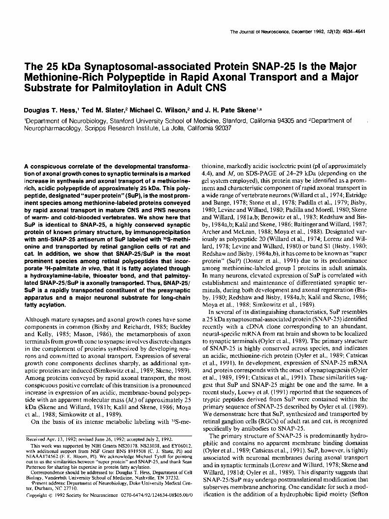
Results Identity between SUP and SNAP-25 In adult rats and cats, monocular injection of Y+methionine was employed to label proteins synthesized by RGCs and trans- ported through their axons in the ON to synaptic terminals in primary optic targets. In membrane samples derived from ON, LGN, and SC of both rat (Fig. 1) and cat (Fig. 2), SDS-PAGE revealed a prominent labeled polypeptide with A4, of approxi- mately 25 kDa. This polypeptide was precipitated by anti-SNAP- 25 antiserum (Oyler et al., 1989) but not by preimmune serum (Figs. 1, 2). The absence of the polypeptide in samples derived from the ON contralateral to the injected eye indicates that it is present as a result of axonal transport without a contribution from local synthesis, and its arrival in the SC contralateral to the injected eye as early as 3.5 hr following intravitreal admin- istration indicates that it is conveyed among group I proteins in the most rapid phase of transport (Fig. 1). The increase over time in the abundance of labeled polypeptide in the SC indicates that it accumulates in synapses formed by RGCs in primary optic targets (Fig. 1). The presence of substantial levels of labeled polypeptide in the ON as long as 33 hr following eye injection
4636 Hess et al. l SNAP-25: Identity with “Super Protein” and Fatty Acylation
ON contra ON ipsi SC contra TOtal Anti-SNAP-25 Preimm TOtal Anti-SNAP-25 Total Anti-SNAP-25
15h, 3.5 15 33 15 15 3.5 15 33 15 3.5 15 33
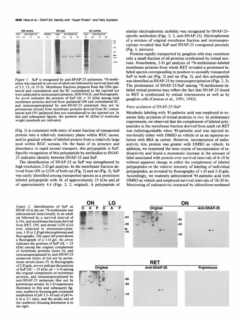
Figure 1. SUP is recognized by anti-SNAP-25 antiserum. %-methi- onine was injected in one eye of adult rats followed by survival intervals of 3.5, 15, or 33 hr. Membrane fractions prepared from the ONs ipsi- lateral and contralateral and the SC contralateral to the injected eye were subjected to immunoprecipitation, SDS-PAGE, and fluorography. An arrow indicates the position of SUP (M, = 25 kDa) among total membrane proteins derived from ipsilateral ON and contralateral SC, and immunoprecipitated by anti-SNAP-25 antiserum (but not by preimmune serum) from membrane proteins derived from SC contra- lateral and ON ipsilateral (but not contralateral) to the injected eye. In this and subsequent figures, the position and M, &Da) of molecular weight standards are indicated.
(Fig. 1) is consistent with entry of some fraction of transported protein into a relatively stationary phase within RGC axons, and/or gradual release of labeled protein from a relatively large pool within RGC somata. On the basis of its presence and abundance in rapid axonal transport, this polypeptide is SUP. Specific recognition of this polypeptide by antibodies to SNAP- 25 indicates identity between SNAP-25 and SUP.
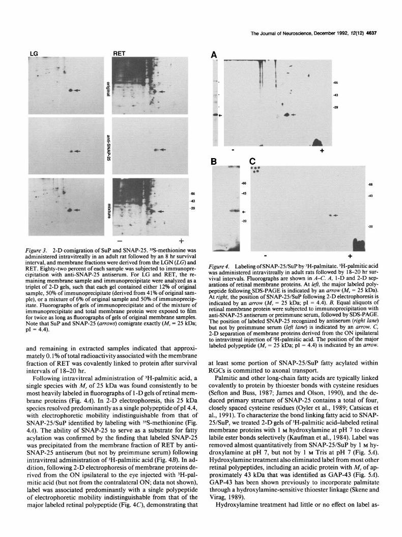
The identification of SNAP-25 as SUP was strengthened by high-resolution 2-D gel analysis. In the membrane fraction de- rived from ON or LGN of both cat (Fig. 2) and rat (Fig. 3), SUP was easily identified among transported species as a prominent labeled polypeptide with M, of approximately 25 kDa and p1 of approximately 4.4 (Figs. 2, 3, original). A polypeptide of
Figure 2. Identification of SUP as SNAP-25 in the cat. 35S-methionine was administered intravitreally in an adult cat followed by a survival interval of 6.5 hr, and membrane fractions derived from RET, ON, and dorsal LGN (LG) were subjected to immunoprecipita- tion, 1 -D or 2-D gel electrophoresis and fluorography. The upper left panel shows a fluorograph of a 1-D gel. An arrow indicates the position of SUP (M, = 25 kDa) among the original complement of membrane proteins (lanes 0), and immunoprecipitated by anti-SNAP-25 antiserum (lanes A) but not by preim- mune serum (lanes P). In fluorographs of 2-D gels, arrows indicate the position of SUP (M, = 25 kDa; p1 = 4.4) among the original complement of membrane proteins, and immunoprecipitated by anti-SNAP-25 antiserum (but not by preimmune serum). In 2-D separations illustrated in this and subsequent fig- ures, isoelectric focusing gels contained ampholines of pH 3.5-10 and of pH 4- 6 in a 2:l ratio, and the acidic end of the isoelectric focusing dimension is to the right.
ON LG OAPOAP
. . .
similar electrophoretic mobility was recognized by SNAP-25- specific antibodies (Figs. 2, 3, anti-SNAP-25). Electrophoresis of a mixture of original membrane fraction and immunopre- cipitate revealed that SUP and SNAP-25 comigrated precisely (Fig. 3, mixture).
Proteins rapidly transported by ganglion cells may constitute only a small fraction of all proteins synthesized by retinal neu- rons. Nonetheless, 2-D gel analysis of 35S-methionin+labeled membrane proteins from whole RET revealed a prominent la- beled species corresponding in position to axonally transported SUP in both cat (Fig. 2) and rat (Fig. 3), and this polypeptide was identified as SNAP-25 by immunoprecipitation (Figs. 2,3). The prominence of SNAP-25/SuP among 35S-methionine-la- beled retinal proteins may reflect the fact that SNAP-25 found in RET is synthesized by retinal interneurons as well as by ganglion cells (Catsicas et al., 199 1, 1992).
Fatty acylation of SNAP-25/&P
Metabolic labeling with 3H-palmitic acid was employed to ex- amine fatty acylation of retinal proteins in vivo. In preliminary experiments, we observed that the complement of labeled poly- peptides in the membrane fraction derived from adult rat RET was indistinguishable when 3H-palmitic acid was injected in- travitreally either with DMSO as vehicle or as an aqueous so- lution with BSA as carrier. However, incorporation of radio- activity into protein was greater with DMSO as vehicle. In addition, we examined the time course of incorporation of ra- dioactivity and found a monotonic increase in the amount of label associated with protein over survival intervals of 4-l 8 hr without apparent change in either the complement of labeled polypeptides or the relative intensity of labeling of individual polypeptides, as revealed by fluorography of 1 -D and 2-D gels. Accordingly, we routinely administered 3H-palmitic acid with DMSO as vehicle and employed survival intervals of 18-20 hr. Monitoring of radioactivity extracted by chloroform:methanol
Original ON
Anti-SNAP-25
Original RET
Anti-SNAP-25 Prqimmune
-66 -43
The Journal of Neuroscience, December 1992, 72(12) 4637
t
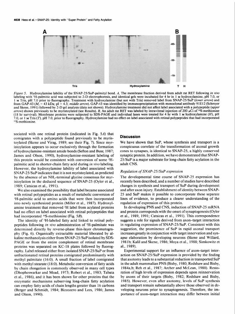
+ Figure 3. 2-D comigration of SUP and SNAP-25. 3SS-methionine was administered intravitreallv in an adult rat followed bv an 8 hr survival interval, and membrane fractions were derived from the LGN (LG) and RET. Eighty-two percent of each sample was subjected to immunopre- cinitation with anti-SNAP-25 antiserum. For LG and RET, the re- maining membrane sample and immunoprecipitate were analyzed as a triplet of 2-D gels, such that each gel contained either 12% of original sample, 50% of immunoprecipitate (derived from 4 1% of original sam- ple), or a mixture of 6% of original sample and 50% of immunoprecip- itate. Fluorographs of gels of immunoprecipitate and of the mixture of immunoprecipitate and total membrane protein were exposed to film for twice as long as fluorographs of gels of original membrane samples. Note that SUP and SNAP-25 (arrows) corn&ate exactly (M, = 25 kDa; PI = 4.4).
and remaining in extracted samples indicated that approxi- mately 0.1% of total radioactivity associated with the membrane fraction of RET was covalently linked to protein after survival intervals of 18-20 hr.
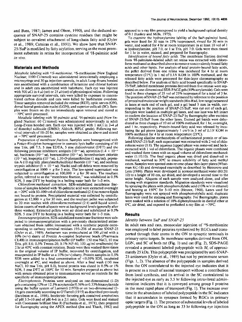
Following intravitreal administration of 3H-palmitic acid, a single species with M, of 25 kDa was found consistently to be most heavily labeled in fluorographs of 1 -D gels of retinal mem- brane proteins (Fig. 4A). In 2-D electrophoresis, this 25 kDa species resolved predominantly as a single polypeptide of p14.4, with electrophoretic mobility indistinguishable from that of SNAP-25/SuP identified by labeling with ?&methionine (Fig. 4A). The ability of SNAP-25 to serve as a substrate for fatty acylation was confirmed by the finding that labeled SNAP-25 was precipitated from the membrane fraction of RET by anti- SNAP-25 antiserum (but not by preimmune serum) following intravitreal administration of 3H-palmitic acid (Fig. 4B). In ad- dition, following 2-D electrophoresis of membrane proteins de- rived from the ON ipsilateral to the eye injected with ‘H-pal- mitic acid (but not from the contralateral ON, data not shown), label was associated predominantly with a single polypeptide of electrophoretic mobility indistinguishable from that of the major labeled retinal polypeptide (Fig. 4C), demonstrating that
*.
-c
B
m +
C ‘..
+
Figure 4. Labeling of SNAP-25/SuP by ‘H-palmitate. IH-palmitic acid was administered intravitreally in adult rats followed by 1 S-20 hr sur- vival intervals. Fluorographs are shown in A-C. A, 1-D and 2-D sep- arations of retinal membrane proteins. At left, the major labeled oolv- peptide following SDS-PAGE is indicated by an arrow (M, = 25 kD&. At riaht. the nosidon of SNAP-25/SuP following 2-D electropboresiq ip - ------- -- indi&ted by-an arrow (M, = 25 kDa; pI = 4.4). B, Equal aliquots of retinal membrane protein were subjected to immunoprecipitation with anti-SNAP-25 antiserum or preimmune serum, followed by SDS-PAGE. The position of labeled SNAP-25 recognized by antiserum (right lane) but not by preimmune serum (left lane) is indicated by an arrow. C, 2-D separation of membrane proteins derived from the ON ipsilateral to intravitreal injection of JH-palmitic acid. The position of the major labeled polypeptide (M, = 25 kDa; pI = 4.4) is indicated by an arrow.
at least some portion of SNAP-25/SuP fatty acylated within RGCs is committed to axonal transport.
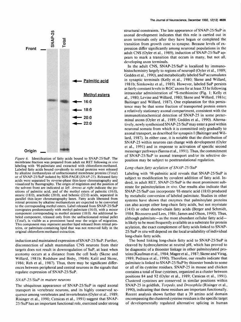
Palmitic and other long-chain fatty acids are typically linked covalently to protein by thioester bonds with cysteine residues (Sefton and Buss, 1987; James and Olson, 1990), and the de- duced primary structure of SNAP-25 contains a total of four, closely spaced cysteine residues (Oyler et al., 1989; Catsicas et al., 199 1). To characterize the bond linking fatty acid to SNAP- 25/SuP, we treated 2-D gels of 3H-palmitic acid-labeled retinal membrane proteins with 1 M hydroxylamine at pH 7 to cleave labile ester bonds selectively (Kaufman et al., 1984). Label was removed almost quantitatively from SNAP-25/SuP by 1 M hy- droxylamine at pH 7, but not by 1 M Tris at pH 7 (Fig. 5A). Hydroxylamine treatment also eliminated label from most other retinal polypeptides, including an acidic protein with M, of ap- proximately 43 kDa that was identified as GAP-43 (Fig. 5A). GAP-43 has been shown previously to incorporate palmitate through a hydroxylamine-sensitive thioester linkage (Skene and Virag, 1989).
Hydroxylamine treatment had little or no effect on label as-
4638 Hess et al. l SNAP-25: Identity with “Super Protein” and Fatty Acylation
Tris Hydroxylamine T H
Figure 5. Hydroxylamine lability of the SNAP-25/SuP-palmityl bond. A, The membrane fraction derived from adult rat RET following in viva labeling with ‘H-palmitic acid was subjected to 2-D electrophoresis, and identical gels were incubated for 4 hr in 1 M hydroxylamine, pH 7.0, or 1 M Tris, pH 7.0 prior to fluorography. Treatment with hydroxylamine (but not with Tris) removed label from SNAP-25/SuP (lower arrow) and from GAP-43 (M, = 43 kJ3a; p1 = 4.3; middle arrow). GAP-43 was identified by immunoprecipitation with monoclonal antibody 91E12 (Schreyer and Skene, 199 1) followed by 2-D gel analysis (data not shown). Hydroxylamine treatment did not affect label associated with a polypeptide (upper arrow) shown previously to be myristoylated (see Results). B, An adult rat RET was labeled by intravitreal injection of 200 &i of %-methionine (18 hr survival). Membrane proteins were subjected to SDS-PAGE and individual lanes were treated for 4 hr with 1 M hydroxylamine (H), pH 7.0, or 1 M Tris (n, pH 7.0, prior to fluorography. Hydroxylamine had no effect on label associated with retina1 polypeptides that had incorporated %-methionine.
sociated with one retinal protein (indicated in Fig. SA) that comigrates with a polypeptide found previously to be myris- toylated (Skene and Virag, 1989; see their Fig. 7). Since myr- istoylation appears to occur exclusively through the formation of hydroxylamine-resistant amide bonds (Sefton and Buss; 1987; James and Olson, 1990), hydroxylamine-resistant labeling of this protein would be consistent with conversion of some 3H- palmitic acid to shorter-chain fatty acid during in vivo labeling. However, the hydroxylamine lability of label associated with SNAP-2YSuP indicates that it is not myristoylated, as predicted by the absence of an NH,-terminal glycine consensus for myr- istoylation in the deduced sequence of SNAP-25 (Oyler et al., 1989; Catsicas et al., 199 1).
We also examined the possibility that label became associated with retinal polypeptides as a result of metabolic conversion of 3H-palmitic acid to amino acids that were then incorporated into newly synthesized protein (Miller et al., 1987). Hydroxyl- amine treatment that removed 3H label from acylated proteins had no effect on label associated with retinal polypeptides that had incorporated 35S-methionine (Fig. 5B).
The identity of 3H-labeled fatty acid linked to retinal poly- peptides following in vivo administration of 3H-palmitate was determined directly by reverse-phase thin-layer chromatogra- phy (Fig. 6). Organically extractable material liberated by al- kaline methanolysis either from SNAP-2YSuP isolated by SDS- PAGE or from the entire complement of retinal membrane proteins was separated on KC-18 plates followed by fluorog- raphy. Label released either from isolated SNAP-25/SuP or from unfractionated retinal proteins corn&rated predominantly with methyl palmitate (l&O). A small fraction of label comigrated with methyl stearate (18:O). Metabolic conversion of fatty acids by chain elongation is commonly observed in many cell types (Dhopheswarkar and Mead, 1973; Robert et al., 1983; Tabata et al., 1986), and it has been shown for other proteins that the enzymatic mechanisms subserving long-chain fatty acylation can employ fatty acids of chain lengths greater than 16 carbons (Berger and Schmidt, 1984; Bizzozero and Lees, 1986; James and Olson, 1990).
Discussion We have shown that SUP, whose synthesis and transport is a conspicuous correlate of the transformation of axonal growth cones to synapses, is identical to SNAP-25, a highly conserved synaptic protein. In addition, we have demonstrated that SNAP- 25/SuP is a major substrate for long-chain fatty acylation in the adult CNS.
Regulation of SNAP-25/&P expression The developmental time course of SNAP-25 expression has recently been described, and a number of studies have described changes in synthesis and transport of SUP during development and after axon injury. Establishment of identity between SNAP- 25 and SUP makes it possible to concorporate these separate lines of evidence, to produce a clearer understanding of the regulation of expression of this protein.
In developing PNS and CNS, induction of SNAP-25 mRNA and protein corresponds with the onset of synaptogenesis (Oyler et al., 1989, 1991; Catsicas et al., 1991). This correspondence suggests a role for signals derived from axon-target interaction in regulating expression of SNAP-25/SuP. Consistent with this suggestion, the prominence of SUP in rapid axonal transport increases greatly in conjunction with target innervation and syn- apse elaboration by developing neurons (Skene and Willard, 1981b; Kalil and Skene, 1986; Moya et al., 1988; Simkowitz et al., 1989).
Experimental support for an influence of axon-target inter- action on SNAP-25/SuP expression is provided by the finding that axotomy leads to a substantial reduction in transported SUP in the adult mammalian PNS (Bisby, 1980; Redshaw and Bisby, 1984a,b; Reh et al., 1987; Archer and McLean, 1988). Resto- ration of high levels of expression depends upon reinnervation by axons of their targets (Bisby, 1982; Redshaw and Bisby, 1985). However, even after axotomy, levels of SUP synthesis and transport remain substantially above those observed in de- veloping neurons prior to synaptogenesis. Therefore, the im- portance of axon-target interaction may differ between initial
The Journal of Neuroscience, December 1992, 12(12) 4639
4- Palmitic acid
Methvl esters
4 16:0
+ 18:0
4 20:o
4 22:o
Origin I
Figure 6. Identification of fatty acids bound to SNAP-25/SuP. The membrane fraction was prepared from adult rat RET following in vivo labeling with ‘H-palmitate and extracted with chloroform:methanol. Labeled fatty acids bound covalently to retinal proteins were released by alkaline methanolysis of unfractionated membrane proteins (Total) or of SNAP-2YSuP isolated by SDS-PAGE (SNAP-25). Released fatty acids were separated by reverse-phase thin-layer chromatography and visualized by fluorography. The origin of migration and the position of the solvent front are indicated at left. Arrows at right indicate the po- sitions of palmitic acid, and of the methyl esters of palmitic (16:0), stearic (l&O), arachidic (20:0), and behenic (22:0) acids, separated in parallel thin-layer chromatography lanes. Fatty acids liberated from retinal proteins by alkaline methanolysis are expected to be converted to the corresponding methyl esters. Label released from SNAP-25/SuP comigrates predominantly with methyl palmitate (16:0), with a minor component corresponding to methyl stearate (18:O). An additional la- beled component, released only from the unfractionated retinal pellet (Total), is visible as a prominent band near the origin of migration. This component may represent another lipid released from retinal pro- teins, or palmitate-containing lipid that was not removed fully in the original chlorofornnmethanol extraction.
induction and maintained expression of SNAP-25/SuP. Further, disconnection of adult mammalian CNS neurons from their targets does not result in downregulation of SUP, at least when axotomy occurs at a distance from the cell body (Skene and Willard, 198 lb; Redshaw and Bisby, 1984b; Kalil and Skene, 1986; Reh et al., 1987). Thus, there may be significant differ- ences between peripheral and central neurons in the signals that regulate expression of SNAP-2YSuP.
SNAP-25/SuP in mature neurons
The ubiquitous appearance of SNAP-2YSuP in rapid axonal transport in vertebrate neurons, and its highly conserved se- quence among vertebrates and invertebrates (Oyler et al., 1989; Risinger et al., 1990; Catsicas et al., 1991) suggest that SNAP- 25/SuP has an important functional role, exercised under strong
structural constraints. The late appearance of SNAP-25/SuP in axonal development indicates that this role is carried out in axon terminals only after they have begun or completed the transition from growth cone to synapse. Because levels of ex- pression differ significantly among neuronal populations in the adult CNS (Oyler et al., 1989) induction of SNAP-25/SuP ap- pears to mark a transition that occurs in many, but not all, developing axon terminals.
In the adult CNS, SNAP-25/SuP is localized by immuno- histochemistry largely to regions of neuropil (Oyler et al., 1989; Geddes et al., 1990), and metabolically labeled SUP accumulates in synaptic terminals (Kelly et al., 1980; Skene and Willard, 198 lb; Simkowitz et al., 1989). However, labeled SUP persists at fairly constant levels in RGC axons for at least 33 hr following intraocular administration of %-methionine (Fig. 1; Kelly et al., 1980; Levine and Willard, 1980; Skene and Willard, 198 lc; Baitinger and Willard, 1987). One explanation for this persis- tence may be that some fraction of transported protein enters a relatively stationary axonal compartment, consistent with the immunohistochemical detection of SNAP-25 in some preter- minal axons (Oyler et al., 1989; Geddes et al., 1990). Altema- tively, newly synthesized SNAP-25/SuP may enter a pool within neuronal somata from which it is committed only gradually to axonal transport, as described for synapsin I (Baitinger and Wil- lard, 1987). In either case, it is notable that the distribution of SNAP-25 within neurons can change with development (Oyler et al., 1991) and in response to activation of specific second messenger pathways (Sanna et al., 199 1). Thus, the commitment of SNAP-25/SuP to axonal transport and/or its selective de- position may be subject to posttranslational regulation.
Long-chain fatty acylation of SNAP-25/SuP
Labeling with 3H-palmitic acid reveals that SNAP-2YSuP is subject to modification by covalent addition of fatty acid. In- deed, in adult RET, SNAP-25/SuP is the most prominent sub- strate for palmitoylation in vivo. Our results also indicate that SNAP-25/SuP can incorporate 3H-stearic acid (18:O) produced by metabolic conversion of labeled palmitate. Studies in other systems have shown that enzymes that palmitoylate proteins can also accept other long-chain fatty acids, but not myristate (14:0) or other shorter-chain fatty acids (Berger and Schmidt, 1984; Bizzozero and Lees, 1986; James and Olson, 1990). Thus, although palmitate-as the most abundant cellular fatty acid- is likely to be most frequently available as a substrate for protein acylation, the exact complement of fatty acids linked to SNAP- 25/SuP in situ will depend on the local availability of individual long-chain fatty acids.
The bond linking long-chain fatty acid to SNAP-25/SuP is cleaved by hydroxylamine at neutral pH, which has proved to be diagnostic of a thioester linkage in other palmitoylated pro- teins (Kaufman et al., 1984; Magee et al., 1987; Skene and Virag, 1989; Pedraza et al., 1990). Therefore, our results indicate that palmitate is linked to SNAP-25/SuP by thioester bonds to some or all of its cysteine residues. SNAP-25 in mouse and chicken contains a total of four cysteines, organized as a cluster between positions 84 and 92 (Oyler et al., 1989; Catsicas et al., 1991). Clustered cysteines are conserved in similar positions within SNAP-25 in goldfish, Torpedo, and Drosophila (Risinger et al., 1990) indicating that these residues are important functionally. Recent analysis shows further that a 39 amino acid domain encompassing the clustered cysteine residues is the specific target of developmentally regulated alternative splicing in human
4640 Hess et al. * SNAP-25: Identity with “Super Protein” and Fatty Acylation
SNAP-25 (Bark and Wilson, 199 1). Thus, the domain of SNAP- 25&P that is the probable locus of posttranslational modifi- cation by long-chain fatty acid may have particular regulatory significance.
Functional implications offatty acylation
The membrane affinity of a number of eukaryotic proteins is altered by posttranslational, covalent addition of one or more lipid moieties (Sefton and Buss, 1987; James and Olson, 1990). Long-chain fatty acylation in particular contributes to the mem- brane association of some members of the ras superfamily of small G-proteins (Willumsen et al., 1984; Magee et al., 1987; Hancock et al., 1989, 1990), and is required for membrane attachment by the neural-specific growth cone protein GAP-43 (Skene and Virag, 1989; Zuber et el., 1989; Liu et al., 1991).
The association of SNAP-25/SuP with the membrane com- partment of axons and synapses is disrupted by detergents, but not by treatments known to release only peripheral proteins (Lorenz and Willard, 1978; Skene and Willard, 198 Id; Oyler et al., 1989). The primary structure of SNAP-25/SuP, however, is unusually hydrophilic and lacks transmembrane or other strong- ly hydrophobic domains, although a moderately hydrophobic domain may emerge through formation of an amphiphilic helix at the amino terminus (Oyler et al., 1989). Covalent attachment of one or more long-chain fatty acid moieties to the clustered cysteine residues of SNAP-2YSuP would establish a strongly hydrophobic domain that could mediate or substantially en- hance the interaction of this hydrophilic protein with neuronal membranes. In this respect, SNAP-25/SuP resembles GAP-43, which is predominantly hydrophilic but tightly membrane as- sociated by virtue of palmitoylation (Skene and Virag, 1989; Zuber at al., 1989; Liu et al., 199 1). If the hydrophobic domain created by fatty acylation anchors SNAP-25/SuP to the mem- brane, then more hydrophilic regions of the molecule would likely remain exposed to the cytoplasmic environment, facili- tating interaction with cytoskeletal or other cytoplasmic ele- ments.
SNAP-25/SuP en route to synaptic terminals, like other group I proteins, is conveyed in association with vesicular or other membranes (Lorenz and Willard, 1978; Grafstein and Forman, 1980). The appearance of labeled SNAP-25/SuP in the ON following intravitreal administration of 3H-palmitic acid indi- cates that the protein is fatty acylated in ganglion cells prior to its commitment to axonal transport. Thus, fatty acylation might either directly mediate or enhance the association of SNAP-25/ SUP with membranous structures targeted for rapid axonal transport. It has recently been shown that site-directed muta- genesis that eliminates palmitoylation of another group I pro- tein, GAP-43, attenuates its delivery into growing neurites and prevents its localization to growth cones (Liu et al., 199 1). Long- chain fatty acylation may play similar roles in modulating the commitment of SNAP-25/SuP to rapid axonal transport and its selective accumulation in synaptic terminals.
References Archer DR, McLean GW (1988) Changes in fast axonally transported
proteins in the regenerating rabbit vagus nerve. Neurosci Lett 87: 15 l- 156.
Baitinger C, Willard M (1987) Axonal transport of synapsin I-like pro&ins in rabbit retinal ganglion cells. J Neurosci 7:373%3735.
Bark C. Wilson MC 1199 1) Alternative snlicina generates a variant SNAP-25 protein d&ng development. Sic Ne&sci Abstr 17:53 1.
Benowitz LI, Lewis ER ( 1983) Increased transport of 44,000- to 49,000-
dalton acidic proteins during regeneration of the goldfish optic nerve: a two-dimensional gel analysis. J Neurosci 3:2 153-2163.
Berger M, Schmidt MFG (1984) Cell-free fatty acid acylation of Sem- liki Forest viral polypeptides with microsomal membranes from eu- karyotic cells. J Biol Chem 259:7245-7252.
Bisby MA ( 1980) Changes in the composition of labelled protein trans- ported in motor axons during their regeneration. J Neurobiol 11:345- 360.
Bisby MA (1982) Prolonged alteration in composition of fast-trans- ported proteins in axons prevented from regenerating after injury. J Neurobiol 13:377-381.
Bixby JL, Reichardt LF (1985) The expression and localization of synaptic vesicle antigens at neuromuscular junctions in vitro. J Neu- rosci 5:3070-3080.
Bizzozero OA, Lees MB (1986) Fatty acid acylation of brain myelin proteolipid protein in vitro: identification of the lipid donor. J Neu- rochem 46:630-636.
Buckley K, Kelly RB (1985) Identification of a transmembrane gly- coprotein specific for secretory vesicles of neural and endocrine cells. J Cell Biol 100: 1284-l 294.
Catsicas S, Larhammar D, Blomqvist A, Sanna PP, Milne RJ, Wilson MC (199 1) Expression of a conserved cell-type-specific protein in nerve terminals coincides with synaptogenesis. Proc Nat1 Acad Sci USA 88:785-789.
Catsicas S, Catsicas M, Keyser KT, Karten HJ, Wilson MC, Milner RJ (1992) Differential expression of the presynaptic protein SNAP-25 in mammalian retina. J Neurosci Res, in press.
Dhopheswarkar GA, Mead JF (1973) Uptake and transport of fatty acids into the brain and the role of the blood-brain barrier. Adv Lipid Res 11:109-142.
Doster SK, Lozano AM, Aguayo AJ, Willard MB (199 1) Expression of the growth-associated protein GAP-43 in adult rat retinal ganglion cells following axon injury. Neuron 6:635447.
Estridne M, Bunge R ( 1978) Compositional analysis of growing axons from rat.sympathet& neuions. J-Cell Biol 793138-l 551 -
Fairbanks G. Steck TL. Wallach DFH ( 197 1) Electroohoretic analvsis of the ma&r polvpedtides of the hum‘an e&hrocytemembrane. I&o- chemistry 10:26bb-26 17.
_ _
Geddes JW. Hess EJ. Hart RA. Kesslak JP. Cotman CW. Wilson MC (1990, L&ions ofhippocambal circuiiry’define synaptbsomal-asso- ciated protein-25 as a novel presynaptic marker. Neuroscience 38: 515-525.
Grafstein B, Forman DS (1980) Intracellular transport in neurons. Physiol Rev 60:1167-1283.
Hancock JF, Magee AI, Childs JE, Marshall CJ (1989) All rus proteins are polyisoprenylated but only some are palmitoylated. Cell 57: 1167- 1177.
Hancock JF, Paterson H, Marshall CJ (1990) A polybasic domain or palmitoylation is required in addition to the CAAX motif to localize p2lras to the plasma membrane. Cell 63:133-139.
Heuser D (1968) Thin layer chromatography of fatty acids on Silanised silica gel. J Chromatogr 33:62-69.
Jacobson RD, Virag I, Skene JHP (1986) A protein associated with axon growth, GAP-43, is widely distributed and developmentally regulated in rat CNS. J Neurosci 6: 1843-1855.
James G, Olson EN (1990) Fatty acylated proteins as components of intracellular signaling pathways. Biochemistry 29:2623-2634.
Jen G, Thach RE (1982) Inhibition of host translation in encephal- omyocarditis virus-infected L cells: a novel mechanism. J Virol 43: 250-261.
Kalil K, Skene JHP (1986) Elevated synthesis of an axonally trans- ported protein correlates with axon outgrowth in normal and injured pyramidal tracts. J Neurosci 6:2563-2570.
Kaufman JF, Krangel MS, Strominger JL (1984) Cysteines in the transmembrane region of major histocompatibility complex antigens are fatty acylated via thioester bonds. J Biol Chem 259:723&7238.
Kelly AM, Wagner JA, Kelly RB (1980) Properties of individual nerve terminal proteins identified by two-dimensional gel electrophoresis. Brain Res 185:192-197.
Laemmli UK (1970) Cleavage of structural proteins during the assem- bly of the head of bacteriophage T4. Nature 227:680-685.
Laskey RA, Mills AD (1975) Quantitative film detection of ‘H and 14C in polyacrylamide gels by fluorography. Eur J Biochem 56:335- 341.
Levine J, Willard MB (1980) The composition and organization of
The Journal of Neuroscience, December 1992. 72(12) 4641
axonally transported proteins in the retinal ganglion cells of the guinea pig. Brain Res 194: 137-154.
Liu Y, Chapman ER, Storm DR (1991) Targeting of neuromodulin (GAP-43) fusion proteins to growth cones in cultured rat embryonic neurons. Neuron 6:4 1 l-420.
Loewy A, Liu W-S, Baitinger C, Willard MB (1991) The major %- methionine-labeled rapidly transported protein (superprotein) is iden- tical to SNAP-25 a protein of synaptic terminals. J Neurosci 11: 3412-3421.
Lorenz T, Willard M (1978) Subcellular fractionation ofintra-axonally transported polypeptides in the rabbit visual system. Proc Nat1 Acad Sci USA 75505-509.
Magee AI, Gutierrez L, McKay IA, Marshall CJ, Hall A (1987) Dy- namic fatty acylation of p21N--. EMBO J 6:3353-3357.
Mason CA (1986) Axon development in mouse cerebellum: embry- onic axon forms and expression of synapsin I. Neuroscience 19: 13 19- 1333.
Miller JC, Gnaedinger JM, Rapoport SI (1987) Utilization of plasma fatty acid in rat brain: distribution of F4Clpalmitate between oxidative and synthetic pathways. J Neurochem 49: 150 l-l 5 14.
Mova KL. Benowitz LI. Jhaveri S. Schneider GE (1988) Chanees in \ , rapidly transported proteins in developing hamster retinofugal axons. J Neurosci 8:44454454.
O’Farrell PH (1975) High-resolution two-dimensional electrophoresis of proteins. J Biol Chem 250:4007-402 1.
Oyler GA, Higgins GA, Hart RA, Battenberg E, Billingsley M, Bloom FE, Wilson MC (1989) The identification of a novel synaptosomal- associated protein, SNAP-25, differentially expressed by neuronal subpopulations. J Cell Biol 109:3039-3052.
Oyler GA, Polli JW, Wilson MC, Billingsley ML (1991) Develop- mental expression of the 25-kDa synaptosomal-associated protein (SNAP-25) in rat brain. Proc Nat1 Acad Sci USA 88:5247-525 1.
Padilla SS, Morel1 P (1980) Axonal transport of 35S-methionine-la- beled proteins in two intra-brain tracts of the rat. J Neurochem 35: 436-443.
Padilla SS, Roger LJ, Toews AD, Goodrum JF, Morel1 P (1979) Com- parison of proteins transported in different tracts of the central ner- vous system. Brain Res 176:407411.
Pedraza L, Owens GC, Green LAD, Salzer JL (1990) The myelin- associated glycoproteins: membrane disposition, evidence of a novel disulfide linkage between immunoglobulin-like domains, and post- translational palmitylation. J Cell Biol 111:265 l-266 1.
Redshaw JD, Bisby MA (1984a) Proteins of fast axonal transport in the regenerating hypoglossal nerve ofthe rat. Can J Physiol Pharmacol 62:1387-1393.
Redshaw JD, Bisby MA (1984b) Fast axonal transport in central ner- vous system and peripheral nervous system axons following axotomy. J Neurobiol 15: 109-l 18.
Redshaw JD, Bisby MA (1985) Comparison of the effects of sciatic nerve crush or resection on the proteins of fast axonal transport in rat dorsal root ganglion cell axons. Exp Neurol 88:437446.
Reh TA, Redshaw JD, Bisby MA (1987) Axons of the pyramidal tract do not increase their transport of growth-associated proteins after axotomy. Mol Brain Res 2: l-6.
Risinger C, Blomqvist AG, Lundell I, Catsicas S, Wilson MC, Iarham- mar D (1990) Strong evolutionary conservation of SNAP-25 be- tween Drosophila, goldfish, chicken and mouse. Sot Neurosci Abstr 16:355.
Robert J, Montaudon D, Hugues P (1983) Incorporation and metab- olism of exogenous fatty acids by cultured normal and tumoral glial cells. Biochem Biophys Acta 752:383-395.
Sanna PP, Bloom FE, Wilson MC (199 1) Dibutyryl-CAMP induces SNAP-25 translocation into the neurites of PC12 cells. Dev Brain Res 59: 104-108.
Schreyer DJ, Skene JHP (199 1) Fate of GAP-43 in ascending spinal axons of DRG neurons after peripheral nerve injury: delayed accu- mulation and correlation with regenerative potential. J Neurosci 11: 3738-3751.
Sefton BM, Buss JE (1987) The covalent modification of eukaryotic proteins with lipid. J Cell Biol 104: 1449-l 453.
Simkowitz P, Ellis L, Pfenninger KH (1989) Membrane proteins of the nerve growth cone and their developmental regulation. J Neurosci 9:1004-1017.
Skene JHP (1989) Axonal growth-associated proteins. Annu Rev Neu- rosci 12:127-156.
Skene JHP, Virag I (1989) Posttranslational membrane attachment and dynamic fatty acylation of a neuronal growth cone protein, GAP- 43. J Cell Biol 108:613-624.
Skene JHP, Willard M ( 198 1 a) Changes in axonally transported pro- teins during axon regeneration in toad retinal ganglion cells. J Cell Biol 89:86-95.
Skene JHP, Willard M (198 1 b) Axonally transported proteins asso- ciated with axon growth in rabbit central and peripheral nervous systems. J Cell Biol 89:96-103.
Skene JHP, Willard M (1981~) Electrophoretic analysis of axonally transported proteins in toad retinal ganglion cells. J Neurochem 37: 79-87.
Skene JHP, Willard M (198 Id) Characteristics of growth-associated polypeptides in regenerating toad retinal ganglion cell axons. J Neu- rosci 1:419-426.
Stone GC, Wilson PL, Hall ME (1978) Two-dimensional gel electro- phoresis of proteins in rapid axoplasmic transport. Brain Res 144: 287-302.
Tabata H, Bell JM, Miller JC, Rapoport SI (1986) Incorporation of plasma palmitate into the brain of the rat during development, Dev Brain Res 29: l-8.
Willard M, Cowan WM, Vagelos PR (1974) The polypeptide com- position of intra-axonally transported proteins: evidence for four transport velocities. Proc Nat1 Acad Sci USA 7 1:2 183-2 187.
Willumsen BM, Norris K, Papageorge AG, Hubbert NL, Lowy DR (1984) Harvey murine sarcoma virus p2 1 ras protein: biological and biochemical significance of the cysteine nearest the carboxy terminus. EMBO J 3:258 l-2585.
Zuber MX, Strittmatter SM, Fishman MC (1989) A membrane-tar- geting signal in the amino terminus of the neuronal protein GAP-43. Nature 341:345-348.