Embed Size (px)
Citation preview
SPECIAL ARTICLE
The 2005 International Society of Urological Pathology(ISUP) Consensus Conference on Gleason Grading
of Prostatic Carcinoma
Jonathan I. Epstein, MD,* William C. Allsbrook, Jr, MD,† Mahul B. Amin, MD,‡
and Lars L. Egevad, MD, PhD,§ and the ISUP Grading Committeek
Donald F. Gleason in 1966 created a unique grading systemfor prostatic carcinoma based solely on the architectural
pattern of the tumor.3,14,22 Another innovative aspect of this sys-tem was, rather than assigning the worst grade as the grade ofthe carcinoma, the grade was defined as the sum of the twomost common grade patterns and reported as the Gleasonscore. The original description of this system, based on a studyof 270 patients from the Minneapolis Veterans AdministrationHospital, is seen in Table 1.
Initially, Gleason intended to classify carcinomas intofour patterns, but a small group of distinctive tumors (clear cell)was observed and they were placed in a separate fifth category(pattern 4).14 Certain aspects of the original Gleason systemwould be interpreted differently in today’s practice. The crib-riform pattern described as a component of Gleason’s originalpatterns 2 and 3 would today typically be considered highergrade. Individual cells listed under Gleason’s original pattern 3would also be currently assigned a higher grade. Pattern 4 hasbecome significantly expanded beyond Gleason’s original de-scription of tumors with clear cytoplasm that resembled renalcell carcinoma (Table 1).
By 1974, Gleason and theVeteransAdministrationCoop-erative Urological Research Group expanded their study to
1032 men.15 Gleason pattern 4 was described in a figurelegend as ‘‘raggedly infiltrating, fused-glandular tumor, fre-quently with pale cells, may resemble hypernephroma of kid-ney.’’ The Gleason system was further refined by Mellinger in1977 when the papillary and cribriform tumor under Gleasonpattern 3 was described as having a ‘‘smooth and usuallyrounded edge.’’23 These modifications of the Gleason system aredepicted in Table 1. In describing the breakdown of Gleasonpatterns among 2911 cases, Gleason pattern 1 was seen in3.5%; pattern 2 in 24.4%; pattern 3 in 87.7%; pattern 4 in12.1%; and pattern 5 in 22.6%.23 These percentages added upto approximately 150% because 50% of the tumors showed atleast two different patterns.
In 1977, Gleason provided additional comments con-cerning the application of the Gleason system.16 ‘‘Grading isperformed under low magnification (40–1003).’’ He alsostated ‘‘an occasional small area of fused glands did notchange a pattern 3 tumor to pattern 4. A small focus of dis-organized cells did not change a pattern 3 or 4 tumor to pattern5.’’ The only comment relating to tertiary patterns was ‘‘oc-casionally, small areas of a third pattern were observed.’’
WHY THE NEED FOR A CONSENSUS ONGLEASON GRADING?
It is a testament to the enduring power of the originalGleason grading system that it is the accepted grading systemthroughout the world, despite its inception almost 40 yearsago. How many other things in medicine have stood the test oftime so well? Nonetheless, medicine in general and prostatecarcinoma in specific has changed dramatically since the late1960s, when the Gleason grading system was derived. In the1960s, there was no screening for prostate cancer other than bydigital rectal examination, as serum PSA had not yet been dis-covered. In Gleason’s 1974 study, most (86%) of the men hadadvanced disease with either local extension out of the prostateon clinical examination or distant metastases. Only 6% of pa-tients had nonpalpable tumor diagnosed by transurethralresection and 8% of patients were diagnosed with a localizednodule on rectal examination.15 The method of obtainingprostate tissue was also very different from today’s practice.Typically, only a couple of thick-gauge needle biopsies weredirected into an area of palpable abnormality. The use of 18-gauge thin biopsy needles and the concept of sextant needlebiopsies to more extensively sample the prostate were notdeveloped until the 1980s.17 Consequently, the grading of
From the *Department of Pathology, Urology and Oncology, Johns HopkinsHospital, Baltimore, MD; †Departments of Pathology & Surgery(Urology), Medical College of Georgia, Augusta, GA; ‡Department ofPathology and Laboratory Medicine, Urology, Hematology & Oncology,Emory University School of Medicine, Atlanta, GA; and §Department ofPathology and Cytology, Karolinska Hospital, Stockholm, Sweden.
kSheldon Bastacky, USA; Antonio Lopez Beltran, Spain; Aasmund Berner,Norway; Athanase Billis, Brazil; Liliane Boccon-Gibod, France; LiangCheng, USA; Francisco Civantos, USA; Cynthia Cohen, USA; Michael B.Cohen, USA; Milton Datta, USA; Charles Davis, USA; Brett Delahunt,New Zealand; Warick Delprado, Australia; John N. Eble, USA;Christopher S. Foster, UK; Masakuni Furusato, Japan; Paul B. Gaudin,USA; David J. Grignon, USA; Peter A. Humphrey, USA; Kenneth A.Iczkowski, USA; Edward C. Jones, Canada; Scott Lucia, USA; Peter A.McCue, USA; Tipu Nazeer, USA; Esther Oliva, USA; Chin-Chen Pan,Taiwan; Galina Pizov, Israel; Victor Reuter, USA; Hemamali Samar-atunga, Australia; Thomas Sebo, USA; Isabell Sesterhenn, USA; MariaShevchuk, USA; John R. Srigley, Canada; Sueli Suzigan, Brazil; HiroyukiTakahashi, Japan; Pheroze Tamboli, USA; Puay Hoon Tan, Singapore;Bernard Tetu, Canada; Satish Tickoo, USA; John E. Tomaszewski, USA;Patricia Troncoso, USA; Toyonori Tsuzuki, Japan; Lawrence D. True,USA; Theo van der Kwast, Canada; Thomas M. Wheeler, USA; Kirk J.Wojno, USA; Robert H. Young, USA.
Reprints: Jonathan I. Epstein, MD, 401 N. Broadway Street, Johns HopkinsHospital, Weinberg Building, Rm 2242, Baltimore, MD 21231 (e-mail:[email protected]).
Copyright � 2005 by Lippincott Williams & Wilkins
1228 Am J Surg Pathol � Volume 29, Number 9, September 2005
prostate cancer in thin cores and in multiple cores from dif-ferent sites of the prostate were not issues in Gleason’s era.
In the 1960s, radical prostatectomy was relatively un-common, prostates were not as often removed intact, andglands were not processed in their entirety or as extensivelyand systematically to the degree currently seen. Further issuesrelating to radical prostatectomy specimens such as the grad-ing of multiple nodules within the same prostate or dealingwith tertiary patterns were not addressed within the originalGleason system.
The Gleason system also predated the use of immuno-histochemistry. It is likely that with immunostaining for basalcells many of Gleason’s original 1 + 1 = 2 adenocarcinomas ofthe prostate would today be regarded as adenosis (atypicaladenomatous hyperplasia). Similarly, many of the cases in1967 diagnosed as cribriform Gleason pattern 3 carcinomawould probably be currently referred to as cribriform highgrade prostatic intraepithelial neoplasia, if labeled with basalcell markers.1
Another issue not dealt with in the original Gleason grad-ing system is how to grade newly described variants of adeno-carcinoma of the prostate. Some of the more common variantswhere grading controversy exists include: mucinous carci-noma, ductal adenocarcinoma, foamy gland carcinoma, andpseudohyperplastic adenocarcinoma of the prostate. In addition,there are certain patterns of adenocarcinoma of the prostatesuch as thosewith glomeruloid features andmucinous fibroplasia(collagenous micronodules) where the use of Gleason gradingwas not defined.
The application of the Gleason system for all of thereasons noted above varies considerably in contemporary sur-gical pathology practice and has led to several recent attemptsto achieve consensus on Gleason grading.
RECENT WORLD HEALTH ORGANIZATIONCONSENSUS STATEMENTS ON THE
GLEASON SYSTEMThe 2004 World Health Organization Classification of
Tumors: Pathology and Genetics: Tumors of the Urinary
System and the Male Genital Organs summarized the currentstate of Gleason grading.12 A follow-up international con-sensus meeting on ‘‘International Consultation on Predictorsof Patient Outcome in Prostate Cancer’’ sponsored by theWorld Health Organization took place in 2004 in Stockholm,Sweden.2,13 Although both of these meetings provided acurrent analysis of Gleason system, they were restricted to arelatively limited number of participants for both financial andlogistical reasons. In an attempt to elicit input from a greaterrepresentation of the urologic pathology community, a surveyon Gleason grading was sent to 91 pathologists with 67 re-spondents.9 Although the survey provided useful informationas to the worldwide practice of the Gleason grading systemamong urologic pathologists, certain questions were ambig-uous and were interpreted differently by different respondents.In addition, the survey provided unidirectional responses toquestions, not allowing for back-and-forth discussions ofcontroversial areas.
2005 ISUP CONSENSUS CONFERENCEThe authors of the Gleason grading survey convened
a group of urologic pathologists at the 2005 United States andCanadian Academy meeting in San Antonio in an attempt toachieve consensus in controversial areas relating to theGleason grading system. The goal of the meeting was toachieve consensus among leading urologic pathologists inspecific areas of Gleason grading, including areas where thereis currently either a lack of data or scant information as to theoptimal method of grading. In the latter instances, the con-sensus was based on personal and institutional experience witha large number of cases. More than 70 urologic pathologistswere invited to attend, with most not in attendance having aconflict in their schedule. Only one invitee declined par-ticipation, stating that the Gleason system in its original formshould not be altered and hence there was no need for aconsensus conference. For the purposes of this meeting, wedefined ‘‘consensus’’ when two thirds of the participants werein agreement, although for almost all of the issues discusseda much higher degree of agreement was reached. With rare
TABLE 1. Gleason System
Original Gleason System: 1966 & 1967
Pattern 1: Very well differentiated, small, closely packed, uniform, glands in essentially circumscribed masses
Pattern 2: Similar (to pattern 1) but with moderate variation in size and shape of glands and more atypia in the individual cells; cribriform pattern may be present,still essentially circumscribed, but more loosely arranged
Pattern 3: Similar to pattern 2 but marked irregularity in size and shape of glands, with tiny glands or individual cells invading stroma away from circumscribedmasses, or solid cords and masses with easily identifiable glandular differentiation within most of them
Pattern 4: Large clear cells growing in a diffuse pattern resembling hypernephroma; may show gland formation
Pattern 5: Very poorly differentiated tumors; usually solid masses or diffuse growth with little or no differentiation into glands
Gleason’s Modifications: 1974 & 1977
Patterns 1 & 2: Unchanged
Pattern 3: Adds to earlier description: may be papillary or cribriform (1974), which vary in size and may be quite large, but the essential feature is the smooth andusually rounded edge around all the circumscribed masses of tumor (1977)
Pattern 4: Adds to earlier description: raggedly infiltrating, fused-glandular tumor (1974); glands are not single and separate, but coalesce and branch (1977)
Pattern 5: Adds to earlier description: can resemble comedocarcinoma of the breast (1977); almost absent gland pattern with few tiny glands or signet cells (1977)
q 2005 Lippincott Williams & Wilkins 1229
Am J Surg Pathol � Volume 29, Number 9, September 2005 ISUP Consensus Conference
exception, there was uniformity of opinion between theconsensus opinion from the Gleason survey and those whoattended the 2005 ISUP consensus conference. The currentmanuscript was circulated to all those listed in Table 2 whohave accepted their name to be associated with this consensusstatement, recognizing that by doing so it does not mean thatthey are in agreement with all of the consensus statements,but rather that they are in overall acceptance of most of theviews expressed by both the survey and the ISUP consensusmeeting.
GENERAL APPLICATIONS OF THE GLEASONGRADING SYSTEM
As described by Gleason, the initial grading of prostatecarcinoma should be performed at low magnification usinga 43 or 103 lens.16 After one assesses the case at scanningmagnification, one may proceed to use the 203 lens to verifythe grade. For example, at low magnification one may have theimpression of fused glands or necrosis but may require highermagnification at 203 to confirm its presence. However, oneshould not initially use the 203 or 403 objectives to look for
TABLE 2. Participants in Gleason Survey and ISUP Consensus Meeting
Participant Country Participant Country
Mahul B. Amin*† USA Peter A. McCue† USA
Ferran Algaba* Spain John McNeal* USA
William Allsbrook*† USA Gregor Mikuz* Austria
Alberto Ayala* USA Rodolfo Montironi* Italy
Sheldon Bastacky*† USA Robin Moseley* UK
Aasmund Berner*† Norway Ray Nagle* USA
Athanase Billis*† Brazil Tipu Nazeer*† USA
Antonio Lopez Beltran† Spain Stig Nordling* Finland
Liliane Boccon-Gibod*† France Gerald O’Dowd* USA
Christer Busch* Norway Esther Oliva*† USA
Liang Cheng† USA Roberto Orozco* Guatemala
John Cheville* USA Kathleen O’Toole* USA
Stephen Cina* USA Chin-Chen Pan*† Taiwan
Francisco Civantos*† USA Constance Parkinson* UK
Cynthia Cohen*† USA Robert O. Petersen* USA
Michael B. Cohen*† USA Carl-Gustaf Pihl* Sweden
Milton Datta† USA Galina Pizov† ISRAEL
Charles Davis*† USA Andrew Renshaw† USA
Brett Delahunt*† New Zealand Victor Reuter*† USA
Warick Delprado*† Australia Jae Ro* South Korea
Anthony di Sant’Agnese* USA Mark Rubin* USA
John N. Eble*† USA Hemamali Samaratunga*† Australia
Lars Egevad*† Sweden Thomas Sebo*† USA
Jonathan I. Epstein*† USA Isabell Sesterhenn*† USA
Fang Fan* USA Maria Shevchuk*† USA
Christopher S. Foster† UK John R. Srigley*† Canada
Masakuni Furusato† Japan Sueli Suzigan† Brazil
Paul B. Gaudin*† USA Hiroyuki Takahashi† Japan
Neal Goldstein* USA Pheroze Tamboli† USA
David J. Grignon*† USA Bernard Tetu*† Canada
Hans Hamberg* Sweden Satish Tickoo† USA
Burkhard Helpap* Germany John E. Tomaszewski† USA
Puay Hoon Tan*† Singapore Patricia Troncoso*† USA
Peter A. Humphrey*† USA Lawrence D. True*† USA
Kenneth A. Iczkowski*† USA Toyonori Tsuzuki*† Japan
Sonny Johansson* USA Theo van der Kwast*† Canada
Edward C. Jones*† Canada Thomas M. Wheeler*† USA
Hillel Kahane* USA Kirk J. Wojno*† USA
Howard Levin* USA Ximing Yang* USA
Scott Lucia† USA Robert H. Young*† USA
*Gleason Survey participant.†ISUP Consensus Meeting participant.
1230 q 2005 Lippincott Williams & Wilkins
Epstein et al Am J Surg Pathol � Volume 29, Number 9, September 2005
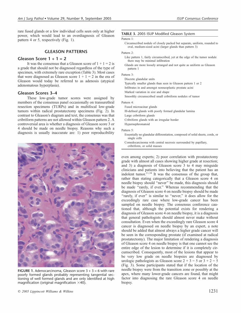
rare fused glands or a few individual cells seen only at higherpower, which would lead to an overdiagnosis of Gleasonpattern 4 or 5, respectively (Fig. 1).
GLEASON PATTERNS
Gleason Score 1 + 1 = 2It was the consensus that a Gleason score of 1 + 1 = 2 is
a grade that should not be diagnosed regardless of the type ofspecimen, with extremely rare exception (Table 3). Most casesthat were diagnosed as Gleason score 1 + 1 = 2 in the era ofGleason would today be referred to as adenosis (atypicaladenomatous hyperplasia).
Gleason Scores 3–4These low-grade tumor scores were assigned by
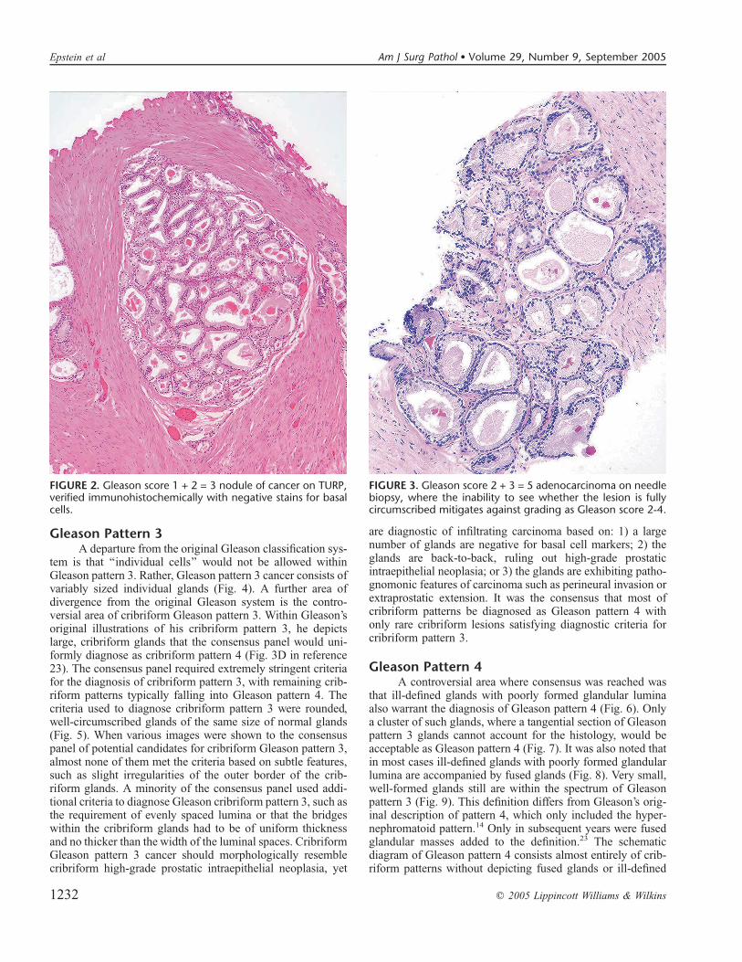
members of the consensus panel occasionally on transurethralresection specimens (TURPs) and in multifocal low-gradetumors within radical prostatectomy specimens (Fig. 2). Incontrast to Gleason’s diagram and text, the consensus was thatcribriform patterns are not allowed within Gleason pattern 2. Acontroversial area is whether a diagnosis of Gleason score 3 or4 should be made on needle biopsy. Reasons why such adiagnosis is usually inaccurate are: 1) poor reproducibility
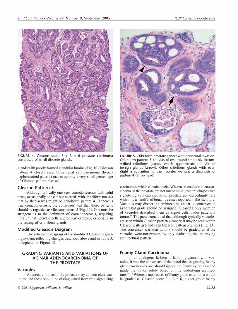
even among experts; 2) poor correlation with prostatectomygrade with almost all cases showing higher grade at resection;and 3) a diagnosis of Gleason score 3 to 4 may misguideclinicians and patients into believing that the patient has anindolent tumor.11,34 It was the consensus of the group that,rather than stating categorically that a Gleason score 4 onneedle biopsy should ‘‘never’’ be made, this diagnosis shouldbe made ‘‘rarely, if ever.’’ Whereas recommending that thediagnosis of Gleason score 4 on needle biopsy should be made‘‘rarely, if ever’’ is similar to ‘‘never,’’ it does allow for theexceedingly rare case where low-grade cancer has beensampled on needle biopsy. The consensus conference cau-tioned that, although the potential exists for rendering adiagnosis of Gleason score 4 on needle biopsy, it is a diagnosisthat general pathologists should almost never make withoutconsultation. Even when the exceedingly rare Gleason score 4cancer is diagnosed on needle biopsy by an expert, a noteshould be added that almost always a higher grade cancer willbe seen in the corresponding prostate (if examined at radicalprostatectomy). The major limitation of rendering a diagnosisof Gleason score 4 on needle biopsy is that one cannot see theentire edge of the lesion to determine if it is completely cir-cumscribed. Consequently, most of the lesions that appear tobe very low grade on needle biopsies are diagnosed byurologic pathologists as Gleason score 2 + 3 = 5 or 3 + 2 = 5(Fig. 3). Some participants stated that if the location of theneedle biopsy were from the transition zone or possibly at theapex, where many lower-grade cancers are found, that mightfactor into diagnosing the rare Gleason score 4 on needlebiopsy.
FIGURE 1. Adenocarcinoma, Gleason score 3 + 3 = 6 with rarepoorly formed glands probably representing tangential sec-tioning of well formed glands and are only identified at highmagnification (original magnification 340).
TABLE 3. 2005 ISUP Modified Gleason System
Pattern 1:
Circumscribed nodule of closely packed but separate, uniform, rounded tooval, medium-sized acini (larger glands than pattern 3)
Pattern 2:
Like pattern 1, fairly circumscribed, yet at the edge of the tumor nodulethere may be minimal infiltration
Glands are more loosely arranged and not quite as uniform as Gleasonpattern 1
Pattern 3:
Discrete glandular units
Typically smaller glands than seen in Gleason pattern 1 or 2
Infiltrates in and amongst nonneoplastic prostate acini
Marked variation in size and shape
Smoothly circumscribed small cribriform nodules of tumor
Pattern 4:
Fused microacinar glands
Ill-defined glands with poorly formed glandular lumina
Large cribriform glands
Cribriform glands with an irregular border
Hypernephromatoid
Pattern 5:
Essentially no glandular differentiation, composed of solid sheets, cords, orsingle cells
Comedocarcinoma with central necrosis surrounded by papillary,cribriform, or solid masses
q 2005 Lippincott Williams & Wilkins 1231
Am J Surg Pathol � Volume 29, Number 9, September 2005 ISUP Consensus Conference
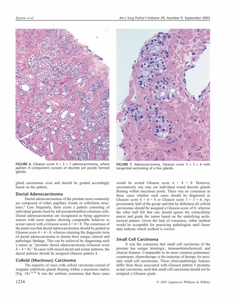
Gleason Pattern 3A departure from the original Gleason classification sys-
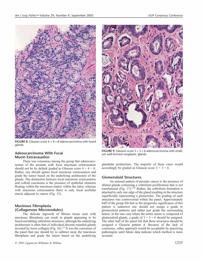
tem is that ‘‘individual cells’’ would not be allowed withinGleason pattern 3. Rather, Gleason pattern 3 cancer consists ofvariably sized individual glands (Fig. 4). A further area ofdivergence from the original Gleason system is the contro-versial area of cribriform Gleason pattern 3. Within Gleason’soriginal illustrations of his cribriform pattern 3, he depictslarge, cribriform glands that the consensus panel would uni-formly diagnose as cribriform pattern 4 (Fig. 3D in reference23). The consensus panel required extremely stringent criteriafor the diagnosis of cribriform pattern 3, with remaining crib-riform patterns typically falling into Gleason pattern 4. Thecriteria used to diagnose cribriform pattern 3 were rounded,well-circumscribed glands of the same size of normal glands(Fig. 5). When various images were shown to the consensuspanel of potential candidates for cribriform Gleason pattern 3,almost none of them met the criteria based on subtle features,such as slight irregularities of the outer border of the crib-riform glands. A minority of the consensus panel used addi-tional criteria to diagnose Gleason cribriform pattern 3, such asthe requirement of evenly spaced lumina or that the bridgeswithin the cribriform glands had to be of uniform thicknessand no thicker than the width of the luminal spaces. CribriformGleason pattern 3 cancer should morphologically resemblecribriform high-grade prostatic intraepithelial neoplasia, yet
are diagnostic of infiltrating carcinoma based on: 1) a largenumber of glands are negative for basal cell markers; 2) theglands are back-to-back, ruling out high-grade prostaticintraepithelial neoplasia; or 3) the glands are exhibiting patho-gnomonic features of carcinoma such as perineural invasion orextraprostatic extension. It was the consensus that most ofcribriform patterns be diagnosed as Gleason pattern 4 withonly rare cribriform lesions satisfying diagnostic criteria forcribriform pattern 3.
Gleason Pattern 4A controversial area where consensus was reached was
that ill-defined glands with poorly formed glandular luminaalso warrant the diagnosis of Gleason pattern 4 (Fig. 6). Onlya cluster of such glands, where a tangential section of Gleasonpattern 3 glands cannot account for the histology, would beacceptable as Gleason pattern 4 (Fig. 7). It was also noted thatin most cases ill-defined glands with poorly formed glandularlumina are accompanied by fused glands (Fig. 8). Very small,well-formed glands still are within the spectrum of Gleasonpattern 3 (Fig. 9). This definition differs from Gleason’s orig-inal description of pattern 4, which only included the hyper-nephromatoid pattern.14 Only in subsequent years were fusedglandular masses added to the definition.23 The schematicdiagram of Gleason pattern 4 consists almost entirely of crib-riform patterns without depicting fused glands or ill-defined
FIGURE 2. Gleason score 1 + 2 = 3 nodule of cancer on TURP,verified immunohistochemically with negative stains for basalcells.
FIGURE 3. Gleason score 2 + 3 = 5 adenocarcinoma on needlebiopsy, where the inability to see whether the lesion is fullycircumscribed mitigates against grading as Gleason score 2-4.
1232 q 2005 Lippincott Williams & Wilkins
Epstein et al Am J Surg Pathol � Volume 29, Number 9, September 2005
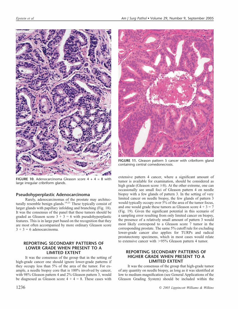
glands with poorly formed glandular lumina (Fig. 10). Gleasonpattern 4 closely resembling renal cell carcinoma (hyper-nephromatoid pattern) makes up only a very small percentageof Gleason pattern 4 cases.
Gleason Pattern 5Although typically one sees comedonecrosis with solid
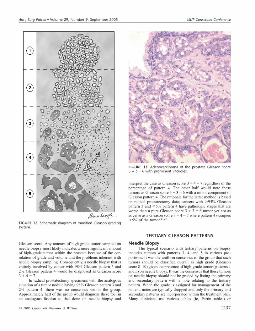
nests, occasionally one can see necrosis with cribriform massesthat by themselves might be cribriform pattern 4. If there istrue comedonecrosis, the consensus was that these patternsshould be regarded as Gleason pattern 5 (Fig. 11). One must bestringent as to the definition of comedonecrosis, requiringintraluminal necrotic cells and/or karyorrhexis, especially inthe setting of cribriform glands.
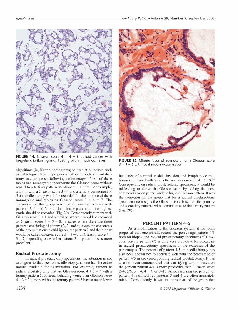
Modified Gleason DiagramThe schematic diagram of the modified Gleason’s grad-
ing system, reflecting changes described above and in Table 3,is depicted in Figure 12.
GRADING VARIANTS AND VARIATIONS OFACINAR ADENOCARCINOMA OF
THE PROSTATE
VacuolesAdenocarcinomas of the prostate may contain clear vac-
uoles, and these should be distinguished from true signet-ring
carcinomas, which containmucin.Whereas vacuoles in adenocar-cinoma of the prostate are not uncommon, true mucin-positivesignet-ring cell carcinomas of prostate are exceedingly rarewith only a handful of bona fide cases reported in the literature.Vacuoles may distort the architecture, and it is controversialas to what grade should be assigned. Gleason’s only mentionof vacuoles described them as signet cells under pattern 5tumor.16 The panel concluded that, although typically vacuolesare seen within Gleason pattern 4 cancer, it may be seen withinGleason pattern 5 and even Gleason pattern 3 tumors (Fig. 13).The consensus was that tumors should be graded, as if thevacuoles were not present, by only evaluating the underlyingarchitectural pattern.
Foamy Gland CarcinomaIn an analogous fashion to handling cancers with vac-
uoles, it was the consensus of the panel that in grading foamygland carcinomas one should ignore the foamy cytoplasm andgrade the tumor solely based on the underlying architec-ture.25,35 Whereas most cases of foamy gland carcinoma wouldbe graded as Gleason score 3 + 3 = 6, higher-grade foamy
FIGURE 4. Gleason score 3 + 3 = 6 prostate carcinomacomposed of small discrete glands.
FIGURE 5. Cribriform prostate cancer with perineural invasion.Cribriform pattern 3 consists of oval-round smoothly circum-scribed cribriform glands, which approximate the size ofbenign glands (arrow). Other cribriform glands with evenslight irregularities to their border warrant a diagnosis ofpattern 4 (arrowhead).
q 2005 Lippincott Williams & Wilkins 1233
Am J Surg Pathol � Volume 29, Number 9, September 2005 ISUP Consensus Conference
gland carcinomas exist and should be graded accordinglybased on the pattern.
Ductal AdenocarcinomaDuctal adenocarcinomas of the prostate most commonly
are composed of either papillary fronds or cribriform struc-tures.6 Less frequently, there exists a pattern consisting ofindividual glands lined by tall pseudostratified columnar cells.Ductal adenocarcinomas are recognized as being aggressivetumors with most studies showing comparable behavior toacinar cancer with a Gleason score 4 + 4 = 8. The consensus ofthe panel was that ductal adenocarcinomas should be graded asGleason score 4 + 4 = 8, whereas retaining the diagnostic termof ductal adenocarcinoma to denote their unique clinical andpathologic findings. This can be achieved by diagnosing sucha tumor as ‘‘prostatic ductal adenocarcinoma (Gleason score4 + 4 = 8).’’ In cases with mixed ductal and acinar patterns, theductal patterns should be assigned Gleason pattern 4.
Colloid (Mucinous) CarcinomaThe majority of cases with colloid carcinoma consist of
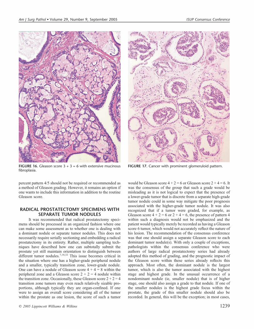
irregular cribriform glands floating within a mucinous matrix(Fig. 14).10,30 It was the uniform consensus that these cases
would be scored Gleason score 4 + 4 = 8. However,uncommonly one may see individual round discrete glandsfloating within mucinous pools. There was no consensus inthese cases whether such cases should be diagnosed asGleason score 4 + 4 = 8 or Gleason score 3 + 3 = 6. Ap-proximately half of the group said that by definition all colloidcarcinomas should be assigned a Gleason score of 8, whereasthe other half felt that one should ignore the extracellularmucin and grade the tumor based on the underlying archi-tectural pattern. Given the lack of consensus, either methodwould be acceptable for practicing pathologists until futuredata indicate which method is correct.
Small Cell CarcinomaIt was the consensus that small cell carcinoma of the
prostate has unique histologic, immunohistochemical, andclinical features. Comparable to its more common pulmonarycounterpart, chemotherapy is the mainstay of therapy for pros-tatic small cell carcinomas. These clinicopathologic featuresdiffer from those associated with Gleason pattern 5 prostaticacinar carcinoma, such that small cell carcinoma should not beassigned a Gleason grade.
FIGURE 6. Gleason score 4 + 3 = 7 adenocarcinoma, wherepattern 4 component consists of discrete yet poorly formedglands.
FIGURE 7. Adenocarcinoma, Gleason score 3 + 3 = 6 withtangential sectioning of a few glands.
1234 q 2005 Lippincott Williams & Wilkins
Epstein et al Am J Surg Pathol � Volume 29, Number 9, September 2005
Adenocarcinoma With FocalMucin Extravasation
There was consensus among the group that adenocarci-nomas of the prostate with focal mucinous extravasationshould not be by default graded as Gleason score 4 + 4 = 8.Rather, one should ignore focal mucinous extravasation andgrade the tumor based on the underlying architecture of theglands. The distinction between focal mucinous extravasationand colloid carcinoma is the presence of epithelial elementsfloating within the mucinous matrix within the latter, whereaswith mucinous extravasation there is only focal acellularmucin adjacent to cancer (Fig. 15).
Mucinous Fibroplasia(Collagenous Micronodules)
The delicate ingrowth of fibrous tissue seen withmucinous fibroplasia can result in glands appearing to befused resembling cribriform structures, although the underlyingarchitecture is often that of individual discrete rounded glandsinvested by loose collagen (Fig. 16).4,5 It was the consensus ofthe panel that one should try to subtract away the mucinousfibroplasia and grade the tumor based on the underlying
glandular architecture. The majority of these cases wouldaccordingly be graded as Gleason score 3 + 3 = 6.
Glomeruloid StructuresAn unusual pattern of prostate cancer is the presence of
dilated glands containing a cribriform proliferation that is nottransluminal (Fig. 17).4,26 Rather, the cribriform formation isattached to only one edge of the gland resulting in the structuresuperficially representing a glomerulus. The grading of suchstructures was controversial within the panel. Approximatelyhalf of the group felt that as the prognostic significance of thispattern is unknown one should not assign a grade toglomeruloid patterns and rather just grade the surroundingtumor; in the rare case where the entire tumor is composed ofglomeruloid glands, a grade of 3 + 3 = 6 should be assigned.The other half of the panel felt that these structures should beassigned a Gleason pattern 4. Because of the lack ofconsensus, either approach would be acceptable by practicingpathologists until future data indicate which method is moreaccurate.
FIGURE 8. Gleason score 4 + 4 = 8 adenocarcinoma with fusedglands.
FIGURE 9. Gleason score 3 + 3 = 6 adenocarcinoma with small,yet well-formed neoplastic glands.
q 2005 Lippincott Williams & Wilkins 1235
Am J Surg Pathol � Volume 29, Number 9, September 2005 ISUP Consensus Conference
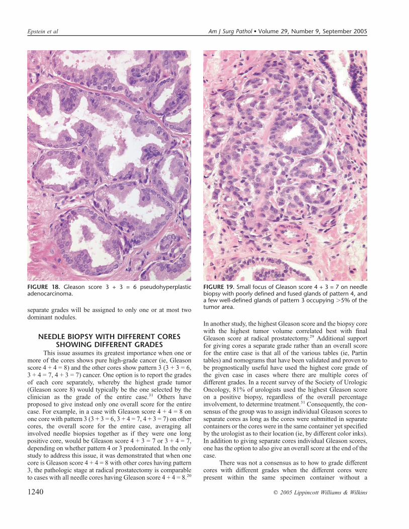
Pseudohyperplastic AdenocarcinomaRarely, adenocarcinomas of the prostate may architec-
turally resemble benign glands.19,21 These typically consist oflarger glands with papillary infolding and branching (Fig. 18).It was the consensus of the panel that these tumors should begraded as Gleason score 3 + 3 = 6 with pseudohyperplasticfeatures. This is in large part based on the recognition that theyare most often accompanied by more ordinary Gleason score3 + 3 = 6 adenocarcinoma.
REPORTING SECONDARY PATTERNS OFLOWER GRADE WHEN PRESENT TO A
LIMITED EXTENTIt was the consensus of the group that in the setting of
high-grade cancer one should ignore lower-grade patterns ifthey occupy less than 5% of the area of the tumor. For ex-ample, a needle biopsy core that is 100% involved by cancer,with 98% Gleason pattern 4 and 2% Gleason pattern 3, wouldbe diagnosed as Gleason score 4 + 4 = 8. These cases with
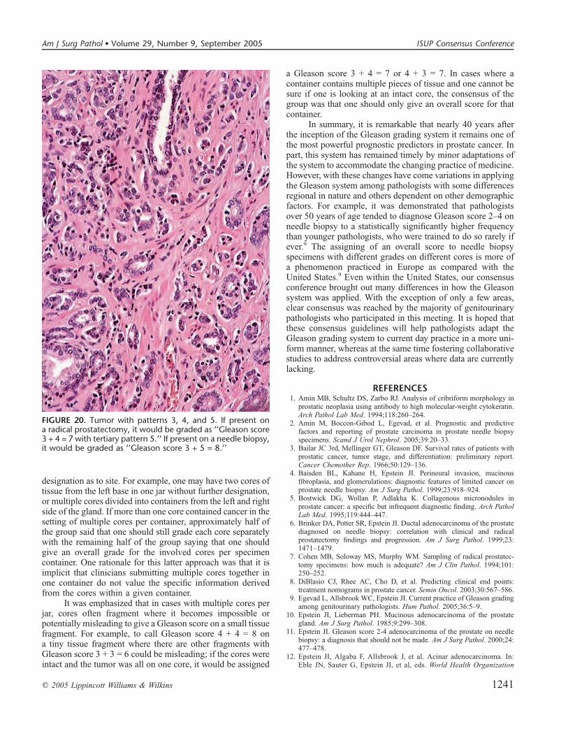
extensive pattern 4 cancer, where a significant amount oftumor is available for examination, should be considered ashigh grade (Gleason score $8). At the other extreme, one canoccasionally see small foci of Gleason pattern 4 on needlebiopsy with a few glands of pattern 3. In the setting of verylimited cancer on needle biopsy, the few glands of pattern 3would typically occupy over 5% of the area of the tumor focus,and one would grade these tumors as Gleason score 4 + 3 = 7(Fig. 19). Given the significant potential in this scenario ofa sampling error resulting from only limited cancer on biopsy,the presence of a relatively small amount of pattern 3 wouldmost likely correspond to a Gleason score 7 tumor in thecorresponding prostate. The same 5% cutoff rule for excludinglower-grade cancer also applies for TURPs and radicalprostatectomy specimens, which in most cases would relateto extensive cancer with .95% Gleason pattern 4 tumor.
REPORTING SECONDARY PATTERNS OFHIGHER GRADE WHEN PRESENT TO A
LIMITED EXTENTIt was the consensus of the group that high-grade tumor
of any quantity on needle biopsy, as long as it was identified atlow to medium magnification (see General Applications of theGleason Grading System) should be included within the
FIGURE 10. Adenocarcinoma Gleason score 4 + 4 = 8 withlarge irregular cribriform glands.
FIGURE 11. Gleason pattern 5 cancer with cribriform glandcontaining central comedonecrosis.
1236 q 2005 Lippincott Williams & Wilkins
Epstein et al Am J Surg Pathol � Volume 29, Number 9, September 2005
Gleason score. Any amount of high-grade tumor sampled onneedle biopsy most likely indicates a more significant amountof high-grade tumor within the prostate because of the cor-relation of grade and volume and the problems inherent withneedle biopsy sampling. Consequently, a needle biopsy that isentirely involved by cancer with 98% Gleason pattern 3 and2% Gleason pattern 4 would be diagnosed as Gleason score3 + 4 = 7.
In radical prostatectomy specimens with the analogoussituation of a tumor nodule having 98% Gleason pattern 3 and2% pattern 4, there was no consensus within the group.Approximately half of the group would diagnose these foci inan analogous fashion to that done on needle biopsy and
interpret the case as Gleason score 3 + 4 = 7 regardless of thepercentage of pattern 4. The other half would note thesetumors as Gleason score 3 + 3 = 6 with a minor component ofGleason pattern 4. The rationale for the latter method is basedon radical prostatectomy data; cancers with .95% Gleasonpattern 3 and ,5% pattern 4 have pathologic stages that areworse than a pure Gleason score 3 + 3 = 6 tumor yet not asadverse as a Gleason score 3 + 4 = 7 where pattern 4 occupies.5% of the tumor.24,27
TERTIARY GLEASON PATTERNS
Needle BiopsyThe typical scenario with tertiary patterns on biopsy
includes tumors with patterns 3, 4, and 5 in various pro-portions. It was the uniform consensus of the group that suchtumors should be classified overall as high grade (Gleasonscore 8–10) given the presence of high-grade tumor (patterns 4and 5) on needle biopsy. It was the consensus that these tumorson needle biopsy should not be graded by listing the primaryand secondary pattern with a note relating to the tertiarypattern. When the grade is assigned for management of thepatient, notes are typically dropped and only the primary andsecondary patterns are incorporated within the treatment plan.Many clinicians use various tables (ie, Partin tables) or
FIGURE 12. Schematic diagram of modified Gleason gradingsystem.
FIGURE 13. Adenocarcinoma of the prostate Gleason score3 + 3 = 6 with prominent vacuoles.
q 2005 Lippincott Williams & Wilkins 1237
Am J Surg Pathol � Volume 29, Number 9, September 2005 ISUP Consensus Conference
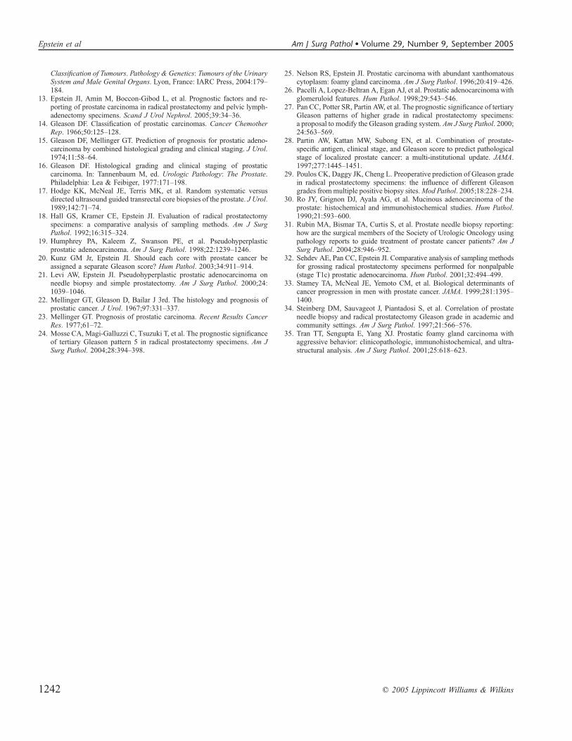
algorithms (ie, Kattan nomograms) to predict outcomes suchas pathologic stage or prognosis following radical prostatec-tomy, and prognosis following radiotherapy.8,28 All of thesetables and nomograms incorporate the Gleason score withoutregard to a tertiary pattern mentioned in a note. For example,a tumor with a Gleason score 3 + 4 and a tertiary component of5 on needle biopsy would be recorded for the purpose of thesenomograms and tables as Gleason score 3 + 4 = 7. Theconsensus of the group was that on needle biopsies withpatterns 3, 4, and 5, both the primary pattern and the highestgrade should be recorded (Fig. 20). Consequently, tumors withGleason score 3 + 4 and a tertiary pattern 5 would be recordedas Gleason score 3 + 5 = 8. In cases where there are threepatterns consisting of patterns 2, 3, and 4, it was the consensusof the group that one would ignore the pattern 2 and the biopsywould be called Gleason score 3 + 4 = 7 or Gleason score 4 +3 = 7, depending on whether pattern 3 or pattern 4 was moreprevalent.
Radical ProstatectomyIn radical prostatectomy specimens, the situation is not
analogous to that seen on needle biopsy, as one has the entirenodule available for examination. For example, tumors atradical prostatectomy that are Gleason score 4 + 3 = 7 with atertiary pattern 5, whereas behaving worse than Gleason score4 + 3 = 7 tumors without a tertiary pattern 5 have a much lower
incidence of seminal vesicle invasion and lymph node me-tastases compared with tumors that are Gleason score 4 + 5 = 9.24
Consequently, on radical prostatectomy specimens, it would bemisleading to derive the Gleason score by adding the mostcommon Gleason pattern and the highest Gleason pattern. It wasthe consensus of the group that for a radical prostatectomyspecimen one assigns the Gleason score based on the primaryand secondary patterns with a comment as to the tertiary pattern(Fig. 20).
PERCENT PATTERN 4–5As a modification to the Gleason system, it has been
proposed that one should record the percentage pattern 4/5both on biopsy and radical prostatectomy specimens.33 How-ever, percent pattern 4/5 is only very predictive for prognosisin radical prostatectomy specimens at the extremes of thepercentages. The percent of pattern 4/5 on needle biopsy hasalso been shown not to correlate well with the percentage ofpattern 4/5 in the corresponding radical prostatectomy. It hasalso not been demonstrated that classifying tumors based onthe percent pattern 4/5 is more predictive than Gleason score2–4, 5/6, 3 + 4, 4 + 3, or 8–10. Also, assessing the percent ofpattern 4 is difficult as patterns 3 and 4 are often intimatelymixed. Consequently, it was the consensus of the group that
FIGURE 14. Gleason score 4 + 4 = 8 colloid cancer withirregular cribriform glands floating within mucinous lakes. FIGURE 15. Minute focus of adenocarcinoma Gleason score
3 + 3 = 6 with focal mucin extravasation.
1238 q 2005 Lippincott Williams & Wilkins
Epstein et al Am J Surg Pathol � Volume 29, Number 9, September 2005
percent pattern 4/5 should not be required or recommended asa method of Gleason grading. However, it remains an option ifone wants to include this information in addition to the routineGleason score.
RADICAL PROSTATECTOMY SPECIMENS WITHSEPARATE TUMOR NODULES
It was recommended that radical prostatectomy speci-mens should be processed in an organized fashion where onecan make some assessment as to whether one is dealing witha dominant nodule or separate tumor nodules. This does notnecessarily require serially sectioning and embedding a radicalprostatectomy in its entirety. Rather, multiple sampling tech-niques have described how one can subtotally submit theprostate yet still maintain orientation to distinguish betweendifferent tumor nodules.7,18,32 This issue becomes critical inthe situation where one has a higher-grade peripheral noduleand a smaller, typically transition zone, lower-grade nodule.One can have a nodule of Gleason score 4 + 4 = 8 within theperipheral zone and a Gleason score 2 + 2 = 4 nodule withinthe transition zone. Occasionally, these Gleason score 2 + 2 = 4transition zone tumors may even reach relatively sizable pro-portions, although typically they are organ-confined. If onewere to assign an overall score considering all of the tumorwithin the prostate as one lesion, the score of such a tumor
would be Gleason score 4 + 2 = 6 or Gleason score 2 + 4 = 6. Itwas the consensus of the group that such a grade would bemisleading as it is not logical to expect that the presence ofa lower-grade tumor that is discrete from a separate high-gradetumor nodule could in some way mitigate the poor prognosisassociated with the higher-grade tumor nodule. It was alsorecognized that if a tumor were graded, for example, asGleason score 4 + 2 = 6 or 2 + 4 = 6, the presence of pattern 4within such a diagnosis would not be emphasized and thepatient would typically merely be recorded as having a Gleasonscore 6 tumor, which would not accurately reflect the nature ofhis lesion. The recommendation of the consensus conferencewas that one should assign a separate Gleason score to eachdominant tumor nodule(s). With only a couple of exceptions,pathologists within the consensus conference who wereauthors of large radical prostatectomy series had alreadyadopted this method of grading, and the prognostic impact ofthe Gleason score within these series already reflects thisapproach. Most often, the dominant nodule is the largesttumor, which is also the tumor associated with the higheststage and highest grade. In the unusual occurrence of anondominant nodule (ie, smaller nodule) that is of higherstage, one should also assign a grade to that nodule. If one ofthe smaller nodules is the highest grade focus within theprostate, the grade of this smaller nodule should also berecorded. In general, this will be the exception; in most cases,
FIGURE 16. Gleason score 3 + 3 = 6 with extensive mucinousfibroplasia.
FIGURE 17. Cancer with prominent glomeruloid pattern.
q 2005 Lippincott Williams & Wilkins 1239
Am J Surg Pathol � Volume 29, Number 9, September 2005 ISUP Consensus Conference
separate grades will be assigned to only one or at most twodominant nodules.
NEEDLE BIOPSY WITH DIFFERENT CORESSHOWING DIFFERENT GRADES
This issue assumes its greatest importance when one ormore of the cores shows pure high-grade cancer (ie, Gleasonscore 4 + 4 = 8) and the other cores show pattern 3 (3 + 3 = 6,3 + 4 = 7, 4 + 3 = 7) cancer. One option is to report the gradesof each core separately, whereby the highest grade tumor(Gleason score 8) would typically be the one selected by theclinician as the grade of the entire case.31 Others haveproposed to give instead only one overall score for the entirecase. For example, in a case with Gleason score 4 + 4 = 8 onone core with pattern 3 (3 + 3 = 6, 3 + 4 = 7, 4 + 3 = 7) on othercores, the overall score for the entire case, averaging allinvolved needle biopsies together as if they were one longpositive core, would be Gleason score 4 + 3 = 7 or 3 + 4 = 7,depending on whether pattern 4 or 3 predominated. In the onlystudy to address this issue, it was demonstrated that when onecore is Gleason score 4 + 4 = 8 with other cores having pattern3, the pathologic stage at radical prostatectomy is comparableto cases with all needle cores having Gleason score 4 + 4 = 8.20
In another study, the highest Gleason score and the biopsy corewith the highest tumor volume correlated best with finalGleason score at radical prostatectomy.29 Additional supportfor giving cores a separate grade rather than an overall scorefor the entire case is that all of the various tables (ie, Partintables) and nomograms that have been validated and proven tobe prognostically useful have used the highest core grade ofthe given case in cases where there are multiple cores ofdifferent grades. In a recent survey of the Society of UrologicOncology, 81% of urologists used the highest Gleason scoreon a positive biopsy, regardless of the overall percentageinvolvement, to determine treatment.31 Consequently, the con-sensus of the group was to assign individual Gleason scores toseparate cores as long as the cores were submitted in separatecontainers or the cores were in the same container yet specifiedby the urologist as to their location (ie, by different color inks).In addition to giving separate cores individual Gleason scores,one has the option to also give an overall score at the end of thecase.
There was not a consensus as to how to grade differentcores with different grades when the different cores werepresent within the same specimen container without a
FIGURE 19. Small focus of Gleason score 4 + 3 = 7 on needlebiopsy with poorly defined and fused glands of pattern 4, anda few well-defined glands of pattern 3 occupying .5% of thetumor area.
FIGURE 18. Gleason score 3 + 3 = 6 pseudohyperplasticadenocarcinoma.
1240 q 2005 Lippincott Williams & Wilkins
Epstein et al Am J Surg Pathol � Volume 29, Number 9, September 2005
designation as to site. For example, one may have two cores oftissue from the left base in one jar without further designation,or multiple cores divided into containers from the left and rightside of the gland. If more than one core contained cancer in thesetting of multiple cores per container, approximately half ofthe group said that one should still grade each core separatelywith the remaining half of the group saying that one shouldgive an overall grade for the involved cores per specimencontainer. One rationale for this latter approach was that it isimplicit that clinicians submitting multiple cores together inone container do not value the specific information derivedfrom the cores within a given container.
It was emphasized that in cases with multiple cores perjar, cores often fragment where it becomes impossible orpotentially misleading to give a Gleason score on a small tissuefragment. For example, to call Gleason score 4 + 4 = 8 ona tiny tissue fragment where there are other fragments withGleason score 3 + 3 = 6 could be misleading; if the cores wereintact and the tumor was all on one core, it would be assigned
a Gleason score 3 + 4 = 7 or 4 + 3 = 7. In cases where acontainer contains multiple pieces of tissue and one cannot besure if one is looking at an intact core, the consensus of thegroup was that one should only give an overall score for thatcontainer.
In summary, it is remarkable that nearly 40 years afterthe inception of the Gleason grading system it remains one ofthe most powerful prognostic predictors in prostate cancer. Inpart, this system has remained timely by minor adaptations ofthe system to accommodate the changing practice of medicine.However, with these changes have come variations in applyingthe Gleason system among pathologists with some differencesregional in nature and others dependent on other demographicfactors. For example, it was demonstrated that pathologistsover 50 years of age tended to diagnose Gleason score 2–4 onneedle biopsy to a statistically significantly higher frequencythan younger pathologists, who were trained to do so rarely ifever.9 The assigning of an overall score to needle biopsyspecimens with different grades on different cores is more ofa phenomenon practiced in Europe as compared with theUnited States.9 Even within the United States, our consensusconference brought out many differences in how the Gleasonsystem was applied. With the exception of only a few areas,clear consensus was reached by the majority of genitourinarypathologists who participated in this meeting. It is hoped thatthese consensus guidelines will help pathologists adapt theGleason grading system to current day practice in a more uni-form manner, whereas at the same time fostering collaborativestudies to address controversial areas where data are currentlylacking.
REFERENCES1. Amin MB, Schultz DS, Zarbo RJ. Analysis of cribriform morphology in
prostatic neoplasia using antibody to high molecular-weight cytokeratin.Arch Pathol Lab Med. 1994;118:260–264.
2. Amin M, Boccon-Gibod L, Egevad, et al. Prognostic and predictivefactors and reporting of prostate carcinoma in prostate needle biopsyspecimens. Scand J Urol Nephrol. 2005;39:20–33.
3. Bailar JC 3rd, Mellinger GT, Gleason DF. Survival rates of patients withprostatic cancer, tumor stage, and differentiation: preliminary report.Cancer Chemother Rep. 1966;50:129–136.
4. Baisden BL, Kahane H, Epstein JI. Perineural invasion, mucinousfibroplasia, and glomerulations: diagnostic features of limited cancer onprostate needle biopsy. Am J Surg Pathol. 1999;23:918–924.
5. Bostwick DG, Wollan P, Adlakha K. Collagenous micronodules inprostate cancer: a specific but infrequent diagnostic finding. Arch PatholLab Med. 1995;119:444–447.
6. Brinker DA, Potter SR, Epstein JI. Ductal adenocarcinoma of the prostatediagnosed on needle biopsy: correlation with clinical and radicalprostatectomy findings and progression. Am J Surg Pathol. 1999;23:1471–1479.
7. Cohen MB, Soloway MS, Murphy WM. Sampling of radical prostatec-tomy specimens: how much is adequate? Am J Clin Pathol. 1994;101:250–252.
8. DiBlasio CJ, Rhee AC, Cho D, et al. Predicting clinical end points:treatment nomograms in prostate cancer. Semin Oncol. 2003;30:567–586.
9. Egevad L, Allsbrook WC, Epstein JI. Current practice of Gleason gradingamong genitourinary pathologists. Hum Pathol. 2005;36:5–9.
10. Epstein JI, Lieberman PH. Mucinous adenocarcinoma of the prostategland. Am J Surg Pathol. 1985;9:299–308.
11. Epstein JI. Gleason score 2-4 adenocarcinoma of the prostate on needlebiopsy: a diagnosis that should not be made. Am J Surg Pathol. 2000;24:477–478.
12. Epstein JI, Algaba F, Allsbrook J, et al. Acinar adenocarcinoma. In:Eble JN, Sauter G, Epstein JI, et al, eds. World Health Organization
FIGURE 20. Tumor with patterns 3, 4, and 5. If present ona radical prostatectomy, it would be graded as ‘‘Gleason score3 + 4 = 7 with tertiary pattern 5.’’ If present on a needle biopsy,it would be graded as ‘‘Gleason score 3 + 5 = 8.’’
q 2005 Lippincott Williams & Wilkins 1241
Am J Surg Pathol � Volume 29, Number 9, September 2005 ISUP Consensus Conference
Classification of Tumours. Pathology & Genetics: Tumours of the UrinarySystem and Male Genital Organs. Lyon, France: IARC Press, 2004:179–184.
13. Epstein JI, Amin M, Boccon-Gibod L, et al. Prognostic factors and re-porting of prostate carcinoma in radical prostatectomy and pelvic lymph-adenectomy specimens. Scand J Urol Nephrol. 2005;39:34–36.
14. Gleason DF. Classification of prostatic carcinomas. Cancer ChemotherRep. 1966;50:125–128.
15. Gleason DF, Mellinger GT. Prediction of prognosis for prostatic adeno-carcinoma by combined histological grading and clinical staging. J Urol.1974;11:58–64.
16. Gleason DF. Histological grading and clinical staging of prostaticcarcinoma. In: Tannenbaum M, ed. Urologic Pathology: The Prostate.Philadelphia: Lea & Feibiger, 1977:171–198.
17. Hodge KK, McNeal JE, Terris MK, et al. Random systematic versusdirected ultrasound guided transrectal core biopsies of the prostate. J Urol.1989;142:71–74.
18. Hall GS, Kramer CE, Epstein JI. Evaluation of radical prostatectomyspecimens: a comparative analysis of sampling methods. Am J SurgPathol. 1992;16:315–324.
19. Humphrey PA, Kaleem Z, Swanson PE, et al. Pseudohyperplasticprostatic adenocarcinoma. Am J Surg Pathol. 1998;22:1239–1246.
20. Kunz GM Jr, Epstein JI. Should each core with prostate cancer beassigned a separate Gleason score? Hum Pathol. 2003;34:911–914.
21. Levi AW, Epstein JI. Pseudohyperplastic prostatic adenocarcinoma onneedle biopsy and simple prostatectomy. Am J Surg Pathol. 2000;24:1039–1046.
22. Mellinger GT, Gleason D, Bailar J 3rd. The histology and prognosis ofprostatic cancer. J Urol. 1967;97:331–337.
23. Mellinger GT. Prognosis of prostatic carcinoma. Recent Results CancerRes. 1977;61–72.
24. Mosse CA, Magi-Galluzzi C, Tsuzuki T, et al. The prognostic significanceof tertiary Gleason pattern 5 in radical prostatectomy specimens. Am JSurg Pathol. 2004;28:394–398.
25. Nelson RS, Epstein JI. Prostatic carcinoma with abundant xanthomatouscytoplasm: foamy gland carcinoma. Am J Surg Pathol. 1996;20:419–426.
26. Pacelli A, Lopez-Beltran A, Egan AJ, et al. Prostatic adenocarcinoma withglomeruloid features. Hum Pathol. 1998;29:543–546.
27. Pan CC, Potter SR, Partin AW, et al. The prognostic significance of tertiaryGleason patterns of higher grade in radical prostatectomy specimens:a proposal to modify the Gleason grading system. Am J Surg Pathol. 2000;24:563–569.
28. Partin AW, Kattan MW, Subong EN, et al. Combination of prostate-specific antigen, clinical stage, and Gleason score to predict pathologicalstage of localized prostate cancer: a multi-institutional update. JAMA.1997;277:1445–1451.
29. Poulos CK, Daggy JK, Cheng L. Preoperative prediction of Gleason gradein radical prostatectomy specimens: the influence of different Gleasongrades from multiple positive biopsy sites. Mod Pathol. 2005;18:228–234.
30. Ro JY, Grignon DJ, Ayala AG, et al. Mucinous adenocarcinoma of theprostate: histochemical and immunohistochemical studies. Hum Pathol.1990;21:593–600.
31. Rubin MA, Bismar TA, Curtis S, et al. Prostate needle biopsy reporting:how are the surgical members of the Society of Urologic Oncology usingpathology reports to guide treatment of prostate cancer patients? Am JSurg Pathol. 2004;28:946–952.
32. Sehdev AE, Pan CC, Epstein JI. Comparative analysis of sampling methodsfor grossing radical prostatectomy specimens performed for nonpalpable(stage T1c) prostatic adenocarcinoma. Hum Pathol. 2001;32:494–499.
33. Stamey TA, McNeal JE, Yemoto CM, et al. Biological determinants ofcancer progression in men with prostate cancer. JAMA. 1999;281:1395–1400.
34. Steinberg DM, Sauvageot J, Piantadosi S, et al. Correlation of prostateneedle biopsy and radical prostatectomy Gleason grade in academic andcommunity settings. Am J Surg Pathol. 1997;21:566–576.
35. Tran TT, Sengupta E, Yang XJ. Prostatic foamy gland carcinoma withaggressive behavior: clinicopathologic, immunohistochemical, and ultra-structural analysis. Am J Surg Pathol. 2001;25:618–623.
1242 q 2005 Lippincott Williams & Wilkins
Epstein et al Am J Surg Pathol � Volume 29, Number 9, September 2005