Embed Size (px)
Citation preview

Thank you for viewing this presentation.
We would like to remind you that this material is the property of the author.
It is provided to you by the ERS for your personal use only, as submitted by the
author.
2016 by the author
10-20 system EEG Placement
Andrew Morley Andrew Morley (BSc Hons, RPSGT)(BSc Hons, RPSGT) , , Lizzie HillLizzie Hill (EST RPSGT)
& Prof. Dr Athanasios
G. Kaditis
Chief Respiratory (Sleep)
Physiologist, Royal Hospital for Children, Glasgow
Specialist Respiratory Clinical Physiologist,Royal Hospital for Sick Children, Edinburgh
Conflict of interest disclosure I have no, real or perceived, direct or indirect conflicts of interest that relate to this
presentation. I have the following, real or perceived direct or indirect conflicts of interest that relate to
this presentation: Affiliation / financial interest Nature of conflict / commercial company name
Tobacco-industry and tobacco corporate affiliate related conflict of interest
Grants/research support (to myself, my institution or department):
Honoraria or consultation fees:
Participation in a company sponsored bureau:
Stock shareholder:
Spouse/partner:
Other support or other potential conflict of interest:
This event is accredited for CME credits by EBAP and speakers are required to disclose their potential conflict of interest going back 3 years prior to this presentation. The intent of this disclosure is not to prevent a speaker with a conflict of interest (any significant financial relationship a speaker has with manufacturers or providers of any commercial products or services relevant to the talk) from making a presentation, but rather to provide listeners with information on which they can make their own judgment. It remains for audience members to determine whether the speaker’s interests or relationships may influence the presentation.Drug or device advertisement is strictly forbidden.
x
10-20 EEG PlacementAIMS
• Demonstrate the International 10‐20 EEG system
• Understand steps required to set‐up a10‐20 EEG montage for a Polysomnography sleep study.
• Give each delegate a practical experience setting up a Sleep EEG montage using the 10‐20 EEG system.
Workshop Plan
• This session is going to be a mainly practical session.
• Brief presentation : 10‐20 basics
• Split into pairs and have a go.
• Slides from the session are available as part of the workshop materials – via website
10-20 EEG Placement
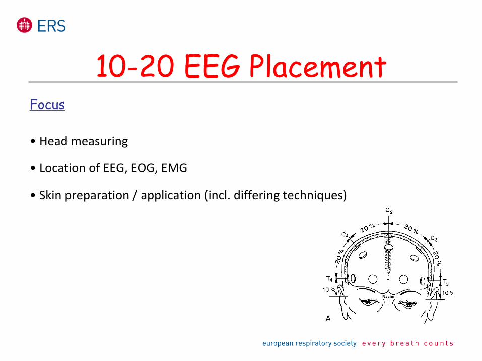
Focus
• Head measuring
• Location of EEG, EOG, EMG
• Skin preparation / application (incl. differing techniques)
10-20 EEG Placement
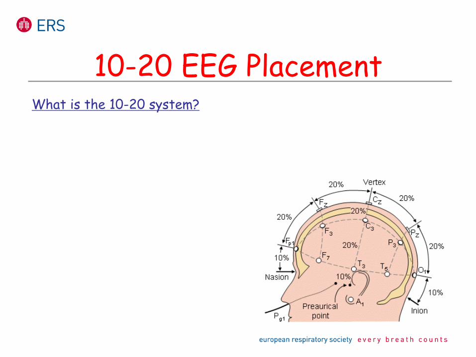
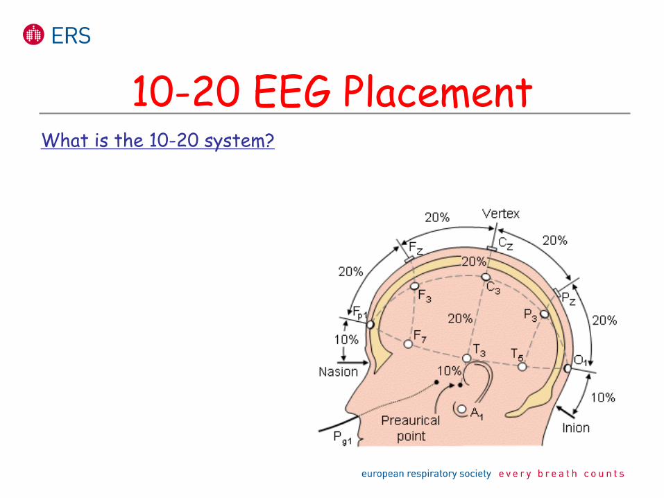
What is the 10-20 system?
10-20 EEG Placement
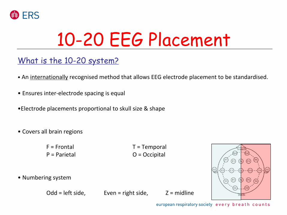
What is the 10-20 system?
• An internationally
recognised method that allows EEG electrode placement to be standardised.
• Ensures inter‐electrode spacing is equal
•Electrode placements proportional to skull size & shape
• Covers all brain regions
F = Frontal
T = Temporal P = Parietal
O = Occipital
• Numbering system
Odd = left side,
Even = right side,
Z = midline
10-20 EEG Placement
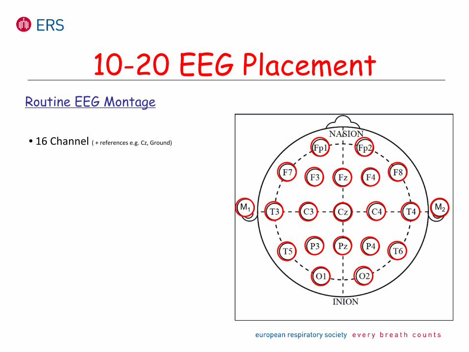
Routine EEG Montage
• 16 Channel ( + references e.g. Cz, Ground)
M1 M2
M1M1 M2
10-20 EEG Placement
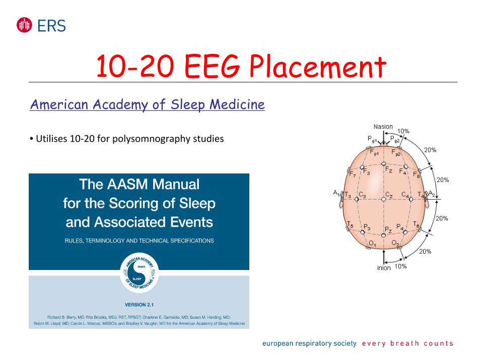
American Academy of Sleep Medicine
• Utilises 10‐20 for polysomnography studies
10-20 EEG Placement
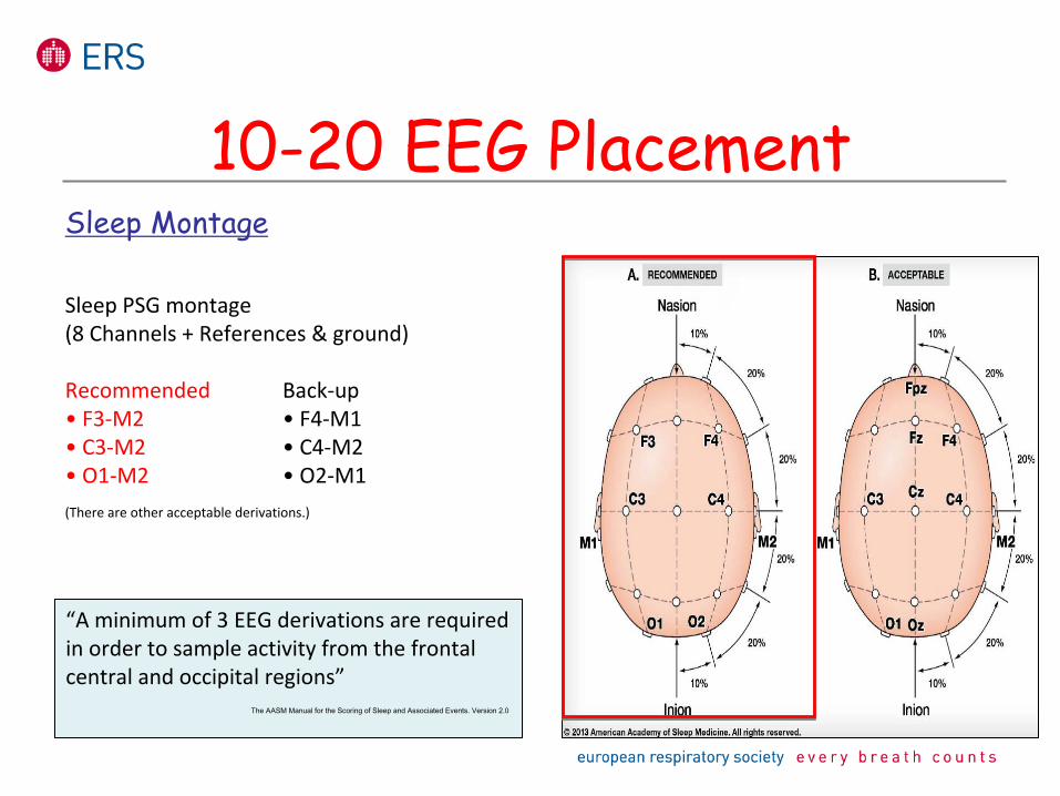
Sleep Montage
Sleep PSG montage (8 Channels + References & ground)
Recommended
Back‐up• F3‐M2
• F4‐M1• C3‐M2
• C4‐M2 • O1‐M2
• O2‐M1 (There are other acceptable derivations.)
“A minimum of 3 EEG derivations are required
in order to sample activity from the frontal
central and occipital regions”The AASM Manual for the Scoring of Sleep and Associated Events. Version 2.0
10-20 EEG Placement
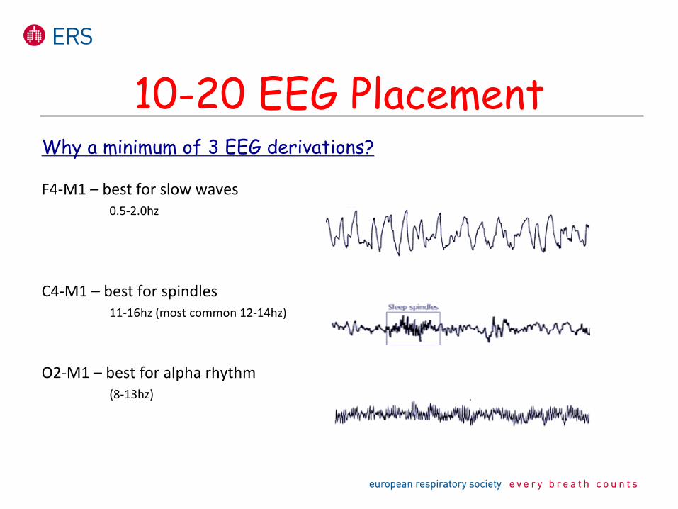
Why a minimum of 3 EEG derivations?
F4‐M1 – best for slow waves0.5‐2.0hz
C4‐M1 – best for spindles11‐16hz (most common 12‐14hz)
O2‐M1 – best for alpha rhythm(8‐13hz)
10-20 EEG Placement
Preparation
10-20 EEG Placement
Be prepared
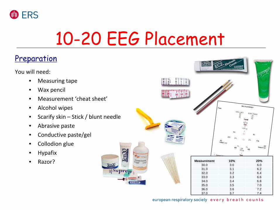
PreparationYou will need:
• Measuring tape• Wax pencil• Measurement ‘cheat sheet’• Alcohol wipes• Scarify skin – Stick / blunt needle• Abrasive paste• Conductive paste/gel• Collodion glue• Hypafix• Razor? Measurement 10% 20%
30.0 3.0 6.031.0 3.1 6.232.0 3.2 6.433.0 3.3 6.634.0 3.4 6.835.0 3.5 7.036.0 3.6 7.237.0 3.7 7.4
10-20 EEG Placement
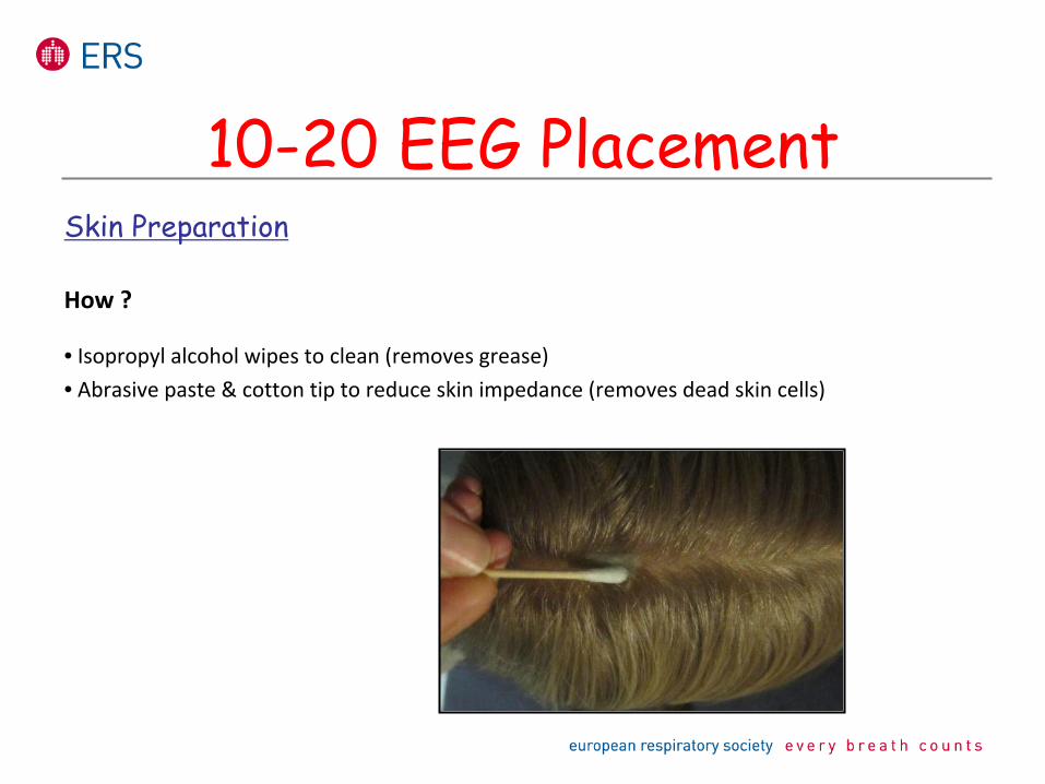
Skin Preparation
How ?
• Isopropyl alcohol wipes to clean (removes grease) • Abrasive paste & cotton tip to reduce skin impedance (removes dead skin cells)
10-20 EEG Placement
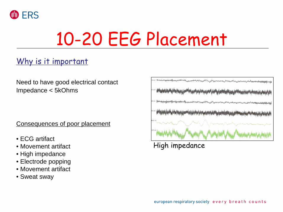
Why is it important
Need to have good electrical contact Impedance < 5kOhms
Consequences of poor placement
• ECG artifact• Movement artifact• High impedance• Electrode popping• Movement artifact• Sweat sway
10-20 EEG Placement
High impedance
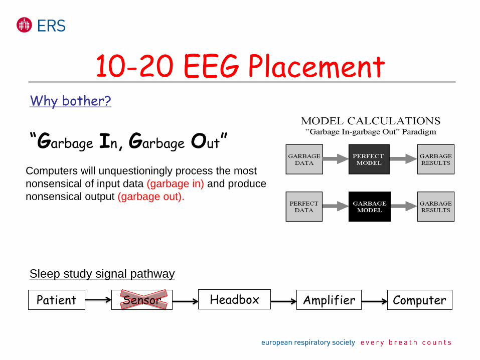
Why bother?
“Garbage
In, Garbage
Out”Computers will unquestioningly process the most nonsensical of input data (garbage in) and produce nonsensical output (garbage out).
Sleep study signal pathway
Patient Sensor Headbox Amplifier Computer
10-20 EEG Placement
What is the 10-20 system?
10-20 EEG Placement
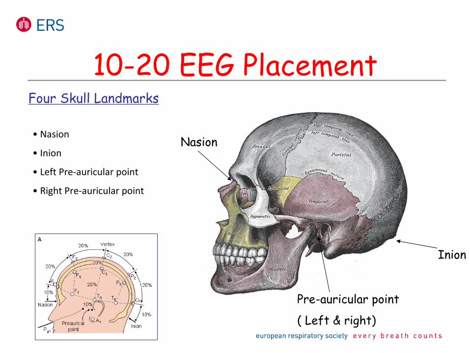
Four Skull Landmarks
Nasion
Inion
Pre-auricular point
( Left & right)
• Nasion
• Inion
• Left Pre‐auricular point
• Right Pre‐auricular point
10-20 EEG Placement
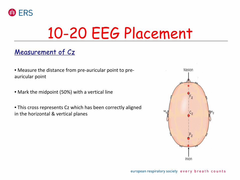
Measurement of Cz
• Measure the distance from pre‐auricular point to pre‐
auricular point
• Mark the midpoint (50%) with a vertical line
• This cross represents Cz which has been correctly aligned
in the horizontal & vertical planes
10-20 EEG Placement
M M
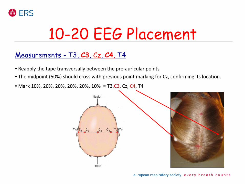
Measurements -
T3, C3, Cz, C4,
T4
• Reapply the tape transversally between the pre‐auricular points • The midpoint (50%) should cross with previous point marking for
Cz, confirming its location.
• Mark
10%, 20%, 20%, 20%, 20%, 10%
= T3,C3, Cz, C4, T4
10-20 EEG Placement
M M
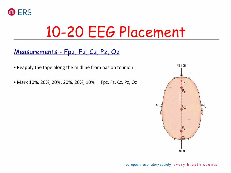
Measurements -
Fpz, Fz, Cz, Pz, Oz
• Reapply the tape along the midline from nasion to inion
• Mark
10%, 20%, 20%, 20%, 20%, 10%
= Fpz, Fz, Cz, Pz, Oz
10-20 EEG Placement
M M
Oz
Fpz
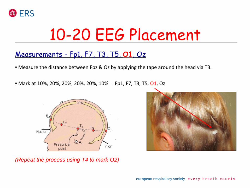
Measurements -
Fp1, F7, T3, T5, O1, Oz
• Measure the distance between Fpz & Oz by applying the tape around the head via T3.
• Mark
at 10%, 20%, 20%, 20%, 20%, 10%
= Fp1, F7, T3, T5, O1, Oz
(Repeat the process using T4 to mark O2)
10-20 EEG Placement
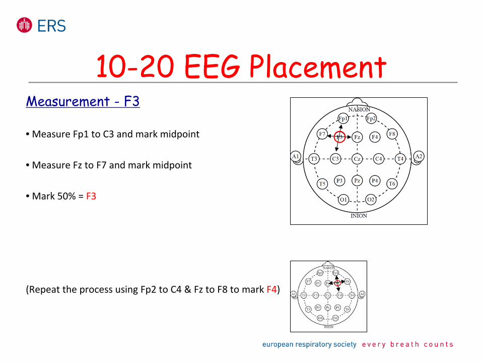
Measurement -
F3
• Measure Fp1 to C3 and mark midpoint
• Measure Fz to F7 and mark midpoint
• Mark 50% = F3
(Repeat the process using Fp2 to C4 & Fz to F8 to mark F4)
10-20 EEG Placement
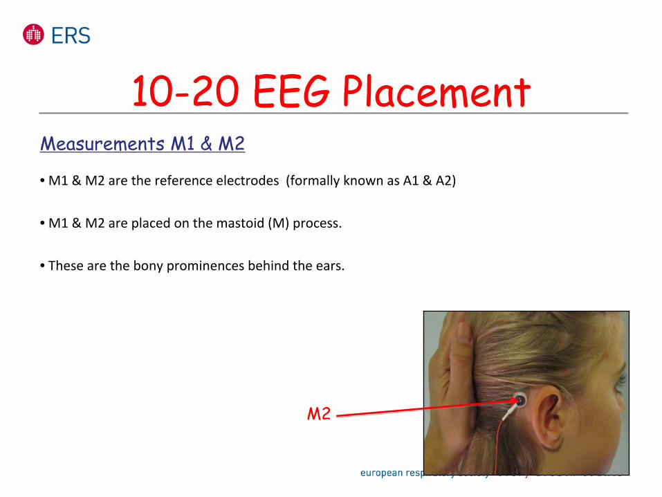
Measurements M1 & M2
• M1 & M2 are the reference electrodes (formally known as A1 & A2)
• M1 & M2 are placed on the mastoid (M) process.
• These are the bony prominences behind the ears.
10-20 EEG Placement
M2
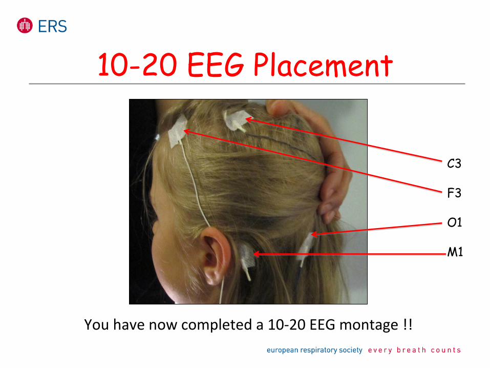
C3
F3
O1
M1
10-20 EEG Placement
You have now completed a 10‐20 EEG montage !!
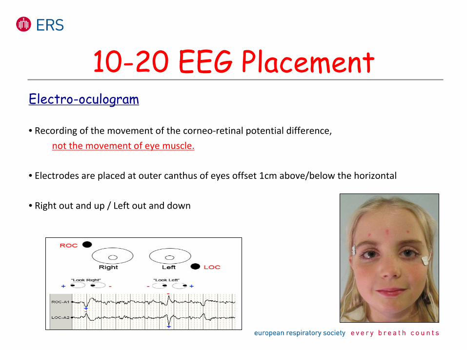
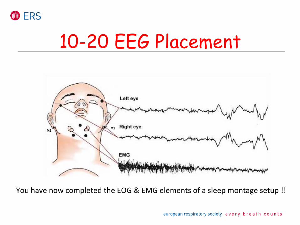
Electro-oculogram
• Recording of the movement of the corneo‐retinal potential difference,not the movement of eye muscle.
• Electrodes are placed at outer canthus of eyes offset 1cm above/below the horizontal
• Right out and up / Left out and down
10-20 EEG Placement
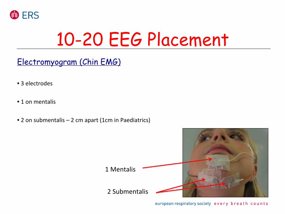
Electromyogram (Chin EMG)
• 3 electrodes
• 1 on mentalis
• 2 on submentalis – 2 cm apart (1cm in Paediatrics)
10-20 EEG Placement
1 Mentalis
2 Submentalis
10-20 EEG Placement
You have now completed the EOG & EMG elements of a sleep montage
setup !!
M1M2
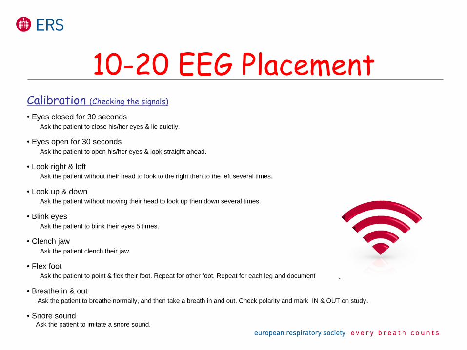
Calibration (Checking the signals)
• Eyes closed for 30 secondsAsk the patient to close his/her eyes & lie quietly.
• Eyes open for 30 secondsAsk the patient to open his/her eyes & look straight ahead.
• Look right & leftAsk the patient without their head to look to the right then to the left several times.
• Look up & downAsk the patient without moving their head to look up then down several times.
• Blink eyesAsk the patient to blink their eyes 5 times.
• Clench jawAsk the patient clench their jaw.
• Flex footAsk the patient to point & flex their foot. Repeat for other foot. Repeat for each leg and document on study.
• Breathe in & outAsk the patient to breathe normally, and then take a breath in and out. Check polarity and mark IN & OUT on study.
• Snore soundAsk the patient to imitate a snore sound.
10-20 EEG Placement
Practical SessionPractical Session
Your turn !!!
10-20 EEG Placement
The AASM annual for the Scoring of Sleep and Associated Events: Rules, Terminology and technical Specifications. Version 2.1 American Academy of Sleep Medicine (2014)
Sleep Medicine Textbook (European Sleep Research Society (ESRS)Claudio Bassetti, Zoran Dogas, Philippe Peigneux, Regensburg, (2014)
Essentials of Polysomnography.William H. Spriggs; Jones & Bartlett Publishers (2008)
Essentials of Sleep TechnologyRichard S. Rosenberg; American Academy of Sleep Medicine (2010)
Atlas of Clinical Polysomnography Second Edition (Two-volume Set) Nic Butkov Media matrix , (2011)
The ten twenty system of the International Federation. Electroencephalography and ClinicalJasper, H.H. , Neurophysiology, 1958, 10:371-375.
Polysomnographic technique: An overview. In: Sleep disorders medicine, 2nd ed. BostonChokroverty S. Butterworth Heinemann (1999)
Fundamentals of EEG technology, Volume 1: Basic concepts and methods. Tyner F, Knott J, Mayer W Jr. New York: Raven Press; (1983).
Sleep medicine.Lee-Chiong T, Sateia M, Carskadon M, (Hanley & Belfus, 2002)
Further Reading
Further Training
• Practical Polysomnography – Edinburgh, UK– Various dates
• Edinburgh Sleep Medicine Course – Edinburgh, UK– March 2016
• European Sleep School – Orihuela Costa, Spain– Various dates
• International Sleep Medicine Course – Cardiff, UK– June 2016


![Chapter 7 Lie Groups, Lie Algebras and the Exponential Mapcis610/cis61005sl8.pdf · Lie Groups, Lie Algebras and the Exponential Map 7.1 Lie Groups and Lie Algebras In Gallier [?],](https://img.pdfslide.us/doc/110x75/5f0c1a337e708231d433c07b/chapter-7-lie-groups-lie-algebras-and-the-exponential-map-cis610-lie-groups.jpg)