Embed Size (px)
Citation preview
ORIGINAL PAPER
Thalidomide influences atherogenesis in aortas of ApoE2/2/LDLR2/2 double knockout mice: a nano-CT study
Marian Kampschulte • Irina Gunkel • Philipp Stieger •
Daniel G. Sedding • Anne Brinkmann • Erik L. Ritman •
Gabriele A. Krombach • Alexander C. Langheinrich
Received: 11 September 2013 / Accepted: 20 January 2014
� The Author(s) 2014. This article is published with open access at Springerlink.com
Abstract Plaque progression in atherosclerosis is closely
connected to angiogenesis due to vasa vasorum (VV)
growth. Objective of this study was to determine the
unknown long-term effect of thalidomide on adventitial
VV neovascularization and plaque progression using nano-
focussed computed tomography (nano-CT). Proliferation
and migration assays in human coronary artery endothelial
cells (HCAEC) measured number of viable cells after
incubation with thalidomide. Male ApoE-/-/LDLR-/-
(AL) mice (n = 5) received a thalidomide containing
western diet (WD) over 29 weeks. Another five male AL
mice (WD without thalidomide) served as control group.
Descending aortas were scanned with nano-CT at
(1.5 lm)3 isotropic voxel size. Number and area of
adventitial VV as well as plaque cross sectional area were
measured. Results were complemented by histology. Tha-
lidomide inhibited proliferation and migration of HCAEC
dose-dependently. VV neovascularization decreased in
number per cross section (7.66 ± 0.301 vs. 8.62 ± 0.164,
p \ 0.001) and in cross sectional area (0.0183 ± 0.0011
vs. 0.0238 ± 0.0008 mm2, p \ 0.001). Cross sectional
area of plaque decreased significantly when treated with
thalidomide (0.57 ± 0.0187 vs. 0.803 ± 0.0148 mm2,
p \ 0.001). Nano-CT imaging revealed a reduced plaque
growth and VV neovascularization after long-term appli-
cation of thalidomide. Therefore, nano-CT can be consid-
ered as a new method to detect therapeutic effects in
experimental models of atherosclerosis.
Keywords Thalidomide � Atherosclerosis �Angiogenesis � Vasa vasorum � Nano-CT
Introduction
Progression of atherosclerotic plaques, adventitial vasa
vasorum (VV) neovascularization and even inflammation
are intrinsically tied processes. VV proliferation contrib-
utes to the progression of atherosclerotic plaque by main-
taining an increased nutrient supply to the vessel wall [1].
In human coronary arteries, VV rupture with concomitant
intraplaque haemorrhage is associated with plaque desta-
bilisation [2]. Vice versa, ruptured aortic plaques show an
increased microvessel density [3]. Imaging of atheroscle-
rotic lesions for the purpose of discrimination of different
plaque stages is imperative for risk estimation in patients
M. Kampschulte (&) � I. Gunkel � A. Brinkmann �G. A. Krombach
Department of Diagnostic and Interventional Radiology,
University Hospital Giessen, Klinikstrasse 33, 35385 Giessen,
Germany
e-mail: [email protected]
P. Stieger
Department of Cardiology, Angiology and Pneumology,
University Hospital ‘Otto-von-Guericke’ Magdeburg,
Magdeburg, Germany
P. Stieger � D. G. Sedding
Department of Internal Medicine I, Cardiology/Angiology,
Giessen University Hospital, Giessen, Germany
D. G. Sedding
Department of Cardiology and Angiology, Hannover Medical
School, Hannover, Germany
E. L. Ritman
Department of Physiology and Biomedical Engineering, Mayo
Clinic College of Medicine, Rochester, MN, USA
A. C. Langheinrich
Department of Radiology, BG Clinic Frankfurt/Main, Frankfurt,
Germany
123
Int J Cardiovasc Imaging
DOI 10.1007/s10554-014-0380-5
with atherosclerosis but still remains a challenge for
experimental and clinical CT imaging. Nano-CT represents
a technical advancement of micro-CT by using nano
focused X-ray tubes. The technique has proven its capa-
bility to identify and characterize vascular pathologies with
different plaque types, demonstrated VV neovascularisa-
tion and plaque growth in aortas of ApoE-/-/LDLR-/-
double knockout mice (AL mice) [4]. Despite of plaque
growth in the ascending aorta and the aortic arch during the
lifespan of AL mice, VV density is known to decrease at
these sites due to regressive fibro calcification of plaque. In
contrast, plaque growth and VV density increases in the
descending aorta during the lifespan of AL mice showing
instable plaque with intraplaque hemorrhage [4]. Never-
theless, there is no experience in the application of this
technique for studying pharmaceutical effects concerning
the progress or arrest of atherosclerosis.
Thalidomide is an anti-angiogenic drug [5] with a broad
spectrum of current clinical applications ranging from
therapy of multiple myeloma [6] to erythema nodosum
leprosum [7]. In addition to these clinical applications,
different studies proved the molecular effects of thalido-
mide to reduce systemic and local tumor necrosis factor
alpha (TNF-a) production and VV neovascularisation in
early atherosclerotic lesions with a concomitant decrease of
lesion growth [8–10]. Irrespective of molecular mecha-
nisms and their inhibition by thalidomide, sprouting of
microvasculature depends on endothelial cell (EC) migra-
tion and proliferation. Currently, the effect of thalidomide
on human arterial ECs concerning migration and prolifer-
ation is not fully known.
The current nano-CT study was designed to tests the
hypothesis that thalidomide (1) influences human coronary
artery endothelial cells (HCAECs) function i.e. migration
and proliferation, (2) reduces adventitial VV neovascul-
atization and (3) reduces plaque formation in the
descending aorta of AL mice with advanced atherosclero-
sis. Thereby this study demonstrates the potential of nano-
CT as a new and suitable imaging method for preclinical
research on therapeutics in atherosclerosis.
Materials and methods
Endothelial cell function
To clarify the effect of thalidomide on EC function, cells
were incubated in FCS containing growth medium. Tha-
lidomide was dissolved in dimethyl sulfoxide (DMSO) and
added in different concentrations to observe whether there
is a dose dependent inhibition of proliferation and migra-
tion of HCAEC.
Proliferation
Human coronary artery EC (HCAEC, Cambrex, Verviers,
Belgium) proliferation was determined using a BrdU
incorporation assay kit (cell proliferation ELISA, Roche,
Mannheim, Germany). Briefly, HCAEC were seeded on
1 % gelatin-coated on 96-well plates at a seeding density of
3 9 103 cells per well in endothelial growth medium
(EGM-2, Cambrex) for 24 h, followed by serum starvation
and pre-treatment with different concentrations of thalid-
omide between 50 and 250 lg/ml for 24 h. Afterwards
cells were stimulated with EGM-2 for 18 h again in the
presence or absence of thalidomide in same concentrations.
BrdU labeling solution was applied for another 6 h and
BrdU incorporation was detected according to the manu-
facturer’s instructions. Number of viable cells in growth
factor enriched buffer alone was set to a value of 100 %.
Number of viable cells resulting from different thalidomide
concentrations was compared to the number of cells in
DMSO-enriched buffer.
Migration
Human coronary artery endothelial cells (Cambrex, Ver-
viers, Belgium) were grown in endothelial growth medium
(EGM-2, Cambrex) at 37 �C and 5 % CO2. ECs from
passage 2–4 were used for all experiments. Cells were
serum starved in 0.5 % fetal bovine serum for 12 h in the
presence or absence of thalidomide in concentrations of
500 or 250 lg/ml respectively. Cell migration was mea-
sured using a modified Boyden chamber assay. For this
purpose, cells were detached with 0.05 % trypsin/
0.53 mmol/l EDTA, washed, and the cell suspension
(250 ll, 5 9 105 cells/well) was added to the transwell
insert (8-lm pore size, Costar, Cambridge, MA, USA).
Then, 750 ll of basal medium containing 50 ng/ml human
VEGF-A165 (PeproTech Inc, Rocky Hill, NJ, USA) was
added to the lower chamber and cells were incubated for
6 h in the presence or absence of different concentrations
of thalidomide (500 or 250 lg/ml).
After incubation, non-migrated cells were removed from
the upper surface of the insert with a cotton swab. Fol-
lowing reinsertion of transwell inserts, WST reagent
(Roche, Mannheim, Germany) was added to the lower
chamber and incubated for further 30 min. After removing
the inserts, the converted formazan, which directly corre-
lates to the number of viable cells, was quantified in a
microplate reader (Tecan sunrise, Crailsheim, Germany) at
450 nm. Number of viable cells in DMSO-enriched buffer
alone were set to a value of 100 % and compered to the
number of cells resulting from different thalidomide
concentrations.
Int J Cardiovasc Imaging
123

Animal experiments
Animal studies were performed according to the guideline
for animal protection and approved by the institutional
animal care and use committee. Thalidomide was added to
a fat and cholesterol enriched western type diet (ssniff TD
88137, ssniff Spezialdiaten GmbH, Soest, Germany) and
administered with a mean daily dose of 200 mg/kg/day to
male AL mice (n = 5, Charles Rivers Wiga, Sulzbach,
Germany) beginning at the age of 6 weeks. Coeval male
AL mice (n = 5) with the same western diet (WD) but
without thalidomide served as controls. Chocolate taste
was added to both diets to conceal potentially disgusting
taste. Animals were housed in a specific pathogen free
environment with access to food and water ad libitum and a
12/12 h day/night cycle. After 29 weeks of diet, i.e. at age
of 35 weeks, the animals were euthanized with a fatal dose
of inhaled isoflourane. The left ventricle was cannulated
and injected with heparinized saline (10 ml of 0.9 %
sodium chloride with 1,000 IU heparin) until the venous
effluent from the right atrium was free from blood. A
plumbiferous radiopaque polymer (Microfil� MV-122,
Flow Tech, Carver, MA, USA) was injected into the left
ventricle at a nominal pressure of 100 mmHg until it
emerged from the inferior vena cava. After polymerization
of the compound, the heart and entire aorta were removed
and immersed in 4 % neutral buffered formalin.
Nano-CT
Descending aortas were scanned along the vertical axis
using a nano-CT (SkyScan_2011, Bruker Micro-CT,
Kontich, Belgium). The scanning system consists of a nano
focus, pumped open type X-ray source with a lanthanium
boride cathode. The electron beam is focused by two
electromagnetic lenses and directed on a tungsten coated
beryllium window with a minimum spot size of 400 nm.
Tube current was 180 lA and tube voltage was set to
produce a 40 kVp transmission X-ray cone beam. Samples
were positioned on a computer controlled rotation stage
and scanned over 180� in steps of 0.3� around their vertical
axis. Acquisition time of each projection image was 2.4 s.
For the purpose of noise reduction projection images were
acquired repeatedly with averaging of four frames leading
to a scan time up to 2 h. The X-ray detector consists of a
12-bit digital CCD high-resolution (1,280 9 1,024 pixel)
camera with fibre optic 3.7:1 coupling to an X-ray scin-
tillator and digital frame-grabber. Tomographic recon-
struction, using the multi-angular projection image data
was performed using a modified Feldkamp cone beam
reconstruction algorithm. The resulting voxel resolution
was (1.5 lm)3 isotropic voxel size.
Image analysis
A total of 1,084 cross sectional nano-CT images was
analyzed. Measurements were performed using the ANA-
LYZE Software packages (ANALYZE 9.0/10.0, Mayo
Clinic, Rochester, MN, USA). Atherosclerotic plaque can
be detected by virtue of the intraluminal encroachment by
the plaque as well as the different X-ray attenuation
properties (i.e. CT gray scale) that differentiate the plaque
from the intraluminal contrast agent and the normal sur-
rounding tissues of the arterial wall. After blinding of
thalidomide-group versus control group to the examiner,
VV and atherosclerotic plaques were manually segmented
and measured in every tenth subsequent slice of each data
set. Quantified parameters were total number of VV, VV
cross sectional area (mm2) and plaque area (mm2) per slice
as described previously [4].
Histology
Subsequent to nano-CT scanning, aortas were dehydrated and
embedded in paraffin. Contiguous serial sections (6 lm slice
thickness) within each sample were mounted on a microscope
slide and stained with haematoxylin and eosin. Masson tri-
chrome identified collagen and elastic fibers used to demon-
strate connective tissue (adventitial border). Sequential nano-
CT slices were examined for best side-by-side matching of
histology and findings of the nano-CT images.
Statistical analysis
All data are presented as mean ± SEM. Statistical analysis
was performed on JMP 6.0 (SAS, Cary, NC, USA). One-
way ANOVA, followed by a Tukey–Kramer post hoc test
with correction for multiple comparisons was used to
identify the statistical differences among groups. Individual
group comparison was performed by an unpaired Student
t test. A value of p \ 0.05 was considered significant in all
analyses. Box–whisker plots were used to illustrate results
of statistical findings of nano-CT image analysis, bar
graphs were used to demonstrate EC function.
Results
Thalidomide inhibits EC proliferation and migration
Compared to growth factor incubated cells, thalidomide
significantly inhibited the proliferation of EC dependent to
the applicated dose in BrdU assay [42.4 ± 6.6 % (control)
vs. 33.4 ± 3 % (thalidomide: 250 lg/m), p \ 0.05] with
findings of significance at concentrations of thalidomide of
100 lg/ml and higher (Table 1). Additionally, migration of
Int J Cardiovasc Imaging
123

ECs was significantly inhibited by thalidomide in a dose
dependent manner (p \ 0.05, Fig. 1) at 250 lg/ml
(90.5 ± 0.53 %) and at 500 lg/ml thalidomide
(81.8 ± 1.4 %).
Thalidomide inhibits VV neovascularization
and growth of atherosclerotic lesions
Images obtained by nano-CT enabled a 3D visualization of
the aorta and an analysis of plaque characteristics over the
entire length of the imaged sample. At the age of 35 weeks,
AL mice demonstrate adventitial VV neovascularization
and atherosclerotic plaques in the descending aorta
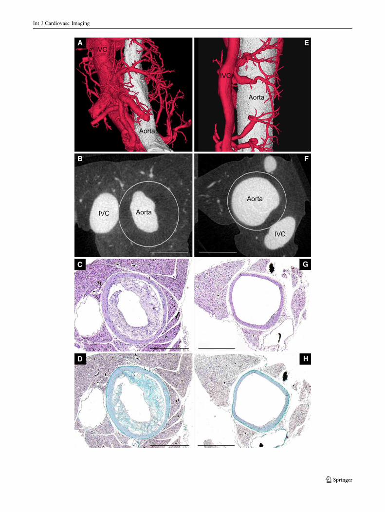
(Fig. 2a, b, e, f). This was confirmed by histology (Fig. 2c,
d, g, h).
Compared to controls, quantitative nano-CT data analysis
revealed that the cross sectional area of adventitial VV was
significantly decreased in thalidomide treated mice
(0.0183 ± 0.0011 vs. 0.0238 ± 0.0008 mm2, p \ 0.001,
Fig. 3a). This was accompanied by a decrease of the total
number of VV per cross sectional image (7.66 ± 0.301 vs.
8.62 ± 0.164, p \ 0.001; Fig. 2a, b vs. e, f; Fig. 3b). Simul-
taneously, thalidomide fed animals demonstrated a significant
decrease of atherosclerotic plaque area (0.57 ± 0.0187 vs.
0.803 ± 0.0148 mm2, p \ 0.001; Fig. 2b–d vs. f–h, Fig. 4).
Discussion
The major findings of this study are that thalidomide inhibits
(1) proliferation and migration in HCAEC in vitro, (2)
adventitial VV growth and (3) growth of atherosclerotic
plaque in the descending aorta of AL mice. It supports tha-
lidomide’s effectiveness in an experimental model of ath-
erosclerosis, which shows morphologically comparable
findings to human atherosclerosis. The study underlines the
value of nano-CT for quantitative imaging of atherosclerosis.
Thalidomide and endothelial function
Thalidomide was introduced to the European market in
1957 as a sedative and withdrawn in 1961 after press
reports about intake of thalidomide during pregnancy and a
Table 1 Impact of thalidomide on HCAEC proliferation: BrdU assay
of HCAECs in presence or absence of different concentrations of
thalidomide
Concentration of
thalidomide (lg/ml)
GF DMSO Number of
cells (%)
Significance
0 ? - 100 –
0 - ? 23.8 ± 5.9 –
0 ? ? 42.4 ± 6.6 –
50 ? ? 40.3 ± 5.3 n.s.
100 ? ? 38.2 ± 4.2 p \ 0.05
150 ? ? 36.9 ± 4.1 p \ 0.05
200 ? ? 34.4 ± 3.2 p \ 0.05
250 ? ? 33.4 ± 3.1 p \ 0.05
It shows a dose dependent significantly reduced replication of DNA at
concentrations of thalidomide at 100 lg/ml and higher, indicating a
reduced proliferation
Fig. 1 Effect of thalidomide on
EC migration: migration of
HCAECs is significantly
inhibited and dose depending
from concentration of
thalidomide. Left column FCS
without thalidomide (control),
middle column
FCS ? thalidomide 250 lg/ml;
right column
FCS ? thalidomide 500 lg/ml
cFig. 2 Descending aortas of AL mice at the age of 35 weeks with
29 weeks of administration of thalidomide (right column) and control
diet (left column). 3D volume rendered nano-CT (a, e) at (1.5 lm)3,
axial single slices (b, f), HE-Staining (c, g) and Masson Trichrome (d,
h) demonstrate the reduced number of VV and the smaller plaque
volume in thalidomide treated animals. Corresponding histology
depicts the plaque size and adventitial VV locations similar to nano-
CT imaging. In contrast to nano-CT, histological preparation incl.
cutting leads to artificial disruption of the plaque (c, d) and tears out
polymerized contrast agent (Microfil) from vessel lumen (c, d, g, h).
The white circle defines the border of tunica media to the adventitial
tissue
Int J Cardiovasc Imaging
123
Int J Cardiovasc Imaging
123

series of birth defects such as phocomelia or aplasia of
extremities. Recent investigations identified cereblon as a
protein, responsible for limb outgrowth, which is inhibited
by binding to thalidomide [11]. It was banned from therapy
over several decades. During the last two decades, it
experiences a renaissance due to re-evaluation of its dif-
ferent therapeutic effect. There is evidence that thalido-
mide influences the expression profile of inflammatory
cytokines and angiogenetic factors in different animal and
disease models [10, 12–14]. Thereby thalidomide inhibited
early neointima formation and reduced VV neovasculari-
zation in coronary arteries of WD fed pigs, combined with
reduced levels of TNF-a and VEGF [9] and inhibition of
early atherogenesis in ApoE deficient mice [8]. Neverthe-
less, these data do not clarify the impact of thalidomide on
function of human ECs neither its influence to plaque and
VV growth over a long time span in a model of enhanced
atherosclerosis starting at an early time point.
Migration and proliferation of ECs are basic require-
ments for angiogenesis [15] as seen in VV neovasculari-
zation. They can be affected in several ways. In the present
study, we demonstrate that thalidomide prevents prolifer-
ation and migration of human HCAECs. These findings
support previous investigations showing that thalidomide
leads to reduced proliferation in human umbilical venous
endothelial cells and influences bone marrow angiogenesis
in multiple myeloma [16, 17]. It expands the knowledge of
thalidomide’s influence on human ECs, showing that it
affects arterial as well as venous ECs.
Quantitative nano-CT for assessment of plaque
formation and VV neovascularization
We hypothesized, that a long time application of thalido-
mide might inhibit lesion progression in concert with VV
neovascularization in the descending aorta of AL mice and
can be monitored using nano-CT. Similar therapeutic
effects were observed for short time application of 3-de-
azaadenosine to AL mice in early atherosclerosis [18]
indicating that growth of atherosclerosic plaque can be (1)
influenced in this model of experimental atherosclerosis
and (2) monitored using micro-CT. Feeding a thalidomide-
enriched WD diet over 29 weeks means a long time
exposure of thalidomide to AL mice when assuming a life
span of 70–90 weeks in this animal model. At the end of
this long time exposure an effect with decreased plaque
size was seen. This fact bridges findings concerning the
inhibition of early atherogenesis by thalidomide [8, 9] and
a late inhibition by angiostatin [19]. Nano-CT proved
thalidomide’s influence on VV neovascularization as well
as plaque growth in (1) advanced stages of the disease and
(2) over a long time span of application. Since plaque type
(fibrotic) was the same in both groups, our results indicate a
lesser neovascularization i.e. nutrition supply of the vessel
wall in concert with thalidomide administration.
For studying this purpose, nano-CT is a versatile and
helpful technique that offers additional features for
Fig. 3 Effects of thalidomide on plaque neovascularization: animals
treated with thalidomide demonstrate a significantly reduced total
lumen cross sectional area (a) and total number (b) of adventitial VV
compared to controls
Fig. 4 Quantitative nano-CT: effects of thalidomide on plaque size:
animals treated with thalidomide demonstrate a significantly reduced
cross sectional area of atherosclerotic lesions compared to controls
Int J Cardiovasc Imaging
123

quantitative assessment of atherosclerosis. Just as micro-
CT, nano-CT combines non-disruptive ex vivo tissue
characterization with 3D visualization and quantification of
major vessels (aorta) and its pathognomonic VV micro
vascularization. It exceeds micro-CTs spatial resolution to
a level of sub micrometer voxel size and provides the
opportunity to assess vessels of capillary size. Thereby
nano-CT depicts the 3D architecture of the complete vas-
cular tree. In the past, micro-CT measured ‘‘density’’ i.e.
changes of gray scale values in atherosclerotic plaque often
remained speculative between cellular plaque composition
and contrast agent within intraplaque microvessels [20].
Due to its superior spatial resolution, nano-CT can assess
pathologies such as intraplaque hemorrhage beyond the
limits of micro-CT.
Limitations
The study was performed in a limited number of animals
(n = 5 in each group). Blinded image analysis revealed
findings with clear statistical significant differences prov-
ing (1) the effect of thalidomide and moreover (2) the
applicability of nano-CT for research in atheroscloris. On
the other hand, feeding a WD means a metabolic stress
leading to obesity and cutaneous xanthoma. Therefore we
abstained from a further enlargement of groups. This study
proves a reduction of HCAECs viability but was not
designed to clarify the specific molecular targets and
effects on cellular metabolism in HCAECs. Application of
thalidomide began prior to the obvious appearance of VV
and atherosclerotic plaque. It needs to be clarified if tha-
lidomide even acts on growth of pre-existing plaque and
VV and if it takes influence on the course of the apparent
disease simulating a pharmacological intervention after the
onset of atherosclerosis. Further research should compar-
atively investigate molecular similarities and differences of
plaque development in AL mice and humans and thalido-
mide’s mechanistic influence on both. Against this back-
ground, 3D imaging with high spatial resolution as
provided by nano-CT has the potential to prove the impact
of molecular findings on morphological alterations in
atherosclerosis.
Conclusions
Vasa vasorum neovascularization is associated to plaque
development. A continued application of thalidomide even
over a longer time course reduces both in WD fed AL
mice. The data demonstrate nano-CT’s value as a tool to
investigate the influence of preclinical drug interventions
on atherosclerosis at a microscopic level with 3D data sets
in isotropic voxel size. Future research in the field of
atherosclerosis inclusive research on novel therapeutics
and other topics of microvascular research such as tumor,
fracture and inflammation should consider nano-CT as a
powerful imaging modality.
Acknowledgments We would like to thank G. Martels, Justus-
Liebig University Giessen, Germany and Steven M. Jorgensen of
Mayo Clinic College of Medicine for technical assistance. The
investigation was supported in part from the faculty of human med-
icine of the Justus-Liebig University Giessen, Germany and the DFG
(German Research Foundation—NST 162/291-1 FUGG).
Conflict of interest None
Open Access This article is distributed under the terms of the
Creative Commons Attribution License which permits any use, dis-
tribution, and reproduction in any medium, provided the original
author(s) and the source are credited.
References
1. Mulligan-Kehoe MJ (2010) The vasa vasorum in diseased and
nondiseased arteries. Am J Physiol Heart Circ Physiol
298(2):H295–H305
2. Kolodgie FD, Gold HK, Burke AP, Fowler DR, Kruth HS, Weber
DK, Farb A, Guerrero LJ, Hayase M, Kutys R, Narula J, Finn
AV, Virmani R (2003) Intraplaque hemorrhage and progression
of coronary atheroma. N Engl J Med 349(24):2316–2325
3. Moreno PR, Purushothaman KR, Fuster V, Echeverri D, Trus-
zczynska H, Sharma SK, Badimon JJ, O’Connor WN (2004)
Plaque neovascularization is increased in ruptured atherosclerotic
lesions of human aorta: implications for plaque vulnerability.
Circulation 110(14):2032–2038
4. Kampschulte M, Brinkmann A, Stieger P, Sedding DG, Dierkes
C, Bohle RM, Krombach G, Ritman EL, Langheinrich AC (2010)
Quantitative CT imaging of the spatio-temporal distribution
patterns of vasa vasorum in aortas of apoE-/-/LDL-/- double
knockout mice. Atherosclerosis 212(2):444–450
5. D’Amato RJ, Loughnan MS, Flynn E, Folkman J (1994) Tha-
lidomide is an inhibitor of angiogenesis. Proc Natl Acad Sci USA
91(9):4082–4085
6. Witzig TE, Laumann KM, Lacy MQ, Hayman SR, Dispenzieri A,
Kumar S, Reeder CB, Roy V, Lust JA, Gertz MA, Greipp PR,
Hassoun H, Mandrekar SJ, Rajkumar SV (2013) A phase III
randomized trial of thalidomide plus zoledronic acid versus
zoledronic acid alone in patients with asymptomatic multiple
myeloma. Leukemia 27(1):220–225
7. Chen M, Doherty SD, Hsu S (2010) Innovative uses of thalido-
mide. Dermatol Clin 28(3):577–586
8. Chew M, Zhou J, Daugherty A, Eriksson T, Ellermann-Eriksen S,
Hansen PR, Falk E (2003) Thalidomide inhibits early athero-
genesis in apoE-deficient mice. APMIS Suppl 109:113–116
9. Gossl M, Herrmann J, Tang H, Versari D, Galili O, Mannheim D,
Rajkumar SV, Lerman LO, Lerman A (2009) Prevention of vasa
vasorum neovascularization attenuates early neointima formation
in experimental hypercholesterolemia. Basic Res Cardiol
104(6):695–706
10. Moreira AL, Wang J, Sarno EN, Kaplan G (1997) Thalidomide
protects mice against LPS-induced shock. Braz J Med Biol Res
30(10):1199–1207
11. Ito T, Ando H, Suzuki T, Ogura T, Hotta K, Imamura Y, Yam-
aguchi Y, Handa H (2010) Identification of a primary target of
thalidomide teratogenicity. Science 327(5971):1345–1350
Int J Cardiovasc Imaging
123

12. Ishikawa M, Kanno S, Takayanagi M, Takayanagi Y, Sasaki K
(1998) Thalidomide promotes the release of tumor necrosis fac-
tor-alpha (TNF-alpha) and lethality by lipopolysaccharide in
mice. Biol Pharm Bull 21(6):638–640
13. Kawai T, Watanabe N, Yokoyama M, Arai K, Oana S, Harayama
S, Yasui K, Oh-Ishi T, Onodera M (2013) Thalidomide attenuates
excessive inflammation without interrupting lipopolysaccharide-
driven inflammatory cytokine production in chronic granuloma-
tous disease. Clin Immunol 147(2):122–128
14. Yasui K, Yashiro M, Tsuge M, Manki A, Takemoto K, Ya-
mamoto M, Morishima T (2010) Thalidomide dramatically
improves the symptoms of early-onset sarcoidosis/Blau syn-
drome: its possible action and mechanism. Arthritis Rheum
62(1):250–257
15. Bauer SM, Bauer RJ, Velazquez OC (2005) Angiogenesis, vas-
culogenesis, and induction of healing in chronic wounds. Vasc
Endovascular Surg 39(4):293–306
16. Marks MG, Shi J, Fry MO, Xiao Z, Trzyna M, Pokala V, Ihnat
MA, Li PK (2002) Effects of putative hydroxylated thalidomide
metabolites on blood vessel density in the chorioallantoic mem-
brane (CAM) assay and on tumor and endothelial cell prolifera-
tion. Biol Pharm Bull 25(5):597–604
17. Kumar S, Witzig TE, Dispenzieri A, Lacy MQ, Wellik LE,
Fonseca R, Lust JA, Gertz MA, Kyle RA, Greipp PR, Rajkumar
SV (2004) Effect of thalidomide therapy on bone marrow angi-
ogenesis in multiple myeloma. Leukemia 18(3):624–627
18. Langheinrich AC, Sedding DG, Kampschulte M, Moritz R,
Wilhelm J, Haberbosch WG, Ritman EL, Bohle RM (2009)
3-Deazaadenosine inhibits vasa vasorum neovascularization in
aortas of ApoE(-/-)/LDL(-/-) double knockout mice. Ath-
erosclerosis 202(1):103–110
19. Moulton KS, Vakili K, Zurakowski D, Soliman M, Butterfield C,
Sylvin E, Lo KM, Gillies S, Javaherian K, Folkman J (2003)
Inhibition of plaque neovascularization reduces macrophage
accumulation and progression of advanced atherosclerosis. Proc
Natl Acad Sci USA 100(8):4736–4741
20. Langheinrich AC, Michniewicz A, Sedding DG, Walker G,
Beighley PE, Rau WS, Bohle RM, Ritman EL (2006) Correlation
of vasa vasorum neovascularization and plaque progression in
aortas of apolipoprotein E(-/-)/low-density lipoprotein(-/-)
double knockout mice. Arterioscler Thromb Vasc Biol
26(2):347–352
Int J Cardiovasc Imaging
123