Embed Size (px)
Citation preview
10/27/14
1
Bone Marrow Aspirate and Biopsy: A Guide for Advanced Practitioners in Oncology Sandra Kurtin, RN, MS, AOCN®, ANP-C The University of Arizona Cancer Center Tucson, Arizona
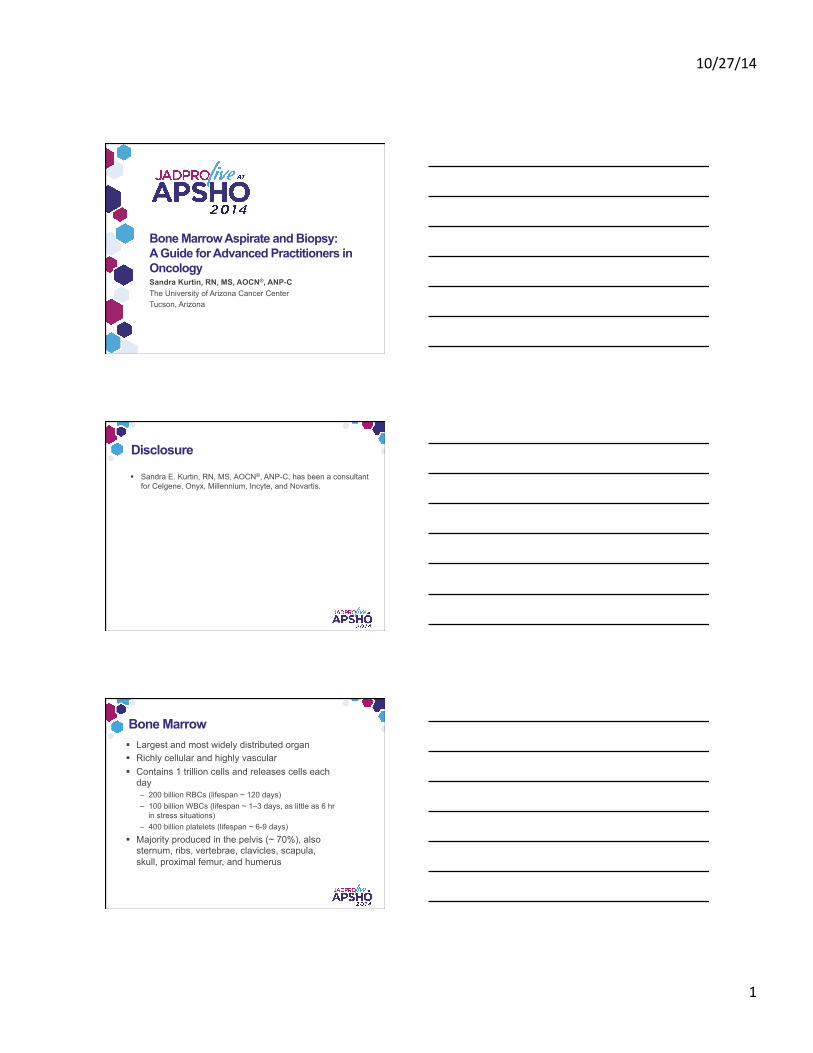
Disclosure
§ Sandra E. Kurtin, RN, MS, AOCN®, ANP-C, has been a consultant for Celgene, Onyx, Millennium, Incyte, and Novartis.
Bone Marrow § Largest and most widely distributed organ § Richly cellular and highly vascular § Contains 1 trillion cells and releases cells each
day – 200 billion RBCs (lifespan ~ 120 days) – 100 billion WBCs (lifespan ~ 1–3 days, as little as 6 hr
in stress situations) – 400 billion platelets (lifespan ~ 6-9 days)
§ Majority produced in the pelvis (~ 70%), also sternum, ribs, vertebrae, clavicles, scapula, skull, proximal femur, and humerus
10/27/14
2
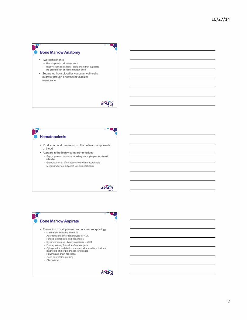
Bone Marrow Anatomy
§ Two components – Hematopoietic cell component – Highly organized stromal component that supports
the proliferation of hematopoietic cells
§ Separated from blood by vascular wall–cells migrate through endothelial vascular membrane
Hematopoiesis
§ Production and maturation of the cellular components of blood
§ Appears to be highly compartmentalized – Erythropoiesis: areas surrounding macrophages (erythroid
islands) – Granulopoiesis: often associated with reticular cells – Megakaryocytes: adjacent to sinus epithelium
Bone Marrow Aspirate
§ Evaluation of cytoplasmic and nuclear morphology – Maturation: including blasts % – Auer rods and other tdt analysis for AML – Ringed sideroblasts and iron stores – Dyserythropoiesis, dysmyelopoiesis – MDS – Flow cytometry for cell surface antigens – Cytogenetics to detect chromosomal aberrations that are
diagnostic and/or prognostic for disease – Polymerase chain reactions – Gene expression profiling – Chimerisms
10/27/14
3
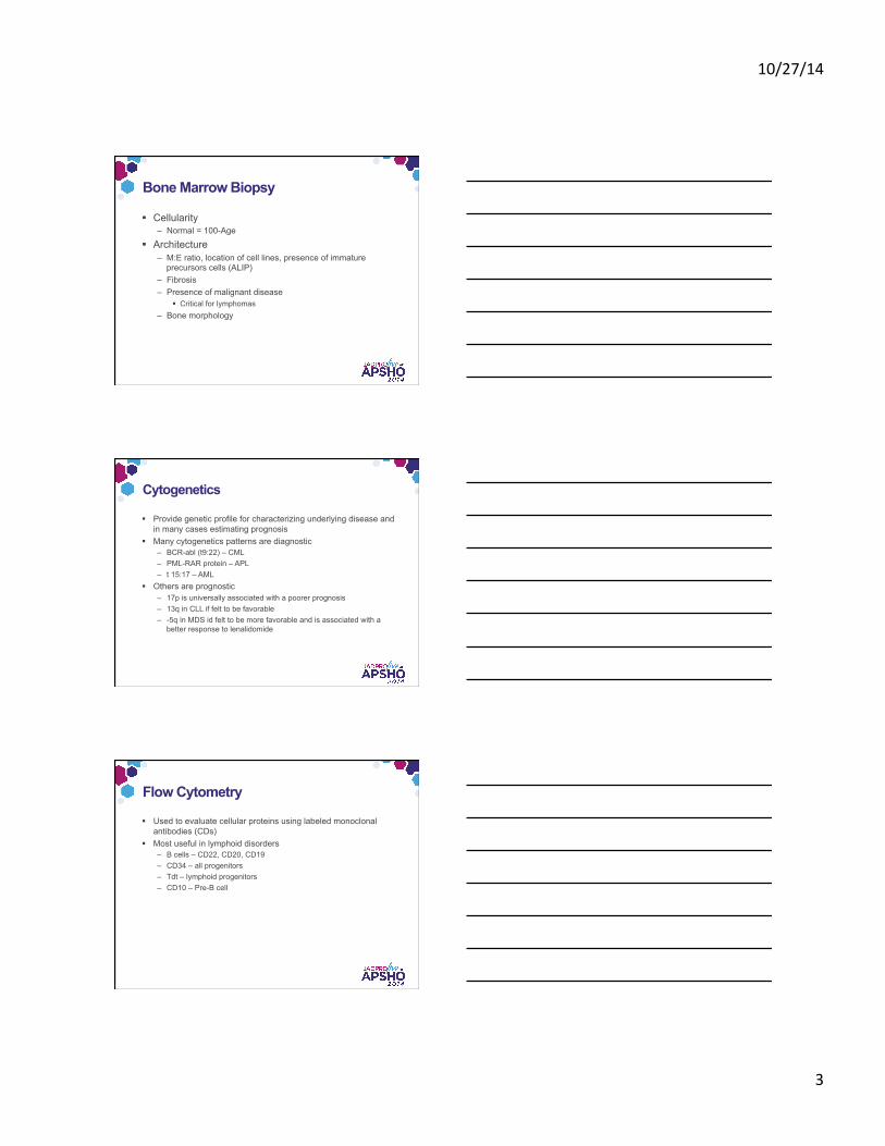
Bone Marrow Biopsy
§ Cellularity – Normal = 100-Age
§ Architecture – M:E ratio, location of cell lines, presence of immature
precursors cells (ALIP) – Fibrosis – Presence of malignant disease
§ Critical for lymphomas – Bone morphology
Cytogenetics
§ Provide genetic profile for characterizing underlying disease and in many cases estimating prognosis
§ Many cytogenetics patterns are diagnostic – BCR-abl (t9:22) – CML – PML-RAR protein – APL – t 15:17 – AML
§ Others are prognostic – 17p is universally associated with a poorer prognosis – 13q in CLL if felt to be favorable – -5q in MDS id felt to be more favorable and is associated with a
better response to lenalidomide
Flow Cytometry
§ Used to evaluate cellular proteins using labeled monoclonal antibodies (CDs)
§ Most useful in lymphoid disorders – B cells – CD22, CD20, CD19 – CD34 – all progenitors – Tdt – lymphoid progenitors – CD10 – Pre-B cell
10/27/14
4
Common Indications for Bone Marrow Biopsy and Aspirate
Other
§ Unexplained anemia § Macrocytic anemia § Unexplained leukopenia § Unexplained thrombocytopenia § Pancytopenia
Staging for Suspected Malignancies
§ Multiple myeloma: due to elevated M-proteins
§ Amyloidosis: usually § Lymphoma: usually due to presence of
adenopathy or organomegaly – Non-Hodgkin's lymphoma – Hodgkin lymphoma – Unexplained splenomegaly – Chronic lymphocytic leukemia (CLL)
§ Myeloid malignancies: usually due to peripheral blasts, cytopenias, or abnormal findings on the peripheral smear
– AML – CML – MDS – Myeloproliferative neoplasms – Hairy cell leukemia – Aplastic anemia
§ Solid tumor metastases to the bone marrow
Cytopenias
§ Fever of unknown origin § Suspected chromosomal disorders
in neonates § Suspected storage disease (eg,
Gaucher disease, Niemann-Pick) § Confirmation of normal marrow in
potential allogeneic donor § Evaluation of iron stores
Key Elements of the Procedure: Preparing the Patient
1. Pre-procedural patient education – Informed Consent – Rationale – Risks – Alternatives – Procedure description – After care
2. Time-out procedure 3. Allow the patient to visit the bathroom prior to the procedure
– You can organize your supplies while they are away 4. Confirm indication, tests to be performed on specimen, amount of
aspirate, and appropriate tubes for processing 5. Assemble supplies: Many trays have extra supplies – I like to keep my
tray tidy – get rid of things you don’t need, and organize you tray to make it easy to find the things you need.
6. Positioning of the patient – Side lying – lateral decubitus – may be used for posterior iliac crest or anterior
iliac spine – Prone – for posterior iliac crest – Supine – sternal aspirate only
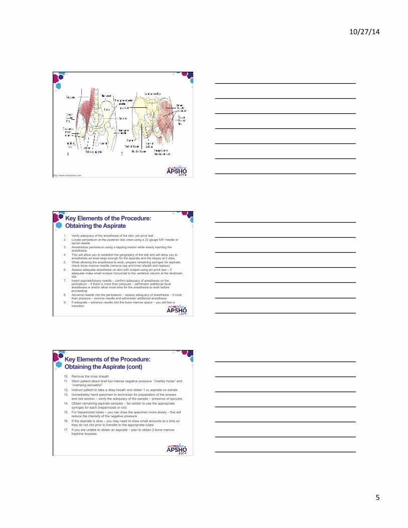
Key Elements of the Procedure: Site Selection and Preparation
1. Establishing landmarks – Posterior iliac crest is most common – Anterior superior iliac spine may be used in selected patients:
§ Extensive radiation to the pelvis (prostate or rectal cancers) – Unable to position to access posterior iliac crest – Sternal site is only used for aspirates – no core can be obtained
§ Generally in morbidly obese patients – http://www.innerbody.com/image_skelfov/skel18_new.html
2. Sterile preparation of the site – most commonly chlorhexidine 3. Anesthetize the skin at the selected site
§ 1%–2% lidocaine
§ Lidocaine may be buffered to decrease the burning associated with injection
Ruegg et al. Oncol Nurs Forum. 2009;36:52–60
10/27/14
5
http://www.endoszkop.com
Key Elements of the Procedure: Obtaining the Aspirate
1. Verify adequacy of the anesthesia of the skin: pin-prick test 2. Locate periosteum at the posterior iliac crest using a 22 gauge 5/8” needle or
spinal needle 3. Anesthetize periosteum using a tapping motion while slowly injecting the
anesthesia 4. This will allow you to establish the geography of the site and will allow you to
anesthetize an area large enough for the aspirate and the biopsy at 2 sites. 5. While allowing the anesthesia to work, prepare remaining syringes for aspirate,
check bone marrow needle (remove cap and inner sheath and replace) 6. Assess adequate anesthesia on skin with scalpel using pin-prick test – if
adequate make small incision horizontal to the vertebral column at the landmark site
7. Insert aspirate/biopsy needle – confirm adequacy of anesthesia on the periosteum – if there is more than pressure – administer additional local anesthesia or and/or allow more time for the anesthesia to work before proceeding
8. Advance needle into the periosteum – assess adequacy of anesthesia – if more than pressure – remove needle and administer additional anesthesia
9. If adequate – advance needle into the bone marrow space – you will feel a transition
Key Elements of the Procedure: Obtaining the Aspirate (cont) 10. Remove the inner sheath 11. Warn patient about brief but intense negative pressure: “charley horse” and
“cramping sensation” 12. Instruct patient to take a deep breath and obtain 1 cc aspirate on exhale 13. Immediately hand specimen to technician for preparation of the smears
and clot section – verify the adequacy of the sample – presence of spicules 14. Obtain remaining aspirate samples – be certain to use the appropriate
syringes for each (heparinized or not) 15. For heparinized tubes – you can draw the specimen more slowly – this will
reduce the intensity of the negative pressure 16. If the aspirate is slow – you may need to draw small amounts at a time so
they do not clot prior to transfer to the appropriate tubes 17. If you are unable to obtain an aspirate – plan to obtain 2 bone marrow
trephine biopsies
10/27/14
6
Key Elements of the Procedure: Obtaining the Biopsy
1. Bring the biopsy needle just out of the periosteum 2. Reposition to a second site within the area of anesthesia 3. Confirm adequacy of local anesthesia 4. Insert needle with inner sheath in place until you are anchored
in the outer cortex, then remove 5. Continue inserting the needle into the bone until you are in the
bone marrow space 6. Insert the inner sheath gently into the needle – this will give you
an idea of now big the core will be – a core of at least 1.5 cm is recommended
7. Rotate the needle clockwise 3-5 turns
Key Elements of the Procedure: Obtaining the Biopsy (cont) 8. While applying pressure to the needle, shake the needle to help
loosen the core 9. Rotate the needle back and forth while removing the core –
instruct the patient to take a deep breath and remove the core on exhale
10. Warn the patient about a quick pinch and a tug 11. Use the shepherd’s hook to remove the core from the needle to
assess adequacy of the sample – The core is used to prepare touch preps and is then placed
into solution 12. Apply pressure to the site 13. Apply sterile dressing
Key Elements of the Procedure: Post-procedural Care and Education § Continue pressure to the site for at least 3-5 minutes § Apply a pressure dressings to the site § Provide the patient with instruction for after care
– Keep the dressing on overnight – The patient may shower (not bathe) 24 hours after the procedure – Keep the dressing on when they get in to the shower – Take the dressing off when they get out of the shower – Apply a Band-Aid to the site for an additional 24–48 hours – No tub-bath, Jacuzzi or swimming (anything that would imply soaking in
water) for at least 48 hours following the procedure § Arrange for a follow-up call the day following the procedure § Reportable signs and symptoms
– Excessive pain at the biopsy site or radiating to the legs – Bleeding or excessive bruising at the biopsy site or in the legs – Pain or swelling above or below the biopsy site – Fevers or chills – Numbness at or below the biopsy site
10/27/14
7
References Al-Ibraheemi, A., Pham. T., Hen, L., et al. (2013). Comparison between 1-needle technique versus 2-needle technique for bone marrow aspiration and biopsy procedures. Arch Pathol Lab Med, 137, 974-978. doi:10.5858/arpa.2012-0046-OA.
American Society of Hematology. (2014). Medicare Hospital Outpatient PPS Final Rule for 2014: Summary of Major Provisions. http://www.hematology.org/Advocacy/Policy-News/2014/3034.aspx
Bain, B.J. (2003). Bone marrow biopsy morbidity and mortality. Br J Haematol, 121, 949-951.
Berber, I., Erkurt, M.A., Kuku., I., et al. ( 2014). An unexpected complication of bone marrow aspiration and trephine biopsy: arteriovenous fistula. Med Princ Pract, 23, 380–383. doi:10.1159/000358173
Malempati, S., Joshi, S., Lai, S., et al. (2009). Bone marrow aspiration and biopsy. N Engl J Med, 361, e28. doi:10.1056/NEJMvcm0804634
Odejide, O.O., Cronin, A.M., DeAngelo, D.J., et al. (2013). Improving the quality of bone marrow assessment. Cancer, 119, 3472-3478. doi:10.1002/cncr.28249.
Ruegg, T.A., Curran, C.R., & Lamb, T. (2009). Use of buffered lidocaine in bone marrow biopsies: A randomized, controlled trial. Oncol Nurs Forum, 36, 52-60.
Sovani, V., Harvey, C., Haynes, A.P., et al. (2014). Bone marrow trephine biopsy involvement by lymphoma: review of histiopathological features in 511 specimens and correlation with diagnostic biopsy, aspirate and peripheral blood findings. J Clin Pathol, 67, 389-395. doi:10.1136/clinpath-2013-201520.
Voigt, J., & Mosier, M. (2013) A powered bone marrow biopsy system vs manual methods: a systematic review and meta-analysis of randomized trials. J Clin Pathol, 13, 66, 792–796. doi:10.1136/jclinpath-2013-201605
Online Resources Landmarks and surrounding structures Anatomy Explorer: Iliac crest
http://www.innerbody.com/image_skelfov/skel18_new.html Anatomy of posterior iliac crest bone marrow biopsy
http://www.youtube.com/watch?v=cUXXWWYj3zo Bones of the pelvis – hip bones – anatomy tutorial
http://www.youtube.com/watch?v=3v5AsAESg1Q Muscles of the hip and thigh - human anatomy
http://www.youtube.com/watch?v=hk8RF-7oy9Y
Bone marrow aspirate and biopsy procedure – two needle technique: http://zp9vv3zm2k.ssscom.ezproxy1.library.arizona.edu/?V=1.0&sid=PubMed:LinkOut&pmid=19812396
Bone marrow aspirate and biopsy using the On-Control device http://www.arrowoncontrol.com/powered-bone-marrow-biopsy/the-procedure
Patient Education Building Blocks of Hope, pp 7-8
http://www.buildingblocksofhope.com What Does the Bone Marrow Do?
http://www.mds-foundation.org/wp-content/uploads/2014/06/Blood-Marrow-Booklet_English_ebook_5.02.2014.pdf