Embed Size (px)
Citation preview
© 2017. K. Venkata Subbaiah & V. Vijay Kumar. This is a research/review paper, distributed under the terms of the Creative Commons Attribution-Noncommercial 3.0 Unported License http://creativecommons.org/licenses/by-nc/3.0/), permitting all non-commercial use, distribution, and reproduction inany medium, provided the original work is properly cited.
Texture Image Segmentation using Morphology in Wavelet Transforms
By K. Venkata Subbaiah & V. Vijay Kumar
Abstract- One of the essential and crucial steps for image understanding, interpretation, analysis and recognition is the image segmentation. This paper advocates a new segmentation scheme using morphology on wavelet decomposed images. The present paper provides a good segmentation on natural images and textures by dividing an image into non overlapping regions, which are homogenous in terms of certain features such as texture, spatial coordinates etc. using simple morphological operations. Morphological enhancement technique based on Top Hat transforms enhances the local contrast in this paper. The morphological treatment and followed by Otsu’s threshold overcomes the problem of noise and thin gaps, and also smooth the final regions. The experimental results on four different databases demonstrate the success of the proposed method, compared to many other methods.
Keywords: morphology, top hat transform, local contrast, otsu threshold.
Classification: I.3.3, I.4.0,I.4.6
TextureImageSegmentationusingMorphologyinWaveletTransforms
Strictly as per the compliance and regulations of:
GJCST-F
Volume 17 Issue 1 Version 1.0 Year 2017
Online ISSN: 0975-4172 & Print ISSN: 0975-4350Publisher: Global Journals Inc. (USA)
Type: Double Blind Peer Reviewed International Research Journal
Graphics & vision
Global Journal of Computer Science and Technology: F
Texture Image Segmentation using Morphology in Wavelet Transforms
K. Venkata Subbaiah α & V. Vijay Kumar σ
Abstract-
One of the essential and crucial steps for image understanding, interpretation, analysis and recognition is the image segmentation. This paper advocates a new segme-
ntation scheme using morphology on wavelet decomposed images. The present paper provides a good segmentation on natural images and textures by dividing an image into non overlapping regions, which are homogenous in terms of certain features such as texture, spatial coordinates etc. using simple morphological operations. Morphological enhancement technique based on Top Hat transforms enhances the
local contrast in this paper. The morphological treatment and followed by Otsu’s threshold overcomes the problem of noise and thin gaps, and also smooth the final regions. The experimental results on four different databases demonstrate the success of the proposed method, compared to many other methods.
Keywords:
morphology, top hat transform, local contrast, otsu threshold.
I.
Introduction
esearch on texture segmentation has been carried out for decades;
this is because image analysis, description, illustration, classification,
image understanding and restoration are largely depen-
dent on the segmentation results. The texture segmen-
tation plays a vital role in a variety of applications such as medical imaging, textile designs,
identification
of human faces and expressions, various military applica-
tions, remote sensing, robot vision, cartography, identifi-
cation of vehicles and quality assurance in industries etc. The
texture is still a relatively poorly
understood phenomenon. It is very easy and natural for human being to understand a texture; it is extremely difficult to define it. That’s why many researchers attempted to define texture based on their application and a catalogue of texture definitions is available in literature [1].Texture segmentation
[2, 3, 4, 5] break ups an image texture into dissimilar
areas depending on a variety of attributes. The attributes can be texture, pixel intensities, color, shape or any other feature of interest according to the particular application.
Researchers contributed significantly to the problem of image segmentation in the literature [6, 7, 8, 9, 10,11]. Color is one of the
important attribute of the texture and there are many segmentation schemes that are based on color [10, 12, 3, 14, 15, 16, 17].
The segmentation methods based on wavelets [18], hidden Markov models [19],multichannel filtering [20], quadtree [21], fractal dimension [22], feature smoo
thing [23], split-and-merge methods [24], autoregre-
ssive models [25], pyramid node linking [26], local linear
transforms [27], Markov random field models [28], and selective feature smoothing with clustering [29] are proposed in the literature. The above methods [18-29] attained fine results for texture like mosaics (a tiny set of fine-grained texture); and failed to achieve a precise segmentation for natural texture images.
The present
paper considered Brodtaz and other natural textures and implemented segmentation on wavelet based ima-
ges using morphological and thresholding techniques to obtain better results.
Edge-based [30, 31], region-based [32] and pixel-based segmentation [33] methods are also popular in the literature. Region-based segmentation can identify partitions in a given image. The region based methods [34, 41-43] arepopular in literature. The segmentation methods based on normalized cuts are also proposed [35-40] and among these, the multi scale normalized cut approach [39] obtained a precise segm-entation.
The histogram based [44-48] methods, fall in
to pixel based segmentation approaches. Thetextonor shape based methods [49-52] also attained good results. The exactness of segmentation method is highly dependent on i) Type of textures ii) The type of attributes considered iii)
The way the attributes are evaluated
(global, local or region wise etc.). This indicates that segmentation methods are application dependent. The present paper initially decomposes the texture images using wavelet transforms. The segmentation scheme is applied on the decomposed image and quality
assess-
ment parameters are evaluated. The present paper is organized as follows: The section 2 describes the related work. The section 3 and 4 describes the propo-
sed method and results and discussions respectively. The section five describes the conclusions.
II.
Related Work
a)
Mathematical Morphology (MM)
Mathematical morphology (MM) is the popular and wide spread non-linear theoretical model and widely used for image investigation, processing, analysis, and
R
© 2017 Global Journals Inc. (US)
Globa
l Jo
urna
l of C
ompu
ter Sc
ienc
e an
d Te
chno
logy
V
olum
e XVII
Issu
e I Versio
n I
1
Year
2017
(
)F
Author α: Research Scholar, Rayalaseema University, Kurnool, AP, India Assoc. Prof. in Dept. of CSE, PBR Visvodaya Institute of Technology & Science, Kavali, A.P., India.e-mail: [email protected] σ: Professor, Dean and Director for CACR, Anurag Group of Institutions (Autonomous), Hyderabad, Telangana, India. e-mail: [email protected]
other applications. Mathematical morphology refers to image components like topology, shape, connectivity etc. The morphological operations are very simple, easy to understand, compute and analyze because they are basically derived from algebraic operators and it is proposed by Matheron and Serra and it is an extension of Minkowski’s set theory [53], [54]. Morphology is gained much attention in solving and analyzing image processing problems related to geometrical variations and aspects of the image, whereas most of the non-morphological image processing methods are mostly unsuccessful in this aspect. These methods have additi- onal advantages in dealing with textures, because the texture is basically nonlinear in nature. The morphology basically deals with shape or topological properties of objects. That is the reason they are most significant in segmentation problems. Using morphological opera- tions one can easily represent or capture the various dissimilarities between geometrical properties such as size, connectivity, shape, which are considered as essential feature parameters that are basically needed to partition or segment an image texture. Many resear- chers used morphological operations extensively in various computer vision and pattern recognition applica- tions like: preprocessing, boundary detection, removal of noise, image segmentation, image enhancement, image smoothening, image understanding and analysis of images. The main reason for the popular usage of mathematical morphology in image processing is they are based on dilation and erosion operations, which can be implemented in binary and gray leveldomains.
b) Gray Value Morphological Processing The dilation operation, in gray level is given by
equation 1. The images grow in size by dilation. The erosion is given by the following equation 2. The image size is reduced in size, based on the specifications of the structure element (SE). The morphological gray level opening and closing are defined in equations 3 and 4.
𝐷𝐷𝐷𝐷𝐷𝐷𝐷𝐷𝐷𝐷𝐷𝐷𝐷𝐷𝐷𝐷 = max[𝑗𝑗 ,𝑘𝑘]𝜖𝜖𝜖𝜖
{𝑃𝑃[𝑚𝑚 − 𝑗𝑗,𝐷𝐷 − 𝑘𝑘]} = max𝜖𝜖
𝑃𝑃 (1)
𝐸𝐸𝐸𝐸𝐷𝐷𝐸𝐸𝐷𝐷𝐷𝐷𝐷𝐷 =
min[𝑗𝑗 ,𝑘𝑘]∈𝜖𝜖
{𝑃𝑃[𝑚𝑚 − 𝑗𝑗,𝐷𝐷 − 𝑘𝑘]} = min𝜖𝜖
(𝑃𝑃)
(2)
𝑂𝑂𝑂𝑂𝑂𝑂𝐷𝐷𝐷𝐷𝐷𝐷𝑂𝑂 − 𝑂𝑂𝐺𝐺(𝑃𝑃,𝜖𝜖) =
max𝜖𝜖
�min𝜖𝜖
(𝑃𝑃)�
(3)
𝑂𝑂𝑂𝑂𝑂𝑂𝐷𝐷𝐷𝐷𝐷𝐷𝑂𝑂 − 𝑂𝑂𝐺𝐺(𝑃𝑃,𝜖𝜖) =
min𝜖𝜖
�max𝜖𝜖
(𝑃𝑃)�
(4)
Where P and Q are the original image and structuring element.
The Structuring element Q contains fixed num-
ber pixels, which are bounded and convex in nature. The erosion followed by dilation is called morphological opening. In opening the erosion of an image removes all structures that cannot fit inside. Further shrinks all other structures. Then by dilating the result of the erosion with the same structuring element, the structures that are
survived by the erosion (were shrunken, not deleted) will be restored. Opening generally smoothes the contour of an image, splits slim isthmuses, and overcomes from thin protrusions affect. The closing smooth the image removes minute holes, and fills gaps in the contour. This retains the uniformity of a local region.
III. Methodology
To derive precise segmentation the present paper initially converts the color image in to gray level image using HSV color quantization. The present paper then convert the gray level image into discrete wavelet transform (DWT) using Harr wavelet transform.
a) Wavelet transforms The mathematical function that is used to divide
a given function into components of different frequency is called wavelet transform. And each component is studied by wavelets with a resolution that matches its scale. An image signal is passed through an analysis filter bank followed by a decimation operation and analyzed in wavelet transforms. This filter bank consists of a low pass and a high pass filter at each decomposition stage. The low pass filter, corresponds to an averaging operation. The low pass filters extracts the coarse information of a signal. The high pass filter extracts the detail information of the signal and it represents to a differencing operation. The image will be divided i.e., decomposed into four sub-bands i.e. denoted by low-low (LL), high-low (HL), low-high (LH) and high-high (HH). The LH1, HL1 and HH1 sub bands correspond to the detail images i.e., finest scale wavelet coefficients. The LL1 sub-band corresponds to approximation image (coarse level coefficients). By decomposing LL1 sub band alone, the next coarse level of wavelet coefficients will be obtained and they are denoted as LL2, LH2, HL2, and HH2. Similarly, to obtain further decomposition, LL2 will be used. The features obtained from these DWT transformed images are useful for texture analysis, namely segmentation. The
(a)
Globa
l Jo
urna
l of C
ompu
ter Sc
ienc
e an
d Te
chno
logy
V
olum
e XVII
Issu
e I Versio
n I
2
Year
2017
(
)
© 20 7 Global Journa ls Inc. (US)1
FTexture Image Segmentation using Morphology in Wavelet Transforms
(b)
(c)
(d)
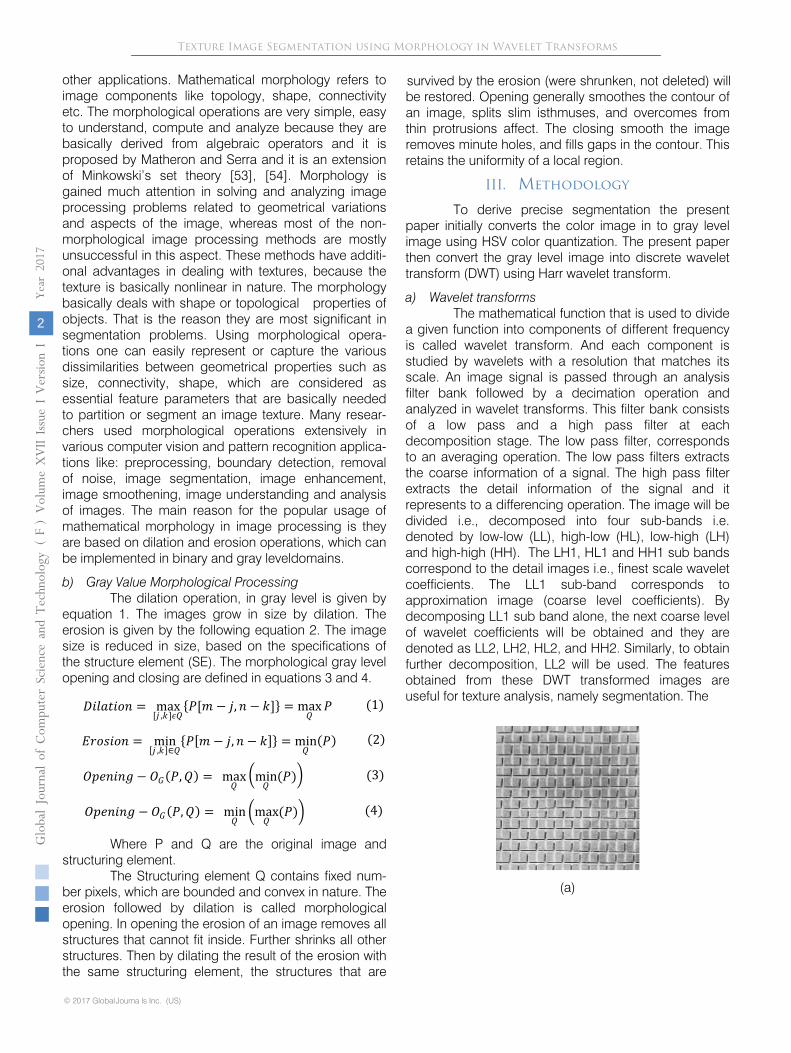
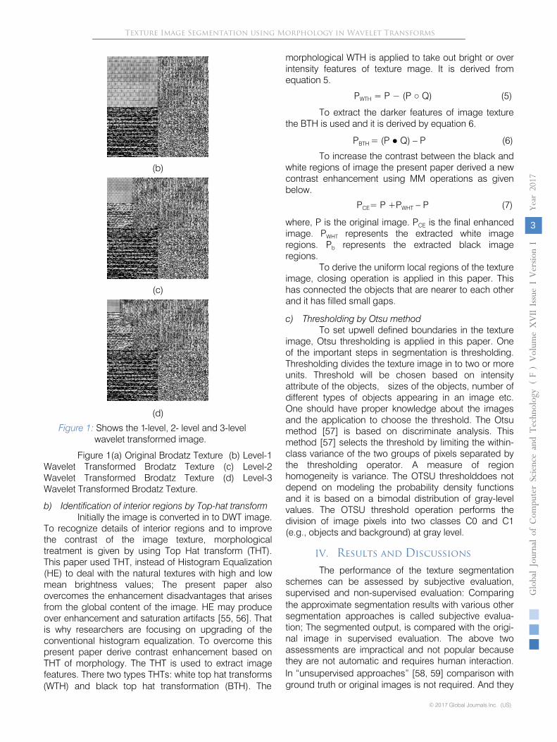
Figure 1: Shows the 1-level, 2- level and 3-level wavelet transformed image.
Figure 1(a) Original Brodatz Texture (b) Level-1 Wavelet Transformed Brodatz Texture (c) Level-2 Wavelet Transformed Brodatz Texture (d) Level-3 Wavelet Transformed Brodatz Texture.
b) Identification of interior regions by Top-hat transform Initially the image is converted in to DWT image.
To recognize details of interior regions and to improve the contrast of the image texture, morphological treatment is given by using Top Hat transform (THT). This paper used THT, instead of Histogram Equalization (HE) to deal with the natural textures with high and low mean brightness values; The present paper also overcomes the enhancement disadvantages that arises from the global content of the image. HE may produce over enhancement and saturation artifacts [55, 56]. That is why researchers are focusing on upgrading of the conventional histogram equalization. To overcome this present paper derive contrast enhancement based on THT of morphology. The THT is used to extract image features. There two types THTs: white top hat transforms (WTH) and black top hat transformation (BTH). The
morphological WTH is applied to take out bright or over intensity features of texture mage. It is derived from equation 5.
PWTH
= P − (P ○
Q) (5)
To extract the darker features of image texture the BTH is used and it is derived by equation 6.
PBTH = (P ● Q) –
P (6)
To increase the contrast between the black and white regions of image the present paper derived a new contrast enhancement using MM operations as given below.
PCE= P +PWHT
–
P (7)
where, P is the original image. PCE
is the final enhanced
image. PWHT
represents the extracted white image
regions. Pb
represents the extracted black image
regions. To derive the uniform local regions of the texture
image, closing operation is applied in this paper. This
has connected the objects that are nearer to each other and it has filled small gaps.
c)
Thresholding by Otsu method To set upwell defined boundaries in the texture
image, Otsu thresholding is applied in this paper. One of the important steps in segmentation is thresholding. Thresholding
divides the texture image in to two or more
units. Threshold will be chosen based on intensity attribute of the objects, sizes of the objects, number of different types of objects appearing in an image etc. One should have proper knowledge about the images and the application to choose the threshold. The Otsu method [57] is based on discriminate analysis. This method [57] selects the threshold by limiting the within-class variance of the two groups of pixels separated by the thresholding operator. A measure of region homogeneity is variance. The OTSU thresholddoes not depend on modeling the probability density functions and it is based on a bimodal distribution of gray-level values. The OTSU threshold operation performs the division of image pixels into two classes C0 and C1 (e.g., objects and background) at gray level.
IV.
Results and Discussions
The performance of the texture segmentation
schemes
can be assessed by subjective evaluation, supervised and non-supervised evaluation: Comparing the approximate segmentation results with various other segmentation approaches is called subjective evalua-
tion; The segmented output, is compared with the origi- nal image in supervised evaluation. The above two
assessments are impractical and not popular because they are not automatic and requires human interaction.
© 2017 Global Journals Inc. (US)
Globa
l Jo
urna
l of C
ompu
ter Sc
ienc
e an
d Te
chno
logy
V
olum
e XVII
Issu
e I Versio
n I
3
Year
2017
(
)F
In “unsupervised approaches” [58, 59] comparison with ground truth or original images is not required. And they
Texture Image Segmentation using Morphology in Wavelet Transforms
take less time in evaluating the performance of the segmentation method. The proposed segmentation scheme is assessed by unsupervised parameters like: Discrepancy, Entropy, Standard deviation, internal region contrast as given below. The value of these indicates the following:
If the value of discrepancy is high, then it indicates a better segmentation.
Using entropy value one can recognize the Over segmentation and under segmentations. Over segmentation will be resulted if entropy
value less than 1 and if it is above 1.5 then represents under segmentation. A better segmentation is estimated with lower values of standard deviation. Region uniformity should not be disturbed while segmenting. If the segmented image results a low internal contrast then it indicates a high uniformity.
𝐷𝐷𝐷𝐷𝐸𝐸𝐷𝐷𝐸𝐸𝑂𝑂𝑂𝑂𝐷𝐷𝐷𝐷𝐷𝐷𝐷𝐷 =
∑ ∑ (𝐴𝐴(𝐸𝐸, 𝐷𝐷) − 𝐵𝐵(𝐸𝐸, 𝐷𝐷))𝑚𝑚𝐷𝐷=1
𝐷𝐷𝐸𝐸=1
(8)
Where A(r,c) and B(r,c) represents the gray level original and segmented image.
Entropy of an image is given as
𝐸𝐸𝐷𝐷𝐷𝐷𝐸𝐸𝐷𝐷𝑂𝑂𝐷𝐷 =
−∑ ∑
𝐵𝐵(𝐸𝐸, 𝐷𝐷)𝐷𝐷𝐷𝐷𝑂𝑂�𝐵𝐵(𝐸𝐸, c)�𝐷𝐷𝐸𝐸
(9)
Where B is segmented image
Standard deviation of a given vector is expressed as
Standared
deviation
S =
� 1n−1
∑ (xi − x�)2ni=1 �
12
(10)
Where xi andx� are the value of vector and average of all values.
Internal region contrast is defined as
Internal
region
contrast
Ij =
1Sj∑ max�cont(𝐸𝐸, t), t ∈s∈Rj
N𝐸𝐸∩Rj
(11)
Where N(s) is the neighborhood and contrast (s,t) = |Cx(s) -
Cx(t)| is the contrast of pixel ‘s’ and ‘t’. The region uniformity is measured by internal contrast, Ij. The Ij
represents “region average Max Contrast”.
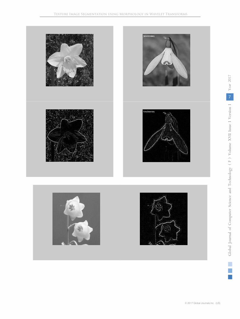
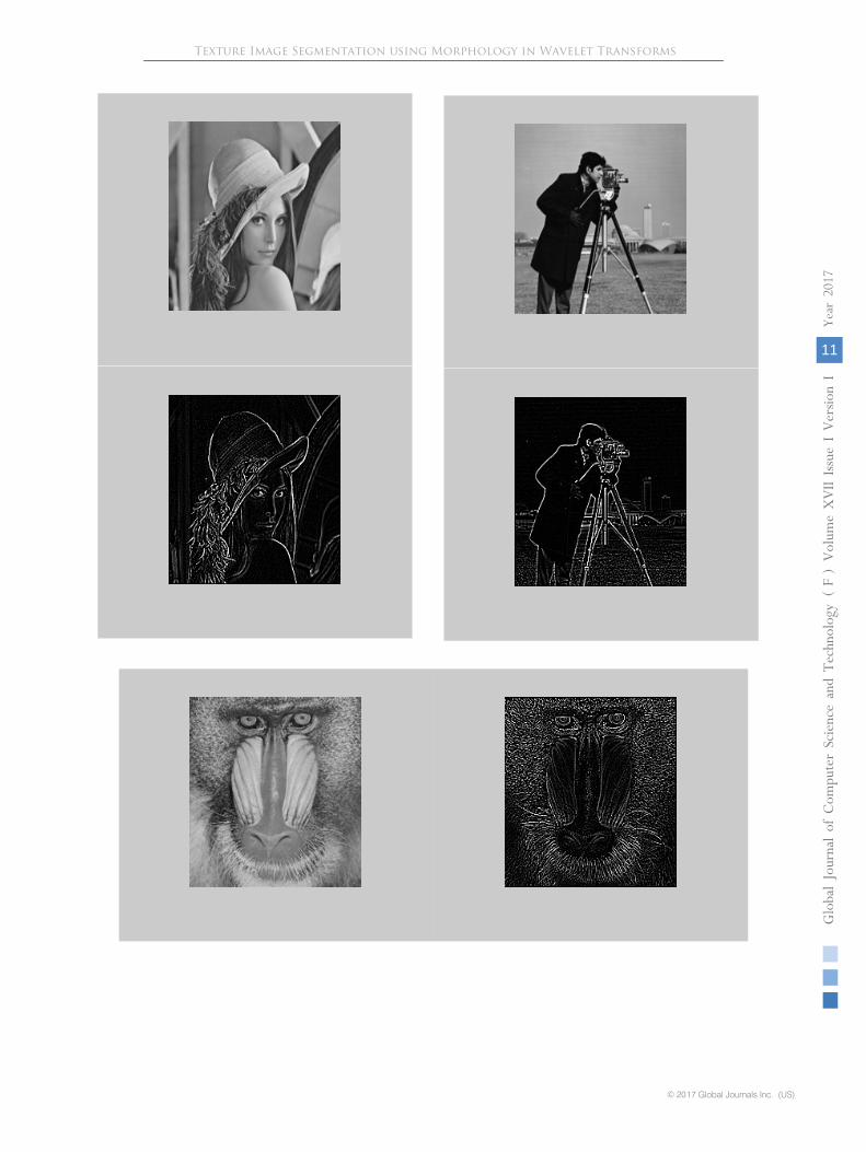
The present method is tested on Brodtaz [60],
Oxford flowers [61], Wang [62] and standard images from Google (Lena, Camera man, House, Mandrill, and Ship) [63]. The experiments are carried out by considering 100 images from each data base, thus it results a total of 400images.There are 1,000 natural images in Wang database and these images are selected manually from Corel stock photo database. These images are divided into 10 categories (each category consists 100 images). There are 17 classes (80 images per class) in Oxford flower database. There are 112 texture images in the Brodatz album with different background intensities. The proposed method is compared with three existing methods ISLGHEM [64], automatic thresholding
method [65], wavelet based watershed method [66] and MULBP method [67]. To show the performance, the proposed integrated DWT segmentation scheme is
applied on input images and
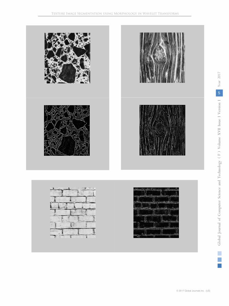
results are shown in Fig.2, 3, 4 and 5 for Brodatz, Oxford, Wang and standard database textures respectively. The following are noted from the segmented outputs and they clearly establish the following facts.
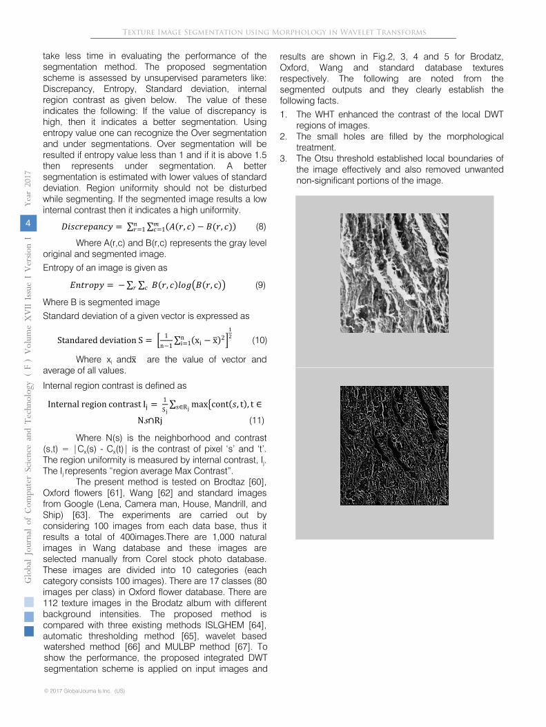
1.
The WHT enhanced the contrast of the local DWT regions of images.
2.
The small holes are filled by the morphological treatment.
3.
The Otsu threshold established local boundaries of the image effectively and also removed unwanted non-significant portions of the image.
Globa
l Jo
urna
l of C
ompu
ter Sc
ienc
e an
d Te
chno
logy
V
olum
e XVII
Issu
e I Versio
n I
4
Year
2017
(
)
© 20 7 Global Journa ls Inc. (US)1
FTexture Image Segmentation using Morphology in Wavelet Transforms
© 2017 Global Journals Inc. (US)
Globa
l Jo
urna
l of C
ompu
ter Sc
ienc
e an
d Te
chno
logy
V
olum
e XVII
Issu
e I Versio
n I
5
Year
2017
(
)F
Texture Image Segmentation using Morphology in Wavelet Transforms
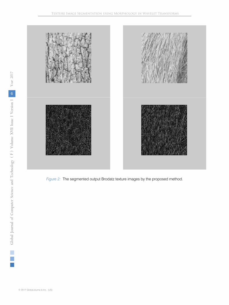
Figure 2: The segmented output Brodatz texture images by the proposed method.
Globa
l Jo
urna
l of C
ompu
ter Sc
ienc
e an
d Te
chno
logy
V
olum
e XVII
Issu
e I Versio
n I
6
Year
2017
(
)
© 20 7 Global Journa ls Inc. (US)1
FTexture Image Segmentation using Morphology in Wavelet Transforms
© 2017 Global Journals Inc. (US)
Globa
l Jo
urna
l of C
ompu
ter Sc
ienc
e an
d Te
chno
logy
V
olum
e XVII
Issu
e I Versio
n I
7
Year
2017
(
)F
Texture Image Segmentation using Morphology in Wavelet Transforms
Figure 3: The segmented output Oxford flower texture images by the proposed method.
Globa
l Jo
urna
l of C
ompu
ter Sc
ienc
e an
d Te
chno
logy
V
olum
e XVII
Issu
e I Versio
n I
8
Year
2017
(
)F
Texture Image Segmentation using Morphology in Wavelet Transforms
© 20 7 Global Journa ls Inc. (US)1
© 2017 Global Journals Inc. (US)
Globa
l Jo
urna
l of C
ompu
ter Sc
ienc
e an
d Te
chno
logy
V
olum
e XVII
Issu
e I Versio
n I
9
Year
2017
(
)F
Texture Image Segmentation using Morphology in Wavelet Transforms
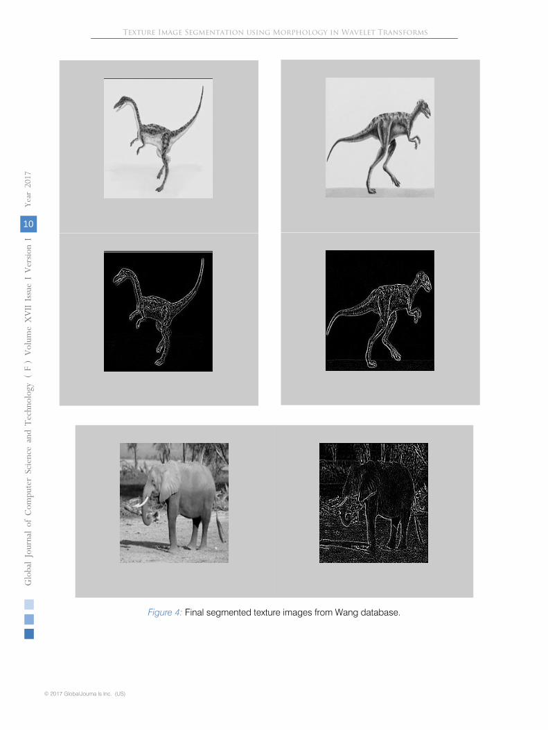
Figure 4:
Final segmented texture images from Wang database.
Globa
l Jo
urna
l of C
ompu
ter Sc
ienc
e an
d Te
chno
logy
V
olum
e XVII
Issu
e I Versio
n I
10
Year
2017
(
)F
Texture Image Segmentation using Morphology in Wavelet Transforms
© 20 7 Global Journa ls Inc. (US)1
© 2017 Global Journals Inc. (US)
Globa
l Jo
urna
l of C
ompu
ter Sc
ienc
e an
d Te
chno
logy
V
olum
e XVII
Issu
e I Versio
n I
11
Year
2017
(
)F
Texture Image Segmentation using Morphology in Wavelet Transforms
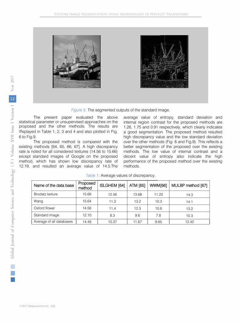
Figure 5:
The segmented outputs of the standard image.
The present paper evaluated the above statistical parameter or unsupervised approaches on the proposed and the other methods. The results are displayed in Table 1, 2, 3 and 4 and also plotted in Fig. 6 to Fig.9.
The proposed method is compared with the existing methods [64, 65, 66, 67]. A high discrepancy rate is noted for all considered textures (14.56 to 15.66) except standard images of Google on the proposed method, which has shown low discrepancy rate of 12.19, and resulted an average value of 14.5.The
average value of entropy, standard deviation and internal region contrast for the proposed methods are 1.26, 1.75 and 0.91 respectively, which clearly indicates a good segmentation. The proposed method resulted high discrepancy value and the low standard deviation over the other methods (Fig: 6 and Fig.9). This reflects a better segmentation of the proposed over the existing methods. The low value of internal contrast and a decent value of entropy also indicate the high performance of the proposed method over the existing methods.
Table 1:
Average values of discrepancy.
Name of the data base
Proposed
method
ISLGHEM [64]
ATM [65]
WWM[66]
MULBP method [67]
Brodatz texture
15.66
12.56
13.68
11.20
14.3
Wang
15.64
11.3
13.2
10.3
14.1
Oxford flower
14.56
11.4
12.3
10.6
13.2
Standard image
12.10
8.3
9.6
7.8
10.3
Average of all databases
14.49
10.37
11.67
9.65
12.42
Globa
l Jo
urna
l of C
ompu
ter Sc
ienc
e an
d Te
chno
logy
V
olum
e XVII
Issu
e I Versio
n I
12
Year
2017
(
)
© 20 7 Global Journa ls Inc. (US)1
FTexture Image Segmentation using Morphology in Wavelet Transforms
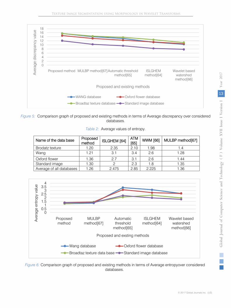
Figure 5:
Comparison graph
of proposed and existing methods in terms of Average discrepancy over considered databases.
Table 2:
Average values of entropy.
Name of the data base
Proposed
method
ISLGHEM [64]
ATM
[65]
WWM [66]
MULBP method[67]
Brodatz texture
1.20
2.35
2.10
1.98
1.4
Wang
1.21
3.1
3.4
2.6
1.28
Oxford flower
1.36
2.7
3.1
2.6
1.44
Standard image
1.30
2
2.3
1.8
1.35
Average of all databases
1.26
2.475
2.85
2.225
1.36
Figure 6:
Comparison graph of proposed and existing methods in terms of Average entropyover considered
databases.
02468
1012141618
Proposed method MULBP method[67]Automatic threshold method[65]
ISLGHEM method[64]
Wavelet based watershed
method[66]
Ave
rage
dis
crep
ancy
val
ue
Proposed and existing methods
WANG database Oxford flower database
Broadtaz texture database Standard image database
00.5
11.5
22.5
33.5
4
Proposed method
MULBP method[67]
Automatic threshold
method[65]
ISLGHEM method[64]
Wavelet based watershed
method[66]
Ave
rage
ent
ropy
val
ue
Proposed and exsting methods
Wang database Oxford flower database
Broadtaz texture data base Standard image database
© 2017 Global Journals Inc. (US)
Globa
l Jo
urna
l of C
ompu
ter Sc
ienc
e an
d Te
chno
logy
V
olum
e XVII
Issu
e I Versio
n I
13
Year
2017
(
)F
Texture Image Segmentation using Morphology in Wavelet Transforms
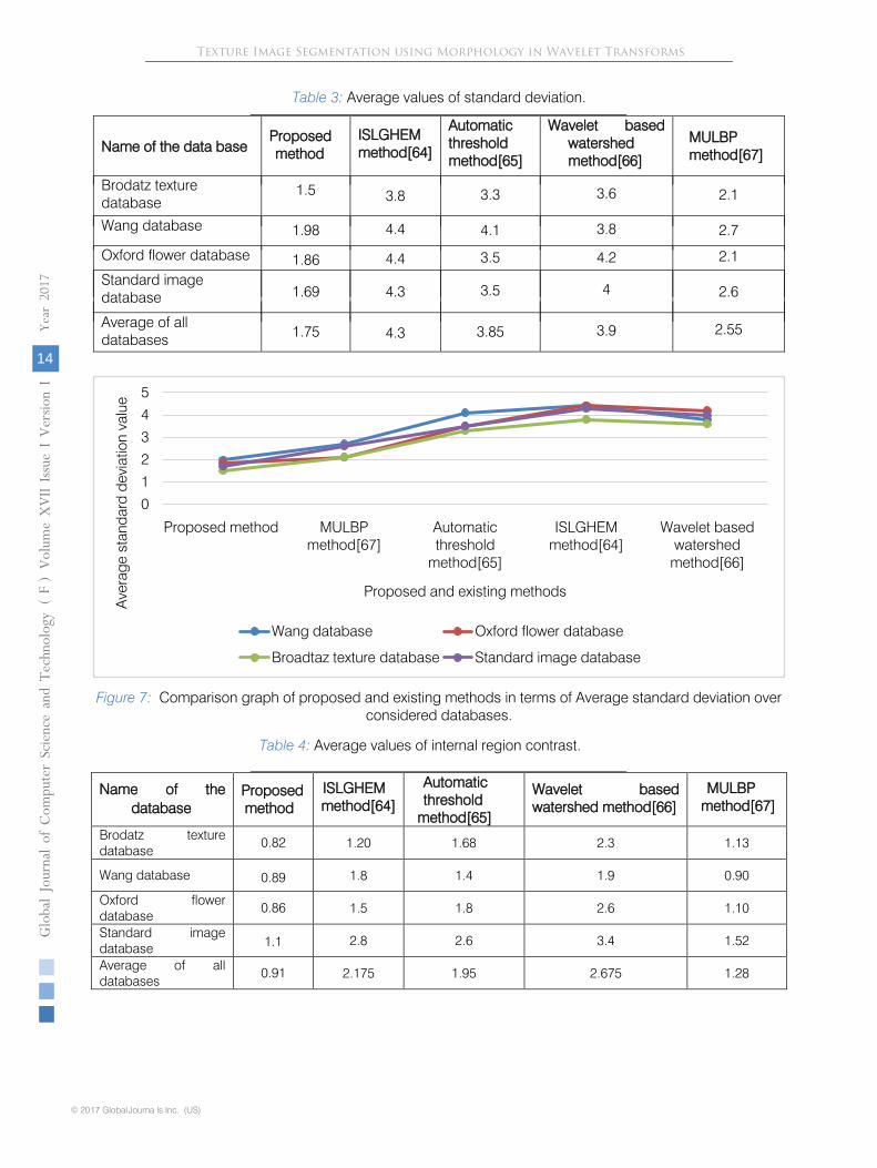
Table 3: Average values of standard deviation.
Name of the data base
Proposed
method ISLGHEM
method[64]
Automatic threshold method[65]
Wavelet based watershed method[66]
MULBP method[67]
Brodatz texture database
1.5 3.8 3.3 3.6 2.1
Wang database
1.98
4.4
4.1
3.8
2.7
Oxford flower database
1.86
4.4
3.5
4.2
2.1
Standard image database
1.69
4.3
3.5
4
2.6
Average of all databases
1.75
4.3
3.85
3.9
2.55
Figure 7: Comparison graph of proposed and existing methods in terms of Average standard deviation over considered databases.
Table 4:
Average values of internal region contrast.
Name of the database
Proposed
method
ISLGHEM method[64]
Automatic threshold
method[65]
Wavelet based watershed method[66]
MULBP method[67]
Brodatz texture database
0.82
1.20
1.68
2.3
1.13
Wang database
0.89
1.8
1.4
1.9
0.90
Oxford flower database
0.86
1.5
1.8
2.6
1.10
Standard image database
1.1
2.8
2.6
3.4
1.52
Average of all databases
0.91
2.175
1.95
2.675
1.28
0
1
2
3
4
5
Proposed method MULBP method[67]
Automatic threshold
method[65]
ISLGHEM method[64]
Wavelet based watershed
method[66]
Ave
rage
sta
ndar
d de
viat
ion
valu
e
Proposed and existing methods
Wang database Oxford flower database
Broadtaz texture database Standard image database
Globa
l Jo
urna
l of C
ompu
ter Sc
ienc
e an
d Te
chno
logy
V
olum
e XVII
Issu
e I Versio
n I
14
Year
2017
(
)
© 20 7 Global Journa ls Inc. (US)1
FTexture Image Segmentation using Morphology in Wavelet Transforms
Figure 8: Comparison graph of proposed and existing methods in terms of Average internal region contrast over
considered databases
V. Conclusions The morphological operations covered the
small holes with intensities and borders are connected for a better segmentation.
The Otsu threshold
established local boundaries efficiently and provided better contrast and also removed the unwanted local scenes of the image. The whole process of segmentation is automatic and requires no supervision. The present paper improves the contrast of sharp details in light and dark areas. The present method is experimented on the four standard databases namely Brodatz, Wang, Oxford flowers and standard images. The present method attained a good segmentation on the four datasets: however the proposed method have shown significantly high performance on Brodatz database textures when compared with other standard datasets; followed by Wang and oxford datasets. Further no over and under segmentation is reported by the present method on the considered databases. The present method is simple and suitable to real time applications because it achieved good segmentation with three basic steps.
References Références Referencias
1.
Kekre.
H.
B, Saylee
Gharge, "Texture Based Segme-
ntation using Statistical Properties for Mammogra- phic Images”, Int. Journal of Advanced Computer
Science and Applications(IJACSA), Vol. 1, No. 5, pp. 102-107, Nov 2010.
2.
Lutz Goldmann, Tomasz Adamek, Peter Vajda, “Towards Fully Automatic , Image Segmentation Evaluation”, in Advanced Concepts for Intelligent Vision Systems Lecture Notes in Comp. Science, vol. 5259, pp. 566–577, 2008.
3.
Singh.
S and Sharma.
M, "Texture experiments with Meastex and Vistex benchmarks," in Proc.
Inter. Conf. on Advances in Pattern recog., Lecture Notes in
Computer Science, 2013.
4.
Wang.B and Zhang.L, “Supervised texture segmen- tation using wavelet transform,” in Proc. of Int. Conf.
on Neural Networks and Signal Processing, vol. 2, pp. 1078–1082, 2003.
5.
M. Wirth, D. Nikitenko and J. Lyon, “Segmentation of the
Breast Region in Mammograms using a Rule-
Based Fuzzy Reasoning Algorithm”, GVIP Journal Special Issue on Mammograms, pp. 13-21, 2007.
6.
Vijaya Kumar, SakaKezia, I. SantiPrabha, “A new texture segmentation approach for medical images”
International Journal of Scientific & Engineering Res- earch (IJSER), Vol. 4, Iss.1, 2013, pp.1-5, ISSN:
2229-5518. 7.
SakaKezia, I.SantiPrabha, V.VijayaKumar, “A color-texture based segmentation method to extract ob-
ject from background”, International Journal Image, And Graphics And Signal Processing (IJIGSP), Vol. 5, Iss. 3, 2013, pp.19-25, ISSN: 2074-
9082.
8.
M. Joseph
Prakash,
V.
Vijayakumar, “A new texture
based segmentation method to extract object from background”, Global Journal of Computer Science And Technology Graphics & Vision (GJCST), Vol.12, Iss.15, 2012, pp;1-6, ISSN: 0975-4350.
9. V.Vijaya Kumar, B. Eswar Reddy, A. Nagaraja Rao, U.S.N. Raju, “Texture segmentation methods based on combinatorial of morphological and statistical operations”, Journal of multimedia (JMM), Academy
00.5
11.5
22.5
33.5
4
Proposed method
MULBP method[67]
Automatic threshold
method[65]
ISLGHEM method[64]
Wavelet based watershed
method[66]
Ave
rage
inte
rnal
regi
on c
ontra
st
Proposed and exsting methods
Wang database Oxford flower database
Broadtaz texture database Standard image database
© 2017 Global Journals Inc. (US)
Globa
l Jo
urna
l of C
ompu
ter Sc
ienc
e an
d Te
chno
logy
V
olum
e XVII
Issu
e I Versio
n I
15
Year
2017
(
)F
publishers, Vol.3, Iss.1, 2008, pp.36-40, ISSN 1796-2048.
Texture Image Segmentation using Morphology in Wavelet Transforms
10.
V.
Vijaya Kumar, A. Nagaraja Rao, U.S.N. Raju, B.
Eswar Reddy, “Pipeline implementation of new seg-
mentation based on cognate neighborhood appro-
ach”, International Journal of Computer Science (IJCS), Science publications, Vol.35, Iss.1, 2008, pp. 1-6, ISSN: 1552-6607.
11.
AshwiniKunte, Anjali Bhalchandra, Efficient DIS Based Region Growing Segmentation Technique for VHR Satellite Images ICGST-GVIP Journal, Volume 10, Issue 3, August 2010 .
12.
R. E. Cummings, P. Pouliquen, and M. A. Lewis, “A Vision Chip for Colour Segmentation and Pattern Matching”, EURASIP Journal on Applied Signal Processing, no. 7, pp. 703-712, 2003
13.
Y. Yang, C. Zhen and P. Lin, “Fuzzy C-means clus-
tering algorithm with a novel
penalty term for image segmentation”, Opto-Electronics Rev., vol. 13, no. 4, 2005.
14.
M. B. Meevathi and K. Rajesh, “Volterra Filter for color image segmentation”, Inter. Journal of Comp-uter Science and Engineering, vol. 2, no. 1, 2008.
15.
H. D. Cheng and Y. Sun, “A hierarchical approach to color image segmentation using homogeneity”, IEEE Transaction on Image Processing, vol. 9, no. 12, pp. 2071-
2082, 2000.
16.
H. Seddik and E. Ben Braiek “Color Medical Images Watermarking, Based Neural Network Segmentation “GVIP
Journal Special Special Issue on (Medical Image Processing), pp. 81-86, 2006.
17.
A. Laine, J. Fan, “Frame representations for texture segmentation”, IEEE Trans. Image Processing, 5 (1996), 771-780.
18.
JP. Mosiganti, JM kezia, V. Vijay Kumar, “Morpho-
logical Multi-scale Stationary Wavelet Transform based Texture Segmentation”, International Journal of Artificial Intelligence & Applications (IJAIA) , Vol. 8, Iss. 1, pp: 32-39, 2014, ISSN: 2074-9082
19.
J.-L. Chen, A. Kundu, “Unsupervised texture segme-
ntation using multichannel decomposition and hidden Markov models”, IEEE Trans. Image Proce-
ssing, 4 (1995), 603-619.
20.
H. Greenspan, R. Goodman, R. Chellappa, C.H. Anderson, “Learning texture discrimination rules in a multiresolution system”, IEEE Trans. Pattern Anal. Machine Intelligence 16 (1994) 894-901
21.
M. Spann, R. Wilson, “A quad-tree approach to image segmentation which combines statistical and spatial information”, Pattern Recognition, 18 (1985) 257-269.
22.
B.B. Chaudhuri, N. Sarkar, “Texture segmentation using fractal dimension”, IEEE Trans. Pattern Anal. Machine Intelligence, 17 (1995) 72-77.
23.
J.Y. Hsiao, A.A. Sawchuk, “Unsupervised texture image segmentation using feature smoothing and probabilistic relaxation techniques”, Computer Visi-
on Graphics Image Processing
48 (1989) 1-21.
24.
P.C. Chen, T. Pavlidis, “Segmentation by texture using a co-occurrence matrix and a split-and-merge algorithm”, Comput. Graphics Image Processing 10 (1979) 172-182.
25.
J. Mao, A.K. Jain, “Texture classification and segm-entation using multi resolution simultaneous autore-
gressive models”, Pattern Recognition 25 (1992) 173-188.
26.
M. Pietika¬ inen, A. Rosenfeld, “Image segmen-
tation by texture using pyramid node linking”, IEEE Trans. Systems Man Cybernet. 11 ,1981, 822-825.
27.
M. Unser, M. Eden, “Multiresolution feature extrac-
tion and selection for texture segmentation”, IEEE Trans. Pattern Anal. Machine Intelligence 11 (1989) , 717-728.
28.
A.K. Jain, F. Farrokhnia, “Unsupervised texture seg-
mentation using Gabor filters”, Pattern Recognition, 24 (1991), 1167-1186.
29.
C.Kervrann and F.Heitz. A Markov Random Field model-based approach to unsupervised texture segmentation using local and global statistics. IEEE Trans. Image Processing, 4(6):856{862, June 1995.
30.
M. Lalitha1 , M. Kiruthiga2 , C. Loganathan3
, A Survey on Image Segmentation through Clustering Algorithm, International Journal of Science and Research (IJSR), Volume 2 Issue 2, February 2013 , India Online ISSN: 2319-7064
31.
Mairal, J., Leordeanu, M., Bach, F., Hebert, M., Ponce, J.: “Discriminative
sparse image models for class-specific edge detection and image interpre-
tation”. Proc. 10th European Conf. Computer Vision, 2008, pp. 43–56.
32.
Peng, B., Zhang, L., Zhang, D.: “Automatic image segmentation by dynamic region merging”, IEEE Trans. Image Process., 2011, 20, (12), pp. 3592–3605
33.
Arbelaez, P., Maire, M., Fowlkes, C., Malik, J.: “Contour detection and hierarchical image segme-
ntation”, IEEE Trans. Patt. Anal. Mach. Intell., 2010, 33, (5), pp. 898–916.
34.
Jähne, B.: “Practical handbook on image proce-
ssing for scientific and technical applications”, (CRC Press, 2004, 2nd Ed.), Ch. 15.
35.
Shi, J., Malik, J.: “Normalized cuts and image seg-
mentation”, IEEE Trans. Patt. Anal. Mach. Intell., 2000, 22, (8), pp. 888–905.
36.
Malik, J., Belongie, S., Leung, T., Shi, J.: ‘Contour and texture analysis for image segmentation’, Int. J. Comput. Vis., 2001, 43, (1), pp. 7–27.
37.
Yu, S.X., Shi, J.: ‘Multiclass spectral clustering’. Proc. Int. Conf. Computer Vision, October 2003, pp. 313–319.
38. Yu, S.X.: ‘Segmentation using multiscale cues’‘. Proc. IEEE Conf. on Comput. Vision Pattern Recognition, June 2004, vol. 1, pp. I-247–I-254.
Globa
l Jo
urna
l of C
ompu
ter Sc
ienc
e an
d Te
chno
logy
V
olum
e XVII
Issu
e I Versio
n I
16
Year
2017
(
)
© 20 7 Global Journa ls Inc. (US)1
F
39. Huang, S.H., Chu, Y.H., Lai, S.H., Novak, C.L.: ‘‘Learning-based vertebra detection and iterative
Texture Image Segmentation using Morphology in Wavelet Transforms
normalized-cut segmentation for spinal MRI’‘, IEEE Trans. Med. Imag., 2009, 28, (8), pp. 1595–1605.
40.
Yu, S.X.: ‘‘Segmentation induced by scale invari-
ance’‘. Proc. IEEE Conf. on Comput. Vision Pattern Recognition, June 2005, vol. 1, pp. 444–451
41.
Cour, T., Benezit, F., Shi, J.: ‘‘Spectral segmentation with
Multiscale graph decomposition’‘. Proc. IEEE Conf. on Computer Vision Pattern Recognition, June 2005, vol. 2, pp. 1124–1131.
42.
Carreira-Perpinan, M.A.: ‘‘Acceleration strategies for Gaussian mean-shift image segmentation’‘. Proc. IEEE Conf. on Computer Vision Pattern Recognition, June 2006, vol. 1, pp. 1160–1167.
43.
3Tao, W., Jin, H., Liu, J.: ‘‘Unified mean shift segmentation and graph region merging algorithm for infrared ship target segmentation’‘, Opt. Eng., 2007, 46, (12), pp. 127002–127002–7.
44.
Mayer, A., Greenspan, H.: ‘‘An adaptive mean-shift framework for MRI brain segmentation’‘, IEEE Trans. Med. Imag., 2009, 28, (8), pp. 1238–1250
45.
Chen, T.W., Chen, Y.L., Chien, S.Y.: ‘‘Fast image segmentation based on K-means clustering with histograms in HSV color space’‘. Proc. IEEE Int. Workshop on Multimedia Signal Processing, October 2008, pp. 322–325.
46.
Tan, K.S., Isa, N.A.M.: ‘‘Color image segmentation using histogram Thresholding –
Fuzzy C-means hybrid approach”, Patt. Recogn., 2011, 44, (1), pp. 1–15.
47.
Tan, K.S., Isa, N.A.M., Lim, W.H.: “Color image segmentation using adaptive unsupervised clustering approach”, Appl. Soft Comput., 2012, 13, pp. 2017–2036.
48.
Tan, K.S., Lim, W.H., Isa, N.A.M.: “Novel initialization scheme for Fuzzy C-Means algorithm on color image segmentation”, Appl. Soft Comput., 2013, 13, pp. 1832–1852.
49.
Ghamisi, P., Couceiro, M.S., Benediktsson, J.A., Ferreira, N.M.: ‘‘An efficient method for segme-
ntation of images based on fractional calculus and natural selection’‘, Expert Syst. Appl., 2012, 39,
(16), pp. 12407–12417.
50.
Donald.
A, Adjeroh and UmasankarKandaswamy, “Texton-based segmentation of retinal vessels”,
Journal of Optical Society of America , vol. 24, no. 5, pp.1384–1393, May 2007.
51.
Idrissisidiyassine, Samir belfkih, "Texture image segmentation using a new descriptor and mathema-
tical morphology”, in Int. Arab Journal of Information Technology, Vol.10, No.2, pp. 204-208, March 2013.
52.
Kezia.S, ShantiPrabha.I, VijayaKumar.V, “Innovative segmentation approach based on LRTM”, Int. Journal of Soft Computing and Engg., vol. 2, no. 5, pp. 229-233, Nov. 2012.
53.
J. Serra, Image Analysis and Mathematical Mor-
phology. London, U.K.: Academic, 1982.
54.
J. Serra , Introduction to mathematical morphology, Comput. Vis., Graph., Image Process., vol. 35, pp. 283–305, 1986.
55.
Antonini, M., Barlaud, M., Mathieu, P., Daubechies, I., 1992. Image coding using wavelet transform. IEEE Trans. Image Process. 1 (2), 205–220.
56.
Derong, Y, Yuanyuan Z, Dongguo L, “Fast Compu-
tation of Multiscale Morphological Operations for Local Contrast Enhancement”, Proceedings of the 2005 IEEE, pp.3090-3092.
57.
Otsu.
N, "A threshold selection method from gray-level histograms", IEEE Trans. Sys., Man., Cyber, Vol.9, pp.62–66, 1979.
58.
Cui.
Y, Dong.
H,
Zhou.
E.
Z, “An Early Fire Detection Method Based on Smoke Texture Analysis and Discrimination”, Journal Congress on Image and Sig. Proc., pp.95–99, 2008.
59.
Mounir
Sayadi, LotfiTlig and Farhat
Fnaiech, “A new texture segmentation method based on the fuzzy C-mean algorithm and statistical features”, Applied Mathematical Sciences, vol. 1, no. 60, pp. 2999 –
3007, 2007.
60.
P. Brodatz, Textures: “A Photographic Album for Artists and Designers “.New York, NY, USA: Dover, 1999.
61.
http://www.robots.ox.ac.uk/~vgg/data/flowers/
62.
[Online]. <http://wang.ist.psu.edu/docs/related.shtml>
63.
http://www.imageprocessingplace.com/root_files_V3/image_databases.htm.
64.
Saka.
Kezia, Dr.
I
.SantiPrabha, Dr.
V.
VijayaKumar, “A New Texture Segmentation Approach for Medical Images”, International Journal of Scientific & Engineering Research, Volume 4, Issue 1, January-2013.
65.
Yuan Been Chen and Oscal.T.C.Chen, "Image Segmentation Method Using Thresholds Automa-
tically Determined from Picture Contents,” in Eurasip journal on image and video processing, vol.2009, pp. 1-16, 2009
66.
Yu-hua Chai, Li-qunGao, and Shun Lu, Lei Tian, “Wavelet-based Watershed for Image Segmenta-
tion”, in Proc. of the 6th World Congress on Intelli-
gent Control and Automation, Dalian, China, June 21 -
23, 2006.
67.
Anuradha S.G, K. Karibasappa, B. Eswar Reddy, “ A Segmentation scheme based on uniform LBP and morphological approach, Graphics, Vision and Image Processing (ICGST-GVIP), Vol. 16, Iss.1, 2016
© 2017 Global Journals Inc. (US)
Globa
l Jo
urna
l of C
ompu
ter Sc
ienc
e an
d Te
chno
logy
V
olum
e XVII
Issu
e I Versio
n I
17
Year
2017
(
)F
Texture Image Segmentation using Morphology in Wavelet Transforms

This page is intentionally left blank
Texture Image Segmentation using Morphology in Wavelet TransformsGloba
l Jo
urna
l of C
ompu
ter Sc
ienc
e an
d Te
chno
logy
V
olum
e XVII
Issu
e I Versio
n I
18
Year
2017
(
)
© 20 7 Global Journa ls Inc. (US)1
F